epr in structural biology enc2016-v2 - bruker · epr samples • liquids and solids • can be...
TRANSCRIPT

Peter HöferProduct Manager EPRPittsburgh – April 2016
EPR in Structural Biology
Innovation with Integrity

EPR speciesnaturally occurring
• Metal Centers: Cu2+, Mn2+, Fe3+, Mo5+, …
• Radicals: tyrosine, tryptophan, quinol, carotenoid……
• Antioxidants: ascorbate, polyphenols, nitroaromatic drugs…
• Small Molecule: NO, H2O2, O2, OH…
• Defect Centers: O-vacancy, irradiation damage…

EPR speciesreporter molecules
• Spin Probes: TEMPOL, Trityl, DPPH…
• Spin Labels: PROXYL, MTSSL…
• Spin Traps: DMPO, DEPMPO, PBN, MNP; CMH…
N
O
SSO2CH3
CH3
CH3H3C
H3CHS Protein
N
O
S
CH3
CH3H3C
H3C
S Protein
+
CC
Cys
Cys
Not binding
Binding
Transforming

EPR samples
• Liquids and Solids
• Can be measured on the same instrument (same probehead)
• For low temperature VT accessories are required. T range 4-300K: He or cryogen-free VT, 100–500K: liq. and gaseous N2
• Sample concentration range and typical volume
• X-band: nM-M & 50 - 150 µlFor aqueous sample @ RT capillaries or flat cells are used instead of tubes
• Q-band: nM-mM & 5 – 15 µl
• No molecular size restriction!
• In-vitro and in-vivo

4/18/2016 5
(Most) proteins and nucleic acids don’t have unpaired electrons ! no EPR?!
... but ...
we can introduce the wanted unpaired electron into almost any system under investigation
and we can do this also (almost) wherever we want
Spin Labelling Bio Molecules
Site‐directed spin labeling (SDSL)
4/18/2016 6
What Is a Spin Label?
…a stable chemical compound which possesses an unpaired electron (i.e. it is a stable radical) and a specific reactive group which binds to (bio)molecules.
The vast majority of spin labels are nitroxides, where the unpaired electron is located at an –NO group, which is usually part of a heterocyclic ring.
Functional groups contained within the spin label allow them to be attached to the molecule under investigation and determine their specificity.
e.g.: Protein thiol groups (SH‐C) specifically react with the functional groups of the spin labelmethanethiosulfonate,maleimide, andiodoacetamide,
creating a covalent bond with cysteine. Any amino acid of the protein, one at the time, can be replaced with cysteine by site‐directed mutagenesis. If a natural Cys present –outside of the region of interest‐additional site‐directed mutagenesis step (Cys to Ser) is required, unless the native Cys is buried and hence not accessible to the spin label.
A little bit of history:Spin labels were first synthesized in the laboratory of H. M. McConnel in 1965.The first spin labelling studies have been performed using thiol‐specific functional groups to label natively occurring cysteins in proteins (e.g. in hemoglobin).Site‐directed spin labeling (SDSL) was pioneered in the laboratory of Dr. W. L. Hubbell in the late 80s/early 90s.

4/18/2016 7
SDSL-EPR-Tools: Overview
• Spin Label Mobilityreflected in the EPR spectral lineshapes
=> provide a fingerprint of the protein foldand its dynamics
• Accessibilitytowards paramagnetic relaxation
enhancement molecules (NiEDDA, CrOx, O2)=> Discrimination between lipid bilayer, aqueous phase and protein interior
• Polarityof the SL microenvironment
=> reflected in the Azz. Increasing Azz points to shifts in polar environment
• Spin‐Spin Distance determination (~ 8 – 100 A)CW‐EPR: 8‐20 ADEER: 15‐100 A
Information about Structure and Dynamics

Spin Label Environment
Free tumbling
Strongly immobilized
Moderately immobilized
Effects from molecular motion N
Effects from relaxation
30 uM
15 mM
50 mM
N
Characterize the paramagnetic center environment

Spin Label MobilityEPR-SDSL & Intrinsically Disordered Proteins
EPR spectral shape is sensitive to the mobility of the label which is described by the rotational correlation time (c ) A spectral modification represents change in
the environment of the label affecting its mobility and thus reveals structural transitions such as folding events
Region of the intrinsically disordered NTAIL C-terminal domain that undergoes an α-helical-induced folding in the presence of the partner protein PXD .
EPR spectral shape broadening in the case of a disorder‐to order transition due to an induced folding mechanism
Before After folding
0.01 0.1 1 10 1000.1
1
P1/2
23.9 mW
11.1 mW
P / mW0.01 0.1 1 10 100
P1/2
3.9 mW
1.8 mW
P / mW
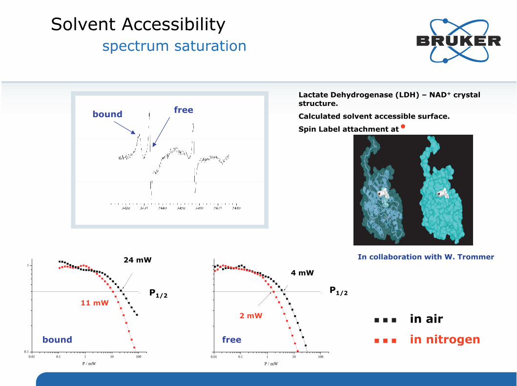
Solvent Accessibilityspectrum saturation
in air
in nitrogen
bound free
bound free
Lactate Dehydrogenase (LDH) – NAD+ crystal structure.
Calculated solvent accessible surface.
Spin Label attachment at
P1/2P1/2
In collaboration with W. Trommer24 mW
11 mW
4 mW
2 mW

April 18, 2016 11
Dependence of the isotropic hyperfine coupling, a0
N, on solvent polarity, for DOXYL (circles) and TOAC (squares) spin labels in proticsolvents
Polarity of the SL microenvironment
a0N

EMXnano, EMXmicro, EMXplusMulti purpose research instruments
EPR Product PortfolioCW-EPR
Innovation with Integrity

Distance measurementsElectron Spin as a Molecular Microscope
Pulse-EPR: ESEEM, HYSCORE, ENDOR
CW-ENDOR
S-I: < 8 Å
Pulse-EPR: DEER/PELDOR
S-S: 15 – 100 Å
CW-EPR
S-S: 5 - 20 ÅS-I: 0 - 4 Å

Electron-Nuclear Hyperfine Coupling
isotropic anisotropic

Pulse EPR: ESEEMLipid Membrane
/2
3p-ESEEM
2p-ESEEM
D2O solvent
#D2O seen by SL at the membrane surface
#D2O

Electron-Electron Dipolar Coupling
pump
D r - 3
EE
observe
0 g1 g2 e2
2 h r3= dd =dd
2 ( 3 cos2 –1 )
r / nm dd / MHz
1.5 15.42.0 6.52.5 3.33.0 1.9

Pulse EPR: Dipolar SpectroscopyDEER/PELDOR
observe
pump
3420 3450 3480 3510Field / G
3530
Dipolar oscillation
Background
/2

Pulse EPR: Dipolar EPR SpectroscopyDEER/PELDOR
0 1 0 0 0 2 0 0 0 3 0 0 0 4 0 0 0 5 0 0 00 .00 .10 .20 .30 .40 .50 .60 .70 .80 .91 .0
1 2 3 4 5 6D is ta n c e [n m ]
Nor
mal
ized
Ech
o A
mpl
itude
t [n s ]
NNOO
1
A BRAB
Pulsed Double Electron Electron Resonance (DEER) Spectroscopy:Measures the dipole–dipole interaction between two unpaired electron spins and is being used to determine long range distances (15 - 100 Å)

DEER vs other techniques
Innovation with Integrity
DEER covers the entire length scale

Pulsed EPR: Dipolar EPR SpectroscopyDEER/PELDOR
site-directedmutagenesis spin labeling
with MTSSLDEERExperiment&Analysis
Wild Type Cys
SH-OH-
0 1 0 0 0 2 0 0 0 3 0 0 0 4 0 0 0 5 0 0 00 .00 .10 .20 .30 .40 .50 .60 .70 .80 .91 .0
1 2 3 4 5 6D is ta n c e [n m ]
Nor
mal
ized
Ech
o A
mpl
itude
t [n s ]
Information content
Distance range: 15 - 100 Å
Distance distribution
Orientation information
Correlate structure and structural changes to
functionality

Pulsed EPR: Dipolar EPR SpectroscopyDEER/PELDOR
Samples: proteins, RNA, DNA,protein-protein, protein-RNA complexes
Types of paramagnetic centers Radicals endogenous: tyrosine tryptophan, quinone…
exogenous: nitroxide and trityl spin labels
Transition ion metals endogenous: Cu, Fe, Mo/W, Ni, Co, Mn…
exogenous: Mn, Cu, Gd, spin labels
Typical concentration 50 - 200 µM
Volume X-Band: 50 - 100 µl
Q-Band: 5 – 15 µl
Temperature: typically 50K-100K
Spin labels
Advantages:
No limitations on molecular size
Works in phospholipids
Works in-cell

Pulsed: Dipolar EPR SpectroscopyPELDOR / DEER: Proteins
The distance between a single pair of spin
labeled mutants is measured at a time
Distance determination between multiple spin
labels is possible however the analysis is
more complicated

Pulsed: Dipolar EPR SpectroscopyPELDOR / DEER: Membrane Proteins
Distance determination in various intermediate states direct observation of large conformational changes
Distance measurements in liposomes Explore structure and conformational dynamics in native-like
environment
Black=open, red=closed

Pulsed EPR: Dipolar EPR SpectroscopyPELDOR / DEER: RNA & DNA
In-Vivo
In-cell: RNA and DNA
In-vivo determination of intramolecular distances in nucleic acids understanding their conformational
flexibility
Strong change in vitro vs in cell

Pulsed EPR: Dipolar EPR SpectroscopyPELDOR / DEER: NMR Meets EPR
Combining NMR and EPR:
Powerful, novel approach for structure determination of large
protein–RNA complexes
Binding of RsmE protein to the RsmZ sRNA:

ELEXYS E580: DEER/PELDOR
Pulse EPR
ELEXYS E580
Q-Band ~ 25 min Acquisition time
X-Band ~ 22 h Acquisition time
Dedicated GUI for ease of use
Recent S/N improvements:
Going from X- to Q-Band (> 50)
Shaped pulse (> 3)
Gain in throughput!

References
• Spin Label Mobility (slide 9)– Martinho, M., et. al., Assessing induced folding within the intrinsically disordered C-terminal
domain of the Henipavirus necleoproteins by site-directed spin labeling EPR spectroscopy (2012) 13(5), p453. doi: 10.1080/07391102.2012.706068
• Polarity of Microenvironemnt (slide 11)– Marsh, D., Spin-Label EPR for Determining Polarity and Proticity in Biomolecular Assemblies:
Transmembrane Profiles, Appl Magn Reson (2010) 37(1-4), p435. doi:10.1007/s00723-009-0078-3
• DEER (slide 22)– Lumme, C., et. al., Nucleoties and Substrates Trigger the Dynamics of the Toc34 GTPase
Homodimer Involved in Chloroplast Preprotein Translocation, Structure (2014), http://dx.doi.org/10.1016/j.str.2014.02.004
• DEER (slide 23)– Duerr, K. L., et. al., Structure and dynamics of AMPA receptor GluA2 in resting, pre-open, and
desensitized states. Cell (2014) 158(4), p 778. doi: 10.1016/j.cell.2014.07.023– Zou, P., et. al., Conformation Cycle of the ABC transporter MsbA in Liposomes. Detailed
Analysis using Double Electron-Electron Resonance Spectroscopy, J Mol. Biol. (2009) 393(3), p586. doi:10.1016/j.jmb.2009.08.050
– Mchaourab, H. S., et. al., Toward the Fourth Dimension of Membrand Protein Structure: Insight into Dynamics from Spin0labeling EPR Spectrscopy, Structure (2011) 19(11), p 1549. doi:10.1016/j.str.2011.10.009
© Copyright 2012 Bruker Corporation.

References
© Copyright 2012 Bruker Corporation.
• DEER (slide 24)– Krstic, I., et. al., Long-Range Distance Measurements on Nucleic Acids in Cells by Pulsed EPR
Spectroscopy. Angew. Chem. Int. Ed. (2011) 50(22), p 5070. doi: 10.1002/anie.201100886– Haensel, R., et. al., In-Cell NMR and EPR Spectroscopy of Biomacromolecules, Angew. Chem.
Int. Ed. (2014) 53(39), p10300. doi: 10.1002/anie.201311320• DEER (slide 25)
– Duss, O., et. al., EPR-aided approach for solution structure determination of large RNAs or protein-RNA complexes, Nature Comm. (2014) 5. doi: 10.1038/ncomms4669
– Duss, O., et. al., Structural basis of the non-coding RNA RsmZ acting as a protein sponge, Nature (2014) 509, p588. doi: 10.1038/nature13271

www.bruker.com
© Copyright Bruker Corporation. All rights reserved.