er conference co* concepcion * conti * corpuz * cosico * cruz * cruz
TRANSCRIPT

ER CONFERENCECo* Concepcion * Conti * Corpuz * Cosico * Cruz * Cruz

General Data
10-month-old, Female From Isabela
Chief Complaint: Pallor and anuria for 2
days

8 days
PTALoose, blood-streaked, mucoid, greenish stools
4-5 x per day ~1 cup per episode
Associated with vomiting of previously ingested milkGood urine outputBrought to a private doctor
Advised ORS after each bowel movement
Metronidazole 30mg/kg/day
History of Present Illness

3 days
PTA 1 episode of bowel movement
Non-blood streaked, formed stools
no vomiting
Decrease in urine output
Highly colored urine
History of present Illness

1 day
PTA 5 episodes of vomiting
Pallor
No urine output
Brought back to private doctor
Referred to USTH for further
management
History of Present Illness
TRANSFER TO USTH

Family History
(-) kidney disease

Physical Examination
Irritable but not in respiratory distress Vital signs were as follows:
BP 90/60 mmHg Anthropometric measurements :
Lt : 75 cm (p75) Wt : 10 kg (p75) Pale, dry skin and petechial rash on the abdomen Anterior fontanel and eyeballs slightly sunken Liver – palpable 3 cm below RSCM Liver span 6cm Urinary bladder not palpable Strong peripheral pulses No signs of edema

Other pertinent information
blahblah

Approach to Diagnosis

Salient Features
10 mo old/F Diarrhea Vomiting Anuria Pallor
Signs of dehydration Slightly sunken
fontanelles and eyeballs
Irritability Dry skin
Petechial rash Hepatomegaly? Strong pulses
Subjective Objective

Presenting Manifestation
Look for a symptom, sign or laboratory finding.. Pathognomonic of a disease Pointing to an organ or part of an organ Pointing to a group of disease Mechanism is well understood Found in the least number of diseases
UST: Pedia (2009). Guideline for History Taking, PE and Diagnosis of Pediatric Patients. 2nd ed.
“GI: Vomiting and diarrhea and w/ signs of dehydration
Renal: anuriaPetechial rash”

Differential Diagnosis
HSP HUS Acute gastroenteritis
Epidemiology Primary childrens’ disease (3-15 y/o)
Affects children under 5
Older children >5 years old
Pathogenesis immune-mediated vasculitis associated with IgA deposition
Ingestion of contaminated foodstuff E-coli producing Shiga-toxin
Campylobacter, Salmonella, Shigella, E. coli, E histolitica inflammation of intestines
Clin Manifestation
-Palpable purpura w/o thrombocytopenia and coagulopathy -Arthritis/arthralgia -Abdominal pain -Renal disease
prodromal illness: abdominal pain, vomiting, and diarrheaSudden onset of:Hemolytic anemia with fragmented erythrocytes Thrombocytopenia Acute renal injury
FeverDiarrhea with blood or mucusVomitingAbdominal pain+/- signs of dehydration

IMPRESSION:HEMOLYTIC UREMIC SYNDROME

Hemolytic uremic syndrome (HUS) One of the main causes of AKI in children
under 3 years of age and an important cause of chronic renal failure and shock during youth
Classical triad: Microangiopathic hemolytic anemia Thrombocytopenia Acute kidney injury
Niaudet, Patrick (2009). Clinical Manifestation and diagnosis of Shiga-like toxin associated (typical) Hemolytic Uremic Syndrome in children.
Uptodate 17.1 desktop

Hemolytic uremic syndrome (HUS) 90% of cases are preceded by a
prodrome of bloody diarrhea toxin-mediated endothelial cell damage,
resulting in thrombotic microangiopathy and intraluminal thrombosis of small vessels, with subsequent tissue ischemia and necrosis
Niaudet, Patrick (2009). Clinical Manifestation and diagnosis of Shiga-like toxin associated (typical) Hemolytic Uremic Syndrome in children.
Uptodate 17.1 desktop

Shiga-like toxin associated HUS (90%) Aka typical, classical, or diarrhea-
associated HUS Escherichia coli and type 1 Shigella
dysenteriae Enterohemorrhagic E. coli (EHEC) produces a
toxin called verotoxin and accounts for 70% of post-diarrheal HUS
80% of EHEC are caused by E. coli O157:H7 Shigella dysenteriae type 1: more severe HUS
Niaudet, Patrick (2009). Clinical Manifestation and diagnosis of Shiga-like toxin associated (typical) Hemolytic Uremic Syndrome in children.
Uptodate 17.1 desktop
Types of HUS

Types of HUS
Non-shiga-like toxin associated HUS (10%) aka atypical, nondiarrhea-associated HUS absence of diarrhea or Shiga toxin-
producing E. coli infection Most commonly due to Strep pneumoniae Prodromal features of URTI, fever or
vomiting
Niaudet, Patrick (2009). Clinical Manifestation and diagnosis of Shiga-like toxin associated (typical) Hemolytic Uremic Syndrome in children.
Uptodate 17.1 desktop

Clinical Manifestation
Prodromal illness with abdominal pain, vomiting and diarrhea immediately precedes HUS Prodromal diarrhea was present in 91%
Diarrhea was bloody in 57% of cases Vomiting occurs in 30 to 60% of cases Fever occurs in 30%.
HUS begins 5-10 days after the onset of diarrhea. There is sudden onset of: Anemia (microangiopathic hemolytic anemia) Thrombocytopenia Acute Renal injury

Clinical Manifestation
• Early symptoms:– Blood in the stools – Irritability – Fever – Lethargy and Weakness– Vomiting and diarrhea
• Later symptoms– Bruising/Skin rash that looks like fine red spots (petechiae) – Jaundice– Decreased consciousness – Oliguria/Anuria– Pallor – Seizures

Clinical Course
Niaudet, Patrick (2009). Clinical Manifestation and diagnosis of Shiga-like toxin associated (typical) Hemolytic Uremic Syndrome in children.
Uptodate 17.1 desktop

Pathogenesis


Pathogenesis
The primary event in pathogenesis of the syndrome appears to be endothelial cell injury.
Capillary and arteriolar endothelial injury in the kidneys leads to localized clotting.
The microangiopathic anemia results from mechanical damage to the red blood cells as they pass to the altered vasculature.
Thrombocytopenia is due to intrarenal platelet adhesion or damage.

Summary of the Pathophysiology
Ingestion of EHEC containing shiga toxin
Attaches to endothelial lining
Circulatory system
RBC WBCPlatelet
Apoptosis
Hemolysis
Hemolytic anemia
Pallor
Highly colored urine
Platelet aggregation
Damage Inflammation
CytokinesTNF aIL-8
Microthrombi
Consumptive thrombocytopen
ia
PetechiaeDeposited in various
organs particularly Kidney

Microangiopathic hemolytic anemia

Microangiopathic hemolytic anemia endothelial cell injury leading to
intravascular coagulation, fibrin deposition, and platelet adherence to microthrombi within the vascular lumen Altered blood flow results in red blood cell destruction.
Low hemoglobin Negative coomb’s test Peripheral blood smear
schistocytes (up to 10 percent of red cells)Tzipori S, Sheoran A, Akiyoshi D, Donohue-Rolfe A, and Trachtman H. Antibody Therapy in the Management of Shiga Toxin-Induced Hemolytic Uremic Syndrome. Clinical Micro
Reviews, 2004 V17 No4, p. 926–941

Thrombocytopenia

Thrombocytopenia
platelet count below 140,000/mm3 and usually about 40,000/mm3.
Despite this, there is usually no purpura or active bleeding
Niaudet, Patrick (2009). Clinical Manifestation and diagnosis of Shiga-like toxin associated (typical) Hemolytic Uremic Syndrome in children.
Uptodate 17.1 desktop

Acute Renal Injury

Acute Kidney Injury
clinical syndrome in which a sudden deterioration in renal function results in the inability of the kidneys to maintain fluid and electrolyte homeostasis
Present in 55-70% of HUS renal function recovers in most of them
(up to 70% in various series)

Rifle Criteria


Barlettaa, G.M.& Bunchman T.E. Acute renal failure in children and infants. Curr Opin Crit Care 10:499–504., 2004

Barlettaa, G.M.& Bunchman T.E. Acute renal failure in children and infants. Curr Opin Crit Care 10:499–504., 2004

Barlettaa, G.M.& Bunchman T.E. Acute renal failure in children and infants. Curr Opin Crit Care 10:499–504., 2004

Etiology of Acute Renal Failure
Biljon, GV. Causes, Prognostic Factors and Treatment Results of Acute Renal Failure in Children Treated in a Tertiary Hospital in South Africa. Journal of Tropical Pediatrics Vol.
54, No. 4, 13 March 2008

Urinalysis, Urine Chemistries, and Osmolality in Acute Renal Failure
HYPOVOLEMIAACUTE TUBULAR NECROSIS
ACUTE INTERSTITIAL NEPHRITIS
GLOMERULONEPHRITIS OBSTRUCTION
Sediment Bland Broad, brownish granular casts
White blood cells, eosinophils, cellular casts
Red blood cells, red blood cell casts Bland or bloody
Protein None or low None or lowMinimal but may be increased with NSAIDs
Increased, >100 mg/dL Low
Urine sodium, mEq/L[*] <20 >30 >30 <20 <20 (acute)
>40 (few days)
Urine osmolality, mOsm/kg >400 <350 <350 >400 <350
FE Na <1 >1 Varies <1 <1 (acute) >1 (few days)
Kliegman: Nelson Textbook of Pediatrics, 18th ed.

Complications of AKI
Hyponatremia Most common cause is accumulation of fluid in
excess of solute Hyperkalemia
Result of the inability to excrete quantitatively the potassium derived from the diet, as well as that released from catabolism, necrotic tissue, and hemolyzed erythrocytes
Hypocalcemia &Hyperphosphatemia Almost constant early finding in ARF Due to phosphate retention, coupled with release
of phosphate from tissue breakdown, contribute to the depression of the serum calcium concentration
Kliegman: Nelson Textbook of Pediatrics, 18th ed.

Complications of AKI
Metabolic Acidosis May occur via 3 basic mechanisms:
Loss of bicarbonate from the body Impaired ability to excrete acid by the kidney Addition of acid to the body (exogenous or
endogenous)
Kliegman: Nelson Textbook of Pediatrics, 18th ed.

Complications of AKI
Hypertension May result from hyperreninemia associated
with the primary disease process and/or expansion of the extracellular fluid volume and is most common in ARF patients with acute glomerulonephritis or HUS
Kliegman: Nelson Textbook of Pediatrics, 18th ed.

Complications of AKI
Uremia Accumulation of nitrogenous wastes w/c are
normally excreted in the urine Occurs in GFR <50% Early symptoms: anorexia and lethargy Late symptoms: decreased mental acuity
and coma

Other Systems Involved

Other Systems involved
Central nervous system (20%): seizures, coma, stroke, hemiparesis, and cortical blindness. Severe CNS involvement is associated with increased mortality.
Gastrointestinal tract: Any area from the esophagus to the perianal area can be involved. The more serious manifestations include severe hemorrhagic colitis, bowel necrosis and perforation, rectal prolapse, peritonitis, and intussusception.
Cardiac dysfunction: Cardiac dysfunction can be due to cardiac ischemia detected by elevated levels of troponin I, uremia, and fluid overload.

Other Systems involved
Pancreas: transient diabetes mellitus may occur, and rarely permanent diabetes mellitus, which may develop years later.
Liver: Hepatomegaly and/or increased serum transaminases are frequent findings.
Hematology: In addition to anemia and thrombocytopenia, leukocytosis is common in diarrhea-induced HUS; the prognosis is worse with increased white blood cell counts

Diagnostics

Diagnosis
Based on presenting signs and symptoms. The classic diagnostic criteria:
microangiopathic hemolytic anemia thrombocytopenia acute renal failureFollowed by an episode of bloody diarrhea
• Physical Examination may show:– Hepatomegaly/ Splenomegaly– Neurologic changes

Diagnosis
• Laboratory tests:– Blood clotting tests (PT and PTT) – BUN and creatinine – Complete blood count – Platelet count – Urinalysis– Urine protein
• Other tests: – Kidney biopsy – Stool culture
McMillan R. Hemorrhagic disorders: abnormalities of platelet and vascular function. In: Goldman L, Ausiello D, eds. Cecil Medicine. 23rd ed. Philadelphia, Pa: Saunders Elsevier; 2007:chap 179.

What labs can we request for our patient
CBC with platelet count Reticulocyte count Peripheral smear Urinalysis PT, aPTT Serum electrolytes Creatinine Blood culture Stool culture

What labs can we request for our patient
CBC with platelet count Hemoglobin=5-9 g/dL White blood cell count may rise to
30.000/mm3 Magnitude related to severity and prognosis of
the syndrome Thrombocytopenia (20.000 - 100.000/mm3)
occurs in more than 90% of patients.

What labs can we request for our patient
Reticulocyte count Reticulocyte count is moderately elevated

What labs can we request for our patient
Peripheral smear Helmet cells, burr cells and fragmented red
blood cells

What labs can we request for our patient
Urinalysis microscopic hematuria and proteinuria

What labs can we request for our patient
PT, aPTT Usually within normal range however may
also show prolonged bleeding time.

What labs can we request for our patient
Serum electrolytes Hyperkalemia Hyperphosphatemia Hypocalcemia Metabolic acidosis

What labs can we request for our patient
BUN, Creatinine Markedly elevated BUN and creatinine
What labs can we request for our patient
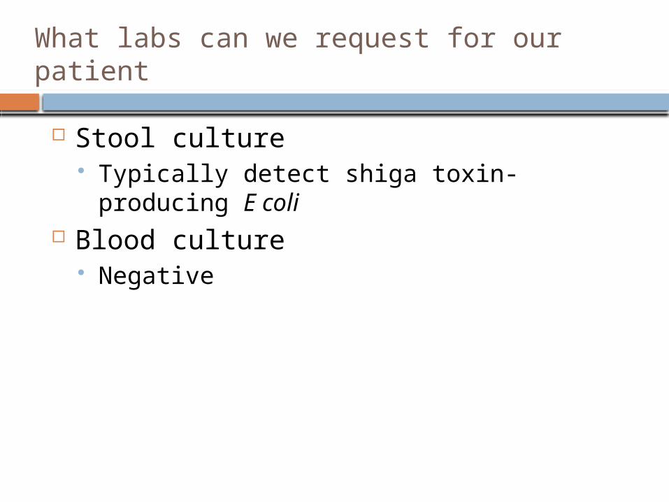
Stool culture Typically detect shiga toxin-producing E coli
Blood culture Negative

Treatment

Treatment
Current treatment of STEC induced HUS targets gastrointestinal, hematologic, vascular and renal complications. It includes isotonic volume replacement or
expansion, red blood cell and platelet transfusion and, for severe AKI, hemo- or peritoneal dialysis
Nelson’s Textbook of Pediatrics, 18th edition
Kidney International (2009) 75, S62–S66; doi:10.1038/ki.2008.624Treatment options for HUS secondary to Escherichia coli O157:H7
1Division of Nephrology, Montreal Children's Hospital and McGill University, Montreal, Quebec, CanadaCorrespondence: Martin Bitzan, Division of Nephrology, Montreal Children's Hospital, 2300, rue Tupper –
E222, Montreal, Quebec, Canada H3H 1P3.

Treatment
Supportive care with meticulous attention to Anemia Thrombocytopenia AKI and complications
Fluid and electrolytes Hypertension Uremia
Wong CS, Jelacic S, Habeeb RL, Watkins SL, Tarr PI: The risk of the hemolytic-uremic syndrome after antibiotic treatment of Escherichia coli O157:H7 infections. N Engl J Med 342 : 1930 –1936, 2000
Bihar Pedicon 2006 - Conference Abstracts.Pediatric Oncall [serial online] 2006 [cited 15 September 2006(Supplement 9)];3.

Anemia
packed red blood cells (RBC) should be transfused when the hemoglobin (Hgb) level is <7 g/dL or hematocrit <18
About 80 percent of patients w/ HUS require PRBC transfusions
guidelines for children >4 months of age are similar to those used in adults. In general, RBC transfusion is recommended for Hgb levels <7 g/dL, since most such children become symptomatic with malaise, irritability, and/or lassitude
Teruya, Jun (2009). Indications for RBC transfusion in infants and children. Uptodate 17.1 Desktop

Thrombocytopenia
Platelet transfusion is reserved for patients with HUS who have significant clinical bleeding or if an invasive procedure is required.
Rare bec platelet count rarely <10,000/mm3
based upon unsubstantiated concerns that consumption of infused platelets would contribute to new or expanding thrombi as reported in adults with thrombotic thrombocytopenic purpura
Niaudet, Patrick (2009). Clinical Manifestation and diagnosis of Shiga-like toxin associated (typical) Hemolytic Uremic Syndrome in children.
Uptodate 17.1 desktop

Fluids
Fluid management is based upon the intravascular fluid status of the patient and renal function. Decreased intravascular volume are repleted
to an euvolemic state. Increased volume and diminished urine
output are fluid restricted. Diuretics rarely avert anuria but a trial of
furosemide (2 to 5 mg/kg per dose) may be attempted to induce a diuresis
Dialysis therapy is required if fluid restriction and/or diuretic therapy fail
Niaudet, Patrick (2009). Clinical Manifestation and diagnosis of Shiga-like toxin associated (typical) Hemolytic Uremic Syndrome in children.
Uptodate 17.1 desktop

Fluids
Once the patient is in an euvolemic state, administration of fluids should be given as insensible losses plus urine output until renal function returns to normal
Niaudet, Patrick (2009). Clinical Manifestation and diagnosis of Shiga-like toxin associated (typical) Hemolytic Uremic Syndrome in children.
Uptodate 17.1 desktop

Electrolytes
Common due to acute kidney injury/failure Hyperkalemia Hyperphosphatemia Metabolic acidosis
Niaudet, Patrick (2009). Clinical Manifestation and diagnosis of Shiga-like toxin associated (typical) Hemolytic Uremic Syndrome in children.
Uptodate 17.1 desktop

Electrolytes
Hyperkalemia Stabilization of the cardiac membrane with the intravenous
calcium (calcium gluconate 10 percent solution in a dose 0.5 to 1.0 mL per kilogram intravenously over 5 to 15 minutes).
Promotion of potassium movement from the extracellular fluid (ECF) into the cells via three different therapies: Glucose and insulin (0.5 to 1.0 g of glucose per kilogram over 30
minutes and 0.1 unit of insulin per kilogram intravenously or subcutaneously);
intravenous sodium bicarbonate (in a dose of 1 to 2 milliequivalent per kilogram over 30 to 60 minutes);
beta agonists, such as albuterol, via nebulization (2.5 mg if the child weighs less than 25 kilogram or 5 mg if the child weighs more).
Niaudet, Patrick (2009). Clinical Manifestation and diagnosis of Shiga-like toxin associated (typical) Hemolytic Uremic Syndrome in children.
Uptodate 17.1 desktop

Electrolytes
Hyperphosphatemia and hypocalcemia Oral phosphate binders and dietary
restriction of phosphorus are commonly used to decrease intestinal absorption of phosphorus.
Intravenous administration of calcium gluconate should be considered if hypocalcemia is severe and/or if bicarbonate therapy is required for severe acidosis and hyperkalemia
Niaudet, Patrick (2009). Clinical Manifestation and diagnosis of Shiga-like toxin associated (typical) Hemolytic Uremic Syndrome in children.
Uptodate 17.1 desktop

Electrolytes
Acidosis Due to inability to excrete acid. Patients with serum bicarbonate levels that
are above 14 mEq/L or with arterial pH greater than 7.2 do not require intervention.
Niaudet, Patrick (2009). Clinical Manifestation and diagnosis of Shiga-like toxin associated (typical) Hemolytic Uremic Syndrome in children.
Uptodate 17.1 desktop

Dialysis
2/3 of HUS patients will require dialysis Indications for dialysis include the following:
Volume overload with evidence of hypertension and/or pulmonary edema refractory to diuretic therapy
Persistent hyperkalemia Severe metabolic acidosis unresponsive to medical
management Neurologic symptoms (altered mental status, seizures) Blood urea nitrogen greater than 100–150 mg/dL (or
lower if rapidly rising) Calcium/phosphorus imbalance, with hypocalcemic
tetany Inability to provide adequate nutritional intake because
of the need for severe fluid restrictionKliegman: Nelson Textbook of Pediatrics, 18th ed.

Dialysis
Peritoneal dialysis may contribute to the dissolution of vascular thrombi by removing fibrinolysis inhibitors and circulating plasminogen activating inhibitor-1, thereby activating normal endogenous fibrinolytic pathways.
Nelson’s Textbook of Pediatrics, 18th edition

Hypertension
caused by overexpansion of intravascular volume and/or ischemia-induced activation of the renin-angiotensin system calcium channel blockers (such as
nifedipine or nicardipine) as the initial choice of antihypertensive
changed to ACE inhibitors in patients who appear to have long-term renal sequelae
Niaudet, Patrick (2009). Clinical Manifestation and diagnosis of Shiga-like toxin associated (typical) Hemolytic Uremic Syndrome in children.
Uptodate 17.1 desktop

Prevention

Antibiotics
Prevention of HUS by antibiotic therapy to treat E.coli intestinal infection or early HUS is a very controversial subject. antibiotic therapy at the stage of gastrointestinal infection
with Stx-E. coli increases—by approximately 17-fold—the risk for full-blown HUS. I antibiotic-induced injury to the bacterial membrane might favor
the acute release of large amounts of toxins. Other reports failed to show a higher risk for HUS associated
with antibiotic administration.
On the basis of available data, we suggest that in patients with Stx-E. coli gastrointestinal infection, antibiotics should be avoided unless in cases with sepsis.
Scheiring J, Andreoli SP and Zimmerhackl LB. Treatment and outcome of Shiga-toxin-associated hemolytic uremic syndrome (HUS). Pediatr Nephrol (2008) 23:1749–1760

Anti-motility agents
In children with hemorrhagic colitis due EHEC infection, the use of antimotility agents has been associated with a greater risk for developing HUS. Thus, antidiarrheal agents are usually avoided contributes to retention of Stx within the
colon, which could enhance absorption of the toxin
Scheiring J, Andreoli SP and Zimmerhackl LB. Treatment and outcome of Shiga-toxin-associated hemolytic uremic syndrome (HUS). Pediatr Nephrol (2008) 23:1749–1760

THANK YOU

Peritoneal Dialysis (PD) vs Intermittent Hemodialysis (IHD) vs Continual Renal Replacement Therapy (CRRT)PD IHC CRRT
BENEFITS Fluid removal + ++ ++ Urea and creatinine clearance + ++ + Potassium clearance ++ ++ + Toxin clearance + ++ + COMPLICATIONS Abdominal pain + - - Bleeding - + + Dysequilibrium - + - Electrolyte imbalance + + + Need for heparinization - + + Hyperglycemia + - - Hypotension + ++ + Hypothermia - - + Central line infection - + + Inguinal/abdominal hernia + - - Peritonitis + - - Protein loss + - - Respiratory compromise + - - Vessel thrombosis - + +
Nelson’s Textbook of Pediatrics, 18th edition