er stress activates nf-kb by integrating functions of ... · er stress activates nf-kb by...
TRANSCRIPT

ER Stress Activates NF-kB by Integrating Functions ofBasal IKK Activity, IRE1 and PERKArvin B. Tam1, Ellen L. Mercado2, Alexander Hoffmann2, Maho Niwa1*
1 Division of Biological Sciences, Section of Molecular Biology, University of California, San Diego, La Jolla, California, United States of America, 2 Department of Chemistry
& Biochemistry, University of California San Diego, La Jolla, California, United States of America
Abstract
NF-kB, a transcription factor, becomes activated during the Unfolded Protein Response (UPR), an endoplasmic reticulum(ER) stress response pathway. NF-kB is normally held inactive by its inhibitor, IkBa. Multiple cellular pathways activate IKK(IkBa Kinase) which phosphorylate IkBa leading to its degradation and NF-kB activation. Here, we find that IKK is requiredfor maximum activation of NF-kB in response to ER stress. However, unlike canonical NFkB activation, IKK activity does notincrease during ER stress, but rather the level of basal IKK activity is critical for determining the extent of NF-kB activation.Furthermore, a key UPR initiator, IRE1, acts to maintain IKK basal activity through IRE1’s kinase, but not RNase, activity.Inputs from IRE1 and IKK, in combination with translation repression by PERK, another UPR initiator, lead to maximal NF-kBactivation during the UPR. These interdependencies have a significant impact in cancer cells with elevated IKK/NF-kBactivity such as renal cell carcinoma cells (786-0). Inhibition of IKK by an IKK inhibitor, which significantly decreases NF-kBactivity, is overridden by UPR induction, arguing for the importance of considering UPR activation in cancer treatment.
Citation: Tam AB, Mercado EL, Hoffmann A, Niwa M (2012) ER Stress Activates NF-kB by Integrating Functions of Basal IKK Activity, IRE1 and PERK. PLoSONE 7(10): e45078. doi:10.1371/journal.pone.0045078
Editor: Marianne Koritzinsky, University Health Network, Canada
Received January 16, 2012; Accepted August 16, 2012; Published October 26, 2012
Copyright: � 2012 Tam et al. This is an open-access article distributed under the terms of the Creative Commons Attribution License, which permits unrestricteduse, distribution, and reproduction in any medium, provided the original author and source are credited.
Funding: This work was supported by National Institutes of Health (NIH) (GM087415), and American Cancer Society (RSG-10-027-01-CSM & RSG-05-01-GMC) toMN and by NIH (GM071573) to AF. The funders had no role in study design, data collection and analysis, decision to publish, or preparation of the manuscript.
Competing Interests: The authors have declared that no competing interests exist.
* E-mail: [email protected]
Introduction
After emerging from the ribosome, secreted and membrane
proteins are initially targeted and translocated into the ER as
nascent polypeptides. To ensure their proper function, these
polypeptides will have to be folded into specific conformations and
modified properly within the lumen of the ER. As production of
unfolded or partially folded proteins will cause deliterious effects,
the ER has a quality control mechanism to ensure only properly
folded protiens can leave the ER. In response to increased
demands of producing secreted or membrane proteins, collectively
termed ‘ER stress,’ the ER functional capacity is adjusted by a
signal transduction pathway called the Unfolded Protein Response
(UPR). Activation of the UPR leads to not only changes in the
transcription profile, but also global translation repression. These
events together allow adjustment of overall ER functions [1,2,3].
Ultimately, cells that fail to re-establish the proper ER protein
folding capacity will be eliminated by induction of apoptosis. UPR
signaling is emerging as a contributing factor to the pathology of
several human diseases including diabetes and cancer [4,5,6]. The
UPR contributes to the growth and survival of tumors, and tumor
microenvironments have been found to induce UPR signaling
[4,5].
In higher eukaryotes, UPR signaling is initiated by three ER
transmembrane sensors, the kinase PERK, the kinase/RNase
IRE1, and the transcription factor ATF6 (reviewed in [1,2,3]).
Activation of PERK leads to global protein translation inhibition
by phosphorylation of eIF2a, a translation initiation factor [7]. At
the same time, PERK also promotes transcription of UPR-specific
genes by increasing translation of the transcription factor ATF4
[7]. IRE1 excises an intron from XBP1 mRNA [8], generating a
spliced version of mRNA coding for a more potent form of a UPR
transcription factor. The third UPR sensor, ATF6, is an ER
transmembrane protein with a transcription activation domain on
its cytoplasmic side. In response to ER stress, ATF6 undergoes
proteolysis within the transmembrane domain to release its
cytoplasmic transactivation domain from the ER membrane,
allowing it to enter into the nucleus [9]. Thus, each ER proximal
sensor is ultimately responsible for activation of a transcription
factor. Activation of ATF6, ATF4, and XBP1 by the UPR result in
a complex pattern of gene regulation [2,3]. UPR signaling aims to
alleviate the high levels of misfolded proteins in the ER by
increasing protein folding capacity through up-regulation of ER
chaperones such as BiP, GRP94, calreticulin, and Erdj4 [10,11]. If
proper protein folding capacity in the ER cannot be restored, the
UPR up-regulates genes such as CHOP that result in activation of
apoptotic pathways. [10]
In addition, activation of the highly studied transcription factor
NF-kB has been reported to be a consequence of ER stress
[12,13,14], although its role during the UPR has yet to be
determined. NFkB consists of a family of dimer forming
transcription factors that include RelA(p65), p50, p52, RelB, and
c-Rel with RelA(p65)/p50 being the canonical form. Normally
held in the cytoplasm in complex with IkBa, an inhibitor of NF-
kB, canonical activation of NF-kB involves phosphorylation of
IkBa by IkB kinase (IKK), followed by proteasome-mediated
degradation of IkBa. IKK is comprised of IKKa, IKKb, and
IKKc (NEMO) subunits, and during canonical activation, IKKbphosphorylates IkBa on serines at position 32 and 36, leading to
PLOS ONE | www.plosone.org 1 October 2012 | Volume 7 | Issue 10 | e45078

its polyubiquitination and proteosomal degradation [15]. This
frees NF-kB for nuclear accumulation, binding to consensus kB
promoter sites, and transcriptional activation of target genes.
Genes regulated by NF-kB primarily promote survival, making
NF-kB a key player in the development of invasive tumors and
metastases, and in resistance to certain chemotherapeutic agents
[16].
NF-kB can be activated by several stimuli including inflamma-
tory signals such as tumor necrosis factor alpha (TNFa),
interleukin-1 (IL-1), lipopolysaccharide (LPS), and internal cell
stresses such as DNA damage [17]. Signals initiated by these
stimuli converge by activating the IKK complex resulting in IKK
mediated degradation of IkBa. Thus, IKK activation is a key
regulatory step of NF-kB activation. Recent reports reveal that the
mechanism of NF-kB activation during ER stress may differ from
this conventional activation. Specifically, studies linking NF-kB
activation to PERK-mediated phosphorylation of eIF2a proposed
NF-kB activation during ER stress was independent of IKK
activation since IkBa phosphorylation was not detected in mouse
embryonic fibroblasts (MEFs) [12,13]. However, a rapid reduction
in IkBa takes place via translation inhibition induced by eIF2aphosphorylation. Since IkBa has a short half life, this consequently
results in an increase in free NF-kB, leading to its activation.
More recently, another report using a human breast cancer cell
line found IRE1 physically interacts with IKK upon ER stress
induction, leading to an increase in P-IkBa and the concomitant
decrease in total IkBa levels, resulting in NF-kB activation.
Furthermore, the initial activation of NF-kB led to production of
TNFa leading to further cannonical NF-kB acitvation, ultimately
leading to cell death [18]. Currently, the relationships between
these reports, specifically IKK phosphorylation of IkBa during ER
stress, are not clear, as different cell lines were used for their
experiments. IKK is a master regulator of NF-kB activation, and
signaling through IKK is critical for almost all NF-kB activating
pathways. Therefore, understanding the involvement of IKK and
its relationship to both IRE1 and PERK is critical for determining
how NF-kB is activated during ER stress.
Using mouse embryonic fibroblasts (MEFs) knocked out for
components of the pathway, we identified the individual roles of
IRE1, PERK, and IKK, and showed that full activation of NF-kB
druing ER stress requires contributions from all three components.
Interestingly, we show that IKKb is required for NF-kB activation,
however no increase in IKK activity is seen during ER stress. This
seemingly paradoxical result led us to the discovery that basal IKK
kinase activity is sufficient and necessary for NF-kB signalling
during ER stress. Furthermore, while PERK mediated transla-
tional inhibition is sufficient to activate NF-kB, cells lacking IRE1
cannot fully activate NF-kB due to decreased basal IKK activity.
Also, increasing basal IKK activity in cells lacking IRE1 rescues
NF-kB activity. Thus, basal IKK activity, maintained by IRE1,
and PERK mediated translation inhibition work together to
activate NF-kB during ER stress. Finally, we demonstrate a
functional implication for this observation using cancer cells that
have increased basal IKK activity where induction of the UPR can
reverse the action of an IKK inhibiting drug.
Results
IKK is Required for Activation of NF-kB during UPRTo probe the mechanistic details of NF-kB activation during
ER stress, in particular, potential involvement of IKK-mediated
IkBa phosphorylation, we examined ER stress induced NF-kB
activation in wild type (WT) mouse embryonic fibroblasts (MEFs)
and MEFs lacking the a and b subunits of IKK (ikk2/2) [19,20].
ER stress was induced by DTT and Thapsigargin (Tg), two well
established UPR activating agents. DTT disrupts disulfide bonds
resulting in unfolded proteins, while Tg ultimately blocks ER
chaperone function by disrupting ER calcium levels. NF-kB was
measured by electrophoretic mobility shift assay (EMSA) [14],
which involves incubation of a radio labeled DNA probe
containing the NF-kB binding site with nuclear extracts prepared
from WT MEFs treated with DTT or Tg. We found that UPR
induction resulted in NF-kB activation (Figure 1A, IKK+/+). Both
competition and supershift assays confirmed that the activated
form of NF-kB is the cannonical p65/p50 form (Figure S1A–S1C).
Furthermore, consistent with the EMSA results, immunofluores-
cence detected NF-kB translocation into the nucleus during ER
stress (Figure S1D). NF-kB activation was also observed with DTT
and Tg in multiple cell types (Figure S2A–S2D). While wild type
MEFs showed robust activation of NF-kB in different cell types,
we found that NF-kB activation was significantly diminished in
ikk2/2 MEFs (Figure 1A, ikk2/2), indicating a requirement for
IKK during UPR induced activation of NF-kB. To further
determine which IKK subunit is required for NF-kB activation,
we used MEFs lacking either ikka2/2 or ikkb2/2 (Figure S1E). We
found that cells lacking IKKa still induced NF-kB upon ER stress
induction while cells lacking IKKb were not able to induce NF-
kB. Furthermore, we found that the kinase activity of IKKb was
required, as only kinase active IKKb, not kinase dead IKKb, can
rescue NF-kB activity in ikkb2/2 cells (Figure S1E). These results
revealed the importance of IKKb during ER stress, a similar
requirement for cannonical NF-kB activation.
To further test involvement of IKK, we examined the
requirement of IkBa phosphorylation. We took IkBa2/2 cells
and added back either wild type IkBa (IkBa-WT) or a non-
phosphorylatable IkBa where the IKK phosphorylation sites
(Ser32/36) were mutated to Alanine (IkBa-SR), preventing its
phosphorylation (Figure 1B lower panel, P- IkBa) [21]. Cells
expressing IkBa-SR showed significantly reduced NF-kB activa-
tion during the UPR (Figure 1B upper panel). As IkBa was
expressed at similar levels between cells expressing IkBa-WT and
IkBa-SR (Figure 1B lower panels, IkBa), an inability to activate
NF-kB in cells expressed IkBa-SR was not due to difference in its
expression level from that of WT IkBa. Thus, together with
diminished NF-kB activation in ikk2/2 or ikkb2/2 MEFs
(Figure 1A & S1E), these data suggested the importance of IKK
mediated IkBa phosphorylation in UPR induced NF-kB activa-
tion.
During canonical NF-kB activation, IkBa phosphorylation
leads to decreased IkBa levels due to proteasomal degradation.
Similarly, total IkBa was decreased after UPR induction
(Figure 1C, IkBa). Curiously, however, we found that phosphor-
ylation of IkBa was not induced during the UPR time course
(Figure 1C, P- IkBa), while canonical NF-kB activating agent,
TNFa treatment clearly increased P- IkBa as expected. These
results were replicated in another cell type (CHO) (Figure S2E).
Since we did not detect increased phosphorylation of IkBa during
ER stress, we wanted to further investigate whether the rate of
IkBa phosphorylation changed during ER stress. Since phosphor-
ylation of IkBa causes proteasome mediated degradation [22], we
added a proteasome inhibitor, MG132, to perk2/2 cells in order to
block proteasomal degradation and measured the rate of
accumulation of P- IkBa throughout the time course of ER stress
(+DTT) or without (2DTT) ER stress (Figure 1D). It should be
noted that these experiments were performed in perk2/2 MEFs to
avoid any change in IkBa levels caused by PERK induced
translation repression in UPR induced cells, so that the rate of
IkBa phosphorylation or IKK activity can be assessed. Rates of
Integrated Activation for NF-kB during the UPR
PLOS ONE | www.plosone.org 2 October 2012 | Volume 7 | Issue 10 | e45078

Figure 1. Basal IKK activity is required for NF-kB activation during ER stress. (A) NF-kB cannot be activated in the absence of IKK. Wild typeMEFs (IKK+/+) and MEFs knockedout for both IKKa and IKKb (ikk2/2) were treated with DTT or Tg, and EMSA was performed using a radiolabelledprobe containing an NF-kB binding site (free oligo). ‘*’ denotes a background band (Figure S1) and free oligo shows that NF-kB probe was not limitedfor this assay. (B) IkBa phosphorylation is required for full NF-kB activation during ER stress. IkBa2/2 cells were reconstituted with either wild typeIkBa (IkBa WT) or a non-phosphorylatable IkBa (IkBa SR) with Serines 32/36 were mutated to Alanines and were treated with Tg. NF-kB activationwas analyzed by EMSA. Bottom Panel: Western blots against P-IkBa, total IkBa, and actin. IkBa was expressed equivalently in both cell types, andphosphorylation was only detected in IkBa WT but not IkBa SR. (C) IkBa phosphorylation does not increase during ER stress. Protein levels ofphosphorylated IkBa (P-IkBa), total IkBa, and actin (loading control) were analyzed by western blot in WT MEFs treated wih DTT, Tg, or TNFa forindicated lengths of time (hrs). No increase in P-IkBa was detected, but a IkBa levels decreased. (D) IRE1 does not contribute to an increase in IKKactivity during the UPR. In order to prevent the effect of the decreased IkBa levels due to translation inhibition, we used perk2/2 MEFs. Cells weretreated with MG132 in the absence (2DTT, closed circle) or presence (+DTT, open circle) of UPR induction. Levels of P-IkBa, total IkBa, and actin weredetermined by western blot. Accumulation of P-IkBa normalized to total IkBa was determined as a measure of basal IKK activity. At least, threeindependent experiments are performed to calculate standard error. (E) IKK kinase activity does not increase during ER stress. IKK kinase assay usingIP’d IKK complexes from WT MEFs treated with either Tg, DTT or TNFa. IP’d complexes were incubated with recombinant IkBa and c32P-ATP.Radiolabelled IkBa was then detected by autoradiography. Efficiencies of IKK IP were shown by depletion of IKKa (bottom panel). Total extracts andextracts after depletion were probed for IKKa protein levels to determine the efficiency of IKK complex IP. No antibody denotes a negative controlusing no antibody during IP (lanes 2, 10, 15). (2) shows a negative control where no kinase was added (lane 8).doi:10.1371/journal.pone.0045078.g001
Integrated Activation for NF-kB during the UPR
PLOS ONE | www.plosone.org 3 October 2012 | Volume 7 | Issue 10 | e45078

accumulation of P- IkBa in the presence of MG132 were
equivalent in untreated and UPR-induced cells (Figure 1D),
indicating that UPR activation does not change either the rate of
IkBa phosphorylation or IKK activity. Therefore, UPR decreased
the overall level of IkBa even though no significant increase in its
phosphorylation was observed.
To further investigate these apparent discrepancies in the IKK
involvement in NF-kB activation during UPR, we examined IKK
kinase activity using immunoprecipitated (IP) IKK from total cell
extracts, followed by incubation with recombinant IkBa substrate
and (c32P)-ATP (Figure 1E, top panel) using in vitro kinase assay.
Efficient IP of the IKK complex occurred as seen by depletion of
IKKa levels from extracts (Figure 1E, bottom panel, compare
lanes 9–13 (total) with lanes 14–18 (after depletion)). TNFashowed high IKK kinase activity (Figure 1E, lane 3) [22], while
UPR-induced cells showed IKK activity equal to uninduced levels
(Figure 1E, lanes 1 and 4–7), indicating that IKK activity did not
increase during ER stress. Taken together, our results revealed
that IKK mediated IkBa phosphorylation was required for NFk-B
activation (Figure 1A & 1B), despite no increase in IKK activity or
phosphorylaton of IkBa during the UPR (Figure 1C, 1D & 1E).
PERK-Mediated Translation Repression is Not Sufficient toFully Activate NF-kB During UPR in Cells Lacking IRE1
Another puzzling observation is the relative contribution of
IRE1 and PERK. To date, there is no direct molecular event
reported that simultaneously requires both IRE1 and PERK for its
activation. To identify functional contributions of each compo-
nent, we used MEFs knocked out in either IRE1 (ire12/2) or
PERK (perk2/2). Previous studies reported that PERK mediated
translational inhibition was sufficient to induce NF-kB during the
UPR [12]. Consequently, perk2/2 MEFs which cannot mount
UPR-induced translation inhibition (Figure 2A), resulted in
significantly diminished activation of NF-kB during UPR activa-
tion when compared with WT (Figure 2B), in agreement with
previous reports [12]. Furthermore, we found that the extent of
translation inhibition determines the level of NF-kB activation
(Figure S3A) in wild type cells. This indicates that the contribution
of PERK, translation inhibition, is sufficient to activate NF-kB in
wild type cells.
To determine the functional role of IRE1, we asked if PERK
mediated translation inhibition was sufficient to activate NF-kB in
cells lacking IRE1. We first determined that PERK mediated
translation inhibition is equivalent in WT and ire12/2 cells
(Figure 2C). If translation repression mediated reduction of IkBawas sufficient for maximum NF-kB activation during UPR, we
anticipated NF-kB activation in ire12/2 cells to take place
normally during ER stress, similar to that in WT cells. However,
despite normal PERK-dependent translational inhibition, we
found that the overall NF-kB activity was greatly diminished in
ire12/2 cells (Figure 2D). Additionally, we used cycloheximide
(CHX), a pharmacological inhibitor of translation, to substitute for
PERK mediated translation inhibition [7], and found that CHX
activated NF-kB in WT (IRE1+/+) cells, while CHX treatment of
ire12/2 cells had significantly reduced NF-kB (Figure 2E). Taken
together, these data demonstrated a functional contribution of
IRE1 to NF-kB activation beyond PERK dependent translation
inhibition. To further confirm that IRE1 does have a functional
contribution, NF-kB activity in ire12/2 cells was restored by
transfection of IRE1, where the extent of NF-kB recovery after
IRE1 transfection was directly proportional to that of IRE1-
mediated XBP1 splicing (Figure S3B & S3C). Together, these
results showed that loss of either IRE1 or PERK resulted in
diminished NF-kB signaling during the UPR, suggesting com-
bined inputs from the IRE1 and PERK signaling branches were
needed for NF-kB activation.
The importance of both IRE1 and PERK for NF-kB activation
during the UPR was also evaluated at the downstream transcrip-
tion level. Using an NF-kB luciferase reporter assay, reduced levels
of luciferase was detected in ire12/2 cells (Figure 2F), in agreement
with the reduced level of activated NF-kB binding to the DNA
detected by EMSA (Figure 2D). To further test the functional
significance of NF-kB activation during the UPR, we examined
the induction of a UPR target gene, BiP/GRP78 in MEFs
knocked out for p65 (nf-kb2/2). These cells did not show active
NF-kB (data not shown). We found that in p65 knockout cells,
BiP/GRP78 expression was diminished compared to wild type
cells, indicating that NF-kB is required for full activation of BiP
(Figure 2G). Using chromatin IP (ChIP), we found that p65 is able
to bind to the promoter of BiP/GRP78 (Figure 2H), indicating
that NF-kB activates transcription of BiP/GRP78 through direct
interaction with BiP/GRP78 promoter. Furthermore, NF-kB
binding to the promoter was IRE1 and PERK dependent
demonstrating the importance of these components in activation
of functional NF-kB during the UPR.
The Kinase Activity of IRE1 Is Required for NF-kBActivation During ER Stress
Since cells lacking IRE1 showed severely decreased NF-kB
during UPR, and IRE1 is a multi-functional protein, we wanted to
examine the importance of IRE1 kinase and RNase activities. To
separate each individual activity, we used a kinase dead form of
IRE1 (IRE1-KD) and a nuclease dead IRE1 (IRE1-ND). IRE1-
KD (K599A) was previously shown to inactivate IRE1 kinase both
in vivo and in vitro [23]. Similarly, IRE1-ND (K907A), inactivated
IRE1 RNase without affecting IRE1 kinase activity [24]. We
transfected WT IRE1, kinase dead IRE1 (IRE1-KD), or nuclease
dead IRE1 (IRE1-ND) into ire12/2 MEFs and determined NF-kB
activation by NF-kB reporter luciferase assay. Western blotting
shows that transfected IRE1 were expressed at similar levels as
WT (Figure 3A, bottom panel). As shown previously (Figure 2F),
ire12/2 MEFs displayed decreased NF-kB activation, however,
transfecting wild type IRE1 rescues NF-kB activity (Figure 3A).
Expression of IRE1-ND in ire12/2 MEFs essentially restored most
of the NF-kB activity achieved by wild type IRE1 (Figure 3A). In
contrast, expression of IRE1-KD was not able to rescue NF-kB
activation in ire12/2 MEFs, demonstrating the importance of
IRE1 kinase for NF-kB activation during the UPR.
While UPR activation results in IRE1 kinase activation, the
functional contribution of IRE1’s kinase in the UPR response has
not been well characterized. However, the kinase function of IRE1
has been associated with its interaction with TRAF2, an adaptor
protein that links IRE1 to c-JUN N-terminal kinase (JNK)
activation [25]. Thus, we tested the involvement of TRAF2 and
the JNK pathway in ER stress induced NF-kB activation. To test
involvement of TRAF2, we used a dominant negative form of
TRAF2 (TRAF2-DN) (D1–87), that has been shown to block IKK
activation during canonical NF-kB pathway [26]. Transfection of
TRAF2-DN into WT cells resulted in decreased levels of UPR-
induced NF-kB activation (Figure 3B), indicating the importance
of TRAF2 in the pathway. We also noticed that TRAF2 protein
level itself was reduced in ire12/2 cells (Figure 3B). Thus, these
results revealed that (i) TRAF2 is required for NF-kB activation
and (ii) IRE1 may regulate cellular concentration of TRAF2. In
contrast to TRAF2, however, loss of JNK in either jnk12/2 or
jnk22/2 MEFs did not affect NF-kB activation significantly
(Figure 3C). Furthermore, we inhibited both JNK1 and JNK2
using a well-established JNK Inhibitor (JNKi, SP600125) known to
Integrated Activation for NF-kB during the UPR
PLOS ONE | www.plosone.org 4 October 2012 | Volume 7 | Issue 10 | e45078
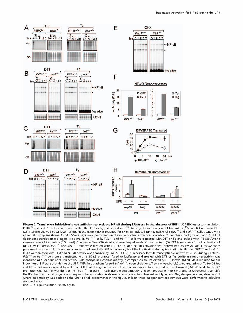
Figure 2. Translation inhibition is not sufficient to activate NF-kB during ER stress in the absence of IRE1. (A) PERK represses translation.PERK+/+ and perk2/2 cells were treated with either DTT or Tg and pulsed with 35S-Met/Cys to measure level of translation (35S panel). Coomassie Blue(CB) staining showed equal levels of total protein. (B) PERK is required for ER stress induced NF-kB. EMSAs of PERK+/+ and perk2/2 cells treated witheither DTT or Tg are shown. Oct-1 EMSA assays were performed on the same nuclear extracts as a control. ‘*’ denotes a background band. (C) PERKdependent translation repression is normal in ire12/2 cells. IRE1+/+ and ire12/2 cells were treated with DTT or Tg and pulsed with 35S-Met/Cys tomeasure level of translation (35S panel). Coomassie Blue (CB) staining showed equal levels of total protein. (D) IRE1 is necessary for full activation ofNF-kB by ER stress. IRE1+/+ and ire12/2 cells were treated with DTT or Tg, and NF-kB activation was determined by EMSA. Oct-1 EMSAs wereperformed as a control. ‘*’ denotes a background band. (E) IRE1 is necessary for NF-kB activation during translation inhibition. IRE1+/+ and ire12/2
MEFs were treated with CHX and NF-kB activity was analyzed by EMSA. (F) IRE1 is necessary for full transcriptional activity of NF-kB during ER stress.IRE1+/+ or ire12/2 cells were transfected with a 3X kB promoter fused to luciferase and treated with DTT or Tg. Luciferase reporter activity wasmeasured as a readout of NF-kB activity. Fold change in luciferase activity in comparison to untreated cells is shown. (G) NF-kB is required for fullinduction of BiP transcript during the UPR. MEFs knocked out for p65 (nf-kb2/2, open circle) or WT cells (closed circle) were treated with Tg for 24 hrsand BiP mRNA was measured by real time PCR. Fold change in transcript levels in comparison to untreated cells is shown. (H) NF-kB binds to the BiPpromoter. Chomatin IP was done on WT, ire12/2, or perk2/2 cells using a-p65 antibody, and primers against the BiP promoter were used to amplifythe IP’d fraction. Fold change in relative promoter association is shown in comparison to untreated wild type cells. Neg designates a negative controlwhere no antibody was added to the ChIP. For all experiments in this figure, at least three independent experiments were performed to calculatestandard error.doi:10.1371/journal.pone.0045078.g002
Integrated Activation for NF-kB during the UPR
PLOS ONE | www.plosone.org 5 October 2012 | Volume 7 | Issue 10 | e45078

inhibit both forms of JNK [27], which has been previously shown
to inhibit IRE1 dependent JNK activation during ER stress [28].
Treatment of cells with JNKi, which inhibited both JNK1 and
JNK2 (Figure S4A), showed no effect on NF-kB activation
(Figure 3C, +JNKi). Therefore, these data suggests that NF-kB
activation during ER stress is mediated via IRE1-TRAF2 but not
via JNK.
TRAF2 has been shown to regulate the activity of IKK in other
signalling pathways [29–32]. We reasoned that diminished NF-kB
activity in ire12/2 MEFs may come from altered compositions or
states of IKK complex components, and thus, we determined the
state of the IKK complex in WT versus ire12/2 cells. We first
looked at protein levels of the IKK components to determine if
IRE1 regulated their cellular concentrations. We found that
protein levels of IKKa, IKKb, and IKKc were equivalent in WT
and ire12/2 cells (Figure 3D), indicating that IRE1 does not affect
protein levels of IKK components. Also, IRE1 may affect the
composition of the IKK complex, so to test this, we immunopre-
cipitated the IKKc and determined if equivalent amounts of
IKKa and IKKb were associated with it. We found that equal
amounts of IKKa, IKKb, and IKKc were associated with each
other in WT and ire12/2 cells (Figure 3E), indicating that IRE1
does not affect the physical composition of the IKK complex.
Despite the presence of the intact IKK complex, however, IKKbactivity measured by its autophosphorylation was significantly
diminished in ire12/2 cells (Figure 3F). Furthermore, the
diminished phosphorylation can be rescued by WT and ND
IRE1, but not KD IRE1, correllating with NF-kB activation
(compare Figure 3A & 3F). Taken together, these results indicate
that the kinase domain of IRE1 regulates NF-kB activation
through TRAF2-IKK signalling.
Basal IKK Kinase Activity is Diminished in the Absence ofIRE1
At the first glance, the lack of changes in either IKK complex
itself or activity of IKK during ER stress seemed contradictory to
involvement of IKK (Figure 1). Thus, we decided to further dissect
the relationships between IKK, IRE1, and PERK. Although IKK
is required for NF-kB activation, IKK activity did not increase
during the UPR (compare Figure 1A/1B vs 1C/1D/1E). These
observations suggested that the uninduced or basal IKK activity
was sufficient to achieve NF-kB activation during the UPR.
Recently, the importance of basal IKK activity in NF-kB activation
by the ribotoxic stimulus UV was reported [21] [33], and thus, we
tested the involvement of basal IKK kinase activity for UPR
induced NF-kB. Since the traditional IKK kinase assay was not
sensitive enough to detect changes in basal IKK activity
(Figure 1E), we used accumulated P-IkBa levels as a readout for
basal IKK activity. We measured basal IKK activity in ire12/2 and
perk2/2 cells, without any UPR inducing agents, to determine if
loss of IRE1 or PERK affected basal IKK activity (Figure 4A&4B).
We found that in ire12/2 MEFs, the rate of P-IkBa accumulation
was 2-fold lower over the time course compared to WT
(Figure 4A). In contrast, P-IkBa accumulation in perk2/2 MEFs
was identical to that of WT cells (Figure 4B), suggesting a role for
IRE1, but not PERK, in retaining basal IKK activity.
Since IKK-mediated phosphorylation of IkBa leads to its
degradation, these results predicted that IkBa in ire12/2 cells
would have a longer half-life in uninduced resting or basal
conditions. We assessed the half-life of IkBa in the presece of
CHX, which prevented synthesis of new IkBa, and measured the
rate of degradation of existing IkBa. Indeed, we found that IkBadisappeared more rapidly in WT than ire12/2 MEFs (Figure 4C),
consistent with the idea that the half life of IkBa was longer in
ire12/2 MEFs than WT cells. Together, these results suggested the
exciting possibility that robust activation of NF-kB during the
UPR requires a certain level of basal IKK activity, which needs to
be present even prior to UPR induction. Furthermore, these
results also argued that IRE1 affected basal IKK activity.
Restoring Basal IKK Activity Can Rescue NF-kB Activationin ire12/2 Cells
The relationship between IRE1 and IKK suggests that
decreased NF-kB activation in ire12/2 MEFs is caused by
decreased basal IKK activity. To further test this idea, we
increased basal IKK activity in ire12/2 MEFs by increasing the
amount of IKK and determined if efficient NF-kB activation was
restored. To this end, ire12/2 cells were transfected with extra
copies of either IKKa or IKKb. Indeed, we found that expression
of IKKß, but not IKKa, in ire12/2 MEFs restored the basal level
of IKK activity as measured by accumulation of P-IkBa in the
presence of MG132 (Figure 4D). Consequently, IKKb, but not
IKKa, was able to rescue NF-kB activation to wild type levels in
the ire12/2 cells (Figure 4E). Since IKKb, but not IKKa, is the
main kinase phosphorylating IkBa in cannonical NF-kB activa-
tion, this result suggests that the UPR utilzes cannonical IKK
activity for NF-kB activation. In addition, the kinase activity of
IKK is needed for activation of NF-kB by UPR, because
transfection of a kinase-dead mutant of IKKb did not rescue
NF-kB activity to wild type levels in ire12/2 MEFs (Figure 4E,
+IKKb DN). Overall, these results highlight the importance of
IKKb kinase function in mediating UPR activated NF-kB.
These experiments reveal that basal IKK activity stimulates NF-
kB activation during the UPR. To demonstrate the functional
importance of regulating basal IKK activity, we altered levels of
basal IKK activity by transfecting of increasing amounts of IKKbinto ire12/2 MEFs and inducing the UPR (Figure 4F). Transfect-
ing 1 mg of IKKb plasmid resulted in NF-kB activation similar to
WT IRE1+/+ cells. Curiously, at high levels of IKK activity, while
active NF-kB level was elevated at the basal level prior to UPR,
activation of the UPR did not result in further increased NF-kB
activity (Figure 4F, 2 mg, compare 0 hr vs 1, 2, 5 & 7 hrs). Levels
of transfected IKKb were confirmed by western blot (Figure S4B).
These results highlight the importance of considering basal IKK
activity in mediating UPR induced NF-kB activation. Taken
together, our results show that basal IKK activity, maintained by
IRE1, is critical to activate NF-kB when PERK-induced
translation inhibition occurs. Furthermore, this finding has
implications in systems which alter IKK basal activity such as
drug development of IKK inhibitors.
ER Stress Can Override Inhibition of an IKK InhibitingDrug
Certain cancer cells have been reported to display elevated NF-
kB/IKK activity [34], and drugs have been developed that which
aim to decrease IKK activity back to basal levels [35]. However,
our results suggest that basal IKK activity is sufficient to activate
NF-kB during the UPR, so we wanted to test if UPR activation
can counteract the effect of the IKK inhibiting drug. 786-0 renal
carcinoma cells were previously characterized to have constitu-
tively active NF-kB activity [36,37]. EMSA assays revealed that
basal NF-kB activity was significantly elevated in 786-0 cells as
compared to MEFs (Figure 5A, 0 h). UPR induction in 786-0 cells
was unable to further activate NF-kB (Figure 5A), similar to
ire12/2 MEFs transfected with high levels of IKKb (Figure 4F,
2 mg) providing additional support for the significance of
uninduced IKK activity in determining the outcome of NF-kB
Integrated Activation for NF-kB during the UPR
PLOS ONE | www.plosone.org 6 October 2012 | Volume 7 | Issue 10 | e45078

Figure 3. The kinase activity of IRE1 is required for NF-kB activation during the UPR. (A) Kinase dead IRE1 cannot rescue NF-kB activity inire12/2 cells. ire12/2 cells were transfected with either WT IRE1, kinase dead IRE1 (IRE1-KD), or nuclease dead IRE1 (IRE1-ND). Western blotsdemonstrate successful transfection of IRE1 (bottom panel). IRE1+/+, ire12/2, and ire12/2transfected with different IRE1 mutants were treated withDTT and NF-kB luciferase reporter assays were performed. Fold change in luciferase activity in comparison to untreated IRE1+/+ cells is shown. (B) NF-kB activation during the UPR requires TRAF2. WT cells were transfected with either WT TRAF2 (TRAF2) or dominant negative TRAF2 (TRAF2-DN),treated with DTT, and EMSA was used to determine the level of active NF-kB. Protein extracts from IRE1+/+ and ire12/2 cells were probed for TRAF2 byWestern Blot. Fold change in TRAF2 levels in comparison to IRE1+/+ is shown. (C) JNK is not involved in ER stress activation of NF-kB. WT, jnk12/2 orjnk22/2 cells were treated with DTT, and again EMSA was used to determine NF-kB activity. Furthermore, incubation of WT cells with 25 mM ofSP600125 (JNKi), a well-established inhibitor of both JNK1/2 for up to 7 hrs did not affect activiation of NF-kB. (D) Protein levels of IKKa, IKKb, andIKKc in ire12/2 cells are equal to wild type levels. IKKa, IKKb, and IKKc protein levels in IRE1+/+ vs. ire12/2 cells were measured using Western Blot. Foldchanges in protein levels in comparison to IRE1+/+ are shown. (E) IRE1 does not affect the composition of the IKK complex. IKK complex wasimmunoprecipitated from IRE1+/+ and ire12/2 cells using anti-IKKc antibody. The Input, depleted (Dep), and immunoprecipitated (IP) fractions areshown. Western blots were performed using antibodies against IKKa, IKKb, and IKKc. Efficient immunoprecipitation occurred as seen by low levels leftbehind in the depleted fraction. Equivalent amounts of each IKK subunit can be immunoprecipitated in WT and ire12/2 cells. (F) Basalphosphorylation of IKKb is reduced in ire12/2 cells and cannot be rescued by kinase dead IRE1. ire12/2 cells were transfected with either WT IRE1,IRE1-KD, or IRE1-ND. P-IKKb levels were determined by western blot in IRE1+/+, ire12/2, and transfected cells. Fold change in P-IKKb in comparison toIRE1+/+ is shown. For all experiments in this figure, quantitations are shown with standard error for at least three independent experiments.doi:10.1371/journal.pone.0045078.g003
Integrated Activation for NF-kB during the UPR
PLOS ONE | www.plosone.org 7 October 2012 | Volume 7 | Issue 10 | e45078

signaling. Also, 786-0 cells were able to activate PERK normally
during the UPR as measured by P-eIF2a (Figure 5A), indicating a
normal UPR response. Uninduced 786-0 cells also show elevated
IKK activity as measured by IKK kinase assay, increased P-IkBa,
and decreased total IkBa levels (Figure 5B), making these cells a
good candidate for targeting with IKK inhibitors.
Our current study predicts that, in 786-0 cells, NF-kB becomes
activatable by UPR if elevated basal IKK activity is reduced to its
normal basal level by an IKK inhibitor. Since UPR becomes
induced by the tumor microenvironment, using an IKK inhibitor
as treatment of such cancer cells may not be an effective cancer
treatment. Instead, the reduction in elevated IKK activity by the
inhibitor would allow cells to activate NF-kB in response to the
UPR inducing microenvrionment of the tumor, rendering the
inhibitor ineffective. To test our hypothesis, we treated 786-0 cells
for 1 hr with a well-characterized IKK inhibitor, SC-514 [38]
(Figure 5C, schematic), and found that SC-514 treatment
diminished both basal IKK (Figure 5D) and NF-kB activities
and continued to suppress NF-kB throughout the time course
(Figure 5F, lanes 1–4). In contrast, induction of UPR by DTT after
SC-514 treatment resulted in robust activation of NF-kB
(Figure 5F, compare lanes 2–4 for no UPR, vs lanes 6–8 with
DTT), even in the continued presence of the IKK inhibitor. Note
that SC-514 did not alter activity of PERK, judging from p-eIF2a(Figure 5E vs 5A), and thus, differences in these effects were not
due to disruptions in translational regulation. These results are
consistent with our hypothesis that the decrease in elevated IKK/
NF-kB to basal levels in cancer cells by an IKK inhibitor allowed
them to respond to UPR by activating NF-kB. Taken together,
these results provide additional support for the idea that basal IKK
activity is a critical factor in UPR induced NF-kB activation.
Discussion
The NF-kB transcription factor family regulates genes involved
in a wide array of processes including inflammation, immune
responses, apoptosis, and turmorigenesis [17]. NF-kB is also
activated by certain types of ER stress [14] [13] [12]. These
previous studies have reported involvement of either PERK or
IRE1 in UPR-induced NF-kB activation. However, studies
examining the role of PERK had never investigated significance
of IRE1, and conversely, studies reported the role of IRE1 did not
examine its relationships to involvement of PERK. Furthermore,
those studies were performed in different cell types. Thus the
relationships between PERK and IRE1 for NF-kB activation in
response to ER stress remained unclear. Here, we have shown that
optimal activation of NF-kB during ER stress requires inputs from
both IRE1 and PERK activities (Figure 6). In agreement with
previous studies, we found that global translation repression
induced by PERK-dependent phosphorylation of eIF2a plays a
critical role in activation of NF-kB during ER stress
(Figure 2A&2B). Furthermore, we have shown that the extent of
transltional inhibition propotionally correllates with the level of
NF-kB activation (Figure S3A). One of the key findings of our
study is that IRE1-dependent homeostatic regulation of basal IKK
activity is necessary for effective activation of NF-kB by PERK. An
increase or decrease in basal IKK activity affects the ability of the
PERK signaling branch to effectively activate NF-kB when ER
stress is imposed. Although we do not detect an increase in IKK
activity during the UPR in MEFs, a previous study reported that
UPR-activated IRE1 caused activation of IKK in MCF-7 cells, a
breast cancer cell line [18], raising the possibility that oncogenic
transformation may intensify the IRE1 contribution to IKK
activity. Furthermore, our subsequent analyses showing the ability
of both a nuclease dead IRE1 and IKKb to restore NF-kB
activation in ire12/2 knockout cells in response to ER stress
(Figures 3A and 4D–4F) have provided additional support for the
involvement of IRE1 in basal IKK regulation. The ability of IRE1
to modulate basal IKK activity suggests that IRE1 is never
completely inactive or ‘‘OFF’’, even in the absence of overt ER
stress, and that IRE1 has an intrinsic housekeeping function.
Previously, we reported that a basal state IRE1 function is also
required for efficient cytokinesis during mitosis in S. cerevisiae [39].
There, we proposed that IRE1 activity is modulated in a ‘‘dimmer
switch’’ fashion, rather than an ‘‘ON/OFF switch’’. Furthermore,
we have reported the role of basal activity in the NF-kB pathway
during the ribotoxic stimulus UV [21]. Taken all together,
therefore, we propose that the optimal activation of NF-kB during
the UPR is mediated by inputs from both PERK and IRE1, and
by altering either one or both of these inputs, the extent of NF-kB
activation can be fine-tuned, allowing for a dynamic response to
ER stress.
One of the questions that remains to be answered is the
molecular mechanism by which IRE1 regulates basal IKK
activity. Previously, UPR activated IRE1 has been shown to
associate with TRAF2 and JNK through its kinase, but not RNase,
domain [25]. In our current study, we found that the cellular
TRAF2 level was significantly diminished in ire12/2 knockout cells
(Figures 3B). Similarly, NF-kB activation upon UPR induction was
also diminished in the absence of IRE1 (Figure 2D). We have also
shown that the IRE1 kinase, but not RNase, is required for basal
IKKb phosphorylation (Figure 3F) and NF-kB activation
(Figure 3A). Curiously, however, JNK plays little role in UPR-
induced NF-kB activation (Figure 3C). Taken together, these data
are consistent with TRAF2 involvement in IRE1 activation of
IKK and NF-kB, pointing to the presence of a physical
interactions between IRE1, IKK and TRAF2. While previous
studies have reported a physical interaction between IRE1,
TRAF2, and IKK only takes place once UPR is activated [18],
results described here suggest that these interactions, if they occur,
should be present at the basal level. Thus, this complex may exist
transiently or be present at low levels during basal conditions. In
fact, our attempts to detect physical interactions between IRE1
and either IKKb or the IKK complex at the basal state by co-
immunoprecipitation with IRE1 have failed even in the presence
of reversible crosslinker (data not shown), and more sensitive
experimental methods may require for detection of such interac-
tion.
eIF2a can be phosphorylated by multiple kinases activated by
various stresses. For example, during viral infection, double-
stranded RNA can induce eIF2a phosphorylation by PKR while
generation of un-charged tRNAs during nutrient deprivation
causes eIF2a phosphorylation by CGN2 [40]. Furthermore,
celluar stresses such as UV damage and hypoxia may activate
multiple eIF2a kinases [41–42]. A common feature between these
stress responses is that they all contain a translational inhibition
component and have all been shown to activate NF-kB [43]. Our
study here suggests that activation of NF-kB in such cases that
repress global translation including viral infection or nutrient
deprivation also requires the contribution of basal IKK activity. In
these cases, an additional component(s) other than IRE1 may exist
to regulate basal IKK activity. Furthermore, basal IKK activity
may also be regulated via TRAF2, similarly to IRE1.
Recently, links between UPR and cancer development are
becoming increasingly clear. For example, overexpression of
spliced XBP1 is tightly correlated with multiple myelomas [44].
Furthermore, cells lacking PERK or XBP1 form significantly
smaller tumors in nude mice [5] [4]. Also, a variety of
Integrated Activation for NF-kB during the UPR
PLOS ONE | www.plosone.org 8 October 2012 | Volume 7 | Issue 10 | e45078

Figure 4. Basal IKK activity is decreased in cells lacking IRE1. (A) Cells lacking IRE1 have lower basal IKK activity. IRE1+/+ (closed circle) andire12/2 (open circle) MEFs were treated with MG132 for up to 60 min, and P-IkBa, total IkBa, and actin were measured by Western Blot. The rate of P-IkBa accumulation normalized to total IkBa was used as a measure of basal IKK activity. (B) PERK does not affect basal IKK activity. PERK+/+ (circle) andperk2/2 (square) MEFs were treated with MG132 for up to 60 min, and P-IkBa, total IkBa, and actin were measured by Western Blot. The rate of P-IkBa accumulation normalized to total IkBa was used as a measure of basal IKK activity. (C) IkBa is more stable in ire12/2 cells. Translation wascompletely blocked in IRE1+/+ (closed circle) and ire12/2 (open circle) using 50 mg/ml cycloheximide (CHX). Decay of existing IkBa, in comparison tountreated conditions, was measured by western blot. For all experiments in this figure, quantitations are shown with standard error from at leastthree independent experiments. (D) Basal IKK activity can be rescued by expression of IKKb. ire12/2 cells were transfected with IKKb or IKKa. MG132was added to either IRE1+/+, ire12/2, or transfected cells for 60 min and accumulation of P-IkBa by western was used to measure basal IKK activity.Westerns for total IkBa and actin are shown. (E) NF-kB activation can be rescued by expression of IKKb. ire12/2 cells were transfected with either IKKb,IKKa, or dominant negative IKKb (IKKb DN). Either IRE1+/+, ire12/2, or transfected cells were treated with Tg for up to 7 hrs. EMSA was used todetermine levels of active NF-kB. (F) Modulating of basal IKK activity correspondingly activates NF-kB. ire12/2 cells were transfected with either0.5 mg, 1 mg, or 2 mg of IKKb. Either IRE1+/+, ire12/2, or transfected cells were treated with DTT for up to 7 hrs and EMSA was used to determine NF-kBactivity.doi:10.1371/journal.pone.0045078.g004
Integrated Activation for NF-kB during the UPR
PLOS ONE | www.plosone.org 9 October 2012 | Volume 7 | Issue 10 | e45078
microenvironments conducive to tumor development including
hypoxia, nutrient starvation, and acidosis, all have been shown to
induce UPR [45,46,47,48]. Together, these demonstrate that the
UPR is a critical component of tumor development. As NF-kB
also promotes tumor survival, NF-kB inhibitors are often used to
suppress elevated NF-kB in cancer cells, in order to make such
tumor more susceptible to chemotherapies [49]. However, our
studies suggest that administration of NF-kB inhibitors could
lender tumors more ‘‘responsive’’ to UPR and thus, to UPR
inducing microenvironments specifically through PERK. Our
results suggest that PERK inhibitors in combination with IKK
inhibitors would provide rather more effective NF-kB suppression.
Materials and Methods
Cell culture and treatmentIKKa/b+/+ and ikka/b2/2 [19], ikka2/2 [50], ikkb2/2 [21],
IkBa WT and IkBaSR [21], IRE1+/+ and ire12/2 [51], PERK+/+
and perk2/2 [52], p652/2 [53], JNK+/+, jnk12/2 and jnk22/2 [54]
MEFs were cultured in DMEM media (Cellgro). 786-O [55] cells
were cultured in RPMI 1640 (Cellgro). All media was supple-
mented with 10% fetal calf serum (Gibco), 100 U/ml penicillin,
and 100 mg/ml streptomycin (Mediatech). Cells were grown in 5%
CO2 at 37uC. Cells were treated with 200 nM thapsigargin
(Calbiochem), 1 mM DTT (Fisher), 50 mg/ml cycloheximide
(Sigma), 25 mM MG-132 (Calbiochem), 100 mM SC-514 (Calbio-
chem), or 20 ng/ml tumor necrosis factor a (TNFa) (Sigma) for
the indicated amount of time. JNK inhibitor SP600125 (Cayman
Chemical) was used at concentration of 25 mM.
EMSANF-kB activity was measured by an electrophoretic mobility
shift assay (EMSA) as previously described [56]. Nuclear protein
was incubated at room temperature for 15 min with 0.01 pmol of32P labeled probe containing an NF-kB binding site (AGTT-
GAGGGGACTTTCCCAGGC) in binding buffer (10 mM Tris
(pH 7.5), 50 mM NaCl, 10% glycerol, 1% NP-40, 1 mM EDTA,
0.1 mg/ml poly-dI.dC) as described detail in Pahl and Baeuerle,
1995. Complexes were separated by gel electrophoresis using a 5%
non-denaturing polyacrylamide gel and visualized by autoradiog-
raphy. Competition assays were performed using a single stranded
probe that forms a double stranded hairpin structure containing
an NF-kB binding site. The sequences of the wild type and mutant
competitors are WT:(CTGGGGACTTTCCAGGTTAGCTTC-
CTGGAAAGTCCCCAG) and the mutant:(CTGTCTACTT-TCCAGGTTAGCTTCCTGGAAAGTAGACAG). Supershift
assays were performed using antibodies against p65 and p50 of
NF-kB (Santa Cruz Biotech) added to the binding mixture and
incubated on ice for 1 hr at 4uC, and separated by electrophoresis
using a 5% non-denaturing polyacrylamide gel and visualized by
autoradiography. As a control, an Oct-1 probe (TGTCGAATG-CAAATCACTAGAA) was used on the same nuclear extracts to
determine that total protein in the extract was similar.
Western blotAfter treatment, cells were washed with ice cold PBS twice and
the lysed using RIPA buffer (20 mM HEPES (pH 7.4), 150 mM
NaCl, 1 mM EDTA, 1% NP-40, 0.25% NaDeoxycholate, 0.1%
SDS, 10 mM NaF, 1 mM NaVO4, 1 mM PMSF, 1 mM PMSF,
100 u/ml Aprotinin, 1.4 mg/ml Pepstatin, 1 mg/ml Leupeptin).
The protein concentration was then normalized by BCA Assay
(Pierce). Samples were analyzed by SDS-PAGE and transferred to
nitrocellulose and probed with antibodies against NF-kB p65
subunit (Santa Cruz), p50 (Santa Cruz), IkBa (Santa Cruz),
phospho-IkBa (Cell Signaling), eIF2a (Cell Signaling), phospho-
eIF2a (Stressgen), IKKa (Santa Cruz), IKKb (Biosource), IKKc(Santa Cruz), P-IKK (Cell Signalling), TRAF2, (Santa Cruz), P-
IKKb (Cell Signalling), P-JNK (Santa Cruz), and actin (Sigma).
35S LabelingAfter treatment, cell were labeled with 50 mCi/ml 35S (Trans
35S-Label, MP Biomedicals) for 10 min, and washed twice with
cold PBS containing non radio-labeled methionine. 15 mg of total
protein was separated by SDS-PAGE and visualized by staining
with coomassie blue staining (CB). Cellular translation levels were
measured by incorporation of radiolabeled amino acids using
autoradiography.
TransfectionsCells were plated to 50% confluency in 10 cm plates the day
before transfection. Plasmids were transfected using the Effectene
(Qiagen) system according to manufacturer’s instructions for
48 hrs.
Dual Luciferase AssaysCells were transfected with 1 mg of a plasmid containing 3XkB-
Luc reporter. As an internal control, cells were also co-transfected
with 0.05 mg of a plasmid with a Renilla luciferase gene driven by
an SV40 promoter. Transfections were done in triplicate, and
after 48 h cells were treated as indicated. Luciferase assays were
carried out using the Dual-Luciferase Reporter Assay (Promega)
according to manufacturer’s instructions. Luciferase activity was
measured by luminomiter (Analytical Luminescence Laboratory,
Monolight, model 2010). Values of samples were normalized to
Renilla luciferase. Values shown are averages and standard error
from at least three independent experiments.
IKK Kinase AssayIKK activity was measured by IKK kinase assays as previously
described [56]. After treatment, cells were washed twice with cold
PBS, and cytoplasmic extracts were taken using Cytolplasmic
Extract Buffer (10 mM HEPES-KOH pH 7.9, 250 mM NaCl,
1 mM EDTA, 0.5% NP-40, 0.2% Tween 20, 2 mM DTT, 1 mM
PMSF, 20 mM b-glycerophosphate, 10 mM NaF, 0.1 mM
Na3VO4). IKK complexes were then immunoprecipitated using
an antibody against IKKc (Pharmingen). Cytoplasmic extracts
before and after immunoprecipitation were run on a western blot
and probed for IKKa (Santa Cruz) to check for immunoprecip-
itation efficiency. IKK complexes were then incubated with 10uCi
c32P-ATP, recombinant IkBa in Kinase Buffer (20 mM HEPES
pH 7.7, 20 mM b-glycerophosphate, 100 mM NaCl, 100 mM
Na3VO4, 10 mM MgCl2, 10 mM NaF, 1 mM PMSF, 2 mM
DTT, 20 mM cold ATP) at 30uC for 30 min. Samples were then
run on SDS-PAGE gels and phosphorylated IkBa was visualized
by autoradiography.
RNA extraction, RT, and quantitative PCRTotal RNA was prepared using RNeasy Mini Kit (Qiagen) and
treated with DNase (Qiagen) according to manufacturer’s
instructions. One microgram of total RNA was then reverse
transcribed using ThermoScript reverse transcriptase (Invitrogen)
according to manufacturer’s instructions to obtain cDNA. For
quantitative PCR, 5 ng of input cDNA was analyzed in triplicate
per sample for each primer pair. All reactions were performed
using SYBR Green PCR Master Mix (Applied Biosystems) and
400 nM of each primer per reaction in a total volume of 25 ml.
The reaction was performed using default cycling parameters on
Integrated Activation for NF-kB during the UPR
PLOS ONE | www.plosone.org 10 October 2012 | Volume 7 | Issue 10 | e45078

an ABI Prism 7200 Sequence Detector. A standard curve
composed of five-fold serial dilutions of concentrated cDNA was
included in each qPCR run for each primer. GRP78/BiP primer: F-
CCATCCCGTGGCATAAACC, R-GGAATCAGTTTGGT-
CATGACACC. Since the SYBR Green system is used, it is
important to have a single product being amplified so a melting
curve analysis was performed after each run to confirm
amplification of a single product. Expression of each gene was
normalized to 18S and expressed as fold induction. Values are
mean6s.e.m of at least three independent experiments.
Chromatin ImmunoprecipitationChIP was performed essentially as described [57]. Briefly, wild
type, ire12/2, perk2/2, and nf-kB2/2 cells were treated with
200 nM Thapsigargin and fixed with 1% formaldehyde (Sigma).
Figure 5. Activation of the UPR can override the inhibition of NF-kB by IKK inhibitors in cancer cells with elevated IKK activity. (A)786-0 cells have constitutively active NF-kB. WT MEFs or 786-O cells were induced with DTT for up to 7 hrs. NF-kB activity was measured by EMSA.Also, levels of P-eIF2a or total eIF2a were measured by western blot. MEFs and 786-0 cells show similar patterns of eIF2a phosphorylation indicatingan intact UPR in 786-0 cells. (B) 786-0 cells have increased basal IKK activity. IKK activity was measured two ways in WT MEFs or 786-0 cells. IKK activity(i) was measured by incubating IKK complex IP’d from cells with recombinant IkBa and c32P-ATP, whereas IKK activity (ii) shows P-IkBa present in cellextracts measured by western blot. Also, protein levels of P-IkBa, total IkBa, IKKß, and actin were determined by Western Blot. (C) Schematic forfollowing experiments. 786-0 cells were treated with an IKK inhibitor, SC514 for 1 h to reduce IKK activity, then cells were treated without or with DTTfor up to 5 hours. (D) SC514 decreases IKK activity. 786-O cells were treated with SC514 for 1 hour, and westerns against P-IkBa, total IkBa, and actinwere performed. A reduction in P-IkBa and increase in total IkBa were seen corresponding to a decrease in IKK activity. (E) SC514 does not affect UPRsignalling. 786-0 cells were incubated with SC514 for 1 h (‘0’), then DTT was added up to 5 hrs. Westerns against P-eIF2a, total eIF2a, and actin weredone. P-eIF2a levels did not change upon addition of SC514, but increased with DTT, indicating SC514 does not alter the normal UPR response. ‘U’indicates untreated conditions. (F) UPR signalling overrides inhibition by SC514. 786-0 cells were incubated with SC514 for 1 hr (‘0’: lanes 2 and 6).Cells were then incubated either without DTT (lanes 3–4) or with DTT (lanes 7–8) still in the presence of SC514. EMSA was performed to measure NF-kB activity. SC514 resulted in a decrease of NF-kB activity (lanes 2–4), but induction of UPR caused activation of NF-kB even in presence of SC514(lanes 6–8). ‘U’ indicates untreated conditions. Quantitation with standard error is shown using NF-kB activity in untreated 786-0 cells as 100% andrepresents at least three independent experiments.doi:10.1371/journal.pone.0045078.g005
Integrated Activation for NF-kB during the UPR
PLOS ONE | www.plosone.org 11 October 2012 | Volume 7 | Issue 10 | e45078

Cells were then sonicated to obtain approximately 500 bp
fragments, and lysate was cleared of debris by centrifugation.
Sample was then precleared with protein-G (Upstate) beads
blocked with sheared salmon sperm DNA (New for 2 h twice.
Immunoprecipitation was done overnight at 4uC with 1 mg p65
antibody (Santa Cruz Biotech). For the negative control, a mouse
IgG (Bio-Rad) was used instead p65 antibody. Complexes were
recovered by protein-G beads blocked with salmon sperm DNA,
washed under stringent conditions, then decrosslinked at 65uCovernight. While decrosslinking, samples were also eluted with 1%
SDS. Samples were then treated with Proteinase K at 55uC for
2 hrs. DNA was extracted by phenol/chloroform and PCR was
performed. Primers were designed targeting the promoter regions
of genes and contain at least one putative NF-kB binding site. BiP/
GRP78 primer F- GGCGTAGCAATGACGTGAG, R-
GCCACTCGCCTTATATACCC.
Supporting Information
Figure S1 ER stress activated NF-kB contains p65/p50.(A) DTT activates NF-kB. Wild type MEFs were treated with
DTT for up to 7 hrs and NF-kB activation was measured by
EMSA (left panel). Right panel: For the Competition Assay, the
5 hr DTT sample was incubated with a non-radiolabelled oligo
containing a wild type NF-kB binding site (kB wt). The correct
NF-kB band is then competed out by the cold probe resulting in a
loss of the signal. The band labeled (*) cannot be competed out by
the cold probe containing a wild type NF-kB binding site, hence it
is designated as a background band. Its identity as a background
band is further confirmed by supershift assays as described below.
When a cold competitor containing a mutation in critical residues
of the kB binding sites are added (kB mt), this does not compete
out the NF-kB band indicating that NF-kB is binding to the
radiolabelled wild type probe. (B) EMSA and Competition Assay
as described in (A) for cells treated with Thapsigarin (Tg) instead of
DTT. Results are similar to the DTT treated cells as described
above. (C) Supershift assays identifying the active form of NF-kB
contains p65 and p50. Eihter the 5 hr DTT or TG (thapsigargin)
samples were incubated with antibodies against p65, p50 or both
p65 and p50 for 1 hr. Samples were then run on a gel.
Incuabation of p65 resulted in a loss of the NF-kB band, and
the appearance of a higher band corresponding to the probe/p65/
antibody complex which has a slower mobility on the gel (shifted
p65). Incubation with p50 also resulted in disappearance of the
NF-kB band and the appearance of a shifted band (shifted p50).
Incubation of both p65 and p50 antibodies resulted in disappear-
ance of the NF-kB band and appearance of two shifted bands.
This indicates that the activated NF-kB contains p65 and p50.
The band labeled (*) could not be shifted by either p65 or p50. (D)
Nuclear localization of NF-kB during TG and TNFa treatment.
Cells were treated with TG for 1 hr or TNFa for 15 min.
Immunofluorescence was then performed with antibodies against
p65 and DAPI to detect nuclei. Untreated cells show cytosolic
localization of p65, but treatment of both TG or TNFa resulted in
nulcear localization of p65. (E) IKKß kinase activity is required for
ER stress induced NF-kB activation. (Top) IKKß, but not IKKa,
Figure 6. Model for NF-kB activation during ER stress. Under unstressed conditions, IkBa is being synthesized, binds, and inhibits NF-kB. IRE1,through TRAF2, is maintaining basal IKK activity which is responsible for phosphorylating a subset of IkBa leading to proteosomal degradation andbasal NF-kB activity. However, most of the NF-kB is sequestered by IkBa. During ER stress, PERK phosphorylation of eIF2a leads to translationrepression which then prevents synthesis of new IkBa and contributes to a decrease in IkBa levels and corresponding increase in free NF-kB levels.Additionally, basal IKK activity is responsible for phosphorylating and degrading the existing IkBa, including IkBa bound to NF-kB, causing a moredramatic decrease in IkBa levels resulting in an even greater amount of free NF-kB. Free NF-kB can then translocate to the nucleus to assist intranscriptional activation of stress response genes. During ER stress in cells with decreased basal IKK, such as ire12/2 cells, basal IKK is considerablyreduced. PERK mediated translation inhibition alone is unable to reduce IkBa levels enough to allow for a significant amount of free NF-kB. Thus,combined inputs from both PERK are IRE1 are required for full activation of NF-kB during ER stress. It should be noted that the possibility remains thatadditional element(s) beyond both PERK induced translation repression and IRE1 regulation of basal IKK/IkBa stability, may also contribute to overallactivation of NF-kB during ER stress.doi:10.1371/journal.pone.0045078.g006
Integrated Activation for NF-kB during the UPR
PLOS ONE | www.plosone.org 12 October 2012 | Volume 7 | Issue 10 | e45078

is required for NF-kB activity. WT, ikka2/2, or ikkß2/2 cells were
treated with Tg for up to 5 hrs. Nuclear extracts were collected
and EMSA was performed using radiolabelled DNA containing an
NF-kB binding site. ikka2/2 cells induced NF-kB similar to WT
cells, however, ikkb2/2 cells were unable to induce NF-kB,
demonstrating that IKKb, but not IKKa is required for NF-kB
activation during ER stress. (Bottom) IKKb kinase activity is
required for NF-kB activation during ER stress. ikkb2/2 cells were
transfected with either WT IKKb or kinase dead IKKb (IKKbDN) and treated with Tg for 2 hrs. ikkb2/2 and ikkb2/2
transfected with kinase dead IKKb are unable to induce NF-kB.
However, ikkb2/2 cells transfected with WT IKKb are able to
induce NF-kB during ER stress, indicating that IKKb kinase
activity is required for NF-kB activation during ER stress.
(TIF)
Figure S2 NF-kB activation in multiple cell types: NIH3T3 and CHO. (A) NF-kB is activated by ER stress in NIH 3T3
cells. NIH 3T3 cells were treated with either DTT or TG for up to
5 hrs, nuclear extracts were prepared, and EMSAs were
performed to measure NF-kB activation. NF-kB was activated in
treatments with both DTT and TG. (*) and (**) denote
background bands as determined by competition assays. (B)
Competition assay for NIH 3T3 cells. The 2 hr sample was taken
and incubated with a cold oligo containing a wild type or mutant
NF-kB binding site. The p65/p50 can be competed out by the
wild type competitor but not the mutant competitor demonstrating
that the complex observed was p65/p50. Although the band
labeled (*) can be competed out by the wild type cold competitor,
it can also be competed out by the mutant competitor signifying
that it is unlikely to be NF-kB. The band labeled (**) cannot be
competed out by the wild type competitor demonstrating that it is
a background band. (C) CHO cells were treated with either DTT,
TG, or TNFa for up to 5 hrs, nuclear extracts were prepared and
EMSAs were performed to measure NF-kB activation. The main
form of NF-kB activated by DTT was p50/p50 while TG
activated the p65/p50 form of NF-kB as determined by
competition and supershift assays in (D). (D) Competition and
supershift assays of NF-kB activated in CHO cells. The 2 hr
sample was used and incubated with a wild type or mutant cold
competitor. Two bands (p65/p50 and p50/p50) were able to be
competed by the cold wild type competitor, but were not able to
be competed by the cold mutant competitor, indicating that these
bands are NF-kB bands. Also, supershift assays reveal the presence
of two forms of NF-kB, p65/p50 and p50/p50 as indicated. The
p65/p50 band can be shifted by antibodies against both p65 and
p50. The p50/p50 band does not shift when only p65 antibody is
added, but can be shifted by the p50 antibody, indicating the
composistion of the band contains p50 but not p65. (E) Total IkBalevels decrease but P-IkBa does not increase during the UPR in
CHO cells. CHO cells were treated with DTT or Tg for up to
5 hrs and protein was extracted. Western blots were performed
against P-IkBa, total IkBa, and actin. P-IkBa levels do not
increase, similar to results using MEFs. Also, IkBa levels decrease
during both DTT and Tg treatment similar to MEFs. Actin was
used as a loading control.
(TIF)
Figure S3 Translation inhibition determines NF-kBactivation and addition of IRE1 can rescue NF-kBactivation and XBP1 splicing in ire12/2 cells. (A) Degree
of translation inhibition determines the strength of NF-kB
activation. Increasing concentrations of CHX were used to inhibit
translation at different levels. We found that 0.01 mg/ml CHX will
inhibit 40% of global translation, 0.1 mg/ml CHX inhibits 75% of
global translation, and 50 mg/ml inhibits 100% of global
translation using 35S labelling experiments (data not shown). Wild
type MEFs were then treated with these concentrations of CHX,
and NF-kB activation was determined by EMSA. Increasing the
degree of global translation inhibition results in increasing NF-kB
activity. 40% translation inhibtion does not increase NF-kB
activation significantly. 75% translation inhibition shows a
moderate activation of NF-kB. 100% translation inhibition shows
the strongest NF-kB activation. (B) Adding back IRE1 can rescue
NF-kB activation in ire12/2 cells. ire12/2 cells were transfected
with increasing amounts of IRE1 plasmid up to 2 mg for 48 hrs.
Cells were then treated with DTT for 5 hours, nuclear extracts
were prepared, and EMSA assays were performed to determine
NF-kB activation in IRE1+/+ or transfected cells during DTT
treatment. Transfecting increasing amounts of IRE1 led to
increasing activation of NF-kB. Quantitations are shown, and at
least three independent experiments were performed. (C) Adding
back IRE1 can rescue XBP1 splicing in ire12/2 cells. RNA was
collected from samples in (B) and RT-PCR was performed to
determine XBP1 splicing. Unspliced (u) XBP1 contains a 26 nt
intron which results in a higher band as compared to the spliced (s)
XBP1. ire12/2 cells are unable to splice XBP1 while WT cells
splice XBP1 efficiently during DTT treatment. Adding back IRE1
resulted in increasing rescue of XBP1 splicing in ire12/2 cells
corresponding to increasing rescue of NF-kB activation.
(TIF)
Figure S4 JNK Inhibition and Transfecting IKKb in-creases basal IKK activity. (A) JNK inhibitor (JNKi,
SP600125) inhibits JNK activation during ER stress. JNKi was
added to cells as in Figure 3C. Protein extracts were taken and
western blots were performed using antibody detecting active,
phosphorylated JNK. Cells treated with DTT showed activation of
both JNK1 and JNK2, but when JNKi was added, both JNK1 and
JNK2 activation was inhibited. (B) In Figure 4F, transfecting
increasing amounts of IKKb led to increasing basal IKK activity.
In order to determine how much IKKb was transfected, western
blots were performed to derermine if equivalent amounts of IKKbwere expressed. Transfecting 0.5 mg plasmid expressed an amount
similar to endogenous IKKb, while 1 and 2 mg expressed
significantly more IKKb.
(TIF)
Acknowledgments
We thank Dr. Doug Cavener for providing perk2/2 and PERK+/+ MEFs,
Dr. Randal Kaufman for ire12/2 and the cognate IRE1+/+ MEFs, Dr.
Randall Johnson for 786-O cells, Dr. Inder Verma for ikka/b2/2 and
IKKa/b+/+ MEFs, Dr. Michael Karin for the jnk12/2, jnk22/2, and
JNK+/+ MEFs, Dr. Richard Morimoto for the IKK plasmids, and Dr.
Kazutoshi Mori for the Luciferase reporters. We also thank Dr. Douglass
Forbes for valuable suggestions and critical reading of the manuscript.
Author Contributions
Conceived and designed the experiments: AT AH MN. Performed the
experiments: AT EM. Analyzed the data: AT EM AH MN. Contributed
reagents/materials/analysis tools: AT EM AH MN. Wrote the paper: AT
MN.
Integrated Activation for NF-kB during the UPR
PLOS ONE | www.plosone.org 13 October 2012 | Volume 7 | Issue 10 | e45078

References
1. Kaufman RJ (2002) Orchestrating the unfolded protein response in health and
disease. J Clin Invest 110: 1389–1398.
2. Mori K (2000) Tripartite management of unfolded proteins in the endoplasmicreticulum. Cell 101: 451–454.
3. Ron D, Walter P (2007) Signal integration in the endoplasmic reticulum
unfolded protein response. Nat Rev Mol Cell Biol 8: 519–529.
4. Bi M, Naczki C, Koritzinsky M, Fels D, Blais J, et al. (2005) ER stress-regulatedtranslation increases tolerance to extreme hypoxia and promotes tumor growth.
Embo J 24: 3470–3481.
5. Romero-Ramirez L, Cao H, Nelson D, Hammond E, Lee AH, et al. (2004)XBP1 is essential for survival under hypoxic conditions and is required for tumor
growth. Cancer Res 64: 5943–5947.
6. Ozcan U, Yilmaz E, Ozcan L, Furuhashi M, Vaillancourt E, et al. (2006)Chemical chaperones reduce ER stress and restore glucose homeostasis in a
mouse model of type 2 diabetes. Science 313: 1137–1140.
7. Harding HP, Zhang Y, Bertolotti A, Zeng H, Ron D (2000) Perk is essential for
translational regulation and cell survival during the unfolded protein response.Mol Cell 5: 897–904.
8. Calfon M, Zeng H, Urano F, Till JH, Hubbard SR, et al. (2002) IRE1 couples
endoplasmic reticulum load to secretory capacity by processing the XBP-1mRNA. Nature 415: 92–96.
9. Haze K, Yoshida H, Yanagi H, Yura T, Mori K (1999) Mammalian
transcription factor ATF6 is synthesized as a transmembrane protein andactivated by proteolysis in response to endoplasmic reticulum stress. Mol Biol
Cell 10: 3787–3799.
10. Okada T, Yoshida H, Akazawa R, Negishi M, Mori K (2002) Distinct roles ofactivating transcription factor 6 (ATF6) and double-stranded RNA-activated
protein kinase-like endoplasmic reticulum kinase (PERK) in transcription duringthe mammalian unfolded protein response. Biochem J 366: 585–594.
11. Yoshida H, Haze K, Yanagi H, Yura T, Mori K (1998) Identification of the cis-
acting endoplasmic reticulum stress response element responsible for transcrip-
tional induction of mammalian glucose-regulated proteins. Involvement of basicleucine zipper transcription factors. J Biol Chem 273: 33741–33749.
12. Deng J, Lu PD, Zhang Y, Scheuner D, Kaufman RJ, et al. (2004) Translational
repression mediates activation of nuclear factor kappa B by phosphorylatedtranslation initiation factor 2. Mol Cell Biol 24: 10161–10168.
13. Jiang HY, Wek SA, McGrath BC, Scheuner D, Kaufman RJ, et al. (2003)
Phosphorylation of the alpha subunit of eukaryotic initiation factor 2 is requiredfor activation of NF-kappaB in response to diverse cellular stresses. Mol Cell Biol
23: 5651–5663.
14. Pahl HL, Baeuerle PA (1995) A novel signal transduction pathway from theendoplasmic reticulum to the nucleus is mediated by transcription factor NF-
kappa B. Embo J 14: 2580–2588.
15. DiDonato J, Mercurio F, Rosette C, Wu-Li J, Suyang H, et al. (1996) Mappingof the inducible IkappaB phosphorylation sites that signal its ubiquitination and
degradation. Mol Cell Biol 16: 1295–1304.
16. Basseres DS, Baldwin AS (2006) Nuclear factor-kappaB and inhibitor of kappaB
kinase pathways in oncogenic initiation and progression. Oncogene 25: 6817–6830.
17. Hayden MS, Ghosh S (2004) Signaling to NF-kappaB. Genes Dev 18: 2195–
2224.
18. Hu P, Han Z, Couvillon AD, Kaufman RJ, Exton JH (2006) Autocrine tumornecrosis factor alpha links endoplasmic reticulum stress to the membrane death
receptor pathway through IRE1alpha-mediated NF-kappaB activation anddown-regulation of TRAF2 expression. Molecular and cellular biology 26:
3071–3084.
19. Li Q, Lu Q, Hwang JY, Buscher D, Lee KF, et al. (1999) IKK1-deficient miceexhibit abnormal development of skin and skeleton. Genes Dev 13: 1322–1328.
20. Tergaonkar V, Bottero V, Ikawa M, Li Q, Verma IM (2003) IkappaB kinase-
independent IkappaBalpha degradation pathway: functional NF-kappaB activityand implications for cancer therapy. Mol Cell Biol 23: 8070–8083.
21. O’Dea EL, Kearns JD, Hoffmann A (2008) UV as an amplifier rather than
inducer of NF-kappaB activity. Molecular cell 30: 632–641.
22. DiDonato JA, Hayakawa M, Rothwarf DM, Zandi E, Karin M (1997) Acytokine-responsive IkappaB kinase that activates the transcription factor NF-
kappaB. Nature 388: 548–554.
23. Tirasophon W, Welihinda AA, Kaufman RJ (1998) A stress response pathwayfrom the endoplasmic reticulum to the nucleus requires a novel bifunctional
protein kinase/endoribonuclease (Ire1p) in mammalian cells. Genes Dev 12:
1812–1824.
24. Tirasophon W, Lee K, Callaghan B, Welihinda A, Kaufman RJ (2000) Theendoribonuclease activity of mammalian IRE1 autoregulates its mRNA and is
required for the unfolded protein response. Genes Dev 14: 2725–2736.
25. Urano F, Wang X, Bertolotti A, Zhang Y, Chung P, et al. (2000) Coupling ofstress in the ER to activation of JNK protein kinases by transmembrane protein
kinase IRE1. Science 287: 664–666.
26. Habelhah H, Takahashi S, Cho SG, Kadoya T, Watanabe T, et al. (2004)Ubiquitination and translocation of TRAF2 is required for activation of JNK but
not of p38 or NF-kappaB. Embo J 23: 322–332.
27. Bennett BL, Sasaki DT, Murray BW, O’Leary EC, Sakata ST, et al. (2001)SP600125, an anthrapyrazolone inhibitor of Jun N-terminal kinase. Proceedings
of the National Academy of Sciences of the United States of America 98: 13681–
13686.
28. Ozcan U, Cao Q, Yilmaz E, Lee AH, Iwakoshi NN, et al. (2004) Endoplasmicreticulum stress links obesity, insulin action, and type 2 diabetes. Science 306:
457–461.
29. Takeuchi M, Rothe M, Goeddel DV (1996) Anatomy of TRAF2. Distinctdomains for nuclear factor-kappaB activation and association with tumor
necrosis factor signaling proteins. The Journal of biological chemistry 271:
19935–19942.30. Nguyen LT, Duncan GS, Mirtsos C, Ng M, Speiser DE, et al. (1999) TRAF2
deficiency results in hyperactivity of certain TNFR1 signals and impairment of
CD40-mediated responses. Immunity 11: 379–389.
31. Habelhah H, Takahashi S, Cho SG, Kadoya T, Watanabe T, et al. (2004)Ubiquitination and translocation of TRAF2 is required for activation of JNK but
not of p38 or NF-kappaB. The EMBO journal 23: 322–332.
32. Lee TH, Shank J, Cusson N, Kelliher MA (2004) The kinase activity of Rip1 isnot required for tumor necrosis factor-alpha-induced IkappaB kinase or p38
MAP kinase activation or for the ubiquitination of Rip1 by Traf2. The Journalof biological chemistry 279: 33185–33191.
33. O’Dea EL, Barken D, Peralta RQ, Tran KT, Werner SL, et al. (2007) A
homeostatic model of IkappaB metabolism to control constitutive NF-kappaBactivity. Molecular systems biology 3: 111.
34. Karin M (2006) Nuclear factor-kappaB in cancer development and progression.
Nature 441: 431–436.
35. Karin M, Yamamoto Y, Wang QM (2004) The IKK NF-kappa B system: atreasure trove for drug development. Nature reviews Drug discovery 3: 17–26.
36. Jackson-Bernitsas DG, Ichikawa H, Takada Y, Myers JN, Lin XL, et al. (2007)
Evidence that TNF-TNFR1-TRADD-TRAF2-RIP-TAK1-IKK pathway me-diates constitutive NF-kappaB activation and proliferation in human head and
neck squamous cell carcinoma. Oncogene 26: 1385–1397.
37. Yang J, Richmond A (2001) Constitutive IkappaB kinase activity correlates with
nuclear factor-kappaB activation in human melanoma cells. Cancer Res 61:4901–4909.
38. Kishore N, Sommers C, Mathialagan S, Guzova J, Yao M, et al. (2003) A
selective IKK-2 inhibitor blocks NF-kappa B-dependent gene expression ininterleukin-1 beta-stimulated synovial fibroblasts. J Biol Chem 278: 32861–
32871.
39. Bicknell AA, Babour A, Federovitch CM, Niwa M (2007) A novel role incytokinesis reveals a housekeeping function for the unfolded protein response.
J Cell Biol 177: 1017–1027.
40. Wek RC (1994) eIF-2 kinases: regulators of general and gene-specific translationinitiation. Trends in biochemical sciences 19: 491–496.
41. van den Beucken T, Koritzinsky M, Wouters BG (2006) Translational control of
gene expression during hypoxia. Cancer biology & therapy 5: 749–755.
42. Parker SH, Parker TA, George KS, Wu S (2006) The roles of translationinitiation regulation in ultraviolet light-induced apoptosis. Molecular and cellular
biochemistry 293: 173–181.
43. Laszlo CF, Wu S (2009) Old target new approach: an alternate NF-kappaBactivation pathway via translation inhibition. Molecular and cellular biochem-
istry 328: 9–16.
44. Papandreou I, Denko NC, Olson M, Van Melckebeke H, Lust S, et al.
Identification of an Ire1alpha endonuclease specific inhibitor with cytotoxicactivity against human multiple myeloma. Blood.
45. Ma Y, Hendershot LM (2004) The role of the unfolded protein response in
tumour development: Friend or foe? Nature Reviews Cancer 4: 966–977.46. Lee E, Nichols P, Spicer D, Groshen S, Yu MC, et al. (2006) GRP78 as a novel
predictor of responsiveness to chemotherapy in breast cancer. Cancer Research
66: 7849–7853.
47. Ranganathan AC, Zhang L, Adam AP, Aguirre-Ghiso JA (2006) Functionalcoupling of p38-induced up-regulation of BiP and activation of RNA-dependent
protein kinase-like endoplasmic reticulum kinase to drug resistance of dormantcarcinoma cells. Cancer Research 66: 1702–1711.
48. Feldman DE, Chauhan V, Koong AC (2005) The unfolded protein response: A
novel component of the hypoxic stress response in tumors. Molecular CancerResearch 3: 597–605.
49. Chaturvedi MM, Sung B, Yadav VR, Kannappan R, Aggarwal BB (2011) NF-
kappaB addiction and its role in cancer: ‘one size does not fit all’. Oncogene 30:1615–1630.
50. Basak S, Kim H, Kearns JD, Tergaonkar V, O’Dea E, et al. (2007) A fourth
IkappaB protein within the NF-kappaB signaling module. Cell 128: 369–381.
51. Lee K, Tirasophon W, Shen X, Michalak M, Prywes R, et al. (2002) IRE1-mediated unconventional mRNA splicing and S2P-mediated ATF6 cleavage
merge to regulate XBP1 in signaling the unfolded protein response. Genes &
development 16: 452–466.
52. Zhang P, McGrath B, Li S, Frank A, Zambito F, et al. (2002) The PERKeukaryotic initiation factor 2 alpha kinase is required for the development of the
skeletal system, postnatal growth, and the function and viability of the pancreas.Molecular and cellular biology 22: 3864–3874.
53. Hoffmann A, Leung TH, Baltimore D (2003) Genetic analysis of NF-kappaB/
Rel transcription factors defines functional specificities. The EMBO journal 22:5530–5539.
Integrated Activation for NF-kB during the UPR
PLOS ONE | www.plosone.org 14 October 2012 | Volume 7 | Issue 10 | e45078

54. Chang L, Jones Y, Ellisman MH, Goldstein LS, Karin M (2003) JNK1 is
required for maintenance of neuronal microtubules and controls phosphoryla-tion of microtubule-associated proteins. Developmental cell 4: 521–533.
55. Strewler GJ, Williams RD, Nissenson RA (1983) Human renal carcinoma cells
produce hypercalcemia in the nude mouse and a novel protein recognized byparathyroid hormone receptors. The Journal of clinical investigation 71: 769–
774.
56. Werner SL, Barken D, Hoffmann A (2005) Stimulus specificity of gene
expression programs determined by temporal control of IKK activity. Science309: 1857–1861.
57. Rayman JB, Takahashi Y, Indjeian VB, Dannenberg JH, Catchpole S, et al.
(2002) E2F mediates cell cycle-dependent transcriptional repression in vivo byrecruitment of an HDAC1/mSin3B corepressor complex. Genes & development
16: 933–947.
Integrated Activation for NF-kB during the UPR
PLOS ONE | www.plosone.org 15 October 2012 | Volume 7 | Issue 10 | e45078