ermak styela clava renewal blood cells experientia 1975
TRANSCRIPT

a-
Separatum TXPERITNTIA 3/, 837 (1975)Birkhduser Verlag, Basel (Schweiz)
An Autoradio$raphic Demonstration of Blood Cell Renewal in Styela claaa (tJrochordata:Ascidiacea)
The blood cells of ascidians circulate in the blood chan-nels and wander throughout the tissues and the tunic.Although some blood cell types are common to all asci-dians, other blood ceIl types often differ from species tospecies. The number of blood cells described in any onespecies also varies with the morphological criteria of theauthors. With iight microscopy, 8 types have been describ-ed in Styela claual while 5 types have been described inStyela pl'icataz
"
The origin and renewal of ascidian blood cells have beenthe subject of controversy. The neural gtand3 and haemo-blasts in the connective tissue a have been reportecL assites of biood cell formation. Several authors haveremarked on the absence of mitotic figures in the bloodspaces 5, 6" There is general agreement that the lymphocyteis the progenitor biood cell type. \Vhether l1'mphocyte celldivision occurs only in the lymph nodules of the body walland digestive tract6 or both in the lymph nodules and inthe circulating blood 1, 2 has not been established. Ineither case, the lymphocytes are presumed to differentiateinto the other cetrI types by the loss of the nucleolus, anincrease in the amount of crrtoplasm, and the acquisitionof various cytoplasmic inclusions and vacuoles. Thetransformations of one blood cell type to another havebeen deduced from morphological criteria alon e2, ?
.
rn the present investigation, autoradiography withtritiated thymidine was used to locahze sites of blood cellproliferation and determine possibie blood cell transforma-tions in the ascidian Styela claua. A short exposure totritiated thymidine labeled blood cells engaged in pre-mitotic DNA synthesis. By sampling tissues at increasingtime intervals after this short exposure, the fate of theproliferating cells was f ollowed
"
Mnterials and meth,ods. Specimens of Styeta claua werecollected from Mission Bry, San Diego, California, IJSAand inj ected intra-atrially with 1 pCi of tritiated thymidine(New England Nuclear corp.) per g fresh weight. Theaqueous solution of tritiated thymidine (specific activitv6.7 Cilrrrlw) was diluted with an equal volume of 2 timesconcentrated sea water before use" 3 individuals weresacrificed by fixation in Bouin's fluid at each of the follow-ing time intervals : J- h, La, 20 and 60 days. The body walland digestive tract were dissected out, dehydrated,, andembedded in paraffin" 7 pm sections were cor.ered withKodak Nuclear Track Emulsion type NBT-2 by thedipping method and stored at 4 oC for periods of 2 weeksto 2 months" Autoradiograms were developed in KodakD-l9 developer (3 min), and sections were stained throughthe emulsion with hematoxylin"
Resr't'lts. 3 types of blood cells can be recognrzed inautoradiograms: the lymphocyte, the leucocyte, and thevacuolated cell. A11 3 blood cell types occur free in thecirculating blood and clustered in lymph nodules withinthe connective tissue of the body. In the body wall, thelymph nodules occur in patches immediateiy adjacent tothe atrial epithelium (the internal lining of the body wa1l).In the digestive tract, they are most concentrated in thebranchiai wall"
r' W. C. GronGE, Q. J1. microsc. Sci. Bl,391_ (1939).2 T" onuvn, Sci. Rep. Res. Insts Tohuku t]niv., Bio1. l l, rgr (1936).3 L. Cuf Nor, Archs ZooI" exp. gdn. g, 13 (189j ).* J. M. Pfnis, Annis Inst. oc6anogr., Monaco 2/,229 (19,t3).5 M. Korr.lra.x, Annls Sci. nat., Zoo7.8, 1 (1908).6 R" H" Mrrran, L.h'I.B.c. \{ern. typ.Br. mar. pl. Anim. ss, (1953).7 R" ExDEAN, Q. Jl.microsc. Sci. /0l , M (1960).
"u* *=*"+*n
.jii,ffi:,,,,,,,,$::::Iri::::::::::::::: ] :: :::::::i:::
F ig. 1. An autoradiogram of the lymphStyela claua t h after the injection of
nodules in the body wall oftritiated thymidine. x 600"
Fig. 2. An autoradiogram of the lymph nodules in the body wali ofStyela claua 60 days after the injection of tritiated thymidine. Theblood celis are no longer labeled although several atrial epithelialcelis (arrows) are still labeled. m, muscle. x 600.
/-
838
Lymphocytes are small, round to oval cells approximate-ly 6-8 pm in diameter with a large nucleolated nucleus.The nucleus fills most of the cell and is surrounded by asmall amount of basophilic cytoplasm. Several lymphocy-tes frequently cluster together in the interior of the lymphnodules. Cell boundaries are difficult to distinguish as theIarge basophilic nuclei of adjacent cells crowd each otherwith little intervening cytoplasm.
Leucocytes are large blood cells about L}-L+ pm indiameter with a small, basophilic nucleus which is eccen-trically displaced and lacks a conspicuous nucleolus.Most of the cell is filled with slightly basophilic cytoplasm.Leucocytes range in shape from spherical to oval andmay have several cytoplasmic processes. The cytoplasmmay be granular or transparent and may contain a largebasophilic inclusion. Leucocytes probably include severalcell types which are difficult to distinguish from eachother. In the lymph nodules, the abundant cytoplasmaround each nucleus produces regions of light basophiliaand makes the leucocyte nucleus easily distinguished fromthe lymphocyte nucleus.
Vacuolated cells are the largest of the blood cells, beingroughly 16-18 pm long. The nucleus is eccentricallyplaced and lacks a conspicuous nucleolus. Within thecytoplasm, the vacuolated cells contain numerous vacu-oles which are clear or yellow in hematoxylin stainedsections and which have a high index of refraction.Vacuolated cells frequently lie embed d ed between theatrial epithelium and the connective tissue of the bodywall.
A t h exposure to tritiated thymidine labeled manyblood cells in the body. Blood cells were labeled. both inthe lymph nodules and in the blood channels. In theIymph nodules, both lymphocytes and leucocytes werolabeled (Figure 1), and labeled nuclei were frequentlvclustered in small groups. Probably no vacuolated cellswere labeled at this time ; however, positive reaction swere difficult to detect in autoradiograms as the vacuolesappear very similar to out-of-focus silver grains.
By 20 days after injection, most of the labeled cells inthe lymph nodules occurred in the peripheral parts of thenodules. Some vacuolated cells were now unequivocally
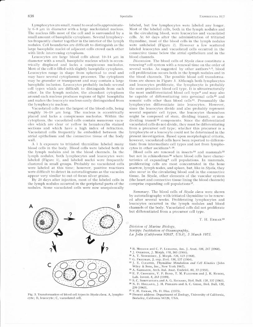
Fig. 3. Transformation of blood cell typesin Styela claaa. A, lympho-cyte; B, leucocyte; C, vacuolated cell.
ExpBnrBNrrA 3tl7
labeled, but few lymphocytes were labeled any longer.Most of the labeled cells, both in the lymph nodules andin the circulating blood, were leucocytes and vacuolatedcells. At 60 days after the administration of tritiatedthymidine, most of the blood cells in the lymph noduleswere unlabeled (Figure 2). llowever a few scatteredlabeled leucocytes and vacuolated cells occurred in theconnective tissue below the atrial epithelium and in theblood channels.
D'iscuss'ion. The blood cells of. Styela claua constitute arenewing 8 cell system with a renewal time on the order ofseveral weeks. As suggested by other authors 1' 2, bloodcell proliferation occurs both in the lymph nodules and inthe blood channels. The possible blood cell transforma-tions are shown in Figure 3. Although both lymphocytesand leucocytes proliferate, the lymphocyte is probablythe more primitive blood cell type. It is ultrastructurallythe most undifferentiated blood cell type e and may alsobe capable of differentiating into germinal cells 10 andsomatic cells other than blood cells 11. Presumably thelymphocytes differentiate into leucocytes. llowever,since the leucocytes divide and also probably representseveral separate cell types, the leucocytes themselvesmight be composed of stem, dividing transit, or non-dividing transit 12 components. Since the differentiatedvacuolated cells do not divide, they must be differentiatingfrom a precursor cell type; whether this precursor is alymphocyte or a leucocyte could not be determined in thepresent investigation. Based upon morphological criteria,however, vacuolated cells have been reported to differen-tiate from intermediate cell types and not from lympho-cytes in other ascidians 7 , L3.
Blood cells are renewed in insects 1a and mammals 15
but not in echinoderms 16 where blood cells have charac-teristics of expandirg* cell populations. In mammals,proliferating cells are most concentrated in the bonemarrow, lymph nodes, and spleen, but, like tn Styela, theyalso occur in the circulating blood and in the connectiveti$sue. In Styela, other elements of the vascular system(the heart and connective tissue lining the blood channels)comprise expanding cell populations 17.
Surnrnary. The blood cells of Styela claua were shownby autoradiography r,vith tritiated thymidine to be renew-ed after several weeks. Proliferating lymphocytes andleucocytes occurred in the lymph nodules and bloodchannels of the body. Vacuolated cells did not proliferatebut differentiated from a precursor cell type.
T. H. ERnrax 18
D'iu'is'ion, of Marine B'iology,Scri,pps Inst'itut'ion of Oceanography,La Jolla (Cali,forn'ia 92037, USA), 5 March 7 975.
8 B. MBSSTER and C. P. LBnroND, Am. J. Anat. 106,247 (1960).e J. OvnRroN, J. Morph.779,305 (1966).
10 A. T. NBwBERRv, J. Morph.726, t23 (1,968).11 G. F'nnBnreN, J. exp. Zool.756,I57 (7964).t'J. E. Cr,Bevnn, Thyrnid,ine Metabol,ism and, CeII, Kinetics (John
Wiley & Sons, fnc., New York 1,967).13 A. SansA.nrN, Arch. ital. Anat. Embriol . 60,33 (1955).14 E. P. CnoNKrrE, V. P. BoNn, T. M. Fr,rBnNnn and J. R. RunrNr,
Lab. fnvest. 8,263 (1959).15 S. C. SsnrvASrAvA and A. G. RrcsARD, Biol. Bull. 728,337 (1965).16 N. D. Holr,AND, J. H. Pnrr-r,rps and A. C. Grnsr, Biol. Bull. 128,
zse (Le6s).L7 T. H. Ennar, Ph. D. Diss. (1975).18 Present address: Department of. ZooIogy, University of California,
Berkeley, California 94720, USA.
Specialia
I f,