ertapenem-induced acute interstitial nephritis (ain) in a ... · pdf filefi v 6 fi 6 85...
TRANSCRIPT
Journal of The Association of Physicians of India ■ Vol. 64 ■ March 2016 85
Ertapenem-Induced Acute Interstitial Nephritis (AIN) in a Case of Protein S Deficiency and Factor V Leiden Mutation with Deep Vein ThrombosisNitin Rathod1, Pavan Pai2
AbstractWe present a case of 58 years old male patient, who presented with high fever for which injection Ertapenem was started empirically at Dubai hospital. Patient was a known case of Deep vein thrombosis of left leg since 5 years on warfarin therapy. Patient came to India for high fever and further management. Patient developed proteinuria with high creatinine and urinary abnormalities. Renal biopsy revealed acute interstitial nephritis (AIN). In addition, he was diagnosed to have protein S deficiency with Factor V Leiden mutation.
1HOD, Medicine Dept., HBT Medical College & R.N. Cooper Municipal Hospital, Mumbai, Maharashtra; 2HOD Nephrology Dept. Nanavati Superspeciality Hospital, Mumbai, MahrarashtraReceived: 03.07.2014; Revised: 26.11.2014; Accepted: 27.12.2014
Introduction
Interstitial nephritis is a usually short-term disorder. In rare cases, it can
cause permanent damage, including chronic kidney failure. Acute interstitial nephritis may be more severe and more likely to lead to long-term or permanent kidney damage in elderly people, if not diagnosed early. Acute interstitial nephritis (AIN) is known to occur following analgesics and certain antibiotics.
Case Report
58 years old male patient presented with history of high fever which was continuous in nature and without chills for six days. He did not give any history of urinary disturbance or frequency of urine. No history of abdominal pain, loose motion or vomiting. No history of cough, chest pain or breathlessness. Patient was in Dubai where he got admitted to the hospital for the same. He is a non smoker, non-alcoholic, having hypertension since last ten years on Tab. Telmisartan 40 mg, Tab. Indapamide and Tab. Lercanidepin 10 mg once a day. He gave history of DVT of left leg (detected on venous Doppler ) 5 years ago and presumed to be idiopathic and started on Tab. Warfarin 5 mg OD. Patient had received Inj. Ertapenem 1 gm IV BD for four days. Along with this antibiotic, patient received paracetamol, vitamins and intravenous fluids. Patient took transfer from Dubai hospital to Mumbai as
there was no improvement in fever. On examinat ion, he was afebri le , haemodynamically stable, minimal oedema feet, JVP not raised, no basal crepitations or rhonchi present.
Investigations revealed were as follow: Hb 10 gm/dL WBC 7520/cu.mm (N 63%/ L 26%/ E 4%/M 7%), Platelet count 3,91,000/cu.mm, rapid malarial test : negative, peripheral smear for malaria parasite: negative, Widal test : negative, CRP 3.3 mg/dL, FBS: 96 mg/dl, BUN 33.6 mg/dL, creatinine 3.0 mg/dL, uric acid 9.4 mg/dL, calcium 8.2 mg/dL phosphorous: 5.7 mg/dL, proteins: 5.3 gm/dL, albumin 2.0 gm/dL sodium: 138 mEq/L, potassium 4.8 mEq/L, chloride: 106 mEq/L,, SGPT 128 U/L, alkaline phosphatase: 100 U/L, bilirubin 0.5 mg/dL, urine protein: 3 plus, RBC 8-10/hpf, leucocytes 2-4/hpf, 24 hours urine for protein: 392 mg/24 hours, urine routine examination did not reveal + eosinophils, HCV IgM antibody- negative, HBsAg –negative and HIV negative. USG (abdomen and pelvis) revealed : both kidneys are normal size and shape. Both kidneys show increased cortical echogenicity suggestive of medical renal disease, Chest X ray: normal, urine culture: negative, blood culture: negative, renal color Doppler: does not show changes of significant renal artery stenosis, ANA 1:80 titre, C-ANCA and P-ANCA
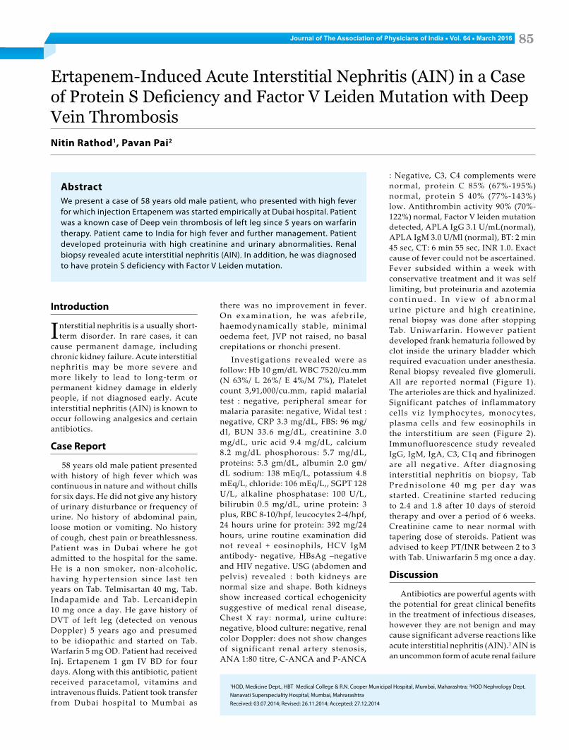
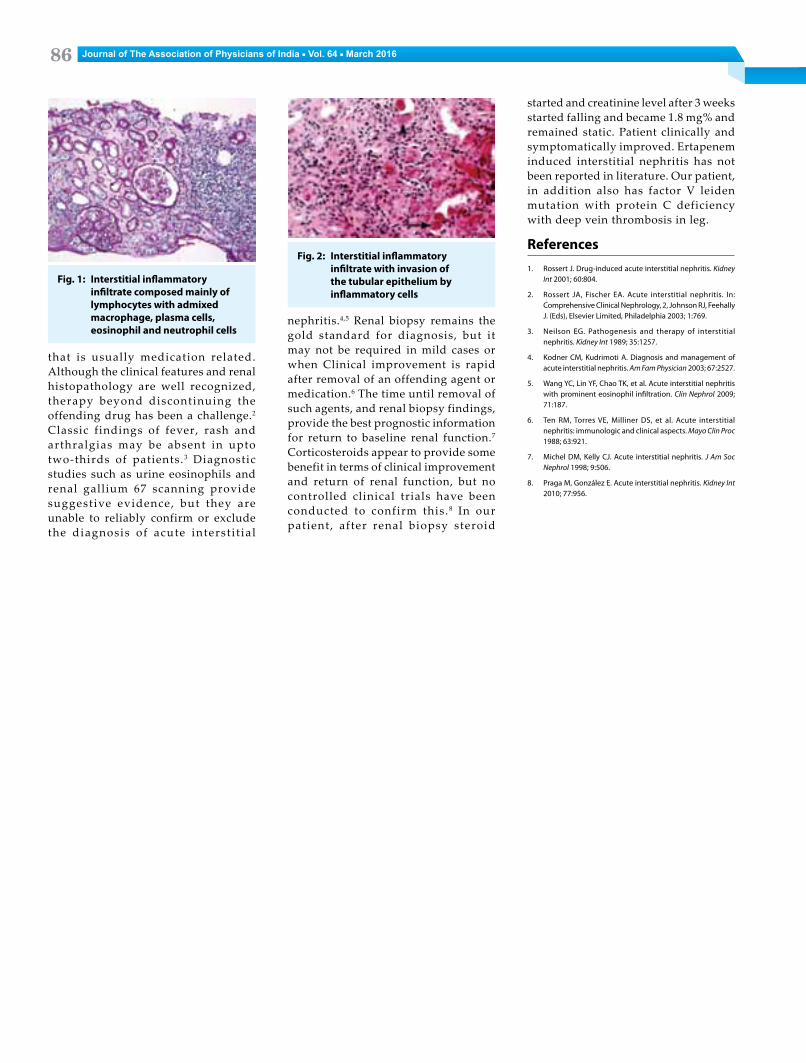
: Negative, C3, C4 complements were normal, protein C 85% (67%-195%) normal, protein S 40% (77%-143%) low. Antithrombin activity 90% (70%-122%) normal, Factor V leiden mutation detected, APLA IgG 3.1 U/mL(normal), APLA IgM 3.0 U/Ml (normal), BT: 2 min 45 sec, CT: 6 min 55 sec, INR 1.0. Exact cause of fever could not be ascertained. Fever subsided within a week with conservative treatment and it was self limiting, but proteinuria and azotemia c o n t i n u e d . I n v i e w o f a b n o r m a l urine picture and high creatinine, renal biopsy was done after stopping Tab. Uniwarfarin. However patient developed frank hematuria followed by clot inside the urinary bladder which required evacuation under anesthesia. Renal biopsy revealed five glomeruli. All are reported normal (Figure 1). The arterioles are thick and hyalinized. Significant patches of inflammatory cells viz lymphocytes, monocytes, plasma cells and few eosinophils in the interstitium are seen (Figure 2). Immunofluorescence study revealed IgG, IgM, IgA, C3, C1q and fibrinogen are all negative. After diagnosing interstitial nephritis on biopsy, Tab Prednisolone 40 mg per day was started. Creatinine started reducing to 2.4 and 1.8 after 10 days of steroid therapy and over a period of 6 weeks. Creatinine came to near normal with tapering dose of steroids. Patient was advised to keep PT/INR between 2 to 3 with Tab. Uniwarfarin 5 mg once a day.
Discussion
Antibiotics are powerful agents with the potential for great clinical benefits in the treatment of infectious diseases, however they are not benign and may cause significant adverse reactions like acute interstitial nephritis (AIN).1 AIN is an uncommon form of acute renal failure
Journal of The Association of Physicians of India ■ Vol. 64 ■ March 201686
that is usually medication related. Although the clinical features and renal histopathology are well recognized, therapy beyond discontinuing the offending drug has been a challenge.2 Classic findings of fever, rash and arthralgias may be absent in upto two-thirds of patients.3 Diagnostic studies such as urine eosinophils and renal gallium 67 scanning provide suggestive evidence, but they are unable to reliably confirm or exclude the diagnosis of acute interst i t ial
nephritis.4,5 Renal biopsy remains the gold standard for diagnosis, but it may not be required in mild cases or when Clinical improvement is rapid after removal of an offending agent or medication.6 The time until removal of such agents, and renal biopsy findings, provide the best prognostic information for return to baseline renal function.7 Corticosteroids appear to provide some benefit in terms of clinical improvement and return of renal function, but no controlled clinical trials have been conducted to confirm this. 8 In our patient, after renal biopsy steroid
Fig. 1: Interstitial inflammatory infiltrate composed mainly of lymphocytes with admixed macrophage, plasma cells, eosinophil and neutrophil cells
started and creatinine level after 3 weeks started falling and became 1.8 mg% and remained static. Patient clinically and symptomatically improved. Ertapenem induced interstitial nephritis has not been reported in literature. Our patient, in addition also has factor V leiden mutation with protein C deficiency with deep vein thrombosis in leg.
References1. Rossert J. Drug-induced acute interstitial nephritis. Kidney
Int 2001; 60:804.
2. Rossert JA, Fischer EA. Acute interstitial nephritis. In: Comprehensive Clinical Nephrology, 2, Johnson RJ, Feehally J. (Eds), Elsevier Limited, Philadelphia 2003; 1:769.
3. Neilson EG. Pathogenesis and therapy of interstitial nephritis. Kidney Int 1989; 35:1257.
4. Kodner CM, Kudrimoti A. Diagnosis and management of acute interstitial nephritis. Am Fam Physician 2003; 67:2527.
5. Wang YC, Lin YF, Chao TK, et al. Acute interstitial nephritis with prominent eosinophil infiltration. Clin Nephrol 2009; 71:187.
6. Ten RM, Torres VE, Milliner DS, et al. Acute interstitial nephritis: immunologic and clinical aspects. Mayo Clin Proc 1988; 63:921.
7. Michel DM, Kelly CJ. Acute interstitial nephritis. J Am Soc Nephrol 1998; 9:506.
8. Praga M, González E. Acute interstitial nephritis. Kidney Int 2010; 77:956.
Fig. 2: Interstitial inflammatory infiltrate with invasion of the tubular epithelium by inflammatory cells