es cell differentiation as a model to study cell biological
TRANSCRIPT

30
ES Cell Differentiation as a Model to Study Cell Biological Regulation of Vascular Development
Kiyomi Tsuji-Tamura, Hiroshi Sakamoto and Minetaro Ogawa Institute of Molecular Embryology and Genetics, Kumamoto University
Japan
1. Introduction
The organized vascular system is established through three successive steps, vasculogenesis,
angiogenesis and vascular remodeling. Vasculogenesis is the initiation of nascent embryonic
vessel formation by the differentiation of mesoderm-derived angioblasts into endothelial
cells. The nascent primitive vascular plexus then expands and reorganizes into hierarchical
vascular structures by the process of angiogenesis, which involves sprouting, bridging, and
intussusceptive division of preexisting vessels. Vascular smooth muscle cells are then
recruited to the vessels and the highly organized mature vascular system is established after
vascular remodeling. By using gene disruption strategies, a number of molecules have been
identified as being involved in the development of the vascular system. These identified
molecules include transcription factors, cell adhesion molecules, secretory molecules and
their receptors, of which deprivation lead to failure of angiogenesis and/ or vascular
remodeling in most cases. Despite the substantial expansion of the list of molecules that are
essential for angiogenesis, we do not yet understand sufficiently how these molecules
regulate the discrete processes of angiogenesis and vascular remodeling. Indeed, we know
very little how normal endothelial cells behave in response to various angiogenic stimuli
and cooperate to generate blood vessels during embryogenesis. It is critically important to
establish an experimental model to study the functions of key molecules at the cellular level
by monitoring the behavior of normal endothelial cells involving in angiogenesis.
In vitro differentiation systems of embryonic stem (ES) cells have been serving as an
excellent experimental maneuver of developmental biology since Doetschman et al. reported
that hematopoietic cells were generated in vitro from ES cells (Doetschman et al., 1985). The
ES cell differentiation systems have three important features. First, as differentiation of cells
and tissues takes place in a culture dish instead of uterus, the process of differentiation can
be directly monitored. Second, intermediate stages of differentiation can be identified and
isolated as a cell population. Third, cell differentiation and proliferation can be controlled by
either adding exogenous signaling molecules, introducing exogenous genes or modifying
endogenous genes. Following the pioneering report of angiogenic differentiation from
mouse ES cells engrafted onto quail chorioallantoic membrane (Risau et al., 1988), in vitro
derivation of vascular endothelial cells from ES cells has a history of almost two decades
(Bloch et al., 1997; Feraud et al., 2001; Vittet et al., 1996; Wang et al., 1992). These earlier
studies relied upon formation of cell aggregates termed cystic embryoid bodies from ES
cells, whereby real-time monitoring of cell differentiation with a high resolution was
www.intechopen.com

Embryonic Stem Cells: The Hormonal Regulation of Pluripotency and Embryogenesis
582
hampered. Since Nishikawa's group developed a culture system in which ES cells
differentiate into hematopoietic and endothelial cell lineages on type IV collagen-coated
substrates or OP9 stromal cell layers (Hirashima et al., 1999; Yamashita et al., 2000), the
simplicity of the culture system allowed detailed analysis of developmental steps essential
in the formation of vascular and hematopoietic systems (Eilken et al., 2009; Kondo et al.,
2009). This system thus provides a useful model to study cell biological regulation of
endothelial cells in the process of angiogenesis and vascular remodeling. The aim of this
chapter is to review our experimental models for the direct examination of differentiation,
cell-cell adhesion, migration and morphological regulation of ES cell-derived endothelial
cells, and discuss the cellular mechanisms underlying the process of vascular development.
2. Endothelial cell-specific gene promoters and enhancers
2.1 Endothelial differentiation from murine ES cells It has been demonstrated that murine ES cells differentiate into Flk-1-expressing lateral
mesodermal cells when cultured on a layer of OP9 stromal cell line (Endoh et al., 2002;
Hashimoto et al., 2007; Kataoka et al., 1997; Nakano et al., 1994). In this culture system,
undifferentiated ES cells are simply added to a culture flask pre-seeded with OP9 cells and
incubated for 4 days in α-MEM supplemented with 2-mercaptoethanol and fetal calf serum.
Cultured cells are dissociated by incubating in a buffer containing chelating reagent, and
stained with a monoclonal antibody against Flk-1. The cells expressing Flk-1, an equivalent
population of proximal lateral mesoderm in the embryo (Kataoka et al., 1997), can be
purified by fluorescence-activated cell sorting (FACS). When the purified Flk-1+
mesodermal cells are further cultured on a freshly prepared OP9 cell layer, they continue to
differentiate into several cell lineages including primitive and definitive hematopoietic cells,
cardiomyocytes, smooth muscle cells and endothelial cells. Vascular endothelial (VE)-
cadherin can be used as a specific marker to identify the endothelial cells. VE-cadherin+ (and
also Flk-1+ CD31+) endothelial cells are finally isolated by FACS with specific monoclonal
antibodies. Alternatively, when undifferentiated ES cells are allowed to differentiate on the
OP9 cell layer for more than 5 days, endothelial cells can be isolated in a single step from the
culture by FACS. In this case, Flk-1+ VE-cadherin- mesodermal cells become detectable on
the 3rd day of differentiation, and Flk-1+ VE-cadherin+ endothelial cells on the 5th day of
differentiation (Endoh et al., 2002; Hashimoto et al., 2007). Consequently, this culture system
is a potent and reproducible way to induce ES cell differentiation into the lateral mesoderm
and its derivatives.
While the lineage-specific markers such as Flk-1 and VE-cadherin can be detected by using
specific monoclonal antibodies thereby providing means to isolate desired cell populations,
the induced expression of reporter genes under the control of lineage-specific gene
promoter/ enhancer is another useful method especially for real-time monitoring of the
process of cell differentiation. For example, Hirai et al. reported that a combination of 5’
flanking region and 3’ portion of the first intron of the Kdr (Flk-1) gene drove the expression
of enhanced green fluorescent protein (EGFP) reporter gene shortly after generation of VE-
cadherin+ endothelial cells during the differentiation of ES cell-derived Flk-1+ mesodermal
cells (Hirai et al., 2003). As the Kdr promoter/ enhancer discriminates mature committed
endothelial cells from immature endothelial cells that still possess a hematopoietic potential,
it should be of use for monitoring the maturation process of endothelial cells. By contrast, a
combination of 5’ flanking region and 5’ half of the first intron of the Cdh5 (VE-cadherin)
www.intechopen.com
ES Cell Differentiation as a Model to Study Cell Biological Regulation of Vascular Development
583
gene was reported to be active exclusively in all of the VE-cadherin+ endothelial cells during
the course of ES cell differentiation, suggesting that the activity of the Cdh5
promoter/ enhancer reflects that of the endogenous Cdh5 gene (Hisatsune et al., 2005).
Therefore, it provides a valuable tool for the endothelial cell-specific induction of a
transgene expression regardless of differentiation stages. In the next section, we show
another example of an endothelial-related enhancer element that might be useful to monitor
endothelial cell differentiation from their precursor stages.
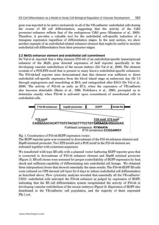
2.2 Mef2c enhancer element and endothelial cell commitment De Val et al. reported that a 44bp element (F10-44) of an endothelial-specific transcriptional
enhancer of the Mef2c gene directed expression of lacZ reporter specifically to the
developing vascular endothelium of the mouse embryo (De Val et al., 2008). The element
consists of a FOX:ETS motif that is present in many known endothelial-specific enhancers.
The F10-44-lacZ reporter mice demonstrated that this element was sufficient to direct
endothelial cell-specific expression from the blood island stage at embryonic day (E) 7.5
through angiogenesis and remodeling at E9.5, and extinguished after E10.5 (De Val et al.,
2008). The activity of F10-44 as early as E7.5, when the expression of VE-cadherin
also becomes detectable (Breier et al., 1996; Nishikawa et al., 1998), prompted us to
determine exactly when F10-44 is activated upon commitment of mesodermal cells to
endothelial cells.
Fig. 1. Construction of F10-44-EGFP expression vector.
The EGFP reporter gene was connected to downstream of the F10-44 enhancer element and
Hsp68 minimal promoter. Two ETS motifs and a FOX motif in the F10-44 element are
indicated together with consensus sequences.
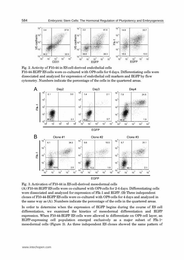
We transfected wild-type ES cells with a plasmid vector harboring EGFP reporter gene that
is connected to downstream of F10-44 enhancer element and Hsp68 minimal promoter
(Figure 1). ES cell clones were screened for proper controllability of EGFP expression by heat
shock and sufficient capability of differentiating into endothelial cell lineage. We obtained
three independent clones that showed essentially the same results. The F10-44-EGFP ES cells
were cultured on OP9 stromal cell layer for 6 days to induce endothelial cell differentiation
as described above. Flow cytometry analyses revealed that essentially all the VE-cadherin+
CD31+ endothelial cells activated the F10-44 enhancer as judged by expression of EGFP,
indicating that the ES cell differentiation system recapitulated the activity of F10-44 in
developing vascular endothelium of the mouse embryos (Figure 2). Expression of EGFP also
distributed in the VE-cadherin- cell population, and the majority of them expressed
Flk-1 yet.
www.intechopen.com
Embryonic Stem Cells: The Hormonal Regulation of Pluripotency and Embryogenesis
584
Fig. 2. Activity of F10-44 in ES cell-derived endothelial cells
F10-44-EGFP ES cells were co-cultured with OP9 cells for 6 days. Differentiating cells were
dissociated and analyzed for expression of endothelial cell markers and EGFP by flow
cytometry. Numbers indicate the percentage of the cells in the quartered areas.
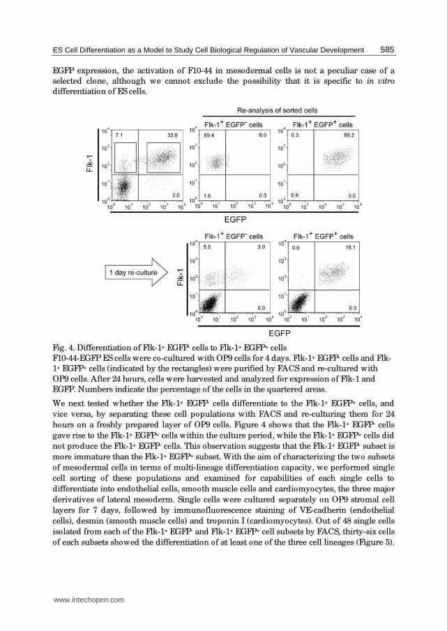
Fig. 3. Activation of F10-44 in ES cell-derived mesodermal cells
(A) F10-44-EGFP ES cells were co-cultured with OP9 cells for 2-4 days. Differentiating cells
were dissociated and analyzed for expression of Flk-1 and EGFP. (B) Three independent
clones of F10-44-EGFP ES cells were co-cultured with OP9 cells for 4 days and analyzed in
the same way as (A). Numbers indicate the percentage of the cells in the quartered areas.
In order to determine when the expression of EGFP begins during the course of ES cell
differentiation, we examined the kinetics of mesodermal differentiation and EGFP
expression. When F10-44-EGFP ES cells were allowed to differentiate on OP9 cell layer, an
EGFP-expressing cell population emerged exclusively as a major subset of Flk-1+
mesodermal cells (Figure 3). As three independent ES clones showed the same pattern of
www.intechopen.com
ES Cell Differentiation as a Model to Study Cell Biological Regulation of Vascular Development
585
EGFP expression, the activation of F10-44 in mesodermal cells is not a peculiar case of a
selected clone, although we cannot exclude the possibility that it is specific to in vitro
differentiation of ES cells.
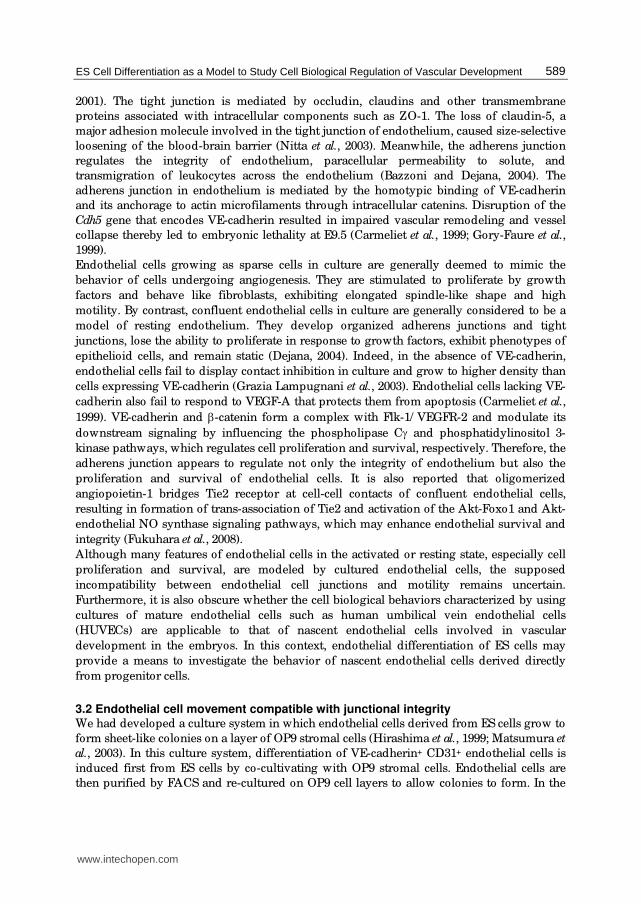
Fig. 4. Differentiation of Flk-1+ EGFP- cells to Flk-1+ EGFP+ cells
F10-44-EGFP ES cells were co-cultured with OP9 cells for 4 days. Flk-1+ EGFP- cells and Flk-
1+ EGFP+ cells (indicated by the rectangles) were purified by FACS and re-cultured with
OP9 cells. After 24 hours, cells were harvested and analyzed for expression of Flk-1 and
EGFP. Numbers indicate the percentage of the cells in the quartered areas.
We next tested whether the Flk-1+ EGFP- cells differentiate to the Flk-1+ EGFP+ cells, and
vice versa, by separating these cell populations with FACS and re-culturing them for 24
hours on a freshly prepared layer of OP9 cells. Figure 4 shows that the Flk-1+ EGFP- cells
gave rise to the Flk-1+ EGFP+ cells within the culture period, while the Flk-1+ EGFP+ cells did
not produce the Flk-1+ EGFP- cells. This observation suggests that the Flk-1+ EGFP- subset is
more immature than the Flk-1+ EGFP+ subset. With the aim of characterizing the two subsets
of mesodermal cells in terms of multi-lineage differentiation capacity, we performed single
cell sorting of these populations and examined for capabilities of each single cells to
differentiate into endothelial cells, smooth muscle cells and cardiomyocytes, the three major
derivatives of lateral mesoderm. Single cells were cultured separately on OP9 stromal cell
layers for 7 days, followed by immunofluorescence staining of VE-cadherin (endothelial
cells), desmin (smooth muscle cells) and troponin I (cardiomyocytes). Out of 48 single cells
isolated from each of the Flk-1+ EGFP- and Flk-1+ EGFP+ cell subsets by FACS, thirty-six cells
of each subsets showed the differentiation of at least one of the three cell lineages (Figure 5).
www.intechopen.com

Embryonic Stem Cells: The Hormonal Regulation of Pluripotency and Embryogenesis
586
Among of them, eighty percent of the Flk-1+ EGFP+ cells produced unipotential colonies
containing only endothelial cells, although the rest of them retained a potential to
differentiate into smooth muscle cells. By contrast, the Flk-1+ EGFP- cells produced a
bipotential and a tripotential colony that contained cardiomyocytes, in addition to colonies
containing endothelial cells and/ or smooth muscle cells. This observation supports the
above assumption that the Flk-1+ EGFP- subset represents earlier differentiation stage of
lateral mesoderm than the Flk-1+ EGFP+ subset.
Fig. 5. Multi-lineage differentiation potential of mesodermal cells
F10-44-EGFP ES cells were co-cultured with OP9 cells for 4 days. Forty-eight single cells of
Flk-1+ EGFP- and Flk-1+ EGFP+ subsets were separately deposited into wells pre-seeded
with OP9 cells. After 7 days cultivation, wells were stained with antibodies against VE-
cadherin (red), desmin (green) and troponin I (blue). (A) Number of wells that were positive
for any of the three cell lineages. (B) A colony containing only endothelial cells. (C) A colony
containing endothelial cells and smooth muscle cells. (D) A colony containing all the three
cell lineages. Scale bars indicate 100 μm.
Our results suggest that the F10-44 enhancer element is first activated in a subset of lateral
mesoderm, of which differentiation potential becomes restricted to the endothelial and
smooth muscle cell lineages. As smooth muscle cells derived from Flk-1+ EGFP+ cells did not
retain the expression of EGFP (data not shown), the F10-44 activity persists specifically in
the endothelial cell lineage. It remains to be elucidated whether the early mesodermal
activation of F10-44 also takes place in the mouse embryos, and whether the initiation of the
F10-44 activity in the mesoderm and its maintenance in the endothelium are regulated by
the same trans-activators.
www.intechopen.com

ES Cell Differentiation as a Model to Study Cell Biological Regulation of Vascular Development
587
2.3 Monitoring endothelial differentiation of mesodermal cells As mentioned above, F10-44 directed EGFP expression from endothelial precursors through
differentiation of mature endothelial cells in ES cell cultures. Taking advantage of high
visibility of EGFP fluorescence in living cells, the developmental process of endothelial
precursors can be directly monitored by time-lapse analysis of differentiating ES cells. In
order to detect the initiation of the F10-44 activity in mesodermal cells, we seeded F10-44-
EGFP ES cells onto OP9 stromal cell layer and performed time-lapse imaging analyses of
differentiating cells.
Fig. 6. Time-lapse analysis of differentiating mesodermal cells
F10-44-EGFP ES cells were seeded on the OP9 cell layer to induce the mesodermal
differentiation. After 3 days, the cultures were subjected to time-lapse analyses under a
fluorescence microscope. Numbers indicate elapsed time counted by hours. (A) EGFP
fluorescence images. (B) Phase contrast images.
As shown in Figure 6, progression of mesodermal cell differentiation as revealed by the
emergence of EGFP fluorescence occurred within a group of cells in a relatively
synchronous manner, rather than a clonal expansion of a single ancestral EGFP+ cells. This
observation suggests that a group of mesodermal cells has determined their fate toward the
endothelial cell lineage at a certain point, and then a combination of trans-activators activate
F10-44 enhancer element synchronously in this population. The same sequence of events
may also take place independently in other groups of mesoderm cells in culture.
www.intechopen.com

Embryonic Stem Cells: The Hormonal Regulation of Pluripotency and Embryogenesis
588
Fig. 7. Time-lapse analysis of differentiating endothelial cells
F10-44 EGFP ES cells were co-cultured with OP9 cells for 4 days, and Flk-1+ EGFP+ cells
were purified by FACS. Cells were seeded on OP9 cell layer, and cultured in the presence of
VEGF-A. After 36 hours, the cultures were subjected to time-lapse analyses under a
fluorescence microscope. Numbers indicate elapsed time counted by hours. (A) EGFP
fluorescence images. (B) Phase contrast images.
The F10-44 enhancer element also provides means of tracking how endothelial precursors are
organized into endothelial structures in culture. F10-44-EGFP ES cells were co-cultured with
OP9 stromal cells for 4 days. FACS-purified Flk-1+ EGFP+ cells were re-cultured on OP9 cell
layer in the presence of 50 ng/ mL vascular endothelial growth factor A (VEGF-A), and
subjected to time-lapse analyses (Figure 7). The EGFP+ endothelial precursors first generated
small groups of endothelial cells, and then the cells were rearranged to form large elongated
clusters. Endothelial cells were further organized into a network of cord-like structures
through splitting of elongated clusters, sprouting of endothelial cells with lamellipodial
protrusions, migration and interconnection of elongated endothelial cells. These processes are
reminiscent of those found in vasculogenesis and angiogenesis in the yolk sac vasculatures.
Therefore, by combining the endothelial cell differentiation on OP9 stromal cell layer with the
expression of fluorescent molecular probes, ES cell differentiation systems provide powerful
experimental models to investigate the behavior of normal endothelial cells in response to
angiogenic stimuli. We will discuss this point a little bit more in the next section.
3. Cell-cell adhesion and motility of endothelial cells
3.1 Cell-cell junction as a regulating factor of endothelial cells Endothelial cells adhere to each other through tight junctions and adherens junctions. The
tight junction regulates paracellular permeability and maintains cell polarity (Tsukita et al.,
www.intechopen.com
ES Cell Differentiation as a Model to Study Cell Biological Regulation of Vascular Development
589
2001). The tight junction is mediated by occludin, claudins and other transmembrane
proteins associated with intracellular components such as ZO-1. The loss of claudin-5, a
major adhesion molecule involved in the tight junction of endothelium, caused size-selective
loosening of the blood-brain barrier (Nitta et al., 2003). Meanwhile, the adherens junction
regulates the integrity of endothelium, paracellular permeability to solute, and
transmigration of leukocytes across the endothelium (Bazzoni and Dejana, 2004). The
adherens junction in endothelium is mediated by the homotypic binding of VE-cadherin
and its anchorage to actin microfilaments through intracellular catenins. Disruption of the
Cdh5 gene that encodes VE-cadherin resulted in impaired vascular remodeling and vessel
collapse thereby led to embryonic lethality at E9.5 (Carmeliet et al., 1999; Gory-Faure et al.,
1999).
Endothelial cells growing as sparse cells in culture are generally deemed to mimic the
behavior of cells undergoing angiogenesis. They are stimulated to proliferate by growth
factors and behave like fibroblasts, exhibiting elongated spindle-like shape and high
motility. By contrast, confluent endothelial cells in culture are generally considered to be a
model of resting endothelium. They develop organized adherens junctions and tight
junctions, lose the ability to proliferate in response to growth factors, exhibit phenotypes of
epithelioid cells, and remain static (Dejana, 2004). Indeed, in the absence of VE-cadherin,
endothelial cells fail to display contact inhibition in culture and grow to higher density than
cells expressing VE-cadherin (Grazia Lampugnani et al., 2003). Endothelial cells lacking VE-
cadherin also fail to respond to VEGF-A that protects them from apoptosis (Carmeliet et al.,
1999). VE-cadherin and β-catenin form a complex with Flk-1/ VEGFR-2 and modulate its
downstream signaling by influencing the phospholipase Cγ and phosphatidylinositol 3-
kinase pathways, which regulates cell proliferation and survival, respectively. Therefore, the
adherens junction appears to regulate not only the integrity of endothelium but also the
proliferation and survival of endothelial cells. It is also reported that oligomerized
angiopoietin-1 bridges Tie2 receptor at cell-cell contacts of confluent endothelial cells,
resulting in formation of trans-association of Tie2 and activation of the Akt-Foxo1 and Akt-
endothelial NO synthase signaling pathways, which may enhance endothelial survival and
integrity (Fukuhara et al., 2008).
Although many features of endothelial cells in the activated or resting state, especially cell
proliferation and survival, are modeled by cultured endothelial cells, the supposed
incompatibility between endothelial cell junctions and motility remains uncertain.
Furthermore, it is also obscure whether the cell biological behaviors characterized by using
cultures of mature endothelial cells such as human umbilical vein endothelial cells
(HUVECs) are applicable to that of nascent endothelial cells involved in vascular
development in the embryos. In this context, endothelial differentiation of ES cells may
provide a means to investigate the behavior of nascent endothelial cells derived directly
from progenitor cells.
3.2 Endothelial cell movement compatible with junctional integrity We had developed a culture system in which endothelial cells derived from ES cells grow to
form sheet-like colonies on a layer of OP9 stromal cells (Hirashima et al., 1999; Matsumura et
al., 2003). In this culture system, differentiation of VE-cadherin+ CD31+ endothelial cells is
induced first from ES cells by co-cultivating with OP9 stromal cells. Endothelial cells are
then purified by FACS and re-cultured on OP9 cell layers to allow colonies to form. In the
www.intechopen.com
Embryonic Stem Cells: The Hormonal Regulation of Pluripotency and Embryogenesis
590
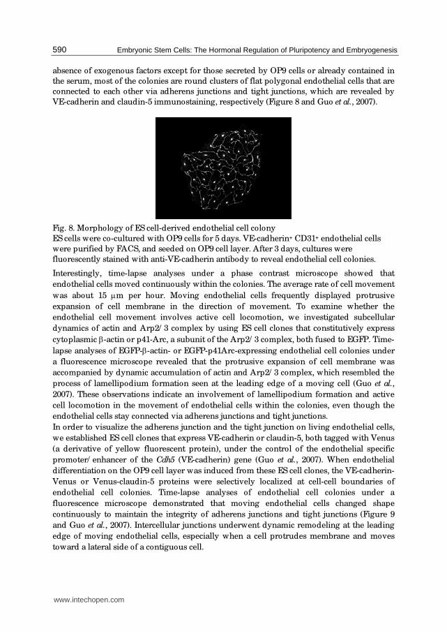
absence of exogenous factors except for those secreted by OP9 cells or already contained in
the serum, most of the colonies are round clusters of flat polygonal endothelial cells that are
connected to each other via adherens junctions and tight junctions, which are revealed by
VE-cadherin and claudin-5 immunostaining, respectively (Figure 8 and Guo et al., 2007).
Fig. 8. Morphology of ES cell-derived endothelial cell colony
ES cells were co-cultured with OP9 cells for 5 days. VE-cadherin+ CD31+ endothelial cells
were purified by FACS, and seeded on OP9 cell layer. After 3 days, cultures were
fluorescently stained with anti-VE-cadherin antibody to reveal endothelial cell colonies.
Interestingly, time-lapse analyses under a phase contrast microscope showed that
endothelial cells moved continuously within the colonies. The average rate of cell movement
was about 15 μm per hour. Moving endothelial cells frequently displayed protrusive
expansion of cell membrane in the direction of movement. To examine whether the
endothelial cell movement involves active cell locomotion, we investigated subcellular
dynamics of actin and Arp2/ 3 complex by using ES cell clones that constitutively express
cytoplasmic β-actin or p41-Arc, a subunit of the Arp2/ 3 complex, both fused to EGFP. Time-
lapse analyses of EGFP-β-actin- or EGFP-p41Arc-expressing endothelial cell colonies under
a fluorescence microscope revealed that the protrusive expansion of cell membrane was
accompanied by dynamic accumulation of actin and Arp2/ 3 complex, which resembled the
process of lamellipodium formation seen at the leading edge of a moving cell (Guo et al.,
2007). These observations indicate an involvement of lamellipodium formation and active
cell locomotion in the movement of endothelial cells within the colonies, even though the
endothelial cells stay connected via adherens junctions and tight junctions.
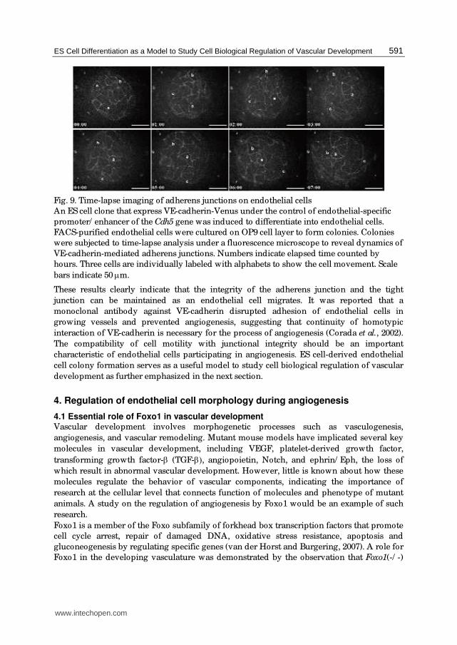
In order to visualize the adherens junction and the tight junction on living endothelial cells,
we established ES cell clones that express VE-cadherin or claudin-5, both tagged with Venus
(a derivative of yellow fluorescent protein), under the control of the endothelial specific
promoter/ enhancer of the Cdh5 (VE-cadherin) gene (Guo et al., 2007). When endothelial
differentiation on the OP9 cell layer was induced from these ES cell clones, the VE-cadherin-
Venus or Venus-claudin-5 proteins were selectively localized at cell-cell boundaries of
endothelial cell colonies. Time-lapse analyses of endothelial cell colonies under a
fluorescence microscope demonstrated that moving endothelial cells changed shape
continuously to maintain the integrity of adherens junctions and tight junctions (Figure 9
and Guo et al., 2007). Intercellular junctions underwent dynamic remodeling at the leading
edge of moving endothelial cells, especially when a cell protrudes membrane and moves
toward a lateral side of a contiguous cell.
www.intechopen.com
ES Cell Differentiation as a Model to Study Cell Biological Regulation of Vascular Development
591
Fig. 9. Time-lapse imaging of adherens junctions on endothelial cells
An ES cell clone that express VE-cadherin-Venus under the control of endothelial-specific
promoter/ enhancer of the Cdh5 gene was induced to differentiate into endothelial cells.
FACS-purified endothelial cells were cultured on OP9 cell layer to form colonies. Colonies
were subjected to time-lapse analysis under a fluorescence microscope to reveal dynamics of
VE-cadherin-mediated adherens junctions. Numbers indicate elapsed time counted by
hours. Three cells are individually labeled with alphabets to show the cell movement. Scale
bars indicate 50 μm.
These results clearly indicate that the integrity of the adherens junction and the tight
junction can be maintained as an endothelial cell migrates. It was reported that a
monoclonal antibody against VE-cadherin disrupted adhesion of endothelial cells in
growing vessels and prevented angiogenesis, suggesting that continuity of homotypic
interaction of VE-cadherin is necessary for the process of angiogenesis (Corada et al., 2002).
The compatibility of cell motility with junctional integrity should be an important
characteristic of endothelial cells participating in angiogenesis. ES cell-derived endothelial
cell colony formation serves as a useful model to study cell biological regulation of vascular
development as further emphasized in the next section.
4. Regulation of endothelial cell morphology during angiogenesis
4.1 Essential role of Foxo1 in vascular development Vascular development involves morphogenetic processes such as vasculogenesis,
angiogenesis, and vascular remodeling. Mutant mouse models have implicated several key
molecules in vascular development, including VEGF, platelet-derived growth factor,
transforming growth factor-β (TGF-β), angiopoietin, Notch, and ephrin/ Eph, the loss of
which result in abnormal vascular development. However, little is known about how these
molecules regulate the behavior of vascular components, indicating the importance of
research at the cellular level that connects function of molecules and phenotype of mutant
animals. A study on the regulation of angiogenesis by Foxo1 would be an example of such
research.
Foxo1 is a member of the Foxo subfamily of forkhead box transcription factors that promote
cell cycle arrest, repair of damaged DNA, oxidative stress resistance, apoptosis and
gluconeogenesis by regulating specific genes (van der Horst and Burgering, 2007). A role for
Foxo1 in the developing vasculature was demonstrated by the observation that Foxo1(-/ -)
www.intechopen.com

Embryonic Stem Cells: The Hormonal Regulation of Pluripotency and Embryogenesis
592
mice die around E11 due to defects in the development of branchial arches and
malformation in major vessels of the embryo and the yolk sac (Furuyama et al., 2004; Hosaka
et al., 2004). Foxo1(-/ -) embryos developed a small first branchial arch, but no second
branchial arch, and often exhibited marked pericardial swelling. The dorsal aorta was
severely underdeveloped and irregularly formed. Hypoplasia of aortic arch artery was also
observed. Foxo1(-/ -) yolk sacs showed primitive vascular plexus similar to that of wild-type
yolk sacs at E8.75, but failed to develop a normal vasculature at E9.5. These observations
suggested that vasculogenesis, but not angiogenesis and remodeling, proceeds without
Foxo1. Expression of the Foxo1 gene was detected in developing vasculature of normal
mouse embryos including the dorsal aorta, the intersomitic vessels, the vitelline and
umbilical vessels and others (Furuyama et al., 2004; Hosaka et al., 2004). Although Foxo1 has
been considered as a key regulator of energy metabolism and lifespan, the role of this
transcription factor in the vascular development can hardly be understood from the well-
known target genes. There was no significant difference between wild-type and Foxo1(-/ -)
yolk sac in the expression levels of genes involved in vascular development including
VEGF-A, VEGFR-1, VEGFR-2, angiopoietin-1, angiopoietin-2, Tie-1, Tie-2 and EphB4. Still,
Foxo1(-/ -) yolk sac vasculature appeared to had lost an arterial property as indicated by
reduced expression of arterial markers such as ephrinB2 and connexin-40 (Furuyama et al.,
2004).
In order to delineate the role of Foxo1 in angiogenesis, we employed an in vitro
differentiation system of ES cells for investigating the expression and function of Foxo1 in
endothelial cells. Foxo1 transcripts were detected in the VE-cadherin+ CD31+ endothelial
cells derived from wild-type ES cells differentiating in the co-culture with OP9 stromal cells
(Furuyama et al., 2004). Comparable number of endothelial cells were able to be obtained
from the cultures initiated from wild-type and Foxo1(-/ -) ES cells. Endothelial cells of the
two genotypes also gave rise to comparable number and size of monostratal endothelial
colonies when purified by FACS and re-seeded on the OP9 cell layer, suggesting that the
loss of Foxo1 did not influence the proliferation and survival of endothelial cells. This notion
may contrast with a previous report that Foxo1 induced apoptosis in HUVECs, which was
inhibited by angiopoietin-1 signaling via activation of Akt pathway (Daly et al., 2004). This
discrepancy could reflect the difference in the differentiation stage of endothelial cells,
namely, nascent immature endothelial cells derived from ES cells and mature endothelial
cells isolated from established endothelium. The former may resemble the cells that
undertake vascular development in the embryos.
We took advantage of the ES cell-derived endothelial cell colony formation assay to
compare behavior of wild-type and Foxo1(-/ -) endothelial cells in response to various
angiogenic stimuli. In the absence of exogenously added factors, endothelial cells of both
genotypes formed monostratal colonies composed of rough-edged flat cells as described
above. When VEGF-A was added in the medium during colony formation, however, wild-
type and Foxo1(-/ -) endothelial cells exhibited quite different morphological responses.
VEGF-A induced elongation of wild-type endothelial cells, thereby led to form colonies with
corded structure composed of long spindle-shaped endothelial cells entwining with each
other. In contrast to the response of wild-type endothelial cells, Foxo1(-/ -) endothelial cells
remained flatten in the presence of exogenous VEGF-A. Yet they changed morphology from
rough-edged to polygonal shape with rather straight adherens junctions and partially
overlapped to the neighboring cells (Figure 10). (Endothelial cell overlapping as a stable
condition is rarely observed in unstimulated monostratal colonies.) Induced expression of
www.intechopen.com

ES Cell Differentiation as a Model to Study Cell Biological Regulation of Vascular Development
593
Foxo1 cDNA in the Foxo1(-/ -) endothelial cells by using the endothelial-specific
promoter/ enhancer of the Cdh5 (VE-cadherin) gene restored cell elongation in response to
VEGF-A, implying a cell-autonomous function of Foxo1 in the morphological response of
endothelial cells (Furuyama et al., 2004). Taken together, these results suggest that Foxo1
regulates endothelial cell elongation in response to VEGF-A signaling, and the failure of
proper morphological response of endothelial cells to angiogenic stimuli likely accounts for
the compromised angiogenesis in the Foxo1(-/ -) embryos.
Fig. 10. Morphological response of endothelial cells to VEGF-A
Wild-type and Foxo1(-/ -) ES cells were co-cultured with OP9 cells for 5 days. VE-cadherin+
CD31+ endothelial cells were purified by FACS and seeded onto OP9 cell layer. Cells were
cultured for 3 days in the presence or absence of exogenously added VEGF-A. Endothelial
cell colonies were revealed by immunofluorescent staining of VE-cadherin.
4.2 Endothelial cell elongation induced by angiogenic factors Severe dilation of the yolk sac blood vessels in Foxo1(-/ -) mice are reminiscent of mice
lacking components of the TGF-β signaling pathway including Alk5, Alk1, endoglin and
Smad5 (Larsson et al., 2001; Li et al., 1999; Oh et al., 2000; Yang et al., 1999). It is well known
that endothelial cells provide TGF-β signaling to neighboring mesenchymal cells and
promote their differentiation into smooth muscle cells which support and stabilize the
vessels (ten Dijke and Arthur, 2007). However, endothelial cell-specific disruption of the
Tgfbr2 or Alk5 gene resulted in the phenotype indistinguishable from that of the Tgfbr2-null
or Alk5-null embryos, suggesting an endothelial cell-intrinsic role of TGF-β signaling in the
regulation of angiogenesis (Carvalho et al., 2004; Jiao et al., 2006). In order to examine
whether Foxo1 and TGF-β are involved in a common process of angiogenesis, we tested the
effect of TGF-β on the morphology of ES cell-derived endothelial cells. VE-cadherin+ CD31+
endothelial cells derived from wild-type and Foxo1(-/ -) ES cells differentiating in the co-
culture with OP9 cells were purified by FACS and re-seeded on the OP9 cell layer in the
presence of TGF-β. Wild-type endothelial cells produced cord-like colonies composed of
spindle-shaped cells in response to TGF-β. By contrast, Foxo1(-/ -) endothelial cells failed to
produce cord-like colonies, but they exhibited partial overlapping as in the case of VEGF-A
treatment (Matsukawa et al., 2009). These observations indicate that TGF-β induces
elongation of endothelial cells in a Foxo1-dependent manner.
Sequestration of a low concentration of endogenous VEGF-A pre-existing in the culture
(mainly secreted by OP9 cells) by addition of Flt1-Fc chimeric protein diminished TGF-β-
induced elongation of wild-type endothelial cells, indicating that endothelial cell elongation
induced by TGF-β depends upon VEGF-A signaling (Matsukawa et al., 2009). As the amount
www.intechopen.com

Embryonic Stem Cells: The Hormonal Regulation of Pluripotency and Embryogenesis
594
of Vegfa transcripts in OP9 cells was not influenced by the stimulation with TGF-β, two
possible mechanisms might be implied. First, TGF-β may synergize with VEGF-A to
regulate endothelial cell morphology, and Foxo1 is possibly involved in the TGF-β signaling.
It is indeed reported that Foxo proteins interacted with Smad3 and Smad4 to form a p21Cip1
transactivation complex in the regulation of neuroepithelial and glioblastoma cell
proliferation by TGF-β (Seoane et al., 2004). Alternatively, TGF-β may reduce the threshold
of VEGF-A-responsiveness of endothelial cells, thereby facilitates cell elongation under the
presence of a limited amount of VEGF-A, which is dependent upon Foxo1. Nevertheless, the
observations that TGF-β induced endothelial cell elongation in a Foxo1-dependent manner
and that disruption of Foxo1 and TGF-β signaling resulted in a similar phenotype in
vascular development suggest important roles of Foxo1-dependent endothelial cell
elongation in the process of angiogenesis.
4.3 Distinct functions of Foxo subfamily members The Foxo subfamily of forkhead box transcription factors consists of four members (Foxo1,
Foxo3, Foxo4 and Foxo6). Somatic deletion of the Foxo1, Foxo3 and Foxo4 genes in adult mice
revealed that Foxo1 is the major regulator of endothelial stability which suppresses
endothelial growth and hemangioma development, yet a certain degree of functional
redundancy appears to exist among the Foxo members (Paik et al., 2007). Indeed, both Foxo1
and Foxo3 were reported to induce apoptosis in HUVECs (Daly et al., 2004; Kim et al., 2005).
Foxo1 and Foxo3, but not Foxo4, were also shown to comparably inhibit endothelial cell
migration and tube formation in vitro (Potente et al., 2005). However, Foxo1 appears to have
nonredundant functions in vascular development during embryogenesis. In contrast to the
defect of angiogenesis in Foxo1(-/ -) embryos, both Foxo3(-/ -) and Foxo4(-/ -) mice are born
alive with no detectable abnormality of vascular development (Castrillon et al., 2003; Hosaka
et al., 2004). Dispensability of these two genes for vascular development possibly due to
differential tissue distribution of the Foxo members in developing embryos, since the
expression of Foxo1, Foxo3 and Foxo4 mRNA in the embryos were complementary to each
other (Furuyama et al., 2000). Alternatively, molecular functions of the Foxo members in the
endothelium are possibly diversified. For instance, expression of several genes that are
related to inflammation and angiogenesis (e.g. angiopoietin-2 and interleukin-8) are
differentially regulated by Foxo1 and Foxo3 in HUVECs (Potente et al., 2005).
We examined expression of Foxo3 during the course of ES cell differentiation into
endothelial cell lineage. While Foxo1 was expressed constantly by undifferentiated ES cells,
Flk-1+ mesodermal cells and various stages of VE-cadherin+ CD31+ endothelial cells,
expression of Foxo3 was not detected in any of these cell populations (Matsukawa et al.,
2009). Therefore, Foxo1 appears to be a primary and nonredundant factor that regulates
morphological response of developing endothelial cells to angiogenic stimulations. In order
to gain insight into the molecular mechanisms underlying the cell morphological regulation
by Foxo1, we investigated whether or not Foxo3 is able to functionally replace Foxo1 when
expressed as a transgene in Foxo1(-/ -) endothelial cells (Matsukawa et al., 2009). We
recruited a tetracycline-regulated gene expression system to induce the expression of either
Foxo1 or Foxo3 protein in Foxo1(-/ -) endothelial cells. In this system, a tetracycline
responsive promoter drives expression of either the Foxo1 or Foxo3 transgene when activated
by a tetracycline transactivator in the absence of tetracycline, or suppresses it in the presence
of tetracycline. The expression system was introduced into Foxo1(-/ -) ES cells, and tightly
www.intechopen.com
ES Cell Differentiation as a Model to Study Cell Biological Regulation of Vascular Development
595
controllable clones were selected. The Foxo1- or Foxo3-inducible Foxo1(-/ -) ES cell clones
were allowed to differentiate into endothelial cells by co-culturing with OP9 stromal cells
for 5 days in the presence of tetracycline (to suppress the transgene expression), followed by
FACS purification of endothelial cells. The sorted endothelial cells were seeded onto OP9
cell layer in the presence of VEGF-A, and the transgene was activated by withdrawal of
tetracycline. Morphological examination of endothelial cell colonies revealed that induction
of Foxo1 in Foxo1(-/ -) endothelial cells led to cell elongation that is almost comparable to
wild-type endothelial cells. In contrast to the effect of Foxo1, induction of Foxo3 in Foxo1(-/ -
) endothelial cells failed to restore the VEGF-A-dependent elongation, suggesting that Foxo3
is not able to exert the same function as does Foxo1 in promoting endothelial cell elongation
in response to VEGF-A signaling (Matsukawa et al., 2009).
ES cell-derived endothelial cell cultures represent ongoing processes of endothelial cell
differentiation such as the upregulation of CD34 and Flt1 expression and activation of the
promoter/ enhancer element of the Flk-1 gene (Hirai et al., 2003; Hirashima et al., 1999;
Hirashima et al., 2003). Interestingly, when endothelial cells were isolated from 6 days
culture of differentiating Foxo3-inducible ES cell clones (i.e. 24 hours later than the above-
described experiments) and examined for morphological response to VEGF-A, induction of
Foxo3 restored the VEGF-A-dependent endothelial cell elongation (Matsukawa et al., 2009).
It is thus suggested that endothelial cells serially activate two different mechanisms of
morphological regulation during the differentiation process. Foxo1 is involved in both
mechanisms, while Foxo3 is able to participate only in the late-acting mechanism. Taken
together, we hypothesize that distinct functions as well as distribution of Foxo1 and Foxo3
in endothelial cells may account for the difference of the phenotypes between Foxo1(-/ -) and
Foxo3(-/ -) embryos.
The functional disparity of Foxo members in endothelial cells should provide valuable clue
to clarify the molecular mechanisms underlying the regulation of endothelial cell
morphology by Foxo1. Identification of Foxo1 target genes that are responsible for the
morphological regulation is of critical importance. We performed DNA microarray analysis
to compare gene expression profiles of the endothelial cells derived from wild-type and
Foxo1(-/ -) ES cells by using the 3D-Gene Mouse Oligo chip 24 (Toray Industries Inc.).
Among a total of 25,392 genes, we identified 207 genes that were differentially expressed in
the wild-type and Foxo1(-/ -) endothelial cells. From the 207 genes, we further excluded
genes of which expression was influenced in the same direction (increased or decreased) by
the induction of Foxo1 or Foxo3 in Foxo1(-/ -) endothelial cells. Thirty-one genes were finally
selected as candidate genes, and in vitro functional screening is currently underway. Once a
target gene of Foxo1 that is responsible for the regulation of endothelial cell morphology is
identified, expression of the candidate gene should be genetically manipulated in the Foxo1(-
/ -) embryos to examine whether or not it restores the abnormal angiogenesis caused by
Foxo1-deficiency, which will finally testify to the importance of Foxo1-dependent
morphological regulation of endothelial cells in vascular development.
4.4 Vessel-like structure formation from ES cells In order to understand the role of Foxo1 in the regulation of endothelial cell morphology, it
is important to investigate how the cytoskeletal organization of endothelial cells is
influenced in the absence of functional Foxo1. However, the OP9 cell-dependent two-
dimensional culture described in the preceding sections is not suitable for microscopic
www.intechopen.com

Embryonic Stem Cells: The Hormonal Regulation of Pluripotency and Embryogenesis
596
observation of cytoskeletal structures of endothelial cells due to interference of the robust
cytoskeleton of OP9 cells. In addition, endothelial cell behavior is induced not only by
exogenously added factors but might also be influenced by unknown molecules secreted by
OP9 cells. It is thus necessary to establish an experimental model in which endothelial cell
behavior can be examined in the absence of stromal cells. Yamashita et al. previously
reported that ES cell-derived Flk-1+ mesodermal cells produced vessel-like structures
consisting of endothelial tubes supported by smooth muscle cells in a type I collagen gel
culture with VEGF-A (Yamashita et al., 2000). This three-dimensional culture system is able
to circumvent the need of OP9 feeder cells and provide an in vitro model for studying the
morphological change of endothelia cells into a capillary-like structure and its association
with smooth muscle cells. We employed and further improved the culture system to
investigate the impact of Foxo1-deficiency on the cytoskeletal organization of endothelial
cells and the formation of vessel-like structures (Park et al., 2009).
In the culture system, ES cells were cultured with OP9 cells for 4 days to induce
differentiation of Flk-1+ mesodermal cells. Flk-1+ cells were purified by FACS and allowed
to aggregate in suspension culture. Flk-1+ cell aggregates were embedded in type I collagen
gel and cultured for 4 days in the presence of VEGF-A to form vessel-like structures. Dome-
like collagen gels were finally flattened by liquid absorption for fixation and
immunostaining. Gel flattening greatly facilitated immunofluorescence staining and
confocal microscopic observation of the vessel-like structures deeply embedded in the gels
(Park et al., 2009). Wild-type endothelial cells showed spindle-shaped elongation as revealed
by rectilinear staining of VE-cadherin along the long axis of the cells, and organized into
long cord-like structures. On the other hand, Foxo1(-/ -) endothelial cells failed to elongate
and produced only short bundles with irregularly kinked adherens junctions,
demonstrating that Foxo1 is essential for elongation of endothelial cells that are organized
into vessel-like structures in vitro (Figure 11, 12 and Park et al., 2009).
Fig. 11. Abnormal vessel-like structure formation from Foxo1(-/ -) mesodermal cells
Wild-type and Foxo1(-/ -) ES cells were co-cultured with OP9 cells for 4 days. FACS-purified
mesodermal cells were cultured in type I collagen gel in the presence of VEGF-A for 4 days.
Vessel-like structures were fluorescently stained with phalloidin, anti-VE-cadherin antibody
and DAPI. Scale bars indicate 10μm.
www.intechopen.com
ES Cell Differentiation as a Model to Study Cell Biological Regulation of Vascular Development
597
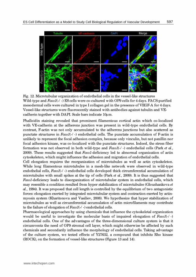
Fig. 12. Microtubular organization of endothelial cells in the vessel-like structures
Wild-type and Foxo1(-/ -) ES cells were co-cultured with OP9 cells for 4 days. FACS-purified
mesodermal cells were cultured in type I collagen gel in the presence of VEGF-A for 4 days.
Vessel-like structures were fluorescently stained with antibodies against tubulin and VE-
cadherin together with DAPI. Scale bars indicate 10μm.
Phalloidin staining revealed that prominent filamentous cortical actin which co-localized
with VE-cadherin at the adherens junction was present in wild-type endothelial cells. By
contrast, F-actin was not only accumulated to the adherens junctions but also scattered as
punctate structures in Foxo1(-/ -) endothelial cells. The punctate accumulation of F-actin is
unlikely to represent the focal adhesion complex, because only vinculin, but not paxillin nor
focal adhesion kinase, was co-localized with the punctate structures. Indeed, the stress fiber
formation was not observed in both wild-type and Foxo1(-/ -) endothelial cells (Park et al.,
2009). These results suggested that Foxo1-deficiency led to abnormal organization of actin
cytoskeleton, which might influence the adhesion and migration of endothelial cells.
Cell elongation requires the reorganization of microtubules as well as actin cytoskeleton.
While long filamentous microtubules in a mesh-like network were observed in wild-type
endothelial cells, Foxo1(-/ -) endothelial cells developed thick circumferential accumulation of
microtubules with small spikes at the tip of cells (Park et al., 2009). It is thus suggested that
Foxo1-deficiency leads to disorganization of microtubular system in endothelial cells, which
may resemble a condition resulted from hyper stabilization of microtubules (Gloushankova et
al., 1994). It was proposed that cell length is controlled by the equilibrium of two antagonistic
forces: elongation exerted by integrated microtubular system and contraction exerted by actin-
myosin system (Kharitonova and Vasiliev, 2008). We hypothesize that hyper stabilization of
microtubules as well as circumferential accumulation of actin microfilaments may contribute
to the failure of elongation of Foxo1(-/ -) endothelial cells.
Pharmacological approaches by using chemicals that influence the cytoskeletal organization
would be useful to investigate the molecular basis of impaired elongation of Foxo1(-/ -)
endothelial cells. One of the advantages of the three-dimensional culture system is that it
circumvents the need of OP9 stromal cell layer, which might otherwise be affected by such
chemicals and secondarily influence the morphology of endothelial cells. Taking advantage
of the culture system, we tested effects of Y27632, a compound that inhibits Rho kinase
(ROCK), on the formation of vessel-like structures (Figure 13 and 14).
www.intechopen.com

Embryonic Stem Cells: The Hormonal Regulation of Pluripotency and Embryogenesis
598
Fig. 13. Aberrant dependency on RhoA/ ROCK pathway in Foxo1(-/ -) endothelial cells
Wild-type and Foxo1(-/ -) ES cells were co-cultured with OP9 cells for 4 days. FACS-purified
mesodermal cells were cultured in type I collagen gel in the presence of VEGF-A and
presence or absence of Y27632. Vessel-like structures were fluorescently stained with
phalloidin, anti-VE-cadherin antibody and DAPI. Scale bars indicate 10μm.
Flk-1+ cells isolated from differentiating wild-type or Foxo1(-/ -) ES cells were allowed to
aggregate and embedded in type I collagen gel and cultured in the presence of 50 ng/ mL
VEGF-A and presence or absence of 10 μM Y27632. VE-cadherin immunofluorescence
staining of flattened gels revealed that wild-type Flk-1+ cells generated capillary-like
structures consisting of spindle-shaped endothelial cells regardless of whether Y27632 was
present or absent in the culture. Actin microfilaments and microtubule networks were not
also affected by Y27632 treatment. These observations indicate that ROCK inhibition does
not affect the elongation of wild-type endothelial cells and the formation of capillary-like
structures in vitro. By contrast, treatment with Y27632 resulted in extreme disintegration of
cell organization in the cultures of Foxo1(-/ -) endothelial cells. Adherens junctions revealed
by VE-cadherin and F-actin staining became fragmentary and distributed in a disorderly
manner. Microtubules also exhibited short fluffiness with random distribution.
Consequently, Foxo1(-/ -) endothelial cells no longer maintained even short bundles in the
presence of Y27632. Foxo1(-/ -) endothelial cells thus appear to be abnormally dependent on
the RhoA/ ROCK pathway.
www.intechopen.com
ES Cell Differentiation as a Model to Study Cell Biological Regulation of Vascular Development
599
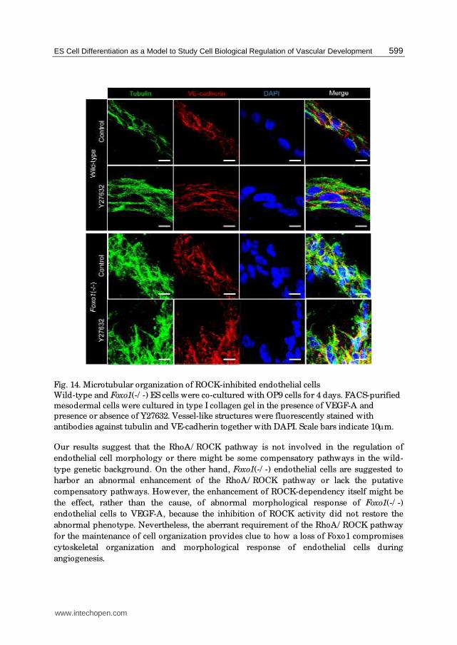
Fig. 14. Microtubular organization of ROCK-inhibited endothelial cells
Wild-type and Foxo1(-/ -) ES cells were co-cultured with OP9 cells for 4 days. FACS-purified
mesodermal cells were cultured in type I collagen gel in the presence of VEGF-A and
presence or absence of Y27632. Vessel-like structures were fluorescently stained with
antibodies against tubulin and VE-cadherin together with DAPI. Scale bars indicate 10μm.
Our results suggest that the RhoA/ ROCK pathway is not involved in the regulation of
endothelial cell morphology or there might be some compensatory pathways in the wild-
type genetic background. On the other hand, Foxo1(-/ -) endothelial cells are suggested to
harbor an abnormal enhancement of the RhoA/ ROCK pathway or lack the putative
compensatory pathways. However, the enhancement of ROCK-dependency itself might be
the effect, rather than the cause, of abnormal morphological response of Foxo1(-/ -)
endothelial cells to VEGF-A, because the inhibition of ROCK activity did not restore the
abnormal phenotype. Nevertheless, the aberrant requirement of the RhoA/ ROCK pathway
for the maintenance of cell organization provides clue to how a loss of Foxo1 compromises
cytoskeletal organization and morphological response of endothelial cells during
angiogenesis.
www.intechopen.com
Embryonic Stem Cells: The Hormonal Regulation of Pluripotency and Embryogenesis
600
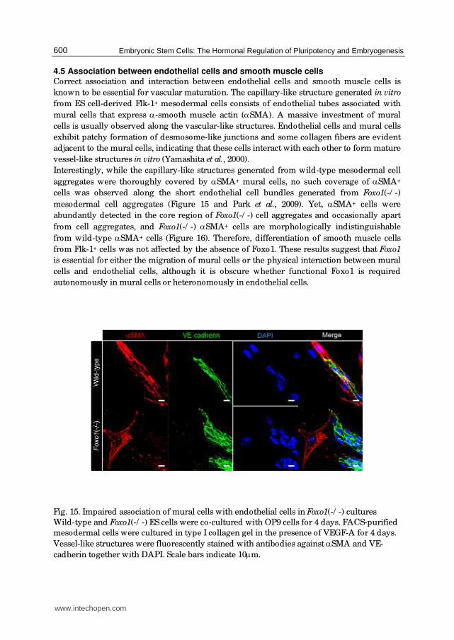
4.5 Association between endothelial cells and smooth muscle cells Correct association and interaction between endothelial cells and smooth muscle cells is
known to be essential for vascular maturation. The capillary-like structure generated in vitro
from ES cell-derived Flk-1+ mesodermal cells consists of endothelial tubes associated with
mural cells that express α-smooth muscle actin (αSMA). A massive investment of mural
cells is usually observed along the vascular-like structures. Endothelial cells and mural cells
exhibit patchy formation of desmosome-like junctions and some collagen fibers are evident
adjacent to the mural cells, indicating that these cells interact with each other to form mature
vessel-like structures in vitro (Yamashita et al., 2000).
Interestingly, while the capillary-like structures generated from wild-type mesodermal cell
aggregates were thoroughly covered by αSMA+ mural cells, no such coverage of αSMA+
cells was observed along the short endothelial cell bundles generated from Foxo1(-/ -)
mesodermal cell aggregates (Figure 15 and Park et al., 2009). Yet, αSMA+ cells were
abundantly detected in the core region of Foxo1(-/ -) cell aggregates and occasionally apart
from cell aggregates, and Foxo1(-/ -) αSMA+ cells are morphologically indistinguishable
from wild-type αSMA+ cells (Figure 16). Therefore, differentiation of smooth muscle cells
from Flk-1+ cells was not affected by the absence of Foxo1. These results suggest that Foxo1
is essential for either the migration of mural cells or the physical interaction between mural
cells and endothelial cells, although it is obscure whether functional Foxo1 is required
autonomously in mural cells or heteronomously in endothelial cells.
Fig. 15. Impaired association of mural cells with endothelial cells in Foxo1(-/ -) cultures
Wild-type and Foxo1(-/ -) ES cells were co-cultured with OP9 cells for 4 days. FACS-purified
mesodermal cells were cultured in type I collagen gel in the presence of VEGF-A for 4 days.
Vessel-like structures were fluorescently stained with antibodies against αSMA and VE-
cadherin together with DAPI. Scale bars indicate 10μm.
www.intechopen.com
ES Cell Differentiation as a Model to Study Cell Biological Regulation of Vascular Development
601
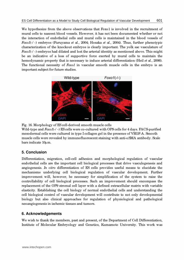
We hypothesize from the above observations that Foxo1 is involved in the recruitment of
mural cells to nascent blood vessels. However, it has not been documented whether or not
the interaction of endothelial cells and mural cells is maintained in the blood vessels of
Foxo1(-/ -) embryos (Furuyama et al., 2004; Hosaka et al., 2004). Thus, further phenotypic
characterization of the knockout embryos is clearly important. The yolk sac vasculature of
Foxo1(-/ -) embryos had dilated and lost the arterial identity as mentioned above. This might
be an indicative of a loss of supportive force exerted by mural cells to maintain the
hemodynamic property that is necessary to induce arterial differentiation (Heil et al., 2006).
The functional necessity of Foxo1 in vascular smooth muscle cells in the embryo is an
important subject for future studies.
Fig. 16. Morphology of ES cell-derived smooth muscle cells
Wild-type and Foxo1(-/ -) ES cells were co-cultured with OP9 cells for 4 days. FACS-purified
mesodermal cells were cultured in type I collagen gel in the presence of VEGF-A. Smooth
muscle cells were revealed by immunofluorescent staining with anti-αSMA antibody. Scale
bars indicate 10μm.
5. Conclusion
Differentiation, migration, cell-cell adhesion and morphological regulation of vascular
endothelial cells are the important cell biological processes that drive vasculogenesis and
angiogenesis. In vitro differentiation of ES cells provides useful means to elucidate the
mechanisms underlying cell biological regulation of vascular development. Further
improvement will, however, be necessary for simplification of the system to raise the
controllability of cell biological processes. Such an improvement should encompass the
replacement of the OP9 stromal cell layer with a defined extracellular matrix with variable
elasticity. Establishing the cell biology of normal endothelial cells and understanding the
cell biological control of vascular development will contribute to not only developmental
biology but also clinical approaches for regulation of physiological and pathological
neoangiogenesis in ischemic tissues and tumors.
6. Acknowledgements
We wish to thank the members, past and present, of the Department of Cell Differentiation,
Institute of Molecular Embryology and Genetics, Kumamoto University. This work was
www.intechopen.com

Embryonic Stem Cells: The Hormonal Regulation of Pluripotency and Embryogenesis
602
supported by grants from the Ministry of Education, Culture, Sports, Sciences and
Technology (MEXT) of Japan and Japan Society for the Promotion of Science. This work was
supported in part by Global COE Program (Cell Fate Regulation Research and Education
Unit), MEXT, Japan.
7. References
Bazzoni, G. & Dejana, E. (2004). Endothelial cell-to-cell junctions: molecular organization
and role in vascular homeostasis. Physiol Rev, Vol. 84, No. 3, 869-901.
Bloch, W., Forsberg, E., Lentini, S., Brakebusch, C., Martin, K., Krell, H.W., Weidle, U.H.,
Addicks, K. & Fassler, R. (1997). Beta 1 integrin is essential for teratoma growth and
angiogenesis. J Cell Biol, Vol. 139, No. 1, 265-278.
Breier, G., Breviario, F., Caveda, L., Berthier, R., Schnurch, H., Gotsch, U., Vestweber, D.,
Risau, W. & Dejana, E. (1996). Molecular cloning and expression of murine vascular
endothelial-cadherin in early stage development of cardiovascular system. Blood,
Vol. 87, No. 2, 630-641.
Carmeliet, P., Lampugnani, M.G., Moons, L., Breviario, F., Compernolle, V., Bono, F.,
Balconi, G., Spagnuolo, R., Oostuyse, B., Dewerchin, M., et al. (1999). Targeted
deficiency or cytosolic truncation of the VE-cadherin gene in mice impairs VEGF-
mediated endothelial survival and angiogenesis. Cell, Vol. 98, No. 2, 147-157.
Carvalho, R.L., Jonker, L., Goumans, M.J., Larsson, J., Bouwman, P., Karlsson, S., Dijke, P.T.,
Arthur, H.M. & Mummery, C.L. (2004). Defective paracrine signalling by TGFbeta
in yolk sac vasculature of endoglin mutant mice: a paradigm for hereditary
haemorrhagic telangiectasia. Development, Vol. 131, No. 24, 6237-6247.
Castrillon, D.H., Miao, L., Kollipara, R., Horner, J.W. & DePinho, R.A. (2003). Suppression of
ovarian follicle activation in mice by the transcription factor Foxo3a. Science, Vol.
301, No. 5630, 215-218.
Corada, M., Zanetta, L., Orsenigo, F., Breviario, F., Lampugnani, M.G., Bernasconi, S., Liao,
F., Hicklin, D.J., Bohlen, P. & Dejana, E. (2002). A monoclonal antibody to vascular
endothelial-cadherin inhibits tumor angiogenesis without side effects on
endothelial permeability. Blood, Vol. 100, No. 3, 905-911.
Daly, C., Wong, V., Burova, E., Wei, Y., Zabski, S., Griffiths, J., Lai, K.M., Lin, H.C., Ioffe, E.,
Yancopoulos, G.D., et al. (2004). Angiopoietin-1 modulates endothelial cell function
and gene expression via the transcription factor FKHR (FOXO1). Genes Dev, Vol. 18,
No. 9, 1060-1071.
De Val, S., Chi, N.C., Meadows, S.M., Minovitsky, S., Anderson, J.P., Harris, I.S., Ehlers,
M.L., Agarwal, P., Visel, A., Xu, S.M., et al. (2008). Combinatorial regulation of
endothelial gene expression by ets and forkhead transcription factors. Cell, Vol. 135,
No. 6, 1053-1064.
Dejana, E. (2004). Endothelial cell-cell junctions: happy together. Nat Rev Mol Cell Biol, Vol. 5,
No. 4, 261-270.
Doetschman, T.C., Eistetter, H., Katz, M., Schmidt, W. & Kemler, R. (1985). The in vitro
development of blastocyst-derived embryonic stem cell lines: formation of visceral
yolk sac, blood islands and myocardium. J. Embryol. Exp. Morphol., Vol. 87, 27-45.
www.intechopen.com
ES Cell Differentiation as a Model to Study Cell Biological Regulation of Vascular Development
603
Eilken, H.M., Nishikawa, S. & Schroeder, T. (2009). Continuous single-cell imaging of
blood generation from haemogenic endothelium. Nature, Vol. 457, No. 7231, 896-
900.
Endoh, M., Ogawa, M., Orkin, S. & Nishikawa, S. (2002). SCL/ tal-1-dependent process
determines a competence to select the definitive hematopoietic lineage prior to
endothelial differentiation. Embo J, Vol. 21, No. 24, 6700-6708.
Feraud, O., Cao, Y. & Vittet, D. (2001). Embryonic stem cell-derived embryoid bodies
development in collagen gels recapitulates sprouting angiogenesis. Lab Invest, Vol.
81, No. 12, 1669-1681.
Fukuhara, S., Sako, K., Minami, T., Noda, K., Kim, H.Z., Kodama, T., Shibuya, M., Takakura,
N., Koh, G.Y. & Mochizuki, N. (2008). Differential function of Tie2 at cell-cell
contacts and cell-substratum contacts regulated by angiopoietin-1. Nat Cell Biol,
Vol. 10, No. 5, 513-526.
Furuyama, T., Kitayama, K., Shimoda, Y., Ogawa, M., Sone, K., Yoshida-Araki, K.,
Hisatsune, H., Nishikawa, S., Nakayama, K., Nakayama, K., et al. (2004). Abnormal
angiogenesis in Foxo1 (Fkhr)-deficient mice. J Biol Chem, Vol. 279, No. 33, 34741-
34749.
Furuyama, T., Nakazawa, T., Nakano, I. & Mori, N. (2000). Identification of the differential
distribution patterns of mRNAs and consensus binding sequences for mouse DAF-
16 homologues. Biochem J, Vol. 349, No. Pt 2, 629-634.
Gloushankova, N.A., Lyubimova, A.V., Tint, I.S., Feder, H.H., Vasiliev, J.M. & Gelfand, I.M.
(1994). Role of the microtubular system in morphological organization of normal
and oncogene-transfected epithelial cells. Proc Natl Acad Sci U S A, Vol. 91, No. 18,
8597-8601.
Gory-Faure, S., Prandini, M.H., Pointu, H., Roullot, V., Pignot-Paintrand, I., Vernet, M. &
Huber, P. (1999). Role of vascular endothelial-cadherin in vascular morphogenesis.
Development, Vol. 126, No. 10, 2093-2102.
Grazia Lampugnani, M., Zanetti, A., Corada, M., Takahashi, T., Balconi, G., Breviario, F.,
Orsenigo, F., Cattelino, A., Kemler, R., Daniel, T.O., et al. (2003). Contact inhibition
of VEGF-induced proliferation requires vascular endothelial cadherin, beta-catenin,
and the phosphatase DEP-1/ CD148. J Cell Biol, Vol. 161, No. 4, 793-804.
Guo, R., Sakamoto, H., Sugiura, S. & Ogawa, M. (2007). Endothelial cell motility is
compatible with junctional integrity. J Cell Physiol, Vol. 211, No. 2, 327-335.
Hashimoto, K., Fujimoto, T., Shimoda, Y., Huang, X., Sakamoto, H. & Ogawa, M. (2007).
Distinct hemogenic potential of endothelial cells and CD41(+) cells in mouse
embryos. Dev Growth Differ, Vol. 49, No. 4, 287-300.
Heil, M., Eitenmuller, I., Schmitz-Rixen, T. & Schaper, W. (2006). Arteriogenesis versus
angiogenesis: similarities and differences. Journal of cellular and molecular medicine,
Vol. 10, No. 1, 45-55.
Hirai, H., Ogawa, M., Suzuki, N., Yamamoto, M., Breier, G., Mazda, O., Imanishi, J. &
Nishikawa, S. (2003). Hemogenic and nonhemogenic endothelium can be
distinguished by the activity of fetal liver kinase (Flk)-1 promoter/ enhancer during
mouse embryogenesis. Blood, Vol. 101, No. 3, 886-893.
www.intechopen.com

Embryonic Stem Cells: The Hormonal Regulation of Pluripotency and Embryogenesis
604
Hirashima, M., Kataoka, H., Nishikawa, S., Matsuyoshi, N. & Nishikawa, S. (1999).
Maturation of embryonic stem cells into endothelial cells in an in vitro model of
vasculogenesis. Blood, Vol. 93, No. 4, 1253-1263.
Hirashima, M., Ogawa, M., Nishikawa, S., Matsumura, K., Kawasaki, K., Shibuya, M. &
Nishikawa, S. (2003). A chemically defined culture of VEGFR2+ cells derived from
embryonic stem cells reveals the role of VEGFR1 in tuning the threshold for VEGF
in developing endothelial cells. Blood, Vol. 101, No. 6, 2261-2267.
Hisatsune, H., Matsumura, K., Ogawa, M., Uemura, A., Kondo, N., Yamashita, J.K., Katsuta,
H., Nishikawa, S. & Chiba, T. (2005). High level of endothelial cell-specific gene
expression by a combination of the 5' flanking region and the 5' half of the first
intron of the VE-cadherin gene. Blood, Vol. 105, No. 12, 4657-4663.
Hosaka, T., Biggs, W.H., 3rd, Tieu, D., Boyer, A.D., Varki, N.M., Cavenee, W.K. & Arden,
K.C. (2004). Disruption of forkhead transcription factor (FOXO) family members in
mice reveals their functional diversification. Proc Natl Acad Sci U S A, Vol. 101, No.
9, 2975-2980.
Jiao, K., Langworthy, M., Batts, L., Brown, C.B., Moses, H.L. & Baldwin, H.S. (2006). Tgfbeta
signaling is required for atrioventricular cushion mesenchyme remodeling during
in vivo cardiac development. Development, Vol. 133, No. 22, 4585-4593.
Kataoka, H., Takakura, N., Nishikawa, S., Tsuchida, K., Kodama, H., Kunisada, T., Risau,
W., Kita, T. & Nishikawa, S.I. (1997). Expressions of PDGF receptor alpha, c-Kit and
Flk1 genes clustering in mouse chromosome 5 define distinct subsets of nascent
mesodermal cells. Dev. Growth. Differ., Vol. 39, No. 6, 729-740.
Kharitonova, M.A. & Vasiliev, J.M. (2008). Controlling cell length. Seminars in cell &
developmental biology, Vol. 19, No. 6, 480-484.
Kim, H.S., Skurk, C., Maatz, H., Shiojima, I., Ivashchenko, Y., Yoon, S.W., Park, Y.B. &
Walsh, K. (2005). Akt/ FOXO3a signaling modulates the endothelial stress response
through regulation of heat shock protein 70 expression. Faseb J, Vol. 19, No. 8, 1042-
1044.
Kondo, N., Ogawa, M., Wada, H. & Nishikawa, S. (2009). Thrombin induces rapid
disassembly of claudin-5 from the tight junction of endothelial cells. Exp Cell Res,
Vol. 315, No. 17, 2879-2887.
Larsson, J., Goumans, M.J., Sjostrand, L.J., van Rooijen, M.A., Ward, D., Leveen, P., Xu, X.,
ten Dijke, P., Mummery, C.L. & Karlsson, S. (2001). Abnormal angiogenesis but
intact hematopoietic potential in TGF-beta type I receptor-deficient mice. Embo J,
Vol. 20, No. 7, 1663-1673.
Li, D.Y., Sorensen, L.K., Brooke, B.S., Urness, L.D., Davis, E.C., Taylor, D.G., Boak, B.B. &
Wendel, D.P. (1999). Defective angiogenesis in mice lacking endoglin. Science, Vol.
284, No. 5419, 1534-1537.
Matsukawa, M., Sakamoto, H., Kawasuji, M., Furuyama, T. & Ogawa, M. (2009). Different
roles of Foxo1 and Foxo3 in the control of endothelial cell morphology. Genes Cells,
Vol. 14, No. 10, 1167-1181.
Matsumura, K., Hirashima, M., Ogawa, M., Kubo, H., Hisatsune, H., Kondo, N., Nishikawa,
S., Chiba, T. & Nishikawa, S. (2003). Modulation of VEGFR-2-mediated endothelial-
cell activity by VEGF-C/ VEGFR-3. Blood, Vol. 101, No. 4, 1367-1374.
www.intechopen.com

ES Cell Differentiation as a Model to Study Cell Biological Regulation of Vascular Development
605
Nakano, T., Kodama, H. & Honjo, T. (1994). Generation of lymphohematopoietic cells from
embryonic stem cells in culture. Science, Vol. 265, No. 5175, 1098-1101.
Nishikawa, S.I., Nishikawa, S., Kawamoto, H., Yoshida, H., Kizumoto, M., Kataoka, H. &
Katsura, Y. (1998). In vitro generation of lymphohematopoietic cells from
endothelial cells purified from murine embryos. Immunity, Vol. 8, 761-769.
Nitta, T., Hata, M., Gotoh, S., Seo, Y., Sasaki, H., Hashimoto, N., Furuse, M. & Tsukita, S.
(2003). Size-selective loosening of the blood-brain barrier in claudin-5-deficient
mice. J Cell Biol, Vol. 161, No. 3, 653-660.
Oh, S.P., Seki, T., Goss, K.A., Imamura, T., Yi, Y., Donahoe, P.K., Li, L., Miyazono, K., ten
Dijke, P., Kim, S., et al. (2000). Activin receptor-like kinase 1 modulates
transforming growth factor-beta 1 signaling in the regulation of angiogenesis. Proc
Natl Acad Sci U S A, Vol. 97, No. 6, 2626-2631.
Paik, J.H., Kollipara, R., Chu, G., Ji, H., Xiao, Y., Ding, Z., Miao, L., Tothova, Z., Horner, J.W.,
Carrasco, D.R., et al. (2007). FoxOs are lineage-restricted redundant
tumor suppressors and regulate endothelial cell homeostasis. Cell, Vol. 128, No. 2,
309-323.
Park, S.H., Sakamoto, H., Tsuji-Tamura, K., Furuyama, T. & Ogawa, M. (2009). Foxo1 is
essential for in vitro vascular formation from embryonic stem cells. Biochem Biophys
Res Commun, Vol. 390, No. 3, 861-866.
Potente, M., Urbich, C., Sasaki, K., Hofmann, W.K., Heeschen, C., Aicher, A., Kollipara, R.,
DePinho, R.A., Zeiher, A.M. & Dimmeler, S. (2005). Involvement of Foxo
transcription factors in angiogenesis and postnatal neovascularization. J Clin Invest,
Vol. 115, No. 9, 2382-2392.
Risau, W., Sariola, H., Zerwes, H.G., Sasse, J., Ekblom, P., Kemler, R. & Doetschman, T.
(1988). Vasculogenesis and angiogenesis in embryonic-stem-cell-derived embryoid
bodies. Development, Vol. 102, No. 3, 471-478.
Seoane, J., Le, H.V., Shen, L., Anderson, S.A. & Massague, J. (2004). Integration of Smad and
forkhead pathways in the control of neuroepithelial and glioblastoma cell
proliferation. Cell, Vol. 117, No. 2, 211-223.
ten Dijke, P. & Arthur, H.M. (2007). Extracellular control of TGFbeta signalling in vascular
development and disease. Nat Rev Mol Cell Biol, Vol. 8, No. 11, 857-869.
Tsukita, S., Furuse, M. & Itoh, M. (2001). Multifunctional strands in tight junctions. Nat Rev
Mol Cell Biol, Vol. 2, No. 4, 285-293.
van der Horst, A. & Burgering, B.M. (2007). Stressing the role of FoxO proteins in lifespan
and disease. Nat Rev Mol Cell Biol, Vol. 8, No. 6, 440-450.
Vittet, D., Prandini, M.H., Berthier, R., Schweitzer, A., Martin Sisteron, H., Uzan, G. &
Dejana, E. (1996). Embryonic stem cells differentiate in vitro to endothelial cells
through successive maturation steps. Blood, Vol. 88, No. 9, 3424-3431.
Wang, R., Clark, R. & Bautch, V.L. (1992). Embryonic stem cell-derived cystic embryoid
bodies form vascular channels: an in vitro model of blood vessel development.
Development, Vol. 114, No. 2, 303-316.
Yamashita, J., Itoh, H., Hirashima, M., Ogawa, M., Nishikawa, S., Yurugi, T., Naito, M.,
Nakao, K. & Nishikawa, S. (2000). Flk1-positive cells derived from embryonic stem
cells serve as vascular progenitors. Nature, Vol. 408, No. 6808, 92-96.
www.intechopen.com

Embryonic Stem Cells: The Hormonal Regulation of Pluripotency and Embryogenesis
606
Yang, X., Castilla, L.H., Xu, X., Li, C., Gotay, J., Weinstein, M., Liu, P.P. & Deng, C.X. (1999).
Angiogenesis defects and mesenchymal apoptosis in mice lacking SMAD5.
Development, Vol. 126, No. 8, 1571-1580.
www.intechopen.com

Embryonic Stem Cells: The Hormonal Regulation of Pluripotencyand EmbryogenesisEdited by Prof. Craig Atwood
ISBN 978-953-307-196-1Hard cover, 672 pagesPublisher InTechPublished online 26, April, 2011Published in print edition April, 2011
InTech EuropeUniversity Campus STeP Ri Slavka Krautzeka 83/A 51000 Rijeka, Croatia Phone: +385 (51) 770 447 Fax: +385 (51) 686 166www.intechopen.com
InTech ChinaUnit 405, Office Block, Hotel Equatorial Shanghai No.65, Yan An Road (West), Shanghai, 200040, China
Phone: +86-21-62489820 Fax: +86-21-62489821
Pluripotency is a prerequisite for the subsequent coordinated differentiation of embryonic stem cells into alltissues of the body. This book describes recent advances in our understanding of pluripotency and thehormonal regulation of embryonic stem cell differentiation into tissue types derived from the ectoderm,mesoderm and endoderm.
How to referenceIn order to correctly reference this scholarly work, feel free to copy and paste the following:
Kiyomi Tsuji-Tamura, Hiroshi Sakamoto and Minetaro Ogawa (2011). ES Cell Differentiation as a Model toStudy Cell Biological Regulation of Vascular Development, Embryonic Stem Cells: The Hormonal Regulation ofPluripotency and Embryogenesis, Prof. Craig Atwood (Ed.), ISBN: 978-953-307-196-1, InTech, Available from:http://www.intechopen.com/books/embryonic-stem-cells-the-hormonal-regulation-of-pluripotency-and-embryogenesis/es-cell-differentiation-as-a-model-to-study-cell-biological-regulation-of-vascular-development

© 2011 The Author(s). Licensee IntechOpen. This chapter is distributedunder the terms of the Creative Commons Attribution-NonCommercial-ShareAlike-3.0 License, which permits use, distribution and reproduction fornon-commercial purposes, provided the original is properly cited andderivative works building on this content are distributed under the samelicense.