essential role for trpc5-containing extracellular vesicles ... · essential role for...
TRANSCRIPT

Essential role for TrpC5-containing extracellularvesicles in breast cancer withchemotherapeutic resistanceXin Maa,b,1,2, Zhen Chena,2, Dong Huac,2, Dongxu Hed,2, Linjun Wangc, Peng Zhangb, Junqi Wange, Yanfei Caia,Caiji Gaoe, Xiaodong Zhanga, Fangfang Zhanga, Teng Wangc, Tingting Hongc, Linfang Jinc, Xiaowei Qic, Shuxian Chena,Xiaoting Gua, Dangtong Yanga, Qiongxi Pana, Yifei Zhua, Yun Chena, Daozhen Chenf, Liwen Jiange, Xiaofeng Hang,Yanyun Zhangh, Jian Jina,h,1, and Xiaoqiang Yaob
aSchool of Pharmaceutical Sciences, cAffiliated Hospital, dNational Engineering Laboratory for Cereal Fermentation Technology, and gWuxi Medical School,Jiangnan University, Wuxi 214122, China; bSchool of Biomedical Sciences and eSchool of Life Sciences, Centre for Cell and Developmental Biology, ChineseUniversity of Hong Kong, Shatin, NT, Hong Kong; fCentral Laboratory, Wuxi Maternity and Child Health Care Hospital Affiliated to Nanjing MedicalUniversity, Wuxi 214002, China; and hInstitute of Health Sciences, Shanghai Institutes for Biological Sciences, Chinese Academy of Sciences and Shanghai JiaoTong University School of Medicine, Shanghai 200025, China
Edited by Tak W. Mak, The Campbell Family Institute for Breast Cancer Research at Princess Margaret Cancer Centre, Ontario Cancer Institute, UniversityHealth Network, Toronto, Canada, and approved March 19, 2014 (received for review January 8, 2014)
A critical challenge for chemotherapy is the development of chemo-resistance in breast cancer. However, the underlying mechanismsand validated predictors remain unclear. Extracellular vesicles (EVs)have gained attention as potential means for cancer cells to shareintracellular contents. In adriamycin-resistant human breast cancercells (MCF-7/ADM), we analyzed the role of transient receptorpotential channel 5 (TrpC5) in EV formation and transfer as well asthe diagnostic implications. Up-regulated TrpC5, accumulated inEVs, is responsible for EV formation and trapping of adriamycin(ADM) in EVs. EV-mediated intercellular transfer of TrpC5 allowedrecipient cells to acquire TrpC5, consequently stimulating multi-drug efflux transporter P-glycoprotein production through a Ca2+-and activated T-cells isoform c3-mediated mechanism and thus,conferring chemoresistance on nonresistant cells. TrpC5-contain-ing circulating EVs were detected in nude mice bearing MCF-7/ADM tumor xenografts, and the level was lower after TrpC5–siRNA treatment. In breast cancer patients who underwent che-motherapy, TrpC5 expression in the tumor was significantly higherin patients with progressive or stable disease than in patients witha partial or complete response. TrpC5-containing circulating EVswere found in peripheral blood from patients who underwentchemotherapy but not patients without chemotherapy. Takentogether, we found that TrpC5-containing circulating EVs maytransfer chemoresistance property to nonchemoresistant recipi-ent cells. It may be worthwhile to further explore the potential ofusing TrpC5-containing EVs as a diagnostic biomarker for che-moresistant breast cancer.
The development of chemotherapeutic resistance in breastcancer is a serious problem (1, 2). To date, the mechanisms
underlying chemoresistance are still largely unknown, and novalidated predictive factor of chemoresistance is available in theclinic. Therefore, it is important to identify the signaling path-ways and search for circulating markers in breast cancer resistantto chemotherapy.The extracellular environment contains a large number of
mobile membrane-limited vesicles named extracellular vesicles(EVs). Major EV populations include exosomes, microvesicles,and apoptotic bodies (1, 3–5). These dynamic EVs may haveessential function in intercellular communication and immuneregulation (5). Tumor cells also generate EVs (3, 4). Largequantities of tumor-derived circulating EVs have been foundin the blood of patients with glioblastoma multiforme (4), pan-creatic cancer (6), gastric cancer (7), and acute myeloid leukemia(8). They contain cell surface proteins, RNA, and DNA (3, 4,9, 10). They mediate intercellular cross-talk by transferringtheir intravesicular contents from donor to recipient cells and
participating in tumor invasion and metastasis (11–13). How-ever, how these structures are generated and their importancein chemotherapeutic resistance in breast cancer are poorlyunderstood.On the basis of our previous finding that transient receptor
potential channel 5 (TrpC5) regulates the multidrug transporterP-glycoprotein (P-gp) (13), we investigated the possible role ofTrpC5 in the formation and release of EVs in the context ofchemotherapeutic drugs. TrpC5 is one of the mammalian tran-sient receptor potential proteins. It is a nonselective cationchannel with Ca2+ permeability (14). Functionally, TrpC5 is in-volved in growth factor-regulated vesicular trafficking throughPI3K, Rac1, and PIP-5-kinase–mediated pathways (15). Here,we describe findings that suggest a unique role for TrpC5 intransfer of chemoresistance and its diagnostic implications.
Significance
A critical challenge for chemotherapy is development of che-moresistance, but underlying molecular mechanisms remainunclear. In this study, we found that drug-resistant adriamycin-resistant human breast cancer cells possessed numerous tran-sient receptor potential channel 5 (TrpC5) -containing extra-cellular vesicles (EVs) on the cell surface. Suppressing TrpC5expression diminished the formation of EVs. Incubation ofdrug-sensitive recipient cells with EVs endowed recipients withdrug-resistant properties. In both human samples and a mousemodel of breast cancer, the expression of TrpC5 proteins washigh in the tumor, and the levels of TrpC5-positive EVs werehigh in the circulation. These data suggest a critical role ofTrpC5-containing EVs in the transfer of drug resistance. In thefuture, monitoring TrpC5-containing EVs in the circulationcould potentially be used to predict the clinical outcome ofchemotherapy.
Author contributions: X.M., D. Hua, D. He, L.W., J.W., X.Q., Y. Chen, D.C., L. Jiang, X.H.,Y. Zhang, J.J., and X.Y. designed research; X.M., Z.C., D. Hua, D. He, L.W., P.Z., J.W.,Y. Cai, C.G., X.Z., F.Z., T.W., T.H., L. Jin, S.C., X.G., D.Y., Q.P., and Y. Zhu performedresearch; X.M., Z.C., D. He, P.Z., and T.W. analyzed data; and X.M., J.J., and X.Y.wrote the paper.
The authors declare no conflict of interest.
This article is a PNAS Direct Submission.1To whom correspondence may be addressed. E-mail: [email protected] [email protected].
2X.M., Z.C., D. Hua, and D. He contributed equally to this work.
This article contains supporting information online at www.pnas.org/lookup/suppl/doi:10.1073/pnas.1400272111/-/DCSupplemental.
www.pnas.org/cgi/doi/10.1073/pnas.1400272111 PNAS | April 29, 2014 | vol. 111 | no. 17 | 6389–6394
MED
ICALSC
IENCE
S

ResultsTrpC5 Is Required for EV Formation in Adriamycin-Resistant HumanBreast Cancer Cells. We established adriamycin-resistant humanbreast cancer cells (MCF-7/ADM). These cells, with greatlyup-regulated multidrug efflux transporter P-gp (13), displayeda 630-fold greater resistance to adriamycin (ADM) than theparental line MCF-7/WT. They also displayed enhanced re-sistance to paclitaxel (146-fold), epirubicin (56-fold), vincristine(5-fold), and mitoxantrone (127-fold).Interestingly, we found a great enhancement of EV structures
on the surface of MCF-7/ADM cells by transmission EM (TEM)(Fig. 1A) compared with the WT (Fig. 1B). ADM has natural redfluorescence, and it functions by intercalating into DNA (16). InMCF-7/WT cells, as expected, ADM mainly accumulated insidethe nucleus, which was shown by confocal microscopy (redautofluorescence) (Fig. 1C). However, in MCF-7/ADM cells,ADM accumulation was much less, with most of the residual
drug in the periphery and not in the nucleus (Fig. 1C). Immuno-TEM and an ADM-specific monoclonal antibody were furtherused to verify its accumulation in EVs. Images clearly showed anEV distribution of ADM (Fig. 1D). Immunostaining for the EVmarker flotillin2 (17) in intact MCF-7/ADM cells confirmed theidentity of puncta as EVs (Fig. 1E). These results clearly in-dicated that ADM was trapped in EVs (Fig. 1E), where itspharmacological effect would be reduced.Cytosolic Ca2+ ([Ca2+]i) is an important signal for EV for-
mation (18–20), whereas TrpC5 is a Ca2+-permeable channel(21, 22) that is involved in local vesicular trafficking (15) withsubstantially higher expression in chemoresistant MCF-7/ADMcells (21). Thus, we further tested whether TrpC5 is involved inthe formation of EVs. In chemoresistant MCF-7/ADM cells,immunostaining as well as immuno-TEM clearly showed theaccumulation of TrpC5 in EVs (Fig. 1F). Immunostainingshowed that TrpC5 is colocalized with flotillin2-positive EVs
Fig. 1. TrpC5 is required for EV formation in MCF-7/ADM cells. (A and B) Transmission electron micrographs of (A) ADM-resistant (MCF-7/ADM) and (B) ADM-sensitive (MCF-7/WT) human breast cancer cells. (C) Confocal immunostaining micrographs of ADM (autofluorescence; red) distribution in EVs in MCF-7/ADMand MCF-7/WT cells. DIC, differential interference contrast. (D) Immunotransmission electron micrograph of ADM distribution (arrows) in EVs. (E) Confocalimmunostaining micrograph of flotillin2 expression in EVs from MCF-7/ADM cells. (F) Confocal immunostaining micrographs (Left) and immunotransmissionelectron micrograph (arrows; Right) showing distribution of TrpC5 in EVs. (G) Transmission electron micrographs showing effects of TrpC5 knockdown on EVformation in MCF-7/ADM cells (Left, scrambled; Right, TrpC5-siRNA). (H) Confocal immunostaining micrographs showing the effect of TrpC5 knockdown onADM distribution in EVs in MCF-7/ADM cells (Left, scrambled; Right, TrpC5-siRNA). TrpC5-siRNA or scrambled siRNA (200 nM); ADM (10 μg/mL; overnight) wasadded to the culture medium 1 d before fluorescence measurement. (Scale bars: 1 μm.)
6390 | www.pnas.org/cgi/doi/10.1073/pnas.1400272111 Ma et al.
near cell periphery (Fig. S1). Up-regulated P-gp (21), whichis coded by the mdr1 gene and pumps cytotoxic drugs fromMCF-7/ADM cells, also had an EV distribution (Fig. S2). InMCF-7/ADM cells, TEM showed that TrpC5-specific siRNAgreatly inhibited the generation of EVs compared with scram-bled siRNA by TEM (Fig. 1G), and confocal microscopy showedthat ADM reaccumulated in the nucleus (Fig. 1H). We furtherconstructed an MCF-7 cell line stably expressing TrpC5 andfound that up-regulation of TrpC5 greatly increased the gener-ation of EVs (Fig. S3). These results showed that TrpC5 playsan essential role in EVs formation by chemoresistant breastcancer cells.
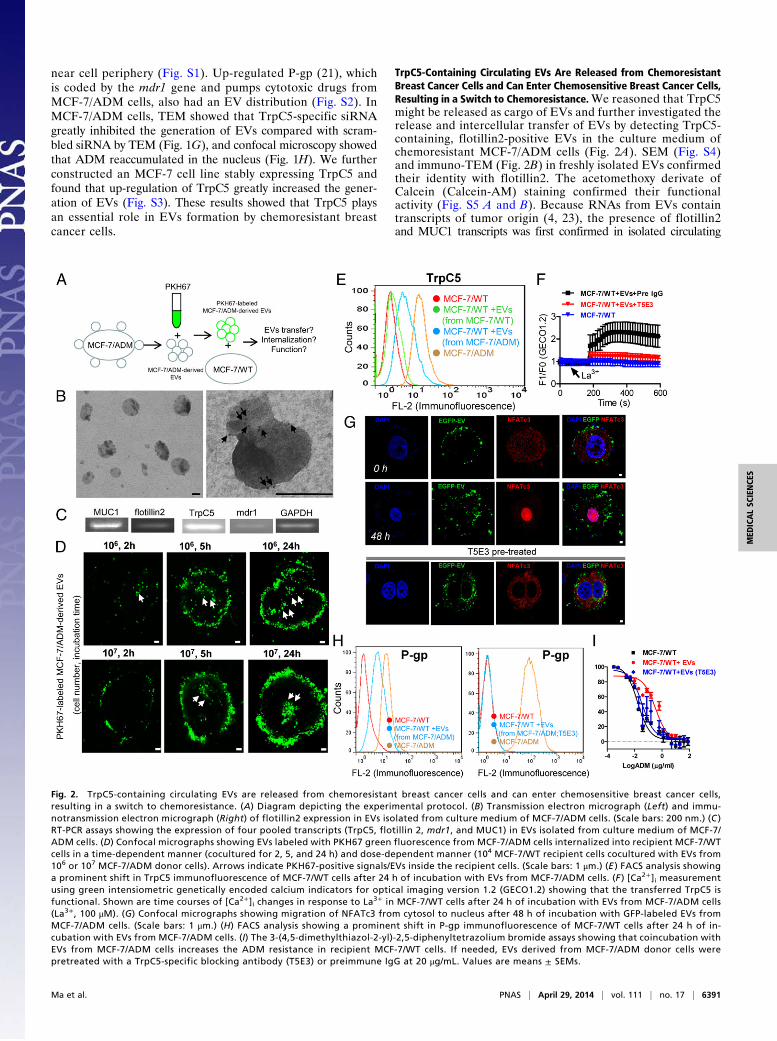
TrpC5-Containing Circulating EVs Are Released from ChemoresistantBreast Cancer Cells and Can Enter Chemosensitive Breast Cancer Cells,Resulting in a Switch to Chemoresistance.We reasoned that TrpC5might be released as cargo of EVs and further investigated therelease and intercellular transfer of EVs by detecting TrpC5-containing, flotillin2-positive EVs in the culture medium ofchemoresistant MCF-7/ADM cells (Fig. 2A). SEM (Fig. S4)and immuno-TEM (Fig. 2B) in freshly isolated EVs confirmedtheir identity with flotillin2. The acetomethoxy derivate ofCalcein (Calcein-AM) staining confirmed their functionalactivity (Fig. S5 A and B). Because RNAs from EVs containtranscripts of tumor origin (4, 23), the presence of flotillin2and MUC1 transcripts was first confirmed in isolated circulating
Fig. 2. TrpC5-containing circulating EVs are released from chemoresistant breast cancer cells and can enter chemosensitive breast cancer cells,resulting in a switch to chemoresistance. (A) Diagram depicting the experimental protocol. (B) Transmission electron micrograph (Left) and immu-notransmission electron micrograph (Right) of flotillin2 expression in EVs isolated from culture medium of MCF-7/ADM cells. (Scale bars: 200 nm.) (C )RT-PCR assays showing the expression of four pooled transcripts (TrpC5, flotillin 2, mdr1, and MUC1) in EVs isolated from culture medium of MCF-7/ADM cells. (D) Confocal micrographs showing EVs labeled with PKH67 green fluorescence from MCF-7/ADM cells internalized into recipient MCF-7/WTcells in a time-dependent manner (cocultured for 2, 5, and 24 h) and dose-dependent manner (104 MCF-7/WT recipient cells cocultured with EVs from106 or 107 MCF-7/ADM donor cells). Arrows indicate PKH67-positive signals/EVs inside the recipient cells. (Scale bars: 1 μm.) (E ) FACS analysis showinga prominent shift in TrpC5 immunofluorescence of MCF-7/WT cells after 24 h of incubation with EVs from MCF-7/ADM cells. (F ) [Ca2+]i measurementusing green intensiometric genetically encoded calcium indicators for optical imaging version 1.2 (GECO1.2) showing that the transferred TrpC5 isfunctional. Shown are time courses of [Ca2+]i changes in response to La3+ in MCF-7/WT cells after 24 h of incubation with EVs from MCF-7/ADM cells(La3+, 100 μM). (G) Confocal micrographs showing migration of NFATc3 from cytosol to nucleus after 48 h of incubation with GFP-labeled EVs fromMCF-7/ADM cells. (Scale bars: 1 μm.) (H) FACS analysis showing a prominent shift in P-gp immunofluorescence of MCF-7/WT cells after 24 h of in-cubation with EVs from MCF-7/ADM cells. (I) The 3-(4,5-dimethylthiazol-2-yl)-2,5-diphenyltetrazolium bromide assays showing that coincubation withEVs from MCF-7/ADM cells increases the ADM resistance in recipient MCF-7/WT cells. If needed, EVs derived from MCF-7/ADM donor cells werepretreated with a TrpC5-specific blocking antibody (T5E3) or preimmune IgG at 20 μg/mL. Values are means ± SEMs.
Ma et al. PNAS | April 29, 2014 | vol. 111 | no. 17 | 6391
MED
ICALSC
IENCE
S

EVs. MUC1 is frequently expressed in breast cancer; it is sortedinto rafts by a flotillin-dependent mechanism and exported throughEVs (24–26). Its expression in MCF-7/ADM cells was confirmed byimmunostaining (Fig. S6). RT-PCR was then performed to simul-taneously identify the transcript expression of flotillin2, MUC1,TrpC5, and mdr1. We found that these transcripts were positive inEVs derived from the culture medium of MCF-7/ADM cells, in-dicating the release of TrpC5-containing, flotillin2-positive EVs bythese cells (Fig. 2C). To determine whether the EVs were in-ternalized by recipient cells, we labeled the isolated EVs witha PKH67 labeling kit and incubated them with MCF-7/WT cells inculture (4, 27). The results showed that the labeled EVs bound toMCF-7/WT cells in a time- and dose-dependent manner (Fig. 2D).We further determined the transfer of EVs containing protein bymonitoring GFP that was stably expressed in MCF-7/ADM cells.Uptake of GFP fluorescence into recipient cells was observed onexposure to the GFP-containing EVs (Fig. S7). In this regard,we asked whether TrpC5 can be transferred in this manner andremain functional. Fluorescence-activated cell sorting analysisshowed that MCF-7/WT cells incubated with donor cell (MCF-7/ADM) EVs displayed an up-regulated expression of TrpC5, in-dicating the microvesicular transfer of TrpC5 (Fig. 2E). Thefunctional presence of TrpC5 was determined by [Ca2+]i mea-surement. Lanthanum (La3+) is known to potentiate TrpC5 ac-tivity but inhibit many other Ca2+-permeable channels (21, 22).Application of La3+ elicited a rise in [Ca2+]i in recipient MCF-7/WT cells cocultured with EVs derived from MCF-7/ADM donorcells but not MCF-7/WT cells alone (Fig. 2F). This La3+-elicited[Ca2+]i rise was reduced by a TrpC5-specific blocking antibody,T5E3, confirming the involvement of functional TrpC5 (Fig. 2F).We showed previously that [Ca2+]i influx through TrpC5 is cru-cial for nuclear translocation of activated T-cells isoform c3(NFATc3) and that NFATc3 is the transcriptional factor thatlinks the TrpC5 activity to P-gp production (21). Here, after 48h of coculture with EVs, NFATc3 translocated into the nucleusin one-half of the cells (Fig. 2G), and P-gp increased in recipientMCF-7/WT cells (Fig. 2H). With EV transfer, the recipientchemosensitive MCF-7/WT cells became more resistant toADM-induced cell death, displaying a 20-fold higher resistance
to ADM (Fig. 2I). Importantly, NFATc3 translocation, P-gpincrease, and up-regulation of drug resistance were markedlyinhibited by pretreatment of EVs with T5E3 (Fig. 2 G–I), in-dicating that the up-regulated expression of P-gp was mainlycaused by the transferred TrpC5-induced TrpC5–NFATc3–P-gpsignal pathway (Fig. 2 E–I).
TrpC5 Is Required for the Release of Circulating EVs from HumanBreast Tumor Xenografts in Athymic Nude Mice. After validatingthe generation and release of EVs from chemoresistant MCF-7/ADM cells, we next assessed circulating EVs in nude micebearing MCF-7/ADM xenograft tumors. Immunohistochemistryshowed that TrpC5, flotillin2, and P-gp were abundantly expressedin MCF-7/ADM xenograft tumors (Fig. 3A and Fig. S8) and thatflotillin2 and P-gp expressions were substantially reduced inTrpC5–siRNA-treated tumor xenografts (Fig. 3A). RT-PCR wasperformed to determine the expression with four pooled tran-scripts TrpC5, flotillin2, mdr1, and MUC1 in circulating EVs. Alltranscripts were positive in seven of seven nude mice, indicatingthat TrpC5-containing EVs are released into the peripheral bloodfrom MCF-7/ADM cells (Fig. 3B). Importantly, in the TrpC5–siRNA-treated tumor xenografts, the TrpC5, flotillin2, and P-gptranscript levels were lower than with scrambled siRNA, furthershowing the requirement of TrpC5 for the release of EVs andtheir cargos.
Circulating TrpC5-Containing EVs Predict Clinical Outcome ofChemotherapy for Breast Cancer. To explore the clinical potentialof TrpC5 in the formation of EVs in breast cancer, we analyzedpaired breast cancer tissue from 26 patients before and afteranthracycline/taxane-based chemotherapy (Table S1). Immuno-histochemistry showed that both TrpC5 and flotillin2 expressionswere significantly up-regulated after chemotherapy (Fig. 4 A andB). Moreover, TrpC5 expression was positively correlated withthat of flotillin2 (Fig. 4C). Treatment response was assessedby the response evaluation criteria in solid tumors (28).Among 26 patients, 13 patients responded to chemotherapy[partial response/complete response (PR/CR)], whereas 13patients were not responsive [progressive disease/stable disease
Fig. 3. TrpC5 is required for the release of circulating EVs from human breast tumor xenografts in athymic nude mice. (A) Flotillin2 expression was sub-stantially reduced in TrpC5–siRNA-treated tumor xenografts. Immunohistochemical staining for TrpC5, flotillin2, and P-gp in sections of MCF-7/ADM tumorxenografts with TrpC5-siRNA (scrambled siRNA as control). Immunohistochemical staining in sections of MCF-7/WT tumor xenografts was taken as the control(n = 5 in each group). (B) RT-PCR assays showing the expression of four pooled transcripts (TrpC5, flotillin 2, mdr1, and MUC1) in peripheral blood from nudemice bearing chemoresistant MCF-7/ADM tumor xenografts. Female nude mice bearing xenograft tumors derived from MCF-7/ADM were injected at thetumor sites with TrpC5-siRNA (40 pmol; scrambled siRNA as control; n = 7 in each group). MCF-7/ADM tumor continued to grow in size under ADM treatment,indicating ADM resistance. Data were analyzed by Student t test. Values are means ± SEMs. #P < 0.05 compared with scrambled. (Scale bars: 100 μm.)
6392 | www.pnas.org/cgi/doi/10.1073/pnas.1400272111 Ma et al.

(PD/SD)]. Importantly, TrpC5 expression was significantly greaterin patients with PD/SD than in patients with PR/CR (Fig. 4D),indicating its close association with the induction of chemo-resistance. Flotillin2 expression was also higher in patients withPD/SD than patients with PR/CR (Fig. 4E). RT-PCR was nextused to assess the features of TrpC5-containing circulating EVs inperipheral blood from 33 patients with chemotherapy. TrpC5 aswell as flotillin2, mdr1, and MUC1 were simultaneously amplifiedfrom EVs in 17 of 33 samples from these patients but not in 12patients without chemotherapy (Fig. 4F and Table S2). Flotillin2and TrpC5 expression or MUC1 and TrpC5 expression in circu-lating EVs isolated from patients was further confirmed by FACS(Fig. S9). Calcein-AM staining in circulating EVs confirmed theirfunctional activity (Fig. S5B). Thus, identification of tumor-spe-cific TrpC5 in circulating EVs may provide a window on theclinical outcome of chemotherapy (Fig. 4G).
DiscussionTo achieve more effective and individualized chemotherapeutictreatment of breast cancer patients, it is essential to understandthe mechanisms responsible for drug resistance and define
reliable indicators for response to therapy. The findings de-scribed here shed light on an unconventional and poorlyunderstood mechanism of cell-to-cell communication in thecontext of chemotherapeutic drugs and how that communi-cation may have significant consequences in the developmentand transfer of the chemoresistant phenotype in breast can-cer. In particular, we have shown that exposing chemosensitivebreast cancer recipient cells to bioactive TrpC5-containing EVsthat are constitutively shed by certain chemoresistant breast can-cer donor cells can cause the recipient cells to acquire a chemo-resistant phenotype. To the best of our knowledge, this is the firststudy investigating the association of an ion channel in circu-lating EVs with chemotherapeutic response.It has been suggested that EVs can transfer P-gp from
drug-resistant lymphoblastic leukemia cells to nonresistantcells, conferring multidrug resistance on nonresistant cells, butits capacity is not sustained (29). We recently reported that up-regulation of TrpC5 protein is crucial for P-gp induction and thedevelopment of chemoresistance in breast cancer cells (21).Here, we show that the same is true for recipient cells after theirEV-mediated acquisition of resistance. Transfer of TrpC5 allowed
Fig. 4. Circulating TrpC5-containing EVs predict clinical outcome of breast cancer with chemotherapy. (A) Representative images and summary data fromimmunohistochemical staining of TrpC5 in paired pre- and postchemotherapy breast cancer tissue from patients showing elevated TrpC5 expression (n = 26).(B) Representative images and summary data from immunohistochemical staining for flotillin2 in paired pre- and postchemotherapy breast cancer tissueshowing that flotillin2 expression is elevated in breast cancer patients (n = 26). Data were analyzed by Student t test. (C) Pearson correction of TrpC5 ex-pression with flotillin2 (n = 26). Data were analyzed using Pearson correlation test. (D) TrpC5 expression was significantly greater in patients with PD/SD (n =13) compared with patients with PR/CR (n = 13). (E) Flotillin2 expression was higher in patients with PD/SD (n = 13) than in patients with PR/CR (n = 13). Datawere analyzed by Student t test. (F) RT-PCR assays showing the expression of four pooled transcripts (TrpC5, flotillin 2, mdr1, and MUC1) in peripheral bloodwere elevated in patients who received chemotherapy with PD/SD (n = 17) but not elevated in patients who did not received chemotherapy (n = 12). (G)Proposed model highlighting TrpC5 in EVs formation and transfer and the diagnostic implications. (Scale bars: 100 μm.)
Ma et al. PNAS | April 29, 2014 | vol. 111 | no. 17 | 6393
MED
ICALSC
IENCE
S
the recipient cells to acquire/produce this Ca2+-permeablechannel, consequently stimulating P-gp production in the recipientcells through a Ca2+- and NFATc3-mediated mechanism and asa result, endowing nonresistant cells with chemoresistance. Ca2+
entry is also known to stimulate the transcription of TrpCs them-selves in a positive feedback manner through the NFAT tran-scription factor (30, 31). In this way, TrpC5 and P-gp can becontinuously produced, resulting in long-lasting drug resistance.One aspect of our work that merits additional consideration
involves the potential mechanisms of abundant accumulationof TrpC5 in EVs in the context of chemotherapeutic drugs. Agrowing body of evidence has confirmed the translocation of Trpchannels. A particularly good example comes from a recentstudy, which showed that EGF induces rapid vesicular trans-location of TrpC5 channels, dramatically increasing membrane-associated functional TrpC5 and resulting in tight spatial–temporalcontrol of TrpC5 (15). Rho-GTPases regulate vesicular traf-ficking and membrane processes, and Rac1 plays a role in thetranslocation of functional TrpC5 (15). Thus, more work needsto be done to determine the molecular mechanism of EV ac-cumulation of TrpC5 in chemoresistant breast cancer cells.Collectively, our data suggest that up-regulated TrpC5 accu-
mulated in EVs is responsible for EV formation and EV trappingof chemotherapeutic drugs. More importantly, we also showeda critical role of TrpC5-containing EVs in the transfer of drugresistance property to nonchemoresistant recipient cells. Nota-bly, we found a striking association between TrpC5-containingEVs circulating in peripheral blood and the clinical response tochemotherapy. Although there is no reason to think that theseeffects are limited to TrpC5, TrpC5-containing circulating EVsmay enable the monitoring of chemotherapeutic efficacy. Thus,it may be worthwhile to further explore the potential of using
TrpC5-containing EVs as a diagnostic biomarker for chemo-resistant breast cancer.
Materials and MethodsAdditional methods are described in SI Materials and Methods.
TEM. Sample preparation, thin sectioning, and immunolabeling on sectionswere performed with antibodies against ADM, TrpC5, and flotillin2 and gold-coupled secondary antibodies. Sections were viewed with a HitachiHT7700 TEM.
ADM Accumulation. Subcellular distribution of ADM was determined usinga laser scanning confocal microscope.
Labeling of EVs with PKH67 and Confocal Microscopy. EVs were labeled witha PKH67 green fluorescent labeling kit (Sigma-Aldrich) following the man-ufacturer’s instructions. MCF-7/WT cells were incubated with the PKH67-labeled MCF-7/ADM-derived EVs for the indicated time. Cells were washedand subjected to confocal microscopy (Zeiss LSM 510 confocal laserscanning microscopy).
ACKNOWLEDGMENTS. We thank Prof. Iain C. Bruce for critical reading ofthe manuscript and Profs. B. Jiang, J. Xu, and K. Y. Lu of the State KeyLaboratory of Food Science and Technology, Jiangnan University for insight-ful comments and technical assistance. This work was supported by Programfor New Century Excellent Talents in University of The Ministry of Educationof China Grant NCET-12-0880 (to X.M.); Fundamental Research Funds forCentral Universities Grant JUSRP51311A (to X.M. and J.J.); China NationalNatural Science Foundation Grants 81100185 (to X.M.), 31200126 (to D. He),31371317 (to D. He), and 81273437 (to J.J.); Jiangsu Province National Nat-ural Science Foundation Grant BK2010161 (to D. Hua); Ministry of Scienceand Technology of China Grant 2011CB966200 (to Y. Zhang); National Nat-ural Science Foundation of China (NSFC)-Hong Kong Research Grants Council(RGC) Joint Grant 81361168001 (to J.J.); Chinese Academy of Sciences Stra-tegic Priority Research Program Grant XDA01040000 (to J.J.); RGC-NSFC JointGrant N_CUHK439/13 (to X.Y.); and Hong Kong RGC Grant AoE/M-05-12(to X.Y).
1. Théry C, Ostrowski M, Segura E (2009) Membrane vesicles as conveyors of immuneresponses. Nat Rev Immunol 9(8):581–593.
2. Chen LP, Cai SM, Fan JX, Li ZT (1995) PEBA regimen (cisplatin, etoposide, bleomycin,and adriamycin) in the treatment of drug-resistant choriocarcinoma. Gynecol Oncol56(2):231–234.
3. Al-Nedawi K, et al. (2008) Intercellular transfer of the oncogenic receptor EGFRvIII bymicrovesicles derived from tumour cells. Nat Cell Biol 10(5):619–624.
4. Skog J, et al. (2008) Glioblastoma microvesicles transport RNA and proteins thatpromote tumour growth and provide diagnostic biomarkers. Nat Cell Biol 10(12):1470–1476.
5. György B, et al. (2011) Membrane vesicles, current state-of-the-art: Emerging role ofextracellular vesicles. Cell Mol Life Sci 68(16):2667–2688.
6. Zwicker JI, et al. (2009) Tumor-derived tissue factor-bearing microparticles are asso-ciated with venous thromboembolic events in malignancy. Clin Cancer Res 15(22):6830–6840.
7. Baran J, et al. (2010) Circulating tumour-derived microvesicles in plasma of gastriccancer patients. Cancer Immunol Immunother 59(6):841–850.
8. Van Aalderen MC, et al. (2011) Procoagulant myeloblast-derived microparticles inAML patients: Changes in numbers and thrombin generation potential during che-motherapy. J Thromb Haemost 9(1):223–226.
9. Sanderson MP, et al. (2008) Generation of novel, secreted epidermal growth factorreceptor (EGFR/ErbB1) isoforms via metalloprotease-dependent ectodomain sheddingand exosome secretion. J Cell Biochem 103(6):1783–1797.
10. Graner MW, et al. (2009) Proteomic and immunologic analyses of brain tumor exo-somes. FASEB J 23(5):1541–1557.
11. Ratajczak J, Wysoczynski M, Hayek F, Janowska-Wieczorek A, Ratajczak MZ (2006)Membrane-derived microvesicles: Important and underappreciated mediators of cell-to-cell communication. Leukemia 20(9):1487–1495.
12. Al-Nedawi K, Meehan B, Rak J (2009) Microvesicles: Messengers and mediators oftumor progression. Cell Cycle 8(13):2014–2018.
13. Cocucci E, Racchetti G, Meldolesi J (2009) Shedding microvesicles: Artefacts no more.Trends Cell Biol 19(2):43–51.
14. Beech DJ (2007) Canonical transient receptor potential 5. Handbook Exp Pharmacol(179):109–123.
15. Bezzerides VJ, Ramsey IS, Kotecha S, Greka A, Clapham DE (2004) Rapid vesiculartranslocation and insertion of TRP channels. Nat Cell Biol 6(8):709–720.
16. Hittelman WN, Rao PN (1975) The nature of adriamycin-induced cytotoxicity in Chi-nese hamster cells as revealed by premature chromosome condensation. Cancer Res35(11 Pt 1):30–35.
17. Antonyak MA, et al. (2011) Cancer cell-derived microvesicles induce transformationby transferring tissue transglutaminase and fibronectin to recipient cells. Proc NatlAcad Sci USA 108(12):4852–4857.
18. Freyssinet JM (2003) Cellular microparticles: What are they bad or good for? J ThrombHaemost 1(7):1655–1662.
19. Morel O, Jesel L, Freyssinet JM, Toti F (2011) Cellular mechanisms underlying theformation of circulating microparticles. Arterioscler Thromb Vasc Biol 31(1):15–26.
20. Hamon Y, et al. (2000) ABC1 promotes engulfment of apoptotic cells and transbilayerredistribution of phosphatidylserine. Nat Cell Biol 2(7):399–406.
21. Ma X, et al. (2012) Transient receptor potential channel TRPC5 is essential for P-glycoprotein induction in drug-resistant cancer cells. Proc Natl Acad Sci USA109(40):16282–16287.
22. Xu SZ, et al. (2008) TRPC channel activation by extracellular thioredoxin. Nature451(7174):69–72.
23. Arroyo JD, et al. (2011) Argonaute2 complexes carry a population of circulating mi-croRNAs independent of vesicles in human plasma. Proc Natl Acad Sci USA 108(12):5003–5008.
24. Staubach S, Razawi H, Hanisch FG (2009) Proteomics of MUC1-containing lipid raftsfrom plasma membranes and exosomes of human breast carcinoma cells MCF-7.Proteomics 9(10):2820–2835.
25. Brouckaert O, et al. (2013) The prognostic role of preoperative and (early) post-operatively change in CA15.3 serum levels in a single hospital cohort of primaryoperable breast cancers. Breast 22(3):254–262.
26. Gion M, Mione R, Leon AE, Dittadi R (1999) Comparison of the diagnostic accuracy ofCA27.29 and CA15.3 in primary breast cancer. Clin Chem 45(5):630–637.
27. Liu C, et al. (2006) Murine mammary carcinoma exosomes promote tumor growth bysuppression of NK cell function. J Immunol 176(3):1375–1385.
28. Therasse P, et al. (2000) New guidelines to evaluate the response to treatment in solidtumors. European Organization for Research and Treatment of Cancer, NationalCancer Institute of the United States, National Cancer Institute of Canada. J NatlCancer Inst 92(3):205–216.
29. Bebawy M, et al. (2009) Membrane microparticles mediate transfer of P-glycoproteinto drug sensitive cancer cells. Leukemia 23(9):1643–1649.
30. Fantozzi I, et al. (2003) Hypoxia increases AP-1 binding activity by enhancing ca-pacitative Ca2+ entry in human pulmonary artery endothelial cells. Am J Physiol LungCell Mol Physiol 285(6):L1233–L1245.
31. Kuwahara K, et al. (2006) TRPC6 fulfills a calcineurin signaling circuit during patho-logic cardiac remodeling. J Clin Invest 116(12):3114–3126.
6394 | www.pnas.org/cgi/doi/10.1073/pnas.1400272111 Ma et al.