essentials and guidelines for hospital-based medical ... · aapm report no. 90 (revision of aapm...
TRANSCRIPT
AAPM REPORT NO. 90(Revision of AAPM Report No. 36)
Essentials and Guidelines for Hospital-BasedMedical Physics Residency Training Programs
Report of the Subcommittee on Residency Trainingand Promotion
of the
Education and Training of Medical Physics Committeeof the AAPM Education Council
August 2006
© 2006 by American Association of Physicists in Medicine
DISCLAIMER: This publication is based on sources and information believed to be reliable, but the AAPM and the editors disclaim any warranty or
liability based on or relating to the contentsof this publication.
The AAPM does not endorse any products, manufacturers, or suppliers. Nothing in this
publication should be interpreted as implying such endorsement.
DISCLAIMER: This publication is based on sources and information believed to be reliable, but the AAPM, the editors, and the publisher disclaim any warranty or liability based on or relating to
the contents of this publication.
The AAPM does not endorse any products, manufacturers, or suppliers. Nothing in this publication should be interpreted as implying such endorsement.
ISBN-13: 978-1-888340-62-4ISBN-10: 978-1-888340-62-2
ISSN: 0271-7344
© 2006 by American Association of Physicists in Medicine
All rights reserved. No part of this publication may be reproduced, stored in a retrieval system, or transmitted in any form or by any means (electronic, mechanical, photocopying, recording, or
otherwise) without the prior written permission of the publisher.
Published byAmerican Association of Physicists in Medicine
One Physics EllipseCollege Park, MD 20740-3846
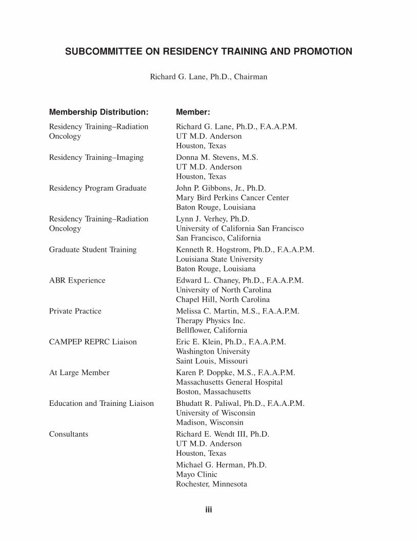
SUBCOMMITTEE ON RESIDENCY TRAINING AND PROMOTION
Richard G. Lane, Ph.D., Chairman
Membership Distribution: Member:
Residency Training–Radiation Richard G. Lane, Ph.D., F.A.A.P.M.Oncology UT M.D. Anderson
Houston, Texas
Residency Training–Imaging Donna M. Stevens, M.S.UT M.D. AndersonHouston, Texas
Residency Program Graduate John P. Gibbons, Jr., Ph.D.Mary Bird Perkins Cancer CenterBaton Rouge, Louisiana
Residency Training–Radiation Lynn J. Verhey, Ph.D.Oncology University of California San Francisco
San Francisco, California
Graduate Student Training Kenneth R. Hogstrom, Ph.D., F.A.A.P.M.Louisiana State UniversityBaton Rouge, Louisiana
ABR Experience Edward L. Chaney, Ph.D., F.A.A.P.M.University of North CarolinaChapel Hill, North Carolina
Private Practice Melissa C. Martin, M.S., F.A.A.P.M.Therapy Physics Inc.Bellflower, California
CAMPEP REPRC Liaison Eric E. Klein, Ph.D., F.A.A.P.M.Washington UniversitySaint Louis, Missouri
At Large Member Karen P. Doppke, M.S., F.A.A.P.M.Massachusetts General HospitalBoston, Massachusetts
Education and Training Liaison Bhudatt R. Paliwal, Ph.D., F.A.A.P.M.University of WisconsinMadison, Wisconsin
Consultants Richard E. Wendt III, Ph.D.UT M.D. AndersonHouston, Texas
Michael G. Herman, Ph.D.Mayo ClinicRochester, Minnesota
iii

This page intentionally left blank.
v
Foreword .......................................................................................................................... ix
CHAPTER 1
Essentials and Guidelines for Diagnostic Imaging PhysicsResidency Training Programs
1.1 Introduction ............................................................................................................... 1
1.2 Objective of a Diagnostic Imaging Physics ResidencyTraining Program ....................................................................................................... 1
1.3 Didactic Knowledge Requirements ............................................................................. 1
1.4 Structure and Conduct of a Diagnostic Imaging PhysicsResidency Program..................................................................................................... 2
1.5 Expected Areas of Competence for a Clinical Medical Physicistin Diagnostic Imaging ................................................................................................ 6
1.6 Education Requirements for Residents in Diagnostic Imaging Physics ........................ 9
1.7 Radiation Physics Knowledge of Specific Importancefor Diagnostic Imaging Physics Residents ................................................................... 10
1.8 Clinical Knowledge of Specific Importance for Diagnostic ImagingPhysics Residents........................................................................................................ 16
1.9 Radiation Biology Knowledge of Specific Importancefor Diagnostic Imaging Physics Residents ................................................................... 18
CHAPTER 2
Essentials and Guidelines for Nuclear MedicinePhysics Residency Training Programs
2.1 Introduction ............................................................................................................... 20
2.2 Objective of a Nuclear Medicine Physics Residency Training Program................................................................................................ 20
CONTENTS
vi
2.3 Didactic Knowledge Requirements ............................................................................. 20
2.4 Structure and Conduct of a Nuclear Medicine PhysicsResidency Program..................................................................................................... 21
2.5 Expected Areas of Competence for a Clinical Medical Physicistin Nuclear Medicine................................................................................................... 25
2.6 Education Requirements for Residents in Nuclear Medicine Physics .......................... 28
2.7 Radiation Physics Knowledge of Specific Importance for Nuclear Medicine Physics Residents ........................................................................... 29
2.8 Clinical Knowledge of Specific Importance for Nuclear MedicinePhysics Residents........................................................................................................ 32
2.9 Radiation Biology Knowledge of Specific Importancefor Nuclear Medicine Physics Residents...................................................................... 34
CHAPTER 3
Essentials and Guidelines for Radiation OncologyPhysics Residency Training Programs
3.1 Introduction ............................................................................................................... 36
3.2 Objective of a Radiation Oncology Physics ResidencyTraining Program ....................................................................................................... 36
3.3 Didactic Knowledge Requirements ............................................................................. 36
3.4 Structure and Conduct of a Radiation Oncology PhysicsResidency Program..................................................................................................... 37
3.5 Expected Areas of Competence for a Clinical Medical Physicistin Radiation Oncology................................................................................................ 41
3.6 Education Requirements for Residents in Radiation Oncology Physics........................ 50
3.7 Radiation Physics Knowledge of Specific Importancefor Radiation Oncology Physics Residents .................................................................. 51
3.8 Clinical Knowledge of Specific Importance for Radiation OncologyPhysics Residents........................................................................................................ 54
3.9 Radiation Biology Knowledge of Specific Importancefor Radiation Oncology Physics Residents .................................................................. 56
CONTENTS

vii
Epilogue ........................................................................................................................... 62Acronyms......................................................................................................................... 64References....................................................................................................................... 67
CONTENTS

This page intentionally left blank.
ix
The need for established standards of post graduate education and training of medical physicists
is clear. The complexity of techniques in diagnostic imaging as well as in treatment simulation,
planning, and dose delivery in radiotherapy has increased significantly in just the last 5 years. A
few years ago, the hardware and software that is currently being used routinely in hospitals and
clinics across the country today was then found only in academic institutions. In radiation
oncology, terms that were known to but a few medical physicists 10 years ago are used routinely
today, such as “multileaf collimators,” “collapsed cone convolution algorithms,” “image-guided
radiation treatment,” “dose-volume constraints,” “simulated annealing optimization,” as well as
“respiratory gated planning and treatment.” In diagnostic imaging, recent advances include com-
puted radiography, direct digital radiography, multi-slice computed tomography (CT) scanning,
CT fluoroscopy, CT–magnetic resonance imaging (CT–MRI) co-registration, and three-dimen-
sional imaging. In nuclear medicine, positron emission tomography (PET) imaging and PET–CT
are rapidly finding routine use in patient care.
It has never been possible to learn medical physics by unstructured self-study or by
observation alone. It is now no longer possible to become a fully competent, qualified medical
physicist through on-the-job training, even under the mentorship of a single, experienced medical
physicist. Over the past few years, it has become increasingly clear that the training standards
and documentation associated with accreditation are needed for proper training of individuals to
be capable of practicing medical physics independently. It is also clear that high-quality training
can take place effectively in a hospital setting as well as in an academic environment.
Significant progress has been made in implementing standards for the clinical training
of medical physicists since 1992 when the AAPM published the report of the Ad Hoc
Committee on Clinical Training of Radiological Physicists (AAPM Report Number 36) enti-
tled “Essentials and Guidelines for Hospital-Based Medical Physics Residency Training
Programs.” Soon thereafter, the Commission on Accreditation of Medical Physics Education
Programs, Inc. (CAMPEP) was formed and these guidelines were adopted for the accredita-
tion of medical physics residency programs. Since then, the CAMPEP Residency Education
Program Review Committee (CAMPEP-REPRC) has been working with institutions striving
to meet these accreditation standards. In 1997 CAMPEP accredited the first residency pro-
gram in Radiation Oncology Physics at Washington University. A total of 12 programs in
Radiation Oncology Physics and 2 in Diagnostic Imaging Physics have been accredited at the
time of this publication.
FOREWORD
x
Funding is a critically important aspect of clinical training programs. Significant progress
has been made here as well. The AAPM Development Committee supervises the disbursement
of several vendor-sponsored training grants in support of medical physics residencies. In addi-
tion, funding is available to accredited programs from the Centers for Medicare Services. This
funding is in proportion to the Medicare services provided by the institution. Existing medical
physics residency programs are demonstrating that clinical service provided by the resident off-
sets most if not all of the costs associated with establishing an accredited program.
In 2003, the AAPM Committee on the Education and Training of Medical Physicists was
charged with revisiting and updating the original AAPM Report Number 36. The Medical
Physics Residency and Promotion Subcommittee was formed of AAPM members with extensive
experience in clinical, professional, and educational aspects of medical physics. The members of
the subcommittee recognized that the recent publication of the AAPM Report No. 79 entitled
“Academic Program Recommendations for Graduate Degrees in Medical Physics”* covered the
didactic training requirements of a medical physics resident. Therefore, the subcommittee could
concentrate on the clinical and professional knowledge needed to function independently as a
practicing medical physicist in the areas of radiation oncology, diagnostic imaging, and nuclear
medicine.
It is the sincere hope of the subcommittee that this revision will serve the medical
physics community as well as and for as long as did the original document published almost
15 years ago.
FOREWORD
*AAPM Report No. 79, Academic Program Recommendations for Graduate Degrees in Medical Physics, B.R.Paliwal (Chairman, Education and Training of Medical Physicists Committee), Madison, WI: Medical PhysicsPublishing, Madison, WI (2002).
1
1.1 INTRODUCTION
Diagnostic Imaging Physics is a subspecialty of medical physics related to all aspects of medical
imaging used for diagnosis and treatment of human disease. In the clinical setting, diagnostic
imaging physicists are responsible for those aspects of diagnostic imaging where physics plays a
role in safe and accurate diagnostic imaging procedures for patient care. Other major roles of the
diagnostic imaging physicist include teaching, research, and administration. Section 1.5 provides
an extensive list of specific activities and duties.
1.2 OBJECTIVE OF A DIAGNOSTIC IMAGING PHYSICS RESIDENCYTRAINING PROGRAM
The objective of the diagnostic imaging physics residency training program is to educate and to
train medical physicists to a level of competency sufficient to practice diagnostic imaging physics
independently. To accomplish this goal, adequate structure, facilities, staff, patient resources, and
educational environment must be provided.
1.3 DIDACTIC KNOWLEDGE REQUIREMENTS
Upon satisfactory completion of the diagnostic imaging physics residency program, the graduate
will have a knowledge of medical physics equivalent to that of a graduate of a Commission on
Accreditation of Medical Physics Education Programs, Inc. (CAMPEP)-accredited medical
physics graduate program as appropriate for a diagnostic imaging physics specialty. This is
accomplished most directly by accepting into the residency program applicants who have gradu-
ated from an accredited medical physics graduate program.
Alternatively, graduates of non-accredited medical physics graduate programs and grad-
uates of physics or related graduate programs shall be expected to attend appropriate medical
physics graduate courses and/or participate in a structured program of self-study based on the
AAPM Report No. 79, “Academic Program Recommendation for Graduate Degrees in Medical
CHAPTER 1
ESSENTIALS AND GUIDELINES FOR DIAGNOSTIC IMAGING PHYSICS RESIDENCY TRAINING PROGRAMS
2
Physics.” Of critical importance is the regularly scheduled assessment of the resident’s medical
physics knowledge. For attendance at graduate courses, passing grades on examinations provide
required documentation. For self-study, results of written or oral examinations of subject matter
may be required. Basic education eligibility requirements for diagnostic imaging residents are
found in section 1.6.
Specific radiation physics knowledge for diagnostic imaging residents is found in section
1.7. Specific clinical knowledge for diagnostic imaging residents is found in section 1.8. Specific
radiation biology knowledge for diagnostic imaging residents is found in section 1.9.
1.4 STRUCTURE AND CONDUCT OF A DIAGNOSTIC IMAGING PHYSICS RESIDENCY PROGRAM
1.4.1 Length of Training
A clinical training period of at least 2 years is required following graduate school (see section 1.3).
The first resident year should provide a broad experience in clinical diagnostic imaging physics.
The purpose of the first year is to provide the physicist with the capability of managing, either
alone or with others, the broad range of imaging physics tasks for patients under care in a diag-
nostic radiology facility.
The second year of training builds on the first year, both in level of responsibility and in
undertaking training in special topics such as specification, acceptance testing, and quality
assurance of imaging equipment.
During these 2 years, clinical research and development projects may be included as part
of the clinical training program. In addition, a reasonable and justifiable amount of the clinical
training experience may take place at affiliated institutions.
1.4.2 Program Director
The program director is responsible for the whole of the diagnostic imaging physics training pro-
gram. The program director:
(1) Must contribute sufficient time to the program to ensure adequate direction.
(2) Is responsible for program organization and direction as well as instruction and
supervision of physics residents.
(3) Must arrange for the provision of adequate facilities, teaching staff, clinical
resources, and educational resources.
(4) Is responsible for the recruitment and appointment of physics residents and must
ensure that the appointed residents meet the eligibility requirements listed in section
1.6.
CHAPTER 1
3
(5) Is responsible for ensuring the resident is making satisfactory progress and for pro-
viding appropriate disciplinary action should this not be the case.
The qualifications of the program director are as follows:
(1) Must be certified in Diagnostic Imaging Physics by an appropriate certifying board.
(2) Must have at least 7 years of full-time experience as a qualified medical physicist
practicing in diagnostic imaging physics.
(3) Must be a full-time staff member, qualified in and practicing diagnostic imaging
physics at the training facility.
1.4.3 Staff
The program must provide adequate numbers of staff for the teaching of diagnostic imaging
physics, clinical diagnostic imaging, and radiation biology. The teaching staff must be qualified
in those areas in which they are assigned to instruct and supervise physics residents, and they
must devote the necessary time and effort to the educational program. Commitment to the
physics resident training program by the staff is essential to the success of the program. The staff
should be engaged in scholarly activities, such as:
(1) Participation in regional and national scientific societies;
(2) Participation in their own continuing education;
(3) Scientific publication and presentation.
An adequate staff must include at least two (2) full-time diagnostic imaging physicists, both cer-
tified by an appropriate certifying board, and a full time diagnostic radiologist, certified by the
American Board of Radiology (ABR) or its equivalent. It is recommended that access to train-
ing from a radiation biologist be available.
1.4.4 Training Content
Training in the clinical and technical areas of diagnostic imaging physics should include the fol-
lowing: principles and procedures involved in the production of clinical diagnostic images;
methods of image evaluation; techniques for optimization of radiation exposure for diagnostic
examination; methods of calculating specific organ doses and risk estimations; calibration and
monitoring of diagnostic imaging equipment; and radiation safety procedures. Residents must
obtain an in depth knowledge in the clinical physics areas listed in section 1.5.
The clinical physics training staff should provide for a systematic course of instruction
that encourages progressive supervised resident responsibility for patient care and must ensure
that the physics resident personally performs the commonly accepted clinical physics procedures
in diagnostic imaging. The resident must keep a detailed list of clinical physics procedures he or
ESSENTIALS AND GUIDELINES FOR DIAGNOSTIC IMAGING PHYSICS
4
she has performed. This list must be reviewed periodically by the program director and the pro-
gram steering committee, and must be available for external review of the program.
1.4.5 Training Complement
The number of residents in the training program must be commensurate with the total capacity
of the program to offer an adequate educational experience in diagnostic imaging physics. The
maximum number of residents in the 24 months of clinical diagnostic imaging must not exceed
the number of full-time equivalent staff diagnostic imaging physicists.
1.4.6 Training Evaluation
The program director is responsible for the continuing evaluation of the program as well as for
the documentation of the educational progress and performance of each resident. To assure reg-
ular progress assessment, the resident should meet at least biweekly with the clinical coordinator
or rotation supervisor. Monthly meetings of the resident with the program director are recom-
mended. Proper documentation of these meetings will assure compliance and continuity of
assessment. Written evaluations must be performed at the completion of each rotation.
In addition, resident performance and progress must be documented at least yearly
using an oral examination conducted by appropriate members of the program steering com-
mittee and faculty. The results of all evaluations must be discussed with the resident and must
be documented.
The program director should document any prior training from another institution that is
to be used to satisfy the training criteria of the program. It is the program director’s responsi-
bility to counsel, to censure, and, after due process, to dismiss residents who fail to demonstrate
appropriate industry, competence, responsibility, learning abilities, and ethical behavior.
1.4.7 Facilities
Adequate space must be available for the conduct of a good clinical physics practice and train-
ing program. The following facilities must be available:
(1) Radiographic and/or fluoroscopic systems for general radiography, mammography,
cardiac catheterization, and special procedures;
(2) Computed tomography scanner;
(3) Magnetic resonance imaging scanner;
(4) Ultrasound imager;
(5) Digital imaging system;
(6) Physics laboratory.
CHAPTER 1
5
If any of the required facilities are not available on-site, the program must provide clinical train-
ing on such equipment at another approved institution. In addition, electronics and machine
shops should be available.
1.4.8 Clinical Resources
The training program in diagnostic imaging physics must provide a sufficient volume and variety
of patients for adequate resident experience. The number of diagnostic imaging examinations per
year should be at least 50,000.
1.4.9 Institutional Support
The institution sponsoring the program of clinical training in diagnostic imaging physics should
provide administrative support in terms of budget and space in addition to clinical and educa-
tional resources. Adequate conference room and audiovisual facilities should be provided.
Commitment to long-term funding of the program is essential.
1.4.10 Educational Environment
The clinical training in diagnostic imaging physics should occur in an environment that encour-
ages exchange of knowledge and experience among physics residents in the diagnostic imaging
physics program and with medical residents located in the same institution participating in the
residency program in diagnostic radiology.
1.4.11 Conferences
Conferences and teaching rounds must provide for progressive resident participation. Adequate
frequency of conferences and attendance by imaging physics residents, diagnostic imaging
physicists, diagnostic radiologists, and other staff should be documented. Conferences available
to the resident should include intradepartmental clinical conferences, such as staff radiology con-
ferences, interesting case conferences, and physics conferences. Other conferences should
include radiation safety, radiation biology, and journal review.
1.4.12 Library Resources
A sufficient variety of journals, reference books, and resource materials pertinent to diagnostic
imaging physics and associated fields in diagnostic radiology and basic sciences should be pro-
vided and must be immediately accessible for resident study. A complete bibliography can be
found in AAPM Report No. 79, “Academic Program Recommendations for Graduate Degrees in
Medical Physics.” Physics residents must have access to a general medical library. In addition,
physics residents must have access to the educational resources available on the Internet.
ESSENTIALS AND GUIDELINES FOR DIAGNOSTIC IMAGING PHYSICS
6
1.5 EXPECTED AREAS OF COMPETENCE FOR A CLINICALMEDICAL PHYSICIST IN DIAGNOSTIC IMAGING
Competence must be demonstrated in the following major areas of responsibility:
(1) Specification, acceptance testing, and quality assurance of imaging equipment.
(2) Measurement and calculation of radiation exposure and dose.
(3) Improving and maintaining medical image quality.
(4) Training of physicists, clinical diagnostic imaging residents, radiological and ultra-
sound technologists, and/or other allied health professionals in diagnostic radiology.
(5) Education of health professionals in diagnostic imaging physics and radiation
effects.
Competency in clinical and laboratory research in diagnostic imaging physics is recommended.
The specific competencies are listed below.
1.5.1 Imaging Systems
Radiographic, fluoroscopic, special procedures, conventional tomographic, mammographic,
computed tomography (CT), ultrasound (US), and magnetic resonance imaging (MRI), includ-
ing associated print and electronic display media.
A. Design and fundamentals
B. Selection
1. Performance specification
2. Feature comparison
3. Siting issues
4. Performance test design
C. Acceptance testing/quality assurance
1. Mechanical
2. Radiation output
3. Shielding adequacy, siting
4. Baseline performance measurements
5. Imaging techniques
6. Quantitative evaluation
7. Measures of image quality
CHAPTER 1
7
D. Quality control
1. Imaging equipment
2. Film processors
3. Printers: laser, dry laser, thermal
4. Film densitometer
5. Computer equipment
6. Image transmission devices
7. Quantitative procedures
8. Digital Imaging and Communications in Medicine (DICOM); (storage, print, etc.)
a. Work list management (WLM)
b. Query/Retrieve (Q/R)
E. Dose determination
1.5.2 Computer Systems for Image Display and Processing
A. Hardware and operation
B. Software
C. Acceptance testing
D. Interfacing/peripherals
E. Image transmission devices
F. Clinical applications
1.5.3 Radiation Protection
A. Shielding design
B. Survey
1. X-ray
2. Radiofrequency (RF)
C. Regulations/recommendations
1. National/state/local
2. As low as reasonably achievable (ALARA)
3. Joint Commission on Accreditation of Healthcare Organizations (JCAHO)
4. Radiation safety committee
5. American College of Radiology (ACR) standards
D. Personnel monitoring
1. Thermoluminescence dosimetry (TLD)
2. Film badges
3. Other dosimeters
ESSENTIALS AND GUIDELINES FOR DIAGNOSTIC IMAGING PHYSICS
8
E. Guidelines/instructions for personnel
1. Residents
2. Medical students
3. Technology students
4. Hospital, medical, and nursing staff
5. Maintenance, custodial staff
F. Hazards of low levels of radiation
G. Anatomical awareness
1. Radiographic anatomy
2. Physiology (functional imaging, interventional procedures, image-guided therapies)
3. Patient shielding (gonadal and fetal shields, etc.)
1.5.4 Dosimetry
A. Techniques (design/calibration/use)
1. Ionization chamber
2. Other (TLD, film, etc.)
a. Optically stimulated luminescence (OSL)
b. Metal oxide semiconductor field-effect transistor (MOSFET)
B. Patient dose values
1. Sensitive tissues
2. Assessment of doses (risk analysis)
3. Fetal dose estimates
1.5.5 Additional Duties
A. Educational
1. Teaching
2. Seminars
B. Developmental studies
1. Imaging techniques
2. Dose reduction
3. Computational techniques
4. Dosimetric techniques
5. Equipment performance evaluation
6. Evaluation of system upgrades
C. Clinical development
CHAPTER 1
9
1.6 EDUCATION REQUIREMENTS FOR RESIDENTS IN DIAGNOSTIC IMAGING PHYSICS
1.6.1 Degree
The required degree is a master of science (M.S.) degree or a doctorate (Ph.D.) in:
A. Medical physics from a CAMPEP-accredited program, or
B. Medical physics from a non-accredited program, or
C. Physics, or
D. A discipline closely related to physics.
1.6.2 Curriculum
The applicant’s undergraduate and/or graduate education should demonstrate knowledge
acquired in the following areas:
A. Fundamental physics
B. Advanced mathematics
C. Advanced atomic and nuclear physics
D. Electronics
E. Computers
F. Physical Chemistry
1.6.3 Background Knowledge
Graduates of programs in medical physics should have demonstrated knowledge in topics con-
sidered to be minimal by the AAPM guidelines for the M.S. degree in Medical Physics
Academic Programs. Graduates of physics or related graduate programs are expected to acquire
this knowledge as part of their residency training.
This includes knowledge in the following areas:
A. Radiation physics
B. Radiation dosimetry
C. Radiation measurement techniques and instrumentation
D. Radiation protection
E. Principles of imaging
F. Radiation biology
G. Human anatomy and physiology
H. Introduction to clinical radiology and radiation oncology
ESSENTIALS AND GUIDELINES FOR DIAGNOSTIC IMAGING PHYSICS
10
1.7 RADIATION PHYSICS KNOWLEDGE OF SPECIFIC IMPORTANCE FOR DIAGNOSTIC IMAGING PHYSICS RESIDENTS
1.7.1 Production of X-rays
A. X-ray tubes
1. Requirements for x-ray production
2. Historical development
3. Focal spot size
4. X-ray targets
5. X-ray production efficiency
6. Characteristic and bremsstrahlung spectra
7. mA and kVp effects
8. Heat production and dissipation (rating charts)
9. Line-focus principle
10. Special tubes
a. Grid controlled
b. Field emission
c. Mammography
d. High heat capacity
B. X-ray generators
1. Primary circuit
2. Secondary circuit
3. Filament circuit
4. Modes of rectification
5. Single-phase and three-phase operation
6. High and medium frequencies
7. Others
a. Falling load
b. Capacitor discharge
c. Constant potential
d. Battery operated
1.7.2 Interactions of X-rays and Gamma Rays
A. Attenuation of a beam of x- or gamma rays
1. Attenuation and absorption coefficients
2. Attenuation in the body
CHAPTER 1
11
B. Modes of interaction
1. Classical/Raleigh scattering
2. Photoelectric absorption
3. Compton scattering
4. Pair production
1.7.3 Measurement of Radiation Exposure
A. Photon and energy flux density and fluence
B. The roentgen
C. Electronic equilibrium
D. Ionization chambers
1. Free-air chambers
2. Thimble chambers
3. Condenser chambers
E. Electrometers
F. Geiger and proportional counters
G. Survey meters
H. Exposure measurements of an x- or gamma ray beam
1. Selection of exposure parameters
2. Selection of chamber
3. Positioning of chamber
4. Corrections to readings
1.7.4 Radiation Quality
A. Measures of quality
1. Half-value layer (HVL) and effective energy
2. Measurement of HVL
B. Factors influencing quality
1. Variations in quality across a beam
2. Filtration and accelerating potential
1.7.5 Determination of Absorbed Dose
A. Units of radiation dose, dose equivalent, quality factor
B. Calculation of dose from exposure
C. Determination of absorbed dose from an ionization chamber measurement (Bragg-Gray
cavity theory)
ESSENTIALS AND GUIDELINES FOR DIAGNOSTIC IMAGING PHYSICS
12
D. Direct measurement of absorbed dose
1. Film
2. TLD
3. Calorimetry
4. Chemical dosimetry
1.7.6 Imaging Concepts
A. Mode
1. Transmission
2. Emission
3. Reflection
4. Reconstruction
B. Image characteristics
1. Density, contrast, latitude
2. Detail, resolution, modulation transfer function (MTF)
3. Noise
4. Speed
5. Dose
6. Interrelationships
C. Viewing conditions
1. Visual receptors
2. Film, soft copy, stationary, live, etc.
3. Variables
D. Analog vs. digital considerations
1.7.7 X-ray Filters and Beam-limiting Devices
A. Filtration
1. Inherent filtration
2. Added filter
3. Special purpose
4. Effect upon image quality and radiation dose
B. Scattered radiation: image quality and dose effects
C. Heel effect
D. Beam-limiting devices
1. Aperture
2. Cones (dental units)
CHAPTER 1
13
3. Collimators
4. Positive beam limitation
5. Performance measurements
1.7.8 X-ray Imaging Geometry
A. Magnification
B. Distortion; unequal magnification
C. Geometric unsharpness
D. Motion unsharpness
1.7.9 Scattered Radiation
A. Grids
1. Construction
2. Nomenclature
3. Types
4. Performance parameters
5. Practical considerations
B. Air gap
C. Slot radiography
D. Equalization radiography
1.7.10 Radiographic Imaging
A. Intensifying screens
1. Uses
2. Construction
3. Principles of operation
4. Conversion efficiency
5. Speed
6. Resolution
7. Interrelationships
B. Film
1. Uses
2. Construction
3. Processing
4. Photographic properties
5. Characteristic curve
ESSENTIALS AND GUIDELINES FOR DIAGNOSTIC IMAGING PHYSICS
14
C. Computed and direct digital radiography
1. Storage phosphor plates
2. Plate readers
3. Direct digital capture devices
4. Digital image display
5. Processing algorithms
1.7.11 Fluoroscopy
A. System design
B. Image intensifiers/digital receptors
C. Image quality measures
D. Automatic brightness control (ABC)
E. Television
F. Spot films
G. Photospots
H. Video recording
I. Other fluoroscopic imaging modes:
1. Ciné (cardiac)
2. Pulsed
3. High dose-rate (HDR)
1.7.12 Special Techniques
A. Stereoradiography
B. Xeroradiography
C. Subtraction techniques
D. Three-dimensional (3-D) imaging
E. Duplication
1.7.13 Computed Tomography (CT)
A. Basic principles
1. Conventional detectors
2. Multichannel detectors
B. Data acquisition
C. Image reconstruction
D. Image display
E. Image analysis
CHAPTER 1
15
F. Artifacts
G. Quantitative CT
H. Dual energy CT
I. Fast CT
J. CT fluoroscopy
1.7.14 Ultrasound (US)
A. Basic principles
B. Physical characteristics
C. Transducers
D. Modes
E. Real time
F. Doppler
G. Duplex systems
H. Image quality measurements
I. Scan converter
1.7.15 Magnetic Resonance Imaging (MRI)
A. Basic principles
B. Nature of nuclear magnetic resonance (NMR) signal
C. Pulse sequences
D. Spin system encoding
E. Image reconstruction
F. lmage contrast
G. Equipment
1. Magnets
2. RF systems
3. Gradient systems
4. Coils
H. Bioeffects
I. Fast scan techniques
J. Flow imaging
K. Chemical shift imaging
L. Spectroscopy
M. Image quality measurements
N. Artifacts
ESSENTIALS AND GUIDELINES FOR DIAGNOSTIC IMAGING PHYSICS
16
O. Site planning
P. Patient and personnel protection issues
1.7.16 Mammography
A. Basic principles
B. Imaging equipment
1. Screen/film
2. Digital mammography
3. Stereotactic biopsy
C. Quality control
1. Technologist
2. Physicist
3. Radiologist
D. Mammography Quality Standards Act (MQSA)
1.7.17 ACR Programs in Diagnostic Radiology
1.8 CLINICAL KNOWLEDGE OF SPECIFIC IMPORTANCE FOR DIAGNOSTIC IMAGING PHYSICS RESIDENTS
1.8.1 Medical Terminology
1.8.2 Anatomy
A. Normal structures and appearance
B. Normal variants
C. Image quality and artifacts
1. Radiographic
2. Fluoroscopic
3. CT
4. US
5. MRI
6. Mammographic
1.8.3 Physiology
A. Normal organ function
B. Normal organ variation
CHAPTER 1
17
C. Pathophysiology of disease
D. Metabolic cycles and interactions
E. Laboratory tests
1.8.4 Patient Procedures
A. Radiographic and fluoroscopic
1. Neurologic
2. Chest
3. Musculoskeletal
4. Gastrointestinal (GI)
5. Genitourinary (GU)
6. Pediatric
7. Obstetric
8. Vascular/interventional
9. Cardiac
10. Emergency/trauma
B. Special imaging
1. CT
2. Ultrasound
3. Nuclear medicine
4. MRI
5. Mammograpy
6. Special procedures: vascular/interventional radiology (VIR)
1.8.5 Contrast Media
A. Contrast enhancement
1. X-ray imaging modalities
2. Ultrasound
3. MRI
B. Biochemistry
C. Physiology reactions
1.8.6 Regulatory Requirements and Guidelines
ESSENTIALS AND GUIDELINES FOR DIAGNOSTIC IMAGING PHYSICS
18
1.9 RADIATION BIOLOGY KNOWLEDGE OF SPECIFIC IMPORTANCE FOR DIAGNOSTIC IMAGING PHYSICS RESIDENTS
1.9.1 Late Effects
A. Nonspecific life shortening
1. Definition
2. In animals
3. In man
B. Carcinogenesis
1. The latent period
2. Dose response curve in animals
3. Leukemia
4. Breast cancer
5. Lung cancer
6. Other cancers and tumors
7. Malignancies in prenatally exposed children
8. Mechanisms for radiation carcinogenesis
C. Genetics of irradiation
1. Point mutations
2. Relationship to dose
3. Chromosome aberrations
4. Doubling dose
5. Genetically significant dose (GSD)
6. Genetic effect in humans
7. Background radiation in relation to GSD
1.9.2 Radiation Effects in the Developing Embryo and Fetus
A. Intrauterine death
B. Congenital abnormalities including neonatal death
C. Growth retardation
D. Dependence of the above effects on dose, dose-rate, and stage in gestation
E. Carcinogenesis following in utero exposure
F. Human experience of pregnant women exposed to therapeutic doses
G. Occupational exposure of potentially pregnant women
H. Elective booking or “10-day rule”
I. The “practical threshold” theories for therapeutic abortion
CHAPTER 1
19
1.9.3 Radiation Epidemiology
A. Prevalence, incidence, mortality
B. Cohort and case control studies
C. Relative risk, excess risk, absolute risk, attributable risk, odds ratio
D. Standard mortality rate
E. Association vs. causation
F. Major human epidemiology studies
G. Adaptive response to large doses of radiation
1.9.4 Risk Analysis for Low-Level Radiation Exposure
ESSENTIALS AND GUIDELINES FOR DIAGNOSTIC IMAGING PHYSICS
20
2.1 INTRODUCTION
Nuclear Medicine Physics is a subspecialty of medical physics related to the diagnostic, thera-
peutic, and investigational use of radionuclides in medicine. In the clinical setting, nuclear med-
icine physicists are responsible for those aspects of nuclear medicine where physics plays a role
in safe and accurate nuclear medicine diagnostic and therapeutic patient procedures. Other
major roles of the nuclear medicine physicist include teaching, research, and administration.
Section 2.5 provides an extensive list of specific activities and duties.
2.2 OBJECTIVE OF A NUCLEAR MEDICINE PHYSICS RESIDENCY TRAINING PROGRAM
The objective of the nuclear medicine physics residency training program is to educate and train
medical physicists to a competency level sufficient to practice nuclear medicine physics inde-
pendently. To accomplish this goal, adequate structure, facilities, staff, patient resources, and
educational environment must be provided.
2.3 DIDACTIC KNOWLEDGE REQUIREMENTS
Upon satisfactory completion of the nuclear medicine physics residency program, the graduate will
have a knowledge of medical physics equivalent to that of a graduate of a CAMPEP-accredited
medical physics graduate program as appropriate for a nuclear medicine physics specialty. This
is accomplished most directly by accepting into the residency program applicants who have
graduated from an accredited medical physics graduate program.
Alternatively, graduates of non-accredited medical physics graduate programs and grad-
uates of physics or related graduate programs shall be expected to attend appropriate medical
physics graduate courses and/or participate in a structured program of self-study based on the
AAPM Report No. 79, “Academic Program Recommendation for Graduate Degrees in Medical
Physics.” Of critical importance is the regularly scheduled assessment of the resident’s medical
CHAPTER 2
ESSENTIALS AND GUIDELINES FOR NUCLEAR MEDICINE PHYSICS RESIDENCY TRAINING PROGRAMS
21
physics knowledge. For attendance at graduate courses, passing grades on examinations provide
required documentation. For self-study, results of written or oral examinations of subject matter
may be required. Basic education eligibility requirements for nuclear medicine residents are
found in section 2.6.
Specific radiation physics knowledge for nuclear medicine residents is found in section
2.7. Specific clinical knowledge for nuclear medicine residents is found in section 2.8. Specific
radiation biology knowledge for nuclear medicine residents is found in section 2.9.
2.4 STRUCTURE AND CONDUCT OF A NUCLEAR MEDICINE PHYSICS RESIDENCY PROGRAM
2.4.1 Length of Training
A clinical training period of at least 2 years is required following graduate school (see section
2.3). The first resident year must provide a broad experience in clinical nuclear medicine physics.
The purpose of the first year is to provide the physicist with the capability of managing, either
alone or with others, the broad range of clinical physics tasks regarding patients under care in a
nuclear medicine facility.
The second year of training builds on the first year, both in level of responsibility and in
undertaking training in special topics such as specification, acceptance testing, and quality
assurance of nuclear medicine equipment.
During these 2 years, clinical research and development projects may be included as part
of the clinical training program. In addition, a reasonable and justifiable amount of the clinical
training experience may take place at affiliated institutions.
2.4.2 Program Director
The program director is responsible for the whole of the nuclear medicine physics residency
training program. The program director:
(1) Must contribute sufficient time to the program to ensure adequate direction.
(2) Is responsible for program organization and direction as well as the instruction and
supervision of the physics residents.
(3) Must arrange for the provision of adequate facilities, teaching staff, clinical
resources, and educational resources.
(4) Is responsible for the recruitment and appointment of physics residents and must
ensure that the appointed residents meet the eligibility requirements listed in
section 2.6.
ESSENTIALS AND GUIDELINES FOR NUCLEAR MEDICINE PHYSICS
22
(5) Is responsible for ensuring the resident is making satisfactory progress and for providing
appropriate disciplinary action should this not be the case.
The qualifications of the program director are as follows:
(1) Certification in nuclear medicine physics by an appropriate certifying board.
(2) At least 7 years of full-time experience as a qualified medical physicist practicing in
nuclear medicine physics.
(3) A full-time staff member qualified in and practicing nuclear medicine physics at the
training facility.
2.4.3 Staff
The program must provide adequate numbers of staff for the teaching of clinical nuclear medi-
cine physics, clinical nuclear medicine, and radiation biology. The teaching staff must be quali-
fied in those areas in which they are assigned to instruct and to supervise physics residents, and
must devote the necessary time and effort to the educational program. Commitment to the
physics resident training program by the staff is essential to the success of the program. The staff
should be engaged in scholarly activities, such as:
(1) Participation in regional and national scientific societies;
(2) Participation in their own continuing education; and
(3) Scientific publication and presentation.
An adequate staff must include at least one full time nuclear medicine physicist, certified by an
appropriate certifying board, and one full time nuclear medicine physician, certified by the
appropriate certifying board. It is recommended that additional staff include access to a nuclear
pharmacist, a radiation biologist, and a qualified medical physicist practicing in diagnostic imag-
ing physics and/or in radiation oncology physics.
2.4.4 Training Content
The training must include a systematic course of instruction with demonstrations on clinical
and technical subjects pertinent to the various phases of nuclear medicine physics, including
the calibration and monitoring of nuclear medicine equipment, assay of radiopharmaceuticals,
image processing, computer applications, and radiation safety procedures. Residents must
obtain an in-depth knowledge in the clinical physics areas listed in section 2.5. The clinical
physics training staff must provide for a systematic course of instruction that encourages pro-
gressive, supervised resident responsibility for patient care and must ensure that the physics
resident personally performs those clinical physics procedures commonly accepted in all
aspects of nuclear medicine.
CHAPTER 2
23
The resident must maintain a detailed list of clinical physics procedures that he or she has
performed. This list will be reviewed periodically by the program director and the program
steering committee and must be available for external review of the program.
2.4.5 Training Complement
The number of residents in the training program must be commensurate with the capacity of the
program to offer an adequate educational experience in nuclear medicine physics. It is desirable
to have two positions. However, the maximum number of residents in the 24 months of clinical
nuclear medicine must not exceed the number of full-time equivalent staff nuclear medicine
physicists.
2.4.6 Training Evaluation
The program director is responsible for the continuing evaluation of the program and for the
documentation of the educational progress and performance of each resident. To assure contin-
uous progress assessment, the resident should meet at least biweekly with the clinical coordina-
tor or rotation supervisor. Monthly meetings of the resident with the program director are
recommended. Proper documentation of these meetings will assure compliance and continuity
of assessment.
Resident performance and progress must be assessed at least yearly using an oral exam-
ination conducted by appropriate members of the program steering committee and faculty. The
results of these evaluations must be discussed with the resident and must be documented.
The program director should document any prior training from another institution that is
to be used to satisfy the training criteria of the program. It is the program director’s responsi-
bility to counsel, to censure, and, after due process, to dismiss residents who fail to demonstrate
appropriate industry, competence, responsibility, learning abilities, and ethical behavior.
2.4.7 Facilities
Space adequate for the conduct of a good clinical physics practice and training program must be
available. There must be:
(1) Two or more gamma cameras;
(2) A single photon emission computed tomography (SPECT) unit;
(3) Access to a positron emission tomography (PET) unit or facility;
(4) A computer for image analysis;
(5) Nuclear medicine dose calibration instrumentation;
(6) Thyroid probe and a gamma well counter; and
(7) A physics laboratory.
ESSENTIALS AND GUIDELINES FOR NUCLEAR MEDICINE PHYSICS
24
Electronics and machine shops should be available. Training on SPECT-CT and PET-CT systems
should be available. If any of the required facilities are not available on-site, the program must
provide clinical training on such equipment at another approved institution.
2.4.8 Clinical Resources
The training program in nuclear medicine physics must provide a sufficient volume and variety
of patients for adequate resident experience. There must be at least 3000 nuclear medicine pro-
cedures performed per year.
2.4.9 Institutional Support
The institution sponsoring the program of clinical training in nuclear medicine physics should
provide administrative support in terms of budget and space in addition to clinical, and educa-
tional resources. Adequate conference room and audiovisual facilities should be provided.
Commitment to long term funding of the program is essential.
2.4.10 Educational Environment
The clinical training in nuclear medicine physics should occur in an environment that encourages
exchange of knowledge and experience among physics residents in the nuclear medicine physics
program and with medical residents located in the same institution participating in the residency
program in nuclear medicine.
2.4.11 Conferences
Conferences and teaching rounds must provide for progressive resident participation. Adequate
frequency of conferences and attendance by nuclear medicine physics residents, nuclear medi-
cine physicists, nuclear medicine physicians, and other staff should be documented.
Adequate conference room and audiovisual facilities must be provided. There must be
intradepartmental clinical conferences including new patient conferences, problem case confer-
ences, and physics conferences; other conferences should include radiation safety, radiation biol-
ogy, and journal review.
2.4.12 Library Resources
A sufficient variety of journals, reference books, and resource materials pertinent to nuclear
medicine physics and associated fields in medicine, oncology, and basic sciences should be pro-
vided and must be immediately accessible for resident study. A complete bibliography can be
found in AAPM Report No. 79, “Academic Program Recommendations for Graduate Degrees in
CHAPTER 2
25
Medical Physics.” Physics residents must have access to a general medical library. In addition,
physics residents must have access to the educational resources available on the Internet.
2.5 EXPECTED AREAS OF COMPETENCE FOR A CLINICALMEDICAL PHYSICIST IN NUCLEAR MEDICINE
Competence must be demonstrated in the following major areas of responsibility:
(1) Specification, acceptance testing, and calibration of nuclear medicine equipment.
(2) Measurement and calculation of activity and dose.
(3) Quality assurance and radiation safety.
(4) Training of medical physicists, diagnostic radiology residents, nuclear medicine res-
idents, nuclear medicine technologists, and/or other allied health professionals in
nuclear medicine.
(5) Education of health professionals and the public in nuclear medicine physics and
radiation effects.
Competency in clinical and laboratory research in nuclear medicine physics is recommended.
Specific competencies are listed below.
2.5.1 Equipment
Gamma cameras, uptake study equipment, well-type gamma scintillation counters, liquid scin-
tillation counter, tomographic cameras, SPECT and PET systems, computer analysis systems,
multi-image format cameras, and film processors. Picture archiving and communication system
(PACS) equipment and hybrid equipment such as SPECT-CT and PET-CT should be available.
A. Selection
1. Performance specification, including National Electrical Manufacturers Association
(NEMA) specifications
2. Feature comparison
3. Mechanical/architectural considerations
4. Performance test design
B. Acceptance testing
1. Mechanical/safety
2. Baseline performance measurements
3. Imaging techniques
4. Quantitative evaluations
ESSENTIALS AND GUIDELINES FOR NUCLEAR MEDICINE PHYSICS
26
C. Quality Assurance
1. Daily
2. Weekly to monthly
3. Semiannual to annually
D. Calibration
1. Scintillation counters
2. Multichannel analyzers
3. Survey meters
4. Gamma cameras; planar and SPECT
5. PET
6. Sealed sources
7. Dose calibrators
2.5.2 Computer Systems
A. Hardware operations
B. Display devices: cathode ray tubes (CRTs) and liquid crystal displays (LCDs), grayscale, color
C. Software
D. Quality assurance
E. Peripheral connections/operations
F. Image transmission devices
G. Computer networks and security
H. Clinical applications
I. Digital Imaging and Communications in Medicine (DICOM)
1. Workload management (WLM)
2. Query/Retrieve (Q/R)
3. Storage, print, modality, information/object definition (IOD)
4. DICOM Standard Grayscale Display Function
2.5.3 Radiation Safety
A. Radiation control
1. Area surveys
2. Surface wipes
3. Radionuclide receipt
4. Radioactive waste disposal
B. Protection
1. Shielding design; hot lab, patient holding, imaging systems
CHAPTER 2
27
2. Patient, staff, public
3. Darkrooms
C. Radiation incidents
1. Decontamination
2. Medical events
3. Dose to fetus/embryo
D. Therapeutic procedures
E. Regulations/recommendations
1. National/state/local
2. As low as reasonably achievable (ALARA)
3. Joint Commission on Accreditation of Healthcare Organizations (JCAHO)
4. Radiation Safety Office
5. Radioactive Materials License
F. Monitoring
1. TLD/OSL
2. Film badges
3. Other personnel dosimeters
G. Guidelines/instructions for personnel
1. Medical residents
2. Medical students
3. Technology students
4. Hospital, medical, and nursing staff
5. Maintenance, custodial staff
H. Hazards of low levels of radiation
2.5.4 Room Design
A. Air exhaust
B. Hot laboratory
C. Materials storage
D. Darkroom
E. Safety features
F. Patient preparation and holding
2.5.5 Patient Dosimetry
A. Internal organ dose calculations
B. Fetal dose calculation
C. Therapeutic procedures
ESSENTIALS AND GUIDELINES FOR NUCLEAR MEDICINE PHYSICS
28
2.5.6 Radiopharmacy
A. Kit preparation
B. Quality control
C. Activity assay
D. Therapeutics (including beta-emitters)
2.5.7 Clinical Studies
A. Anatomy/physiology/pharmaceutical uptake and elimination
B. Isotope/activity
C. Organ dosages
D. Computer analysis/techniques
E. Improvements
1. Existing studies
2. New studies
2.5.8 Additional Duties
A. Educational
1. Teaching
2. Extramural lectures
B. Developmental studies
1. Treatment techniques
2. Treatment aids
3. Computational techniques
4. Dosimetric techniques
5. Equipment performance evaluation
2.6 EDUCATION REQUIREMENTS FOR RESIDENTS IN NUCLEAR MEDICINE PHYSICS
2.6.1 Degree
The required degree is a master of science (M.S.) degree or a doctorate (Ph.D.) in:
A. Medical physics from a CAMPEP-accredited program; or
B. Medical physics from a non-accredited program; or
C. Physics; or
D. A discipline closely related to physics.
CHAPTER 2
29
2.6.2 Curriculum
The applicant’s undergraduate and/or graduate education should demonstrate knowledge
acquired in the following areas:
A. Fundamental physics
B. Advanced mathematics
C. Advanced atomic and nuclear physics
D. Electronics
E. Computers
F. Physical chemistry (desirable)
G. Organic chemistry (desirable)
2.6.3 Background Knowledge
Graduates of programs in medical physics should have demonstrated knowledge in topics con-
sidered to be minimal by the AAPM guidelines for M.S. in Medical Physics Academic
Programs. Graduates of physics or related graduate programs are expected to acquire this knowl-
edge as part of their residency training, which includes knowledge in the following areas:
A. Radiation physics
B. Radiation dosimetry
C. Radiation measurement techniques and instrumentation
D. Radiation protection
E. Principles of imaging
F. Radiation biology
G. Human anatomy and physiology
H. Introduction to clinical radiology and radiation oncology
2.7 RADIATION PHYSICS KNOWLEDGE OF SPECIFIC IMPORTANCE FOR NUCLEAR MEDICINE PHYSICS RESIDENTS
2.7.1 Nuclear Medicine Equipment
A. Isotope calibrators
B. Common components
1. Pre-amplifiers and amplifiers
2. Discriminators and scalers
3. Analog-to-digital converters (ADCs)
4. Rate meters
5. Pulse-height analyzers
ESSENTIALS AND GUIDELINES FOR NUCLEAR MEDICINE PHYSICS
30
C. Well counters
D. Probe systems
E. Pulse height analysis
1. Photopeak
2. Compton plateau
3. Compton edge
4. Secondary peaks
5. Calibration
6. Comparison among detectors
7. Full width at half maximum (FWHM)
F. Scintillation camera
1. History
2. Collimation
3. Crystals and photomultiplier tubes
4. Electronic components, corrections, and display
5. Camera-computer interface
6. Performance characteristics
a. Spatial, energy, and temporal resolution
b. Sensitivity
c. Uniformity
7. Static vs. dynamic acquisition
8. Artifacts and methods for correction
a. Uniformity correction
b. Energy correction
c. Dual-isotope correction
9. Multi-crystal devices
G. Tomographic imaging
1. Pinhole and slant-hole tomography
2. Single photon emission computed tomography (SPECT)
a. Calibrations
b. Reconstruction techniques
c. Display
d. Reformation
3. Positron emission tomography (PET)
a. Acquisition principles
b. Radiopharmaceuticals
CHAPTER 2
31
c. Scanner designs
d. Time-of-flight systems
e. Matching of performance characteristics with clinical examination
H. Survey instruments
1. Area monitoring
a. Hot lab/preparation areas
b. Long-term area monitoring
2. Personnel monitoring
2.7.2 Radiopharmaceuticals
A. Biologically important radionuclides
B. Physicochemical properties and biodistribution patterns
C. Purities
D. Assays for radioactivity
E. Mechanisms for localization and release
F. Uptake and elimination; physical, biological, and effective half-life
G. Monoclonal antibodies
2.7.3 Radiopharmaceutical Dosimetry
A. Sources of internal radionuclides
B. Standard man model
C. Critical organ
D. Body burden
E. Medical internal radiation dose (MIRD) method
1. Cumulated activity
2. Equilibrium dose constant
3. S-factor
4. Absorbed fraction
5. Organ- and cellular-based approaches
6. Effective half-lives for uptake and elimination
F. Factors affecting internal dose
G. Bioassays
H. Effective dose, effective dose equivalent
2.7.4 Radiation Safety
A. Regulatory agencies
ESSENTIALS AND GUIDELINES FOR NUCLEAR MEDICINE PHYSICS
32
B. Licensing procedures
C. Dose limits;
1. Annual Limit on Intake (ALI)
2. Derived air concentration (DAC)
D. ALARA
1. De minimus
2. Action levels
E. Protection principles
1. Time
2. Distance
3. Shielding
F. Laboratory procedures
1. Handling
2. Patient administration
3. Decontamination
4. Treatment of accidental ingestion
5. Procedures for radionuclide therapy
G. License requirements
1. Labeling of areas
2. Surveys and wipe test
3. Waste disposal
4. Personnel monitoring
5. Records and reports
6. Personnel instruction
7. Emergency procedures: spill protocol
8. Shielding requirements
9. Medical event definitions and procedures
10. Radiation safety officer
11. Radiation safety committee
2.8 CLINICAL KNOWLEDGE OF SPECIFIC IMPORTANCE FOR NUCLEAR MEDICINE PHYSICS RESIDENTS
2.8.1 Medical Terminology
2.8.2 Anatomy and Physiology
CHAPTER 2
33
2.8.3 Considerations for the Clinical Use of Radiopharmaceuticals
A. Normal biodistribution of diagnostic radiopharmaceuticals
B. Radiopharmacokinetics in nuclear medicine
C. Metabolic fate of radiopharmaceuticals
D. Selection of radiopharmaceuticals
E. Radiopharmaceutical kits and quality control
F. Adverse reactions associated with radiopharmaceuticals
2.8.4 Instrumentation and Procedural Problems in Nuclear Medicine
2.8.5 Patient Preparation for Nuclear Medicine Studies
2.8.6 Nuclear Medicine Diagnostic Procedures
A. Central nervous system
B. Lung
C. Reticuloendothelial system
D. Bone
E. Renal
F. Cardiovascular
G. Thyroid
H. Tumor
2.8.7 Assessment of Tests
A. Sensitivity, specificity, and accuracy
B. Predictive value; positive and negative
C. Modulation Transfer Function (MTF)
D. Detective Quantum Efficiency (DQE)
E. Receiver operating curve
2.8.8 Nuclear Medicine Therapy
A. Therapeutic applications of radiopharmaceuticals
B. Nuclear medicine procedures for monitoring patient therapy
C. Treatment planning
ESSENTIALS AND GUIDELINES FOR NUCLEAR MEDICINE PHYSICS
34
2.8.9 Role of the Federal Drug Administration (FDA) and the Nuclear RegulatoryCommission (NRC) in Nuclear Pharmacy and Medicine
2.8.10 Cardiopulmonary Resuscitation (CPR)
2.9 RADIATION BIOLOGY KNOWLEDGE OF SPECIFIC IMPORTANCEFOR NUCLEAR MEDICINE PHYSICS RESIDENTS
2.9.1 Late Effects
A. Nonspecific life shortening
1. Definition
2. In animals
3. In man
B. Carcinogenesis
1. The latent period
2. Dose-response curve in animals
3. Leukemia
4. Breast cancer
5. Lung cancer
6. Other cancers and tumors
7. Malignancies in prenatally exposed children
8. Mechanisms for radiation carcinogenesis
C. Genetics of irradiation
1. Point mutations; relationship to dose
2. Chromosome aberrations; relationship to dose
3. Doubling dose
4. Genetically significant dose (GSD)
5. Genetic effect in humans
6. Background radiation in relation to GSD
2.9.2 Radiation Effects in the Developing Embryo and Fetus
A. Intrauterine death
B. Congenital abnormalities including neonatal death
C. Growth retardation
D. Dependence of the above effects on dose, dose-rate, and stage in gestation
E. Carcinogenesis following in utero exposure
CHAPTER 2
35
F. Human experience of pregnant women exposed to therapeutic doses
G. Occupational exposure of potentially pregnant women
H. Elective booking or “10-day rule”
I. The “practical threshold” for therapeutic abortion
J. Effects of irradiation of human embryo
2.9.3 Radiation Epidemiology
A. Prevalence, incidence, mortality
B. Cohort and case control studies
C. Relative risk, excess risk, absolute risk, attributable risk, odds ratio
D. Standard mortality rate
E. Association vs. causation
F. Major human epidemiology studies
G. Adaptive response to large doses of radiation
2.9.4 Risk Analysis for Low-Level Radiation Exposure
ESSENTIALS AND GUIDELINES FOR NUCLEAR MEDICINE PHYSICS
36
3.1 INTRODUCTION
Radiation Oncology Physics is a subspecialty of medical physics related to the treatment of
human disease (mainly cancer) with ionizing radiation. In the clinical setting, radiation oncol-
ogy physicists are responsible for those aspects of radiation oncology where physics plays a role
in safe and accurate planning and delivery of radiation therapy to the patient. Other major roles
of the radiation oncology physicist include teaching, research, and administration. Section 3.5
provides an extensive list of specific activities and duties.
3.2 OBJECTIVE OF A RADIATION ONCOLOGY PHYSICS RESIDENCY TRAINING PROGRAM
The objective of the radiation oncology physics residency training program is to educate and to
train physicists to a competency level sufficient to practice radiation oncology physics indepen-
dently. To accomplish this goal, adequate structure, facilities, staff, patient resources, and educa-
tional environment must be provided.
3.3 DIDACTIC KNOWLEDGE REQUIREMENTS
Upon satisfactory completion of the radiation oncology physics residency program, the graduate will
have a knowledge of medical physics equivalent to that of a graduate of a CAMPEP-accredited
medical physics graduate program as appropriate for a radiation oncology physics specialty. This
is accomplished most directly by accepting into the residency program applicants who have
graduated from an accredited medical physics graduate program.
Alternatively, graduates of non-accredited medical physics graduate programs and grad-
uates of physics or related graduate programs shall be expected to attend appropriate medical
physics graduate courses and/or participate in a structured program of self-study based on the
AAPM Report No. 79, “Academic Program Recommendation for Graduate Degrees in Medical
Physics.” Of critical importance is the regularly scheduled assessment of the resident’s medical
CHAPTER 3
ESSENTIALS AND GUIDELINES FOR RADIATION ONCOLOGYPHYSICS RESIDENCY TRAINING PROGRAMS
37
physics knowledge. For attendance at graduate courses, passing grades on examinations provide
required documentation. For self-study, results of written or oral examinations of subject matter
may be required. Basic education eligibility requirements for radiation oncology physics resi-
dents are found in section 3.6.
Specific radiation physics knowledge for radiation oncology physics residents is found in
section 3.7. Specific clinical knowledge for radiation oncology physics residents is found in sec-
tion 3.8. Specific radiation biology knowledge for radiation oncology physics residents is found
in section 3.9.
3.4 STRUCTURE AND CONDUCT OF A RADIATION ONCOLOGYPHYSICS RESIDENCY PROGRAM
3.4.1 Length of Training
A clinical training period of at least 2 years is required following graduate school (see section
3.3). The organization of the training will depend somewhat on the organization of the clinical
activities of the radiation oncology facility. However, in general, the first resident year should
provide a broad experience in clinical radiation oncology physics. The purpose of the first year
is to provide the physicist with the capability of managing, either alone or with others, the broad
range of clinical physics tasks for patients under care in a radiation oncology department.
The second year of training builds on the first year, both in level of responsibility and in
undertaking training in special topics such as commissioning of treatment machines and treat-
ment planning systems. In addition, training in special treatment procedures, such as intensity-
modulated radiation therapy (IMRT), stereotactic radiosurgery (SRS), total body irradiation
(TBI), total skin electron treatment (TSET), intravascular brachytherapy (IVB), and prostate
seed implants (PSI) may be delayed until the second year.
During these 2 years, clinical research and development projects may be included as part
of the clinical training program. In addition, a reasonable and justifiable amount of the clinical
training experience may take place at affiliated institutions.
3.4.2 Program Director
The program director is responsible for the whole of the radiation oncology physics train-
ing program. The program director:
(1) Must contribute sufficient time to the program to ensure adequate direction.
(2) Is responsible for program organization and direction as well as instruction and
supervision of physics residents.
ESSENTIALS AND GUIDELINES FOR RADIATION ONCOLOGY PHYSICS
38
(3) Must arrange for the provision of adequate facilities, teaching staff, clinical
resources, and educational resources.
(4) Is responsible for the recruitment and appointment of physics residents and must ensure
that the appointed residents meet the eligibility requirements listed in section 3.6.
(5) Is responsible for ensuring the resident is making satisfactory progress, and for pro-
viding appropriate disciplinary action should this not be the case.
The qualifications of the program director are as follows:
(1) Must be certified in radiation oncology physics by an appropriate certifying board.
(2) Must have at least 7 years of full-time experience as a qualified medical physicist
practicing in radiation oncology physics.
(3) Must be a full-time staff member, qualified in and practicing radiation oncology
physics at the training facility.
3.4.3 Staff
The program must provide adequate numbers of staff for the teaching of clinical radiation oncol-
ogy physics, clinical radiation oncology, and radiation biology. The teaching staff must be quali-
fied in those areas in which they are assigned to instruct and supervise physics residents, and
staff members must be able to devote the necessary time and effort to the educational program.
Commitment to the physics resident training program by the staff is essential to the success of
the program. The staff should be engaged in scholarly activities, such as:
(1) Participation in regional and national scientific societies;
(2) Participation in their own continuing education; and
(3) Scientific publication and presentation.
An adequate staff must include at least two (2) full-time radiation oncology physicists, certified
by an appropriate certifying board, and a full time radiation oncologist, certified by the
American Board of Radiology (ABR) or its equivalent. It is recommended that additional staff
include a full-time medical dosimetrist and access to training from a radiation biologist.
3.4.4 Training Content
Training in clinical and technical subjects pertinent to the various areas of radiation oncology
physics should include the following: interstitial and intracavitary irradiation, radiopharmaceuti-
cals, external beam megavoltage irradiation [both with low energy and high energy (15 MV or
greater)], electron beam therapy, radiographic/fluoroscopic simulation and CT-based virtual sim-
ulation, computerized dose planning, physical treatment planning, construction of treatment aids,
calibration and monitoring of radiation therapy equipment, and radiation safety procedures.
Residents must obtain an in-depth knowledge in the clinical physics areas listed in section 3.5.
CHAPTER 3
39
The clinical physics training staff must provide a systematic course of instruction that
encourages progressive supervised resident responsibility for patient care and must ensure that
the physics resident personally performs the commonly accepted clinical physics procedures in
all aspects of radiation oncology. The resident must keep a detailed list of clinical physics pro-
cedures that he or she has performed. This list must be reviewed periodically by the program
director and the program steering committee, and must be available for external review of the
program.
3.4.5 Training Complement
The complement of residents in the training program must be commensurate with the total
capacity of the program to offer an adequate educational experience in radiation oncology
physics. The maximum number of residents in the 24 months of clinical radiation oncology must
not exceed one-half the number of full-time equivalent staff radiation oncology physicists.
3.4.6 Training Evaluation
The program director is responsible for the continuing evaluation of the program and documen-
tation of the educational progress and performance of each resident. To assure continuous
progress assessment, the resident should meet at least biweekly with the rotation supervisor.
Monthly meetings of the resident with the program director are recommended. Proper docu-
mentation of these meetings will assure compliance and continuity of assessment. Written eval-
uations must be preformed at the completion of each training rotation. In addition, resident
performance and progress must be documented at least yearly using an oral examination con-
ducted by appropriate members of the program steering committee and faculty. The results of all
evaluations must be discussed with the resident and documented. The program director should
document any prior training from another institution that is to be used to satisfy the training cri-
teria of the program. It is the program director’s responsibility to counsel, to censure, and, after
due process, to dismiss residents who fail to demonstrate appropriate industry, competence,
responsibility, learning abilities, and ethical behavior.
3.4.7 Facilities
Space adequate for the conduct of a good clinical physics practice and training program must be
available. Clinical facilities must include the following:
(1) Two or more megavoltage machines including high energy (15 MV or greater) and
electron beam treatment capability.
(2) Access to a radiographic/fluoroscopic therapy simulator and access to a CT scanner
used for virtual simulation.
ESSENTIALS AND GUIDELINES FOR RADIATION ONCOLOGY PHYSICS
40
(3) Necessary equipment to do interstitial and intracavitary brachytherapy procedures,
including an HDR afterloading treatment unit.
(4) Equipment for computer-assisted treatment planning and construction of special
treatment aids.
(5) A physics dosimetry laboratory with dosimeters including ionization chambers,
diodes, and thermoluminescence dosimetry (TLD) for calibration and measurement.
Availability of electronics and machine shops is desirable. If any of the required facilities are not
available on-site, the program must provide clinical training on such equipment at another
approved institution.
3.4.8 Clinical Resources
The training program in radiation oncology physics must provide a sufficient volume and variety
of cancer patients for adequate resident experience. The number of new external beam and
brachytherapy patients treated per year should be sufficient that the resident is adequately
trained in all of the medical physics appropriate for these treatments. The number of new exter-
nal beam patients treated per year should be at least 500 and must include an adequate number
treated with IMRT. The number of brachytherapy patients treated per year should be at least 30
and include high dose-rate (HDR) and seed implants. The number of special treatment proce-
dures, such as TBI and SRS performed each year should be adequate to permit clinical training
of the resident. Arrangements should be made for the resident to get a minimum training on
those treatment procedures that are not performed at the training facility.
3.4.9 Institutional Support
The institution sponsoring the program of clinical training in radiation oncology physics should
provide administrative support in terms of budget and space in addition to clinical and educa-
tional resources. Adequate conference room and audiovisual facilities should be provided.
Commitment to long-term funding of the program is essential.
3.4.10 Educational Environment
The clinical training in radiation oncology physics should occur in an environment that encour-
ages exchange of knowledge and experience between the residents in the radiation oncology
physics program and the medical residents located in the same institution participating in the
radiation oncology program.
CHAPTER 3
41
3.4.11 Conferences
Conferences and teaching rounds must provide for progressive resident participation. Adequate
frequency of conferences and attendance by radiation oncology physics residents, radiation
oncology physicists, radiation oncologists, and other staff should be documented. There must be
intradepartmental clinical oncology conferences including new patient conferences, weekly chart
reviews, problem case conferences, and physics/dosimetry conferences. Other conferences should
include morbidity and mortality, radiation biology, and journal review.
3.4.12 Library Resources
A sufficient variety of journals, reference books, and resource materials pertinent to radiation
oncology physics and associated fields in oncology and basic sciences should be provided and
must be readily accessible for resident study. A complete bibliography can be found in AAPM
Report No. 79, “Academic Program Recommendations for Graduate Degrees in Medical
Physics.” Physics residents also should have access to a general medical library. In addition,
physics residents must have access to the educational resources available on the Internet.
3.5 EXPECTED AREAS OF COMPETENCE FOR A CLINICAL MEDICAL PHYSICISTIN RADIATION ONCOLOGY
Competence must be demonstrated in the following major areas of responsibility:
(1) Calibration of therapy equipment.
(2) Measurement and calculation of dose.
(3) Computer-assisted radiation treatment planning.
(4) Physical treatment planning.
(5) Design and fabrication of treatment aids.
(6) Quality assurance, including acceptance testing and commissioning of hardware and
software used in planning and treating patients.
(7) Training of physicists, radiation oncology residents, medical dosimetrists, radiation
therapists, and/or other allied health professionals in radiation oncology.
(8) Education of health professionals and the public in radiation oncology physics and
radiation effects.
Competency in clinical and laboratory research in radiation oncology physics as well as in
implementation of new treatment procedures and devices is recommended. Specific competen-
cies are listed below.
ESSENTIALS AND GUIDELINES FOR RADIATION ONCOLOGY PHYSICS
42
3.5.1 Treatment Equipment
Megavoltage photons (linear accelerators and cobalt-60 units) and electrons, kilovoltage, and/or
superficial x-rays.
A. Selection
1. Performance specification
2. Feature comparison
3. Mechanical/architectural considerations
4. Performance test design
B. Protection
1. Room design and shielding calculations
2. Licensing by Nuclear Regulatory Commission (NRC) and/or state
3. Construction supervision and site planning
4. Radiation survey; including low energy (4–6 MV) and high energy (15–25 MV)
units.
C. Acceptance/commissioning
1. Mechanical, safety, and radiation tests
2. Treatment planning data
D. Calibration
1. Instrumentation and phantoms
2. Photons (protocols: AAPM TG 21, TG 51, TG 61)
3. Electrons (protocols: AAPM TG 51, TG 25)
E. Quality assurance
1. Daily
2. Weekly and/or monthly
3. Annual
4. Recommendations (AAPM TG 40)
3.5.2 Conventional Simulator (Radiographic/Fluoroscopic)
A. Selection
1. Performance specification
2. Feature comparison
3. Mechanical/architectural considerations
4. Performance test design
B. Protection/design/architectural
1. Walls/ceiling/floor
2. Control area
CHAPTER 3
43
3. Darkroom
4. Room Survey
5. Regulations: federal, state, local
C. Acceptance testing
1. Mechanical
2. Image quality and characteristics
3. Dose
D. Quality assurance
1. Mechanical and radiation
2. Radiographic/fluoroscopic
3. Processor
E. Radiographic techniques
3.5.3 CT Simulator (CT-based [AAPM TG 66])
A. Selection
1. Performance specification
2. Feature comparison
3. Mechanical/architectural considerations
4. Performance test design
B. Protection/design/architectural
1. Walls/ceiling/floor
2. Control area
3. Darkroom
4. Room survey
5. Regulations: federal, state, local
C. Acceptance testing
1. Diagnostic image quality tests
2. Dose calculations
3. Geometry tests (digitally reconstructed radiographs [DRRs], etc.)
4. Networking tests
D. Quality assurance
1. Geometric accuracy
2. Imaging
3. Networking
E. CT protocols
ESSENTIALS AND GUIDELINES FOR RADIATION ONCOLOGY PHYSICS
44
3.5.4 Patient Treatment
A. Treatment techniques
1. Coplanar beam treatment techniques
2. Non-coplanar beams (3-D)
3. Image-guided radiation therapy
a. Computed tomography (CT)
b. Magnetic resonance imaging (MRI)
c. Positron emission tomography (PET)
d. Ultrasound
e. Image registration and fusion
4. Site-specific techniques
a. Breast
b. Central nervous system (CNS)
c. Genitourinary (GU)
d. Gynecological (GYN)
e. Gastrointestinal (GI)
f. Head and Neck
g. Lymphoma
h. Melanoma
i. Pediatrics
j. Sarcoma
k. Thoracic
B. Treatment planning
1. Patient positioning, immobilization, and localization
2. Tumor localization/patient contours (radiographic/fluoroscopic, CT simulation)
3. Custom blocking and multileaf collimators (MLCs)
4. Computer-assisted isodose generation
C. Monitor unit (MU) calculations
1. Source-skin distance (SSD) and percentage depth dose (PDD)
2. Source-axis distance (SAD)
a. Tissue-air ratio (TAR)
b. Tissue-maximum ratio (TMR)
c. Tissue-phantom ratio (TPR)
3. Extended SSD
4. Off-axis points
5. Inhomogeneity (heterogeneity) corrections
CHAPTER 3
45
6. Tissue compensation
7. Asymmetric collimation
8. Sc and Sp
9. Enhanced dynamic wedge
10. Virtual wedge
D. Quality assurance
1. Treatment plan verification
2. Treatment record verification
3. Monitor unit calculation rechecks
4. Patient positioning
a. Ultrasound (US)
b. Electronic portal imaging device (EPID)
5. Portal imaging (film, EPID, computed radiography [CR])
6. Tissue compensators and field-in-field techniques
7. MU calculators
8. Information systems data entry and integrity
9. Record and verify systems
10. Fetal dose and pacemakers
11. Treatment delivery verification
12. In-vivo dosimetry
E. Special procedures
1. Total body photon irradiation (TBI)
2. Total skin electron treatment (TSET)
3. lntraoperative (electrons)
4. Small field
a. Stereotactic radiosurgery (SRS)
b. Stereotactic radiation therapy (SRT)
5. Electron arc
6. Tissue compensation
7. Bolus and beam spoiler
8. Respiratory correlated planning and delivery
F. Treatment planning workstations
1. Data acquisition
2. Acceptance testing
3. Quality assurance
4. Computer algorithms (models)
5. Treatment techniques
ESSENTIALS AND GUIDELINES FOR RADIATION ONCOLOGY PHYSICS
46
6. Normalization
7. Inhomogeneity (heterogeneity) corrections
8. Beam modeling
9. Data transfer
G. Patient safety
1. Mechanical
a. Blocks and trays
b. Patient couch
c. Gantry–patient collision
d. Accessories
2. Electrical
3. Ozone
4. Cerrobend
3.5.5 Radiation Safety
A. Regulations/recommendations/licensing
1. National/state/local
2. Nuclear Regulatory Commission (NRC)
3. As low as reasonably achievable (ALARA)
4. Joint Commission on Accreditation of Healthcare Organizations (JCAHO)
5. Radiation safety committee
6. ACR recommendations
B. Survey meters (ionization chamber, Geiger Müller (GM), scintillation)
1. Calibration
2. Quality assurance (constancy)
3. Characteristics
C. Personnel monitoring
1. Badges (film, TLD)
2. Other (pocket, chirper, etc.)
3. Reports, assessment
D. Guidelines and instructions for personnel
1. Therapists/medical dosimetrists
2. Other staff (nursing, maintenance, etc.)
3. Medical residents (radiation oncology, other)
4. Medical students
E. Hazards of low levels of radiation
CHAPTER 3
47
3.5.6 Intensity-Modulated Radiation Therapy
A. Inverse planning
1. Objective functions
2. Optimization
B. IMRT delivery
1. Tomotherapy
2. MLC sliding window
3. MLC step and shoot
4. Intensity modulated arc therapy (IMAT)
C. IMRT quality assurance
1. Field fluence maps
a. MLC positioning accuracy
2. Phantom plan and delivery
3. Isodose verification
4. Dose delivery verification
D. Radiation safety
1. Leakage radiation
2. Vault shielding
3.5.7 Brachytherapy
A. Radionuclides
1. Sealed sources
2. Unsealed sources
B. Sealed sources
1. Form/construction
2. Activities
3. Protection/storage/handling
4. Standardization/calibration
5. Activity check
6. Leak checks
7. Licensing
8. Most appropriate survey instrument
C. Radiation protection
1. Shielding design
2. Surveys
ESSENTIALS AND GUIDELINES FOR RADIATION ONCOLOGY PHYSICS
48
3. Personal radiation safety badges
4. Shipping and receiving
5. Patients with implanted radioactive material
D. Clinical applications
1. Radionuclide selection
2. Applicator choice
a. Low dose-rate (LDR)
b. High dose-rate (HDR)
3. Activity considerations
4. Protection (staff, visitors)
5. Procedure requirements
E. Treatment planning
1. Source spacing
2. Activities
3. Dose rates and dose calculation formalisms
4. Source localization
5. Computerized planning
F. Quality assurance (QA)
3.5.8 Detectors and Dosimeters
A. Ionization chambers
1. Cylindrical
2. Parallel-plate
B. TLD
C. Diodes
D. Film (silver bromide, radiochromic)
E. MOSFET detectors
3.5.9 Imaging
A. CT
1. Scanning systems and techniques
2. Geometric accuracy
3. Density tables
4. 4-D CT (four-dimensional CT)
CHAPTER 3
49
B. MRI
1. Scanning systems and techniques
2. Geometric accuracy
3. MRI-CT image registration
C. Ultrasound
1. Scanning systems and techniques
2. Tumor positioning
D. PET
1. Scanning systems and techniques
2. Tumor localization
3. Image registration
E. Picture archiving and communication system (PACS)
1. Digital Imaging and Communications in Medicine (DICOM)
2. DICOM RT (DICOM in Radiation Therapy)
3.5.10 Additional Duties
A. Educational
1. Teaching
2. Seminar and journal club presentations
B. Developmental studies
1. Treatment techniques
2. Treatment aids
3. Computational techniques
4. Dosimetric techniques
5. Equipment performance evaluation
6. Treatment delivery systems
7. Patient positioning systems
8. Other
C. Administrative
1. Personnel management (staffing models)
2. Budgeting (billing)
3. Legislative and regulatory activity/involvement
4. Professional societies and activities
5. Professional liability
6. U.S. Food and Drug Administration (FDA)/safe medical devices act
ESSENTIALS AND GUIDELINES FOR RADIATION ONCOLOGY PHYSICS
50
3.6 EDUCATION REQUIREMENTS FOR RESIDENTS IN RADIATION ONCOLOGY PHYSICS
3.6.1 Degree
The required degree is a master of science (M.S.) degree or a doctorate (Ph.D.) in:
A. Medical physics from a CAMPEP-accredited program, or
B. Medical physics from a non-accredited program, or
C. Physics, or
D. A discipline closely related to physics.
3.6.2 Curriculum
The applicant’s undergraduate and/or graduate education should demonstrate knowledge
acquired in the following areas:
A. Fundamental physics
B. Advanced mathematics
C. Advanced atomic and nuclear physics
D. Electronics
E. Computers
F. Physical chemistry
3.6.3 Background Knowledge
Graduates of programs in medical physics should have demonstrated knowledge in topics con-
sidered to be minimal by AAPM guidelines for M.S. in Medical Physics Academic Programs.
Graduates of physics or related graduate programs are expected to acquire this knowledge as
part of their residency training. This includes knowledge in the following areas:
A. Radiation physics
B. Radiation dosimetry
C. Radiation measurement techniques and instrumentation
D. Radiation protection
E. Principles of imaging
F. Radiation biology
G. Human anatomy and physiology
H. Introduction to clinical radiology and radiation oncology
CHAPTER 3
51
3.7 RADIATION PHYSICS KNOWLEDGE OF SPECIFIC IMPORTANCEFOR RADIATION ONCOLOGY PHYSICS RESIDENTS
3.7.1 Particulate Radiations
A. Properties of particulate radiations
1. Specific ionization
2. Energy loss per ion pair (W)
3. Linear energy transfer (LET)
B. Interactions of heavy charged particles and pions
1. Bragg peak
2. Possibilities for radiation therapy
C. Interactions of electrons
1. Interactions with electrons
2. Interactions with nuclei
3. Applications to radiation therapy
D. Neutron interactions
1. Slow neutron interactions
2. Fast neutron interactions
3. Applications to radiation therapy
3.7.2 High-Energy Treatment Machines
A. Cobalt units
B. Van de Graaff generators
C. Linear accelerators
D. Betatrons
E. Resonance transformers
F. Cyclotrons for neutron therapy
3.7.3 Measurement of Radiation Exposure
A. Photon and energy flux density and fluence
B. The roentgen
C. Electronic equilibrium
D. Ionization chambers
1. Free-air chambers
2. Thimble chambers
3. Condenser chambers
ESSENTIALS AND GUIDELINES FOR RADIATION ONCOLOGY PHYSICS
52
4. Extrapolation chambers
5. Parallel-plate chambers
E. Exposure calibration of an x- or gamma ray beam
1. Selection of calibration variables
2. Selection of chamber
3. Positioning of chamber
4. Electrometers
5. Corrections to readings
F. Quality assurance checks on radiation therapy units
3.7.4 Radiation Quality
A. Measures of quality
1. HVL and effective energy
2. Measurement of HVL
B. Factors influencing quality
1. Variations in quality across a beam
2. Filtration and accelerating potential
3.7.5 Determination of Absorbed Dose
A. Units of radiation dose, dose equivalent
B. Calculation of dose from exposure
C. Measurement of absorbed dose with an ionization chamber
1. Bragg-Gray cavity theory
2. Spencer-Attix cavity theory
D. Direct measurement of absorbed dose
1. Film
2. TLD
3. Calorimetry
4. Chemical dosimetry
3.7.6 Calibration of High-Energy Photon and Electron Beams
A. Photons
1. Stopping power ratios, energy transfer coefficients, and energy absorption coefficients
2. AAPM TG 51 protocol
B. Electrons
1. AAPM TG 51 protocol
CHAPTER 3
53
3.7.7 Dose Distributions: External Beam Therapy
A. Dosimetric variables
1. Backscatter factor (BSF) and peak scatter factor (PSF)
2. Percent depth dose (PDD)
3. Tissue-air ratio (TAR)
4. Scatter-air ratio (SAR)
5. Tissue-maximum ratio (TMR) and tissue-phantom ratio (TPR)
6. lsodose distributions
7. Treatment time and MU calculations
8. Fixed SSD and isocentric treatment techniques
9. Sc and Sp
B. Single and multiple field dose distributions
1. Wedge field distributions (physical, motorized, dynamic, virtual)
2. Design for compensating filters
3. Corrections for surface obliquities
4. Corrections for heterogeneities
5. Dose perturbations at interfaces
6. Adjoining fields
7. Plan evaluation:
a. Integral dose
b. Dose-volume histogram (DVH)
c. Tumor control probability (TCP)
d. Normal tissue complication probability (NTCP)
C. Dose distributions for rotational therapy
D. Calculation of dose in large, irregular fields
E. Electron beam planning
3.7.8 Dose Distributions, Sealed Source Therapy
A. Handling of sealed radioactive sources
B. Dose distributions for sealed implant sources
C. Design of sealed source implants
D. Radium and its substitutes
E. Special techniques for Ir-192 and I-125
F. Other sealed sources in therapy
G. Implant systems: interstitial/intracavitary
H. HDR
ESSENTIALS AND GUIDELINES FOR RADIATION ONCOLOGY PHYSICS
54
3.7.9 Radiation Protection from External Sources
A. Concepts and units
1. Quality factors
2. Dose equivalent
3. Protection regulations
B. Treatment room design
1. Primary radiation
2. Scatter
3. Leakage
4. Special problems with high-energy photon and electron beams
C. Sealed source storage
D. Protection surveys
E. Personnel monitoring
F. Implant systems: interstitial/intracavitary
3.7.10 Radiation Protection from Unsealed Sources
A. Body burdens and critical organs
1. Maximum permissible body burden (MPBB)
2. Effective half lives for uptake and elimination
B. Internal dose computations
1. Locally absorbed radiation
2. Penetrating radiation
C. Handling radionuclide therapy patients
D. Licensing procedures for using radionuclides
3.8 CLINICAL KNOWLEDGE OF SPECIFIC IMPORTANCE FOR RADIATION ONCOLOGY PHYSICS RESIDENTS
Each topic listed should be covered pertaining to primary malignancies of anatomical sites.
3.8.1 Medical Terminology
3.8.2 Anatomy and Physiology
3.8.3 Epidemiology (Influence of Sex, Age, Occupation, Geography, Etc.)
CHAPTER 3
55
3.8.4 Pathologic Classification
A. Relative incidence of each type
B. Radiation response relative to histology
3.8.5 Site(s) of Primary Occurrence
A. Anatomy of region
B. Relative incidence of such occurrence
C. Physical findings
D. Diagnostic procedures to evaluate primary disease
3.8.6 Modes of Metastases
A. Anatomical considerations
B. Incidence of types of metastases
3.8.7 Sites of Metastases
A. Anatomy of spread
B. Incidence of spread to various sites
C. Diagnostic studies to evaluate metastases
3.8.8 Extent of Disease
A. Clinical staging
B. Systems of clinical staging
C. Pathologic staging when applicable
D. Studies available to aid in clinical staging
E. Physical findings in the different clinical staging
3.8.9 Complications of Primary and/or Secondary Disease
A. Anatomical considerations
B. Pathologic considerations
C. Physiologic considerations
D. Methods of evaluating complications
3.8.10 Discussions of Indicated Treatment: Primary Disease
A. Surgery
B. Radiation therapy
C. Chemotherapy
ESSENTIALS AND GUIDELINES FOR RADIATION ONCOLOGY PHYSICS
56
D. Combinations of above
E. Immunology
F. Hyperthermia
G. Other dose modifiers
3.8.11 Indicated treatment: Metastatic Disease
A. Nodal locations
B. Nodal spread
C. Design of portals
3.8.12 Radiation Dosimetry and Treatment Planning
A. Systems available for dosimetry
B. Methods of use of dosimetry
C. Techniques of treatment planning
D. Optimal beams and/or radionuclides
3.8.13 Cardiopulmonary Resuscitation (CPR)
3.9 RADIATION BIOLOGY KNOWLEDGE OF SPECIFIC IMPORTANCEFOR RADIATION ONCOLOGY PHYSICS RESIDENTS
3.9.1 Factors That Modify Radiation Response
A. The oxygen effect
1. Effect of oxygen concentration
a. Oxygen enhancement ratio (OER)
2. Time of action of oxygen
3. Mechanism of the oxygen effect
4. Implications for radiotherapy
5. Methods to overcome problems of hypoxic cells
B. Potentially lethal damage (PLD)
1. Repair in vitro
2. Repair in vivo
3. PLD and high LET radiations
4. Implications in radiotherapy
CHAPTER 3
57
C. Sublethal damage
1. Split-dose experiments with cells in vitro
2. Sublethal damage repair in normal tissues
3. Sublethal damage repair in tumors
4. Sublethal damage and hypoxia
5. Sublethal damage and high LET radiations
6. DQ as a measure of repair
7. Apoptosis
E. Dose-rate
1. Dose-rate effect in cells in vitro
2. Dose-rate effect in normal tissues
3. Dose-rate effect in tumors
4. Interstitial therapy
5. Beam therapy at low dose-rate
3.9.2 Linear Energy Transfer (LET)
A. Definition
B. Track and energy average
C. LET for different types of radiation
D. OER as a function of LET
3.9.3 Relative Biological Effectiveness (RBE)
A. Definition
B. RBE for different cells and tissues
C. RBE as a function of dose
D. RBE and fractionation
E. RBE as a function of LET
F. Quality Factor (QF)
3.9.4 Tissue Radiosensitivity
A. Classification based on radiation pathology
B. Types of cell populations
1. Self renewal
2. Conditional renewal
3. Stem cell
4. Differentiated
ESSENTIALS AND GUIDELINES FOR RADIATION ONCOLOGY PHYSICS

58
3.9.5 Time–Dose and Fractionation
A. The 4 Rs of radiobiology
B. The basis of fractionation
C. The Strandquist plot
D. Nominal standard dose
3.9.6 Other Radiation Modalities
A. Protons
1. Production
2. Processes of absorption
3. Depth dose patterns
4. Advantages compared with x-rays
5. Facilities available
B. Neutrons
1. Production
2. Processes of absorption
3. Depth dose patterns
4. Advantages compared with x-rays
5. Facilities available
C. Pions
1. Production
2. Processes of absorption
3. Depth dose patterns
4. Advantages compared with x-rays
5. Facilities available
D. High-energy heavy ions
1. Production
2. Processes of absorption
3. Depth dose patterns
4. Advantages compared with x-rays
5. Facilities available
3.9.7 Chemotherapeutic Agents Used as Adjuvants with Radiation
A. Antibiotics
B. Alkylating agents
C. Antimetabolites
CHAPTER 3
59
D. Plant alkaloids
E. Other synthetic agents
3.9.8 Carcinogenesis
A. The latent period
B. Dose response curve in animals
C. Leukemia
D. Breast cancer
E. Thyroid cancer
F. Bone cancer
G. Skin cancer
H. Lung cancer
I. Other tumors
J. Malignancies in prenatally exposed children
K. Mechanisms for radiation carcinogenesis
3.9.9 Radiation Effects in the Developing Embryo and Fetus
A. Intrauterine death
B. Congenital abnormalities including neonatal death
C. Growth retardation
D. Dependence of the above effects on dose, dose-rate, and stage in gestation
E. Carcinogenesis following in utero exposure
F. Human experience of pregnant women exposed to therapeutic doses
G. Occupational exposure of potentially pregnant women
H. The “practical threshold” for therapeutic abortion
3.9.10 Radiophysiology of Human Tissues
A. Effects of irradiation of the skin
1. Clinical manifestations
2. Histological substratum of effects
3. Repair
4. Degrees of sequelae
5. Injurious effects
B. Effects of irradiation of bone and cartilage
1. Effects of growing bones and cartilage
2. Effects on adult bones and cartilage
ESSENTIALS AND GUIDELINES FOR RADIATION ONCOLOGY PHYSICS
60
3. Clinical manifestations
4. Histological substratum of effects
5. Functional consequences and sequelae
C. Effects of irradiation of the kidney
1. Clinical manifestations
2. Histological substratum of effects
3. Acute and chronic functional repercussions
4. Permanent sequelae
D. Effects of irradiation of the lung
1. Acute clinical effects
2. Ultimate effects
3. Histologic substratum of effects
4. Measures to reduce final effects
5. Sequelae
E. Effects of irradiation of nervous tissues
1. Effects on the brain
2. Effects on spinal cord
3. Effects on peripheral nerves
4. Clinical manifestations
5. Histological substratum
6. Sequelae
F. Effects of irradiation of the ovaries
1. Clinical manifestations
2. Histological substratum
3. Reversibility of effects
4. Therapeutic implications
G. Effects of irradiation of the testes
1. Clinical consequences
2. Histological substratum
3. Reversibility
4. Protective measures
H. Effects of irradiation of the eye
1. Clinical consequences
2. Histological substratum
3. Protective measures
4. Time–dose connotations
5. Sequelae of therapy
CHAPTER 3
61
I. Effects of irradiation of lymphoid tissues
1. Clinical manifestations
2. Histological substratum
3. Reversibility
J. Effects of irradiation of the bone marrow
1. Clinical and laboratory manifestations
2. Chronology of effects
3. Histologic substratum
4. Recovery
5. Therapeutic applications
K. Effects of irradiation of the oral, pharyngolaryngeal, and esophageal mucous membranes
1. Clinical manifestations
2. Histological substratum
3. Repair
4. Sequelae
L. Effects of irradiation of the salivary glands
1. Acute manifestations
2. Histological substratum
3. Dental consequences
4. Prophylaxis
M. Untoward effects observable in clinical radiotherapy
1. Technological protection
2. Role of total dose
3. Role of fractionation
4. Measures of prevention
5. Therapeutic measures
N. Effects of irradiation of human embryo
1. Role of age
2. Role of dose
3. Teratogenic effects
4. Measures of prevention
ESSENTIALS AND GUIDELINES FOR RADIATION ONCOLOGY PHYSICS
62
The goal of a clinical medical physics residency program is to train medical physicists to a level
of knowledge and competence to permit them to practice independently in one of the three main
branches of medical physics. This report represents the collected wisdom of many experienced
clinical medical physicists of the design of a residency program that will achieve this goal. It is
probable that any professional medical physicist will take several years to attain all the knowl-
edge listed in this document or to be competent in all the activities listed in this document.
Therefore we have tried to note the difference between knowledge, activities, and competencies
that are required of a graduate of a residency program and those that are not necessary but are
highly desirable.
We strongly believe in the training model of clinical residency and strongly encourage the
development of residency programs in any facility that meets a set of minimum requirements
and has the will to meet the goal of independent practice for their graduates. The recommenda-
tions of this report suggest that these minimums can also be obtained through collaboration
agreements with two or more facilities.
The minimum requirements for a diagnostic imaging physics residency program are as follows:
(1) At least two full-time board-certified diagnostic imaging physics faculty/staff.
(2) At least one radiologist to assist in training.
(3) Film and digital imaging systems for radiography, fluoroscopy, and mammography.
(4) CT and MRI scanners.
(5) Access for training on cardiac catheterization and ultrasound imaging.
(6) A minimum of 50,000 diagnostic imaging procedures a year.
(7) A sufficient volume and variety of procedures for adequate resident experience.
The minimum requirements for a nuclear medicine physics residency program are as follows:
(1) At least one full-time board-certified nuclear medicine physicist faculty/staff. (And
another medical physicist board certified in another branch of medical physics.)
(2) At least one nuclear medicine physician to assist in training.
(3) At least two gamma cameras, a SPECT unit, and access to a PET unit.
(4) Nuclear medicine dose calibrator, thyroid probe, and gamma well-counter.
(5) Current state-of-the art computer-assisted image analysis system.
(6) A minimum patient load of 3000 nuclear medicine patients per year.
(7) A sufficient volume and variety of procedures for adequate resident experience.
EPILOGUE
63
The minimum requirements for a radiation oncology physics residency program are as follows:
(1) At least two full-time board-certified radiation oncology physics faculty/staff.
(2) At least one radiation oncologist to assist in training.
(3) At least two dual-photon, dual-modality accelerators.
(4) Current state-of-the art computer-assisted treatment planning.
(5) A minimum external beam patient load of 500 patients per year, of which at least
100 should be treated using the IMRT.
(6) A minimum brachytherapy patient load of 30 patients per year.
EPILOGUE
64
AAPM American Association of Physicists in Medicine
ABC Automatic brightness control
ABR American Board of Radiology
ACR American College of Radiology
ADC Analog-to-digital converter
ALARA As Low As Reasonably Achievable
ALI Annual limit on intake
BSF Back scatter factor
CAMPEP Commission on Accreditation of Medical Physics Educational Programs
CNS Central nervous system
CPR Cardiopulmonary resuscitation
CR Computed radiography
CRT Cathode ray tube
CT Computed tomography
DAC Derived air concentration
DICOM Digital Imaging and Communications in Medicine
DICOM RT Digital Imaging and Communications in Medicine in Radiation Therapy
DQE Detective quantum efficiency
DRR Digitally reconstructed radiograph
DVH Dose-volume histogram
EPID Electronic portal imaging device
FDA Food and Drug Administration (U. S.)
FWHM Full width at half maximum
GI Gastrointestinal
GM Geiger-Müller (counter)
GSD Genetically significant dose
ACRONYMS
65
GU Genitourinary
GYN Gynecological, gynecology
HDR High dose-rate
HVL Half-value layer
IMAT Intensity-modulated arc therapy
IMRT Intensity-modulated radiation therapy
IOD Information/object definition
IVB Intravascular brachytherapy
JCAHO Joint Commission on Accreditation of Health Organizations
LCD Liquid crystal display
LDR Low dose-rate
LET Linear energy transfer
MIRD Medical interval radiation dose
MLC Multileaf collimator
MOSFET Metal oxide semiconductor field-effect transistor
MPBB Maximum permissible body burden
MQSA Mammography Quality Standards Act
MR Magnetic resonance
MRI Magnetic resonance imaging
MTF Modulation transfer function
MU Monitor unit
NEMA National Electrical Manufacturers Association
NMR Nuclear magnetic resonance
NRC Nuclear Regulatory Commission
NTCP Normal tissue complication probability
OER Oxygen enhancement ratio
OSL Optically stimulated luminescence
PACS Picture Archiving and Communication Systems
ACRONYMS
66
PDD Percentage depth dose
PET Position emission tomography
PLD Potentially lethal damage
PSF Peak scatter factor
PSI Prostate seed implants
QA Quality assurance
QF Quality factor
Q/R Query/Retrieve
RBE Relative Biological Effectiveness
REPRC Residency Education Program Review Committee
RF Radiofrequency
RT Radiotherapy/radiation therapy
SAD Source-axis distance
SAR Scatter-air ratio
SPECT Single photon emission computed tomography
SRS Stereotactic radiosurgery
SRT Stereotactic radiotherapy
SSD Source-skin distance
TAR Tissue-air ratio
TBI Total body irradiation
TCP Tumor control probability
TG Task Group
TLD Thermoluminescent dosimetry
TMR Tissue-maximum ratio
TPR Tissue-phantom ratio
TSET Total skin electron therapy
US Ultrasound
VIR Vascular interventional radiology
WLM Work list management
ACRONYMS
67
AAPM Report 79AAPM Report No. 79, Academic Program Recommendations for Graduate Degrees in MedicalPhysics, B.R. Paliwal, J. C.-H. Chu, P. M. DeLuca, Jr., A. Feldman, E. E. Grein, D. E. Herbert,E. F. Jackson, F. M. Khan, R. L. Maughan, V. Natarajan, E. B. Podgorsak, E. R. Ritenour, andM. K. Zaidi. Report of the Education and Training of Medical Physicists Committee. Madison,WI: Medical Physics Publishing, Madison, WI, 2002.
AAPM TG-21AAPM TG-21. “A protocol for the determination of absorbed dose from high-energy photonand electron beams.” Med Phys 10(6):741–771.
AAPM TG-25Khan, F. M., K. P. Doppke, K. R. Hogstrom, G. J. Kutcher, R. Nath, S. C. Prasad, J. A. Purdy,M. Rozenfeld, and B. L. Werner. (1991). “Clinical electron-beam dosimetry: Report of AAPMRadiation Therapy Committee Task Group No. 25.” Med Phys 18(1):73–109. Also available asAAPM Report No. 32.
AAPM TG-40Kutcher, G. J., L. Coia, M. Gillin, W. F. Hanson, S. Leibel, R. J. Morton, J. R. Palta, J. A.Purdy, L. E. Reinstein, G. K. Svensson, M. Weller, and L. Wingfield. (1994). Comprehensive QAfor radiation oncology: Report of AAPM Radiation Therapy Committee Task Group 40.” MedPhys 21(4):581–618. Also available as AAPM Report No. 46.
AAPM TG-51Almond, P. R., P. J. Biggs, B. M. Coursey, W. F. Hanson, M. Saiful Huq, R. Nath, and D. W. O.Rogers. (1999). “AAPM’s TG-51 protocol for clinical reference dosimetry of high-energy pho-ton and electron beams.” Med Phys 26(9):1847–1870. Also available as AAPM Report No. 67.
AAPM TG-61Ma, C.-M., C. W. Coffey, L. A. DeWerd, C. Liu, R. Nath, S. M. Seltzer, and J. P. Seuntjens.(2001). “AAPM protocol for 40–300 kV x-ray beam dosimetry in radiotherapy and radiobiol-ogy.” Med Phys 28(6):868–893. Also available as AAPM Report No. 76.
AAPM TG-66Mutic, S., J. R. Palta, E. K. Butker, I. J. Das, M. Saiful Huq, L.-N. D. Loo, B. J. Salter, C. H.McCollough, and J. Van Dyk. (2003). “Quality assurance for computed-tomography simulatorsand the computed-tomography-simulation process: Report of the AAPM Radiation TherapyCommittee Task Group No. 66.” Med Phys 30(10):2762–2792. Also available as AAPM ReportNo. 83.
REFERENCES
