estimation of sex through metric measurements of …
TRANSCRIPT
ESTIMATION OF SEX THROUGH METRIC MEASUREMENTS OF THE PETROUS
PORTION OF THE TEMPORAL BONE IN CONTEMPORARY POPULATIONS
THESIS
Presented to the Graduate Council of
Texas State University-San Marcos
in Partial Fulfillment
of the Requirements
for the Degree
Master of ARTS
by
Cristina Chartier Watson, B.A.
San Marcos, Texas
May 2013

ESTIMATION OF SEX THROUGH METRIC MEASUREMENTS OF THE PETROUS
PORTION OF THE TEMPORAL BONE IN CONTEMPORARY POPULATIONS
Committee Members Approved:
_______________________________
Michelle D. Hamilton, Chair
_______________________________
Kerrie Lewis Graham
________________________________
Corina M. Kellner
________________________________
M. Katherine Spradley
Approved:
________________________________
J. Michael Willoughby
Dean of the Graduate College

COPYRIGHT
by
Cristina Chartier Watson
2013

FAIR USE AND AUTHOR’S PERMISSION STATEMENT
Fair Use
This work is protected by the Copyright Laws of the United States (Public Law 94-553,
section 107). Consistent with fair use as defined in the Copyright Laws, brief quotations
from this material are allowed with proper acknowledgement. Use of this material for
financial gain without the author’s express written permission is not allowed.
Duplication Permission
As the copyright holder of this work I, Cristina Chartier Watson, refuse permission to
copy in excess of the “Fair Use” exemption without my written permission

v
ACKNOWLEDGEMENTS
I would first like to thank my parents and big brother for all of their love,
unwavering support, and patience during this research. You all have kept me emotionally
grounded and constantly inspired to grow with the experiences and opportunities I have
been generously provided. To the rest of my family and friends, thank you so much for
your support and for believing in me. I am so truly thankful to have each and every one
of you in my life.
To my thesis committee members- there are no words for how incredibly grateful
I am to each of you for your time, patience, and guidance. First and foremost, thank you
Dr. Michelle Hamilton for your guidance, wisdom, and for instilling in me a passion for
forensic anthropology and bioarchaeology. You have constantly challenged me to
become better with every passing day and have inspired a curiosity that will continue to
drive me throughout the rest of my academic career. Thank you a million times to Dr.
Corina Kellner for igniting the spark that fueled my interest and love in biological
anthropology and for providing me with the opportunity of a lifetime to learn and grow
through research. To Dr. Kerrie Lewis Graham and Dr. Katherine Spradley, thank you so
much for your guidance and wisdom throughout the entire process of this research.
Finally, I would like to thank the other faculty who assisted me in my endeavors:
Thank you Dr. Britt Bousman for answering my ever-persistent questions and Dr. Beth

vi
Erhart for helping me get my ideas for my thesis up and running. To Rosie Bongiovanni
who helped give me direction, your time and assistance will forever be appreciated. Next,
thank you to The University of Tennessee-Knoxville for allowing me to utilize the
William M. Bass skeletal collection for this research. And last but certainly not least, no
words can ever convey the gratitude I have to Mr. Grady Early whose generous donations
helped to provide for the amazing facilities and equipment I have had the privilege of
using these past two years and for assisting in the funding of this project. Without you
this research would not have been possible.
This manuscript was submitted on April 15, 2013.
vii
TABLE OF CONTENTS
Page
ACKNOWLEDGEMENTS .................................................................................................v
LIST OF TABLES ............................................................................................................. ix
LIST OF FIGURES .............................................................................................................x
ABSTRACT ....................................................................................................................... xi
CHAPTER
I. INTRODUCTION .......................................................................................1
Current Methods for the Estimation of Sex ...........................................2
Methods for Sex Estimation of Fragmentary Remains ..........................3
Previous Studies Involving the Petrous Portion.....................................6
II. MATERIALS AND METHODS .................................................................9
Collections ..........................................................................................9
Measurements ...................................................................................12
Statistical Methods ............................................................................18
III. RESULTS ..................................................................................................21
Descriptive Statistics ............................................................................21
Intraobserver Error ...............................................................................21
General Liner Model MANOVA .........................................................23
Discriminant Function Analysis ..........................................................26
Stepwise Discriminant Function Analysis ...........................................26
IV. DISCUSSION ............................................................................................28
V. CONCLUSION ..........................................................................................34
Limitations ........................................................................................36

viii
Advantages ........................................................................................38
Applications and Future Research ....................................................38
REFERENCES ..................................................................................................................41

ix
LIST OF TABLES
Title Page
1. Sample Group Information for Study ....................................................................11
2. Sample Group Information for William M. Bass Collection at The University of
Tennessee-Knoxville..............................................................................................11
3. Sample Group Information for Texas State University-San Marcos Donated
Skeletal Collection .................................................................................................11
4. List of Measurements with Descriptions and Instructions for each .......................13
5. Descriptive Statistics of Individuals from UTK ....................................................22
6. Descriptive Statistics of Individuals from TSU-SM ..............................................22
7. Descriptive Statistics of Pooled Data .....................................................................23
8. Relationships Between Metric Measurements Paired with Variables ...................25
9. Combinations of Paired Variables with Metric Measurements .............................25
10. Combinations of Three Variables with Metric Measurements ..............................25
11. Linear Discriminant Function Equation for Sex Estimation..................................27

x
LIST OF FIGURES
Figure Page
1. Lateral left ectocranial view of a human skull with arrows pointing to left
temporal bone.........................................................................................................14
2. Transverse endocranial view of the human skull with the temporal bones denoted
in purple and other identifying features labelled (The McGraw-Hill Companies,
Inc.) ........................................................................................................................14
3. Posterior view of the endocranium of a human skull with both petrous portions
demarcated with black circles ................................................................................15
4. Diagram of a left temporal bone (Gray’s Anatomy taken from
www.prohealthsys.com) ........................................................................................15
5. Superior view of the left petrous portion with measurement L .............................16
6. Posterior medial view of the left petrous portion with measurements C, B,
and E ......................................................................................................................16
7. Posterior medial view of the left petrous portion with measurements G, HI, D,
and F.......................................................................................................................17
8. Anterior view of the left petrous portion with measurement W ............................17
9. Original diagrams of measurements from Kalmey and Rathbun 1996 ..................18

xi
ABSTRACT
ESTIMATION OF SEX THROUGH METRIC MEASUREMENTS OF THE PETROUS
PORTION OF THE TEMPORAL BONE IN CONTEMPORARY POPULATIONS
by
Cristina Chartier Watson, B.A.
Texas State University-San Marcos
May 2013
SUPERVISING PROFESSOR: MICHELLE D. HAMILTON
The current study seeks to determine if metric measurements of the petrous
portion of the temporal bone is an appropriate method of sex estimation for
contemporary individuals. Skeletal remains utilized in this study were derived from
the William M. Bass Donated Skeletal Collection at The University of Tennessee-
Knoxville and the Texas State University-San Marcos donated skeletal collection.
Methods utilized for this study were derived from Kalmey and Rathbun (1996) who
compiled 9 measurements of the petrous portion and applied them to historic
individuals. Intraobserver error was calculated via a 10% approach, comprised of
xii
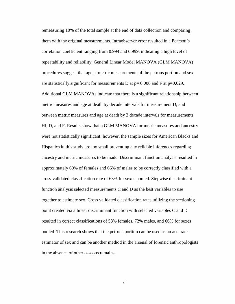
remeasuring 10% of the total sample at the end of data collection and comparing
them with the original measurements. Intraobserver error resulted in a Pearson’s
correlation coefficient ranging from 0.994 and 0.999, indicating a high level of
repeatability and reliability. General Linear Model MANOVA (GLM MANOVA)
procedures suggest that age at metric measurements of the petrous portion and sex
are statistically significant for measurements D at p= 0.000 and F at p=0.029.
Additional GLM MANOVAs indicate that there is a significant relationship between
metric measures and age at death by decade intervals for measurement D, and
between metric measures and age at death by 2 decade intervals for measurements
HI, D, and F. Results show that a GLM MANOVA for metric measures and ancestry
were not statistically significant; however, the sample sizes for American Blacks and
Hispanics in this study are too small preventing any reliable inferences regarding
ancestry and metric measures to be made. Discriminant function analysis resulted in
approximately 60% of females and 66% of males to be correctly classified with a
cross-validated classification rate of 63% for sexes pooled. Stepwise discriminant
function analysis selected measurements C and D as the best variables to use
together to estimate sex. Cross validated classification rates utilizing the sectioning
point created via a linear discriminant function with selected variables C and D
resulted in correct classifications of 58% females, 72% males, and 66% for sexes
pooled. This research shows that the petrous portion can be used as an accurate
estimator of sex and can be another method in the arsenal of forensic anthropologists
in the absence of other osseous remains.

1
CHAPTER I
INTRODUCTION
This thesis research focuses on estimating the sex of individuals through metric
analysis of the petrous portion of the temporal bone for applications within forensic
anthropology. Forensic anthropology is a sub-field of study within biological
anthropology concerned with the applications of skeletal biological knowledge and
techniques to forensic medicolegal cases. One important endeavor of these forensic
anthropological investigations is the identification of individuals who may otherwise go
unidentified due to advanced stages of decomposition, skeletonization, commingling or
severe osteological fragmentation. In such cases, it is the responsibility of the forensic
anthropologist to create a biological profile through osteological study of the remains
which includes estimations of sex, age, ancestry, stature, possible trauma, overall health,
and any evidence of unique pathological conditions.
The process of creating a biological profile typically begins first with the
estimation of the sex of a given individual, as most methods of estimation of other
biological information are affected by sex assessment (SWGANTH 2010). This is due in
large part to the presence of sexual dimorphism in the human skeleton due to different
hormone secretions during puberty which help form anatomical and physiological
differences between males and females (White and Folkens 2005). As a general rule,
female skeletal elements are characterized by smaller size and more gracile features,

2
whereas male skeletal elements are characterized by larger, more robust elements. Not
taking sex into account prior to the estimation of stature, for instance, will confound the
process due to different morphological changes in the skeleton between men and women.
It is imperative, then, that a multitude of sex estimation techniques be available to a
practitioner of forensic anthropology in order to accurately assist in the identification of
the deceased.
Current Methods for the Estimation of Sex
Techniques for the estimation of sex generally fall under two categories of
methods: morphological trait assessment and metric analysis (SWGANTH 2010).
Morphological trait assessment refers to the visual assessment of skeletal characteristics
as they are present in osteological remains. Certain traits and features of skeletal elements
are known to display marked differences caused by sexual dimorphism in the appearance
or absence of certain traits (Norén et al. 2005). Morphological assessment is usually
performed on the pelvis and skull, as these two regions of the human skeleton exhibit the
most visually observable sexual dimorphism in the body second to secondary sexual
characteristics present in fresh human remains (Bruzek 2002; Houghton 1974; Phenice
1969; White and Folkens 2005). These morphological trait assessment methods utilizing
the pelvis and skull generally requires extensive training and experience and can result in
approximately 80-90% accuracy (White and Folkens 2005).
Metric analysis refers to taking metric measurements of various features and
elements on the human skeleton and comparing them to known variation of
measurements for males and females. Metric analysis can be performed on any bones
present in the human skeletal system and can also be used to estimate other aspects of the
3
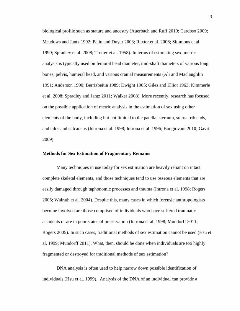
biological profile such as stature and ancestry (Auerbach and Ruff 2010; Cardoso 2009;
Meadows and Jantz 1992; Pelin and Duyar 2003; Raxter et al. 2006; Simmons et al.
1990; Spradley et al. 2008; Trotter et al. 1958). In terms of estimating sex, metric
analysis is typically used on femoral head diameter, mid-shaft diameters of various long
bones, pelvis, humeral head, and various cranial measurements (Ali and Maclaughlin
1991; Anderson 1990; Berrizbeitia 1989; Dwight 1905; Giles and Elliot 1963; Kimmerle
et al. 2008; Spradley and Jantz 2011; Walker 2008). More recently, research has focused
on the possible application of metric analysis in the estimation of sex using other
elements of the body, including but not limited to the patella, sternum, sternal rib ends,
and talus and calcaneus (Introna et al. 1998; Introna et al. 1996; Bongiovani 2010; Gavit
2009).
Methods for Sex Estimation of Fragmentary Remains
Many techniques in use today for sex estimation are heavily reliant on intact,
complete skeletal elements, and those techniques tend to use osseous elements that are
easily damaged through taphonomic processes and trauma (Introna et al. 1998; Rogers
2005; Walrath et al. 2004). Despite this, many cases in which forensic anthropologists
become involved are those comprised of individuals who have suffered traumatic
accidents or are in poor states of preservation (Introna et al. 1998; Mundorff 2011;
Rogers 2005). In such cases, traditional methods of sex estimation cannot be used (Hsu et
al. 1999; Mundorff 2011). What, then, should be done when individuals are too highly
fragmented or destroyed for traditional methods of sex estimation?
DNA analysis is often used to help narrow down possible identification of
individuals (Hsu et al. 1999). Analysis of the DNA of an individual can provide a

4
determination of sex with high precision regardless of age (White and Folkens 2005).
However, DNA analysis is often a costly, time consuming procedure, taking many
months in most modern crime laboratories due to backlog (Crime Lab Report 2008). In
cases where there are multiple numbers of individuals and remains to be sent to crime
laboratories for analysis, DNA analysis is often not a realistic option and is both cost and
time prohibitive. Likewise, reference samples from possible living relatives must be
collected in order for a DNA match to be possible. Without DNA samples from the living
to compare with the deceased, the individual may not be successfully identified.
Additionally, for remains that are too poorly preserved, DNA extraction is not always
possible.
When identification via DNA analysis is not possible or is not a feasible option in
a given situation, morphological visual assessment and metric analysis can be performed
on fragmentary remains. To the extent that certain osseous elements are still present and
preserved, visual morphological assessment can be used. Metric studies tend to focus on
regions of skeletal elements that tend to preserve, such as the study of fragmentary
skeletal remains by Albanese et al. 2008. This study examined two different methods for
two scenarios forensic practitioners often face in the field: the presence of only the
proximal femur, and the presence of the proximal femur and a portion of the hip with a
damaged os pubis. Both methods tested resulted in approximately 90-97% accuracy in
sex estimation without being population specific.
The portions of the human skeleton that tend to survive contexts of high impact or
poor preservation are those comprised of the densest bone (Lynnerup et al. 2006; Isçan
2005). One of the densest areas of bone in the cranium and often the only portion of bone

5
well-preserved in highly traumatized or disintegrated human remains is the petrous
portion of the temporal bone (Jorkov et al. 2009; Lynnerup et al. 2006). It has been
suggested in previous studies that the petrous portion of the temporal bone may yield
accurate estimations of sex due to its density and location in the endocranial region of the
cranium (Isçan 2005; Todd et al. 2010). The petrous portion of the temporal bone
according to White and Folkens (2005) is:
“… the massive, dense bony part that dominates the endocranial aspect of the
temporal. The sharp superior edge of the endocranial petrous surface angles
anteromedially, separating the temporal and occipital lobes of the brain and
housing the internal ear. The petrous is wedged between the occipital and the
sphenoid… The petrous part of the bone houses the delicate organs of hearing and
equilibrium…” (White and Folkens 2005: 95).
Due to its location on the endocranial portion of the skull, it is expected that the petrous
portion will not be affected morphologically by changes that occur due to cultural
activities or environmental stressors in life. Further, previous research demonstrates that
the petrous portion does not undergo bone remodeling throughout life after 2 years of age
except in the outer periosteal layers (Jorkov et al. 2009; Zehnder et al. 2005). This
suggests that the petrous portion is not as susceptible to changes over time as long bones,
which undergo bone remodeling every 7-10 years and allows the petrous portion to be
used as a proxy in bioarchaeological contexts when tooth enamel is absent (Jorkov et al.
2009; Zehnder et al. 2005). Reliable methods to estimate sex of an individual from the
petrous portion that are both widely available to forensic practitioners in the field and are
cost effective would be highly beneficial in the endeavor to identify individuals.
Previous Studies Involving the Petrous Portion
Recent research has utilized a variety of methods to estimate sex from the internal
acoustic meatus on the petrous portion of the temporal bone. One approach involves the

6
creation of casts of the lateral angle of the internal acoustic meatus utilizing dental
casting materials (Graw et al. 2005; Norén et al. 2005). This technique has demonstrated
some promise in showing the present sexual dimorphism of this feature; however, this
method can be quite costly, time consuming, and requires a large amount of expertise as
casts must be created accurately and left to set for an appropriate amount of time in order
for a cast to be used for angle measurements. If a cast from this technique is created and a
mistake is made in the formation process or is not allowed to set for the appropriate
allotment of time, the entire cast must be discarded. In two separate studies, this method
has shown approximately 62.5-66.3% accuracy in females and 68.5-73.3% accuracy in
males (Graw et al. 2005) and 82.3% accuracy overall for sex estimation (Norén et al.
2005).
Other techniques of sex estimation of the petrous portion have been created and
tested that are less costly. These focus on measurements of the diameter of the internal
acoustic meatus via the insertion and fit of various standard sizes of Dremel© drill bits
(Lynnerup et al. 2006). The accuracy of this method was approximately 70%. This
method of sex estimation is both time and cost efficient, and Dremel© bits are widely
available; however, this method seems to reflect more regarding shape rather than
diameter measurement. As the internal acoustic meatus is more oval in shape in
comparison to rounded or angled Dremel© bits, and because the internal acoustic meatus
shape is not constant across individuals, this method does not account for the remainder
of the meatus that cannot be measured which may provide important information and its
inclusion may allow sex to be estimated more accurately. Alternate avenues of sex
estimation, then, are still needed.

7
In 2001, Wahl and Graw utilized a metric method of estimating the sex of
individuals from the petrous portion. The authors utilized a collection of cadaver remains
(of which the origin and description are not provided), and created a large set of
landmarks and measurement combinations. However, the definitions of these landmarks
are confusing and utilize a system of arrows and numbers that are difficult for anyone to
use without extensive experience. Their method resulted in approximately 67% correct
estimation of individuals in their collection which can be a useful method to estimate sex
in a situation where the petrous portion is the only remaining osseous element (Wahl and
Graw 2001).
If possible, a cost and time prohibitive method for measuring the petrous portion
of the temporal bone that is more accurate and precise enough to be used in the field
should be developed. The most accurate and applicable metric method was developed by
Kalmey and Rathbun (1996) and involves taking metric measurements of 9 landmarks of
the petrous portion. It was based on individuals from the historic Terry Collection at the
Smithsonian Institution which is comprised of American individuals of European and
African ancestry with dates of birth ranging from 1842-1943. Their results revealed sex
estimation accuracy rates of 66 to 74% which will be useful in contexts where other
osseous remains are not recovered. As their method has not yet been applied to modern
forensic populations, my thesis research project attempts to further test this method to
achieve greater accuracy.
The present research examines documented skeletal collections of contemporary
adult American populations to assess patterns of sexual dimorphism in the petrous
portion of the temporal bone. It explores whether or not it is possible to accurately

8
estimate sex of an individual through metric examination of 9 measurements of the
petrous portion developed and refined by Kalmey and Rathbun (1996). Two hypotheses
are tested, which are:
1) This research will test if there is a significant relationship between metric
measurements of the petrous portion of the temporal bone and sex.
2) If a significant relationship between metric measurements and sex is present,
this research seeks to provide a classification function for estimating sex for professional
anthropologists to utilize in real world forensic and bioarchaeological situations.
The present research utilizes contemporary populations from the William M. Bass
Donated Skeletal Collection housed at The University of Tennessee-Knoxville and the
Texas State University donated skeletal collection. These collections comprise of
individuals with birth years after 1910 and attempts to expand the Kalmey and Rathbun
(1996) study by incorporating American White, Black, and Hispanic individuals.

9
CHAPTER II
MATERIALS AND METHODS
Collections
The skeletal remains utilized in this study were of known sex, age at death, and
ancestry. Age at death ranged from 20 years to 91 years, birth years ranged from 1892 to
2012, and population groups included American Whites, Blacks, and Hispanics. Data
compilation began in June of 2012 from two skeletal research collections: The William
M. Bass Donated Skeletal Collection at The University of Tennessee-Knoxville, and the
Texas State University-San Marcos donated skeletal collection. These collections were
utilized due to the number of available documented donated skeletal remains featuring
cranial autopsies. In addition, these collections are composed of individuals who were
born in the last 100 years, which allows this research to represent contemporary
individuals of forensic context.
The William M. Bass Donated Skeletal Collection at The University of
Tennessee-Knoxville was founded in 1981 and houses several hundred donated
individuals. All individuals in the collection have birth years ranging from 1892 to 2011,
though the majority of individuals have birth years after 1940. Of the available 228
autopsied crania, 140 individuals were randomly chosen with birth years after 1910 via a
random number generator. Biological profile information, which is provided on the front

10
of each donated individual’s box, was not viewed until after data collection was complete
in order to avoid observer bias.
Data collection at The University of Tennessee-Knoxville was performed one
individual at a time to ensure each cranium was delivered to its correct box after metric
measurements were taken. The cranium was removed and placed atop a donut-shaped
beanbag to protect the basicranium and features of the skull from breakage. The callote of
the cranium, which is the autopsied top portion of the skull, was removed and the
exposed endocranium was visually assessed for any damage to the left petrous portion; if
the damage was too severe for metric measurements to be taken, the right side was used.
However, if both right and left petrous portions were too damaged for measurements to
be taken with confidence, the skull was replaced in its collection box and removed from
the data collection sample. Likewise, if the crania were falsely noted as autopsied which
occurred in 22 instances, they were removed from the study sample.
Each of the 9 metric measurements was taken with a digital Kobalt 6” Metric and
SAE Caliper and a General Tools & Instruments 3” Metric and SAE Caliper (available at
local hardware stores) accurate to the nearest hundredth and recorded first in a scientific
composition notebook and then input into a spreadsheet. Calibration checks were
performed for both calipers at the beginning of every data collection day and were
checked throughout the data collection process. Photographs were taken of the posterior,
superior, and anterior views of the petrous portion of each cranium with corresponding
comments on condition and feature presentation recorded into the composition notebook
and spreadsheet. The individual was then returned to their collection box. The data
11
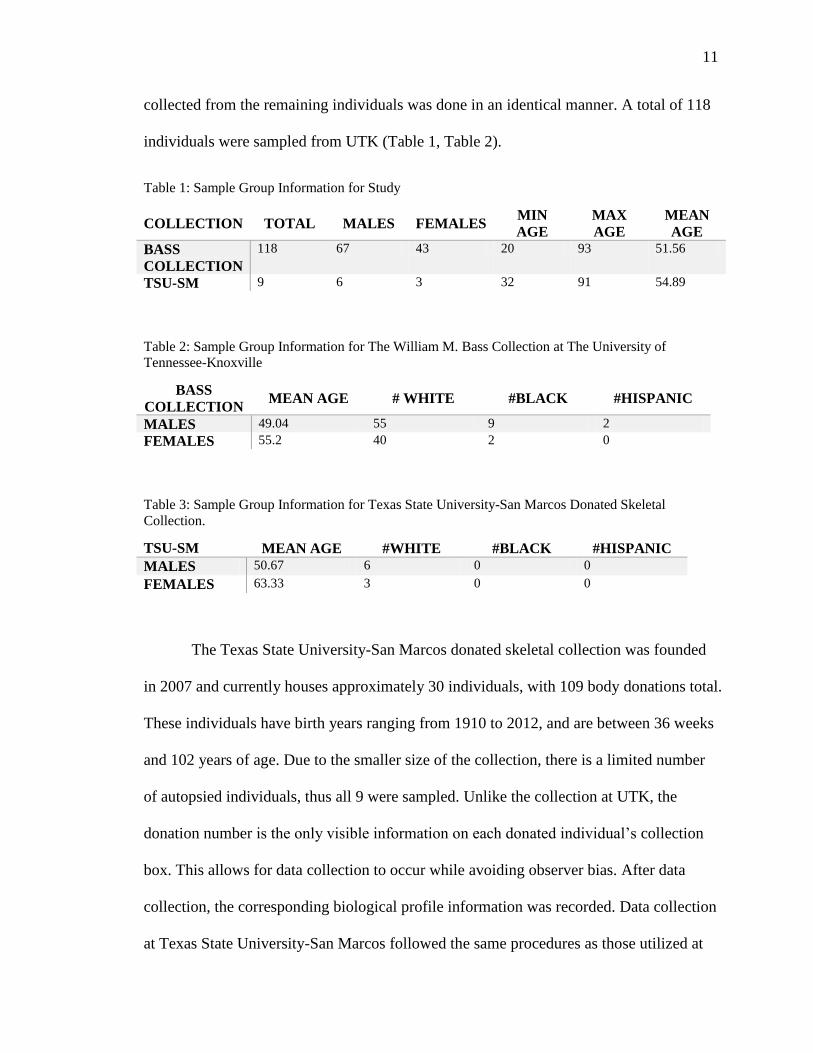
collected from the remaining individuals was done in an identical manner. A total of 118
individuals were sampled from UTK (Table 1, Table 2).
Table 1: Sample Group Information for Study
COLLECTION TOTAL MALES FEMALES MIN
AGE
MAX
AGE
MEAN
AGE
BASS
COLLECTION
118 67 43 20 93 51.56
TSU-SM 9 6 3 32 91 54.89
Table 2: Sample Group Information for The William M. Bass Collection at The University of
Tennessee-Knoxville
BASS
COLLECTION MEAN AGE # WHITE #BLACK #HISPANIC
MALES 49.04 55 9 2
FEMALES 55.2 40 2 0
Table 3: Sample Group Information for Texas State University-San Marcos Donated Skeletal
Collection.
TSU-SM MEAN AGE #WHITE #BLACK #HISPANIC
MALES 50.67 6 0 0
FEMALES 63.33 3 0 0
The Texas State University-San Marcos donated skeletal collection was founded
in 2007 and currently houses approximately 30 individuals, with 109 body donations total.
These individuals have birth years ranging from 1910 to 2012, and are between 36 weeks
and 102 years of age. Due to the smaller size of the collection, there is a limited number
of autopsied individuals, thus all 9 were sampled. Unlike the collection at UTK, the
donation number is the only visible information on each donated individual’s collection
box. This allows for data collection to occur while avoiding observer bias. After data
collection, the corresponding biological profile information was recorded. Data collection
at Texas State University-San Marcos followed the same procedures as those utilized at

12
UTK (above). A total of 9 individuals were sampled and included in the total sample
(Table 1, Table 3).
At the end of data collection at each institution, 10% of the sample was randomly
selected and the measurements for that individual retaken in order to calculate the
intraobserver error. These measurements were recorded both in the scientific composition
notebook and spreadsheet for record keeping and later compared to assess the variability
and repeatability for the methods utilized.
Measurements
Metric measurements taken during data collection follow the points and
definitions of Kalmey and Rathbun (1996) with one of their measurements taken from
Wahl and Graw (2001) and additional requirements added during data collection to
ensure standardization (Table 4; Figures 1-5). Each measurement was taken with a
Kobalt 6" Metric and SAE Caliper and a General Tools & Instruments 3” Metric and
SAE Caliper to the nearest hundredth and were re-zeroed after each measurement and
checked for calibration after each individual. These tools were chosen due to their public
availability and low cost in an effort to avoid methods that are cost prohibitive for
researchers and professionals in the field.

13
Table 4: List of Measurements with Descriptions and Instructions for each. Original Descriptions are
Derived from Kalmey and Rathbun (1996) and Wahl and Graw (2001); Modified Additional
Descriptions are Provided on the Right Side by the Author.
MEASUREMENT DESCRIPTION (KALMEY
AND RATHBUN 1996)
ADDITIONAL
DESCRIPTION
L Sigmoid sulcus-petrous apex
intersection (SS) to the most
medial point on the petrous (med.
Pt.)(sliding caliper)
Taken at suture closest to the
endocranial wall for SS; if med. Pt
is fused to sphenoid, do not insert
caliper end inside the suture
C SS to posterior (lateral) margin of
internal acoustic meatus (IAM)
(Sliding caliper)
Fit caliper jaw inside IAM for
measurement—this becomes
difficult with variation in cranial
autopsy cuts
E Posterior margin of IAM to
eminentia arcuata (EA). Taken on
EA at highest point; if EA is
plateau or if it has two peaks at
either end then take the
measurement in the center (sliding
caliper).
HI Height of IAM (taken at center of
meatus)(vernier dial caliper)
Use inside jaws of calipers
B Cochlear aqueduct (CA) to EA
(sliding caliper)
This measurement may be impeded
by extra bony growth around the
CA.
D CA to superior margin at IAM
(center of superior IAM) (vernier
dial caliper)
F CA to posterior (lateral) margin of
IAM (from Wahl) (vernier dial
calipers)
G EA to med pt. (sliding caliper) if med. Pt is fused to sphenoid, do
not insert caliper end inside the
suture
W CA to hiatus of facial canal
(sliding caliper)
This measurement may be impeded
by extra bony growth around the
hiatus of the facial canal or the
formation of a bony bridge.

14
Figure 1: Lateral left ectocranial view of a human skull with arrows pointing to left temporal bone.
Figure 2: Transverse endocranial view of the human skull with the temporal bones denoted in purple and
other identifying features labelled (The McGraw-Hill Companies, Inc.).

15
Figure 3: Posterior view of the endocranium of a human skull with both petrous portions demarcated with
black circles.
Figure 4: Diagram of a left temporal bone (Gray’s Anatomy taken from www.prohealthsys.com).

16
Figure 5: Superior view of the left petrous portion with measurement L.
Figure 6: Posterior medial view of the left petrous portion with measurements C, B, and E.

17
Figure 7: Posterior medial view of the left petrous portion with measurements G, HI, D, and F.
Figure 8: Anterior view of the left petrous portion with measurement W.

18
Figure 9: Original diagrams of measurements from Kalmey and Rathbun 1996.
Statistical Methods
Statistical methods applied in this study followed the protocol established by
Bongiovanni’s analysis of the sternum, which included similar questions, materials, and
hypotheses (Bongiovanni 2010). These statistical methods were chosen due to the
similarities in metric analysis and similar goals involving the estimation of sex from
metric measurements. These methods include descriptive statistics of petrous portion
measurements (frequencies, means, standard variations, and variances), generalized linear
model MANOVA procedures for the 9 measurements in the study, discriminant function
analysis and a stepwise discriminant function analysis. All statistical analyses were

19
performed with the program Statistical Package for the Social Sciences (SPSS) on
February 14th, 2013.
The Generalized Linear Model MANOVA (GLM MANOVA) procedure was
performed to assess if a statistically significant relationship existed between each variable
utilized in this study and sex. This procedure also discerned which individual variables
provide the best measures for the estimation of sex. Measures that provide significant
results at the 0.05 level were considered statistically significant and acceptable in
proceedings of court as evidence according to Daubert rulings (Grivas et al. 2008).
Additional GLM MANOVA procedures were performed in order to discern any other
relationships between metric measurements and age, ancestry, and sex. Significant results
at the 0.05 level for these analyses gave insight into other factors that affect morphology
and measurements that can confound estimations of sex from the petrous portion.
A discriminant function analysis was performed to determine which
measurements are the best estimators of sex and provide classification rates for males,
females, and pooled sex.
A stepwise discriminant function analysis was then performed to discern if a
combination of measurements provide a better estimator of sex. This was followed by
linear discriminant function analysis to develop an equation for the estimation of sex
using those metric measurements selected in the stepwise discriminant function.
The linear discriminant function equation was derived following the stepwise
discriminant function analysis from the output through the subtraction of the group means
of selected measurements of males and females. The resulting equation will provide a
quick and easy way to estimate the sex of an individual, as those who fall above the

20
discriminant value would result in a male sex designation and those who fall below the
discriminant value would result in a female sex designation. Individuals who fall on the
discriminant value will be assigned an ambiguous sex designation. Creating a linear
discriminant function equation with these measurements will be important for contexts in
which forensic remains of individuals are not complete and only a few of the
measurements may be taken. Discriminant function analysis and sectioning points
resulting from analysis must provide cross-validated rates of classification and be
statistically significant at the 0.05 level to be considered significant in this study and to be
used as evidence in court proceedings as a result of the Daubert rulings (Grivas et al.
2008).
21
CHAPTER III
RESULTS
Statistical analysis performed on the data collected from osteological collections
at The University of Tennessee-Knoxville and Texas State University-San Marcos were
performed via Microsoft Excel to compute intraobserver error and descriptive statistics
for each measurement. The statistical software package SPSS was used for the remaining
statistical analyses.
Descriptive Statistics
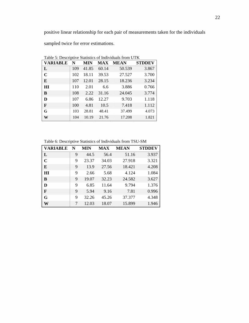
Descriptive statistics for each of the 9 measurements from The University of
Tennessee-Knoxville are available in Table 5, and from Texas State University-San
Marcos in Table 6. Table 7 features descriptive statistics from pooled data from both
skeletal collections.
Intraobserver Error
Intraobserver error was performed for this study utilizing a 10% approach. This
means that at the end of the data collection process at each institution, 10% of the total
sample at each institution was measured an additional time. On the measurements that
were retaken as part of this procedure and the original measurements, a Pearson’s
correlation coefficient analysis was performed. This analysis provided a near perfect
22
positive linear relationship for each pair of measurements taken for the individuals
sampled twice for error estimations.
Table 5: Descriptive Statistics of Individuals from UTK
VARIABLE N MIN MAX MEAN STDDEV
L 109 41.85 60.14 50.539 3.867
C 102 18.11 39.53 27.527 3.700
E 107 12.01 28.15 18.236 3.234
HI 110 2.01 6.6 3.886 0.766
B 108 2.22 31.16 24.045 3.774
D 107 6.86 12.27 9.703 1.118
F 100 4.81 10.5 7.418 1.112
G 103 28.81 48.41 37.499 4.073
W 104 10.19 21.76 17.208 1.821
Table 6: Descriptive Statistics of Individuals from TSU-SM
VARIABLE N MIN MAX MEAN STDDEV
L 9 44.5 56.4 51.16 3.937
C 9 23.37 34.03 27.918 3.321
E 9 13.9 27.56 18.421 4.208
HI 9 2.66 5.68 4.124 1.084
B 9 19.07 32.23 24.582 3.627
D 9 6.85 11.64 9.794 1.376
F 9 5.94 9.16 7.81 0.996
G 9 32.26 45.26 37.377 4.348
W 7 12.03 18.07 15.899 1.946
23
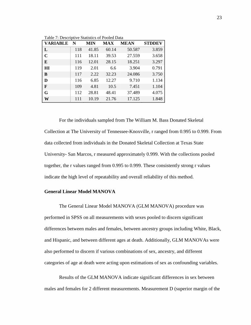
Table 7: Descriptive Statistics of Pooled Data
VARIABLE N MIN MAX MEAN STDDEV
L 118 41.85 60.14 50.587 3.859
C 111 18.11 39.53 27.559 3.658
E 116 12.01 28.15 18.251 3.297
HI 119 2.01 6.6 3.904 0.791
B 117 2.22 32.23 24.086 3.750
D 116 6.85 12.27 9.710 1.134
F 109 4.81 10.5 7.451 1.104
G 112 28.81 48.41 37.489 4.075
W 111 10.19 21.76 17.125 1.848
For the individuals sampled from The William M. Bass Donated Skeletal
Collection at The University of Tennessee-Knoxville, r ranged from 0.995 to 0.999. From
data collected from individuals in the Donated Skeletal Collection at Texas State
University- San Marcos, r measured approximately 0.999. With the collections pooled
together, the r values ranged from 0.995 to 0.999. These consistently strong r values
indicate the high level of repeatability and overall reliability of this method.
General Linear Model MANOVA
The General Linear Model MANOVA (GLM MANOVA) procedure was
performed in SPSS on all measurements with sexes pooled to discern significant
differences between males and females, between ancestry groups including White, Black,
and Hispanic, and between different ages at death. Additionally, GLM MANOVAs were
also performed to discern if various combinations of sex, ancestry, and different
categories of age at death were acting upon estimations of sex as confounding variables.
Results of the GLM MANOVA indicate significant differences in sex between
males and females for 2 different measurements. Measurement D (superior margin of the

24
internal acoustic meatus to the cochlear aqueduct) demonstrates significance differences
between males and females at p= 0.000 and measurement F (cochlear aqueduct to
posterior margin of the internal acoustic meatus) demonstrates significant differences
between males and females at p= 0.029.
Additional GLM MANOVA procedures were performed to discern if significant
relationships existed between measurements of the petrous portion and age at death, sex,
ethnicity and multiple combinations of age at death, sex, and ancestry. The results of
these procedures provided interesting insights into the relationship of endocranial
morphology and biological variants. Results demonstrate no statistically significant
differences between metric measurements and ancestry, but reveals significant
relationships between metric measurements of the petrous portion and age at death parsed
by decade intervals for measurement D at p=0.024. Results for GLM MANOVA
performed between metric measurements and age at death parsed by 2 decade intervals
resulted in statistically significant differences between age and metric measurements for
HI at p=0.004, D at p=0.003, and F at p=0.02.
To discern if age at death, sex, and ethnicity were acting as dependent variables
rather than independent non-complementary variables, GLM MANOVAs were also
performed for all possible combinations of sex, age at death by decade intervals, age at
death by 2 decade intervals, and ancestry. These combinations can be found in Table 8,
Table 9, and Table 10.

25
Table 8: Relationships Between Metric Measurements Paired with Variables
COMBINATION SIGNIFICANT? MEASURES
MEASURES, SEX SIGNIFICANT
D: 0.000
F: 0.029
MEASURES, AGE
(1 DECADE) SIGNIFICANT D: 0.024
MEASURES, AGE
(2 DECADES) SIGNIFICANT
HI: 0.004
D: 0.003
F: 0.02
MEASURES, ANCESTRY NOT SIGNIFICANT N/A
Table 9: Combinations of Paired Variables with Metric Measurements
COMBINATION SIGNIFICANT? MEASURES
MEASURES, SEX, AGE (1
DECADE) SIGNIFICANT D: 0.018
MEASURES, SEX, AGE
(2 DECADES) SIGNIFICANT
HI: 0.010
B: 0.037
D: 0.001
F: 0.031
G: 0.048
MEASURES, SEX,
ANCESTRY SIGNIFICANT D: 0.006
MEASURES, AGE (1
DECADE), ANCESTRY NOT SIGNIFICANT N/A
MEASURES, AGE (2
DECADES), ANCESTRY SIGNIFICANT
HI: 0.023
B: 0.028
D: 0.037
Table 10: Combinations of Three Variables with Metric Measurements
COMBINATION SIGNIFICANT? MEASURES
MEASURES, SEX, AGE (1
DECADE), ANCESTRY NO SIGNIFICANCE N/A
MEASURES, SEX, AGE (2
DECADES), ANCESTRY SIGNIFICANT
H: 0.028
B: 0.029
D: 0.008
F: 0.059
26
Discriminant Function Analysis
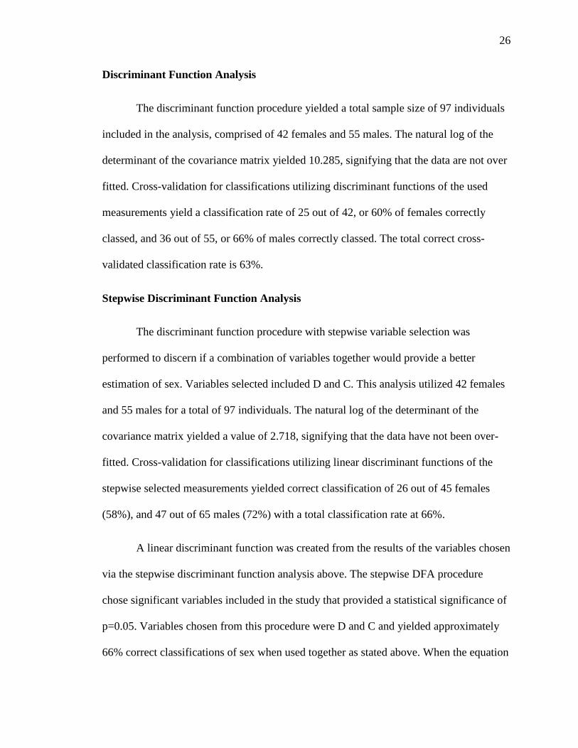
The discriminant function procedure yielded a total sample size of 97 individuals
included in the analysis, comprised of 42 females and 55 males. The natural log of the
determinant of the covariance matrix yielded 10.285, signifying that the data are not over
fitted. Cross-validation for classifications utilizing discriminant functions of the used
measurements yield a classification rate of 25 out of 42, or 60% of females correctly
classed, and 36 out of 55, or 66% of males correctly classed. The total correct cross-
validated classification rate is 63%.
Stepwise Discriminant Function Analysis
The discriminant function procedure with stepwise variable selection was
performed to discern if a combination of variables together would provide a better
estimation of sex. Variables selected included D and C. This analysis utilized 42 females
and 55 males for a total of 97 individuals. The natural log of the determinant of the
covariance matrix yielded a value of 2.718, signifying that the data have not been over-
fitted. Cross-validation for classifications utilizing linear discriminant functions of the
stepwise selected measurements yielded correct classification of 26 out of 45 females
(58%), and 47 out of 65 males (72%) with a total classification rate at 66%.
A linear discriminant function was created from the results of the variables chosen
via the stepwise discriminant function analysis above. The stepwise DFA procedure
chose significant variables included in the study that provided a statistical significance of
p=0.05. Variables chosen from this procedure were D and C and yielded approximately
66% correct classifications of sex when used together as stated above. When the equation

27
was applied to the entire sample group used in this study, including those that were not
included in the statistical analysis due to missing measurements, resulted in
approximately 72.8% correct sex classifications. For each individual, the measurements
for C and D are taken and input into the equation, then the variables and the constant for
this equation are added together (Table 11). Ending values less than 0 are considered
female, while ending values greater than 0 are considered male. Values at 0 are
considered to be ambiguous
Table 11: Linear Discriminant Function Equation for Sex Estimation
VARS METRIC 1 METRIC 2 CONSTANT
C, D -0.146 (C) 0.799(D) -3.423
28
CHAPTER IV
DISCUSSION
The goals of this research determined if methods utilizing metric measurements of
the petrous portion of the temporal bone are appropriate to estimate sex of individuals.
This study utilized the established measurements of Kalmey and Rathbun (1996) based
on historical individuals and tested if the metric measurements were significant in the
estimation of sex of contemporary American individuals. Measurements are considered
significant at the p= 0.05 level, which is the required statistical level of significance to be
admitted as scientific evidence in a court of law based on Daubert rulings (Grivas et al.
2008).
As much forensic work for mass fatality and traumatic accident identification is
done in the field and many techniques in use today such as those that involve DNA
analysis are too costly for large case amounts, using and testing more cost-effective
materials is imperative. The purpose of this research tested a set of established
measurements based on historical individuals housed within the Terry Collection at the
Smithsonian Institution and applied them to contemporary individuals so as to provide an
additional method of sex estimation to the arsenal of forensic anthropologists. Previous
research demonstrated that aspects of the biological profile such as long bone length,
which can be used as a proxy for stature, vary throughout time due to secular change
(Jantz and Jantz 1999). Secular change is shown to be affected by both environmental

29
factors, such as improved nutrition and health status (Jantz and Jantz 1999). By
examining contemporary individuals in the current study and comparing results to the
previous study which utilized historical collections, an inference can be made regarding
possible change over time of the morphology of structures of the endocranium such as the
petrous portion. Additionally, as the petrous portion is one of the densest regions of bone
in the human body and preserves well in cases of traumatic accidents and poor
preservation environments, this method could be an important tool in medicolegal
investigations where methods such as DNA analysis and other methods of sex estimation
cannot be utilized due to cost or due to poor quality of remains.
The results of this study show that metric measurement of the petrous portion can
estimate sex in modern populations and is statistically significant at the 0.05 level.
Correct classifications of sex reached upwards of 73% in this study. Intraobserver error
measured through a Pearson’s correlation coefficient provides between 0.995 and 0.999,
indicating an extremely high positive correlation and thus high repeatability. Although
the results are not as high as other methods of sex estimation, such as visual
morphological trait assessment of the pelvis and skull and metric measurements of
various long bone features and cranial measurements utilizing complete osteological
elements or a combination of elements (Dwight 1905; Giles and Elliot 1963; Spradley
and Jantz 2011; Walker 2008; White and Folkens 2005), the results show significant
association between metric measures and sex that are greater than chance (50%). As this
method is based on measurements of only the petrous portion of the temporal bone
without relying on any other related structures or features of the skull, this method has the
potential to provide forensic professionals with another method of estimating sex of

30
individuals with highly fragmentary remains where other osseous elements are not
present or well preserved.
The PROC GLM MANOVA procedure in SPSS was performed on all
measurements to discern the presence of significant differences between males and
females, between ancestral groups including White, Black, and Hispanic, and to see if
relationships between sex, ancestry, and age at death affected sex estimation. The
procedure yielded no significant differences for any individual variable with relation to
ancestry or ancestry and sex paired together; however, due to the small number of Black
and Hispanic individuals that were available for inclusion in this study it cannot be said
that ancestry does not affect measurements of sex in the petrous portion. More research
with a higher number of individuals from other ancestral groups should be included
before considering sex estimation using the petrous portion to be unrelated to an
individual’s ancestry.
This GLM MANOVA procedure, however, revealed other statistically significant
relationships between metric measurements used in this study and various other variables.
First, age at death of individuals included in this study were parsed into decade intervals
in 8 categories: 20-29, 30-39, 40-49, 50-59, 60-69, 70-79, 80-89, and 90-99. When
compared only with metric measures, age at death in decade intervals was only
significant for measurement D (cochlear aqueduct to superior margin at the internal
acoustic meatus) at p=0.024. When analyzed with both metric measures and sex, age at
death in decade intervals was also only significant for measurement D at p=0.018. Age at
death in decade intervals was not significant, however, when compared with ancestry or
when compared with sex and ancestry. Due to the small sample size present when age at

31
death was parsed into these eight categories and due to only one variable being
significant, age at death by single decade intervals may not be the best way to parse and
examine age at death and measures used.
Broader decade categories for age at death were then examined in order to see if
more individuals in each category would result in significant results. Age at death was
parsed into four categories by 2 decade intervals, beginning with 20-39, 40-59, 60-79,
and finally ages 80-99 comprising the final group. The PROC GLM MANOVA reveal a
much more statistically meaningful relationship between age at death by 2 decade
intervals and the measurements in this study, with significant measurements including HI
(height of the internal acoustic meatus) at p= 0.004, D at p= 0.003, and F (cochlear
aqueduct to posterior lateral margin of the internal acoustic meatus) at p=0.02. When
compared with measurements and sex together, age at death by 2 decade intervals is
significant for 5 different measures, including HI, B (cochlear aqueduct to arcuate
eminence), D, F, and G (arcuate eminence to medial point). When paired with metric
measures and ancestry, and with metrics, sex and ancestry, measures HI, B, and D were
all significant. HI, D, F all comprise measurements that include the internal acoustic
meatus with B and G including the arcuate eminence. The location of these significant
measures along the outer periosteal layers of the petrous portion as well as their general
location in the endocranium suggests that measurements including these 2 features may
be the most susceptible to age related changes in morphology of the petrous portion.
Finally, the results of the primary focus of this study, which was to determine if
there exists a significant relationship between metric measures and sex, resulted in
significant differences in sex between males and females for measurements D and F at

32
the 0.05 level. The statistically significant relationship between these two measurements
and sex suggest that sexual dimorphism is present among males and females in the
petrous portion of the temporal bone. Cross-validation for classifications utilizing linear
discriminant functions through the discriminant function analysis yield a classification
rate of 25 out of 42 of females (60%) correctly classed and 36 out of 55 of males (66%)
correctly classed and a total classification rate of 63% cross-validated. Though these are
not high classification rates, they are higher than chance.
The discriminant function procedure with stepwise variable selection was
performed to discern if a combination of variables together would provide a better
estimation of sex. The stepwise procedure chose the variables that were the better
estimators of sex which included measurements C (sigmoid sulcus to posterior lateral
margin of the internal acoustic meatus) and D. Classifications utilizing these two
measurements together to estimate sex as a linear discriminant function yielded correct
classification of 26 out of 45 females (58%), and 47 out of 65 males (72%). Utilizing the
stepwise selected variables together as an estimator of sex resulted in lower classification
rates for females, higher classification for males, and a higher total cross-validated
correct classification rates rising to 66%. The stepwise discriminant function analysis
gave a slightly higher classification rate for males and females than did the discriminant
function analysis without stepwise variable selection.
It is clear, however, from these results that the petrous portions of females are
most often misclassified when compared to males, especially at the results of the stepwise
discriminant function analysis. This could be caused by several factors including an
underrepresentation of females in the sample or the possibility that there could be an age

33
related bias present in the sample. Age-related effects have been known to affect the
morphology or structure of bony elements of the female skeleton, such as masculinization
of the cranial features with increasing age in females; however, more research is needed
to fully understand this process (Rogers 2005). As the average age at death of females in
this study is ~55 years with a median of 54 years, and the male average age at death at
~49 years with a median also at 49 years, the older age of women in this sample
compared to men could indeed be pulling the estimations of sex upwards toward higher
end values that classify the individual as male when utilizing the discriminant function
equation which could reflect masculinization processes of the endocranium in females.
Overall, Kalmey and Rathbun’s (1996) methods for metric measurements of the
petrous portion yielded between 66-74% accuracy in correctly estimating the sex of
individuals of historic significance housed in the Terry Collection at the Smithsonian
Institution. This percentage represents the range of correct classifications for males and
females in the previous study. The results of this current study show that similar accuracy
percentages from 58-73% can be achieved utilizing the same metric measurements of the
petrous portion on contemporary individuals. Regardless of historical or contemporary
provenience, measurements of the petrous portion in this research or previous research
have not exceeded 74%. This research, however, represents the first time the petrous has
been measured with these numbers on modern individuals. This method still affords
forensic practitioners another accurate method to estimate sex in situations that
demonstrate an absence of other bones or presence of other fragmentary osseous elements.

34
CHAPTER V
CONCLUSION
This research focused on applying the metric methods of Kalmey and
Rathbun (1996) based on the historic Terry Collection housed at the Smithsonian
Institution to modern individuals from two donated skeletal collections. The purpose
of this research focused on two questions: Can metric measurements of the petrous
portion be used to estimate the sex of an individual in a forensic medicolegal
context? If so, how accurate is this method?
This research began during the summer of 2012 and sampled individuals
from the William M. Bass Donated Skeletal Collection at The University of
Tennessee-Knoxville and the donated skeletal collection at Texas State University-
San Marcos. A total of 118 individuals with cranial autopsies were randomly
sampled and tested. Included in the study are individuals of White, Black, and
Hispanic ancestry. Age at death for individuals included in the sample ranged from
20 years to 91 years.
Results showed a significant relationship between metric measurements
utilized in this study compiled by Kalmey and Rathbun (1996) and estimation of sex.
Variables D and F were found through the GLM MANOVA procedure to have the
highest significant relationship out of all 9 variables. Discriminant Function
35
Analysis resulted in a cross-validated classification rate of 59.5% of females
correctly classed and 65.5% of males correctly classified. Stepwise Discriminant
Function Analysis selected variables D and C as the greatest predictors of sex and
resulted in a linear equation to determine a sectioning point:
-0.146 (C) + 0.799 (D) -3.423
Ending values that are positive classify as male while negative values classify as
female.
This linear discriminant function equation resulted in the correct
classification of 57.8% of females, 72.3% of males, and 66.4% total correct
classifications of sex cross validated. When applied to the entire sample group used
in this study, using this equation resulted in approximately 72.8% correct sex
classifications.
In addition to exploring the relationship of sex and the metric measurements
utilized in this study, GLM MANOVA procedures were also performed for all
combinations of ancestry, age by decade intervals, age by 2 decade intervals, and sex.
Ancestry showed no significant relationship with any combination of variables
except when combined with age at death by two decades and age at death by two
decades and sex, but sample sizes of other ancestral groups must be increased in
future studies for further inferences to be made. Age at death parsed by decade
intervals with 8 categories was only significant at measurement D.
Age at death parsed by 2 decade intervals with 4 broad categories, however,
showed a significant relationship between metric measurements, sex, ancestry, and

36
when compared with sex and ancestry together. Parsing by 2 decade intervals
allowed sample sizes for each category to be larger and more representative of the
sample collected. The resulting significance of every combination utilizing age
parsed into categories of 2 decade intervals has very important implications. This
suggests there may be a relationship between the morphology of the petrous portion
of the temporal bone and increasing age. As the mean total age at death for females
in this study was approximately 56 years and the mean age for males was 49, a bias
toward older females may be present in this sample. Along those same lines, these
results may suggest that a form of masculinization of the endocranium with
increasing age may be present. More research should be conducted to further explore
the relationship of age with the morphology and appearance of the petrous portion to
better understand possible age related changes that could affect identification
processes.
Limitations
One major assumption that should be addressed is that the individuals in this
study sample are representative of the population. This sample, though as broad as
possible with the applied limitations of sampling only autopsied crania, cannot hope to be
representative of the entire population of the United States. With an increase in the
number of foreign immigrants into the United States, more ancestral groups aside from
Whites and the small group of Blacks and Hispanics here must be included to provide a
more inclusive technique and ensure that ancestry, indeed, does not play a part in the
morphological differences in the petrous portion. Further research utilizing more sub-
populations can help to affirm or refute the possibility that metric analysis of the petrous
37
portion is a non-population specific technique that can be applied on any ancestral group
in the United States.
Factors noted above that could also affect the measurements include an
underrepresentation of females in the sample, an age-related bias in the sample with
females at age 55 and males at age 49. As shown, however, for the small sample
available for use in this study there is no statistically significant relationship between
ancestry and the metric measurements taken in this study or a combination of sex and
ethnicity except when compared with age at death in broader 20 year intervals. A more
equal ratio of males and females in the sample may, however, improve the percentages of
correct classifications.
Other factors not discussed above can also include the sampling process. Though
all samples were randomly selected based on the presence of cranial autopsies, not all
individuals could be sampled due to the quality or dimensions of the cranial autopsies
performed. Additionally, the sample was reduced due to misdocumentation of cranial
autopsies performed on several individuals. In terms of those individuals with cranial
autopsies, craniotomy procedures resulting with higher endocranial walls were much
more difficult to measure than individuals with skewed cranial autopsies or larger callotes.
Indeed, many times a measurement was not possible due to the interior dimensions of an
individual’s autopsy. Further, it is unclear if there is asymmetry present among the right
and left petrous portions of individuals. Future research should address this possibility.
.

38
Advantages
This method utilizes an element of the human body that does not have direct
association with the outside environment or individual musculature. As such, occupation
and musculoskeletal markers or MSMs are not likely to show and have not in previous
studies shown any effect on the morphology of the petrous portion of the temporal bone.
Through the study of contemporary individuals, the results of the current study does not
vary remarkably from the previous study by Kalmey and Rathbun (1996) utilizing
historical individuals. This suggests that secular change over time, which can be affected
by genetics and environmental factors such as improved nutrition and health status, is
most likely not present in the petrous portion of the temporal bone. This suggest that
features of the endocranium such as the petrous portion, in addition to not being
susceptible to occupational MSMs and other environmental agents, may not be affected
through over time by secular change.
Applications and Future Research
Forensic practitioners can utilize this methods through the use of digital
sliding metric calipers that are widely available at most hardware and home
improvement stores. Practitioners should view the original definitions provided by
Kalmey and Rathbun (1996) as well as the additional notes provided by the author of
this research project (Table 4). Together, measurements C and D are the two best
estimators of sex when used in the equation above for the sectioning point.
The results of sex estimation utilizing these methods mirrors the results of
Kalmey and Rathbun (1996), resulting in between 65-74% accurate classification

39
rates of males and females utilizing the stepwise discriminant function results and
sectioning point equation. This method provides a method of sex estimation that can
be used as an estimator of sex in the absence of other osseous elements. This can be
particularly useful in real world applications in cases of high impact accidents and
mass fatalities, such as plane crashes, commingling, and other situations of poor
preservation or high fragmentation of osteological remains of individuals where the
chance of finding intact skeletal remains may be slim. Since the petrous portion is
one of the densest elements of bone (Isçan 2005) and is often the only element
present in such cases, this method will provide another technique useful in aiding the
identification of individuals when other, more diagnostic elements may not be
present or are too damaged to be used with integrity. Additionally, as there appears
to be no secular change in the measurements of the petrous portion through time for
historical and contemporary individuals, this method may also be applicable in
bioarchaeological contexts where soils and other taphonomic factors result in poor
preservation. Further research should explore this possibility.
Future studies involving the petrous portion should investigate the possible
presence of asymmetry between the right and left petrous portions. Larger sample
sizes of Blacks, Hispanics, and the inclusion of other ancestral populations found in
the United States should also be included to either accept or refute the possibility that
this method of sex estimation may not be ancestry dependent. The constraints of this
study will limit the use of known collections to include only those individuals that
have cranial autopsies performed before skeletal donation at death or cranial
fragmentation that would allow access to the petrous portions. Despite this limitation,

40
such collections, though small in sample size, do exist. Additionally, research should
also be conducted to test bioarchaeological collections that have the petrous portions
and pelves of individuals available to further test the presence or absence of secular
change in the endocranium through the passage of time. Finally, researchers should
investigate further into the relationship between age and morphology of the petrous
portion, especially with the possibility of masculinization of the female endocranium
with increasing age.

41
REFERENCES
Akansel G, Inan N, Kurtas O, Sarisoy HT, Arslan A, Demirci A. 2008. Gender and the
Lateral Angle of the Internal Acoustic Canal Meatus as Measured on
Computerized Tomography of the Temporal Bone. Forensic Science
International 178: 93-95.
Albanese J, Eklics G, Tuck A. 2008. A Metric Method for Sex Determination Using the
Proximal Femur and Fragmentary Hipbone. Journal of Forensic Sciences 53(6):
1283-1288.
Ali RS, Maclaughlin SM. 1991 Sex Identification from the Auricular Surface of the
Adult Human Ilium. International Journal of Osteoarchaeology 1(1): 57-61.
Anderson BE. 1990. Ventral Arc of the Os Pubis: Anatomical and Developmental
Considerations. American Journal of Physical Anthropology 83(4): 449-458.
Auerbach BM, Ruff CB. 2010. Stature Estimation Formulae for Indigenous North
American Populations. American Journal of Physical Anthropology 141: 190-207.
Berrizbeitia EL. 1989. Sex Determination with the Head of the Radius. Journal of
Forensic Sciences 34(5): 1206-1213.
Bruzek J. 2002. A Method for Visual Determination of Sex Using the Human Hip Bone.
American Journal of Physical Anthropology 117(2): 157-168.
Bongiovanni, R. 2010. Estimating Sex of the Human Skeleton Based on Metrics of the
Sternum. M.A. Thesis, Department of Anthropology, Texas State University-San
Marcos.
Cardoso HFV. 2009. A Test of Three Methods for Estimating Stature from Immature
Skeletal Remains Using Long Bone Lengths. Journal of Forensic Sciences 54: 13-
19.
Crime Lab Report. The Mechanics of Crime Laboratories. Online Posting. 2008. Crime
Lab Report Headlines. 2011 December 2
http://www.crimelabreport.com/media_accuracy/mechanics.htm.
Dwight T. 1905. The Size of the Articular Surfaces of the Long Bones as Characteristic
of Sex: An Anthropological Study. American Journal of Anatomy 4(1): 19-31.
Giles E, Elliot O. 1963 Sex Determination by Discriminant Function Analysis of Crania.
American Journal of Physical Anthropology 21(1):53-68.
Graw M, Wahl J, Ahlbrecht M. 2005. Course of the Meatus Acusticus Internus as
Criterion for Sex Differentiation. Forensic Science International 147: 113-117.

42
Grivas CR, Komar DA. 2008. Kumho, Daubert, and the Nature of Scientific Inquiry:
Implications for Forensic Anthropology. Journal of Forensic Sciences 53 (4): 771-
776.
Houghton P. 1974. The Relationship of the Pre-Auricular Grrove of the Ilium to
Pregnancy. American Journal of Physical Anthropology 41(3): 381-389.
Hsu CM, Huang NE, Tsai LC, Kao LG, Chao CH, Linacre A, Lee JC-I. 1999.
Identification of Victims of the 1998 Taoyuan Airbus Crash Accident Using DNA
Analysis. International Journal of Medicine 133: 43-46.
Introna FJ, Di Vella G, Campobasso CP. 1998. Sex Determination by Discriminant
Analysis of Patella Measurements. Forensic Science International 95: 39-45.
Introna FJ, Di Vella G, Campobasso CP, Dragone M. 1996. Sex Determination By
Discriminant Analysis of Calcanei Measurements. Journal of Forensic Sciences:
725-72Iscan YM. 2005. Forensic Anthropology of Sex and Body Size. Forensic
Science International 147: 107-112.
Jantz LM, Jantz RL. 1999. Secular Change in Long Bone Length and Proportion in the
United States, 1800–1970. American Journal of Physical Anthropology 110: 57-
67.
Jørkov MLS, Heinemeier J, Lynnerup N. 2009. The Petrous Bone—A new Sampling Site
for Identifying Early Dietary Patterns in Stable Isotopic Studies. American
Journal of Physical Anthropology 138: 199–209.
Kalmey, Rathbun. 1996. Sex Determination by Discriminant Function Analysis of the
Petrous Portion of the Temporal Bone. Journal of Forensic Sciences 41(5): 865-
867.
Kimmerle EH, Ross A, Slice D. 2008. Sexual Dimorphism in America: Geometric
Morphometric Analysis of the Craniofacial Region. Journal of Forensic Sciences
53(1): 54-57.
Lynnerup N, Schulz M, Madelung A, Graw M. 2006. Diameter of the Human Internal
Acoustic Meatus and Sex Determination. International Journal of
Osteoarchaeology 16: 118-123.
Meadows L, Jantz RL. 1992. Estimation of Stature from Metacarpal Lengths. Journal of
Forensic Sciences 37: 147-154.
Mundorff AZ. 2011. Integrating Forensic Anthropology into Disaster Victim
Identification. Forensic Science Medical Pathology: Online Publication.
Norén A, Lynnerup N, Czarnetzki A, Graw M. 2005. Lateral Angle: A Method for
Sexing Using the Petrous Bone. American Journal of Physical Anthropology 128:
318-323.
Phenice TW. 1969. A Newly Developed Visual Method of Sexing the Os Pubis.
American Journal of Physical Anthropology 30(2): 297-301.

43
Pelin IC, Duyar I. 2003. Estimating Stature from Tibia Length: A Comparison of
Methods. Journal of Forensic Sciences 48: 708-712.
Raxter MH, Auerbach BM, Ruff CB. 2006. Revision of the Fully Technique for
Estimating Statures. American Journal of Physical Anthropology 130: 374-384.
Simmons T, Jantz RL, Bass WM. 1990. Stature Estimation from Fragmentary Femora: A
Revision of the Steele Method. Journal of Forensic Sciences 35: 628-636.
Spradley MK, Jantz RL. 2011. Sex Estimation in Forensic Anthropology: Skull Versus
Postcranial Elements. Journal of Forensic Sciences 56(2): 289-296.
SWGANTH: The Scientific Working Group for Forensic Anthropology. 2010. Personal
Identification. http://swganth.startlogic.com/Identification%20Rev0.pdf, accessed
February 28, 2013.
Todd NW, Graw M, Dietzel M. 2010. “Lateral Angle”of the Internal Auditory Canal:
Non-Association with Temporal Bone Pneumatization. Journal of Forensic
Sciences 55(1): 141-144.
Trotter M, Gleser GC. 1958. A Re-Evaluation of estimation of Stature Based on
Measurements of Stature Taken During Life and of Long Bones After Death.
American Journal of Physical Anthropology 16: 79-123.
Ubelaker, DH. 2009. The Forensic Evaluation of Burned Skeletal Remains: A synthesis.
Forensic Science International 183: 1-5.
Wahl J, Graw M. 2001. Metric Sex Differentiation of the Pars Petrosa Ossis Temporalis.
International Journal of Legal Medicine 114: 215-223.
Walker PL. 2008. Sexing Skulls Using Discriminant Function Analysis of Visually
Assessed Traits. American Journal of Physical Anthropology 136(1): 39-50.
White TD, Folkens PA. The Human Bone Manual. 1st edition. 2005. San Diego (CA):
Academic Press.
Zehnder AF, Kristiansen AG, Adams JC, Merchant SN, McKenna MJ. 2005.
Osteoprotegerin in the Inner Ear May Inhibit Bone Remodeling in the Otic
Capsule. The Laryngoscope 115: 172-177.

VITA
Cristina Chartier Watson was born in Castro Valley, California on May 15,
1989 and is the daughter of Leslie Jean Watson and Larry Leo Watson, Jr. After
completing her work at Castro Valley High School, Castro Valley, California in
2007, she entered Northern Arizona University. During her undergraduate career,
she spent the summer of 2010 in Peru as part of a bioarchaeological field school
through Pontificia Universidad Católica del Peru. She received the degree of
Extended Bachelor of Arts in Anthropology with an emphasis in Biological
Anthropology from Northern Arizona University in May 2011 along with minors in
Biology, Spanish, and Japanese. In August 2011, she entered the Graduate College at
Texas State University-San Marcos.
Permanent Address: [email protected]
This thesis was typed by Cristina C. Watson.