et ai, it et ai., (dat) et al., et ai.,shodhganga.inflibnet.ac.in/bitstream/10603/5981/9/09_chapter...
TRANSCRIPT

DISCUSSION
Diabetes Mellitus is a metabolic disorder that not only causes a decrease in
efticiency of the pancreatic p cells to secrete insulin but also is accompanied by altered
monoamine levels and their turnover rates in the CNS (Bhattacharya and Saraswathi,
1991, Garris, 1990, Lackovic et ai, 1990). It is characterized by hyperphagia, polydypsia
and activation of the hypothalamic pituitary axis (HPA) producing·(Mohan Kumar, et aI.,
2003) a marked increase in food and water intake. Hyperglycemia is reported to be a
major factor that damages the CNS monoaminergic activity as a result of neuronal
degeneration in different regions of the brain. Onset of diabetes has been reported to
inhibit the firing of dopaminergic neurons (Sailer, 1984) with alteration in its metabolism.
The magnitude and duration of dopamine signalling during diabetes is reported to be
altered as a result of decreased activity of DA transporter (DAT) causing a low clearance
of DA (Galli, et al., 2002; Figlewicz, et aI., 1996; 2003). Hyperglycemia as a result of
destruction in the pancreatic islets during diabetes is suggested to have an important role
in the impairment of dopamine and other neurotransmitter functions.
Increased blood glucose and decreased body weight during diabetes is similar
with previous reports as a result of the marked destruction of insulin secreting pancreatic
islet p-cells by streptozotocin (Junod, et. aI, 1969). Hyperglycemia occurs as a result of
increased glycogenolysis, decreased glycogenesis, increased gluconeogenesis, impaired
glucose transport across membranes and almost complete suppression of the conversion
of glucose into fatty acids via acetyl-CoA. During diabetes there is decrease in body
weight as a result of altered metabolic function. Insulin treatment normalised the
increased blood glucose level and decreased body weight to control values.
BRAIN MORPHOLOGICAL CHANGES DURING DIABETES
Microscopic sections from the brain regions using periodic acid stain in
the CS, CC and HYPO revealed that there was an accumulation of glycogen granules in
14 day diabetic rats. Previous reports had only cited the accumulation of glycogen
granules in the hypothalamus of 12 months diabetic rats (Bestetti &. Rossi, 1980).
Glycogen granules are reponed to be absent or present in minimal quantities in the
neuronal cytoplasm in normal conditions as these cells are reponed to posses enzymes
57

involved in glycogen synthesis. Hyperglycemia is reported to cause an increased activity
of these enzymes. Our results in the CS CC and HYPO of14 day diabetic rats showed
dense glycogen accumulation suggesting an increased formation of glycogen with
destruction in the cells. This indicates the degenerative change in these brain regions
which occurs in the early days of diabetes itself. Previous reports suggest that
accumulation occured in 12 months diabetic rats (Bestetti & Rossi, 1980). Insulin
treatment mobilized the accumulated glycogen but did not completely improve the
degeneration.
PANCREA TIC MORPHOLOGICAL CHANGES DURING DIABETES
Pancreatic tissue section from control rats showed cells with distinct
nuclei with hematoxylin and eosin stain. In the diabetic pancreatic section distorted
nuclei and cells were observed. The damage in the pancreas as a result of diabetes
resulted in hyperglycemia which we observed by the increase in blood glucose levels.
Treatment with insulin brought a significantly elevated blood glucose level to control
values. Onset of hyperglycemia caused severe progressive cell destruction
Streptozotocin induced diabetes is reported to cause marked degeneration in the pancreas
(Bora & Srivastava, 1985; Ani, et al., 1996). Destruction in the pancreas is suggested to
cause insulities as a result of lack of insulin. Insulin treatment reduced the damage to the
tissue
CENTRAL NERVOUS SYSTEM ALTERATIONS OF DOPAMINE AND
HOMOVANILLIC ACID DURING DIABETES
Several experimental models have been described which provide
information on the etiology of lOOM. Sterptozotocin (STZ) is a toxic agent selective to
pancreatic p-cells that induces IDDM by causing the p-cell destruction (Like & Rossini,
1976; Paik, et aI., 1980).
In experimental models various doses of STZ have been used to induce diabetes
and 65mg have been found to produce (Sumiyoshi, et aI., 1996) maximum
hyperglycemia. DA content in the striatum was affected by different do~es of STZ. As
the dose increased there was an increase in DA content and 65mg of STZ gave the
maximum elevated DA content. When different doses of STZ were injected, an increase
58

in DA content was observed as a result of damage in the pancreatic islet population. The
substantia nigra (SN) is an autonomic area in the central nervous system which plays an
important role in controlling structure and activity of pancreatic islets. Lesions in the
substantia nigra not only resulted in reduced size and number of islets cell populations
but also decreased the content of insulin and glucagon in the pancreas (Smith & Davis,
1983). It has been established that central nervous system cell groups projecting into the
pancreatic vagal motor neurons received inputs from the adrenergic, noradrenergic and
serotonergic neurons from the lower brain stem and a dopaminergic input from
paraventricular nucleus of hypothalamus (Lowey, et aI., 1994). Lesions in these brain
regions are reported to affect the pancreatic islet cell population and growth. Similarly
srz damage of the islets disturbed the central dopaminergic connections altering the
dopamine metabolism. Alteration in HVA has always been considered as an index ofDA
metabolism in the brain (Eisenhofer, et aI., 1991; 1993; Roth, et al., 1983). In our results
a significant decrease in HV A content on STZ injection indicates that the dopaminergic
alterations are due to damages in the pancreatic islets. There was also a decrease in the
conversion of DA to HV A as a result of STZ injection showing a decrease in the
metabolism of dopamine.
In the time dependent study, DA content in the striatum began showing signs of
increase 12 hrs after the injection of STZ and increased significantly at the end of 48 hrs.
Dopamine content is reported to increase in the brain regions after 48 hrs of STZ
injection (Onegova, et a!., 1980). A concordant decrease in peripheral insulin levels after
injection of STZ has also been reported which suggest the importance of the feedback
mechanism between the metabolic disturbances as a result of insulin insufficiency and the
changes in the monoamines level in the brain regions (Onegova, et al., 1980). The islet
destruction by STZ has a role in causing stress to the brain by increasing the levels ofDA
during the onset of diabetes. The homovanillic acid content and the turnover ratio
decreased as the content ofDA increased. The decrease in HV A was observed 3 hrs after
srz injection when DA content did not show any change. This alteration in the DA
metabolism indicates that damage in the dopaminergic system occurs as. a result of the
pancreatic islet destruction caused by the onset of diabetes. Thus central DA system and
their alterations has a role in the etiology of diabetes.
59
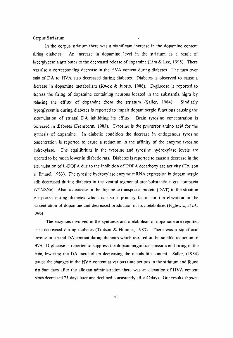
Corpus Striatum
In the corpus striatum there was a significant increase in the dopamine content
during diabetes. An increase in dopamine level in the striatum as a result of
hyperglycemia attributes to the decreased release of dopamine (Lim & Lee, 1995). There
was also a corresponding decrease in the HV A content during diabetes. The turn over
ratio of DA to RV A also decreased during diabetes. Diabetes is observed to cause a
decrease in dopamine metabolism (Kwok & Juorio, 1986). D-glucose is reported to
depress the firing of dopamine containing neurons located in the substantia nigra by
reducing the efllux of dopamine from the striatum (Saller, 1984). Similarly
hyperglycemia during diabetes is reported to impair dopaminergic functions causing .the
accumulation of striatal DA inhibiting its efllux. Brain tyrosine concentration is
decreased in diabetes (Frenstorm, 1983). Tyrosine is the. precursor amino acid for the
synthesis of dopamine. In diabetic condition the decrease in endogenous tyrosine
concentration is reported to cause a reduction in the affinity of the enzyme tyrosine
hydroxylase. The equilibrium in the tyrosine and tyrosine hydroxylase levels are
reported to be much lower in diabetic rats. Diabetes is reported to cause a decrease in the
accumulation of L-DOP A due to the inhibition of DOP A decarboxylase activity (Trulson
& Himmel, 1983). The tyrosine hydroxylase enzyme mRNA expression in dopaminergic
cells decreased during diabetes in the ventral tegmental area/substantia nigra compacta
(VTNSNc). Also, a decrease in the dopamine transporter protein (DAT) in the striatum
is reported during diabetes which is also a primary factor for the elevation in the
concentration of dopamine and decreased production of its metabolites (Figlewiz, et aI.,
1996).
The enzymes involved in the synthesis and metabolism of dopamine are reported
to be decreased during diabetes (Trulson & Himmel, 1983). There was a significant
increase in striatal DA content during diabetes which resulted in the notable reduction of
HV A. D-glucose is reported to suppress the dopaminergic transmission and firing in the
brain, lowering the DA metabolism decreasing the metabolite content. Saller, (1984)
studied the changes in the RV A content at various time periods in the stria,tum and found
that four days after the alloxan administration there was an elevation of HV A content
which decreased 21 days later and declined consistently after 42days. Our results showed
60
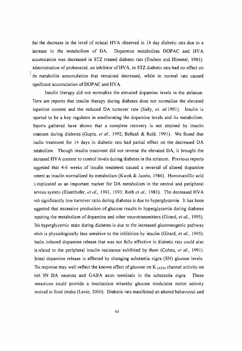
that the decrease in the level of striatal HV A observed in 1'4 day diabetic rats due to a
decrease in the metabolism of DA. Dopamine metabolites DOP AC and HV A
accumulation was decreased in STZ treated diabetic rats (Trulson and Himmel, 1983).
Administration of probenecid, an inhibitor ofHVA, in STZ diabetic rats had no effect on
I the metabolite accumulation that remained decreased, while in normal rats caused
significant accumulation ofDOPAC and HV A.
Insulin therapy did not normalize the elevated dopamine levels in the striatum.
There are reports that insulin therapy during diabetes does not normalize the elevated
dopamine content and the reduced DA turnover rate (Sally, et. al. 1991 ). Insulin is
reported to be a key regulator in ameliorating the dopamine levels and its metabolism.
Reports gathered have shown that a complete recovery is not attained by insulin
treatment during diabetes (Gupta, et a!., 1992; Bellush & Reid, 1991). We found that
insulin treatment for 14 days in diabetic rats had partial effect on the decreased DA
metabolism. Though insulin treatment did not reverse the elevated DA, it brought the
decreased HV A content to control levels during diabetes in the striatum. Previous reports
suggested that 4-6 weeks of insulin treatment caused a reversal of altered dopamine
content as insulin normalized its metabolism (K wok & Juorio, 1986). Homovanillic acid
is implicated as an important marker for DA metabolism in the central and peripheral
nervous system (Eisenhofer, et al., 1991, 1993; Roth et al., 1983). The decreased HV A
with significantly low turnover ratio during diabetes is due to hyperglycemia. It has been
suggested that excessive production of glucose results in hyperglycemia during diabetes
impairing the metabolism of dopamine and other neurotransmitters (Girard, et aI., 1995).
This hyperglycemic state during diabetes is due to the increased gluconeogenic pathway
which is physiologically less sensitive to the inhibition by insulin (Girard, et aI., 1995).
Insulin induced dopamine release that was not fully effective in diabetic rats could also
be related to the peripheral insulin resistance exhibited by them (Cohen, et aI., 1991).
Striatal dopamine release is affected by changing substantia nigra (SN) glucose levels.
This response may well reflect the known effect of glucose on K (ATP) channel activity on
both SN DA neurons and GABA axon terminals in the substantia .nigra. These
interactions could provide a mechanism whereby glucose modulates motor activity
involved in food intake (Levin, 2000). Diabetic rats manifested an altered behavioral and
61

neurochemical response suggesting a dysfunctional biosynthetic capacity for DA as a
result of a decreased neurotransmission (Ahmad & Merali, 1989). The striatal dopamine
content was elevated with a corresponding decrease in its metabolism during diabetes
was only partially restored to control levels with insulin treatment in 14day diabetic rats.
Hypothalamus
Dopaminergic action is important in the regulation of the hypothalamic-pitutary
hormone release. Hypothalamic dopamine content decreased during diabetes. Also,
dopamine and its receptors are implicated in the satiety and hunger aspects and body
weight maintenance. The central vagal connection with dopaminergic innervation is
reported to reach the pancreatic islets through the parahypothalamic ventricular (PH V)
nucleus while aderenergic and serotonergic innervations reach the pancreas through the
brain stem (Smith & Davis, 1983; Lowey, et aI., 1994). A decrease in OA in the
hypothalamus during diabetes is caused due to the reduction in the low synthetic rate of
dopamine as tyrosine levels decrease (Fernstrom, et aI., 1983; Leu, et al., 1986). Altered
dopamine is reported to affect the feeding pattern, as food intake is accompanied by OA
release which differs significantly in the hypothalamus of obese and lean Zucker rats.
The reduction in dopamine, norepinephrine and epinephrine levels in the hypothalamus
suggests a low metabolism of monoamines (Bellush & Henley, 1990). They are
responsible for the development of thermoregulatory deficits when exposed to cold
environment (Leu, er aI., 1986). Dopamine is considered as a hormone of the
hypothalamus involved in the secretion of prolactin. It has an inhibitory effect on the
release of prolactin from the anterior pituitary.
The decrease in dopamine content reduced the hypothalamic HV A with no
alterations in turnover ratio of HV A from OA. This indicates a low synthesis of
dopamine in the hypothalamus during diabetes. Insulin treatment for 14 days caused
partial improvement of the DA and HV A content. The turnover ratio was near control
values in all the groups. The decrease in OA and HV A content could be due to the
depletion of dopamine resulting in a dysfunctional biosynthetic capacity for DA during
diabetes (Ahmad & Merali, 1989; Merali, et aI., 1988). This finding bear importance
62

since hypothalamus is reported to play a role in behavionil and physiological changes
associated with diabetes.
Cerebral Cortex
Extracellular dopamine originates from DA and NE neurons in the prefrontal
cortex (PFC). Recent reports suggested that extracellular DA release in the cortex
depend on NE rather than DA innervation. In the cortex DA acts not only as NE
precursor but also as co-transmitter (Gessa et aI., 2001). The co-release of NE and DA
seems to be controlled by U2 adrenergic receptors located on NE nerve terminals.
COllical dopamine content increased with an increase in HV A content during
diabetes. But the turnover rate of HV A from DA decreased during diabetes which
indicates a decrease in the metabolism of dopamine turnover rate. Though there was a
significant increase in both DA and HV A content, the metabolismof dopamine
decreased as a result of hyperglycemia during diabetes. An increased DA and HVA
content in the CC during diabetes is reported previously (Gupta, et aI., 1992, Van, et al.,
1991). Cerebral cortical dopamine metabolism is reported to decrease because increased
glucose during diabetes (Kwok & Juorio, 1986) affects the dopaminergic activites such as
working, memory, and stress response (Tarn & Roth., 1997). During diabetes a lack of
tyrosine affect markedly the physiology and functions of these DA neurons. The overall
deficit in the availability of the precursor amino acid tyrosine which has been previously
reported has an influence in the functioning of DA neurons. Insulin treatment did not
reverse the elevated DA and HVA to control values. This in compliance with previous
reports that a complete recovery has never been attained by insulin therapy (Gupta, et aI.,
1992). The possible reason could be that during diabetes the alternative metabolic
pathways supply glucose to provide energy. These pathways are reported to be resisting
the inhibition by insulin causing only partial recovery (Girard, et aI., 1995). This
increased dopamine with a decreased turnover during diabetes in the cerebral cortex is
associated with metabolic disturbance and behavioural changes. Efflux of DA in the
prefrontal cortex is reported to stimulate hunger and food intake (Ahn & Phillips, 2002).
Diabetes is marked by hyperphagia causing excessive food intake. Hyperglycemia
causes the reuptake of DA into the brain cells which could possibly stimulate a
63

hyperosmolar state that results in dehydration causing polydypsia (Hirata, et aI., 1992).
Brain Stem
Dopamine and HVA content in the brain stem decreased during diabetes
correspondingly. A significant increase in the NE content in the brain stem from
previous reports (Task, et ai, 1992; Jackson, et aI., 1997, 1999) suggested the decrease in
DA could be because of the increased turnover to NE. This is important as the turnover
to NE causes an increased sympathetic stimulation. This has important relevance in
insulin secretion from the pancreatic islets as the increased sympathetic stimulation could
inhibit the insulin secretion.
ALTERA TIONS IN DOPAMINE AND HVA CONTENT IN THE PLASMA, AND
ADRENALS OF CONTROL, DIABETIC AND INSULIN TREATED RATS
Dopamine concentration in the plasma during diabetes decreased significantly.
This is in concurrence with earlier reports that showed that plasma DA decreased during
diabetes (Chandrashekar-Reddi, et aI., 1994). The plasma concentration of the DA is
used as an indicator of central nervous system dopaminergic activity (Esler, et aI., 1991).
This decrease in plasma DA concentration indicates that diabetes causes an alteration in
the overall dopaminergic function and activity. Peripheral plasma level of HV A, the
deaminated and o-methylated metabolite of dopamine, is often used as an indicator of
central nervous system dopaminergic activity (Esler, et al 1993). Our results show that
HV A decreased significantly underlying the decreased metabolism of DA during
diabetes. Regional HV A production is associated with the metabolism of dopamine in
sympathetic nerves and it is at a rate which appears to be influenced by sympathetic
nervous system. Also the turnover ratio of HVA from DA was also decreased during
diabetes. The decreased DA with concordant decrease in HV A and the turnover ratio has
immense importance as the plasma NE and EPI levels increased significantly. This
increase in NE and EPI levels agrees with previous reports from our laboratory and others
are due to central and peripheral increase in the sympathetic stimulation during diabetes
(Jackson, et aI., 1997; ChaoulotT, et aI., 1990a; Chandrashekar-Reddi, era!., 1994). In 14
day diabetic rats the turnover ratio of NE from DA also decreased as there was a
significant decrease in DA. The activity of dopamine-~-hydroxylase (DBH) is reported
6~

to be increased in blood from diabetic rats (Berkowitz & Head, 1978). Thus, the
increased activity of DBH causes an increased conversion of DA to NE which triggers
the sympathetic nervous system. Insulin treatment for 14 days showed improvements in
the concentrations of DA, NE, EPI and HV A. The turnover ratio of HV A from DA was
restored to control values reflecting changes in brain DA metabolism.
The dopamine and HV A contents in the adrenals decreased significantly
indicating an overall decrease in the metabolism of DA. Increased NE and EPI content in
the adrenal medulla during diabetes is observed as a result of the decreased dopamine
content. Most of the NE released is efficiently removed by neuronal and extraneuronal
uptake (Eisenhofer, et al., 1992). Evidences suggest that in the periphery DA serves not
only as a precursor for active compounds released from sympathetic nerves and the
adrenal medulla is suggested to act as an autocrine or paracrine regulator of local organ
function (Eisenhofer, et aI., 1995). The increase in the sympathetic tone is because of the
increase in the NE and EPI levels in diabetic rats. Reports show a decrease in the NE
levels in the adrenal medulla during diabetes (Patel, et al., 1997). We observed a
decreased turnover of NE from DA during diabetes which could be as a result of
degeneration of the adrenals. A decreased turn over is being reported in the adrenal
medulla as a result of its degeneration during diabetes (Patel, et al., 1997). Also, it has
been suggested that the sympathetic tone is differentially altered in the peripheral tissues
and in the adrenals there is a decreased turnover during diabetes (Patel, et aI., 1997).
Thus a decreased DA and HVA content with an increased NE and EPI levels during
diabetes increased the synpathetic stimulation. At the same time 14 day streptozotocin
diabetes indicated a lower turnover of NE from DA suggesting an overall damage in the
sympatho-adrenal system causing peripheral neuropathy.
Brain Dopamine receptor alterations during diabetes
Diabetes mellitus is often accompanied with emotional, behavioral, mood
disturbances and centrally mediated neurological complications (Salkovic & Lackovic,
1992). Striatal dopamine receptors were markedly decreased with no change in affinity
during diabetes with the accumulation of DA in the striatum and a decreased HV A
metabolism. Striatal dopamine firing during diabetes is decreased affecting
65

dopaminergic functions (Sailer, 1984). The decreased dopamine receptor density during
diabetes is related to the decreased locomotor activity in STZ-induced diabetic rats
(Kobayashi, et aI., 1990; Shimomura, et aI., 1990). This finding correlates with our
present data suggesting that the disturbances in the central dopaminergic receptors during
STZ- induced diabetes affects dopamine related functions. An increase in DA receptors
is reported to cause an increased DA receptor sensitivity during diabetes (Lazovsky, et
al., 1981) due to long term blockade of D A receptors or lesions in the striatal D A
receptors. The firing of DA neurons projecting from the substantia nigra to the striatum
is reported to be rapidly suppressed by hyperglycemia leading to the hypofunction of
dopamine receptors (Sailer, 1984).
The decreased DA receptors during diabetes that we report in the striatum is a
major cause in affecting dopamine related functions. There are hypothesis that suggests
activities related to the functional capacities of DA receptors like stereotypy, ambulation,
behaviour are diminished due to hyperglycemia (Lazovsky, -et ai., 1981). Also a decrease
in DA receptors during diabetes may result in hyporesponsiveness (Kamei, et aI., 1998).
It is suggested that in alloxan treated rats with the onset of diabetes causes metabolic
changes such as weight loss and dehydration are reported to occur which modify the DA
metabolism (Omar, et al., 1985).
Insulin treatment effectively restored the decreased density to control levels but
there was a decrease in the affinity of the receptors. The decrease in the affinity of the
receptors during insulin treatment may be a compensatory mechanism in restoring the
aecreased dopaminergic function to normal state which is in compliance with previous
reports.
The dopamine receptors in the hypothalamus did not alter in number during
aiabetes but there was a decrease in affinity in both Scatchard and displacement analysis.
The Log (EC50) value in diabetes increased with an increase in Ki suggesting a decrease
In affinity state. Studies based on the reports from the anterior pituitary of alloxan treated
aiabetic rats showed no significant changes in the DA receptors and there was no
modification in the binding affinity (Omar, et al., 1985). The pancreatic vagal motor
neurons receive dopaminergic input from paraventricular nucleus of hypothalamus
ILowey, et al., 1994). This demonstrates the importance of CNS dopamine in the
66
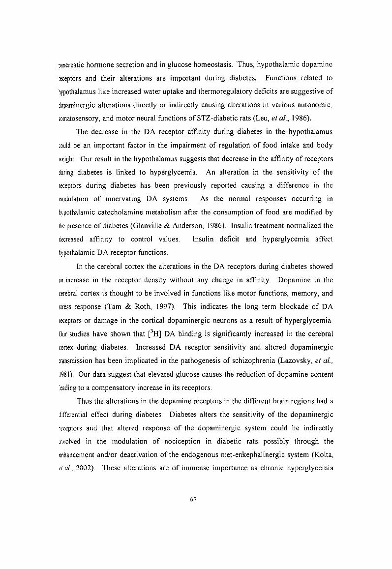
pancreatic honnone secretion and in glucose homeostasis. Thus, hypothalamic dopamine
receptors and their alterations are important during diabetes. Functions related to
hypothalamus like increased water uptake and thermoregulatory deficits are suggestive of
dopaminergic alterations directly or indirectly causing alterations in various autonomic,
somatosensory, and motor neural functions of STZ-diabetic rats (Leu, et ai., 1986).
The decrease in the DA receptor affinity during diabetes in the hypothalamus
could be an important factor in the impairment of regulation of food intake and body
weight. Our result in the hypothalamus suggests that decrease in the affinity of receptors
during diabetes is linked to hyperglycemia. An alteration in the sensitivity of the
receptors during diabetes has been previously reported causing a difference in the
modulation of innervating DA systems. As the normal responses occurring in
hypothalamic catecholamine metabolism after the consumption of food are modified by
Ihe presence of diabetes (Glanville & Anderson, 1986). Insulin treatment normalized the
decreased atlinity to control values.
hypothalamic DA receptor functions.
Insulin deficit and hyperglycemia affect
In the cerebral cortex the alterations in the DA receptors during diabetes showed
an increase in the receptor density without any change in affinity. Dopamine in the
cerebral cortex is thought to be involved in functions like motor functions, memory, and
stress response (Tarn & Roth, 1997). This indicates the long term blockade of DA
receptors or damage in the cortical dopaminergic neurons as a result of hyperglycemia.
Our studies have shown that eH] DA binding is significantly increased in the cerebral
cortex during diabetes. Increased DA receptor sensitivity and altered dopaminergic
transmission has been implicated in the pathogenesis of schizophrenia (Lazovsky, et al.,
1981). Our data suggest that elevated glucose causes the reduction of dopamine content
leading to a compensatory increase in its receptors.
Thus the alterations in the dopamine receptors in the different brain regions had a
ditferential etfect during diabetes. Diabetes alters the sensitivity of the dopaminergic
receptors and that altered response of the dopaminergic system could be indirectly
involved in the modulation of nociception in diabetic rats possibly through the
enhancement and/or deactivation of the endogenous met-enkephalinergic system (Kolta,
l'/ £11., 2001). These alterations are of immense importance as chronic hyperglycemia
67
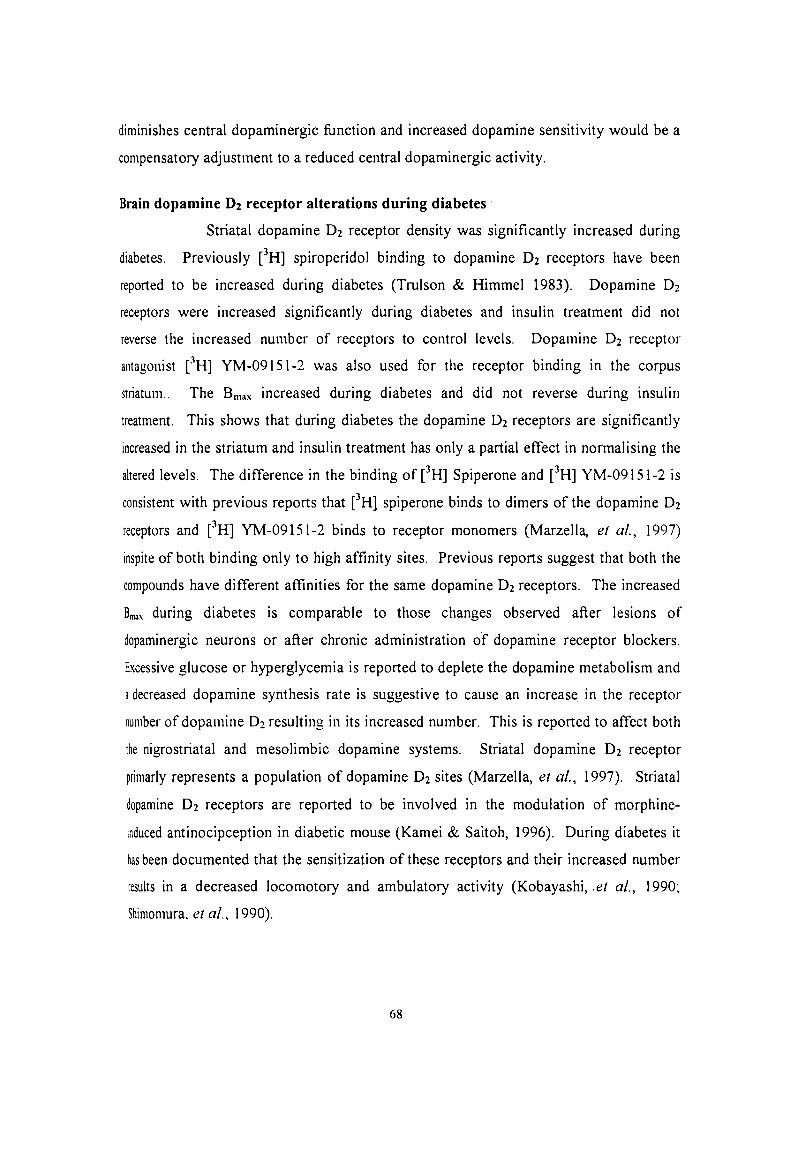
diminishes central dopaminergic function and increased dopamine sensitivity would be a
compensatory adjustment to a reduced central dopaminergic activity.
Brain dopamine D2 receptor alterations during diabetes'
Striatal dopamine D2 receptor density was significantly increased during
diabetes. Previously eH] spiroperidol binding to dopamine D2 receptors have been
reported to be increased during diabetes (Trulson & Himmel 1983). Dopamine O2
receptors were increased significantly during diabetes and insulin treatment did not
reverse the increased number of receptors to control levels. Dopamine O2 receptor
antagonist eH] YM-091S1-2 was also used for the receptor binding in the corpus
striatum.. The Bma .... increased during diabetes and did not reverse during insulin
treatment. This shows that during diabetes the dopamine O2 receptors are significantly
increased in the striatum and insulin treatment has only a partial effect in normalising the
altered levels. The difference in the binding of eH] Spiperone and eH] YM-091S1-2 is
consistent with previous reports that eH] spiperone binds to dimers of the dopamine O2
receptors and eH] YM-091S1-2 binds to receptor monomers (Marzella, et al., 1997)
inspite of both binding only to high affinity sites. Previous reports suggest that both the
compounds have different affinities for the same dopamine O2 receptors. The increased
Bmax during diabetes is comparable to those changes observed after lesions of
dopaminergic neurons or after chronic administration of dopamine receptor blockers.
Excessive glucose or hyperglycemia is reported to deplete the dopamine metabolism and
a decreased dopamine synthesis rate is suggestive to cause an increase in the receptor
number of dopamine O2 resulting in its increased number. This is reported to affect both
the nigrostriatal and mesolimbic dopamine systems. Striatal dopamine O2 receptor
primarly represents a population of dopamine O2 sites (Marzella, et al., 1997). Striatal
dopamine O2 receptors are reported to be involved in the modulation of morphine
induced antinocipception in diabetic mouse (Kamei & Saitoh, 1996). During diabetes it
has been documented that the sensitization of these receptors and their increased number
results in a decreased locomotory and ambulatory activity (Kobayashi, .et aI., 1990;
Shimomura, et aI., 1990).
68

Dopamine through its dopamine Dl receptor stimulates adenylyl cyclase and
inhibits adenylyl cyclase activity through its dopamine D2 receptors. Dopamine DJ
stimulated cAMP production was markedly increased in diabetic rats, whereas ability of
dopamine D2 receptor action to reduce cAMP formation was almost abolished during
diabetes (Gorio, et al., 1989). An imbalance between Gs -proteins and GilGo protein
mediated efficacy of Gs activity as a result of the loss of Gi/Go inhibitory functions has
been found in the striatum and other tissues of diabetic animals (Salkovic & Lackovic,
1992). Doapmine D2 receptors exert their function activating Gi proteins in the brain.
Regulation of the inhibitory G protein-calcium channel complex involves pertusis toxin
(PTX) sensitive and insensitive G proteins (Wiley, et al., 1998). Concomitantly with
such transductional alteration detected in chronic diabetes, caused a marked increase of
the striatal content of met-enkephalin, which is known to utilize Gi/Go proteins for
inhibition of adenylyl cyclase. Diabetes causes the activation of mitogen activated
protein kinase (MAP kinase) p38 as an early step in the signal pathway to dysfunction in
experimental diabetic neuropathy (Agthong & Tomlinson, 2002). Increased content of
dopamine and elevated Bmax of dopamine D2 receptors in the corpus striatum could cause
a transductional defect in diabetic animals leading to diabetic neuropathy.
Diabetic neuropathy is the most common secondary complication of
diabetes mellitus. Evaluation of the effect of levosulpiride, a selective antagonist for O2
dopamine receptors, on the glycemic control of IDDM performed on 40 long-standing
subjects with clinical signs of autonomic neuropathy and delayed gastric emptying
improved glycemic control (Prando, et aI., 1997). The effect of bromocriptine, a potent
dopamine D2 receptor agonist on intraocular pressure in diabetic patients with autonomic
neuropathy revealed that it exerts an ocular hypotensive action through presynaptic
dopamine receptors (Gale, et aI., 1991).
Dopamine D2 receptor gene expression increased in the striatum during diabetes
as a result of the decreased transmission of dopamine. Hyperglycemia depresses the
dopaminergic function and firing. Therefore a decreased dopaminergic activity is always
suggested to increase the dopamine D2 receptors. An increase in the. expression of
dopamine D2 receptors gene results in the increased number.
Dopamine D2 receptor cDNA is described to exist in two isoforms (short and
69
long) as a result of alternative splicing of the same gene that encodes for the receptor.
They are represented as D2s and D2L. The longer form designated as D2L is the
predominant form, although there is some variability among brain regions in the relative
proportions of the two forms (Higgins, et al., 1991; Sealfon, et aI., 1991, Todd, et ai.,
1996). Our RT-PCR data showed that the long D2L form expressed in all conditions and
showed increase in expression during diabetes and insulin treatment. A lesion in the
striatum is reported to increase the expression of dopamine D2L receptor gene (Zang, et
aI., 1994).
In the cerebral cortex also we report an increase in the Bmax of dopamine O2
receptors with out any change in the affinity during diabetes. It is suggested that chronic
treatment with selective dopamine D i or dopamine O2 receptor blockers induces a
receptor-specific increase or decrease of DA receptors (Spano, et al., 1987). There fore
hyperglycemia is reported to bring about an increase in the number of dopamine O2
receptors during diabetes.
The dopamine neurons projecting to the prefrontal cortex (PFC) are thought to
be involved in various motor and behavioural functions (Tarn & Rbth, 1997). This
increased number of dopamine receptors could account for the behavioural
supersensitivity to dopamine agonist as a result of damage in the dopamine functions
(Cresse, et ai., 1976). Diabetes mellitus is also reported to be one important factor for
tardive dyskinesia caused by the chronic treatment with neuroleptic antipsychotic drugs
which exert their action through the dopamine O2 receptors (Meltzer, et ai., 1996). The
increased binding of dopamine 02 receptors in the cerebral cortex with no change in
aftlnity during diabetes has a relevance to the alterations in dopaminergic homeostasis
affecting its function.
The dopamine O2 receptor mRNA in the cerebral cortex increased during diabetes
and remained high after the treatment with insulin. Our receptor studies show an increase
in the receptor number with a decrease in affinity. Therefore such a receptor expression
pattem in the cerebral cortex may be due to differential translational regulation of the
dopamine D2 receptor mRNA. Cortical dopamine D2 receptor expression has never been
previously reported in diabetes. Dopamine D2L receptor mRNA expression was
increased during diabetes in the cerebral cortex. Lesions in the corpus striatum is
70

reported to cause an increased expression of long isoform of dopamine D2L receptor
mRNA (Higgins, et al., 1991, Sealfon, et al., 1991; Todd, et al., 1996).
Thus, dopamine O2 receptors during diabetes are increased in the striatum and
cerebral cortex with an accumulation of DA. Dopamine D2 receptors are reported to
regulate the release of dopamine from dopaminergic neurons originating in the ventral
tegmental area as well as in the substantia nigra (Stoof, et aI., 1987). Hyperglycemia
during diabetes could damage the DA D2 receptors, decreasing the DA related functions
in the striatum and other brain regions.
In the hypothalamus of diabetic rats the binding of eH] YM-091S1-2 to dopamine
D~ receptors decreased significantly with an increase in affinity. The regional difference
in the receptor status is relevant to the role which dopamine plays during various
physiological and behavioural activites. Unis, et al., (1998) reported that eH] YM-
09151-2 binds to the dopamine D2 high affinity receptors. The decrease number of
dopamine O2 receptors in the hypothalamus could result in the sensitization of its
receptors leading to a shift into the higher affinity state. In the intra lateral hypothalamic
area (lntra-LHA) blockade of dopamine O2 receptors by specific antagonist in tumor
bearing (TB) and non tumor bearing (NTB) rats increased food intake indicating the
involvement of dopamine O2 receptors in feeding mechanisms (Zhang, et al., 2001).
Thus during diabetes the decrease in dopamine O2 receptor number or Bmax could disturb
hypothalamic functions. Impairment of dopamine O2 receptor is an important factor that
leads to hyperphagic and polydypsic condition as DA participates in regulating meal size
(Oler, et al., 1997). Dopamine -acetylcholine (DA-Ach) interaction within the lateral
hypothalamus (LH) is involved in the regulation of locomotion, feeding behaviour and
reinforcement (Hoebel, et al., 2000, Baptista, et al., 1990). The cholinergic stimulation
of these activities is regulated by DA through D2 receptors in the hypothalamus. Thus
dopamine in the hypothalamus is related to sensory input, feeding reflexes, food reward
or memory processes (Hernandez & Hoebel, 1988). In the hypothalamus co
administration of dopamine D) and dopamine O2 agonists inhibit the feeding effect
mediated by the action on neuropeptide Y (NPY) (Kuo, 2002). This is effective in the
reduction of food intake in diabetic rats, revealing the efficiency of dopamine D)/ O2
agonist in the improvement of hyperphagia in diabetic animals. Dopamine O2 receptor
71

mRNA expression was in concordant with the receptor data showing a decrease in the
expression of mRNA during diabetes and insulin treatment in diabetic rats caused an
increase in the expression when to control levels. A decrease in dopamine O2 receptors
are reported in obese Zucker rats which contribute to the specific feeding pattern in obese
rats represented by an increased meal size and decreased meal number (Zhang, et al.,
1002). Insulin is reported to regulate the re-uptake of catecholamine transporters.
lntracerebroventricular injection of insulin is reported to cause an increased mRNA
expression of OAT (Figlewicz, et aI., 1994). We report an increased expression of
dopamine D2 receptor mRNA during insulin treatment in diabetic rats. Our receptor
analysis in the hypothalamus showed that insulin treatment did not fully restore the
decreased receptor number and increased affinity to control level. The increased
expression during insulin treatment in the hypothalamus could be a mechanism to
normalise the decreased number to control levels. Thus, decrease in dopamine D2
receptors in the hypothalamus due to lesions rising as a result ofhyperglycemia.
Brain stem is an important part of the brain in monitoring the glucose status and
the regulation of feeding (Guillod, et a/., 2003). In the brain stem also there was a
significant decrease in the dopamine O2 receptor density with a decrease in the affinity
resulting in an overall down regulation of the receptor. Brain stem dopamine O2
receptors have never been reported previously. During diabetes the significant increase
in NE and EPI (Tasaka, et ai, 1992; Jackson, et a/., 1997; 1999) could bind to a2
adrenergic receptors increasing the sympathetic nerve discharge could inhibit insulin
secretion from the pancreatic islets. From our data we suggest that the increased
activation of sympathetic stimulation during diabetes as a result of increased NE and EPI
is because of a decreased OA content in the brain stem with a decrease in the down
regulation of dopamine D2 receptors. This down regulation of dopamine 02 receptors in
the brain stem could have a possible role in the regulation of insulin secretion by
releasing EPI and NE from the adrenal medulla that leads to the inhibition of insulin
secretion in the pancreas. In the brain stem there was a decrease in the expression of
dopamine O2 receptor rnRNA as a result of diabetes which increased further on insulin
treatment. This could be due to the differential transcriptional regulation during diabetes.
Insulin treatment brought the Bmax to control values but there was an increase in affinity
72
of the receptors. Insulin is reported to have a modulatory effect on eNS dopamine and
insulin injection is suggested to cause an increase in dopaminergic function (Figlewicz, et
al., 1996, 1994). It has been reported that damages in the brain can cause an alterations
in the expression of the dopamine D2L isoform which is expressed in the in vivo condition
(Higgins et aI., 1991; Sealfon, et al., 1991; Todd et al., 1996).
Hypothalamic- pituitary -axis and dopaminergic functions during diabetes
The hypothalamus is involved in the monitoring of glucose status and the
regulation of feeding and hunger (Guillod, et al., 2003). Dopamine content during
diabetes decreased in the hypothalamus with a decrease the number of dopamine O2
receptors and corresponding decrease in the dopamine O2 receptor gene expression.
Diabetes activates the HP A axis producing (Mohan Kumar, et al., 2003) a marked
Increase 111 food intake and water intake which is completely reversed by insulin
treatment. Dopaminergic neurons are the direct targets for insulin action which
participate in the reward seeking behaviour (Figlewicz, et al., 2003). Therefore during
diabetes the decreased availability of dopamine could affect these functions. We report a
decreased dopamine content and dopamine 02 receptors in the hypothalamus during
diabetes. This could decrease dopaminergic signalling in the hypothalamus. Diabetes is
reported to decrease the dopamine transporter thus reducing the dopaminergic signaling
affecting dopamine related functions (Galli, et al., 2002). Dopamine O2 receptor
disruption is reported to impair body growth and the somatotroph population (Becu
Villalobos, et al., 2002). We report a decreased bmax of dopamine O2 receptor during
diabetes which could impair the body growth. Thus, a decreased dopamine with a
decreased dopamine D2 receptor expression and receptor number in the hypothalamus as
a result of diabetes affects the metabolic functions of the hypothalamus.
Dopaminergic alteration during diabetes in the pancreas and its significance in
glucose metabolism
Pancreatic islets are sited to contain dopamine in the secretory granules along
with serotonin and calcium (Ahren & Lundquist, 1985). There was a significant decrease
in the turnover ratio of HVA from DA in the pancreas during diabetes. Pancreas is an
important source of non-neuronal dopamine in the body and that this dopamine has a role
73
in protecting the intestinal mucosa (Hoffman, et al.,' 1996 Early sympathetic islet
neuropathy (eSIN) is reported to occur selectively in the islet during diabetes in diabetic
rats (Taborsky, et al., 2002) as a result of monoamine alterations. Increased NE in the
islets could possibly be because of the increased uptake and decreased degradation. The
increased NE and EPI content with a decreased DA and HV A levels in the adrenal
medulla anf plasma during diabetes was observed. Most of the NE released is efficiently
removed by neuronal and extraneuronal uptake (Eisenhofer et al., 1992). Evidences
suggest that in the periphery DA serves not only as a precursor for active compounds
released from sympathetic nerves and the adrenal medulla but also is suggested to act as
an autocrinel paracrine regulator of local organ function (Eisenhofer, et al., 1995). The
central nervous system cell groups project into the pancreatic vagal motor neurons
receive adrenergic, noradrenergic and serotonergic inputs from the lower brain stem and
a dopaminergic input from paraventricular nucleus of hypothalamus demonstrating the
importance of CNS dopamine in the pancreatic hormone secretion and glucose
homeostasis (Lowey, et aI., 1994). Thus, dopamine content in the pancreas showed
marked decrease with a decrease in its metabolism. This could be related to the
sympathetic tone that is increased during diabetes as a result of an increased NE and EPI
levels in the plasma, pancreas and the adrenals. The metabolic clearance rate ofDBH is a
major factor accounting for the increase in DBH activity in the streptozotocin-diabetic rat
(Stolk JM, et aI., 1982, (Watanabe & Nagatsu, 1991). Hyperglycemia could possibly
oecrease the metabolism of DA present in the pancreatic secretory granules that would
affect the pancreatic islet function. As findings suggest that an endogenous alterations in
these hypothalamic monoamines may contribute to islet dysfunction, which is
characteristics of diabetes (Cincotta, et al., 1999).
The dopamine receptors showed an increased Bmax during with a decrease in its
affinity in the pancreatic islets. This was confirmed by both Scatchard and displacement
analysis. The decreased dopamine content and metabolism as a result of hyperglycemia
could be a cause of the increase in the receptor parameters in the pancreatic islets causing
the sensitization of these receptors. Pancreatic dopamine receptors havenoJ been a focus
of studies till date except for a few reports (lmamura, et al., 1990). It has been observed
that the sympathetic alpha receptor and dopamine 0 1 was distributed on the B-cells, the
7~
sympathetic ~2 receptors on the D-cell and the dopamine D2 on the varicosity of the
sympathetic ~2 neuron. Dopamine binding sites are in th,e pancreatic arcinar cells are
suggested to be receptors that mediate the action of dopamine on cAMP accumulation
(Ribet, et al., 1986). Studies in the past indicate that dopamine directly affects pancreatic
islet Band D cell function. Dopamine suppresses somatostatin secretion predominantly
through activation of dopaminergic receptors, whereas it Sl,lppresses insulin release
through an alpha adrenergic mechanism and stimulates glucagon release through a p
adrenergic mechanism (Gerich, et ai" 1982). Dopamine acts on specific dopamine
receptors related to the exocrine pancreatic secretion and sulpiride was found to be a
potent dopamine antagonist in the canine exocrine pancreas (Honda, et al., 1980). The
increased dopamine receptors in the pancreatic islets could be as a result of the decreased
dopamine turnover and as a result of the increased adrenergic activity that is damaged
during diabetes.
Binding of eH] YM-09IS1-2 for dopamine D2 receptors in the pancreatic islets
decreased significantly with an increase in affinity. The Log (ECso) value during diabetes
decreased with an increase in affinity (Table 32, Fig.-34). This is similar to what we
obtained in the hypothalamus for dopamine D2 receptor. The dopamine D2 receptors in
the pancreatic islets demonstrate a down regulation in receptor number with an increase
in affinity. The damage caused as a result of hyperglycemia with an increased
sympathetic stimulation in the pancreatic islets could be a possible cause for the
decreased activity of dopamine D2 receptors in the pancreatic islets during diabetes.
Dopamine D1 receptor alterations will possibly have an effect on the pancreatic islet
population causing a decrease in insulin secretion.
Alterations in the dopamine and dopamine D2 receptors during diabetes
Dopamine receptors decreased significantly in the striatum while the dopamine D2
receptors increased in the number. In the cerebral cortex, dopamine DA and dopamine
D~ receptors showed an increase and the affinity of DA receptors decreased. The
hypothalamic dopamine DA receptor number did not alter but there was a decrease in
aftinity while the dopamine D2 receptors decreased with an increase in affinity. In the
brain stem the dopamine D2 receptors decreased with a decrease in affinity showing an
75
overall down regulation. Thus from our study we conclude that the altered dopamine
receptors and dopamine O2 receptors binding observed in brain region of diabetic rats
increases the sympathetic stimulation. Altered dopamine is reported to mediate an
increased sympathetic nerve discharge (Bauhelal & Mir, 1993). During diabetes in the
pancreatic islets the decrease in dopamine and its turn over increased dopamine receptors.
The dopamine D2 receptors decreased with an increase in affinity. The overall decrease
in the dopaminergic function is as a result of the the increased EPI and NE release from
the adrenals into circulation and pancreas could lead to an inhibition of insulin release.
Effect of norepinephrine on dopamine uptake in the pancreatic islets.
Dopamine is stored in the secretory granules of the pancreatic islets along with
serotonin and calcium (Ahren & Lundquist, 1985). The uptake studies using CH] DA in
the pancreatic islets revealed that DA uptake was maximum in the presence of 10-4 CH]
DA in both the concentrations of glucose (4 & 20mM) and the uptake decreased with a
decrease in DA concentrations. These results indicate that in the presence of glucose " there is an uptake of DA into the pancreatic islets. At high concentrations DA is always
taken up into the pancreatic islets in both the concentrations of glucose. This could have
an implication in insulin secretion as high concentrations of DA in the presence of
glucose stimuli causes a reduction in insulin secretion. Dopamine is reported to modulate
insulin secretion in the pancreatic islets with changes in calcium efflux (Carpinellie, et
aI., 1994). Possibly a high DA concentration in the islets is essential in maintaining the
equilibrium during insulin secretion. The function of islet ~ cells is controlled by a
glucose sensor that operates at physiological glucose concentrations and acts in synergy
with signals originating from hypothalamic neurons. Evidence exists that the extra
pancreatic cells producing and secreting these neuro endocrine signals also exhibit a
glucose sensor and an ability to integrate nutrient and neuro hormonal messages
(Pipeleers, et aI., 2001). From our uptake studies in the pancreatic islets we suggest that
the DA is involved in glucose induced insulin secretion.
We observed that NE at low concentration did not have any effect on the CH]
DA uptake while at high concentration inhibited the uptake of DA in the presence of
4mM and 20mM glucose. Thus, high concentration of NE blocked the uptake ofDA into
76
the pancreatic islets and this could affect the role of DA in glucose induced insulin
secretion. Increased NE level is reported to inhibit the pancreatic islet function (Sheen, et
al.. 2001). The blockade of DA into the pancreatic islets by NE is as a result of its
increased uptake by neuronal and extraneuronal tissue which causes the inhibition of
insulin secretion. The following points are inferred from our uptake studies
1) Dopamine transport into the islets requires glucose and high concentrations ofDA
prevent glucose transport into the pancreatic islets.
2) Dopamine in the secretory granules of the pancreatic islets could be one of the
possible elements that operate at physiological glucose concentrations. It acts in
synergy with signals that integrate messages originating from hypothalamic
neurons and pancreas and damage to this could be a possible cause of the
inhibition of insulin secretion during diabetes.
Effect of dopamine on glucose induced insulin secretion in vitro
Dopamine in the presence of glucose had a dose dependent effect on
insulin secretion. 'We observed that low concentrations ofDA increased glucose (20mM)
induced insulin secretion while high concentration caused the maximum inhibition.
Dopamine iat high concentration s reported to inhibit insulin secretion from the islets
(Carpinelli, et al., 1994). Also, high concentrations of norepinephrine, dopamine, and
serotonin in the pancreatic islets are reported to decrease glucose-stimulated insulin
secretion (Feldman, et al., 1980).
We observed that butaclamol, antagonist for dopamine receptors blocked the
inhibitory and the stimulatory effect of DA in the pancreatic islets mediated. The
addition of sulpiride a potent dopamine D2 receptor antagonist to the pancreatic islets
etJectively blocked the dopaminergic action on insulin secretion. In previous studies
from our laboratory reported that addition of forskolin an activator of cAMP resulted in
overcoming the effect ofDA on insulin secretion (Abraham, 1998).
Dopamine D2 receptors agonists bromocriptine (BRC) and 7 -OH-DP AT were
used to study their effect on glucose induced insulin secretion in the pancreatic islets ill
\'itro. Bromocriptine a potent dopamine D2 agonist at low concentrations stimulated
glucose induced insulin secretion in the presence of 20mM glucose while in high
77

concentrations had an inhibitory effect. The stimulation by BRC at its low concentration
was not as effective in the presence of 4mM glucose. It has been reported previously that
BRC treatment in hyperglycemic state had a strong stimulatory response to insulin
secretion (Oliveira, et aI., 1998). The agonists of dopamine by acting through the
neuroendocrine system Improves peripheral energy metabolism and impaired islet
function. (Lang, et aI., 1998). 7-0H DPAT showed an inhibitory effect on glucose
induced insulin secretion. Previous reports suggest that 7-0H DPAT induced
hyperglycemia decreased insulin secretion (Hillegaart, et al., 1996). In vitro studies
confirmed the stimulatory role of dopamine D2 receptors on insulin secretion.
Norepinephrine is reported to have an antagonist effect on insulin secretion in the
pancreatic islets (Porte, et a!., 1967). Low concentration of NE did not affect the
stimulatory effect ofDA on insulin secretion while high concentrations of NE was found
to be inhibitory. It has been previously reported that high concentrations of NE inhibited
the glucose induced insulin secretion (Zren, et aI., 1980). During diabetes there is an
increased neuronal and extra neuronal uptake of NE that increases the sympathetic "
stimulation (Eisenhofer, et al., 1992). This blocks the insulin secretion as the increased
sympathetic tone elevates peripheral insulin resistance and hyperglycemia. Thus, our ill
vitro results show that increased concentrations of NE blocked the stimulatory effect of
low concentrations DA.
Our ill vitro studies show that low concentration of dopamine is necessary in the
stimulation of insulin by glucose and this is mediated through the dopamine D2 receptors
in the pancreas. We report a decrease in the metabolism of DA with differential
alterations in the dopamine DA and D2 receptors in the brain and pancreas during
diabetes. The decreased dopaminergic tone with a high turnover to NE and EPI results in
an increased sympathetic stimulation decreasing the ~-cell responsiveness to
parasympathetic stimulation to secrete insulin. The increased NE not only blocks the
uptake of DA but also inhibits its stimulatory effect on insulin secretion. Dopaminergic
dysfunction is an important factor during diabetes which not only affects the central
functions but also is a cause for the decreased insulin secretion from the pancreatic islets.
71\