eukaryotic cell replication. the eukaryotic genome the genomes of eukaryotes are much more complex...
TRANSCRIPT
Eukaryotic Cell
Replication

The Eukaryotic Genome• The genomes of eukaryotes are much more complex than those of
prokaryotes. Unlike the genomes of prokaryotes which contain a single chromosome, the genomes of eukaryotes are composed of multiple chromosomes, each containing a linear molecule of DNA.
• Although the number and sizes of chromosomes can vary considerably between species, the basic structure is the same in all eukaryotic cells. The DNA is tightly bound to small basic proteins, called histones, that package the DNA in an orderly way within the nucleus.
• In a human cell the total extended length of DNA is nearly 2 m. However, this DNA must fit into a nucleus with a diameter of only 5 to 10 μm. So in order to fit the entire genome into a eukaryotic cell, the DNA must be very tightly bound around the histones.

Nucleosomes• The complex formed between
eukaryotic DNA and proteins are called chromatin, which typically contains about twice as much protein as DNA. The major proteins found in chromatin are histones, which are small proteins that facilitate binding to the negatively charged DNA molecule. There are five major types of histones (i.e. H1, H2A, H2B, H3, and H4), which are very similar among different species of eukaryotes.
• The basic structural unit of chromatin is the nucleosome, which is a repeating unit comprising 166 base pairs, wrapped twice around a histone core and held in place by a H1 linker histone.

Chromatin Formation• The nucleosomes are
separated by a region of linker DNA that varies from 0 to 80 base pairs in length. Under an electron microscope the nucleosomes appear as 11 nm ‘beads’ on a 2 nm DNA ‘string’.
• The histone H1 molecules cause the nucleosomes to be further condensed into 30 nm thick fibres. This acts as a scaffold onto which loops are formed. These loops are thought to form transcriptional units.

The Arrangement of Chromatin• Although chromatin is often shown forming ‘X’
chromosomes (as in the picture opposite), the extent of chromatin condensation varies during the life cycle of the cell. During the interphase, when the cells are not replicating, most of the chromatin (called euchromatin) is relatively decondensed and is distributed throughout the nucleus. Euchromatin contains the DNA which is active and whose genes can be transcribed. In contrast to euchromatin, about 10% of interphase chromatin (called heterochromatin) is in a highly condensed state resembling the chromatin of cells undergoing mitosis. Heterochromatin is transcriptionaly inactive.
• As cells enter mitosis, their chromosomes become highly condensed so that they can be distributed to daughter cells. The loops of the chromatin fibres further fold upon themselves to form the compact metaphase chromosomes (see picture opposite) of mitotic cells.

Relationship between DNA and Chromosomes

Human Chromosomes
• During mitosis (i.e. cell division) the chromatin fibres condense to form visible ‘X’ chromosomes.
• The human genome comprises 23 pairs of chromosomes.
• In 22 pairs the chromosomes look alike (i.e. both ‘X’ chromosomes). However, in the 23rd pair (i.e. the sex chromosomes) the chromosomes only look alike in females.
• In the 23rd pair of chromosomes, females have an ‘XX’ pair, while males have an ‘XY’ pair.

Chromosomal Structure
1. Telomere
2. Centromere
3. p arm (short arm)
4. q arm (long arm)
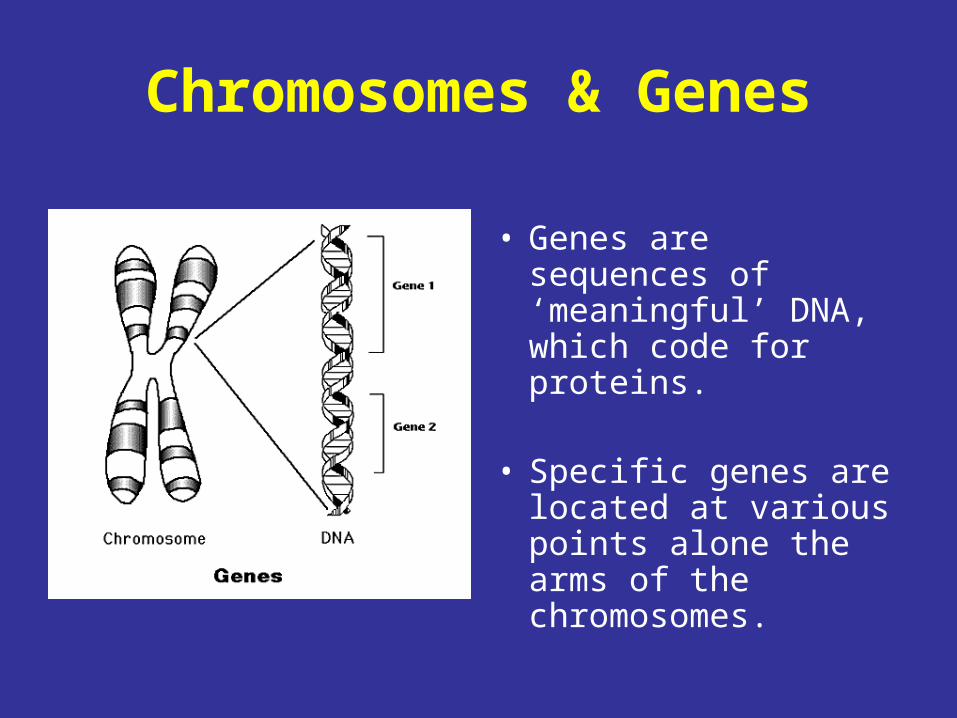
Chromosomes & Genes
• Genes are sequences of ‘meaningful’ DNA, which code for proteins.
• Specific genes are located at various points alone the arms of the chromosomes.
The Cell Cycle
In eukaryotes, the cell cycle is complex and consists of several discrete phases. Although cell growth is usually a continuous process, DNA synthesis only occurs during one phase of the cell cycle. The replicated chromosomes are then distributed to daughter cell by a complex series of events which precede cell division. Progression between the various stages of the cell cycle is controlled by a regulatory system, which coordinates the different events of the cell cycle, and also links the cell cycle with extracellular signals that control cell proliferation.

Mitosis, Cytokinesis & the Interphase
• The cycle of an eukaryotic cell can be divided into two basic parts: mitosis and the interphase. Mitosis is the stage in which nuclear division takes place. It involves separation of the daughter chromosomes and ends up with cell division (a process which is known as cytokinesis). The interphase the stage in which cell growth occurs and DNA synthesis takes place.
• Approximately 95% of the cell cycle is spent in the interphase. During the interphase, the chromosomes decondense and distributed throughout the nucleus, so the nucleus appears morphologically uniform. At the molecular level, however, during the interphase both cell growth and DNA replication occur in preparation for cell division.

Cell Cycle
• Mitosis: Nuclear division takes place. Mitosis comprises four phases, the prophase, the metaphase, the anaphase, and the telophase.
• Cytokinesis: Separation of the two daughter takes place.
• G1 Gap: Cell growth occurs and each cell contains two copies of each chromosome.
• S Phase: DNA replication takes place.
• G2 Gap: Cell growth occurs and each cell contains four copies of each chromosome.
Mitosis
Cytokinesis
G1 First Gap
G2 Second Gap
S phase
Inte
rph
ase
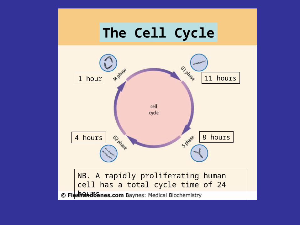
The Cell Cycle
11 hours
8 hours4 hours
1 hour
NB. A rapidly proliferating human cell has a total cycle time of 24 hours

Eukaryotic Cell
Cytoplasm
NB. Cytoskeleton not shown
The centrioles play an important role in mitosis
The Centrioles
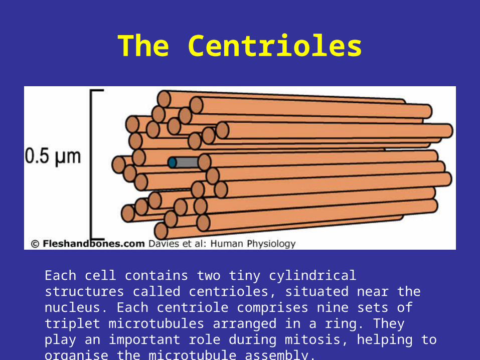
Each cell contains two tiny cylindrical structures called centrioles, situated near the nucleus. Each centriole comprises nine sets of triplet microtubules arranged in a ring. They play an important role during mitosis, helping to organise the microtubule assembly.

Stages of Mitosis
Mitosis comprises four stages:
1. Prophase
2. Metaphase
3. Anaphase
4. Telophase

Prophase
During the prophase the individual strands of chromatin (chromatids) (3) come together to form visible pairs (5) joined at the centromere (i.e. chromosomes). Also, the centrioles duplicate and begin to migrate towards opposite poles of the cell. In the cytoplasm, outside the nucleus, a spindle of microtubules begins to form in between the two centrioles.
Centromere
Visible Chromosome
Metaphase• The nuclear membrane fragments and disappears.• The centrioles position themselves at opposite poles the
cell.• Bundles of microtubules, called spindle fibres, extend
from each pair of polar centrioles towards the equator of the cell.
• The chromosomes shorten and thicken and become attached to the spindle fibres. The area of attachment is called the kinetochore, which is located at the centromere region. Proteins associated with the kinetochore act as “molecular motors” driving the movement of chromosomes along the spindle fibres.
• The chromosomes line up forming an equatorial plate.

Anaphase
• The chromatids separate at the centromeres (i.e. the chromosomes split in two), and are pulled to opposite poles by the spindle fibres. The microtubule fibres shorten as the chromatids approach the cell poles.
• The cell begins to lengthen as the poles move further apart.
• At the end of the anaphase, the two halves of the cell have a complete and identical set of chromatids (now called chromosomes).

Telophase
• A nuclear membrane forms around each group of chromosomes.
• The chromosomes lengthen and begin to uncoil, to form ‘invisible’ chromatin.
• The daughter nuclei begin to form poles.• The microtubule spindle fibres disappear.• Nucleoli reappear.• A cleavage furrow is formed in the cell wall
between the two nuclei.

I. InterphaseII. ProphaseIII. ProphaseIV. MetaphaseV. AnaphaseVI. AnaphaseVII.TelophaseVIII.Telophase
Mitosis
Cytokinesis
• During cytokinesis, the two daughter cells form and separate. The furrow, which began during the telophase, deepens along the equator during cytokinesis until the cell divides in two.
• The two daughter cells are identical to each other and the parent cell.

The Limits of Mitosis
• Not all eukaryotic cells undergo mitosis.
• A special type of cell division, called meiosis, takes place in the ovaries and testes to produce gametes (eggs or sperm containing 23 chromosomes each).
• Some cells, such as neurons, are non-dividing and therefore do not experience mitosis.