european heart journal: acute cardiovascular care€¦ · european heart journal: acute...
TRANSCRIPT
http://acc.sagepub.com/Care
European Heart Journal: Acute Cardiovascular
http://acc.sagepub.com/content/early/2014/02/25/2048872614523348The online version of this article can be found at:
DOI: 10.1177/2048872614523348
published online 25 February 2014European Heart Journal: Acute Cardiovascular CareMakhloufi, Bruno Megarbane, Benoit Vivien, Alain Cohen-Solal, Didier Payen, Frédéric J Baud and Patrick Henry
Gueye, Stéphane Manzo-Silberman, Isabelle Malissin, Damien Logeart, Nikos Magkoutis, Dragos D Capan, Siham Georgios Sideris, Sebastian Voicu, Demetris Yannopoulos, Jean-Guillaume Dillinger, Julien Adjedj, Nicolas Deye, Papa
cardiac arrest, managed with immediate coronary angiogram on admissionFavourable 5-year postdischarge survival of comatose patients resuscitated from out-of-hospital
Published by:
European Society of Cardiology
ESC Working Group on Acute Cardiac Care
and http://www.sagepublications.com
can be found at:European Heart Journal: Acute Cardiovascular CareAdditional services and information for
http://acc.sagepub.com/cgi/alertsEmail Alerts:
http://acc.sagepub.com/subscriptionsSubscriptions:
http://www.sagepub.com/journalsReprints.navReprints:
http://www.sagepub.com/journalsPermissions.navPermissions:
What is This?
- Feb 25, 2014OnlineFirst Version of Record >>
by guest on April 16, 2014acc.sagepub.comDownloaded from by guest on April 16, 2014acc.sagepub.comDownloaded from
European Heart Journal: Acute Cardiovascular Care 1 –10© The European Society of Cardiology 2014Reprints and permissions: sagepub.co.uk/journalsPermissions.navDOI: 10.1177/2048872614523348acc.sagepub.com
EUROPEANSOCIETY OFCARDIOLOGY ®
Favourable 5-year postdischarge survival of comatose patients resuscitated from out-of-hospital cardiac arrest, managed with immediate coronary angiogram on admission
Georgios Sideris1,2,3,*, Sebastian Voicu1,2,3,*, Demetris Yannopoulos4, Jean-Guillaume Dillinger1,2,3, Julien Adjedj1,3, Nicolas Deye1,2,3, Papa Gueye1,3,5, Stéphane Manzo-Silberman1,2,3, Isabelle Malissin1,2,3, Damien Logeart1,2,3, Nikos Magkoutis1, Dragos D Capan6, Siham Makhloufi7, Bruno Megarbane1,3,8, Benoit Vivien5,9,10, Alain Cohen-Solal1,2,3, Didier Payen1,3,5, Frédéric J Baud1,3,8 and Patrick Henry1,2,3
AbstractAims: On-admission coronary angiogram (CA) with angioplasty (percutaneous coronary intervention, PCI) may improve survival in patients resuscitated from out-of-hospital cardiac arrest (OHCA), but long-term survival data are scarce. We assessed long-term survival in OHCA patients managed with on-admission CA and PCI if indicated and compared survival rates in patients with/without acute coronary syndrome (ACS).Methods: Retrospective single-centre study including patients aged ≥18 years resuscitated from an OHCA without noncardiac cause, with sustained return of spontaneous circulation, undergoing on-admission CA with PCI if indicated. ACS was diagnosed angiographically. Survival was recorded at hospital discharge and at 5-year follow up. Survival probability was estimated by Kaplan–Meier survival curves.Results: A total of 300 comatose patients aged 56 years (IQR 48–67 years) were included, 36% with ST-segment elevation. All had on-admission CA; 31% had ACS. PCI was attempted in 91% of ACS patients and was successful in 93%. Hypothermia was performed in 84%. Survival to discharge was 32.3%. After discharge, 5-year survival was 81.7±5.4%. Survival from admission to 5 years was 26.2±2.8%. ACS patients had better survival to discharge (40.8%) compared with non-ACS patients (28.5%, p=0.047). After discharge, 5-year survival was 92.2±5.4% for patients with ACS and 73.4±8.6% without ACS (hazard ratio, HR, 2.7, 95% CI 0.8–8.9, p=0.1). Survival from admission to 5 years was 37.4±5.2% for ACS patients, 20.7±3.0%, for non-ACS patients (HR 1.5, 95% CI 1.12–2.0, p=0.0067).Conclusions: OHCA patients undergoing on-admission CA had a very favourable postdischarge survival. Patients with OHCA due to ACS had better survival to discharge at 5-year follow up than patients with OHCA due to other causes.
1Lariboisière Hospital, Paris, France.2INSERM U942, Paris, France.3Université Denis Diderot, Paris, France.4University of Minnesota, Minneapolis, USA.5SAMU de Paris, APHP, Paris, France.6Workplace Safety and Insurance Board, Toronto, Canada.7Hôpital Fernand Widal, Paris, France.8INSERM U705, Paris, France.9Université Paris Descartes - Paris V, Paris, France
523348 ACC0010.1177/2048872614523348European Heart Journal: Acute Cardiovascular CareSideris et al.research-article2014
Original scientific paper
10Necker Hospital, APHP, 75743 Paris Cedex 15, France.*These authors contributed equally to this work and are co-first authors of this manuscript.
Corresponding author:Sebastian Voicu, Medical and Toxicological Intensive Care Unit, Lariboisière Hospital, APHP, 75475 Paris Cedex 10, France.Email: [email protected]
by guest on April 16, 2014acc.sagepub.comDownloaded from

2 European Heart Journal: Acute Cardiovascular Care
KeywordsAcute coronary syndrome, coronary angiography, heart arrest, long-term survival
Received: 17 October 2013; accepted: 7 January 2014
Introduction
Management of out-of-hospital cardiac arrest (OHCA) is complex, challenging, and continuously evolving, but over-all survival remains poor, and most patients die due to neu-rological or circulatory failure. Despite low in-hospital survival of OHCA patients, long-term survival after hospi-tal discharge is good1 and varies according to the popula-tion studied and the therapeutic interventions.2
Acute coronary artery occlusion is the main cause of OHCA3 and data from nonrandomized studies suggest that immediate coronary angiogram (CA) on admission may be a useful diagnostic and therapeutic procedure.3–8 According to recent guidelines CA is recommended in OHCA patients with ST-elevation myocardial infarction and should be considered in patients with high suspicion of ongoing infarction.9 Although no randomized studies exist to date, CA is performed immediately on admission in several centres.3,4,6–8,10,11
Survival to hospital discharge of patients managed with immediate on-admission CA and angioplasty (percu-taneous coronary intervention, PCI), if indicated, is well documented,3–5 but there is little data on long-term sur-vival. Even though several studies found higher long-term survival in OHCA of cardiac origin compared to noncar-diac origin,12,13 data on patients with angiographically defined acute coronary syndrome (ACS) compared with those without ACS are scarce. This is an important issue since the benefit of CA with PCI occurs especially in this population.
In our centre, routine CA with PCI is performed on admission to the hospital before transfer to the intensive care unit (ICU), for all OHCA patients without an obvious noncardiac cause. The main purpose of the present study was to assess the long-term survival of the patients man-aged with this strategy. The secondary purpose was to eval-uate long-term survival of patients with ACS compared with patients without ACS.
Methods
This study was conducted according to the principles of the Declaration of Helsinki (2008 version) of the World Medical Association. The ethics committee of our institu-tion approved the study and no informed consent was required from the patients or the next of kin.
The prehospital management of the patients in Paris, France has been previously described.14 After successful resuscitation according to guidelines,15 patients are
transferred to our centre directly to the catheterization labo-ratory for routine CA on admission.
Population
We included in this retrospective single-centre study all comatose patients admitted for OHCA between January 2002 and August 2011, ≥18 years old (no upper age limit) with sustained return of spontaneous circulation16 (ROSC), regardless of the initial OHCA rhythm and electrocardio-gram (ECG) changes. In order to avoid the inclusion of a heterogeneous population, we excluded patients with in-hospital cardiac arrest or obvious noncardiac cause (e.g. trauma, drowning, poisoning, drug overdose, hypovolae-mic shock, accidental hypothermia, electrocution). We also excluded patients with Glasgow Coma Scale (GCS)>7 on admission, patients with refractory OHCA (absence of ROSC despite resuscitation attempts until hospital admis-sion), or unsustained ROSC (impossibility to maintain cir-culation with palpable pulse and systolic blood pressure >80 mmHg for >20 min).16
Patient management on admission and CA
On admission to the catheterization laboratory, cardiolo-gists performed CA and PCI if indicated and ICU doctors managed ventilation and circulatory function and initiated/continued therapeutic hypothermia using cold intravenous saline (4°C)17 with target temperature of 32–34°C. Therapeutic hypothermia was maintained during 24 hours and was performed in all patients including those with asystole or pulseless electrical activity as initial rhythm.17
CA was performed through femoral or radial access using standard technique. PCI was attempted if a culprit lesion considered responsible for the OHCA was found. Coronary artery flow was assessed according to Thrombolysis in Myocardial Infarction (TIMI) classification.18 Coronary stenoses were considered significant if ≥50%.3,5,10
ACS was defined angiographically in accordance with pre-vious data11 by the presence of a main coronary artery occlu-sion (TIMI 0 or 1)18 easily crossed by an angioplasty wire3 or lesions with TIMI 2 or 3 flow19 suggestive of ruptured plaques (Ambrose type II)20 with evidence of fresh thrombus.18
PCI was considered successful if postangioplasty blood flow was TIMI 3 and residual stenosis was <50%. CA were retrospectively analysed by two independent experienced observers.
by guest on April 16, 2014acc.sagepub.comDownloaded from

Sideris et al. 3
If CA did not provide the aetiology of the OHCA, a CT-scan pulmonary angiography and brain CT-scan were performed before ICU admission.21
Data collection
Studied variables included clinical characteristics, biochem-ical parameters, and resuscitation intervals: no flow and low flow. We considered no flow as the interval between the patient’s collapse and the beginning of chest compressions and low flow the interval between the beginning of chest compressions and ROSC. The initial rhythm of the OHCA was considered the first ECG trace available after the OHCA, before ROSC. The neurological status was assessed using the Cerebral Performance Category score (CPC).16,22
Survival for three time frames was determined: (i) in-hospital survival was defined as survival from admission to hospital discharge; (ii) postdischarge survival was defined as survival from hospital discharge to long-term follow up; and (iii) long-term overall survival was defined as survival from admission to long-term follow up.
Statistical analysis
Continuous variables were described as median (interquar-tile range, IQR) and compared using the two-tailed
Mann–Whitney U-test. Categorical variables were reported as frequencies and percentages and compared using the chi-squared test and if not applicable, Fisher’s exact test. A sta-tistical difference with p<0.05 was considered significant.
Survival was assessed using Kaplan–Meier survival curves. To establish correlations with survival, clinically significant parameters were introduced into a Cox propor-tional hazards univariate regression. Factors found signifi-cant in univariate regression were introduced into Cox proportional hazards multivariate regression. Statistical analysis was performed using MedCalc version 11.0.1.0 (MedCalc Software, Mariakerke, Belgium).
Results
Between January 2002 and June 2011, 439 OHCA patients were admitted to our institution, and 300 were included (Figure 1). The general characteristics of the patients and the OHCA are given in Tables 1 and 2. All patients were comatose on admission, intubated, and mechanically ventilated.
Angiographic data
CA was performed in all patients on admission. There were 93 patients (31%) who had ACS (Table 3). Only 80% of the
Figure 1. Flow chart and outcome of the included population in the city of Paris (population approximately 2.25 million people).CPC, cerebral performance category; OHCA, out-of-hospital cardiac arrest; PEA, pulseless electrical activity; ROSC, return of spontaneous circula-tion; VF, ventricular fibrillation; VT, ventricular tachycardia.
by guest on April 16, 2014acc.sagepub.comDownloaded from
4 European Heart Journal: Acute Cardiovascular Care
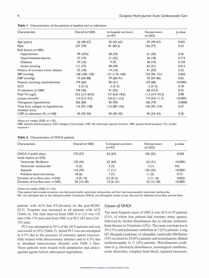
patients with ACS had ST-elevation on the post-ROSC ECG. Troponin was increased in all patients with ACS (Table 4). The time interval from OHCA to CA was 132 min (104–176 min) and from OHCA to PCI 143 min (124–193 min).
PCI was attempted in 91% of the ACS patients and was successful in 93% (Table 3). Initial PCI was not attempted in 4.5% due to the presence of coronary spasm (success-fully treated with intracoronary nitrates) and in 4.5% due to abundant intracoronary thrombi with TIMI 3 flow. These patients were treated with antiplatelet and antico-agulant agents before subsequent angioplasty.
Causes of OHCA
The most frequent cause of OHCA was ACS in 93 patients (31%, of whom four patients had coronary artery spasm), followed by rhythm disturbances due to chronic ischaemic heart disease in 54 patients (18%). The cause was hypoxia in 39 (13%) and pulmonary embolism in 7 (2%) patients. Long QT, Brugada syndrome, or idiopathic ventricular fibrillation (VF) occurred in 24 (8%) patients and nonischaemic dilated cardiomyopathy in 11 (4%) patients. Miscellaneous condi-tions (e g. electrolyte disturbances, neurological conditions, aortic dissection, complete heart block, ruptured aneurysm,
Table 1. Characteristics of the patients at baseline and on admission.
Characteristic Overall (n=300) In-hospital survivors (n=97)
Nonsurvivors (n=203)
p-value
Age (years) 56 (48–67) 50 (45–63) 59 (49–67) 0.001Male 237 (79) 81 (83.5) 156 (77) 0.23Risk factors (n=285) Hypertension 99 (33%) 38 (39) 61 (30) 0.30 Hypercholesterolaemia 57 (19) 21 (22) 36 (18) 0.715 Diabetes 47 (16) 9 (9) 38 (19) 0.102 Active smoking 111 (37) 48 (49) 63 (31) 0.013History of coronary artery disease 55 (18) 14 (14) 41 (20) 0.22SBP (mmHg) 128 (100–150) 131 (110–160) 124 (96–151) 0.062DBP (mmHg) 73 (60–88) 79 (68–91) 70 (59–86) 0.05Patients receiving catecholamines 179 (60) 40 (41) 139 (68) <0.0001GCS 3 (3–3) 3 (3–3) 3 (3–3) 0.18ST-elevation (n=284) 109 (36) 41 (42) 68 (33.5) 0.25Peak TnI (µg/l) 10.2 (2.3–65.0) 9.5 (6.6–18.6) 11.5 (4.9–34.2) 0.98Lactate (mmol/l) 5.4 (2.5–9.2) 2.8 (2.1–3.2) 7.0 (4.0–11.7) <0.0001Therapeutic hypothermia 256 (84) 96 (99) 160 (79) 0.0008Time from collapse to hypothermia initiation (min)
116 (92–148) 112 (89–145) 120 (95–154) 0.27
LVEF on admission (%, n=158) 40 (30–50) 40 (30–50) 45 (24–55) 0.30
Values are median (IQR) or n (%).DBP, diastolic blood pressure; GCS, Glasgow Coma Scale; LVEF, left ventricular ejection fraction.; SBP, systolic blood pressure; TnI, cardiac troponin I.
Table 2. Characteristics of OHCA patients.
Characteristic Overall (n=300) In-hospital survivors (n=97)
Nonsurvivors (n=203)
p-value
OHCA in public place 170 (57) 65 (67) 105 (52) 0.042Initial rhythm (n=270) Ventricular fibrillation 130 (43) 67 (69) 63 (31) <0.0001 Ventricular tachycardiaa 6 (2) 3 (3) 3 (1) NC Asystole 116 (39) 11 (11) 105 (52) <0.0001 Pulseless electrical activity 18 (6) 7 (7) 11 (5) 0.73Duration of no flow (min, n=243) 4 (0–10) 2.5 (0–5) 5 (1–10) 0.0031Duration of low flow (min, n=245) 20 (10–30) 10 (6–21) 22 (12–30) <0.0001
Values are median (IQR) or n (%).aOne patient had torsade de pointe, one had polymorphic ventricular tachycardia, and four had monomorphic ventricular tachycardia.NC, not calculated due to the reduced number of patients; OHCA, out-of-hospital cardiac arrest. See text for definition of no flow and low flow.
by guest on April 16, 2014acc.sagepub.comDownloaded from
Sideris et al. 5
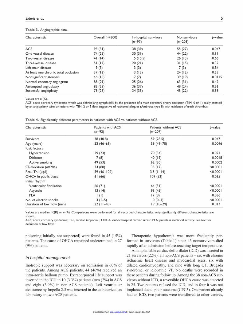
poisoning initially not suspected) were found in 45 (15%) patients. The cause of OHCA remained undetermined in 27 (9%) patients.
In-hospital management
Inotropic support was necessary on admission in 60% of the patients. Among ACS patients, 44 (46%) received an intra-aortic balloon pump. Extracorporeal life support was inserted in the ICU in 10 (3.3%) patients (two (2%) in ACS and eight (3.9%) in non-ACS patients). Left ventricular assistance by Impella 2.5 was inserted in the catheterization laboratory in two ACS patients.
Therapeutic hypothermia was more frequently per-formed in survivors (Table 1) since 43 nonsurvivors died rapidly after admission before reaching target temperature.
An implantable cardiac defibrillator (ICD) was inserted in 21 survivors (22%): all non-ACS patients – six with chronic ischaemic heart disease and myocardial scars, six with dilated cardiomyopathy, and nine with long QT, Brugada syndrome, or idiopathic VF. No deaths were recorded in these patients during follow up. Among the 38 non-ACS sur-vivors without ICD, a reversible OHCA cause was detected in 25. Two patients refused the ICD, and in four it was not implanted due to poor outcome (CPC3). One patient already had an ICD, two patients were transferred to other centres,
Table 3. Angiographic data.
Characteristic Overall (n=300) In-hospital survivors (n=97)
Nonsurvivors (n=203)
p-value
ACS 93 (31) 38 (39) 55 (27) 0.047One-vessel disease 74 (25) 30 (31) 44 (22) 0.11Two-vessel disease 41 (14) 15 (15.5) 26 (13) 0.66Three-vessel disease 51 (17) 20 (21) 31 (15) 0.32Left main disease 9 (3) 3 (3) 7 (3) 0.84At least one chronic total occlusion 37 (12) 13 (13) 24 (12) 0.55Nonsignificant stenosis 46 (15) 7 (7) 39 (19) 0.0115Normal coronary angiogram 88 (29) 25 (26) 63 (31) 0.42Attempted angioplasty 85 (28) 36 (37) 49 (24) 0.56Successful angioplasty 79 (26) 34 (35) 45 (22) 0.59
Values are n (%).ACS, acute coronary syndrome which was defined angiographically by the presence of a main coronary artery occlusion (TIMI 0 or 1) easily crossed by an angioplasty wire or lesions with TIMI 2 or 3 flow suggestive of ruptured plaques (Ambrose type II) with evidence of fresh thrombus.
Table 4. Significantly different parameters in patients with ACS vs. patients without ACS.
Characteristic Patients with ACS (n=93)
Patients without ACS (n=207)
p-value
Survivors 38 (40.8) 59 (28.5) 0.047Age (years) 52 (46–61) 59 (49–70) 0.0046Risk factors Hypertension 29 (23) 70 (34) 0.021 Diabetes 7 (8) 40 (19) 0.0018 Active smoking 49 (53) 62 (30) 0.0002ST-elevation (n=284) 74 (80) 35 (17) <0.0001Peak TnI (µg/l) 59 (46–102) 3.5 (1–14) <0.0001OHCA in public place 61 (66) 109 (53) 0.035Initial rhythm Ventricular fibrillation 66 (71) 64 (31) <0.0001 Asystole 13 (14) 93 (45) <0.0001 PEA 1 (1) 17 (8) 0.036No. of electric shocks 3 (1–5) 0 (0–1) <0.0001Duration of low flow (min) 22 (11–40) 19 (10–29) 0.017
Values are median (IQR) or n (%). Comparisons were performed for all recorded characteristics; only significantly different characteristics are shown.ACS, acute coronary syndrome; Tn I, cardiac troponin I; OHCA, out-of hospital cardiac arrest; PEA, pulseless electrical activity. See text for definition of low flow.
by guest on April 16, 2014acc.sagepub.comDownloaded from
6 European Heart Journal: Acute Cardiovascular Care
and four had ischaemic heart disease with normal electro-physiological study after complete revascularization.
At discharge, all patients with significant coronary artery disease received long-term aspirin, statin therapy, and beta-blockers unless contraindicated. Dual antiplatelet therapy was administered in all ACS patients.
In-hospital, postdischarge, and long-term overall survival
Survival is described in Table 6. There were 97 patients (32%) who were discharged alive from the hospital and 80 (82.5%) were alive at follow up (11 died, six were lost to follow up). Median follow up was 46 months (22–64 months). For the entire population, the probability of
postdischarge survival was 81.7±5.4% and of long-term overall survival was 26.2±2.8% (Table 6).
CPC at discharge was 1 in 44 patients, 2 in 36 patients, and 3 in 17 patients. At long-term follow up, 11 patients died, 15 improved their CPC from 2 to 1, and four improved their CPC from 3 to 2. Thus, 70/97 (72%) patients had good neurological status at long-term follow up: 50 were CPC1, 20 CPC2, and only 10 were CPC3 (Figure 1).
Among patients deceased during follow up, the cause of death was pneumonia in two, severe sepsis in two, meta-static cancer in one, and respiratory failure in one patient. Diastolic heart failure was the cause of death in one patient and cardiogenic shock following ACS due to a coronary lesion different from the initial ACS in another patient. In three patients, the cause of death remained undetermined.
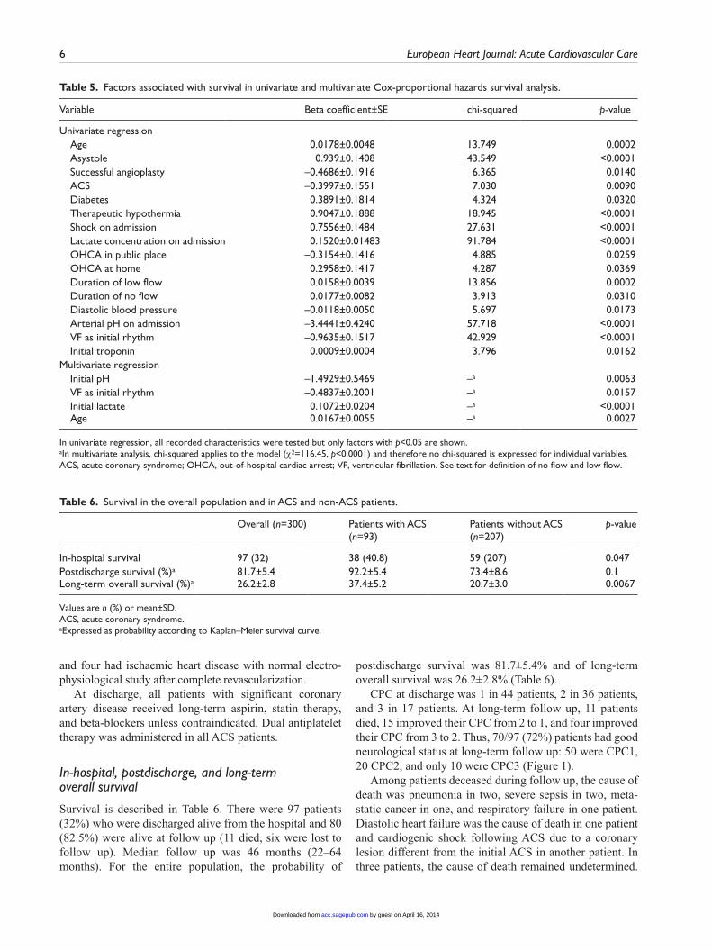
Table 5. Factors associated with survival in univariate and multivariate Cox-proportional hazards survival analysis.
Variable Beta coefficient±SE chi-squared p-value
Univariate regression Age 0.0178±0.0048 13.749 0.0002 Asystole 0.939±0.1408 43.549 <0.0001 Successful angioplasty –0.4686±0.1916 6.365 0.0140 ACS –0.3997±0.1551 7.030 0.0090 Diabetes 0.3891±0.1814 4.324 0.0320 Therapeutic hypothermia 0.9047±0.1888 18.945 <0.0001 Shock on admission 0.7556±0.1484 27.631 <0.0001 Lactate concentration on admission 0.1520±0.01483 91.784 <0.0001 OHCA in public place –0.3154±0.1416 4.885 0.0259 OHCA at home 0.2958±0.1417 4.287 0.0369 Duration of low flow 0.0158±0.0039 13.856 0.0002 Duration of no flow 0.0177±0.0082 3.913 0.0310 Diastolic blood pressure –0.0118±0.0050 5.697 0.0173 Arterial pH on admission –3.4441±0.4240 57.718 <0.0001 VF as initial rhythm –0.9635±0.1517 42.929 <0.0001 Initial troponin 0.0009±0.0004 3.796 0.0162Multivariate regression Initial pH –1.4929±0.5469 –a 0.0063 VF as initial rhythm –0.4837±0.2001 –a 0.0157 Initial lactate 0.1072±0.0204 –a <0.0001 Age 0.0167±0.0055 –a 0.0027
In univariate regression, all recorded characteristics were tested but only factors with p<0.05 are shown.aIn multivariate analysis, chi-squared applies to the model (χ2=116.45, p<0.0001) and therefore no chi-squared is expressed for individual variables.ACS, acute coronary syndrome; OHCA, out-of-hospital cardiac arrest; VF, ventricular fibrillation. See text for definition of no flow and low flow.
Table 6. Survival in the overall population and in ACS and non-ACS patients.
Overall (n=300) Patients with ACS (n=93)
Patients without ACS (n=207)
p-value
In-hospital survival 97 (32) 38 (40.8) 59 (207) 0.047Postdischarge survival (%)a 81.7±5.4 92.2±5.4 73.4±8.6 0.1Long-term overall survival (%)a 26.2±2.8 37.4±5.2 20.7±3.0 0.0067
Values are n (%) or mean±SD.ACS, acute coronary syndrome.aExpressed as probability according to Kaplan–Meier survival curve.
by guest on April 16, 2014acc.sagepub.comDownloaded from

Sideris et al. 7
Four patients required repeat revascularization: two in lesions not related to the initial ACS and four for in-stent restenosis.
Survival in patients with ACS vs. patients without ACS
Patients with ACS had better in-hospital survival than patients without ACS (p=0.047; Table 6). Despite a ten-dency in favour of the ACS group, probability of postdis-charge survival was not different in the two groups (Table 6). The probability of long-term overall survival in patients with ACS was significantly higher than in patients without ACS (hazard ratio 1.5, 95% CI 1.12–2.0, p=0.0067 according to the Kaplan–Meier survival curve; Figure 2).
All recorded characteristics were compared between patients with and without ACS. Characteristics showing significant differences between the two groups are pro-vided in Table 4. The left ventricular ejection fraction at discharge in ACS patients did not differ from non-ACS patients (50%, IQR 42–60 vs. 50%, IQR 37–55%, p=0.4). Among the 58 non-ACS survivors, 31 had cardiomyopa-thy (22 ischaemic heart disease, and nine dilated cardio-myopathy), with left ventricular ejection fraction of 45% (IQR 29–51%).
Univariate and multivariate survival analysis
The factors significantly associated with long-term overall survival in univariate regression are expressed in Table 5. In multivariate regression, four variables were associated with prognosis: presence of VF, age, initial lactate concen-tration, and initial arterial pH (Table 5).
Discussion
The present study is the first to report 5-year probability of survival in a population of resuscitated OHCA patients regardless of the presenting rhythm and presence of ST-elevation on post-ROSC ECG who were managed with an early invasive strategy including routine CA on admis-sion to the hospital and PCI if indicated. The most impor-tant findings are that postdischarge survival was high (81.7%) in the entire OHCA population and that in-hospital survival and, especially, long-term overall survival in patients with OHCA caused by ACS were significantly higher than in patients without ACS.
Patients included in our study had an in-hospital sur-vival of only 32 %, relatively lower than other studies – 38%,3 40%,5 54%4 – but this was due to the severity of our population. Indeed, 197 patients (66%) had at least one very poor prognostic factor: either asystole or pulseless electrical activity as initial rhythm and/or very long resus-citation duration with no flow >5 min and/or low flow >30 min, which may account for the relatively high mortality.
Favourable postdischarge survival in the overall population
Survival for patients with OHCA varies widely across the studies according to the population included. Garot et al.23 reported a 54% 6-month survival in a selected population (OHCA with VF and presumed ST-elevation myocardial infarction referred for primary PCI), but longer follow-up data were not available. A study of 200 OHCA patients with VF as initial rhythm (47% with ACS), reported 79% 60-month postdischarge survival, similar to our data. However, CA was not performed routinely and only 24% of
0 10 20 30 40 50 60
1009080706050403020100
Time (months)S
urvi
val p
roba
bili
ty (
%)
Number at risk
207 53 43 37 30 19 12
93 33 29 23 21 17 13
p=0.0067
No ACS
ACS
No ACS
ACS
Figure 2. Probability of long-term overall survival of ACS patients vs. non-ACS patients according to Kaplan–Meier survival analysis.The probability of long-term overall survival in ACS patients (dashed line) was 37.4±5.2% and in non-ACS patients (solid line) was 20.7±3.0% (hazard ratio 1.5, 95% CI 1.12–2.0; p=0.0067. ACS, acute coronary syndrome.
by guest on April 16, 2014acc.sagepub.comDownloaded from
8 European Heart Journal: Acute Cardiovascular Care
these patients had a myocardial revascularization attempt (by PCI or coronary surgery) during hospitalization.1
Postdischarge survival in our patients seems higher than in OHCA studies before hypothermia and CA were intro-duced. Indeed, Engdahl et al.2 found in 430 OHCA patients with 86% VF as initial rhythm, a 60-month postdischarge survival of only 48%. This difference may be due to thera-peutic hypothermia and possibly to CA with revasculariza-tion in our patients, but also to long-term medical treatment using beta-blockers and lipid-lowering drugs.2,9
A recent study analysed outcome according to the use of CA and PCI and showed improved survival in patients undergoing this procedure.24 In this cohort, 60-month post-discharge survival was 78.7% in patients receiving PCI, compared to 92.7% in our study, a difference that may be explained by therapeutic hypothermia used in 99% of the survivors in our population vs. only 26%.24 Indeed, 9.1% of the patients in this study24 received PCI and therapeutic hypothermia, and the 60-month survival (88.6%) was com-parable to our patients. However, the moment of the coro-nary angiogram in this population was less well defined and was not performed routinely on admission.
Long-term follow up in our population showed not only a good survival, but also an improvement of CPC status, consistent with previous data25 suggesting that neurological improvement continues after hospital discharge.25,26
Favourable prognosis in ACS patients
In the setting of the OHCA the diagnosis of ACS without the use of CA is complex, due to the limited diagnostic value of the ECG changes3,14 and troponin elevation.11 In the absence of CA, ACS cases and severe coronary heart disease may remain undetected and without appropriate treatment. In our population, immediate routine CA avoided this limitation.
Before the era of emergency revascularization, OHCA studies suggested that survival was better in patients with ACS.27,28 A more recent report29 showed better 3-year sur-vival of OHCA patients with ST-elevation ACS, but CA was not performed on a routine basis and details on the selection of patients for PCI are not provided.
Even though in our population the presence of ACS was not independently associated with prognosis in multivariate analysis, the better survival of ACS patients emphasizes the importance of CA with PCI in the initial management of resuscitated OHCA patients. Only four very strong prog-nostic factors (presence of VF as initial rhythm, age, initial lactate concentration, and initial arterial pH) found in many OHCA studies3,14,23 were independently associated with prognosis in multivariate regression. Even though success-ful PCI is not one of them, due to the design of our study, no definite conclusion can be drawn on the influence of PCI on survival, and randomized trials are warranted to evalu-ate this issue.7
Interestingly, the favourable postdischarge prognosis of ACS patients was similar to that recorded in recent ACS studies without OHCA. In a study comparing thrombolysis vs. PCI,30 5-year probability of survival in the PCI group was 87%, similar to 92.2% in our population. This favour-able postdischarge survival may be explained by the good neurological status at discharge in our patients and the pre-served left ventricular function. The median value of left ventricular ejection fraction was 50% in ACS patients, sug-gesting that the overall management including CA with PCI may have a role in preserving cardiac function.
Prognosis of non-ACS patients also compared favoura-bly to previous studies. In a single-centre study including 123 non-ACS OHCA patients, 3-year survival was 19.5%,29 compared to 25.6% in our study. This difference may be due to the routine use of hypothermia in our centre. Thus, despite the severity of our population, postdischarge sur-vival remained favourable, suggesting that an early inva-sive initial management strategy as the one applied in this study, including CA with PCI on admission, hypothermia, medical management according to guidelines,9,17 and use of ICD leads to good long-term neurological survival in most survivors.
Limitations
Our study has several limitations. The study was performed in a single centre highly specialized in OHCA care and therefore the results may not be obtained in other settings.
In our study, 69% of the patients did not have an ACS and may be argued that CA induced a delay in the induction of hypothermia, in the diagnosis of OHCA causes and increased the risk of contrast media-induced nephropathy. However, a benefit may be expected from the knowledge of the coronary status in the subsequent management includ-ing optimization of haemodynamic function using catecho-lamines, antiplatelet treatment, and the subsequent diagnostic algorithm.21
In survivors, hypothermia was performed more fre-quently but this occurred due to the death of some nonsur-vivors before reaching target temperature.
Intra-aortic balloon pumps were used more frequently in ACS than non-ACS patients, but this was performed according to previous recommendations.9 The use of other circulatory assist devices was performed in less than 5% of the patients and their influence on survival in ACS vs. non-ACS patients was probably less significant.
Conclusion
Our study showed that, despite significant in-hospital mor-tality in these severely ill comatose patients resuscitated after OHCA and managed with CA on admission, postdis-charge prognosis remains favourable for up to 5 years. Moreover, long-term overall survival was better in ACS
by guest on April 16, 2014acc.sagepub.comDownloaded from
Sideris et al. 9
patients than in patients without ACS. This type of early invasive in-hospital management seems effective, comple-menting modern out-of hospital resuscitation measures and should be further evaluated.
Conflict of interestThe authors declare that there is no conflict of interest.
Funding
This research received no specific grant from any funding agency in the public, commercial, or not-for-profit sectors.
References
1. Bunch TJ, White RD, Gersh BJ, et al. Long-term outcomes of out-of-hospital cardiac arrest after successful early defi-brillation. N Engl J Med 2003; 348: 2626–2633.
2. Engdahl J, Bang A, Lindqvist J, et al. Time trends in long-term mortality after out-of-hospital cardiac arrest, 1980 to 1998, and predictors for death. Am Heart J 2003; 145: 826–833.
3. Spaulding CM, Joly LM, Rosenberg A, et al. Immediate coronary angiography in survivors of out-of-hospital cardiac arrest. N Engl J Med 1997; 336: 1629–1633.
4. Cronier P, Vignon P, Bouferrache K, et al. Impact of routine percutaneous coronary intervention after out-of-hospital car-diac arrest due to ventricular fibrillation. Crit Care 2011; 15: R122.
5. Dumas F, Cariou A, Manzo-Silberman S, et al. Immediate percutaneous coronary intervention is associated with better survival after out-of-hospital cardiac arrest: insights from the PROCAT (Parisian Region Out of hospital Cardiac ArresT) registry. Circ Cardiovasc Interv 2010; 3: 200–207.
6. Grasner JT, Meybohm P, Caliebe A, et al. Postresuscitation care with mild therapeutic hypothermia and coronary inter-vention after out-of-hospital cardiopulmonary resuscitation: a prospective registry analysis. Crit Care 2011; 15: R61.
7. Larsen JM and Ravkilde J. Acute coronary angiography in patients resuscitated from out-of-hospital cardiac arrest – a systematic review and meta-analysis. Resuscitation 2012; 83: 1427–1433.
8. Tomte O, Andersen GO, Jacobsen D, et al. Strong and weak aspects of an established post-resuscitation treatment proto-col – a five-year observational study. Resuscitation 2011; 82: 1186–1193.
9. Steg PG, James SK, Atar D, et al. ESC Guidelines for the management of acute myocardial infarction in patients pre-senting with ST-segment elevation. Eur Heart J 2012; 33: 2569–2619.
10. Anyfantakis ZA, Baron G, Aubry P, et al. Acute coronary angiographic findings in survivors of out-of-hospital cardiac arrest. Am Heart J 2009; 157: 312–318.
11. Voicu S, Sideris G, Deye N, et al. Role of cardiac troponin in the diagnosis of acute myocardial infarction in comatose patients resuscitated from out-of-hospital cardiac arrest. Resuscitation 2012; 83: 452–458.
12. Dumas F and Rea TD. Long-term prognosis following resus-citation from out-of-hospital cardiac arrest: role of aetiology and presenting arrest rhythm. Resuscitation 2012; 83: 1001–1005.
13. Pell JP, Sirel JM, Marsden AK, et al. Presentation, manage-ment, and outcome of out of hospital cardiopulmonary arrest: comparison by underlying aetiology. Heart 2003; 89: 839–842.
14. Sideris G, Voicu S, Dillinger JG, et al. Value of post-resus-citation electrocardiogram in the diagnosis of acute myo-cardial infarction in out-of-hospital cardiac arrest patients. Resuscitation 2011; 82: 1148–1153.
15. Nolan JP, Deakin CD, Soar J, et al. European Resuscitation Council guidelines for resuscitation 2005. Section 4. Adult advanced life support. Resuscitation 2005; 67 Suppl 1: S39–S86.
16. Jacobs I, Nadkarni V, Bahr J, et al. Cardiac arrest and cardio-pulmonary resuscitation outcome reports: update and simpli-fication of the Utstein templates for resuscitation registries: a statement for healthcare professionals from a task force of the International Liaison Committee on Resuscitation (American Heart Association, European Resuscitation Council, Australian Resuscitation Council, New Zealand Resuscitation Council, Heart and Stroke Foundation of Canada, InterAmerican Heart Foundation, Resuscitation Councils of Southern Africa). Circulation 2004; 110: 3385–3397.
17. Deakin CD, Nolan JP, Soar J, et al. European Resuscitation Council Guidelines for Resuscitation 2010 Section 4. Adult advanced life support. Resuscitation 2010; 81: 1305–1352.
18. Thygesen K, Alpert JS, Jaffe AS, et al. Third universal defi-nition of myocardial infarction. Eur Heart J 2012; 33: 2551–2567.
19. Radsel P, Knafelj R, Kocjancic S, et al. Angiographic char-acteristics of coronary disease and postresuscitation electro-cardiograms in patients with aborted cardiac arrest outside a hospital. Am J Cardiol 2011; 108: 634–638.
20. Ambrose JA, Winters SL, Arora RR, et al. Coronary angio-graphic morphology in myocardial infarction: a link between the pathogenesis of unstable angina and myocardial infarc-tion. J Am Coll Cardiol 1985; 6: 1233–1238.
21. Chelly J, Mongardon N, Dumas F, et al. Benefit of an early and systematic imaging procedure after cardiac arrest: insights from the PROCAT (Parisian Region Out of Hospital Cardiac Arrest) registry. Resuscitation 2012; 83: 1444–1450.
22. Jennett B and Bond M. Assessment of outcome after severe brain damage. Lancet 1975; 1: 480–484.
23. Garot P, Lefevre T, Eltchaninoff H, et al. Six-month outcome of emergency percutaneous coronary intervention in resusci-tated patients after cardiac arrest complicating ST-elevation myocardial infarction. Circulation 2007; 115: 1354–1362.
24. Dumas F, White L, Stubbs BA, et al. Long-term prognosis following resuscitation from out of hospital cardiac arrest: role of percutaneous coronary intervention and therapeutic hypothermia. J Am Coll Cardiol 2012; 60: 21–27.
25. Graves JR, Herlitz J, Bang A, et al. Survivors of out of hos-pital cardiac arrest: their prognosis, longevity and functional status. Resuscitation 1997; 35: 117–121.
26. Bro-Jeppesen J, Kjaergaard J, Horsted TI, et al. The impact of therapeutic hypothermia on neurological function and quality of life after cardiac arrest. Resuscitation 2009; 80: 171–176.
27. Cobbe SM, Dalziel K, Ford I, et al. Survival of 1476 patients initially resuscitated from out of hospital cardiac arrest. BMJ 1996; 312: 1633–1637.
by guest on April 16, 2014acc.sagepub.comDownloaded from

10 European Heart Journal: Acute Cardiovascular Care
28. Kimman GP, Ivens EM, Hartman JA, et al. Long-term survival after successful out-of-hospital resuscitation. Resuscitation 1994; 28: 227–232.
29. Pleskot M, Hazukova R, Stritecka H, et al. Long-term prog-nosis after out-of-hospital cardiac arrest with/without ST
elevation myocardial infarction. Resuscitation 2009; 80: 795–804.
30. Henriques JP, Zijlstra F, van’t Hof AW, et al. Primary percutane-ous coronary intervention vs. thrombolytic treatment: long term follow up according to infarct location. Heart 2006; 92: 75–79.
by guest on April 16, 2014acc.sagepub.comDownloaded from