european heart journal advance access published august … esc af.pdf · mendations or guidelines...
TRANSCRIPT

PMK Cardiology ReviewPMK Cardiology Review
1
ESC GUIDELINES
2016 ESCGuidelines for themanagement of atrialfibrillationdeveloped in collaborationwithEACTSThe Task Force for the management of atrial fibrillation of theEuropean Society of Cardiology (ESC)
Developed with the special contribution of the European HeartRhythm Association (EHRA) of the ESC
Endorsed by the European Stroke Organisation (ESO)
Authors/Task Force Members: Paulus Kirchhof* (Chairperson) (UK/Germany)Stefano Benussi*1 (Co-Chairperson) (Switzerland), Dipak Kotecha (UK),Anders Ahlsson1 (Sweden), Dan Atar (Norway), Barbara Casadei (UK),Manuel Castella1 (Spain), Hans-Christoph Diener2 (Germany), Hein Heidbuchel(Belgium), Jeroen Hendriks (The Netherlands), Gerhard Hindricks (Germany),Antonis S. Manolis (Greece), Jonas Oldgren (Sweden), Bogdan Alexandru Popescu(Romania), Ulrich Schotten (The Netherlands), Bart Van Putte1 (The Netherlands),and Panagiotis Vardas (Greece)Document Reviewers: Stefan Agewall (CPG Review Co-ordinator) (Norway), John Camm (CPG ReviewCo-ordinator) (UK), Gonzalo Baron Esquivias (Spain), Werner Budts (Belgium), Scipione Carerj (Italy),Filip Casselman (Belgium), Antonio Coca (Spain), Raffaele De Caterina (Italy), Spiridon Deftereos (Greece),Dobromir Dobrev (Germany), Jose M. Ferro (Portugal), Gerasimos Filippatos (Greece), Donna Fitzsimons (UK),
* Corresponding authors: Paulus Kirchhof, Institute of Cardiovascular Sciences, University of Birmingham, SWBH and UHB NHS trusts, IBR, Room 136, Wolfson Drive, BirminghamB15 2TT, United Kingdom, Tel: +44 121 4147042, E-mail: [email protected]; Stefano Benussi, Department of Cardiovascular Surgery, University Hospital Zurich, Ramistrasse100, 8091 Zurich, Switzerland, Tel: +41(0)788933835, E-mail: [email protected] Representing the European Association for Cardio-Thoracic Surgery (EACTS)2 Representing the European Stroke Association (ESO)
ESC Committee for Practice Guidelines (CPG) and National Cardiac Societies Reviewers can be found in the Appendix.
ESC entities having participated in the development of this document:
Associations: European Association for Cardiovascular Prevention and Rehabilitation (EACPR), European Association of Cardiovascular Imaging (EACVI), European Heart RhythmAssociation (EHRA), Heart Failure Association (HFA).
Councils: Council on Cardiovascular Nursing and Allied Professions, Council for Cardiology Practice, Council on Cardiovascular Primary Care, Council on Hypertension.
Working Groups: Cardiac Cellular Electrophysiology, Cardiovascular Pharmacotherapy, Grown-up Congenital Heart Disease, Thrombosis, Valvular Heart Disease.
The content of these European Society of Cardiology (ESC) Guidelines has been published for personal and educational use only. No commercial use is authorized. No part of the ESCGuidelines may be translated or reproduced in any form without written permission from the ESC. Permission can be obtained upon submission of a written request to Oxford Uni-versity Press, the publisher of the European Heart Journal and the party authorized to handle such permissions on behalf of the ESC ([email protected]).
Disclaimer. The ESC Guidelines represent the views of the ESC and were produced after careful consideration of the scientific and medical knowledge and the evidence available atthe time of their publication. The ESC is not responsible in the event of any contradiction, discrepancy and/or ambiguity between the ESC Guidelines and any other official recom-mendations or guidelines issued by the relevant public health authorities, in particular in relation to good use of healthcare or therapeutic strategies. Health professionals are encour-aged to take the ESC Guidelines fully into account when exercising their clinical judgment, as well as in the determination and the implementation of preventive, diagnostic ortherapeutic medical strategies; however, the ESC Guidelines do not override, in any way whatsoever, the individual responsibility of health professionals to make appropriate andaccurate decisions in consideration of each patient’s health condition and in consultation with that patient and, where appropriate and/or necessary, the patient’s caregiver. Nordo the ESC Guidelines exempt health professionals from taking into full and careful consideration the relevant official updated recommendations or guidelines issued by the competentpublic health authorities, in order to manage each patient’s case in light of the scientifically accepted data pursuant to their respective ethical and professional obligations. It is also thehealth professional’s responsibility to verify the applicable rules and regulations relating to drugs and medical devices at the time of prescription.
& The European Society of Cardiology 2016. All rights reserved. For permissions please email: [email protected].
European Heart Journaldoi:10.1093/eurheartj/ehw210
European Heart Journal Advance Access published August 27, 2016
by guest on September 1, 2016
http://eurheartj.oxfordjournals.org/D
ownloaded from
นพ ธรณศ จนทรารตนElectrophysiology Unit
รพ พระมงกฎเกลา
Wednesday, December 7, 2016
PMK Cardiology ReviewPMK Cardiology Review
Scope of Presentation
• Diagnosis and screening• General Management• Stroke Prevention• Rate/Rhythm Control• Special Population• To Do and Not to Do Messages• 17 Rules Summary
2
Wednesday, December 7, 2016

PMK Cardiology ReviewPMK Cardiology Review
3
of educational tools and implementation programmes for the re-commendations. To implement the guidelines, condensed pocketguideline versions, summary slides, booklets with essential mes-sages, summary cards for non-specialists and an electronic versionfor digital applications (smartphones, etc.) are produced. These ver-sions are abridged and thus, if needed, one should always refer tothe full text version, which is freely available on the ESC website.The National Societies of the ESC are encouraged to endorse,translate and implement all ESC Guidelines. Implementation pro-grammes are needed because it has been shown that the outcomeof disease may be favourably influenced by the thorough applicationof clinical recommendations.Surveys and registries are needed to verify that real-life daily prac-
tice is in keeping with what is recommended in the guidelines, thuscompleting the loop between clinical research, writing of guidelines,disseminating them and implementing them into clinical practice.Health professionals are encouraged to take the ESC and EACTS
Guidelines fully into account when exercising their clinical judgment,as well as in the determination and the implementation of prevent-ive, diagnostic or therapeutic medical strategies. However, the ESCand EACTS Guidelines do not override in any way whatsoever theindividual responsibility of health professionals to make appropriateand accurate decisions in consideration of each patient’s health con-dition and in consultation with that patient and the patient’s care-giver where appropriate and/or necessary. It is also the healthprofessional’s responsibility to verify the rules and regulationsapplicable to drugs and devices at the time of prescription.
2. IntroductionDespite good progress in the management of patients with atrial fib-rillation (AF), this arrhythmia remains one of the major causes ofstroke, heart failure, sudden death, and cardiovascular morbidityin the world. Furthermore, the number of patients with AF is pre-dicted to rise steeply in the coming years. To meet the growing de-mand for effective care of patients with AF, new information iscontinually generated and published, and the last few years haveseen substantial progress. Therefore, it seems timely to publishthis 2nd edition of the ESC guidelines on AF.Reflecting the multidisciplinary input into the management of
patientswithAF, the Task Force includes cardiologistswith varying sub-specialty expertise, cardiac surgeons, stroke neurologists, and specialistnurses amongst its members. Supplementing the evidence reviewas outlined in the preamble, this Task Force defined three Population,Intervention, Comparison, Outcome, Time (PICOT) questions onrelevant topics for the guidelines. The ESC commissioned external sys-tematic reviews to answer these questions, and these reviews haveinformed specific recommendations.Further to adhering to the standards for generating recommenda-
tions that are common to all ESC guidelines (see preamble), thisTask Force discussed each draft recommendation during web-basedconference calls dedicated to specific chapters, followed by consen-sus modifications and an online vote on each recommendation.Only recommendations that were supported by at least 75% ofthe Task Force members were included in the guidelines.We hope that these guidelines will help to deliver good care to
all patients with AF based on the current state-of-the-art evidencein 2016.
3. Epidemiology and impact forpatients
3.1 Incidence and prevalence of atrialfibrillationIn 2010, the estimated numbers of men and women with AF world-wide were 20.9 million and 12.6 million, respectively, with higher in-cidence and prevalence rates in developed countries.1,2 One in fourmiddle-aged adults in Europe and the USwill develop AF.3–5 By 2030,14–17 million AF patients are anticipated in the European Union,with 120 000–215 000 newly diagnosed patients per year.2,6,7 Esti-mates suggest an AF prevalence of approximately 3% in adults aged20 years or older,8,9 with greater prevalence in older persons1 andin patients with conditions such as hypertension, heart failure, coron-ary artery disease (CAD), valvular heart disease, obesity, diabetesmellitus, or chronic kidney disease (CKD).7,10 – 15 The increasein AF prevalence can be attributed both to better detection ofsilent AF16–18, alongside increasing age and conditions predisposingto AF.19
3.2 Morbidity, mortality, and healthcareburden of atrial fibrillationAF is independently associated with a two-fold increased risk ofall-cause mortality in women and a 1.5-fold increase in men20–22
(Table 3). Death due to stroke can largely be mitigated by anticoa-gulation, while other cardiovascular deaths, for example due toheart failure and sudden death, remain common even in AF pa-tients treated according to the current evidence base.23 AF isalso associated with increased morbidity, such as heart failureand stroke.21,24,25 Contemporary studies show that 20–30% of pa-tients with an ischaemic stroke have AF diagnosed before, during,
Table 3 Cardiovascular morbidity and mortalityassociated with atrial fibrillation
Event Association with AF
Death Increased mortality, especially cardiovascular mortality due to sudden death, heart failure or stroke.
Stroke 20–30% of all strokes are due to AF. A growing number of patients with stroke are diagnosed with ‘silent’, paroxysmal AF.
Hospitalizations 10–40% of AF patients are hospitalized every year.
Quality of life Quality of life is impaired in AF patients independent of other cardiovascular conditions.
Left ventricular dysfunction and heart failure
Left ventricular dysfunction is found in 20–30% of all AF patients. AF causes or aggravates LV dysfunction in many AF patients, while others have completely preserved LV function despite long-standing AF.
Cognitive decline and vascular dementia
Cognitive decline and vascular dementia can develop even in anticoagulated AF patients. Brain white matter lesions are more common in AF patients than in patients without AF.
AF ! atrial fibrillation; LV ! left ventricular.
ESC Guidelines Page 7 of 90
by guest on September 1, 2016
http://eurheartj.oxfordjournals.org/D
ownloaded from
Wednesday, December 7, 2016

PMK Cardiology ReviewPMK Cardiology Review
4
or after the initial event.17,26,27 White matter lesions in the brain,cognitive impairment,28 – 30 decreased quality of life,31,32 anddepressed mood33 are common in AF patients, and between10–40% of AF patients are hospitalized each year.23,34,35
The direct costs of AF already amount to approximately 1% of to-tal healthcare spending in the UK, and between 6.0–26.0 billion USdollars in the US for 2008,36,37 driven by AF-related complications(e.g. stroke) and treatment costs (e.g. hospitalizations). These costswill increase dramatically unless AF is prevented and treated in atimely and effective manner.
3.3 Impact of evidence-basedmanagement on outcomes in atrialfibrillation patientsFigure 1 depicts the major milestones in the management of AF.Despite these advances, substantial morbidity remains. Oral
anticoagulation (OAC) with vitamin K antagonists (VKAs) or non-VKA oral anticoagulants (NOACs) markedly reduces stroke andmortality in AF patients.38,39 Other interventions such as rhythmcontrol and rate control improve AF-related symptoms and maypreserve cardiac function, but have not demonstrated a reductionin long-term morbidity or mortality.40,41
In contemporary, well-controlled, randomized clinical trialsin AF, the average annual stroke rate is about 1.5% and theannualized death rate is around 3% in anticoagulated AF patients.40
In real life, the annual mortality can be different (both higher andlower).42 A minority of these deaths are related to stroke, whilesudden cardiac death and death from progressive heart failureare more frequent, emphasizing the need for interventions beyondanticoagulation.43,44 Furthermore, AF is also associated with highrates of hospitalization, commonly for AF management, but oftenalso for heart failure, myocardial infarction, and treatment-associated complications.34,45
Rate control not inferior to rhythm control
Amiodarone notsuperior to ratecontrol in heart
failure
Dronedarone harmsin permanent AF
Beta-blockerswithout prognostic
benefit in AF patientswith HFrEF
Lenient rate controlacceptable
PVI can suppress AF
PVI maintains SRbetter than
antiarrhythmic drugs
Dronedaroneimproves outcomes in non-permanent AF
AF ablationimproves Qol
First-line PVImaintains SR betterthan antiarrhythmic
drugs
PVI alone aseffective as
complex ablation inpersistent AF
Cryoenergy aseffective as RF
for PVI
VKA superior to aspirinfor stroke prevention in
AF
Dabigatran at least aseffective as VKA in AF
Rixaroxaban andApixaban at least as
effective as VKA in AF
Edoxaban at least aseffective as VKA in AF
Meta-analysis andhealthcare databases:
NOACs safer andslightly more effective
compared to VKA
Ximelagatran aseffective as VKA
VKA reduces stroke inAF by 2/3
1995
2000
2005
2010
2015
ACE-I/ARBs preventAF in heart failure
ARBs prevent AF in hypertension & LVH
ARBs do not preventAF or adverse
outcomes in patientswithout hypertension
PUFA do notprevent AF
ACE-I/ARB preventAF in hypertension
Beta-blockersprevent AF in HFrEFpatients pre-treated
with ACE-I
MRA prevent AF inHFrEF patients pre-treated with ACE-I/
beta-blockers
First maze surgeryfor AF treatment
published
RF based mazemaintains SR after
cardiovascularsurgery
Bipolar RF moreeffective than
conventional RF for stand-alone
AF surgery
Concomitant mazesurgery maintains SRbut increases risk of
permanent pacemaker
LVH = left ventricular hypertrophy; NOAC = non-vitamin K antagonist oral anticoagulant; PUFA = polyunsaturated fatty acid; PVI = pulmonary vein isolation; QoL = quality of life; RF = radiofrequency; SR = sinus rhythm; VKA = vitamin K antagonist.
Figure 1 Timeline of findings from landmark trials in atrial fibrillation management, including treatment of concomitant conditions and preven-tion (green), anticoagulation (blue), rate control therapy (orange), rhythm control therapy (red), and atrial fibrillation surgery (purple).
ESC GuidelinesPage 8 of 90
by guest on September 1, 2016
http://eurheartj.oxfordjournals.org/D
ownloaded from
Wednesday, December 7, 2016
PMK Cardiology ReviewPMK Cardiology Review
Inherited cardiomyopathies, channelopathies, and pathways associated with atrial fibrillation
5
12.2 Combining antiarrhythmic drugsand pacemakersIn selected patients with sick sinus syndrome and fast ventricular re-sponse during AF paroxysms requiring rate control therapy, the add-ition of a pacemaker not only optimizes rate control but may also helpto control rhythm.711,712 Moreover, when antiarrhythmic drug treat-ment leads to sinus node dysfunction and bradycardia, pacingmay per-mit uptitration of the antiarrhythmic drug dose. Such strategies havenever been prospectively investigated and the existing populationsstudied are highly selected.839,840 Some patients with AF-inducedbradycardia may benefit from catheter ablation of AF, obviating theneed for antiarrhythmic drugs and pacemaker implantation.829,830
13. Specific situations
13.1 Frail and ‘elderly’ patientsMany AF patients present at older age (e.g..75 or.80 years). Thereare no studies suggesting that cardiovascular risk reduction is less ef-fective in these ‘elderly’ AF patients than in younger patients. Rather,age is one of the strongest predictors/risk factors for ischaemic strokein AF.382 Good data are available to support the use of anticoagulantsin older patients from BAFTA (Birmingham Atrial Fibrillation Treat-ment of the Aged Study),362 the NOAC trials,39 and from analysesin elderly Americans (Medicare).396 Elderly AF patients are at higherrisk of stroke and, thus, are more likely to benefit from OAC thanyounger patients,841 and yet OAC is still underutilized in the elder-ly.220,842 Although the evidence base is smaller for other treatment
options in AF, the available data support the use of available rateand rhythm control interventions, including pacemakers and catheterablation, without justification to discriminate by age group. Individualpatients at older agemay presentwithmultiple comorbidities includingdementia, a tendency to falls, CKD, anaemia, hypertension, diabetes,and cognitive dysfunction. Such conditions may limit quality of lifemore than AF-related symptoms. Impairment of renal and hepaticfunction and multiple simultaneous medications make drug interac-tions and adverse drug reactions more likely. Integrated AF manage-ment and careful adaptation of drug dosing seem reasonable toreduce the complications of AF therapy in such patients.843
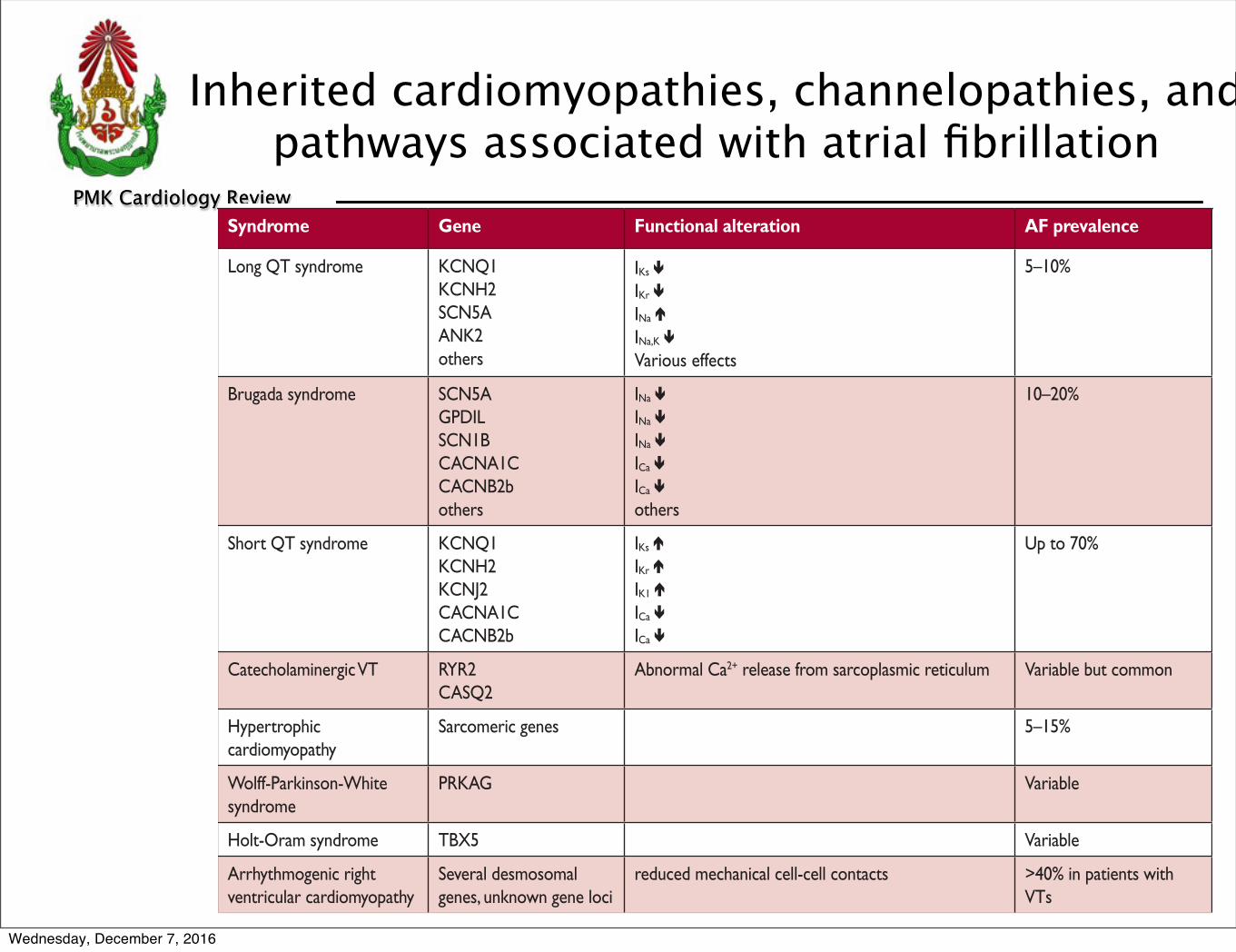
13.2 Inherited cardiomyopathies,channelopathies, and accessory pathwaysSeveral inherited cardiac conditions are associated with early-onsetAF (Table 20). Treatment of the underlying cardiac condition is animportant contribution to AF management in these young patients(see also ESC guidelines on sudden cardiac death844 and hyper-trophic cardiomyopathy845).
13.2.1 Wolff–Parkinson–White syndromePatients with pre-excitation and AF are at risk of rapid conductionacross the accessory pathway, resulting in a fast ventricular rate,possible ventricular fibrillation, and sudden death. In AF patientswith evidence of an antegrade accessory pathway, catheter ablationof the pathway is recommended.869,870 This procedure is safe andeffective and may be considered as a prophylactic treatment strat-egy.871,872 In AF patients surviving a sudden death eventwith evidence
Table 20 Inherited cardiomyopathies, channelopathies, and pathways associated with atrial fibrillation
Syndrome Gene Functional alteration AF prevalence References
Long QT syndrome KCNQ1KCNH2SCN5AANK2others
IKs !
IKr !
INa "
INa,K !
Various effects
5–10% 846–850
Brugada syndrome SCN5AGPDILSCN1BCACNA1CCACNB2bothers
INa !
INa !
INa !
ICa !
ICa !
others
10–20% 851–855
Short QT syndrome KCNQ1 KCNH2KCNJ2CACNA1CCACNB2b
IKs "
IKr "
IK1 "
ICa !
ICa !
Up to 70% 853, 856–858
Catecholaminergic VT RYR2CASQ2
Abnormal Ca2+ release from sarcoplasmic reticulum Variable but common 859–861
Hypertrophic cardiomyopathy
Sarcomeric genes 5–15% 862–864
Wolff-Parkinson-White syndrome
PRKAG Variable 865
Holt-Oram syndrome TBX5 Variable 866
Arrhythmogenic right ventricular cardiomyopathy
Several desmosomal genes, unknown gene loci
reduced mechanical cell-cell contacts >40% in patients with VTs
867, 868
AF ! atrial fibrillation; VT ! .
ESC GuidelinesPage 52 of 90
by guest on September 1, 2016
http://eurheartj.oxfordjournals.org/D
ownloaded from
Wednesday, December 7, 2016

PMK Cardiology ReviewPMK Cardiology Review
6
with paroxysmal AF,111,112 but is less obvious in unselected patientswith persistent AF.113
4.2.2.2 The multiple wavelet hypothesis and rotors as sources of atrialfibrillation
Moe and Abildskov114 proposed that AF can be perpetuated bycontinuous conduction of several independent wavelets propagat-ing through the atrial musculature in a seemingly chaotic manner.As long as the number of wavefronts does not decline below a crit-ical level, they will be capable of sustaining the arrhythmia. Numer-ous experimental and clinical observations can be reconciled withthe multiple wavelet hypothesis.115 All localized sources of AF (ec-topic foci, rotors, or other stable re-entry circuits) cause fibrillatoryconduction remote from the source, which is difficult to distinguishfrom propagation sustaining AF by multiple wavelets, and either of
these phenomena may generate ‘rotors’ picked up by intracar-diac116,117 or body surface117 recordings.
5. Diagnosis and timely detectionof atrial fibrillation
5.1 Overt and silent atrial fibrillationThe diagnosis of AF requires rhythm documentation using an elec-trocardiogram (ECG) showing the typical pattern of AF: Absolutelyirregular RR intervals and no discernible, distinct P waves. ECG-documented AF was the entry criterion in trials forming the evi-dence for these guidelines. By accepted convention, an episode last-ing at least 30 s is diagnostic. Individuals with AF may be
AngII = angiotensin II; TF = tissue factor; FXII = factor XII; IL-6 = interleukin 6; PAI-1 = plasminogen activator inhibitor 1; VCAM-1 = vascular cell adhesion molecule 1.
Diabetes
Heartfailure
Obesity
Coronaryarterydisease
Hypertension
Ageing
Geneticpredisposition
Stroke
Atrialfibrillation
Figure 2 Major mechanisms causing atrial fibrillation that can be considered when choosing therapy. The various aetiological factors (left) causea complex array of pathophysiological changes in the atria, including stretch-induced atrial fibrosis, hypocontractility, fatty infiltration, inflamma-tion, vascular remodelling, ischaemia, ion channel dysfunction, and Ca2+-instability. These changes enhance both ectopy and conduction distur-bances, increasing the propensity of the atria to develop or maintain AF. At the same time, some of these alterations are involved in the occurrenceof the hypercoagulable state associated with AF. For example, hypocontractility reduces local endothelial shear stress, which increases PAI-1 ex-pression, and ischaemia-induced inflammation enhances the expression of endothelial adhesion molecules or promotes shedding of endothelialcells, resulting in tissue factor exposure to the blood stream. These changes contribute to the thrombogenic milieu in the atria of AF patients. AF initself can aggravate many of the mechanisms shown, which may explain the progressive nature of the arrhythmia.
ESC GuidelinesPage 10 of 90
by guest on September 1, 2016
http://eurheartj.oxfordjournals.org/D
ownloaded from
stretch-induced atrial fibrosis, hypocontractility, fatty infiltration, inflammation, vascular remodelling, ischaemia, ion channel dysfunction, and Ca2+-instability
-hypocontractility ---reduces local endothelial shear stress, which increases PAI-1 expression
-ischaemia-induced inflammation---enhances the expression of endothelial adhesion molecules or promotes shedding of endothelial cells, resulting in tissue factor exposure to the blood stream
Wednesday, December 7, 2016

PMK Cardiology ReviewPMK Cardiology Review
7
has been noted with both pharmacological and interventionaltherapies,181 – 185 but there are limited data to compare thebenefit of different treatments.32,186 Assessment of quality oflife is further constrained by a lack of cross-validation of theseveral AF-specific quality of life tools.187 – 191 With regard tosymptom assessment, EHRA suggested the EHRA symptomscale (Table 7) to describe symptom severity in AF patients.192
A similar scale (the Canadian Cardiovascular Society Severity ofAtrial Fibrillation Scale) is used in Canada.193 The EHRA scale hasbeen used and validated.194 – 199 A modification was proposed in2014, subdividing EHRA class 2 into mild (2a) or moderate (2b)impact.199 As symptoms in class 2b (‘troubling’ symptoms) iden-tified patients with a health utility benefit of rhythm control inthat study, this modification may provide a threshold for potentialtreatment decisions, pending independent validation. Whilesome AF patients had no or minimal symptoms (25–40%),many (15–30%) report severe or disabling symptoms.194,196
The modified EHRA scale should be used to guide symptom-orientated treatment decisions and for longitudinal patientprofiling.
Recommendation on use of the modified EuropeanHeart Rhythm Association symptom scale
Recommendation Classa Levelb Ref C
symptom scale is recommended Use of the modified EHRA
in clinical practice and research studies to quantify AF-related symptoms.
I C 192, 199
AF ! atrial fibrillation; EHRA ! European Heart Rhythm Association.aClass of recommendation.bLevel of evidence.cReference(s) supporting recommendations.
7. Detection and management ofrisk factors and concomitantcardiovascular diseasesMany cardiovascular diseases and concomitant conditions increasethe risk of developing AF (Table 8), recurrent AF, and AF-associatedcomplications. The identification of such conditions, their preven-tion and treatment is an important leverage to prevent AF andits disease burden. Knowledge of these factors and their manage-ment is hence important for optimal management of AFpatients.203,204
7.1 Heart failureHeart failure and AF coincide in many patients.215 – 217 They arelinked by similar risk factors and share a common pathophysi-ology.218 Heart failure and AF can cause and exacerbate each otherthrough mechanisms such as structural cardiac remodelling,
Table 8 Cardiovascular and other conditionsindependently associated with atrial fibrillation
Characteristic/comorbidity Association with AF
Genetic predisposition (based on multiple common gene variants associated with AF)64
HR range 0.4–3.2
Older age19
50–59 years60–69 years70–79 years80–89 years
HR:1.00 (reference)4.98 (95% CI 3.49–7.10)7.35 (95% CI 5.28–10.2)9.33 (95% CI 6.68–13.0)
Hypertension (treated) vs. none19 HR 1.32 (95% CI 1.08–1.60)
Heart failure vs. none19 HR 1.43 (95% CI 0.85–2.40)
Valvular heart disease vs. none205 RR 2.42 (95% CI 1.62–3.60)
Myocardial infarction vs. none19 HR 1.46 (95% CI 1.07–1.98)
Thyroid dysfunction206, 207
HypothyroidismSubclinical hyperthyroidism Overt hyperthyroidism
(reference: euthyroid)HR 1.23 (95% CI 0.77–1.97)RR 1.31 (95% CI 1.19–1.44)RR 1.42 (95% CI 1.22–1.63)
Obesity19, 208
None (BMI <25 kg/m!)Overweight (BMI 25–30 kg/m!)Obese (BMI !31 kg/m!)
HR:1.00 (reference)1.13 (95% CI 0.87–1.46)1.37 (95% CI 1.05–1.78)
Diabetes mellitus vs. none19 HR 1.25 (95% CI 0.98–1.60)
Chronic obstructive pulmonary disease209
FEV1 !80%FEV1 60–80%FEV1 <60%
RR:
1.00 (reference)1.28 (95% CI 0.79–2.06)2.53 (95% CI 1.45–4.42)
Obstructive sleep apnoea vs. none210 HR 2.18 (95% CI 1.34–3.54)
Chronic kidney disease211
NoneStage 1 or 2 Stage 3 Stage 4 or 5
OR:1.00 (reference)2.67 (95% CI 2.04–3.48)1.68 (95% CI 1.26–2.24) 3.52 (95% CI 1.73–7.15)
Smoking212
NeverFormerCurrent
HR:1.00 (reference)1.32 (95% CI 1.10–1.57) 2.05 (95% CI 1.71–2.47)
Alcohol consumption213
None1– 6 drinks/week7–14 drinks/week15–21 drinks/week>21 drinks/week
RR:1.00 (reference)1.01 (95% CI 0.94–1.09) 1.07 (95% CI 0.98–1.17)1.14 (95% CI 1.01–1.28) 1.39 (95% CI 1.22–1.58)
Habitual vigorous exercise214
Non-exercisers<1 day/week1"2 days/week3"4 days/week5"7 days/week
RR:1.00 (reference)0.90 (95% CI 0.68"1.20)1.09 (95% CI 0.95"1.26)1.04 (95% CI 0.91"1.19)1.20 (95% CI 1.02"1.41)
AF ! atrial fibrillation; BMI ! body mass index; CI ! confidence interval;FEV1 ! forced expiratory volume in 1 second; HR ! hazard ratio; OR ! oddsratio; RR ! risk ratio.
ESC Guidelines Page 15 of 90
by guest on September 1, 2016
http://eurheartj.oxfordjournals.org/D
ownloaded from
Wednesday, December 7, 2016

PMK Cardiology ReviewPMK Cardiology Review
8
has been noted with both pharmacological and interventionaltherapies,181 – 185 but there are limited data to compare thebenefit of different treatments.32,186 Assessment of quality oflife is further constrained by a lack of cross-validation of theseveral AF-specific quality of life tools.187 – 191 With regard tosymptom assessment, EHRA suggested the EHRA symptomscale (Table 7) to describe symptom severity in AF patients.192
A similar scale (the Canadian Cardiovascular Society Severity ofAtrial Fibrillation Scale) is used in Canada.193 The EHRA scale hasbeen used and validated.194 – 199 A modification was proposed in2014, subdividing EHRA class 2 into mild (2a) or moderate (2b)impact.199 As symptoms in class 2b (‘troubling’ symptoms) iden-tified patients with a health utility benefit of rhythm control inthat study, this modification may provide a threshold for potentialtreatment decisions, pending independent validation. Whilesome AF patients had no or minimal symptoms (25–40%),many (15–30%) report severe or disabling symptoms.194,196
The modified EHRA scale should be used to guide symptom-orientated treatment decisions and for longitudinal patientprofiling.
Recommendation on use of the modified EuropeanHeart Rhythm Association symptom scale
Recommendation Classa Levelb Ref C
symptom scale is recommended Use of the modified EHRA
in clinical practice and research studies to quantify AF-related symptoms.
I C 192, 199
AF ! atrial fibrillation; EHRA ! European Heart Rhythm Association.aClass of recommendation.bLevel of evidence.cReference(s) supporting recommendations.
7. Detection and management ofrisk factors and concomitantcardiovascular diseasesMany cardiovascular diseases and concomitant conditions increasethe risk of developing AF (Table 8), recurrent AF, and AF-associatedcomplications. The identification of such conditions, their preven-tion and treatment is an important leverage to prevent AF andits disease burden. Knowledge of these factors and their manage-ment is hence important for optimal management of AFpatients.203,204
7.1 Heart failureHeart failure and AF coincide in many patients.215 – 217 They arelinked by similar risk factors and share a common pathophysi-ology.218 Heart failure and AF can cause and exacerbate each otherthrough mechanisms such as structural cardiac remodelling,
Table 8 Cardiovascular and other conditionsindependently associated with atrial fibrillation
Characteristic/comorbidity Association with AF
Genetic predisposition (based on multiple common gene variants associated with AF)64
HR range 0.4–3.2
Older age19
50–59 years60–69 years70–79 years80–89 years
HR:1.00 (reference)4.98 (95% CI 3.49–7.10)7.35 (95% CI 5.28–10.2)9.33 (95% CI 6.68–13.0)
Hypertension (treated) vs. none19 HR 1.32 (95% CI 1.08–1.60)
Heart failure vs. none19 HR 1.43 (95% CI 0.85–2.40)
Valvular heart disease vs. none205 RR 2.42 (95% CI 1.62–3.60)
Myocardial infarction vs. none19 HR 1.46 (95% CI 1.07–1.98)
Thyroid dysfunction206, 207
HypothyroidismSubclinical hyperthyroidism Overt hyperthyroidism
(reference: euthyroid)HR 1.23 (95% CI 0.77–1.97)RR 1.31 (95% CI 1.19–1.44)RR 1.42 (95% CI 1.22–1.63)
Obesity19, 208
None (BMI <25 kg/m!)Overweight (BMI 25–30 kg/m!)Obese (BMI !31 kg/m!)
HR:1.00 (reference)1.13 (95% CI 0.87–1.46)1.37 (95% CI 1.05–1.78)
Diabetes mellitus vs. none19 HR 1.25 (95% CI 0.98–1.60)
Chronic obstructive pulmonary disease209
FEV1 !80%FEV1 60–80%FEV1 <60%
RR:
1.00 (reference)1.28 (95% CI 0.79–2.06)2.53 (95% CI 1.45–4.42)
Obstructive sleep apnoea vs. none210 HR 2.18 (95% CI 1.34–3.54)
Chronic kidney disease211
NoneStage 1 or 2 Stage 3 Stage 4 or 5
OR:1.00 (reference)2.67 (95% CI 2.04–3.48)1.68 (95% CI 1.26–2.24) 3.52 (95% CI 1.73–7.15)
Smoking212
NeverFormerCurrent
HR:1.00 (reference)1.32 (95% CI 1.10–1.57) 2.05 (95% CI 1.71–2.47)
Alcohol consumption213
None1– 6 drinks/week7–14 drinks/week15–21 drinks/week>21 drinks/week
RR:1.00 (reference)1.01 (95% CI 0.94–1.09) 1.07 (95% CI 0.98–1.17)1.14 (95% CI 1.01–1.28) 1.39 (95% CI 1.22–1.58)
Habitual vigorous exercise214
Non-exercisers<1 day/week1"2 days/week3"4 days/week5"7 days/week
RR:1.00 (reference)0.90 (95% CI 0.68"1.20)1.09 (95% CI 0.95"1.26)1.04 (95% CI 0.91"1.19)1.20 (95% CI 1.02"1.41)
AF ! atrial fibrillation; BMI ! body mass index; CI ! confidence interval;FEV1 ! forced expiratory volume in 1 second; HR ! hazard ratio; OR ! oddsratio; RR ! risk ratio.
ESC Guidelines Page 15 of 90
by guest on September 1, 2016
http://eurheartj.oxfordjournals.org/D
ownloaded from
Wednesday, December 7, 2016

PMK Cardiology ReviewPMK Cardiology Review
Exercise
9
has been noted with both pharmacological and interventionaltherapies,181 – 185 but there are limited data to compare thebenefit of different treatments.32,186 Assessment of quality oflife is further constrained by a lack of cross-validation of theseveral AF-specific quality of life tools.187 – 191 With regard tosymptom assessment, EHRA suggested the EHRA symptomscale (Table 7) to describe symptom severity in AF patients.192
A similar scale (the Canadian Cardiovascular Society Severity ofAtrial Fibrillation Scale) is used in Canada.193 The EHRA scale hasbeen used and validated.194 – 199 A modification was proposed in2014, subdividing EHRA class 2 into mild (2a) or moderate (2b)impact.199 As symptoms in class 2b (‘troubling’ symptoms) iden-tified patients with a health utility benefit of rhythm control inthat study, this modification may provide a threshold for potentialtreatment decisions, pending independent validation. Whilesome AF patients had no or minimal symptoms (25–40%),many (15–30%) report severe or disabling symptoms.194,196
The modified EHRA scale should be used to guide symptom-orientated treatment decisions and for longitudinal patientprofiling.
Recommendation on use of the modified EuropeanHeart Rhythm Association symptom scale
Recommendation Classa Levelb Ref C
symptom scale is recommended Use of the modified EHRA
in clinical practice and research studies to quantify AF-related symptoms.
I C 192, 199
AF ! atrial fibrillation; EHRA ! European Heart Rhythm Association.aClass of recommendation.bLevel of evidence.cReference(s) supporting recommendations.
7. Detection and management ofrisk factors and concomitantcardiovascular diseasesMany cardiovascular diseases and concomitant conditions increasethe risk of developing AF (Table 8), recurrent AF, and AF-associatedcomplications. The identification of such conditions, their preven-tion and treatment is an important leverage to prevent AF andits disease burden. Knowledge of these factors and their manage-ment is hence important for optimal management of AFpatients.203,204
7.1 Heart failureHeart failure and AF coincide in many patients.215 – 217 They arelinked by similar risk factors and share a common pathophysi-ology.218 Heart failure and AF can cause and exacerbate each otherthrough mechanisms such as structural cardiac remodelling,
Table 8 Cardiovascular and other conditionsindependently associated with atrial fibrillation
Characteristic/comorbidity Association with AF
Genetic predisposition (based on multiple common gene variants associated with AF)64
HR range 0.4–3.2
Older age19
50–59 years60–69 years70–79 years80–89 years
HR:1.00 (reference)4.98 (95% CI 3.49–7.10)7.35 (95% CI 5.28–10.2)9.33 (95% CI 6.68–13.0)
Hypertension (treated) vs. none19 HR 1.32 (95% CI 1.08–1.60)
Heart failure vs. none19 HR 1.43 (95% CI 0.85–2.40)
Valvular heart disease vs. none205 RR 2.42 (95% CI 1.62–3.60)
Myocardial infarction vs. none19 HR 1.46 (95% CI 1.07–1.98)
Thyroid dysfunction206, 207
HypothyroidismSubclinical hyperthyroidism Overt hyperthyroidism
(reference: euthyroid)HR 1.23 (95% CI 0.77–1.97)RR 1.31 (95% CI 1.19–1.44)RR 1.42 (95% CI 1.22–1.63)
Obesity19, 208
None (BMI <25 kg/m!)Overweight (BMI 25–30 kg/m!)Obese (BMI !31 kg/m!)
HR:1.00 (reference)1.13 (95% CI 0.87–1.46)1.37 (95% CI 1.05–1.78)
Diabetes mellitus vs. none19 HR 1.25 (95% CI 0.98–1.60)
Chronic obstructive pulmonary disease209
FEV1 !80%FEV1 60–80%FEV1 <60%
RR:
1.00 (reference)1.28 (95% CI 0.79–2.06)2.53 (95% CI 1.45–4.42)
Obstructive sleep apnoea vs. none210 HR 2.18 (95% CI 1.34–3.54)
Chronic kidney disease211
NoneStage 1 or 2 Stage 3 Stage 4 or 5
OR:1.00 (reference)2.67 (95% CI 2.04–3.48)1.68 (95% CI 1.26–2.24) 3.52 (95% CI 1.73–7.15)
Smoking212
NeverFormerCurrent
HR:1.00 (reference)1.32 (95% CI 1.10–1.57) 2.05 (95% CI 1.71–2.47)
Alcohol consumption213
None1– 6 drinks/week7–14 drinks/week15–21 drinks/week>21 drinks/week
RR:1.00 (reference)1.01 (95% CI 0.94–1.09) 1.07 (95% CI 0.98–1.17)1.14 (95% CI 1.01–1.28) 1.39 (95% CI 1.22–1.58)
Habitual vigorous exercise214
Non-exercisers<1 day/week1"2 days/week3"4 days/week5"7 days/week
RR:1.00 (reference)0.90 (95% CI 0.68"1.20)1.09 (95% CI 0.95"1.26)1.04 (95% CI 0.91"1.19)1.20 (95% CI 1.02"1.41)
AF ! atrial fibrillation; BMI ! body mass index; CI ! confidence interval;FEV1 ! forced expiratory volume in 1 second; HR ! hazard ratio; OR ! oddsratio; RR ! risk ratio.
ESC Guidelines Page 15 of 90
by guest on September 1, 2016
http://eurheartj.oxfordjournals.org/D
ownloaded from
Wednesday, December 7, 2016

PMK Cardiology ReviewPMK Cardiology Review
screening AF
10
5.2.4 Detection of atrial fibrillation in stroke survivorsSequential stratified ECG monitoring detected AF in 24% (95% CI17–31) of stroke survivors,151 and in 11.5% (95% CI 8.9%–14.3%)in another meta-analysis,17 with large variations depending on thetiming, duration, and method of monitoring. AF detection is notuncommon in unselected stroke patients (6.2%, 95% CI 4.4 –8.3),128 but is more likely in patients with cryptogenic stroke im-planted with loop recorders or who have had ECG monitorsfor several weeks.18,128,152 Cryptogenic stroke is defined as astroke in which the cause could not be identified after extensiveinvestigations.153 A broader definition is embolic stroke of un-determined source.154 Several studies have also found AF in pa-tients in whom another competing cause for stroke has beenidentified clinically (e.g. hypertension or carotid artery sten-osis).27,127 Hence, prolonged ECG monitoring seems reasonablein all survivors of an ischaemic stroke without an established diag-nosis of AF.
Recommendations for screening for atrial fibrillation
Recommendations Classa Levelb Ref C
Opportunistic screening for AF is recommended by pulse taking or ECG rhythm strip in patients >65 years of age.
I B 130, 134, 155
In patients with TIA or ischaemic stroke, screening for AF is recommended by short-term ECG recording followed by continuous ECG monitoring for at least 72 hours.
I B 27, 127
It is recommended to interrogate pacemakers and ICDs on a regular basis for atrial high rate episodes (AHRE). Patients with AHRE should undergo further ECG monitoring to document AF before initiating AF therapy.
I B 141, 156
In stroke patients, additional ECG monitoring by long-term non-invasive ECG monitors or implanted loop recorders should be considered to document silent atrial fibrillation.
IIa B 18, 128
Systematic ECG screening may be considered to detect AF in patients aged >75 years, or those at high stroke risk.
IIb B 130, 135, 157
AF ! atrial fibrillation; AHRE ! atrial high rate episodes;ECG ! electrocardiogram; ICD ! implantable cardioverter defibrillator;TIA ! transient ischaemic attack.aClass of recommendation.bLevel of evidence.cReference(s) supporting recommendations.
5.3 Electrocardiogram detection of atrialflutterRight atrial isthmus-dependent flutter has a typical ECG pattern andventricular rate.158 The prevalence of atrial flutter is less than one-tenth of the prevalence of AF.159 Atrial flutter often coexists with orprecedes AF.160 In typical, isthmus-dependent flutter, P waves will
often show a ‘saw tooth’ morphology, especially in the inferior leads(II, III, aVF). The ventricular rate can be variable (usual ratio of atrialto ventricular contraction 4:1 to 2:1, in rare cases 1:1) andmacro-re-entrant tachycardias may be missed in stable 2:1 conduc-tion. Vagal stimulation or intravenous adenosine can thereforebe helpful to unmask atrial flutter. The management of atrial flutteris discussed in section 12.7. Left or right atrial macro re-entranttachycardia is mainly found in patients after catheter ablation forAF, AF surgery, or after open heart surgery.158
6. Classification of atrial fibrillation
6.1 Atrial fibrillation patternIn many patients, AF progresses from short, infrequent episodes tolonger and more frequent attacks. Over time, many patients will de-velop sustained forms of AF. In a small proportion of patients, AFwill remain paroxysmal over several decades (2–3% of AF pa-tients).161 The distribution of paroxysmal AF recurrences is not ran-dom, but clustered.162 AF may also regress from persistent toparoxysmal AF. Furthermore, asymptomatic recurrences of AFare common in patients with symptomatic AF.120
Based on the presentation, duration, and spontaneous termin-ation of AF episodes, five types of AF are traditionally distin-guished: first diagnosed, paroxysmal, persistent, long-standingpersistent, and permanent AF (Table 5). If patients suffer fromboth paroxysmal and persistent AF episodes, the more commontype should be used for classification. Clinically determined AFpatterns do not correspond well to the AF burden measured
Table 5 Patterns of atrial fibrillation
AF pattern Definition
First diagnosed AF
AF that has not been diagnosed before, irrespective of the duration of the arrhythmia or the presence and severity of AF-related symptoms.
Paroxysmal AF Self-terminating, in most cases within 48 hours. Some AF paroxysms may continue for up to 7 days.a
AF episodes that are cardioverted within 7 days should be considered paroxysmal.a
Persistent AF AF that lasts longer than 7 days, including episodes that are terminated by cardioversion, either with drugs or by direct current cardioversion, after7 days or more.
Long-standing persistent AF
Continuous AF lasting for !1 year when it is decided to adopt a rhythm control strategy.
Permanent AF AF that is accepted by the patient (and physician). Hence, rhythm control interventions are, by
AF. Should a rhythm control strategy be adopted, the arrhythmia would be re-classified as ‘long-standing
definition, not pursued in patients with permanent
persistent AF’.
AF ! atrial fibrillation.aThe distinction between paroxysmal and persistent AF is often not made correctlywithout access to long-term monitoring.163 Hence, this classification alone is ofteninsufficient to select specific therapies. If both persistent and paroxysmal episodesare present, the predominant pattern should guide the classification.
ESC Guidelines Page 13 of 90
by guest on September 1, 2016
http://eurheartj.oxfordjournals.org/D
ownloaded from
Wednesday, December 7, 2016

PMK Cardiology ReviewPMK Cardiology Review
Modified European Heart Rhythm Association symptom scale
11
by long-term ECG monitoring.163 Even less is known about theresponse to therapy in patients with long-standing persistentAF or long-standing paroxysmal AF. Despite these inaccuracies,the distinction between paroxysmal and persistent AF has beenused in many trials and therefore still forms the basis of somerecommendations.
There is some evidence suggesting that AF burden may influencestroke risk44,124,164 and could modify the response to rhythm con-trol therapy.76,165 The evidence for this is weak. Therefore, AF bur-den should not be a major factor in deciding on the usefulness of anintervention that is deemed suitable for other reasons.
6.2 Atrial fibrillation types reflectingdifferent causes of the arrhythmiaThe risk of developing AF is increased in a variety of physiologicaland disease states (Figure 2), and the historic term ‘lone AF’ is prob-ably misleading and should be avoided.166 Although the pattern ofAF may be the same, the mechanisms underpinning AF vary substan-tially between patients167 (Table 6). This suggests that stratifying AFpatients by underlying drivers of AF could inform management,for example, considering cardiac and systemic comorbidity (e.g.diabetes and obesity168), lifestyle factors (e.g. activity level, smoking,alcohol intake169,170), markers of cardiac structural remodelling (e.g.fibrosis171–173 or electrocardiographic parameters of AF complex-ity174), or genetic background. Table 6 provides such a taxonomy,informed by expert consensus,76,120,175 but without much evidenceto underpin its clinical use.176 Systematic research defining themajor drivers of AF is clearly needed to better define different typesof AF.176
6.3 Symptom burden in atrial fibrillationPatients with AF have significantly poorer quality of life thanhealthy controls, experiencing a variety of symptoms includinglethargy, palpitations, dyspnoea, chest tightness, sleeping difficul-ties, and psychosocial distress.32,177 – 180 Improved quality of life
Table 6 Clinical types of atrial fibrillationa
AF type Clinical presentation Possible pathophysiology
AF secondary to structural heart disease
AF in patients with LV systolic or diastolic dysfunction, long-standing hypertension with LVH, and/or other structural heart disease. The onset of AF in these patients is a common cause of hospitalization and a predictor of poor outcome.
Increased atrial pressure and atrial structural remodelling, together with activation of the sympathetic and renin-angiotensin system.
Focal AF Patients with repetitive atrial runs and frequent, short episodes of paroxysmal atrial fibrillation. Often highly symptomatic, youngerpatients with distinguishable atrial waves (coarse AF), atrial ectopy, and/or atrial tachycardia deteriorating in AF.
Localized triggers, in most cases originating from the pulmonary veins, initiate AF. AF due to one or a few re-entrant drivers is also considered to be part of this type of AF.
Polygenic AF AF in carriers of common gene variants that have been associated with early onset AF.
Currently under study. The presence of selected gene variants may also influence treatment outcomes.
Postoperative AF New onset of AF (usually self-terminating) after major (typically cardiac) surgery in patients who were in sinus rhythm before surgery and had no prior history of AF.
sympathetic tone, electrolyte changes, and volume overload, Acute factors: inflammation, atrial oxidative stress, high
possibly interacting with a pre-existing substrate.
AF in patients with mitral stenosis or prosthetic heart valves
AF in patients with mitral stenosis, after mitral valve surgery and in some cases other valvular disease.
Left atrial pressure (stenosis) and volume (regurgitation) load are the main drivers of atrial enlargement and structural atrial remodelling in these patients.
AF in athletes Usually paroxysmal, related to duration and intensity of training. Increased vagal tone and atrial volume.
Monogenic AF AF in patients with inherited cardiomyopathies, including channelopathies.
The arrhythmogenic mechanisms responsible for sudden death are likely to contribute to the occurrence of AF in these patients.
Clinical types of AF are modified from the report on the 4th AFNET/EHRA consensus conference76,a. AF ! atrial fibrillation; AFNET ! German Competence NETwork on AtrialFibrillation; EHRA ! European Heart Rhythm Association; LV ! left ventricular; LVH ! left ventricular hypertrophy. It is recognized that these types of AF will overlap in clinicalpractice, and that their impact for management needs to be evaluated systematically.
Table 7 Modified European Heart RhythmAssociation symptom scale (modified from Wynnet al.199)
EHRA scoreModified Symptoms Description
1 None AF does not cause any symptoms
2a Mild Normal daily activity not affected by symptoms related to AFa
2b ModerateNormal daily activity not affectedby symptoms related to AF, but patient troubled by symptomsa
3 Severe Normal daily activity affected by symptoms related to AF
4 Disabling Normal daily activity discontinued
AF ! atrial fibrillation; EHRA ! European Heart Rhythm Association.aEHRA class 2a and 2b can be differentiated by evaluating whether patients arefunctionally affected by their AF symptoms. AF-related symptoms are mostcommonly fatigue/tiredness and exertional shortness of breath, or less frequentlypalpitations and chest pain.42,194,200–202
ESC GuidelinesPage 14 of 90
by guest on September 1, 2016
http://eurheartj.oxfordjournals.org/D
ownloaded from
Wednesday, December 7, 2016

PMK Cardiology ReviewPMK Cardiology Review
Management of AF
12
Recommendations for patients with atrial fibrillationand respiratory diseases
Recommendations Classa Levelb Ref C
Correction of hypoxaemia and acidosis should be considered as initial management for patients who develop AF during an acute pulmonary illness or exacerbation of chronic pulmonary disease.
IIa C
Interrogation for clinical signs of obstructive sleep apnoea should be considered in all AF patients.
IIa B 304, 305, 314, 315
Obstructive sleep apnoea treatment should be optimized to reduce AF recurrences and improve AF treatment results.
IIa B 307–311
AF ! atrial fibrillation.aClass of recommendation.bLevel of evidence.cReference(s) supporting recommendations.
7.7 Chronic kidney diseaseAF is present in 15–20% of patients with CKD.316 The definition ofCKD in most AF trials is relatively strict. Although an estimatedcreatinine clearance (CrCl) rate of ,60 mL/min is indicative ofCKD, a number of trials in AF patients have used CrCl ,50 mL/min to adapt NOAC dosage, usually estimated using the Cock-roft –Gault formula. CrCl in AF patients can deteriorate overtime.317 The management of OAC in patients with CKD is dis-cussed in section 8.2.4.
Recommendations for patients with kidney disease andatrial fibrillation
Recommendations Classa Levelb Ref C
The assessment of kidney function by serum creatinine or creatinine clearance is recommended in all AF patients to detect kidney disease and to support correct dosing of AF therapy.
I A 316, 318!321
All AF patients treated with oral anticoagulation should be considered for at least yearly renal function evaluation to detect chronic kidney disease.
IIa B
AF ! atrial fibrillation.aClass of recommendation.bLevel of evidence.cReference(s) supporting recommendations.
8. Integrated management ofpatients with atrial fibrillationMost patients initially access the healthcare system through pharma-cists, community health workers, or primary care physicians. As AFis often asymptomatic (“silent AF”), these healthcare professionalsare important stakeholders to enable the adequate detection ofAF and to ensure consistent management. The initial assessmentshould be performed at the point of first contact with the healthcaresystem, and is feasible in most healthcare settings (when an ECG isavailable). We propose to consider five domains in the initial
Acute rateand rhythm
control
Manageprecipitating
factors
Assess strokerisk
Assess heartrate
Assesssymptoms
Haemodynamic stability
Cardiovascular riskreduction
Improved lifeexpectancy
Improved quality of life,autonomy, socialfunctioning
Lifestyle changes, treatment ofunderlying cardiovascular conditions
Oral anticoagulation inpatients at risk for stroke
Rate control therapy
Antiarrhythmic drugs,cardioversion, catheter
ablation, AF surgery
Stroke prevention
Symptom improvement,preservation of LV function
Symptomimprovement
Treatment Desired outcome Patient benefit
Figure 5 Acute and chronic management of atrial fibrillation patients, desired cardiovascular outcomes, and patient benefits. Adapted from thereport on the 4th AFNET/EHRA consensus conference.76
ESC Guidelines Page 19 of 90
by guest on September 1, 2016
http://eurheartj.oxfordjournals.org/D
ownloaded from
Wednesday, December 7, 2016

PMK Cardiology ReviewPMK Cardiology Review
Acute AF
13
7.2 HypertensionHypertension is a stroke risk factor in AF; uncontrolled high bloodpressure enhances the risk of stroke and bleeding events and maylead to recurrent AF. Therefore, good blood pressure control shouldform an integral part of the management of AF patients.247 Inhibitionof the renin–angiotensin–aldosterone system can prevent structuralremodelling and recurrent AF.236,244 A recent analysis of the Danishhealthcare database with long-term monitoring of the effect of differ-ent antihypertensive agents on the occurrence of overt AF suggests abeneficial effect of ACE inhibitors or ARBs.245 Secondary analyses ofACE inhibitors or ARBs in patients with heart failure or LVH show alower incidence of new-onset AF.238,246 In patients with establishedAF, but without LV dysfunction or heart failure, ARBs do not preventrecurrent AF better than placebo.240,241 ACE inhibitors or ARBs mayreduce recurrent AF after cardioversion when co-administered withantiarrhythmic drug therapy compared with an antiarrhythmic drugalone.248,249 Meta-analyses driven by these studies suggested a lowerrisk of recurrent AF,236–238,250 but at least one controlled trial failedto demonstrate benefit.240,251
7.3 Valvular heart diseaseValvular heart disease is independently associated withincident AF.252 Approximately 30% of patients with AF have
some form of valvular heart disease, often detected only byechocardiogram.201,253 – 255 AF worsens prognosis in patientswith severe valvular heart disease,256 including those undergoingsurgery or transcatheter interventions for aortic or mitral valvedisease.257–262 Valvular heart disease can be associated with an in-creased thrombo-embolic risk, which probably also adds to thestroke risk in AF patients.263 Similar to heart failure, valvular dis-ease and AF interact with and sustain each other through volumeand pressure overload, tachycardiomyopathy, and neurohumoralfactors.264 – 270 When valve dysfunction is severe, AF can beregarded as a marker for progressive disease, thus favouring valverepair or replacement.271
Traditionally, patients with AF have been dichotomized into‘valvular’ and ‘non-valvular’ AF.272 Although slightly different defini-tions have been used, valvular AF mainly refers to AF patients thathave either rheumatic valvular disease (predominantly mitral sten-osis) or mechanical heart valves. In fact, while AF implies an incre-mental risk for thrombo-embolism in patients with mitral valvestenosis,263,273,274 there is no clear evidence that other valvular dis-eases, including mitral regurgitation or aortic valve disease, need tobe considered when choosing an anticoagulant or indeed to esti-mate stroke risk in AF.275 Therefore, we have decided to replacethe historic term ‘non-valvular’ AF with reference to the specificunderlying conditions.
Cardiovert if unstable
Management of patients presenting acutely with AF and heart failureAcute management Chronic management
Anticoagulate according to stroke risk
Normalise fluid balance with diuretics to improve symptoms
Control rate: Initial rate target <110 bpm; stricter if persistent HF/AF symptoms
Inhibit the renin–angiotensin–aldosterone systema
Early consideration of rhythm control
Advanced HF therapies, including devicesa
Treatment of other cardiovascular disease, especially ischaemia and hypertension
heart failure.aIn patients with heart failure and reduced ejection fraction. Also consider combined ARNI in patients able to tolerate an ACE inhibitor or ARB with ongoing symptoms.*Adapted from Kotecha and Piccini.218
Figure 4 Initial management of newly diagnosed concomitant heart failure and AF. Adapted from Kotecha and Piccini.218
ESC Guidelines Page 17 of 90
by guest on September 1, 2016
http://eurheartj.oxfordjournals.org/D
ownloaded from
Wednesday, December 7, 2016
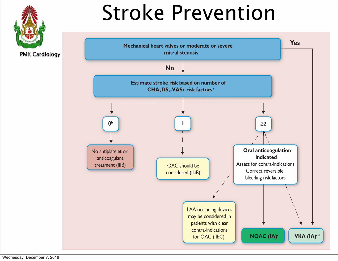
PMK Cardiology ReviewPMK Cardiology Review
Stroke Prevention
14
Recommendations for prediction of stroke andbleeding risk
Recommendations Classa Levelb Ref C
The CHA2DS2-VASc score is recommended for stroke risk prediction in patients with AF.
I A 368, 371, 386
Bleeding risk scores should be considered in AF patients on oral
risk factors for major bleeding.anticoagulation to identify modifiable IIa B
384, 386, 387,
389–392
Biomarkers such as high-sensitivity troponin and natriuretic peptide
stroke and bleeding risk in AF may be considered to further refine
patients.
IIb B 380–382, 387, 393
AF ! atrial fibrillation; CHA2DS2-VASc ! Congestive Heart failure,hypertension, Age !75 (doubled), Diabetes, Stroke (doubled), Vascular disease,Age 65–74, and Sex (female); OAC ! oral anticoagulation.aClass of recommendation.bLevel of evidence.cReference(s) supporting recommendations.
9.2 Stroke prevention9.2.1 Vitamin K antagonistsWarfarin and other VKAs were the first anticoagulants used in AFpatients. VKA therapy reduces the risk of stroke by two-thirds andmortality by one-quarter compared with control (aspirin or no ther-apy).38 VKAs have been used in many patients throughout the worldwith good outcomes,394–396 and this is reflected in the warfarinarms of theNOAC trials (see section 9.2.2.). The use of VKAs is limitedby the narrow therapeutic interval, necessitating frequent monitoringand dose adjustments, but VKAs, when deliveredwith adequate time intherapeutic range (TTR), are effective for stroke prevention in AF pa-tients. Clinical parameters can help to identify patients who are likely toachieve a decent TTR on VKA therapy.397 These have been summar-ized in the SAMe-TT2R2 score. Patients who fare well on this score,when treated with a VKA, have on average a higher TTR than patientswho do not fare well on the score.398,399 VKAs are currently the onlytreatment with established safety in AF patients with rheumatic mitralvalve disease and/or a mechanical heart valve prosthesis.400
9.2.2 Non-vitamin K antagonist oral anticoagulantsNOACs, including the direct thrombin inhibitor dabigatran and thefactor Xa inhibitors apixaban, edoxaban, and rivaroxaban, are
No
YesMechanical heart valves or moderate or severemitral stenosis
Estimate stroke risk based on number ofCHA2DS2-VASc risk factorsa
10b
OAC should beconsidered (IIaB)
LAA occluding devicesmay be considered in
patients with clearcontra-indicationsfor OAC (IIbC) NOAC (IA)c VKA (IA)c,d
No antiplatelet or anticoagulant
treatment (IIIB)
Oral anticoagulationindicated
Assess for contra-indicationsCorrect reversible bleeding risk factors
aCongestive heart failure, Hypertension, Age !75 years (2 points), Diabetes, prior Sstroke/TIA/embolus (2 points), Vascular disease, age 65–74 years, female Sex.bIncludes women without other stroke risk factors. cIIaB for women with only one additional stroke risk factor.dIB for patients with mechanical heart valves or mitral stenosis.
!2
Figure 8 Stroke prevention in atrial fibrillation.
ESC GuidelinesPage 24 of 90
by guest on September 1, 2016
http://eurheartj.oxfordjournals.org/D
ownloaded from
Wednesday, December 7, 2016

PMK Cardiology ReviewPMK Cardiology Review
15
suitable alternatives to VKAs for stroke prevention in AF (Table 13).Their use in clinical practice is increasing rapidly.401 All NOACs havea predictable effect (onset and offset) without need for regular an-ticoagulation monitoring. The phase III trials have been conductedwith carefully selected doses of the NOACs, including clear rulesfor dose reduction that should be followed in clinical practice(Table 13).
9.2.2.1 ApixabanIn the ARISTOTLE (Apixaban for Reduction in Stroke and Other
Thrombo-embolic Events in Atrial Fibrillation) trial,319 apixaban 5 mgtwice daily reduced stroke or systemic embolism by 21% comparedwith warfarin, combined with a 31% reduction in major bleeding andan 11% reduction in all-cause mortality (all statistically significant).Rates of haemorrhagic stroke and intracranial haemorrhage, but notof ischaemic stroke, were lower on apixaban. Rates of gastrointestinalbleeding were similar between the two treatment arms.402
Apixaban is the only NOAC that has been compared with aspirinin AF patients; apixaban significantly reduced stroke or systemic em-bolism by 55% compared with aspirin, with no or only a small differ-ence in rates of major bleeding or intracranial haemorrhage.354,403
9.2.2.2 DabigatranIn the RE-LY (Randomized Evaluation of Long-Term Anticoagula-
tion Therapy) study,318,404 dabigatran 150 mg twice daily reducedstroke and systemic embolism by 35% comparedwith warfarin with-out a significant difference in major bleeding events. Dabigatran110 mg twice daily was non-inferior to warfarin for prevention ofstroke and systemic embolism, with 20% fewer major bleedingevents. Both dabigatran doses significantly reduced haemorrhagicstroke and intracranial haemorrhage. Dabigatran 150 mg twice dailysignificantly reduced ischaemic stroke by 24% and vascular mortality
by 12%, while gastrointestinal bleeding was significantly increased by50%. There was a non-significant numerical increase in the rate ofmyocardial infarction with both dabigatran doses,318,404 which hasnot been replicated in large post-authorization analyses.396 Theseobservational data have also replicated the benefit of dabigatranover VKA found in the RE-LY trial in patients who were mainly trea-ted with the higher dabigatran dose (150 mg twice daily).396
9.2.2.3 EdoxabanIn the ENGAGE AF-TIMI 48 (Effective Anticoagulation with Fac-
tor Xa Next Generation in Atrial Fibrillation–Thrombolysis in
Table 11 Clinical risk factors for stroke, transientischaemic attack, and systemic embolism in theCHA2DS2-VASc score
CHA2DS2-VASc risk factor Points
Congestive heart failure Signs/symptoms of heart failure or objective evidence of reduced left-ventricular ejection fraction
+1
HypertensionResting blood pressure >140/90 mmHg on at least two occasions or current antihypertensive treatment
+1
Age 75 years or older +2
Diabetes mellitusFasting glucose >125 mg/dL (7 mmol/L) or treatment with oral hypoglycaemic agent and/or insulin
+1
Previous stroke, transient ischaemic attack, or thromboembolism
+2
Vascular disease Previous myocardial infarction, peripheral artery disease, or aortic plaque
+1
Age 65–74 years +1
Sex category (female) +1
CHA2DS2-VASc ! Congestive Heart failure, hypertension, Age !75 (doubled),Diabetes, Stroke (doubled), Vascular disease, Age 65–74, and Sex (female).
Table 12 Modifiable and non-modifiable risk factorsfor bleeding in anticoagulated patients based onbleeding risk scores
Hypertension (especially when systolic blood pressure is >160 mmHg)
Modifible bleeding risk factors
Potentially modifible bleeding risk factors
Non-modifible bleeding risk factors
a,b,c
Labile INR or time in therapeutic range <60%a in patients on vitamin K antagonists
Medication predisposing to bleeding, such as antiplatelet drugs and non-steroidal anti-inflammatoy drugsa,d
Excess alcohol (!8 drinks/week)a,b
Anaemiab,c,d
Impaired renal functiona,b,c,d
Impaired liver functiona,b
Reduced platelet count or functionb
Agee (>65 years)a (!75 years)b,c,d
History of major bleedinga,b,c,d
Previous strokea,b
Dialysis-dependent kidney disease or renal transplanta,c
Cirrhotic liver diseasea
Malignancyb
Genetic factorsb
Biomarker-based bleeding risk factors
High-sensitivity troponine
Growth differentiation factor-15e
Serum creatinine/estimated CrCle
ABC ! age, biomarkers, clinical history; ATRIA ! AnTicoagulation and Riskfactors In Atrial fibrillation; CKD ! chronic kidney disease; CrCl ! creatinineclearance; HAS-BLED ! hypertension, abnormal renal/liver function (1 pointeach), stroke, bleeding history or predisposition, labile INR, elderly (.65 years),drugs/alcohol concomitantly (1 point each); HEMORR2HAGES ! hepatic or renaldisease, ethanol abuse, malignancy, older (age .75), reduced platelet count orfunction, rebleeding risk (prior bleed; 2 points), hypertension (uncontrolled),anaemia, genetic factors (CYP 2C9 polymorphisms), excessive fall risk (includingneuropsychiatric disease), and stroke; INR ! international normalized ratio;ORBIT ! Outcomes Registry for Better Informed Treatment of Atrial Fibrillation;TTR ! time in therapeutic range; VKA ! vitamin K antagonist.aDerived from the HAS-BLED score.384bDerived from the HEMORR2HAGES score.383cDerived from the ATRIA score.385dDerived from the ORBIT score.388eDerived from the ABC bleeding score.387
ESC Guidelines Page 25 of 90
by guest on September 1, 2016
http://eurheartj.oxfordjournals.org/D
ownloaded from
Wednesday, December 7, 2016

PMK Cardiology ReviewPMK Cardiology Review
16
5.2.2 Prolonged monitoring for paroxysmal atrialfibrillationParoxysmal AF is often missed.120 Repeated daily ECG recordingsincreased the detection of silent, asymptomatic paroxysmal AF inan unselected Swedish population aged .75 years.120,135 Severalpatient-operated devices136,137 and extended continuous ECGmonitoring using skin patch recorders138 have been validated forthe detection of paroxysmal AF (Web Figure 1).139 The detectionof asymptomatic AF by new technologies, such as smartphone caseswith ECG electrodes, smart watches, and blood pressure machineswith AF detection algorithms, has not yet been formally evaluatedagainst an established arrhythmia detection method.140
5.2.3 Patients with pacemakers and implanted devicesImplanted pacemakers or defibrillators with an atrial lead allowcontinuous monitoring of atrial rhythm. Using this technology, pa-tients with atrial high rate episodes (AHRE) can be identified. De-pending on the risk profile of the population studied, such AHRE are
detected in 10–15% of pacemaker patients.141 AHRE are associatedwith an increased risk of overt AF [hazard ratio (HR) 5.56; 95% con-fidence interval (CI) 3.78–8.17; P, 0.001] and ischaemic stroke orsystemic embolism (HR 2.49; 95% CI 1.28–4.85; P ! 0.007). Thestroke risk in AHRE patients seems lower than the stroke risk in pa-tients with diagnosed AF, and not all AHRE represent AF.142 Strokesoften occur without AHRE detected within 30 days before theevent.143–147 Consequently, it is unclear whether AHRE imply thesame therapeutic requirements as overt AF,148 and the benefit ofOAC in patients with AHRE is tested in ongoing clinical trials [e.g.Apixaban for the Reduction of Thrombo-Embolism in Patients WithDevice-Detected Sub-Clinical Atrial Fibrillation (ARTESiA)(NCT01938248) and Non vitamin K antagonist Oral anticoagulantsin patients with Atrial High rate episodes (NOAH – AFNET 6)(NCT02618577)]. At present, pacemakers and implanted devicesshould be interrogated on a regular basis for AHRE, and patientswith AHRE should undergo further assessment of stroke risk factorsand for overt AF, including ECG monitoring (Figure 3).149
No AF detected
Stro
ke ri
sk lo
w
AF diagnosed
*
Patient without known AF presenting with atrial high rate episode(AHRE, >5–6 min and >180 bpm) detected by an implanted device
Assess eligibility for oral anticoagulation using CHA2DS2-VASc score
Verify presence of AF by ECG documentatione.g. resting ECGAmbulatory ECG recorderPatient-operated devicesReview device electrograms (if available) to determine whether it is AF
Consider patient characteristics(e.g. stroke risk score)and patient preference
Initiate oral anticoagulation(IA)
2DS2-VASc = Congestive Heart failure, hypertension, Age !75 (doubled), Diabetes, Stroke (doubled), Vascular disease, Age 65–74, and Sex (female); ECG = electrocardiogram; EHRA = European Heart Rhythm Association.*In rare individual circumstances, oral anticoagulation may be considered in patients with AHRE, but without diagnosed AF. This clearly needs discussion with the patient and careful
aAdapted from the report of the 3rd AFNET/EHRA consensus conference.150
No antithrombotictherapy (IB)
Figure 3 Management of AHRE detected by an implanted device. Adapted from the report of the 3rd AFNET/EHRA consensus conference.150
ESC GuidelinesPage 12 of 90
by guest on September 1, 2016
http://eurheartj.oxfordjournals.org/D
ownloaded from
Wednesday, December 7, 2016
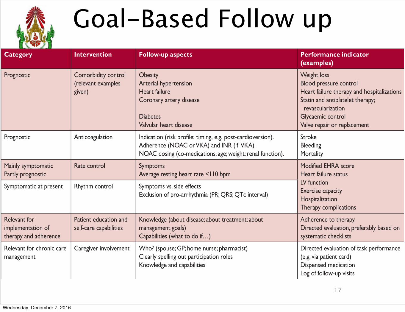
PMK Cardiology ReviewPMK Cardiology Review
Goal-Based Follow up
17
hence there is strong evidence that patients with a CHA2DS2-VASc risk score of 2 or more in men, and 3 or more in women,benefit from OAC. Fortunately, we now have a growing evidencebase regarding stroke risk in patients with one clinical risk factor(i.e. a CHA2DS2-VASc score of 1 for men, and 2 for women), al-though this relies largely on observed stroke rates in patients notreceiving OAC. In many of these patients, anticoagulation seemsto provide a clinical benefit.371 – 375 The rates of stroke andthrombo-embolism vary considerably in patients with CHA2DS2-VASc scores of 1 or 2 due to differences in outcomes, populations,and anticoagulation status (Web Table 1).371,376,377,1041 We there-fore commissioned an analysis of stroke risk in men and womenwith one additional stroke risk factor to inform these guidelines(Web Table 1, last line). OAC should be considered for menwith a CHA2DS2-VASc score of 1 and women with a score of 2,balancing the expected stroke reduction, bleeding risk, and pa-tient preference. Importantly, age (65 years and older) conveysa relatively high and continuously increasing stroke risk that alsopotentiates other risk factors (such as heart failure and sex).Hence, an individualized weighing of risk, as well as patient prefer-ences, should inform the decision to anticoagulate patients withonly one CHA2DS2-VASc risk factor, apart from female sex. Fe-male sex does not appear to increase stroke risk in the absenceof other stroke risk factors (Web Table 1).378,379
Measurement of cardiac troponin (high-sensitivity troponin T or I)andN-terminal pro-B-type natriuretic peptidemay provide additionalprognostic information in selected AF patients.380–382 Biomarker-based risk scores may, in the future, prove helpful to better stratifypatients (e.g. those at a truly low risk of stroke).75,382
9.1.3 Clinical risk scores for bleedingSeveral bleeding risk scores have been developed, mainly in pa-tients on VKAs. These include HAS-BLED [hypertension, abnor-mal renal/liver function (1 point each), stroke, bleeding history orpredisposition, labile INR, elderly (.65 years), drugs/alcohol con-comitantly (1 point each)], ORBIT (Outcomes Registry for BetterInformed Treatment of Atrial Fibrillation), and more recently, theABC (age, biomarkers, clinical history) bleeding score, which alsomakes use of selected biomarkers.383 – 385 Stroke and bleeding riskfactors overlap (compare Tables 11 and 12). For example, olderage is one of the most important predictors of both ischaemicstroke and bleeding in AF patients.386,387 A high bleeding riskscore should generally not result in withholding OAC. Rather,bleeding risk factors should be identified and treatable factorscorrected (see section 8.5). Table 12 provides details of modifi-able bleeding risk factors.
Table 10 Goal-based follow-up
Category Intervention Follow-up aspects Performance indicator (examples)
Prognostic Comorbidity control (relevant examples given)
ObesityArterial hypertensionHeart failureCoronary artery disease
DiabetesValvular heart disease
Weight lossBlood pressure controlHeart failure therapy and hospitalizationsStatin and antiplatelet therapy; revascularizationGlycaemic controlValve repair or replacement
Prognostic AnticoagulationAdherence (NOAC or VKA) and INR (if VKA).Indication (risk profile; timing, e.g. post-cardioversion).
NOAC dosing (co-medications; age; weight; renal function).
Stroke BleedingMortality
Mainly symptomaticPartly prognostic
Rate control SymptomsAverage resting heart rate <110 bpm Heart failure status
Modified EHRA score
LV functionExercise capacityHospitalizationTherapy complications
Symptomatic at present Rhythm control Symptoms vs. side effectsExclusion of pro-arrhythmia (PR; QRS; QTc interval)
Relevant for implementation of therapy and adherence
Patient education and self-care capabilities
Knowledge (about disease; about treatment; about management goals)Capabilities (what to do if…)
Adherence to therapyDirected evaluation, preferably based on systematic checklists
Relevant for chronic care management
Caregiver involvement Who? (spouse; GP; home nurse; pharmacist)Clearly spelling out participation rolesKnowledge and capabilities
Directed evaluation of task performance (e.g. via patient card)Dispensed medicationLog of follow-up visits
b.p.m. ! beats per minute; mEHRA symptoms scale ! modified European Heart Rhythm Association symptoms scale; GP ! general practitioner; INR ! international normalizedratio; LV ! left ventricular; NOAC ! non-vitamin K antagonist oral anticoagulant; VKA ! vitamin K antagonist.
ESC Guidelines Page 23 of 90
by guest on September 1, 2016
http://eurheartj.oxfordjournals.org/D
ownloaded from
Wednesday, December 7, 2016

PMK Cardiology ReviewPMK Cardiology Review
OAC
18
with increased mortality in patients on dialysis.417 There are norandomized trials assessing OAC in haemodialysis patients,418
and no controlled trials of NOACs in patients with severe CKD(CrCl ,25–30 mL/min).318 – 321 Warfarin use was associated ei-ther with a neutral or increased risk of stroke in database analysesof patients on dialysis,419 – 421 including a population-based ana-lysis in Canada (adjusted HR for stroke 1.14; 95% CI 0.78–1.67,adjusted HR for bleeding 1.44; 95% CI 1.13–1.85).422 In contrast,data from Denmark suggest a benefit of OAC in patients on renalreplacement therapy.423 Hence, controlled studies of anticoagu-lants (both VKAs and NOACs) in AF patients on dialysis areneeded.424
9.2.6 Patients with atrial fibrillation requiring kidneytransplantationThere are no randomized trials assessing OAC in patients after kid-ney transplantation. The prescription of NOAC therapy should be
guided by the estimated GFR of the transplanted kidney. Potentialpharmacokinetic interactions of OAC with immunosuppressiveagents should be considered.
9.2.7 Antiplatelet therapy as an alternative to oralanticoagulantsThe evidence supporting antiplatelet monotherapy for stroke pre-vention in AF is very limited.38,428–430 VKA therapy prevents stroke,systemic embolism, myocardial infarction, and vascular death betterthan single or dual antiplatelet therapy with aspirin and clopidogrel(annual risk of 5.6% for aspirin and clopidogrel vs. 3.9% with VKAtherapy).431 Even greater benefits were seen in VKA-treated pa-tients with a high TTR.432 Antiplatelet therapy increases bleedingrisk, especially dual antiplatelet therapy (2.0% vs. 1.3% with antipla-telet monotherapy; P, 0.001),433 with bleeding rates that are simi-lar to those on OAC.354,362,431,434 Thus, antiplatelet therapy cannotbe recommended for stroke prevention in AF patients.
Recommendations for stroke prevention in patients with atrial fibrillation
Recommendations Classa Levelb Ref C
Oral anticoagulation therapy to prevent thromboembolism is recommended for all male AF patients with a CHA2DS2-VASc score of 2 or more. I A
38, 318–321, 354, 404
Oral anticoagulation therapy to prevent thromboembolism is recommended in all female AF patients with a CHA2DS2-VASc score of 3 or more. I A
38,318–321, 354, 404
Oral anticoagulation therapy to prevent thromboembolism should be considered in male AF patients with a CHA2DS2-VASc score of 1, considering individual characteristics and patient preferences. IIa B 371,
375–377
Oral anticoagulation therapy to prevent thromboembolism should be considered in female AF patients with a CHA2DS2-VASc score of 2, considering individual characteristics and patient preferences. IIa B 371, 376,
377
Vitamin K antagonist therapy (INR 2.0–3.0 or higher) is recommended for stroke prevention in AF patients with moderate-to-severe mitral stenosis or mechanical heart valves. I B 274,
435–440
When oral anticoagulation is initiated in a patient with AF who is eligible for a NOAC (apixaban, dabigatran, edoxaban, or rivaroxaban), a NOAC is recommended in preference to a Vitamin K antagonist. I A
39, 318–321,
404
When patients are treated with a vitamin K antagonist, time in therapeutic range (TTR) should be kept as high as possible and closely monitored. I A 395, 432,
441-444
AF patients already on treatment with a vitamin K antagonist may be considered for NOAC treatment if TTR is not well controlled despite good adherence, or if patient preference without contra-indications to NOAC (e.g. prosthetic valve). IIb A
39, 318, 319, 404,
408
Combinations of oral anticoagulants and platelet inhibitors increase bleeding risk and should be avoided in AF patients without another indication for platelet inhibition.
III (harm) B 429, 445
In male or female AF patients without additional stroke risk factors, anticoagulant or antiplatelet therapy is not recommended for stroke prevention.
III (harm) B 368, 371,
376, 377
Antiplatelet monotherapy is not recommended for stroke prevention in AF patients, regardless of stroke risk. III (harm) A 38, 429,
430
NOACs (apixaban, dabigatran, edoxaban, and rivaroxaban) are not recommended in patients with mechanical heart valves (Level of evidence B) or moderate-to-severe mitral stenosis (Level of evidence C).
III (harm) B C 318–321,
400, 404
AF ! atrial fibrillation; CHA2DS2-VASc ! Congestive Heart failure, hypertension, Age !75 (doubled), Diabetes, Stroke (doubled), Vascular disease, Age 65–74, and Sex (female);INR ! international normalized ratio; NOAC ! non-vitamin K antagonist oral anticoagulant; OAC ! oral anticoagulation; TTR ! time in therapeutic range; VKA ! vitamin Kantagonist.aClass of recommendation.bLevel of evidence.cReference(s) supporting recommendations.
ESC GuidelinesPage 28 of 90
by guest on September 1, 2016
http://eurheartj.oxfordjournals.org/D
ownloaded from
Wednesday, December 7, 2016

PMK Cardiology ReviewPMK Cardiology Review
OAC
19
with increased mortality in patients on dialysis.417 There are norandomized trials assessing OAC in haemodialysis patients,418
and no controlled trials of NOACs in patients with severe CKD(CrCl ,25–30 mL/min).318 – 321 Warfarin use was associated ei-ther with a neutral or increased risk of stroke in database analysesof patients on dialysis,419 – 421 including a population-based ana-lysis in Canada (adjusted HR for stroke 1.14; 95% CI 0.78–1.67,adjusted HR for bleeding 1.44; 95% CI 1.13–1.85).422 In contrast,data from Denmark suggest a benefit of OAC in patients on renalreplacement therapy.423 Hence, controlled studies of anticoagu-lants (both VKAs and NOACs) in AF patients on dialysis areneeded.424
9.2.6 Patients with atrial fibrillation requiring kidneytransplantationThere are no randomized trials assessing OAC in patients after kid-ney transplantation. The prescription of NOAC therapy should be
guided by the estimated GFR of the transplanted kidney. Potentialpharmacokinetic interactions of OAC with immunosuppressiveagents should be considered.
9.2.7 Antiplatelet therapy as an alternative to oralanticoagulantsThe evidence supporting antiplatelet monotherapy for stroke pre-vention in AF is very limited.38,428–430 VKA therapy prevents stroke,systemic embolism, myocardial infarction, and vascular death betterthan single or dual antiplatelet therapy with aspirin and clopidogrel(annual risk of 5.6% for aspirin and clopidogrel vs. 3.9% with VKAtherapy).431 Even greater benefits were seen in VKA-treated pa-tients with a high TTR.432 Antiplatelet therapy increases bleedingrisk, especially dual antiplatelet therapy (2.0% vs. 1.3% with antipla-telet monotherapy; P, 0.001),433 with bleeding rates that are simi-lar to those on OAC.354,362,431,434 Thus, antiplatelet therapy cannotbe recommended for stroke prevention in AF patients.
Recommendations for stroke prevention in patients with atrial fibrillation
Recommendations Classa Levelb Ref C
Oral anticoagulation therapy to prevent thromboembolism is recommended for all male AF patients with a CHA2DS2-VASc score of 2 or more. I A
38, 318–321, 354, 404
Oral anticoagulation therapy to prevent thromboembolism is recommended in all female AF patients with a CHA2DS2-VASc score of 3 or more. I A
38,318–321, 354, 404
Oral anticoagulation therapy to prevent thromboembolism should be considered in male AF patients with a CHA2DS2-VASc score of 1, considering individual characteristics and patient preferences. IIa B 371,
375–377
Oral anticoagulation therapy to prevent thromboembolism should be considered in female AF patients with a CHA2DS2-VASc score of 2, considering individual characteristics and patient preferences. IIa B 371, 376,
377
Vitamin K antagonist therapy (INR 2.0–3.0 or higher) is recommended for stroke prevention in AF patients with moderate-to-severe mitral stenosis or mechanical heart valves. I B 274,
435–440
When oral anticoagulation is initiated in a patient with AF who is eligible for a NOAC (apixaban, dabigatran, edoxaban, or rivaroxaban), a NOAC is recommended in preference to a Vitamin K antagonist. I A
39, 318–321,
404
When patients are treated with a vitamin K antagonist, time in therapeutic range (TTR) should be kept as high as possible and closely monitored. I A 395, 432,
441-444
AF patients already on treatment with a vitamin K antagonist may be considered for NOAC treatment if TTR is not well controlled despite good adherence, or if patient preference without contra-indications to NOAC (e.g. prosthetic valve). IIb A
39, 318, 319, 404,
408
Combinations of oral anticoagulants and platelet inhibitors increase bleeding risk and should be avoided in AF patients without another indication for platelet inhibition.
III (harm) B 429, 445
In male or female AF patients without additional stroke risk factors, anticoagulant or antiplatelet therapy is not recommended for stroke prevention.
III (harm) B 368, 371,
376, 377
Antiplatelet monotherapy is not recommended for stroke prevention in AF patients, regardless of stroke risk. III (harm) A 38, 429,
430
NOACs (apixaban, dabigatran, edoxaban, and rivaroxaban) are not recommended in patients with mechanical heart valves (Level of evidence B) or moderate-to-severe mitral stenosis (Level of evidence C).
III (harm) B C 318–321,
400, 404
AF ! atrial fibrillation; CHA2DS2-VASc ! Congestive Heart failure, hypertension, Age !75 (doubled), Diabetes, Stroke (doubled), Vascular disease, Age 65–74, and Sex (female);INR ! international normalized ratio; NOAC ! non-vitamin K antagonist oral anticoagulant; OAC ! oral anticoagulation; TTR ! time in therapeutic range; VKA ! vitamin Kantagonist.aClass of recommendation.bLevel of evidence.cReference(s) supporting recommendations.
ESC GuidelinesPage 28 of 90
by guest on September 1, 2016
http://eurheartj.oxfordjournals.org/D
ownloaded from
Wednesday, December 7, 2016

PMK Cardiology ReviewPMK Cardiology Review
OAC
20
with increased mortality in patients on dialysis.417 There are norandomized trials assessing OAC in haemodialysis patients,418
and no controlled trials of NOACs in patients with severe CKD(CrCl ,25–30 mL/min).318 – 321 Warfarin use was associated ei-ther with a neutral or increased risk of stroke in database analysesof patients on dialysis,419 – 421 including a population-based ana-lysis in Canada (adjusted HR for stroke 1.14; 95% CI 0.78–1.67,adjusted HR for bleeding 1.44; 95% CI 1.13–1.85).422 In contrast,data from Denmark suggest a benefit of OAC in patients on renalreplacement therapy.423 Hence, controlled studies of anticoagu-lants (both VKAs and NOACs) in AF patients on dialysis areneeded.424
9.2.6 Patients with atrial fibrillation requiring kidneytransplantationThere are no randomized trials assessing OAC in patients after kid-ney transplantation. The prescription of NOAC therapy should be
guided by the estimated GFR of the transplanted kidney. Potentialpharmacokinetic interactions of OAC with immunosuppressiveagents should be considered.
9.2.7 Antiplatelet therapy as an alternative to oralanticoagulantsThe evidence supporting antiplatelet monotherapy for stroke pre-vention in AF is very limited.38,428–430 VKA therapy prevents stroke,systemic embolism, myocardial infarction, and vascular death betterthan single or dual antiplatelet therapy with aspirin and clopidogrel(annual risk of 5.6% for aspirin and clopidogrel vs. 3.9% with VKAtherapy).431 Even greater benefits were seen in VKA-treated pa-tients with a high TTR.432 Antiplatelet therapy increases bleedingrisk, especially dual antiplatelet therapy (2.0% vs. 1.3% with antipla-telet monotherapy; P, 0.001),433 with bleeding rates that are simi-lar to those on OAC.354,362,431,434 Thus, antiplatelet therapy cannotbe recommended for stroke prevention in AF patients.
Recommendations for stroke prevention in patients with atrial fibrillation
Recommendations Classa Levelb Ref C
Oral anticoagulation therapy to prevent thromboembolism is recommended for all male AF patients with a CHA2DS2-VASc score of 2 or more. I A
38, 318–321, 354, 404
Oral anticoagulation therapy to prevent thromboembolism is recommended in all female AF patients with a CHA2DS2-VASc score of 3 or more. I A
38,318–321, 354, 404
Oral anticoagulation therapy to prevent thromboembolism should be considered in male AF patients with a CHA2DS2-VASc score of 1, considering individual characteristics and patient preferences. IIa B 371,
375–377
Oral anticoagulation therapy to prevent thromboembolism should be considered in female AF patients with a CHA2DS2-VASc score of 2, considering individual characteristics and patient preferences. IIa B 371, 376,
377
Vitamin K antagonist therapy (INR 2.0–3.0 or higher) is recommended for stroke prevention in AF patients with moderate-to-severe mitral stenosis or mechanical heart valves. I B 274,
435–440
When oral anticoagulation is initiated in a patient with AF who is eligible for a NOAC (apixaban, dabigatran, edoxaban, or rivaroxaban), a NOAC is recommended in preference to a Vitamin K antagonist. I A
39, 318–321,
404
When patients are treated with a vitamin K antagonist, time in therapeutic range (TTR) should be kept as high as possible and closely monitored. I A 395, 432,
441-444
AF patients already on treatment with a vitamin K antagonist may be considered for NOAC treatment if TTR is not well controlled despite good adherence, or if patient preference without contra-indications to NOAC (e.g. prosthetic valve). IIb A
39, 318, 319, 404,
408
Combinations of oral anticoagulants and platelet inhibitors increase bleeding risk and should be avoided in AF patients without another indication for platelet inhibition.
III (harm) B 429, 445
In male or female AF patients without additional stroke risk factors, anticoagulant or antiplatelet therapy is not recommended for stroke prevention.
III (harm) B 368, 371,
376, 377
Antiplatelet monotherapy is not recommended for stroke prevention in AF patients, regardless of stroke risk. III (harm) A 38, 429,
430
NOACs (apixaban, dabigatran, edoxaban, and rivaroxaban) are not recommended in patients with mechanical heart valves (Level of evidence B) or moderate-to-severe mitral stenosis (Level of evidence C).
III (harm) B C 318–321,
400, 404
AF ! atrial fibrillation; CHA2DS2-VASc ! Congestive Heart failure, hypertension, Age !75 (doubled), Diabetes, Stroke (doubled), Vascular disease, Age 65–74, and Sex (female);INR ! international normalized ratio; NOAC ! non-vitamin K antagonist oral anticoagulant; OAC ! oral anticoagulation; TTR ! time in therapeutic range; VKA ! vitamin Kantagonist.aClass of recommendation.bLevel of evidence.cReference(s) supporting recommendations.
ESC GuidelinesPage 28 of 90
by guest on September 1, 2016
http://eurheartj.oxfordjournals.org/D
ownloaded from
Wednesday, December 7, 2016

PMK Cardiology ReviewPMK Cardiology Review
21
Recommendations for combination therapy with oral anticoagulants and antiplatelets
Recommendations Classa Levelb Ref C
After elective coronary stenting for stable coronary artery disease in AF patients at risk of stroke, combination triple therapy with aspirin, clopidogrel and an oral anticoagulant should be considered for 1 month to prevent recurrent coronary and cerebral ischaemic events.
IIa B 522, 524
After an ACS with stent implantation in AF patients at risk of stroke, combination triple therapy with aspirin, clopidogrel and an oral anticoagulant should be considered for 1–6 months to prevent recurrent coronary and cerebral ischaemic events. IIa C 520
After an ACS without stent implantation in AF patients at risk of stroke, dual treatment with an oral anticoagulant and aspirin or clopidogrel should be considered for up to 12 months to prevent recurrent coronary and cerebral ischaemic events. IIa C
The duration of combination antithrombotic therapy, especially triple therapy, should be kept to a limited period, balancing the estimated risk of recurrent coronary events and bleeding. IIa B 520
Dual therapy with any oral anticoagulant plus clopidogrel 75 mg/day may be considered as an alternative to initial triple therapy in selected patients. IIb C 524, 525
ACS ! acute coronary syndromes; AF ! atrial fibrillation; OAC ! oral anticoagulant.aClass of recommendation.bLevel of evidence.cReference(s) supporting recommendations.
AF patient in need of OAC after an ACS
Bleeding risk lowcompared to risk for ACS
or stent thrombosis
Time from ACS0
1 month
3 months
6 months
12 months
lifelong
Triple therapya (IIaB)
OAC
Dual therapyb (IIaC)
OAC monotherapyc (IB)
Bleeding risk highcompared to risk for ACS
or stent thrombosis
Aspirin 75–100 mg daily Clopidogrel 75 mg daily
Triple therapya (IIaB)Triple therapy (IIaB)
Dual therapyb (IIaC)
OAC monotherapyc (IB)
A or C
A or C
PCI = percutaneous coronary intervention.aDual therapy with OAC and aspirin or clopidogrel may be considered in selected patients, especially those not receiving a stent or patients at a longer time from the index event. bOAC plus single antiplatelet. cDual therapy with OAC and an antiplatelet agent (aspirin or clopidogrel) may be considered in patients at high risk of coronary events.
Figure 12 Antithrombotic therapy after an acute coronary syndrome in atrial fibrillation patients requiring anticoagulation.
ESC Guidelines Page 35 of 90
by guest on September 1, 2016
http://eurheartj.oxfordjournals.org/D
ownloaded from
Wednesday, December 7, 2016

PMK Cardiology ReviewPMK Cardiology Review
22
10. Rate control therapy in atrialfibrillationRate control is an integral part of the management of AF patients,and is often sufficient to improve AF-related symptoms. Comparedwith stroke prevention and rhythm control, very little robust evi-dence exists to inform the best type and intensity of rate controltreatment, with the majority of data derived from short-term cross-over trials and observational studies.41,526–528 Pharmacological ratecontrol can be achieved for acute or long-term rate control withbeta-blockers, digoxin, the calcium channel blockers diltiazem andverapamil, or combination therapy (Table 15). A number of antiar-rhythmic drugs also have rate-limiting properties (amiodarone, dro-nedarone, sotalol, and to some extent propafenone), but theyshould only be used in patients needing rhythm control therapy(see Chapter 11).
10.1 Acute rate controlIn the setting of acute new-onset AF, patients are often in need ofheart rate control. Physicians should evaluate underlying causes ofelevated heart rate, such as infection, endocrine imbalance, anaemia,and pulmonary embolism. For acute rate control, beta-blockers and
diltiazem/verapamil are preferred over digoxin because of theirrapid onset of action and effectiveness at high sympathetictone.528 – 532 The choice of drug (Table 15) and target heartrate will depend on patient characteristics, symptoms, LVEF andhaemodynamics, but a lenient initial approach to heart rate seemsacceptable. Combination therapy may be required (Figure 14). Inpatients with HFrEF, beta-blockers, digitalis (digoxin or digitoxin),or their combination should be used,218,533 as diltiazem andverapamil can have negative inotropic effects in patients withLVEF ,40%.222,534,535 In critically ill patients and those withseverely impaired LV systolic function, intravenous amiodaronecan be used where excess heart rate is leading to haemodynamicinstability.536 – 538 Urgent cardioversion should be considered inunstable patients (see chapter 11.2).
10.2 Long-term pharmacological ratecontrol10.2.1 Beta-blockersBeta-adrenoreceptor blocker monotherapy is often the first-linerate-controlling agent,539 largely based on observations of betteracute heart rate control than digoxin. Interestingly, the prognosticbenefit of beta-blockers seen in HFrEF patients with sinus rhythm
AF patient in need of OAC after elective PCI with stent
Bleeding risk lowcompared to risk for ACS
or stent thrombosis
Time from PCI0
1 month
3 months
6 months
12 months
lifelong
OAC
Dual therapyb (IIaC)
OAC monotherapyc (IB)
Bleeding risk highcompared to risk for ACS
or stent thrombosis
Aspirin 75–100 mg daily Clopidogrel 75 mg daily
Triple therapya (IIaB)
Dual therapyb (IIaC)
OAC monotherapyc (IB)
A or C
A or C
percutaneous coronary intervention.aDual therapy with OAC and aspirin or clopidogrel may be considered in selected patients. bOAC plus single antiplatelet. cDual therapy with OAC and an antiplatelet agent (aspirin or clopidogrel) may be considered in patients at high risk of coronary events.
Figure 13 Antithrombotic therapy after elective percutaneous intervention in atrial fibrillation patients requiring anticoagulation.
ESC GuidelinesPage 36 of 90
by guest on September 1, 2016
http://eurheartj.oxfordjournals.org/D
ownloaded from
Wednesday, December 7, 2016

PMK Cardiology ReviewPMK Cardiology Review
Stroke prevention for Cardioversion
23
Recommendations for rhythm control therapy
Recommendations Classa Levelb Ref C
General recommendations
Rhythm control therapy is indicated for symptom improvement in patients with AF. I B 120, 586, 601
Management of cardiovascular risk factors and avoidance of AF triggers should be pursued in patients on rhythm control therapy to facilitate maintenance of sinus rhythm. IIa B 203, 204,
296, 312
With the exception of AF associated with haemodynamic instability, the choice between electrical and pharmacological cardioversion should be guided by patient and physician preferences. IIa C
Cardioversion of AF
Electrical cardioversion of AF is recommended in patients with acute haemodynamic instability to restore cardiac output. I B 612, 702-704
Cardioversion of AF (either electrical or pharmacological) is recommended in symptomatic patients with persistent or long-standing persistent AF as part of rhythm control therapy. I B
584, 601, 627, 628, 648, 705
cardioversion and prevent recurrent AF.Pre-treatment with amiodarone, flecainide, ibutilide, or propafenone should be considered to enhance success of electrical IIa B 248, 584,
633
recommended for pharmacological cardioversion of new-onset AF.In patients with no history of ischaemic or structural heart disease, flecainide, propafenone, or vernakalant are I A
602–605, 614, 618, 622, 706,
707
In patients with no history of ischaemic or structural heart disease, ibutilide should be considered for pharmacological conversion of AF. IIa B
safety assessment.
In selected patients with recent-onset AF and no significant structural or ischaemic heart disease, a single oral dose offlecainide or propafenone (the ‘pill in the pocket’ approach) should be considered for patient-led cardioversion, following IIa B 620, 621
In patients with ischaemic and/or structural heart disease, amiodarone is recommended for cardioversion of AF. I A 597–601
Vernakalant may be considered as an alternative to amiodarone for pharmacological conversion of AF in patients without hypotension, severe heart failure or severe structural heart disease (especially aortic stenosis). IIb B 602–605,
616, 618
Stroke prevention in patients designated for cardioversion of AF
Anticoagulation with heparin or a NOAC should be initiated as soon as possible before every cardioversion of AF or atrial IIa B 708, 709
cardioversion.For cardioversion of AF/atrial flutter, effective anticoagulation is recommended for a minimum of 3 weeks before
flutter.
I B 648, 708
Transoesophageal echocardiography (TOE) is recommended to exclude cardiac thrombus as an alternative to preprocedural anticoagulation when early cardioversion is planned. I B 648, 708
IIa B 648
In patients at risk for stroke, anticoagulant therapy should be continued long-term after cardioversion according to the
Early cardioversion can be performed without TOE in patients with a definite duration of AF <48 hours.
long-term anticoagulation recommendations, irrespective of the method of cardioversion or the apparent maintenance of sinus rhythm. In patients without stroke risk factors, anticoagulation is recommended for 4 weeks after cardioversion.
I B 353, 710
I C
A repeat TOE to ensure thrombus resolution should be considered before cardioversion.
In patients where thrombus is identified on TOE, effective anticoagulation is recommended for at least 3 weeks.
IIa C
AAD for the long-term maintenance of sinus rhythm/prevention of recurrent AF
The choice of AAD needs to be carefully evaluated, taking into account the presence of comorbidities, cardiovascular risk and potential for serious proarrhythmia, extracardiac toxic effects, patient preferences, and symptom burden. I A 41, 580
patients with normal left ventricular function and without pathological left ventricular hypertrophy. Dronedarone, flecainide, propafenone, or sotalol are recommended for prevention of recurrent symptomatic AF in I A
581, 583, 584, 588,
601
Dronedarone is recommended for prevention of recurrent symptomatic AF in patients with stable coronary artery disease, and without heart failure. I A 583, 588
Amiodarone is recommended for prevention of recurrent symptomatic AF in patients with heart failure. I B 596–598
Amiodarone is more effective in preventing AF recurrences than other AAD, but extracardiac toxic effects are common and increase with time. For this reason, other AAD should be considered first.
Patients on AAD therapy should be periodically evaluated to confirm their eligibility for treatment.
IIa C 596–598
IIa C583, 588, 657, 658,
660
Continued
ESC Guidelines Page 45 of 90
by guest on September 1, 2016
http://eurheartj.oxfordjournals.org/D
ownloaded from
Wednesday, December 7, 2016

PMK Cardiology ReviewPMK Cardiology Review
Cardioversion
24
Recommendations for rhythm control therapy
Recommendations Classa Levelb Ref C
General recommendations
Rhythm control therapy is indicated for symptom improvement in patients with AF. I B 120, 586, 601
Management of cardiovascular risk factors and avoidance of AF triggers should be pursued in patients on rhythm control therapy to facilitate maintenance of sinus rhythm. IIa B 203, 204,
296, 312
With the exception of AF associated with haemodynamic instability, the choice between electrical and pharmacological cardioversion should be guided by patient and physician preferences. IIa C
Cardioversion of AF
Electrical cardioversion of AF is recommended in patients with acute haemodynamic instability to restore cardiac output. I B 612, 702-704
Cardioversion of AF (either electrical or pharmacological) is recommended in symptomatic patients with persistent or long-standing persistent AF as part of rhythm control therapy. I B
584, 601, 627, 628, 648, 705
cardioversion and prevent recurrent AF.Pre-treatment with amiodarone, flecainide, ibutilide, or propafenone should be considered to enhance success of electrical IIa B 248, 584,
633
recommended for pharmacological cardioversion of new-onset AF.In patients with no history of ischaemic or structural heart disease, flecainide, propafenone, or vernakalant are I A
602–605, 614, 618, 622, 706,
707
In patients with no history of ischaemic or structural heart disease, ibutilide should be considered for pharmacological conversion of AF. IIa B
safety assessment.
In selected patients with recent-onset AF and no significant structural or ischaemic heart disease, a single oral dose offlecainide or propafenone (the ‘pill in the pocket’ approach) should be considered for patient-led cardioversion, following IIa B 620, 621
In patients with ischaemic and/or structural heart disease, amiodarone is recommended for cardioversion of AF. I A 597–601
Vernakalant may be considered as an alternative to amiodarone for pharmacological conversion of AF in patients without hypotension, severe heart failure or severe structural heart disease (especially aortic stenosis). IIb B 602–605,
616, 618
Stroke prevention in patients designated for cardioversion of AF
Anticoagulation with heparin or a NOAC should be initiated as soon as possible before every cardioversion of AF or atrial IIa B 708, 709
cardioversion.For cardioversion of AF/atrial flutter, effective anticoagulation is recommended for a minimum of 3 weeks before
flutter.
I B 648, 708
Transoesophageal echocardiography (TOE) is recommended to exclude cardiac thrombus as an alternative to preprocedural anticoagulation when early cardioversion is planned. I B 648, 708
IIa B 648
In patients at risk for stroke, anticoagulant therapy should be continued long-term after cardioversion according to the
Early cardioversion can be performed without TOE in patients with a definite duration of AF <48 hours.
long-term anticoagulation recommendations, irrespective of the method of cardioversion or the apparent maintenance of sinus rhythm. In patients without stroke risk factors, anticoagulation is recommended for 4 weeks after cardioversion.
I B 353, 710
I C
A repeat TOE to ensure thrombus resolution should be considered before cardioversion.
In patients where thrombus is identified on TOE, effective anticoagulation is recommended for at least 3 weeks.
IIa C
AAD for the long-term maintenance of sinus rhythm/prevention of recurrent AF
The choice of AAD needs to be carefully evaluated, taking into account the presence of comorbidities, cardiovascular risk and potential for serious proarrhythmia, extracardiac toxic effects, patient preferences, and symptom burden. I A 41, 580
patients with normal left ventricular function and without pathological left ventricular hypertrophy. Dronedarone, flecainide, propafenone, or sotalol are recommended for prevention of recurrent symptomatic AF in I A
581, 583, 584, 588,
601
Dronedarone is recommended for prevention of recurrent symptomatic AF in patients with stable coronary artery disease, and without heart failure. I A 583, 588
Amiodarone is recommended for prevention of recurrent symptomatic AF in patients with heart failure. I B 596–598
Amiodarone is more effective in preventing AF recurrences than other AAD, but extracardiac toxic effects are common and increase with time. For this reason, other AAD should be considered first.
Patients on AAD therapy should be periodically evaluated to confirm their eligibility for treatment.
IIa C 596–598
IIa C583, 588, 657, 658,
660
Continued
ESC Guidelines Page 45 of 90 by guest on Septem
ber 1, 2016http://eurheartj.oxfordjournals.org/
Dow
nloaded from
Wednesday, December 7, 2016

PMK Cardiology ReviewPMK Cardiology Review
25
(,2–4 h), charcoal administration and/or gastric lavage will reducefurther exposure. Dialysis clears dabigatran but is less effective forthe other NOACs.Immediate reversal of the antithrombotic effect is indicated in se-
vere or life-threatening bleeding events. An agreed institutionalprocedure for the management of life-threatening bleeds should bedocumented and accessible at all times to ensure adequate initial man-agement. For VKAs, administration of fresh frozen plasma restores co-agulation more rapidly than vitamin K, and prothrombin complexconcentrates achieve even faster blood coagulation.505 Registry datasuggest that the combination of plasma and prothrombin complexconcentrates is associated with the lowest case fatality following intra-cranial haemorrhage on VKA treatment with an INR !1.3.506 In a mul-ticentre randomized trial of 188 patients, four-factor prothrombincomplex concentrates achieved more rapid INR reversal and effectivehaemostasis than plasma in patients undergoing urgent surgical or in-vasive procedures.507 Administration of prothrombin complex con-centrates may also be considered for severe bleeding on NOACtreatment if specific antidotes are not available.
Several antidotes toNOACs are under development. Idarucizumab(approved in 2015 by the US Food and Drug Administration and theEuropean Medicines Agency) is a clinically available humanized anti-body fragment that binds dabigatran and rapidly and dose-dependentlyreverses its effects without over-correction or thrombin gener-ation.475 Andexanet alpha, a modified recombinant human factor Xathat lacks enzymatic activity, reverses the anticoagulant activity of fac-tor Xa antagonists in healthy subjects within minutes after administra-tion and for the duration of infusion, with a transient increase inmarkers of coagulation activity of uncertain clinical relevance.508 An-other agent under development is ciraparantag (PER977), an antidotedesigned to reverse both direct thrombin and factor Xa inhibitors aswell as the indirect inhibitor enoxaparin.509 The clinical usefulness ofthese specific antidotes needs further evaluation.
9.6.2 Oral anticoagulation in atrial fibrillation patients atrisk of or having a bleeding eventWhile anticoagulation therapy should be paused to control activebleeding, absolute contraindications to long-term OAC after a
VKA NOAC
Delay VKA until INR <2 Minor
Moderate - severe
Severe orlife-threatening
Delay NOAC for 1 dose or 1 day
Add symptomatic treatment:Fluid replacementBlood transfusion
Treat bleeding cause(e.g. gastroscopy)
Consider to addVitamin K (1–10 mg) i.v.
Add symptomatic treatment:Fluid replacementBlood transfusion
Treat bleeding cause(e.g. gastroscopy)
Consider to add oral charcoalif recently ingested NOAC
Consider PCC and FFPConsider replacement of platelets
where appropriate
Consider specific antidote, orPCC if no antidote available
Consider replacement of plateletswhere appropriate
Patient with active bleeding
Compress bleeding sites mechanically
Assess haemodynamic status, blood pressure, basic coagulationparameters, blood count, and kidney function
Obtain anticoagulation history (last NOAC / VKA dose)
FFP = fresh frozen plasma; INR = international normalized ratio; i.v. = intravenous; NOAC = non-vitamin K antagonist oral anticoagulant; OAC = oral anticoagulation; PCC = prothrombin complex concentrates; VKA = vitamin K antagonist.
Figure 11 Management of active bleeding in patients receiving anticoagulation. Institutions should have an agreed procedure in place.
ESC Guidelines Page 33 of 90
by guest on September 1, 2016
http://eurheartj.oxfordjournals.org/D
ownloaded from
Wednesday, December 7, 2016

PMK Cardiology ReviewPMK Cardiology Review
26
stroke are scarce. Parenteral anticoagulants seem to be associatedwith a non-significant reduction in recurrent ischaemic strokewhen administered 7–14 days after the acute stroke [odds ratio(OR) 0.68; 95% CI 0.44–1.06), with a significant increase in symp-tomatic intracranial bleeding (OR 2.89; 95% CI 1.19–7.01), and asimilar rate of death or disability at final follow-up.477 It seems likelythat the bleeding risk on parenteral anticoagulation exceeds thestroke prevention benefit in the first days after a large stroke,whereas patients with a TIA or a small stroke may benefit from early(immediate) initiation or continuation of anticoagulation. Therefore,we propose to initiate anticoagulation in AF patients between 1and 12 days after an ischaemic stroke, depending on stroke severity(Figure 9).478 We suggest repeat brain imaging to determine the op-timal initiation of anticoagulation in patients with a large stroke atrisk for haemorrhagic transformation. Long-term OAC with aVKA363,479–481 or NOAC482 conveys benefits in AF patients whosurvived a stroke. NOACs seem to convey slightly better outcomes,mainly driven by fewer intracranial haemorrhages and haemorrhagic
strokes (OR 0.44; 95%CI 0.32–0.62).482 Detailed data for edoxabanhave not yet been published.321 If a patient suffers a stroke or TIAwhilst taking an anticoagulant, switching to another anticoagulantshould be considered.
9.4.3 Initiation of anticoagulation after intracranialhaemorrhageNo prospective studies have investigated the benefit or risk of the ini-tiation of OAC after intracranial haemorrhage,483 and patients with ahistory of intracranial bleeding were excluded from the randomizedtrials comparing NOACs with VKAs. The available evidence indicatesthat anticoagulation in patients with AF can be reinitiated after 4–8weeks, especially when the cause of bleeding or the relevant risk fac-tor (e.g. uncontrolled hypertension, see Table 12) has been treated,and that such treatment leads to fewer recurrent (ischaemic) strokesand lower mortality.460,484 If anticoagulation is resumed, it seems rea-sonable to consider anticoagulantswith a lowbleeding risk.39 Figure 10depicts a consensus opinion on the initiation or resumption of OAC
StartOAC
Consider additional clinical factors favouring early / delayed initiation of OAC
TIAMild stroke(NIHSS <8)
Moderate stroke(NIHSS 8–15)
Evaluate haemorrhagictransformation by
CT or MRI at day 6
Evaluate haemorrhagictransformation by
CT or MRI at day 12
6 days afteracute event
12 days afteracute event
1 day afteracute event
3 days afteracute event
Factors favouring early initiation of OAC:
Low NIHSS (<8):Small/no brain infarction on imagingHigh recurrence risk, e.g. cardiac thrombus on echoNo need for percutaneous endoscopic gastrostomyNo need for carotid surgeryNo haemorrhagic transformationClinically stableYoung patientBlood pressure is controlled
Factors favouring delayed initiation of OAC:
Large/moderate brain infarction on imagingNeeds gastrostomy or major surgical interventionNeeds carotid surgeryHaemorrhagic transformationNeurologically unstableElderly patientUncontrolled hypertension
Patient with atrial fibrillation and acute TIA or ischaemic strokeExclusion of intracerebral bleeding by CT or MRI
NIH_Stroke_Scale.pdf); OAC = oral anticoagulation; TIA = transient ischaemic attack
Severe stroke(NIHSS !16)
High NIHSS (!8):
Figure 9 Initiation or continuation of anticoagulation in atrial fibrillation patients after a stroke or transient ischaemic attack. This approach isbased on consensus rather than prospective data.
ESC GuidelinesPage 30 of 90 by guest on Septem
ber 1, 2016http://eurheartj.oxfordjournals.org/
Dow
nloaded from
Wednesday, December 7, 2016

PMK Cardiology ReviewPMK Cardiology Review
27
after an intracranial haemorrhage.We recommend amultidisciplinarydecision with input from stroke physicians/neurologists, cardiologists,neuroradiologists, and neurosurgeons.
Recommendations for secondary stroke prevention
Recommendations Classa Levelb Ref C
Anticoagulation with heparin or LMWH immediately after an ischaemic stroke is not recommended in AF patients.
III (harm) A 477
In patients who suffer a TIA or stroke while on anticoagulation, adherence to therapy should be assessed and optimized.
IIa C
In patients who suffer a moderate-to-severe ischaemic stroke while on anticoagulation, anticoagulation should be interrupted for 3–12 days based on a multidisciplinary assessment of acute stroke and bleeding risk.
IIa C
In AF patients who suffer a stroke, aspirin should be considered for prevention of secondary stroke until the initiation or resumption of oral anticoagulation.
IIa B 485
Systemic thrombolysis with rtPA is not recommended if the INR is above 1.7 (or, for patients on dabigatran, if aPTT is outside normal range).
III (harm) C 472, 474
NOACs are recommended in preference to VKAs or aspirin in AF patients with a previous stroke.
I B 363,482
After TIA or stroke, combination therapy of OAC and an antiplatelet is not recommended.
III (harm) B 486
After intracranial haemorrhage, oral anticoagulation in patients with AF may be reinitiated after 4–8 weeks provided the cause of bleeding or the relevant risk factor has been treated or controlled.
IIb B 483, 484, 487
AF ! atrial fibrillation; INR ! international normalized ratio; NOAC !non-vitamin K antagonist oral anticoagulant; OAC ! oral anticoagulation;TIA ! transient ischaemic attack; VKA ! vitamin K antagonist.aClass of recommendation.bLevel of evidence.cReference(s) supporting recommendations.
9.5 Strategies to minimize bleeding onanticoagulant therapyIn a meta-analysis of 47 studies, the overall incidence of major bleedingwith VKAs was 2.1 (range 0.9–3.4) per 100 patient-years in controlledtrials and 2.0 (range 0.2–7.6) per 100 patient-years for observationaldata sets.488 Minimizing treatable bleeding risk factors (see Table 12)seems paramount to reduce the bleeding rate on anticoagulants.
9.5.1 Uncontrolled hypertensionUncontrolled hypertension increases the risk of bleeding onOAC.53 Hence, keeping systolic blood pressure well controlledis of particular relevance in anticoagulated patients with AF.
Treatment according to current guidelines is recommended in pa-tients with known hypertension.489
9.5.2 Previous bleeding eventHistory of bleeding events and the presence of anaemia are import-ant parts of the assessment of all patients receiving OAC. The ma-jority of bleeding events are gastrointestinal. Compared withwarfarin, the risk of gastrointestinal bleeds was increased for dabiga-tran 150 mg twice daily,396,490 rivaroxaban 20 mg once daily,491 andedoxaban 60 mg once daily.321 The risk of gastrointestinal bleedswas comparable to warfarin on dabigatran 110 mg twice daily490
and on apixaban 5 mg twice daily.319 Recent observational analysesdo not replicate these findings, suggesting a smaller effect.396,492,493
In patients in whom the source of bleeding has been identified andcorrected, OAC can be reinitiated. This also appears true for pa-tients who have had an intracranial haemorrhage, once modifiablebleeding risk factors (e.g. uncontrolled hypertension) have beencorrected.460,484
9.5.3 Labile international normalized ratio and adequatenon-vitamin K antagonist oral anticoagulant dosingTTR on VKA therapy is an important predictor of major haemor-rhage.432,441,494 Therefore, we recommend targeting the INR be-tween 2.0 and 3.0 in patients on VKAs, maintaining a high TTR(e.g. !70%494), and to consider switching to a NOAC when ahigh TTR cannot be sustained.444 NOAC dosing should follow thedose-reduction criteria evaluated in the clinical trials, consideringrenal function, age, and weight. Patient information and empower-ment, best delivered through integrated AF management, seemparamount to achieve this goal.
9.5.4 Alcohol abuseAlcohol excess is a risk factor for bleeding in anticoagulated pa-tients,384 mediated by poor adherence, liver disease, variceal bleed-ing, and risk of major trauma. Severe alcohol abuse and bingedrinking habits should be corrected in patients eligible for OAC.
9.5.5 Falls and dementiaFalls and dementia are associated with increased mortality in AF pa-tients,495 without evidence that these conditions markedly increasethe risk of intracranial haemorrhage.495,496 Hence, anticoagulationshould only be withheld from patients with severe uncontrolled falls(e.g. epilepsy or advanced multisystem atrophy with backwardsfalls), or in selected patients with dementia where compliance andadherence cannot be ensured by a caregiver.
9.5.6 Genetic testingIn addition to food and drug interactions, multiple genetic variationsaffect the metabolism of VKAs.497 The systematic use of genetic in-formation for adjustment of VKA dosage has been evaluated in sev-eral controlled clinical studies.498–500 Genetic testing has little effecton TTR or bleeding risk on warfarin, and is not recommended forclinical use at present.501
9.5.7 Bridging periods off oral anticoagulationMost cardiovascular interventions (e.g. percutaneous coronaryintervention or pacemaker implantation) can be performed safelyon continued OAC. When interruption of OAC is required,bridging does not seem to be beneficial, except in patients withmechanical heart valves: In a randomized trial of 1884 patientswith AF, interruption of anticoagulation was non-inferior to
ESC Guidelines Page 31 of 90
by guest on September 1, 2016
http://eurheartj.oxfordjournals.org/D
ownloaded from
Wednesday, December 7, 2016

PMK Cardiology ReviewPMK Cardiology Review
Rhythm-Rate Control Strategies
• Electrical
• Pharmacological
• Radiofrequency ablation
• Upstream therapy28
Wednesday, December 7, 2016

PMK Cardiology ReviewPMK Cardiology Review
29
beta-blockers, which reduced exercise capacity and increasedB-type natriuretic peptide in one small trial of low-risk patientswith preserved LVEF.542
10.2.3 DigitalisCardiac glycosides such as digoxin and digitoxin have been in use forover two centuries, although prescriptions have been decliningsteadily over the past 15 years.543 In the randomized Digitalis Inves-tigation Group (DIG) trial, digoxin had no effect on mortality com-pared to placebo in HFrEF patients in sinus rhythm (RR 0.99; 95% CI0.91–1.07), but reduced hospital admissions (RR 0.72; 95% CI0.66–0.79).544,545 There have been no head-to-head RCTs of di-goxin in AF patients.546 Observational studies have associated di-goxin use with excess mortality in AF patients,547 – 549 but thisassociation is likely due to selection and prescription biases ratherthan harm caused by digoxin,550–553 particularly as digoxin is com-monly prescribed to sicker patients.225 In a crossover mechanistictrial of 47 patients with HFrEF and AF, there were no differencesin heart rate, blood pressure, walking distance, or LVEF betweencarvedilol and digoxin, although beta-blockers did result in
higher B-type natriuretic peptide levels, combination carvedilol/digoxin improved LVEF, and digoxin withdrawal reduced LVEF.554
Comparisons with other rate control therapies are based on small,short-duration studies that identify no or marginal differencesin exercise capacity, quality of life, or LVEF compared todigoxin.526,554–558 Lower doses of digoxin (!250 mg once daily),corresponding to serum digoxin levels of 0.5–0.9 ng/mL, may beassociated with better prognosis.225
10.2.4 AmiodaroneAmiodarone can be useful for rate control as a last resort. The widearray of extracardiac adverse effects associated with amiodaronerenders it a reserve agent in patients whose heart rate cannot becontrolled with combination therapy (e.g. beta-blocker or verap-amil/diltiazem combined with digoxin).
In summary, there is equipoise for the use of different rate controlagents in AF. The choice of beta-blocker, diltiazem/verapamil, di-goxin, or combination therapy should be made on an individual ba-sis, after consideration of patient characteristics and patient
Acute heart rate control of AF
LVEF <40% or signs ofcongestive heart failure
Smallest dose of beta-blocker toachieve rate control
Beta-blocker or diltiazem orverapamil
Amiodarone is an option in patients with haemodynamic instability or severely
reduced LVEF Initial resting heart rate target <110 bpm
Check previous drug history to avoidconcomitant administration
Initial resting heart rate target <110 bpm
Avoid bradycardia
Perform echocardiogram todetermine further management/choice of maintenance therapy
Consider need for anticoagulation
Add digoxin Add digoxin
Initial resting heart rate target <110 bpm Initial resting heart rate target <110 bpm
See Table 15 for medication dosage. Digitoxin is a suitable alternative to digoxin, where available.
LVEF !40%
Figure 14 Acute heart rate control in patients with atrial fibrillation.
ESC GuidelinesPage 38 of 90 by guest on Septem
ber 1, 2016http://eurheartj.oxfordjournals.org/
Dow
nloaded from
Wednesday, December 7, 2016

PMK Cardiology ReviewPMK Cardiology Review
30
monophasic waveforms, and have become the industry stand-ard.626,628 Anterior–posterior electrode positions generate a stron-ger shock field in the left atrium than anterolaterally positionedelectrodes, and restore sinus rhythm more effectively.626,627,630
Pre-treatment with amiodarone (requiring a few weeks of ther-apy),631,632 sotalol,631 ibutilide,633 or vernakalant634 can improvethe efficacy of electrical cardioversion, and similar effects are likelyfor flecainide584 and propafenone.635 Beta-blockers,636 verapamil,diltiazem,637 – 639 and digoxin640,641 do not reliably terminate AFor facilitate electrical cardioversion. When antiarrhythmic drugtherapy is planned to maintain sinus rhythm after cardioversion, itseems prudent to start therapy 1–3 days before cardioversion(amiodarone: a fewweeks) to promote pharmacological conversionand to achieve effective drug levels.584,601
11.1.4 Anticoagulation in patients undergoingcardioversionCardioversion carries an inherent risk of stroke in non-anticoagulated patients,642 which is reduced substantially by the ad-ministration of anticoagulation.643 Immediate initiation of anticoagu-lation is important in all patients scheduled for cardioversion.644–646
Patients who have been in AF for longer than 48 h should start OACat least 3 weeks before cardioversion and continue it for 4 weeksafterwards (in patients without a need for long-term anticoagula-tion). OAC should be continued indefinitely in patients at risk ofstroke. This practice has never been evaluated in controlled trials,but seemed safe in a large observational data set from Finland.647
When early cardioversion is desired, TOE can exclude the majorityof left atrial thrombi, allowing immediate cardioversion.648,649
Ongoing studies will inform about the safety and efficacy of newlyinitiated anticoagulation using NOACs in patients scheduled forcardioversion.
11.2 Long-term antiarrhythmic drugtherapyThe aim of antiarrhythmic drug therapy is improvement inAF-related symptoms.41,580 Hence, the decision to initiate long-term antiarrhythmic drug therapy needs to balance symptom bur-den, possible adverse drug reactions, and patient preferences. Theprinciples of antiarrhythmic drug therapy outlined in the 2010ESC AF guidelines369 are still relevant and should be observed:
(1) Treatment is aimed at reducing AF-related symptoms;(2) Efficacy of antiarrhythmic drugs to maintain sinus rhythm is modest;(3) Clinically successful antiarrhythmic drug therapy may reduce ra-
ther than eliminate the recurrence of AF;(4) If one antiarrhythmic drug ‘fails’, a clinically acceptable response
may be achieved with another agent;(5) Drug-induced pro-arrhythmia or extracardiac side-effects are
frequent;(6) Safety rather than efficacy considerations should primarily guide
the choice of antiarrhythmic drug.
Antiarrhythmic drug therapy approximately doubles sinus rhythmmaintenance compared with no therapy.580 There is no appreciableeffect on mortality or cardiovascular complications, but rhythmcontrol therapy can slightly increase the risk of hospitalizations (of-ten for AF).41,578,579,582,589 – 593 To reduce the risk of side-effects,201,580 a shorter duration of antiarrhythmic drug therapyseems desirable. As an example, short-term treatment (4 weeks)with flecainide for 4 weeks after cardioversion of AF was well-tolerated and prevented most (80%) AF recurrences when com-pared with long-term treatment.584 Short-term antiarrhythmicdrug therapy is also used to avoid early AF recurrences after cath-eter ablation,650 and may be reasonable in patients deemed at
Table 16 Antiarrhythmic drugs for pharmacological cardioversion
Drug Route 1st dose Follow-up dose Risks Reference
Flecainide Oral
IV
200–300 mg
1.5–2 mg/kg over 10 min
N/A 595, 598
Amiodarone IVa 5–7 mg/kg over 1–2 hours
50 mg/hour to a maximum of 1.0 g over 24 hours
Phlebitis, hypotension, bradycardia/AV block. Will slow ventricular rate.
Avoid in patients with IHD and/or significant structural heart disease.Hypotension, atrial flutter with 1:1 conduction, QT prolongation.
Delayed conversion to sinus rhythm (8–12 hours).596–601
Propafenone IV
Oral
1.5–2 mg/kg over 10 min
450–600 mg
(mild). Avoid in patients with IHD and/or significant structural heart disease.
Hypotension, atrial flutter with 1:1 conduction, QRS prolongation 622, 625
Ibutilideb IV 1 mg over 10 min
1 mg over 10 min after waiting for 10 min
QT prolongation, polymorphic ventricular tachycardia/torsades de pointes (3–4% of patients). Will slow ventricular rate. Avoid in patients with QT prolongation, hypokalemia, severe LVH or low ejection fraction.
614, 615
Vernakalant IV 3 mg/kg over 10 min
2 mg/kg over 10 min after waiting for 15 min
Hypotension, non-sustained ventricular arrhythmias, QT and QRS prolongation. Avoid in patients with SBP <100 mmHg, recent (<30 days) ACS, NYHA Class III and IV heart failure, QT interval prolongation (uncorrected QT >440 ms) and severe aortic stenosis.
602–605, 618
ACS ! acute coronary syndromes; AV ! ; IHD ! ischaemic heart disease; i.v. ! intravenous; LVH ! left ventricular hypertrophy; NYHA ! New York Heart Association.aUse a large peripheral vessel and change to oral amiodarone within 24 h of i.v. (central line) administration.bIbutilide is only available in selected European countries.
ESC Guidelines Page 41 of 90
by guest on September 1, 2016
http://eurheartj.oxfordjournals.org/D
ownloaded from
Wednesday, December 7, 2016

PMK Cardiology ReviewPMK Cardiology Review
31
increased risk of antiarrhythmic drug side-effects or in those with alow perceived risk of recurrent AF.
In addition to antiarrhythmic drug therapy and catheter ablation(see section 11.3), management of concomitant cardiovascular con-ditions can reduce symptom burden in AF and facilitate the mainten-ance of sinus rhythm.203,204,296,312 This includes weight reduction,blood pressure control, heart failure treatment, increasing cardio-respiratory fitness, and other measures (see Chapter 7).
11.2.1 Selection of antiarrhythmic drugs for long-termtherapy: safety first!Usually, the safety of antiarrhythmic drug therapy determines theinitial choice of antiarrhythmic drugs (Figure 17). The following ma-jor antiarrhythmic drugs are available to prevent AF:
11.2.1.1 AmiodaroneAmiodarone is an effective multichannel blocker, reduces ven-
tricular rate, and is safe in patients with heart failure.582,651 Torsadesde pointes pro-arrhythmia can occur, andQT interval and TUwavesshould be monitored on therapy (see Table 17).652 Amiodarone of-ten causes extracardiac side-effects, especially on long-term ther-apy,653,654 rendering it a second-line treatment in patients whoare suitable for other antiarrhythmic drugs. Amiodarone appearsless suitable to episodic short-term therapy (unless after catheterablation),655 probably because of its long biological half-life.
11.2.1.2 DronedaroneDronedarone maintains sinus rhythm, reduces ventricular rate,
and prevents cardiovascular hospitalizations (mostly due to AF)and cardiovascular death in patients with paroxysmal or persistentAF or flutter who had at least one relevant cardiovascular co-morbidity.583,588,656 Dronedarone increases mortality in patientswith recently decompensated heart failure (with or withoutAF),657 and in patients with permanent AF in whom sinus rhythmis not restored.658 Dronedarone moderately increases serum cre-atinine, reflecting a reduction in creatinine excretion rather than adecline in kidney function.659
11.2.1.3 Flecainide and propafenoneFlecainide and propafenone are effective in preventing recurrent
AF.581,584,620 They should only be used in patients without significantischaemic heart disease or heart failure to avoid the risk of life-threatening ventricular arrhythmias.660 High ventricular rates result-ing from the conversion of AF into atrial flutter with 1:1 conductionby flecainide or propafenone can be prevented by pre-administeringa beta-blocker, verapamil, or diltiazem.
11.2.1.4 Quinidine and disopyramideQuinidine and disopyramide have been associated with an in-
crease in all-cause mortality (OR 2.39; 95% CI 1.03–5.59; numberneeded to harm 109; 95% CI 34–4985) at 1-year follow-up,580,661
likely due to ventricular arrhythmias (torsades de pointes).580,661
Recent onset AF
Haemodynamicinstability?
Yes No
Elective
Urg
ent
Pharmacologicalcardioversion
Electricalcardioversion (IB)
IntravenousAmiodarone (IA)
IntravenousVernakalant (IIbB)Amiodarone (IA)
IntravenousFlecainide (IA)Ibutilide (IIaB)a
Propafenone (IA)Vernakalant (IA)
Pill in the pocket Flecainide (IIaB)
Propafenone (IIaB)
Severe HFrEF,significant
aortic stenosis
Coronary artery disease,moderate HFrEF or
HFmrEF/HFpEF, abnormal LVH
No relevantstructural
heart disease
Patient choice
fraction; LVH = left ventricular hypertrophy.aIbutilide should not be used in patients with long QT interval.
Figure 16 Rhythm control management of recent onset atrial fibrillation.
ESC GuidelinesPage 42 of 90
by guest on September 1, 2016
http://eurheartj.oxfordjournals.org/D
ownloaded from
Wednesday, December 7, 2016

PMK Cardiology ReviewPMK Cardiology Review
32
preference. All available therapies have the potential for adverse ef-fects and patients should initially be treated with a low dose and up-titrated to achieve symptom improvement. In practice, achieving aheart rate ,110 b.p.m. will often require combination therapy(Figure 15). The benefit of different rate control strategies on symp-toms, quality of life, and other intermediate outcomes is underinvestigation.559
10.3 Heart rate targets in atrialfibrillationThe optimal heart rate target in AF patients is unclear. The RACE(Rate Control Efficacy in Permanent Atrial Fibrillation) II study ran-domized 614 patients with permanent AF to either a target heartrate ,80 b.p.m. at rest and ,110 b.p.m. during moderate exercise,or to a lenient heart rate target of ,110 b.p.m. There was no differ-ence in a composite of clinical events (14.9% in the strict rate con-trol group, 12.9% in the lenient group),560 NYHA class, orhospitalizations.560,561 Similar results were found in a pooled ana-lysis of the AFFIRM (Atrial Fibrillation Follow-up Investigation ofRhythm Management) and RACE trials (1091 participants), albeitwith smaller heart rate differences and without randomization.562
It is worthwhile to note that many ‘adequately rate-controlled’ pa-tients (resting heart rate 60–100 b.p.m.) are severely symptomatic,calling for additional management.194 Nonetheless, lenient rate
control is an acceptable initial approach, regardless of heart failurestatus, unless symptoms call for stricter rate control.
10.4 Atrioventricular node ablation andpacingAblation of the atrioventricular node/His bundle and implantation ofa VVI pacemaker can control ventricular rate when medications failto control rate and symptoms. It is a relatively simple procedurewitha low complication rate and low long-term mortality risk,563,564 es-pecially when the pacemaker is implanted a fewweeks before the AVnodal ablation and the initial pacing rate after ablation is set at 70–90b.p.m.565,566 The procedure does not worsen LV function567 andmay even improve LVEF in selected patients.568 – 570 In selectedHFrEF patients treated with biventricular pacing (cardiac resynchro-nization therapy), AF can terminate,571 although such a ‘rhythm con-trol’ effect of cardiac resynchronization therapy is likely to be smalland clearly needs confirmation.572 AV nodal ablation renders pa-tients pacemaker-dependent for the rest of their lives, limiting AVnodal ablation and pacing to patients whose symptoms cannot bemanaged by rate-controlling medication or by reasonable rhythmcontrol interventions (see AF Heart Team, chapter 12). The choiceof pacing therapy (right ventricular or biventricular pacing with orwithout an implantable defibrillator) will depend on individual pa-tient characteristics, including LVEF.573,574
Long-term heart rate control of AF
Perform echocardiogram (IC)Choose initial rate control therapy (IB) and combination therapy if required (IIaC)
Target initial resting heart rate <110 bpm (IIaB), avoiding bradycardia
Consider early low-dose combination therapy
Add therapy to achieve target heart rate orif ongoing symptoms
Beta-blocker Digoxin Beta-blocker DigoxinDiltiazem/verapamil
Add digoxin Addbeta-blocker Add digoxin
Add diltiazem,verapamil orbeta-blocker
Add digoxin
See Table 15 for medication dosage. Digitoxin is a suitable alternative to digoxin, where available.
LVEF <40% LVEF !40%
Figure 15 Long-term heart rate control in patients with atrial fibrillation.
ESC Guidelines Page 39 of 90
by guest on September 1, 2016
http://eurheartj.oxfordjournals.org/D
ownloaded from
Wednesday, December 7, 2016

PMK Cardiology ReviewPMK Cardiology Review
33
Recommendations for rhythm control therapy
Recommendations Classa Levelb Ref C
General recommendations
Rhythm control therapy is indicated for symptom improvement in patients with AF. I B 120, 586, 601
Management of cardiovascular risk factors and avoidance of AF triggers should be pursued in patients on rhythm control therapy to facilitate maintenance of sinus rhythm. IIa B 203, 204,
296, 312
With the exception of AF associated with haemodynamic instability, the choice between electrical and pharmacological cardioversion should be guided by patient and physician preferences. IIa C
Cardioversion of AF
Electrical cardioversion of AF is recommended in patients with acute haemodynamic instability to restore cardiac output. I B 612, 702-704
Cardioversion of AF (either electrical or pharmacological) is recommended in symptomatic patients with persistent or long-standing persistent AF as part of rhythm control therapy. I B
584, 601, 627, 628, 648, 705
cardioversion and prevent recurrent AF.Pre-treatment with amiodarone, flecainide, ibutilide, or propafenone should be considered to enhance success of electrical IIa B 248, 584,
633
recommended for pharmacological cardioversion of new-onset AF.In patients with no history of ischaemic or structural heart disease, flecainide, propafenone, or vernakalant are I A
602–605, 614, 618, 622, 706,
707
In patients with no history of ischaemic or structural heart disease, ibutilide should be considered for pharmacological conversion of AF. IIa B
safety assessment.
In selected patients with recent-onset AF and no significant structural or ischaemic heart disease, a single oral dose offlecainide or propafenone (the ‘pill in the pocket’ approach) should be considered for patient-led cardioversion, following IIa B 620, 621
In patients with ischaemic and/or structural heart disease, amiodarone is recommended for cardioversion of AF. I A 597–601
Vernakalant may be considered as an alternative to amiodarone for pharmacological conversion of AF in patients without hypotension, severe heart failure or severe structural heart disease (especially aortic stenosis). IIb B 602–605,
616, 618
Stroke prevention in patients designated for cardioversion of AF
Anticoagulation with heparin or a NOAC should be initiated as soon as possible before every cardioversion of AF or atrial IIa B 708, 709
cardioversion.For cardioversion of AF/atrial flutter, effective anticoagulation is recommended for a minimum of 3 weeks before
flutter.
I B 648, 708
Transoesophageal echocardiography (TOE) is recommended to exclude cardiac thrombus as an alternative to preprocedural anticoagulation when early cardioversion is planned. I B 648, 708
IIa B 648
In patients at risk for stroke, anticoagulant therapy should be continued long-term after cardioversion according to the
Early cardioversion can be performed without TOE in patients with a definite duration of AF <48 hours.
long-term anticoagulation recommendations, irrespective of the method of cardioversion or the apparent maintenance of sinus rhythm. In patients without stroke risk factors, anticoagulation is recommended for 4 weeks after cardioversion.
I B 353, 710
I C
A repeat TOE to ensure thrombus resolution should be considered before cardioversion.
In patients where thrombus is identified on TOE, effective anticoagulation is recommended for at least 3 weeks.
IIa C
AAD for the long-term maintenance of sinus rhythm/prevention of recurrent AF
The choice of AAD needs to be carefully evaluated, taking into account the presence of comorbidities, cardiovascular risk and potential for serious proarrhythmia, extracardiac toxic effects, patient preferences, and symptom burden. I A 41, 580
patients with normal left ventricular function and without pathological left ventricular hypertrophy. Dronedarone, flecainide, propafenone, or sotalol are recommended for prevention of recurrent symptomatic AF in I A
581, 583, 584, 588,
601
Dronedarone is recommended for prevention of recurrent symptomatic AF in patients with stable coronary artery disease, and without heart failure. I A 583, 588
Amiodarone is recommended for prevention of recurrent symptomatic AF in patients with heart failure. I B 596–598
Amiodarone is more effective in preventing AF recurrences than other AAD, but extracardiac toxic effects are common and increase with time. For this reason, other AAD should be considered first.
Patients on AAD therapy should be periodically evaluated to confirm their eligibility for treatment.
IIa C 596–598
IIa C583, 588, 657, 658,
660
Continued
ESC Guidelines Page 45 of 90
by guest on September 1, 2016
http://eurheartj.oxfordjournals.org/D
ownloaded from
Wednesday, December 7, 2016

PMK Cardiology ReviewPMK Cardiology Review
34
Although this pro-arrhythmic effect is more common at higherdoses, they are less commonly used for rhythm control in AF. Dis-opyramide may be useful in ‘vagally mediated’ AF (e.g. AF occurringin athletes and/or during sleep76), and has been shown to reduce LVoutflow gradient and improve symptoms in patients with hyper-trophic cardiomyopathy.662–664
11.2.1.5 SotalolSotalol has a relevant risk of torsades de pointes [1% in the Pre-
vention of Atrial Fibrillation After Cardioversion (PAFAC) trial118].Its d-enantiomer is associated with increased mortality compared toplacebo in patients with LV dysfunction after a myocardial infarc-tion,665 probably due to ventricular arrhythmias (OR 2.47; 95% CI1.2–5.05; number needed to harm 166; 95% CI 61–1159).580,665
On the other hand, d,l-sotalol has been used in AF patients withoutsafety signals in two controlled trials.581,601
11.2.1.6 DofetilideDofetilide is another potassium channel blocker that is mainly
available outside of Europe. Dofetilide restores and maintains sinusrhythm in heart failure patients,666 and occasionally in patients re-fractory to other antiarrhythmic drugs.667
Overall, it seems prudent to limit the use of quinidine, disopyra-mide, dofetilide, and sotalol to specific situations. Furthermore,combinations of QT-prolonging antiarrhythmic drugs should gener-ally be avoided for rhythm control in AF (Table 17).
11.2.2 The twelve-lead electrocardiogram as a tool toidentify patients at risk of pro-arrhythmiaIdentifying patients at risk of pro-arrhythmia can help to mitigate thepro-arrhythmic risk of antiarrhythmic drugs.668 In addition to the clinical
characteristics mentioned above, monitoring PR, QT, and QRS dura-tions during initiation of antiarrhythmic drug therapy can identify pa-tients at higher risk of drug-induced pro-arrhythmia on longer-termtreatment.669–671 In addition, the presence of ‘abnormal TU waves’ isa sign of imminent torsades de pointes.652 Periodic ECG analysis forpro-arrhythmia signs has been used successfully in recent antiarrhyth-mic drug trials.118,584,672 Specifically, ECGmonitoring was used system-atically on days 1–3 in patients receiving flecainide, propafenone, orsotalol to identify those at risk of pro-arrhythmia.118,584,601 Based onthis evaluated practice, we suggest to record an ECG in all patientsbefore initiation of antiarrhythmic drugs. Scheduled ECGs during theinitiation period seem reasonable (Table 17).
11.2.3 New antiarrhythmic drugsSeveral compounds that inhibit the ultrarapid potassium current(IKur) and other inhibitors of atypical ion channels are in clinical de-velopment.673–675 They are not available for clinical use at present.The antianginal compound ranolazine inhibits potassium and sodiumcurrents and increases glucose metabolism at the expense of freefatty acid metabolism, thereby enhancing the efficient use of oxy-gen.676,677 Ranolazine was safe in patients with non–ST-segmentelevation myocardial infarction and unstable angina evaluated inthe MERLIN (Metabolic Efficiency With Ranolazine for Less Ische-mia in Non ST-Elevation Acute Coronary Syndrome) trial.678 In apost hoc analysis of continuous ECG recordings obtained duringthe first 7 days after randomization, patients assigned to ranolazinehad a trend towards fewer episodes of AF than those on placebo [75(2.4%) vs. 55 (1.7%) patients; P ! 0.08].679 In the HARMONY(A Study to Evaluate the Effect of Ranolazine and DronedaroneWhen Given Alone and in Combination in Patients With
Initiation of long term rhythm control therapy to improve symptoms in AF
No or minimal signs for structural heart disease
Coronary artery disease, significant valvular heart disease, abnormal LVH
Heart failure
Dronedarone (IA) Flecainide (IA)
Propafenone (IA) Sotalol (IA)a
Catheter ablation (IIaB)b
Dronedarone (IA) Sotalol (IA)a
Amiodarone (IA)d
Catheter ablation (IIaB)b
Catheter ablation (IIaB)b
Amiodarone (IA)
Patient choice Patient choice Patient choicec
aSotalol requires careful evaluation of proarrhythmic risk.bCatheter ablation should isolate pulmonary veins and can be performed using radiofrequency or cryoballoon catheters. c
dAmiodarone is a second-choice therapy in many patients because of its extracardiac side-effects.
Figure 17 Initiation of long term rhythm control therapy in symptomatic patients with atrial fibrillation.
ESC Guidelines Page 43 of 90
by guest on September 1, 2016
http://eurheartj.oxfordjournals.org/D
ownloaded from
Wednesday, December 7, 2016

PMK Cardiology ReviewPMK Cardiology Review
Monitoring
35
Paroxysmal Atrial Fibrillation) trial, the highest tested dose of a com-bination of ranolazine (750 mg twice daily) and dronedarone(225 mg twice daily) slightly reduced AF burden in 134 subjectswith paroxysmal AF and dual-chamber pacemakers.680 Small, open-label studies suggest that ranolazinemight enhance the antiarrhythmiceffect of amiodarone for cardioversion,681–683 whereas the resultsfrom a controlled trial of ranolazine and the ranolazine–dronedaronecombination to prevent AHRE in pacemaker patients were ambigu-ous.684 At present, there is insufficient evidence to recommend rano-lazine as an antiarrhythmic drug, alone or in combination with otherantiarrhythmic drugs. Of note, the ‘funny channel blocker’ ivabradine,which is used for angina and heart failure, increases the risk of AF.685
11.2.4 Antiarrhythmic effects of non-antiarrhythmic drugsACE inhibitors or ARBs appear to prevent new-onset AF in patientswith LV dysfunction and in hypertensive patients with LV hyper-trophy.219,236,237,239,246,250,686 Neprilysin inhibition needs to bestudied further, but does not seem to enhance this effect.224 A Danishcohort study also suggested that initial treatment of uncomplicatedhypertension with ACE inhibitors or ARBs reduces incident AF com-pared with other hypertensive agents.245 ARB therapy did not reducethe AF burden in patients with AF without structural heart disease.241
Thus, ACE inhibitors or ARBs are unlikely to have a relevant directantiarrhythmic effect. However, it might be justified to consider addingACE inhibitors or ARB therapy to antiarrhythmic drugs to reduce AFrecurrences after cardioversion.248,249,687
Compared with placebo, beta-blockers are associated with a re-duced risk of new-onset AF in patients with HFrEF and sinusrhythm.23 Beta-blockers have also been reported to reduce symp-tomatic AF recurrences,580,636,688 but this finding may be drivenby the beneficial effect of rate control, which will render AF moreoften asymptomatic.
Peri-operative statin therapy appeared to reduce the risk of post-operative AF in a number of small RCTs689,690; however, an ad-equately powered placebo-controlled trial has shown no effect ofperi-operative rosuvastatin therapy on post-operative AF.691 Statintreatment does not prevent AF in other settings.692,693 Similarly,polyunsaturated fatty acids failed to show convincing bene-fit.241,694– 698 The role of aldosterone antagonists in the manage-ment of AF has not been extensively investigated in humans.Although preliminary evidence from trials of eplerenone is encour-aging for primary prevention,243 at present there is no robust evi-dence to make any recommendation for the use of aldosteroneantagonists for secondary prevention of AF.699–701
Table 17 Oral antiarrhythmic drugs used for maintaining sinus rhythm after cardioversion
Drug Dose Main contra-indications and precautions Warning signs warranting discontinuation
AV nodal slowing
Suggested ECG monitoring during initiation
Amiodarone 600 mg in divided doses for 4 weeks, 400 mg for 4 weeks, then 200 mg once daily
Caution when using concomitant therapy with QT-prolonging drugs and in patients with SAN or AV node and conduction disease. The dose of VKAs and of digitalis should be reduced. Increased risk of myopathy with statins.Caution in patients with pre-existing liver disease.
QT prolongation >500 ms
10–12 bpm in AF
Baseline, 1 week, 4 weeks
Dronedarone 400 mg twice daily
Contra-indicated in NYHA Class III or IV or unstable heart failure, during concomitant therapy with QT-prolonging drugs, or powerful CYP3A4 inhibitors (e.g. verapamil, diltiazem, azole antifungal agents), and when CrCl <30 mg/mL.The dose of digitalis, beta-blockers, and of some statins should be reduced. Elevations in serum creatinine of 0.1–0.2 mg/dL are common and do not reflect a decline in renal function.Caution in patients with pre-existing liver disease.
QT prolongation >500 ms
10–12 bpm in AF
Baseline, 1 week.
Flecainide
Flecainide slow release
100–150 mg twice daily
200 mg once daily
Contra-indicated if CrCl <50 mg/mL, liver disease, IHD or reduced LV ejection fraction.Caution in the presence of SAN or AV node or conduction disease.CYP2D6 inhibitors (e.g. fluoxetine or tricyclicantidepressants) increase plasma concentration.
QRS duration increases >25% above baseline
None Baseline, day 1, day 2–3
Propafenone
Propafenone SR
150–300 mg three times daily
225–425 mg twice daily
Contra-indicated in IHD or reduced LV ejection fraction.Caution in the presence of SAN or AV node and conduction disease, renal or liver impairment, and asthma.Increases concentration of digitalis and warfarin.
QRS duration increase >25% above baseline
Slight Baseline, day 1, day 2–3
d,l sotalol 80–160 mg twice daily hypertrophy, systolic heart failure, asthma, pre-existing QT
Contra-indicated in the presence of significant LV
prolongation, hypokalaemia, CrCl<50 mg/mL. Moderate renal dysfunction requires careful adaptation of dose.
QT interval >500 ms, QT prolongation by >60 ms upon therapy initiation
Similar to high dose blockers
Baseline, day 1, day 2–3
AF ! atrial fibrillation; b.p.m. ! beats per minute; CrCl ! creatinine clearance; CYP2D6 ! cytochrome P450 2D6; CYP3A4 ! cytochrome P450 3A4; ECG ! electrocardiogram;IHD ! ischaemic heart disease; LV ! left ventricular; LVEF ! left ventricular ejection fraction; NYHA ! New York Heart Association; VKA ! vitamin K antagonist.
ESC GuidelinesPage 44 of 90
by guest on September 1, 2016
http://eurheartj.oxfordjournals.org/D
ownloaded from
Wednesday, December 7, 2016

PMK Cardiology ReviewPMK Cardiology Review
36
simultaneous ablation shows promise, procedural time and rates ofbleeding complications are higher.812,823
11.5 Choice of rhythm control followingtreatment failureThere is insufficient evidence to underpin clear recommendations onhow to treat patients with recurrent AF after catheter ablation. Earlyrecurrences of AF or atrial tachycardias after ablation (occurring with-in 8 weeks) may be treated with cardioversion. Many of the publishedseries of patients undergoing AF ablation included those who failedantiarrhythmic drug therapy. Thus, considering ablation therapy in pa-tients who have symptomatic recurrences on antiarrhythmic drugtherapy is often reasonable. Alternatively, trialling another antiar-rhythmic drug can be considered. Combining an antiarrhythmicdrug with ablation (‘hybrid therapy’, see Chapter 12) should be con-sidered based on the different and possibly synergistic effects of thesedrugs with AF ablation, possibly benefitting patients in whom eithertreatment alone was previously ineffective. Rate control withoutrhythm control, surgical ablation, or repeat catheter ablation shouldbe considered as well (Figure 20). Patient preferences and local accessto therapy are important considerations to inform the therapy choice
in patients who are in need of further rhythm control therapy after aninitial therapy failure.
11.6 The Atrial Fibrillation Heart TeamIn view of the complexity of the different treatment options in patientswith failed rhythm control therapy who still require or demand furtherrhythm control therapy, this Task Force proposes that decisions in-volving AF surgery or extensive AF ablation should be based on advicefrom an AF Heart Team. This will also apply to reversal to a rate con-trol strategy in patients with severe (EHRA III or IV) AF symptoms. AnAF Heart Team should consist of a cardiologist with expertise in anti-arrhythmic drug therapy, an interventional electrophysiologist, and acardiac surgeon with expertise in appropriate patient selection, tech-niques, and technologies for interventional or surgical AF ablation (Fig-ure 20). Such AF Heart Teams—and a collaborative infrastructuresupporting a continued interaction between physicians delivering con-tinued care, AF cardiologists, interventional electrophysiologists, andAF surgeons—should be established to provide optimal advice, and ul-timately to improve rhythmoutcomes for patients in need of advancedand complex rhythm control interventions.
Selection of further rhythm control therapy after therapy failure to improve symptoms of AF
Failure of dronedarone
flecainidepropafenoneor sotalol
Failure of amiodarone
Failure of catheter ablation
Amiodarone (IA)
another AAD (IIa)
Repeat ablation (IA/IIaB)a
another AAD (IIa)
Catheter ablation (IA/IIaB)a
Hybrid therapy(IIaC)c
Patient choice Patient choice
Patient choice informed by AF Heart Team
Rate control (IB)AF surgery (IIaC)b Hybrid therapy (IIaC)c
Patient choice
acatheter ablation should target PVI. IA for paroxysmal AF, IIaB for persistent and long-standing persistent AF. bAF surgery may be PVI (e.g. in paroxysmal AF) or maze surgery (e.g. in therapy-refractory or persistent and long-standing persistent AF). cHybrid therapy involves combination of antiarrhythmic drugs, catheter ablation, and/or AF surgery.
Figure 20 Choice of rhythm control therapy following treatment failure.
ESC GuidelinesPage 50 of 90
by guest on September 1, 2016
http://eurheartj.oxfordjournals.org/D
ownloaded from
Wednesday, December 7, 2016

PMK Cardiology ReviewPMK Cardiology Review
Ablation
37
12. Hybrid rhythm control therapyAF has many different drivers, which are only partially targeted byantiarrhythmic drugs or catheter ablation.96 Hence, combinationor ‘hybrid’ rhythm control therapy seems reasonable, althoughthere is little evidence from controlled trials supporting its use.
12.1 Combining antiarrhythmic drugsand catheter ablationAntiarrhythmic drug therapy is commonly given for 8–12 weeksafter ablation to reduce early recurrences of AF after catheter abla-tion, supported by a recent controlled trial where amiodaronehalved early AF recurrences compared with placebo.650 Prospective
studies have not been done, but a meta-analysis of the available(weak) evidence suggests slightly better prevention of recurrent AFin patients treated with antiarrhythmic drugs after catheter ablation.713
Many patients are treated with antiarrhythmic drug therapy after cath-eter ablation (most often amiodarone or flecainide),587 and this seemsa reasonable option in patients with recurrent AF after ablation. Itseems common sense to consider antiarrhythmic drug therapy in pa-tients who are in need of further rhythm control therapy after catheterablation, but controlled trials to confirm this are desirable.
Combining cavotricuspid isthmus ablation and antiarrhythmicdrugs may lead to improved rhythm control without the need forleft atrial ablation in patients who develop ‘drug-induced atrial flut-ter’ on therapy with flecainide, propafenone, or amiodarone,834–836 al-though recurrent AF is a concern in the long-term.837,838
Recommendations for catheter ablation of atrial fibrillation and atrial fibrillation surgery
Recommendations Classa Levelb Ref C
Catheter ablation of symptomatic paroxysmal AF is recommended to improve AF symptoms in patients who have
sotalol) and who prefer further rhythm control therapy, when performed by an electrophysiologist who has received symptomatic recurrences of AF on antiarrhythmic drug therapy (amiodarone, dronedarone, flecainide, propafenone,
appropriate training and is performing the procedure in an experienced centre.
I A 585–587, 713, 727
documented or occurring during the AF ablation.Ablation of common atrial flutter should be considered to prevent recurrent flutter as part of an AF ablation procedure if IIa B 827
selected patients with symptomatic paroxysmal AF as an alternative to antiarrhythmic drug therapy, considering patient choice, benefit, and risk.
Catheter ablation of AF should be considered as first-line therapy to prevent recurrent AF and to improve symptoms inIIa B 585
All patients should receive oral anticoagulation for at least 8 weeks after catheter (IIaB) or surgical (IIaC) ablation. IIa B C 727
ablation of AF in patients at high-risk of stroke.Anticoagulation for stroke prevention should be continued indefinitely after apparently successful catheter or surgical IIa C
When catheter ablation of AF is planned, continuation of oral anticoagulation with a VKA (IIaB) or NOAC (IIaC) should be considered during the procedure, maintaining effective anticoagulation. IIb B C 760, 768
Catheter ablation should target isolation of the pulmonary veins using radiofrequency ablation or cryothermy balloon catheters. IIa B
585, 715, 716, 734,
735
AF ablation should be considered in symptomatic patients with AF and heart failure with reduced ejection fraction to improve symptoms and cardiac function when tachycardiomyopathy is suspected. IIa C
185, 226–228,
720, 777–779,
828
AF ablation should be considered as a strategy to avoid pacemaker implantation in patients with AF-related bradycardia. IIa C 829, 830
Catheter or surgical ablation should be considered in patients with symptomatic persistent or long-standing persistent AF
Team.refractory to AAD therapy to improve symptoms, considering patient choice, benefit and risk, supported by an AF Heart IIa C
468, 735, 777, 831, 832, 1040
Minimally invasive surgery with epicardial pulmonary vein isolation should be considered in patients with symptomatic AF when catheter ablation has failed. Decisions on such patients should be supported by an AF Heart Team. IIa B 468 812,
819, 823
Maze surgery, possibly via a minimally invasive approach, performed by an adequately trained operator in an experienced centre, should be considered by an AF Heart Team as a treatment option for patients with symptomatic refractory persistent AF or post-ablation AF to improve symptoms.
IIa C 808, 832
Maze surgery, preferably biatrial, should be considered in patients undergoing cardiac surgery to improve symptoms attributable to AF, balancing the added risk of the procedure and the benefit of rhythm control therapy.
IIa A461, 466, 790, 791, 796, 797
Concomitant biatrial maze or pulmonary vein isolation may be considered in asymptomatic AF patients undergoing cardiac surgery. IIb C 796, 797,
833
AF ! atrial fibrillation; NOAC ! non-vitamin K antagonist oral anticoagulant; VKA ! vitamin K antagonist.aClass of recommendation.bLevel of evidence.cReference(s) supporting recommendations.
ESC Guidelines Page 51 of 90
by guest on September 1, 2016
http://eurheartj.oxfordjournals.org/D
ownloaded from
Wednesday, December 7, 2016

PMK Cardiology ReviewPMK Cardiology Review
Hypertrophic cardiomypathy
38
of an accessory pathway, urgent catheter ablation of the pathway isrecommended.869 A documented short pre-excited RR interval(,250 ms) during spontaneous or induced AF is one of the risk mar-kers for sudden death in Wolff–Parkinson–White syndrome(WPW) syndrome, in addition to a history of symptomatic tachycar-dia, the presence of multiple accessory pathways, and Ebstein’s anom-aly. Intravenous procainamide, propafenone, or ajmaline can be usedto acutely slow ventricular rate,873,874 whereas digoxin, verapamil,and diltiazem are contraindicated.875 Intravenous amiodarone shouldbe used with caution, as there are case reports of accelerated ven-tricular rhythms and ventricular fibrillation in patients with pre-excited AF receiving intravenous amiodarone infusion.876
13.2.2 Hypertrophic cardiomyopathyAF is the most common arrhythmia in patients with hypertrophic car-diomyopathy, affecting approximately one-quarter of this popula-tion.877 Observational data highlight a high stroke risk in hypertrophiccardiomyopathy patients with AF, confirming the need for OAC.878
While there ismoreexperiencewithVKAs, therearenodata tosuggestthat NOACs cannot be used in these patients.845 Studies of rate orrhythmcontrolmedications inpatientswithhypertrophic cardiomyop-athy are relatively scarce. Beta-blockers and diltiazem or verapamilseem reasonable treatment options for rate control in these patients.In the absence of significant LV outflow tract obstruction, digoxin canbe used alone or in combination with beta-blockers.845 Amiodaroneseems a safe antiarrhythmic drug in AF patients with hypertrophic car-diomyopathy,879 andexpertopinion suggests thatdisopyramidemaybebeneficial in those with outflow tract obstruction. AF ablation is
effective to suppress symptomatic AF recurrences.880 –884 Surgicaltreatment of AFmay be appropriate in patients with hypertrophic car-diomyopathy undergoing surgery (e.g. for LVoutflow tract obstructionor mitral valve surgery), but experience is limited.
13.2.3 Channelopathies and arrhythmogenic rightventricular cardiomyopathyMany channelopathies and inherited cardiomyopathies are associatedwith AF. AF prevalence ranges from 5–20% in patients with long QTsyndrome or Brugada syndrome, and is up to 70% in short QT syn-drome (Table 20).853,856–858 Penetrance of disease phenotype includ-ing AF is variable.61,852,885,886 Both shortening as well as prolongationof the atrial action potential have been demonstrated as likely me-chanisms underlying AF in these diseases. It seems reasonable to con-sider antiarrhythmic drugs that reverse the suspected channel defectin AF patients with inherited cardiomyopathies (e.g. a sodium channelblocker in LQT3852, or quinidine in Brugada syndrome887). More im-portantly, new-onset AF in young, otherwise healthy individualsshould trigger a careful search for such inherited conditions, includingclinical history, family history, ECG phenotype, and echocardiographyand/or other cardiac imaging.
Monogenic defects only account for 3–5% of all patients with AF,even in younger populations.846,848,888–890 Furthermore, there is noclear link between detected mutations and specific outcomes ortherapeutic needs. For these reasons, genetic testing is not recom-mended in the general AF population.77 Other guidelines have de-scribed the indications for genetic testing in patients withinherited arrhythmogenic diseases.844,891
Recommendations for inherited cardiomyopathies
Recommendations Classa Levelb Ref C
WPW Syndrome
Catheter ablation of the accessory pathway in WPW patients with AF and rapid conduction over the accessory pathway is recommended to prevent sudden cardiac death. I B 892–894
Catheter ablation of the accessory pathway is recommended without delay in WPW patients who survive sudden cardiac death. I C 869
Asymptomatic patients with overt pre-excitation and AF should be considered for accessory pathway ablation after careful counselling. IIa B 872, 892
Hypertrophic cardiomyopathy
Lifelong oral anticoagulation to prevent stroke is recommended in HCM patients who develop AF. I B 878
Restoration of sinus rhythm by electrical or pharmacological cardioversion to improve symptoms is recommended in HCM patients with symptomatic new-onset AF. I B 845
In haemodynamically stable HCM patients with AF, ventricular rate control using beta-blockers and diltiazem/verapamil is recommended. I C 845
IIa B 896
Amiodarone should be considered to achieve rhythm control and maintain sinus rhythm in HCM patients with recurrent
Treatment of LV outflow tract obstruction should be considered in AF patients with HCM to improve symptoms.
symptomatic AF. IIa C 845, 897
Inherited cardiomyopathies and channelopathies
Targeted genetic testing should be considered in patients with AF and a suspicion of inherited cardiomyopathies or channelopathies based on clinical history, family history or electrocardiographic phenotype. IIa A 852
AF ! atrial fibrillation; LV ! left ventricular; WPW !Wolff–Parkinson–White syndrome.aClass of recommendation.bLevel of evidence.cReference(s) supporting recommendations.
ESC Guidelines Page 53 of 90
by guest on September 1, 2016
http://eurheartj.oxfordjournals.org/D
ownloaded from
Wednesday, December 7, 2016

PMK Cardiology ReviewPMK Cardiology Review
Sports
39
13.3 Sports and atrial fibrillationPhysical activity improves cardiovascular health, which translatesinto a lower risk of AF.898 Therefore, physical activity is a corner-stone of preventing AF. Intensive sports practice, especially endur-ance sports (.1500 h of endurance sports practice),899 increasesthe risk of AF later in life,900–902 probably mediated by altered auto-nomic tone, volume load during exercise, atrial hypertrophy, anddilatation.903,904 This results in a U-shaped relationship of physicalactivity and incident AF.214,898,902,905,906 Detraining can reduce AFin models904 and reduces ventricular arrhythmias in athletes,907
but the role of detraining for AF in human athletes is unknown.The management of athletes with AF is similar to general AF man-agement, but requires a few special considerations. Clinical risk fac-tors will determine the need for anticoagulation. Sports with directbodily contact or prone to trauma should be avoided in patients onOAC. Beta-blockers are not well tolerated and at times prohibited,and digoxin, verapamil, and diltiazem are often not potent enough toslow heart rate during exertional AF. Catheter ablation for AF prob-ably has similar outcomes in athletes as in non-athletes,908,909 butfurther data are needed. Pill-in-the-pocket therapy has been usedas well.620 After ingestion of flecainide or propafenone aspill-in-the-pocket, patients should refrain from sports as long asAF persists and until two half-lives of the antiarrhythmic drughave elapsed. Prophylactic ablation of the flutter circuit may beconsidered in athletes treated with sodium channel blockers.910
Recommendations for physical activity in patients withatrial fibrillation
Recommendations Classa Levelb Ref C
Moderate regular physical activity is recommended to prevent AF, while athletes should be counselled that long-lasting intense sports participation can promote AF.
I A214, 898, 900–902, 905, 906
AF ablation should be considered to prevent recurrent AF in athletes. IIa B 908, 909
The ventricular rate while exercising with AF should be evaluated in every athlete (by symptoms and/or by monitoring), and titrated rate control should be instituted.
IIa C
After ingestion of pill-in-the-pocket flecainide or propafenone, patients should refrain from sports as long as AF persists and until two half-lives of the antiarrhythmic drug have elapsed.
IIa C 620
AF ! atrial fibrillation.aClass of recommendation.bLevel of evidence.cReference(s) supporting recommendations.
13.4 PregnancyAF in pregnant women is rare and is usually associated with pre-existing heart disease. AF is associated with increased complicationsfor the mother and foetus.911,912 Better treatment of congenital
heart diseases will probably increase the incidence of AF duringpregnancy in the future.913 Pregnant women with AF should bemanaged as high-risk pregnancies in close collaboration with cardi-ologists, obstetricians, and neonatologists.
13.4.1 Rate controlOwing to a lack of specific data, beta-blockers, verapamil, diltiazem,and digoxin all carry a US Food and Drug Administration pregnancysafety category of C (benefits may outweigh risk), except for atenolol(category D: positive evidence of risk). Their use should be at the low-est dose and for the shortest time required. None of the agents areteratogenic, but they readily cross the placenta.914 Beta-blockers arecommonly used in pregnant women with cardiovascular conditions(e.g. for management of gestational hypertension and pre-eclampsia),but may be associated with intrauterine growth retardation,915 andhence growth scans after 20 weeks’ gestation are recommended. Di-goxin is considered safe for maternal and foetal arrhythmias.916 Thereare insufficient data to comment on verapamil or diltiazem, hence ratecontrol using beta-blockers and/or digoxin is recommended.917 Withregards to breastfeeding, all rate control agents are present in breastmilk, although levels of beta-blockers, digoxin, and verapamil are toolow to be considered harmful. Diltiazem will be present at high levelsand should be considered second-line treatment.918
13.4.2 Rhythm controlRhythm control therapy in pregnant patients with AF has only beenreported in case studies. Amiodarone is associated with severe ad-verse foetal side-effects and should only be considered for emer-gency situations.919 Flecainide and sotalol can both be used forconversion of foetal arrhythmias without major adverse effects,920
and thus are likely to be safe to treat maternal symptomatic AF. Elec-trical cardioversion can be effective for restoration of sinus rhythmwhen tachyarrhythmia is causing haemodynamic instability, with lowrates of adverse outcomes for bothmother and foetus.921 However,in view of the risk of foetal distress, electrical cardioversion shouldonly be carried out where facilities are available for foetal monitor-ing and emergency caesarean section. As with other emergenciesduring pregnancy, patients should receive 100% oxygen, intravenousaccess should be established early, and the mother should be posi-tioned in the left lateral position to improve venous return.922
13.4.3 AnticoagulationVKAs should be avoided in the first trimester because of teratogeniceffects, and in the 2–4 weeks preceding delivery to avoid foetalbleeding. Low-molecular-weight heparins are a safe substitute, asthey do not cross the placenta.923 In the third trimester, frequentlaboratory checks for adequate anticoagulation (e.g. every 10–14days) and corresponding dose adjustments are advised, given thatin somewomen high doses of both VKA and heparin may be neededto maintain adequate anticoagulation. Pregnant patients with AF andmechanical prosthetic valves who elect to stop VKA treatment inconsultation with their specialist team between 6–12 weeks of ges-tation, should receive continuous, dose-adjusted unfractionatedheparin or dose-adjusted subcutaneous low-molecular-weight hep-arin. As only limited data are available about teratogenesis forNOACs, these drugs should be avoided during pregnancy.
ESC GuidelinesPage 54 of 90
by guest on September 1, 2016
http://eurheartj.oxfordjournals.org/D
ownloaded from
Wednesday, December 7, 2016

PMK Cardiology ReviewPMK Cardiology Review
Postoperative AF
40
Recommendations during pregnancy
Recommendations Classa Levelb Ref C
Electrical cardioversion can be performed safely at all stages of pregnancy, and is recommended in patients who are haemodynamically unstable due to AF, and whenever the risk of ongoing AF is considered highfor the mother or the foetus.
I C
Anticoagulation is recommended in pregnant patients with AF at risk of stroke. To minimize teratogenic risk and intrauterine bleeding, dose-adjusted heparin
trimester of pregnancy and in the is recommended during the first
2–4 weeks before delivery. Vitamin K antagonists or heparin can be used in the remaining parts of the pregnancy.
I B 923
NOACs should be avoided in pregnancy and in women planning a pregnancy.
III(harm) C
NOAC ! non-vitamin K antagonist oral anticoagulants.aClass of recommendation.bLevel of evidence.cReference(s) supporting recommendations.
13.5 Post-operative atrial fibrillationAF is common after cardiac surgery (occurring in 15–45% of pa-tients),924 –926 and is associated with increased length of hospitalstay and higher rates of complications and mortality.927 Post-operative AF is also not uncommon after other major surgery,especially in elderly patients. The treatment of post-operative AFis mainly based on studies of patients undergoing cardiac surgery,with much less evidence in the non-cardiac surgery setting.
13.5.1 Prevention of post-operative atrial fibrillationBeta-blockers reduce post-operative AF and supraventricular tachy-cardias, albeit with high heterogeneity and moderate risk of bias in asystematic review of published studies. The most commonly studieddrug was propranolol, with AF in 16.3% of the treatment group vs.31.7% in the control group.925 In the majority of these studies, beta-blockers were administered post-operatively, a regimen supportedin a recent meta-analysis.928 Amiodarone reduced the incidence ofpost-operative AF compared to beta-blocker therapy in severalmeta-analyses, also reducing hospital stay.925,929–931
Despite initial reports from meta-analyses,689,932,933 pre-operativetreatment with statins did not prevent post-operative AF in a prospect-ive controlled trial.934 Other therapies have also been studied in small,hypothesis-generating trials, but have not demonstrated clear beneficialeffects. These include magnesium,925,935,936 n-3 polyunsaturated fattyacids,937–945 colchicine,946 corticosteroids,947,948 and posterior peri-cardectomy.949 Post-operative overdrive biatrial pacing has not gainedwidespread use despite some suggestion of prophylactic effects.925,950
13.5.2 AnticoagulationPost-operative AF is associated with an increased early stroke risk, in-creased morbidity, and 30-day mortality.927,951,952 In the long-term,patients with an episode of post-operative AF have a two-fold increasein cardiovascular mortality, and a substantially increased risk of future
AF and ischaemic stroke, compared with patients that remain in sinusrhythm after surgery.952–958 OAC at discharge has been associatedwith a reduced long-term mortality in patients with post,operativeAF,959 without evidence from controlled trials. Good quality dataare needed to determine whether long-term anticoagulation can pre-vent strokes in patients with post-operative AF at high strokerisk,368,386 and to assess whether short episodes of post-operativeAF (e.g. ,48 h) carry a similar risk as longer episodes.960 The indica-tion and timing of OAC in post-operative AF patients should take intoconsideration the risk of post-operative bleeding.
13.5.3 Rhythm control therapy in post-operative atrialfibrillationIn haemodynamically unstable patients, cardioversion and consider-ation of antiarrhythmic drugs is recommended. Amiodarone or ver-nakalant have been efficient in converting post-operative AF to sinusrhythm.603,950,961 A recent medium-sized trial randomizing patientswith post-operative AF to either rhythm control therapy with amio-darone or to rate control did not find a difference in hospital admis-sions during a 60-day follow-up,962 underpinning that the aim ofrhythm control therapy should be to improve AF-related symptomsin post-operative AF. In asymptomatic patients and in those with ac-ceptable symptoms, rate control or deferred cardioversion pre-ceded by anticoagulation is a reasonable approach.
Recommendations for preventing postoperative atrialfibrillation
Recommendations Classa Levelb Ref C
Peri-operative oral beta-blocker therapy is recommended for the prevention of postoperative AF after cardiac surgery.
I B 925, 928
Restoration of sinus rhythm by electrical cardioversion or antiarrhythmic drugs is recommended in postoperative AF with haemodynamic instability.
I C
Long-term anticoagulation should be considered in patients with AF after cardiac surgery at risk for stroke, considering individual stroke and bleeding risk.
IIa B 368, 386
Antiarrhythmic drugs should be considered for symptomatic postoperative AF after cardiac surgery in an attempt to restore sinus rhythm.
IIa C
Peri-operative amiodarone should be considered as prophylactic therapy to prevent AF after cardiac surgery.
IIa A 905
Asymptomatic postoperative AF should initially be managed with rate control and anticoagulation.
IIa B 962
Intravenous vernakalant may be considered for cardioversion of postoperative AF in patients without severe heart failure, hypotension, or severe structural heart disease (especially aortic stenosis).
IIb B 603
AF ! atrial fibrillation.aClass of recommendation.bLevel of evidence.cReference(s) supporting recommendations.
ESC Guidelines Page 55 of 90
by guest on September 1, 2016
http://eurheartj.oxfordjournals.org/D
ownloaded from
Wednesday, December 7, 2016

PMK Cardiology ReviewPMK Cardiology Review
41
17. A short summary of the management of atrialfibrillation patients
Here, we provide 17 simple rules to guide the diagnosis and managementof AF patients according to the 2016 ESCGuidelines for themanagementof atrial fibrillation developed in cooperation with EACTS.
(1) Use ECG screening in at-risk populations for AF, especially strokesurvivors and the elderly.
(2) Document AF by ECG before starting treatment.(3) Evaluate all AF patients by clinical evaluation, ECG, and
echocardiogram for underlying cardiovascular conditions such ashypertension, heart failure, valvular heart disease, and others.
(4) Provide tailored information and education to AF patients toempower them to support AF management.
(5) Propose lifestyle changes to all suitable AF patients to make theirmanagement more effective.
(6) Treat underlying cardiovascular conditions adequately, e.g. valverepair or replacement in AF patients with significant valvular heartdisease, treatment of heart failure, or management ofhypertension, among others.
(7) Use oral anticoagulation in all AF patients unless they are at lowrisk for stroke based on the CHA2DS2VASc score or have truecontraindications for anticoagulant therapy.
(8) Anticoagulate patients with atrial flutter similar to AF. Offeristhmus ablation to symptomatic flutter patients.
(9) Reduce all modifiable bleeding risk factors in all AF patients onoral anticoagulation, e.g. by treating hypertension, minimizingthe duration and intensity of concomitant antiplatelet andnon-steroidal anti-inflammatory drug therapy, treating anaemiaand eliminating causes for blood loss, maintaining stable INRvalues in patients on VKAs, and moderating alcohol intake.
(10) Check ventricular rate in all AF patients and use rate controlmedications to achieve lenient rate control.
(11) Evaluate AF-related symptoms in all AF patients using themodified EHRA symptoms scale. Whenever patients haveAF-related symptoms, aim to improve symptoms by adjustmentof rate control therapy and by offering antiarrhythmic drugs,cardioversion, or catheter or surgical ablation.
(12) Select antiarrhythmic drugs based on their safety profile andconsider catheter or surgical ablation when antiarrhythmicdrugs fail.
(13) Do not offer routine genetic testing in AF patients unless there issuspicion of an inherited cardiac condition.
(14) Do not use antiplatelet therapy for stroke prevention in AF.(15) Do not permanently discontinue oral anticoagulation in AF
patients at increased risk of stroke unless such a decision is takenby a multidisciplinary team.
(16) Do not use rhythm control therapy in asymptomatic AF patients,nor in patients with permanent AF.
(17) Do not perform cardioversion or catheter ablation withoutanticoagulation, unless an atrial thrombus has been ruled outtransoesophageal echocardiogram.
18. Web addendaThree additional Web figures and two additional Web tables can beaccessed in theWeb addenda to the 2016 ESC AF Guidelines, avail-able at European Heart Journal online and also via the ESCWebsite(www.escardio.org/guidelines).
19. AppendixESC Committee for Practice Guidelines (CPG): Jose LuisZamorano (Chairperson) (Spain), Victor Aboyans (France), StephanAchenbach (Germany), Stefan Agewall (Norway), Lina Badimon(Spain), Gonzalo Baron-Esquivias (Spain), Helmut Baumgartner
(Germany), Jeroen J. Bax (The Netherlands), Hector Bueno (Spain),Scipione Carerj (Italy), Veronica Dean (France), Çetin Erol (Turkey),Donna Fitzsimons (UK), Oliver Gaemperli (Switzerland), PaulusKirchhof (UK/Germany), Philippe Kolh (Belgium), Patrizio Lancellot-ti (Belgium), Gregory Y. H. Lip (UK), Petros Nihoyannopoulos (UK),Massimo F. Piepoli (Italy), Piotr Ponikowski (Poland), Marco Roffi(Switzerland), Adam Torbicki (Poland), Antonio Vaz Carneiro(Portugal), Stephan Windecker (Switzerland).
ESC National Cardiac Societies actively involved in thereview process of the 2016 ESC Guidelines for the managementof atrial fibrillation developed in collaboration with EACTS:
Armenia: Armenian Cardiologists Association, HamletG. Hayrapetyan; Austria: Austrian Society of Cardiology, FranzXaver Roithinger; Azerbaijan: Azerbaijan Society of Cardiology,Farid Aliyev; Belarus: Belorussian Scientific Society of Cardiolo-gists, Alexandr Chasnoits; Belgium: Belgian Society of Cardiology,Georges H. Mairesse; Bosnia and Herzegovina: Association ofCardiologists of Bosnia and Herzegovina, Daniela Loncar Maticevic;Bulgaria: Bulgarian Society of Cardiology, Tchavdar Shalganov;Croatia: Croatian Cardiac Society, Bosko Skoric;Cyprus: CyprusSociety of Cardiology, Loizos Antoniades;Czech Republic:CzechSociety of Cardiology, Milos Taborsky; Denmark: Danish Societyof Cardiology, Steen Pehrson; Egypt: Egyptian Society of Cardi-ology, Said Khaled; Estonia: Estonian Society of Cardiology, PriitKampus; Finland: Finnish Cardiac Society, Antti Hedman;The Former Yugoslav Republic of Macedonia: MacedonianSociety of Cardiology, Lidija Poposka; France: French Society ofCardiology, Jean-Yves Le Heuzey; Georgia: Georgian Society ofCardiology, Kakhaber Estadashvili;Germany: German Cardiac So-ciety, Dietmar Bansch;Hungary: Hungarian Society of Cardiology,Zoltan Csanadi; Ireland: Irish Cardiac Society, David Keane; Israel:Israel Heart Society, Roy Beinart; Italy: Italian Federation of Cardi-ology, Francesco Romeo; Kazakhstan: Association of Cardiolo-gists of Kazakhstan, Kulzida Koshumbayeva; Kosovo: KosovoSociety of Cardiology, Gani Bajraktari;Kyrgyzstan: Kyrgyz Societyof Cardiology, Aibek Mirrakhimov, Latvia: Latvian Society of Car-diology, Oskars Kalejs; Lebanon: Lebanese Society of Cardiology,Samer Nasr; Lithuania: Lithuanian Society of Cardiology, Germa-nas Marinskis; Luxembourg: Luxembourg Society of Cardiology,Carlo Dimmer; Malta: Maltese Cardiac Society, Mark Sammut;Moldova: Moldavian Society of Cardiology, Aurel Grosu;Morocco: Moroccan Society of Cardiology, Salima Abdelali;The Netherlands: Netherlands Society of Cardiology, MartinE. W. Hemels; Norway: Norwegian Society of Cardiology, Ole-Gunnar Anfinsen; Poland: Polish Cardiac Society, Beata Sredniawa;Portugal: Portuguese Society of Cardiology, Pedro Adragao;Romania: Romanian Society of Cardiology, Gheorghe-AndreiDan; Russian Federation: Russian Society of Cardiology, EvgenyN. Mikhaylov; SanMarino: San Marino Society of Cardiology, Mar-co Zavatta; Serbia: Cardiology Society of Serbia, Tatjana Potpara;Slovakia: Slovak Society of Cardiology, Peter Hlivak Slovenia: Slo-venian Society of Cardiology, Igor Zupan; Spain: Spanish Society ofCardiology, Angel Arenal; Sweden: Swedish Society of Cardiology,Frieder Braunschweig; Switzerland: Swiss Society of Cardiology,Dipen Shah; Tunisia: Tunisian Society of Cardiology and Cardio-Vascular Surgery, Ag Sana Ouali; Turkey: Turkish Society of Cardi-ology, Mesut Demir;Ukraine:Ukrainian Association of Cardiology,Oleg Sychov;United Kingdom: British Cardiovascular Society, EdDuncan.
ESC GuidelinesPage 62 of 90
by guest on September 1, 2016
http://eurheartj.oxfordjournals.org/D
ownloaded from
AnticoagulantRate control
Wednesday, December 7, 2016