eurotox basic toxicology course -...
TRANSCRIPT

Emanuela Corsini
IMMUNOTOSSICOLOGYMonday, April 4, 2016

RELEVANCE OF IMMUNOTOXICOLOGY
Industrialized countries had faced a significant increase over the
past few decades of diseases, such as:
cancer (i.e. breast, lung and prostate cancer)
allergy
autoimmunity (i.e. arthritis)
that can be all linked to immune alterations.
Environmental factors are believed to be a major factor
responsible for increased prevalence. Operating in genetically
predisposed individuals, they may directly initiate, facilitate or
exacerbated pathological immune processes.

Objectives
Overview of the immune system
Immunotoxicology: definition
In vivo evaluation

By definition immunology deals with the functioning of the
immune system and its malfunctions in immunological
disorders (i.e. autoimmune diseases, hypersensitivities,
immune deficiency, cancer).
The word immunity derives from the Latin word “immunis”
meaning exempt.
Immunology is the study of how the body fights disease and
infection.
IMMUNOLOGY

Defense against:
Infection (bacteria, viruses, fungi, parasites).
Spontaneously arising neoplasm.
Any foreign material
Self / non-self discrimination
IMMUNE SYSTEM: functions

ORGANIZATION OF THE IMMUNE SYSTEM
A complex, multi-cellular organ system
granulocytes
lymphocytes (B, T)
macrophages
dendritic cells
Location
peripheral blood
lymphatic fluid
Immunocompetent cells, such as T and B lymphocytes, monocytes/macrophages, granulocytes,
that originate from the hematopoietic bone marrow and the thymus, are ubiquitous as they
constantly screen the blood, lymph, tissues and organs for potential pathogens or
neoplastic cells.
40-75% 20-50%
0-6 % 4-13 %
0-3 %


Half life of blood components
* Red cells- 120 days
* Platelets - 10 days
* Granulocytes – 10 h
* Monocytes – 3-4 days
* Lymphocytes – usually they die after having eradicated the
infection (with the exception of the memory cells that live months /
years)

SERUM IMMUNOGLOBULINS
IgM IgG (1-4) IgA (1-2) IgE IgD
Heavy chain m g a e d
MW (Da) 900,000 150,000 385,000 200,000 185,000
% in serum 6 >80 13 0.002 0.25
mg/ml in serum 1.5 13.5 3.5 0.00005 0.03
Half life (days) 6-8 20-25 6 6 h 3
Cross placenta No Yes No No No
Fix complement Yes Yes No No No
Function Main ab of primary
responses
Main blood abof secondary responses, neutralizes
toxins, opsonization
Secreted into mucus, tears,
saliva colostrum
Ab of allergy and
antiparasiticactivity
B cell receptor

LYMPHOID ORGANS
Primary lymphoid organs
Secondary lymphoid organs

Primary lymphoid organs or central lymphoid organs (thymus,
bone marrow)
it is where immature lymphocytes differentiate, proliferate and mature
into immune competent cells (T and B cells)
Secondary lymphoid organs (i.e. spleen, lymph nodes)
it is where antigen is brought so that it can be effectively exposed to
mature lymphocytes. It is where adaptive immune response initiate.
LYMPHOID ORGANS


MATURATION AND DIFFERENTIATION CLONAL EXPANSION
Rearrange TCR/Ig gene segments and acquire specifity
1 out of 100 cells is
successful, the majority of cells are eliminated by apoptosis

SPECIFICITY

Primary lymphoid organs or central lymphoid organs (thymus,
bone marrow)
it is where immature lymphocytes differentiate, proliferate and mature
into immune competent cells (T and B cells)
Secondary lymphoid organs (i.e. spleen, lymph nodes)
it is where antigen is brought so that it can be effectively exposed to
mature lymphocytes. It is where adaptive or specific immune response
initiate.
LYMPHOID ORGANS

EXIT OFEFFECTOR
MECHANISMS
ENTRANCE OF ANTIGENS
AND APCs

Immune Responses
Two major types of immune
response:
1. NATURAL OR INNATE
IMMUNE RESPONSE
2. SPECIFIC OR ACQUISITE
OR ADAPTIVE IMMUNE
RESPONSE

http://www.slideshare.net/sufihannan/humoral-immune-response

Treg
Tr1
(IL-10)
Tr3
(TGF-β)
Induced
CD4CD25 Treg
TH17
Ag
Bone Marrow
Inflammatory CellsPhagocytic Cells
Eos.
Bas. PMNs
Macro-phages NK
InnateNon-SpecificResponses
AcquiredSpecific
ResponsesBT Presentation
Ag
Presentation
AgAg
Presenting Cell
(macrophages,
Langerhans, etc.)
cytokines
Act TIL-2
CD-8
Cyt.T
CD-4Helper Act B
IL-2
TH1
Subsets
TH2IL- 4
IL- 5
IL-6
IL-10
IL-13IFN-g
IL-12 Specific Antibodies
Plasma
Cells
IgM IgG IgAIgE IgD
Cellular immunity Humoral immunity
Thymus

Objectives
Overview of the immune system
Immunotoxicology: definition
In vivo evaluation

DEFINITIONS
IMMUNOTOXICOLOGY studies the adverse effects
of xenobiotics on the immune system.
IMMUNOTOXIC COMPOUND is a compound that
can alter one or more immune functions resulting
in an adverse effect for the host.
Immune enhancement
Immune suppression
hypersensitivity
autoimmunity
susceptibility
to disease
Normalrange
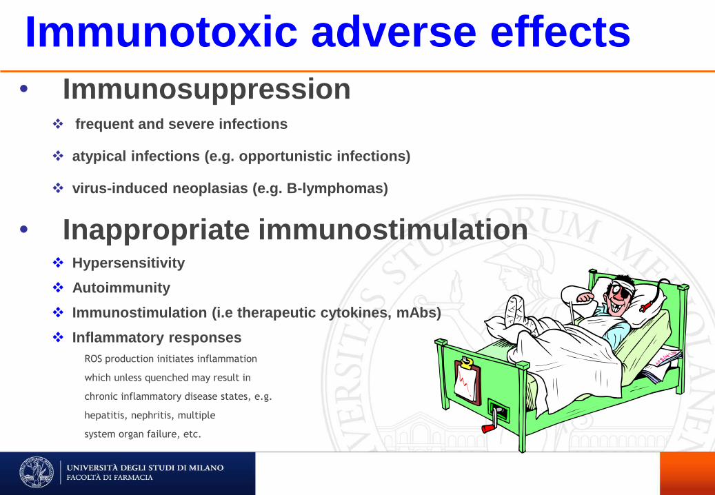
Immunotoxic adverse effects
• Immunosuppression frequent and severe infections
atypical infections (e.g. opportunistic infections)
virus-induced neoplasias (e.g. B-lymphomas)
• Inappropriate immunostimulation Hypersensitivity
Autoimmunity
Immunostimulation (i.e therapeutic cytokines, mAbs)
Inflammatory responses
ROS production initiates inflammation
which unless quenched may result in
chronic inflammatory disease states, e.g.
hepatitis, nephritis, multiple
system organ failure, etc.

FACTORS AFFECTING
SUSCEPTIBILITY TO
IMMUNOTOXICITY

Primary target
(lymphoid tissue)
Secondary target
(non lymphoid tissue)
Conditions of exposure(dose, frequency, duration, route)
IMMUNOTOXICANT
ALTERED IMMUNE FUNCTION
IMMUNOSUPPRESSION IMMUNOENHANCEMENT
Host related factors:
Genetic
Age/sex
Nutrition/disease
Hormonal and CNS status
Chemical related factors:
Chemical reactivity
Biotransfornation
Toxicokinetic

SPANISH FLU: MORTALITY RATE BY AGE RANGE
Age (yrs)
20%
60%
<10%
SPANISH FLU WORDLWIDE
MORTALITY IN ONE YEAR:
OVER 20 MILION PEOPLE

IMMUNE SYSTEM VULNERABILITY
Two properties make the immune system vulnerable to
chemical or physical insults:
1. Its late developing in life (prenatal and neonatal) and
continuous renewal.
2. The delicate control of the balance between activation,
silencing and regulation of immune reactivity after each
pathogen attack, as well as during immunosurveillance.

Objectives
Overview of the immune system
Immunotoxicology: definition
In vivo evaluation
Immune enhancement
Immune suppression
hypersensitivity
autoimmunity
susceptibility
to disease
Normalrange

Immunotoxicology: area of research
• Immunosuppression
• Immunostimulation
• Developmental immunotoxicology
• Immunosenescence
• Molecular immunotoxicology
• In vitro/ in silico immunotoxicology

Tossicologia Generale
EXPERIMENTAL MODELS

ANIMAL MODELS
Toxicity test should be performed with species that will respond
to a test chemical in a toxicological manner similar to that
anticipate in humans (i.e., equivalent metabolism and target
organ).
Rodents, however, still appear to be the most appropriate animal
model for examining the immunotoxicity of non-species specific
compounds.

At present, assessment of immunotoxic effects relies on different animal
models and several assays have been proposed to characterize
immunosuppression and sensitization.
Current available animal models and assays are not valid to assess the
potential for systemic hypersensitivity and, at this time, autoimmunity is
‘not predictable’, with the RA-PLNA holding promise.
CURRENT IN VIVO MODELS

CONDITIONS OF EXPOSURE
They should reflect the most probable route and level
of human exposure.
Treatment conditions should attempt to establish
dose-response curves as well as NOEL.
To be called immunotoxic a xenobiotic should
modulate the immune system at doses below overt
toxicity (stress and malnutrition depress immune
functions).

Immunosuppression hazards induce
Significant morbidity and mortality
Possible unacceptable risks: infections,
lymphomas...

• Current assessment of immunotoxicity rely on animal tests, which include some immune endpoints in repeated dose tests and call for dedicated tests only when certain alerts indicate a problem.
• Different requirements, however, depend on guidelines, i.e. functional tests are required by US-EPA for pesticides; weight of evidence approach for pharmaceuticals (ICH S8).
ASSESSMENT OF IMMUNOTOXICITY

SIGNS OF IMMUNOTOXICITY
From standard toxicity studies the following parameters should be
evaluated for signs of immunotoxicity:
• changes in total and differential white blood cell counts
• alterations in immune organ weights and histology
• decreased basal plasma immunoglobulins
• increased incidence of infections
• increased occurrence of tumors, in the absence of genotoxicity,
hormonal effects, or liver enzyme induction
• chemical retention in organs/cells of the immune system

PARAMETER RECOMMENDATION
Haematology
Total and differential WBC All
Organ weightsSpleen and Thymus All
HistopathologySpleen, Thymus C+H (store all)Draining and distant lymphnodes, bone marrow
C+H (store all)
All = all dose groupsC+H = control and high dose group
ECETOC Monograph No. 21 (1994)

Hazard identification:
- histopathology (C+H)
- haematology
- lymphoid organ weights
Interpretation of TIER I data:
- dose related toxicity
- other toxicity
- most sensitive parameters
magnitude of effect
review histopathology of low
and middle dose groups
Hazard identified STOPNoYes
Conduct Tier II testing
(case by case)
NOTE: to be called immunotoxic a xenobiotic should modulate the
immune system at doses below overt toxicity (stress and malnutrition
depress immune functions).

Relatively well validated models
Relevance of histological changes in
lymphoid organs
One immune function assay absolutely
necessary
Host resistance assays as second-line
assays
METHODS IN IMMUNOTOXICOLOGY
Immunosuppression

Testing Assays
General tests
Organ weights, plasma/serum enzyme levels
Nonfunctional tests: status
Lymphoid organ weights, lymphoid tissue cellularity,
histopathology, immunophenotyping
Functional tests
METHODS IN IMMUNOTOXICOLOGY
Immunosuppression

Functional Tests
Innate Immunity
Humoral-mediated Immunity
Cell-mediated immunity
Host Resistance Assays
METHODS IN IMMUNOTOXICOLOGY
Immunosuppression

Complement + sRBC
in Agar Solution
3 Hour Incubation
Magnified
500 µl Aliquot
Sheep RBC
(AFC)
SRBC around AFC
PLAQUEare hemolyzed =
Antibody Forming Cell
IgM Plaque Forming Cell Assay
Day 4
End Points
PFC / 10 Spleen Cells6
PFC / Spleen

Functional Tests
Innate Immunity
Humoral-mediated Immunity
Cell-mediated immunity
Host Resistance Assays
METHODS IN IMMUNOTOXICOLOGY
Immunomodulation

Host Resistance Assays
B16F10 Melanoma Tumor
Streptococcus pneumoniae
Listeria monocytogenes
Antibody, Complement, PMNs
Macrophages, T Cells
Natural Killer Cells, Macrophages, T Cells
Challenge Model

The early work from these groups, in terms of immunotoxicology assay development, evaluation and implementation, played a critical role in shaping the development of immunotoxicology guidelines for both pharmaceuticals and environmental chemicals.

DATABASE: 50 compounds (NIEHS, CIIT)
OBJECTIVES: to improve future testingstrategies, to provide information to aid inrisk assessment and to examine therelationship between the immune function andhost resistance tests
Risk assessment in immunotoxicology

at So
ciety o
f Tox
icolo
gy
on
Jun
e 11, 2
01
2h
ttp://to
xsci.o
xfo
rdjo
urn
als.org
/D
ow
nlo
aded
from

CONCLUSIONS Fund Appl Toxicol 18: 200, 1992
the performance of only 3 immune tests is sufficient to
predict immunotoxic compounds in rodents (100 %
concordance)
Risk assessment in immunotoxicology
at Society
of T
oxico
logy
on
June 11
, 201
2h
ttp://to
xsci.o
xfo
rdjo
urn
als.org
/D
ow
nlo
aded
from

CONCLUSIONS Fund Appl Toxicol 21: 71, 1993
good correlation between changes in the
immune tests and altered host resistance
no instances where host resistance was
altered without affecting an immune tests.
However, in some instance immune changes
occurred without corresponding changes in
host resistance
Risk assessment in immunotoxicology

US-EPA: ASSESSMENT OF PESTICIDE IMMUNOTOXICITY
IMMUNOTOXIC
Select most appropriate species (rat or mouse) and gender based on (1) structural change in immune system, or (2) general toxicity, or (3) default
female mouse
Perform TDAR Assay (SRBC) in selected species
Negative
Reconsider WoE for potential immunotoxicity
Dose-related positive at < MTD
Some evidence for immunotoxic potential
Perform NK cell activity assay NOT IMMUNOTOXIC
NegativePositive at doses < MTD
Select most appropriate species (rat or mouse) and gender based on (1) structural change in immune system, or (2) general toxicity, or (3) default
female mouse
Initial review of STS to select the most appropriate species (rat or mouse) and gender for immunotoxicity testing
Based on WOE: (1) structural changes in immune system,
(2) general toxicity, or (3) default female mouse
No evidence for immunotoxic potential
ILSI-HESI WORKSHOP SUMMARY 2012

INTERPRETATION
Normal immune system comprises complementary and compensatory
mechanisms (functional reserve).
Failure to identify alterations in host resistance in face of significant
changes in functional ability does not necesseraly mean the absence
of risk to man.
Due to genetic polymorphims the response to immunotoxicants vary
in human beings.
Thus, alterations in immune functions which may be tollerated well in
normal individuals could have more serious consequences for those
who are cronically sick, malnourisehd, or those immune system has
yet to develop or is in decline.

Immune Competence
Perc
en
t o
f P
op
ula
tio
n
Infections
Dose
Exposure to
Immunosuppressant
Population
at Risk
Pathogenicity
The ability to resist pathogen challenge is dependent upon the degree of
immunosuppression and the quantity of pathogen administered.

AGENTS WHICH INHIBIT IMMUNE FUNCTION AND HOST RESISTANCE
Cocaine, marijuana, alcoholDrugs of abuse
T-2, ochratoxinMycotoxins
UV-BUltraviolet light
Asbestos, silica, berylliumParticulates
NO2, O3, SO2Oxidant gases
DMN, benzidine, AAFAromatic amines
Dibutyltin chlorideOrganotins
Carbofuran, chlordanePesticides
DMBA, B(a)P, MCAPolycyclic aromatic hydrocarbons
Benzene, TolueneAromatic hydrocarbons
Lead, Cadmium, ArsenicMetals
TCDD, PCB, PBBPolyhalogenated aromatic hydrocarbons
EXAMPLECLASS

Potential Mechanisms of Immune Suppression
Structural alterations in immune tissues and organs
Alterations in immune cells maturation and differentiation
Alterations in immune cells activation:
Cytotoxicity
Inhibition of cell proliferation (prevent clonal expansion)
Interference with receptor/ligand interaction or transduction of signals to the nucleus
Interference with transcription, translation and release
Antigen
APC
MHC-II
proliferationIL-1
Th CD4+
TCR
CD4CD3
IL-2 Th
activated
B
cells
T
CD8+
Cell mediated
Immunity
Humoral
immunityIL-4, IL-5, IL-6
IL-10, IL-13
IL-2,IL-3 IFN-g,
IL-12, TNF-b
Antigen recognition Clonal expansion Effectors

Antigen
SPECIFIC IMMUNITY
APC
MHC-II
proliferationIL-1
Th CD4+
TCRCD4
CD3
IL-2 Th
activated
B
cells
T
CD8+
Cell mediated
Immunity
Humoral
immunityIL-4, IL-5, IL-6
IL-10, IL-13
IL-2,IL-3 IFN-g,
IL-12, TNF-b
Antigen recognition Clonal expansion Effectors
Siti d’azione degli agenti immunosoppressori:
a. Anticorpo anti Rh
b. Corticosteroidi
c. Globuline anti-timociti, OKT3, anti CD4
d. Ciclosporina, tacrolimus
e. Azatioprina, metotrexato, ciclofosfamide, rapamicina, micofenolato mofetil, corticosteroidi
ab
c
d e
e
e

Immunotoxic adverse effects
• Immunosuppression frequent and severe infections
atypical infections (e.g. opportunistic infections)
virus-induced neoplasias (e.g. B-lymphomas)
• Inappropriate immunostimulation Hypersensitivity
Autoimmunity
Immunostimulation (i.e therapeutic cytokines, mAbs)
Inflammatory responses
ROS production initiates inflammation
which unless quenched may result in
chronic inflammatory disease states, e.g.
hepatitis, nephritis, multiple
system organ failure, etc.
Inappropriate immunostimulation can cause
more frequent autoimmune diseases, allergic
reactions, flu-like syndromes and inhibition
of hepatic metabolism.

CHEMICAL ALLERGY
Hypersensitivity reactions are often considered a major health
problem in relation to environmental chemical exposure.
As a consequence chemical allergy is of considerable
importance to the toxicologist, who has the responsibility
of identifying and characterizing the skin and respiratory
potential of chemicals, and estimating the risk they pose
to human health.
Regulatory authorities worldwide require testing for ACD
potential and appropriate hazard labeling to minimize
exposures

Hypersensitivity
Definition: excessive humoral or cellular response to an antigen which can lead to
tissue damage.Hypersensitivity reactions are the result of normally beneficial
immune responses acting inappropriately.

Hypersensitivity: classification
Type 1: IgE mediated (Immediate type)
Type 2: IgM, IgG, (Cytolysis of cells)
Type 3: IgM, IgG (Immune complex mediated)
Type 4: T-cell mediated (delayed-type)
Pseudoallergic reaction

Two Stages(Distinguishes from irritation)
Induction
Sensitization
(1st exposure)
Elicitation
Challenge
(subsequent exposure)

CHEMICAL ALLERGY
Classification
Mechanisms
Experimental models
Examples

Key passages:•Absorption
•Local trauma –
proinflammatory cytokine
production
•Protein binding
• Antigen processing
• Langerhans cells
maturation and migration
• Antigen presentation to
Th cells and the generation
of memory T cells
(immunogenicity)
STRATUMCORNEUM
Chemical
EPIDERMIS
LYMPH NODE
MIGRATION
MATURATION
TNF- aRANTES MCP-1
IL-1 bGM-CSF
IL-1IL-6
IL-8IL-10IL-12
a
MIP-2IP-10TNF-IFN-g
MHC class II
Th0IL-4, IL-5, IL-6, IL-10
Atopic dermatitis (Type I)
IL-12, INF- g, TNF- b
Allergic contact dermatitis(Type IV)
CD4 +
KC
LC
IL-7
IL-15
IL-18
GM-CSF
TGF-a
TGF-b

• Upon subsequent contact, some LDC migrate to local lymph node as before. Other LDC present processed hapten-carrier to memory T cells in skin.
• Activated memory T cells secrete cytokines that induce release of inflammatory cytokines from other cell types.
• Memory T cells and inflammatory cells are recruited to the epidermis from circulation via chemoattractant cytokines and expression of adhesion molecules.
Hapten
Carrier
Langerhans’ /
Dendritic cell
dermis
Lymphatic Vessel
LocalLymphNode
keratinocytes :
other cytokines
TM
TM
TM
Blood Vessel
TM
-
vascular endothelial cells
inflammation
TM
TM
II
IL-1, IL-6
IFNgIL-1, Il-6
IL-8, TNFa
Allergic Contact Dermatitis: Elicitation

CHEMICAL ALLERGY
Classification
Mechanisms
Experimental models
Examples

EXPERIMENTAL MODELS
Mice Guinea Pigs

Hypersensitivity
Well established methods for contact
hypersensitivity.
Current models and assays as
inadequate predictors for system
hypersensitivity reaction.

Mouse Tests: Local lymph node assay
Mouse Ear Swelling Test
Guinea Pig Tests:
Maximization Test
Occlusive Patch Test
Respiratory Challenge
Systemic Anaphylaxix
METHODS IN IMMUNOTOXICOLOGY
Hypersensitivity Testing

ID injection w/ and without FCA
plus topical application:
Days 5-8
Day 20-22 topical challenge
Read: 48,72 h after challenge
>30% positive
Guinea Pig
Maximization
Test
Topical application - closed patch:
Days 0, 6-8, and 13-15
Day 27-28 topical challenge
of the untreated flank for 6 h
Read: 21, 24, 48 h after removing
patch
> 15% positive
Buehler
Assay
Induction
Challenge
Endpoint erythema
Criteria
20 animals/ group
GUINEA PIG MODELS

Test/vehicle
LOCAL LYMPH NODE ASSAY
Day 0,1,2
Day 5
2 days of rest, 3H TdR
5 hours
T
T
T
T
T
T
T
T
IMMUNE ACTIVATION
Selective clonal expansion of
allergen-responsive T lymphocytes
T
The mouse local lymph node assay (LLNA)
Count DPMs

5
10
15
20
25
0 0.05 0.1 0.25
concentration (%w/v)
stim
ula
tion index
5
10
15
20
25
0 1 2.5 5 10
concentration (%w/v)
stim
ula
tion index
DNCB PABA
LLNA : illustrative results
The proliferation is proportional to
the dose and chemical reactivity.

DNCB
6
12
18
24
30
0 0.05 0.1 0.15 0.2 0.25
Concentration (% w/v)
Sti
mula
tion I
ndex
HCAHCA = hexyl cinnamic aldehyde
4
8
12
16
20
0 10 20 30 40 50
Concentration (% w/v)
Sti
mula
tion I
ndex
EC3 = 0.05% EC3 = 7.9%
RELATIVE SKIN SENSITIZATION POTENCY
Application concentration of test chemical required to provoke a 3-fold increase in LNC
proliferative activity compared with concurrent vehicle treated controls.
The proliferation is proportional to
the dose and to the potency of the
applied allergen.
The EC3 provides a simple means of
obtaining a quantitative measurement
of sensitization.

Classification of Relative Skin Sensitization
Potency Using LLNA EC3 Values
Potency Category EC3 Value µg/cm2EC3 Value %
Concentration
Extreme 25 < 0.1
Strong 25 - 250 ≥ 0.1 - < 1
Moderate 250 - 2500 ≥ 1 - < 10
Weak >2500 ≥ 10
ECETOC technical report #87
1% dose in a LLNA = 250 μg/cm2

•The LLNA is suited well for potency information
whereas Guinea pigs methods are more
challenging due to the inherent design of the
study:
•Measures the elicitation phase
•Not practicable to examine in detail multiple induction
concentrations of a chemical
•Subjective assessment of the frequency of responses
rather than the vigor of the responses
Kimber et al., Toxicology 93: 13, 1994

- Update OECD 429 – Skin sensitization: reduced LLNA
It also includes the Performance Standards that can be used to evaluate the validation status of new and /or modified test methods that are functionall and mechanistically similar to the LLNA.
- OECD 442A - Skin Sensitization: LLNA DA
- OECD 442B - Skin Sensitization: LLNA BrdU-ELISA
All adopted 22nd July 2010
NEW OECD GUIDELINES


Potential Contact Sensitizers
Fragrances
Dyes
Preservatives (kathon CG, CHOH)
Metals (Ni, Co, Be, Cr)

Low Molecular Wt (<3000 Da)
respiratory sensitizers
Toluene diisocyanate
Diphenylmethane diisocyanate
Phthalic anhydride
Trimellitic anhydride
Platinum salts
Reactive dyes

Protein allergens
Detergent Enzymes
Molds and spores
Latex
Microbial pesticides
Animal dander
Food proteins

Definition: autoimmunity is an inappropriate immune response against self-antigens, which can lead to chronic inflammation, tissue destruction
and/or dysfunction.
Autoimmunity

Spectrum of Autoimmune Diseases and
Putative Autoantigens
Hashimoto’s Thyroiditis
Thyrotoxicosis
Pernicious anemia
Autoimmune Atrophic Gastritis
Addison’s Disease
Insulin-Dependent Diabetes Mellitus
Goodpasture’s Syndrome
Myasthenia Gravis
Male Infertility (isolated cases)
Sympathetic Ophthalmia
Multiple Sclerosis
Autoimmune Hemolytic Anemia
Ulcerative Colitis
Rheumatoid Arthritis
Scleroderma
Systemic Lupus Erythematosus (SLE)
Thyroglobulin
Thyroid-stimulating hormone (TSH)
H+/K+-ATPase
Intrinsic factor
21-hydroxylase
Glutamic acid decarboxylase 65
Type IV collagen
Acetyl choline receptor
Epididymal glycoprotein, FA-1
Interphotoreceptor retinol binding protein
Myelin basic protein
X-antigen, glycophorin
Catalase; a-enolase
Rheumatoid factor
Topoisomerase 1; laminins
DNA nucleotides and histones
Organ Specific
Non-Organ Specific More than 60 diseases!

AUTOIMMUNITY
Classification
Mechanisms
Experimental models
Examples

POTENTIAL MECHANISMS OF
AUTOIMMUNITY
1. Failure to remove potentially autoreactive cells
2. Loss of peripheral tolerance
3. Altered self
4. Molecular mimicry (i.e. infections)
5. Exposure of cryptic antigens
6. Altered regulatory protein production

While genotype and gender (> F) appear to be a critical factor in
development of autoimmune disease, contributions by other host factors and
exposure to exogenous agents may induce or exacerbate autoimmune
diseases.
Occupational exposure to silica, solvents, pesticides has been associated
with autoimmune diseases. Three different effects of occupational chemical
exposures have been suggested:
1. Enhanced proinflammatory response
2. Modification of endogenous proteins and subsequent autoantibody
formation
3. Altered production of regulatory factors.
Autoimmunity: multifactorial diseases

AUTOIMMUNITY
Classification
Mechanisms
Experimental models
Examples

Autoimmunity
Poor understanding of basic mechanisms.
No reliable models or general strategy and assays(including the popliteal lymph node assay) are atpresent available (or validated).
Possible strategy:
- tier 1: PLNA or RA-PLNA (hazard identification)
- tier 2: suitable animal models (relevant route of exposure, relevant clinicaloutcomes)

Animal Models
Genetic Predisposition
Autoimmunization
Organic or Chemical Induction
METHODS IN IMMUNOTOXICOLOGY
Autoimmunity

Animal Models
Genetic Predisposition Autoimmune Thyroiditis
MRL (m), BB (r), OS (ch)
Insulin-Dependent Diabetes Mellitus
NOD (m), BB (r), BN (r)
Rheumatoid Arthritis
MRL/lpr (m), SCID (m), HLAB27 (r)
Systemic Lupus Erythematosus
MRL+/+ (m), MRL/lpr (m), NZB/NZW (m)
Scleroderma
TSK (m)
METHODS TO STUDY AUTOIMMUNE DISEASE

AUTOIMMUNITY
Classification
Mechanisms
Experimental models
Examples

Drugs
Infectious Agents
Metals
Particulates (silica)
Pesticides
Solvents
Vaccines
EXOGENOUS FACTORS ASSOCIATED
WITH AUTOIMMUNITY

Particulates
Silica
Strong association between silica exposure (from “dusty trades”) and SLE, rheumatoid arthritis, ANCA-associated vasculitis and glomerulonephritis, and scleroderma
Some individuals appear to develop fibrogenic responses while others develop immunologic responses
Acts as an adjuvant by:
Increasing the longevity of APCs
Increasing antigen processing in APCs
Increasing cytokine production
Inducing a polyclonal activation of B and T cells
EXOGENOUS FACTORS ASSOCIATED WITH AUTOIMMUNITY

CONCLUSION
While it is evident that occupational exposures contribute
in some measure to the overall risk for specific
autoimmune diseases, improved exposure
assessment, better coordination between
experimental models and epidemiological studies
(priori hypothesis) are needed to define these risks
more precisely.