evaluation and management of peripheral nerve injury
TRANSCRIPT
University of Nebraska - Lincoln University of Nebraska - Lincoln
DigitalCommons@University of Nebraska - Lincoln DigitalCommons@University of Nebraska - Lincoln
Uniformed Services University of the Health Sciences U.S. Department of Defense
2008
Evaluation and management of peripheral nerve injury Evaluation and management of peripheral nerve injury
William W. Campbell Uniformed Services University of Health Sciences, [email protected]
Follow this and additional works at: https://digitalcommons.unl.edu/usuhs
Part of the Medicine and Health Sciences Commons
Campbell, William W., "Evaluation and management of peripheral nerve injury" (2008). Uniformed Services University of the Health Sciences. 3. https://digitalcommons.unl.edu/usuhs/3
This Article is brought to you for free and open access by the U.S. Department of Defense at DigitalCommons@University of Nebraska - Lincoln. It has been accepted for inclusion in Uniformed Services University of the Health Sciences by an authorized administrator of DigitalCommons@University of Nebraska - Lincoln.
Invited review
Evaluation and management of peripheral nerve injury q
William W. Campbell*
Department of Neurology, Uniformed Services University of Health Sciences, Room A 1036, 4301 Jones Bridge Road, Bethesda, MD 20814, USA
Accepted 7 March 2008Available online 14 May 2008
Abstract
Common etiologies of acute traumatic peripheral nerve injury (TPNI) include penetrating injury, crush, stretch, and ischemia. Manage-ment of TPNI requires familiarity with the relevant anatomy, pathology, pathophysiology, and the surgical principles, approaches andconcerns. Surgical repair of TPNI is done at varying time intervals after the injury, and there are a number of considerations in decidingwhether and when to operate. In neurapraxia, the compound muscle and nerve action potentials on stimulating distal to the lesion aremaintained indefinitely; stimulation above the lesion reveals partial or complete conduction block. The picture in axonotmesis and neu-rotmesis depends on the time since injury. The optimal timing for an electrodiagnostic study depends upon the clinical question beingasked. Although conventional teaching usually holds that an electrodiagnostic study should not be done until about 3 weeks after theinjury, in fact a great deal of important information can be obtained by studies done in the first week. Proximal nerve injuries are prob-lematic because the long distance makes it difficult to reinnervate distal muscles before irreversible changes occur. Decision makingregarding exploration must occur more quickly, and exploration using intraoperative nerve action potential recording to guide the choiceof surgical procedure is often useful.Published by Elsevier Ireland Ltd on behalf of International Federation of Clinical Neurophysiology.
Keywords: Trauma; Injury; Peripheral nerve; Electrodiagnosis; Electromyography; Surgery
1. Introduction
Etiologies of traumatic peripheral nerve injury (TPNI)include penetrating injury, crush, traction, ischemia, andless common mechanisms such as thermal, electric shock,radiation, percussion, and vibration (Robinson, 2000,2004). In general, stretch-related injuries are the most com-mon type of civilian nerve trauma, especially in motor vehi-cle accidents. Lacerations, as by glass, knife, fan, sawblade, auto metal or long bone fractures make up about30% of serious nerve injuries. Another common injurymechanism is compression, which may involve mechanicaldeformation as well as ischemia (Stanec et al., 1997).Kouyoumdjian reported a 16-year retrospective study of
456 consecutive patients with 557 peripheral nerve injuries(Kouyoumdjian, 2006). Upper-limb injuries occurred in73.5% of cases; the ulnar nerve was most often injured,either singly or in combination. Combined lesions mostcommonly involved the ulnar and median nerves. Motorvehicle accidents, particularly motorcycle crashes, werethe most common cause of injury (Stanec et al., 1997). Ina series of 1167 cases of peripheral nerve injury, 5.7% ofcases were related to sports (Hirasawa and Sakakida,1983).
Peripheral nerve injuries were first studied systematicallyduring the American Civil War by neurologist S. WeirMitchell. Many of the advances in knowledge aboutperipheral nerve injuries have occurred during wartime,from physicians on both sides of the front. In a military set-ting, most peripheral nerve injuries are due to shrapnel(Maricevic and Erceg, 1997). A common cause of TPNIin combat is blast injury, often from bombs or improvisedexplosive devices. Blast typically causes a complex, exten-sive, soft-tissue injury, frequently with vascular injuries
1388-2457/$34.00 Published by Elsevier Ireland Ltd on behalf of International Federation of Clinical Neurophysiology.
doi:10.1016/j.clinph.2008.03.018
q The opinions or assertions contained herein are the private views of theauthor and are not to be construed as official or as reflecting the views ofthe Department of the Army or the Department of Defense.
* Tel.: +1 301 295 3643; fax: +1 301 295 0620.E-mail addresses: [email protected], [email protected]
www.elsevier.com/locate/clinph
Clinical Neurophysiology 119 (2008) 1951–1965
requiring emergency arterial repair. The arterial injury mayproduce limb ischemia requiring fasciotomy. Peripheralnerves may be involved because of the concussive forceof blast overpressure, shrapnel, or limb ischemia with com-partment syndrome. In a series of 151 upper limb war inju-ries seen in Croatia, about one-third of the nerve injurieshad associated arterial injuries. Functional results wereobtained in only 44.8% of cases with both nerve and arte-rial injuries, an outcome likely due in part to nerve ische-mia at the injury site (Stanec et al., 1997; Selecki et al.,1982).
In the current Middle East conflict, the protection affor-ded to coalition soldiers by body armor has resulted in amarkedly increased incidence of peripheral nerve injuries,as combatants survive wounds that would formerly havebeen lethal. The wounded in action to killed in action ratioamong coalition forces in the current conflict is about 8:1,compared to 3:1 in past conflicts, even as recently as Viet-nam. The result has been a marked increase in extremelygrievous extremity wounds, with many amputations andmany TPNIs. Recently developed extremity body armorwill hopefully lessen the incidence of the major extremitywounds. The lightweight, flexible extremity body armor isdesigned to protect the vulnerable areas near major nervesand blood vessels. Non-battle related peripheral nerve syn-dromes are also common in a combat environment (Hart-mann, 2006).
This review will emphasize the clinical and electrodiag-nostic aspects of the care of patients with TPNI.
2. Neuropathology of peripheral nerve injury
To manage patients with TPNI, it is important to beknowledgeable about the relevant anatomy, pathology,pathophysiology, electrodiagnosis, and principles of surgi-cal management. Understanding the anatomy is crucial tograsping the pathophysiologic concepts that underlie theclinical management of patients with peripheral nerve inju-ries (Burnett and Zager, 2004; Maggi et al., 2003). Becausethe clinical neurophysiologist works closely with the sur-geon in managing these cases, it is also important to befamiliar with surgical principles, approaches, and concerns.
The endoneurium surrounds individual myelinatedaxons and groups of unmyelinated ones. Fascicles are col-lections of axons which are surrounded by perineurium.The epifascicular (internal) epineurium lies between fasci-
cles. The peripheral nerve trunk is a collection of fascicles,and the epineurial (external) epineurium surrounds thenerve trunk proper. The endoneurium is longitudinally ori-ented while the perineurium and epineurium are circumfer-ential (Sunderland, 1990). Plexuses of microvessels runlongitudinally in the epineurium, and send transversebranches through the perineurium to form a vascular net-work consisting primarily of capillaries in the endoneu-rium. Nerve trauma increases the permeability of theepineurial vessels, which are more susceptible to compres-sion trauma than the endoneurial vessels. Higher pressurelevels or more prolonged compression will also injure theendoneurial vessels, leading to intrafascicular edema,which may lead to secondary injury to the nerve (Rydevikand Lundborg, 1977).
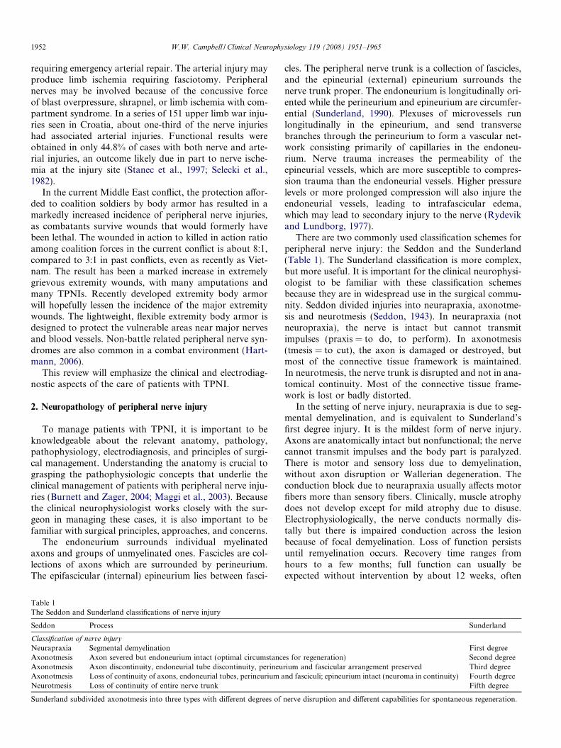
There are two commonly used classification schemes forperipheral nerve injury: the Seddon and the Sunderland(Table 1). The Sunderland classification is more complex,but more useful. It is important for the clinical neurophysi-ologist to be familiar with these classification schemesbecause they are in widespread use in the surgical commu-nity. Seddon divided injuries into neurapraxia, axonotme-sis and neurotmesis (Seddon, 1943). In neurapraxia (notneuropraxia), the nerve is intact but cannot transmitimpulses (praxis = to do, to perform). In axonotmesis(tmesis = to cut), the axon is damaged or destroyed, butmost of the connective tissue framework is maintained.In neurotmesis, the nerve trunk is disrupted and not in ana-tomical continuity. Most of the connective tissue frame-work is lost or badly distorted.
In the setting of nerve injury, neurapraxia is due to seg-mental demyelination, and is equivalent to Sunderland’sfirst degree injury. It is the mildest form of nerve injury.Axons are anatomically intact but nonfunctional; the nervecannot transmit impulses and the body part is paralyzed.There is motor and sensory loss due to demyelination,without axon disruption or Wallerian degeneration. Theconduction block due to neurapraxia usually affects motorfibers more than sensory fibers. Clinically, muscle atrophydoes not develop except for mild atrophy due to disuse.Electrophysiologically, the nerve conducts normally dis-tally but there is impaired conduction across the lesionbecause of focal demyelination. Loss of function persistsuntil remyelination occurs. Recovery time ranges fromhours to a few months; full function can usually beexpected without intervention by about 12 weeks, often
Table 1The Seddon and Sunderland classifications of nerve injury
Seddon Process Sunderland
Classification of nerve injury
Neurapraxia Segmental demyelination First degreeAxonotmesis Axon severed but endoneurium intact (optimal circumstances for regeneration) Second degreeAxonotmesis Axon discontinuity, endoneurial tube discontinuity, perineurium and fascicular arrangement preserved Third degreeAxonotmesis Loss of continuity of axons, endoneurial tubes, perineurium and fasciculi; epineurium intact (neuroma in continuity) Fourth degreeNeurotmesis Loss of continuity of entire nerve trunk Fifth degree
Sunderland subdivided axonotmesis into three types with different degrees of nerve disruption and different capabilities for spontaneous regeneration.
1952 W.W. Campbell / Clinical Neurophysiology 119 (2008) 1951–1965

earlier, provided there is no ongoing compression. Motorparalysis can last as long as 6 months, but most lesionsresolve by 3 months (Dumitru et al., 2001). Since axonsmay be remyelinated at different rates and to differentdegrees, function may be regained unevenly. Commonexamples are Saturday night radial nerve palsy and legcrossing peroneal nerve palsy. Seddon coined the term neu-rapraxia to refer to lesions that recovered in weeks tomonths (Seddon, 1943). Neurapraxia has also been usedto describe rapidly reversible physiological conductionblock lasting only minutes, much too transient to attributeto demyelination, as in one’s foot falling asleep, presum-ably due to focal ischemia without any structural changein the myelin (Wilbourn, 2002). Whether ischemia cancause more prolonged conduction block, lasting days toweeks is controversial (Wilbourn, 2002).
More severe peripheral nerve injuries, such as lacera-tions, contusions, and injuries due to stretch, severe com-pression and injections, cause anatomical disruption ofaxons or the nerve trunk proper. Wallerian degenerationoccurs when there is disruption of the axon (Koeppen,2004; Stoll and Muller, 1999). The distal portion of theaxon degenerates and fragments. Myelin is transformedinto neutral fat and phagocytosed by macrophages. Debrisof the axon and the myelin sheath form ovoids that aregradually digested and disappear (digestion chambers)(Chaudhry et al., 1992). Proximal to the lesion, degenera-tion stops at the first internode in mild injuries, but mayextend further proximally in severe injuries. Within hoursof injury the ends of the severed axons seal over, and thesealed ends swell with cellular organelles because antero-grade axonal transport in the proximal stump and retro-grade axonal transport in the distal stump persist forseveral days (Lunn et al., 1990). Resealing is a necessaryprelude to axon regeneration from the proximal stump.The resealing process requires Ca++ entry into the axo-plasm and occurs rapidly (Yawo and Kuno, 1985). Potentvasoactive peptides accumulate in the axon end bulbs,which along with local mast cell degranulation and angio-genesis cause hyperemia that persists for several weeksafter injury (Hall, 2005). Current evidence indicates thataxonal degeneration is not a passive process, but an activeprogrammed response to disconnection from the cell bodyand the target organ (Hall, 2005). Loss of the axoplasmiccytoskeleton begins within about seven days in humans,accompanied by a caspase-independent program ofautodestruction (Raff et al., 2002). Axonal degenerationis dependent on a local increase in calcium concentrationthat activates calcium-dependent proteolytic enzymes, suchas phospholipases and calpains. These compounds theneffect cytoskeletal dissolution (Hall, 2005).
In the distal stump, although the axon degenerates anddisappears, the connective tissue basement membranesmay remain, forming endoneurial tubes. Schwann cellsproliferate and line the endoneurial tubes (Schwann celltubes, bands of Bungner). These arrays of Schwann cellsand processes within the basement membrane provide the
pathway and scaffolding for axonal regeneration. Walle-rian degeneration begins within hours of injury and is com-plete by 6–8 weeks, leaving a distal stump comprising onlyendoneurial tubes lined by Schwann cells (Hall, 1989; Kanget al., 2003). The Schwann cells are not permanent; theyinvolute and disappear if axonal regeneration does notoccur (Hall, 1999). Experimentally, there is a progressiveinability of chronically denervated Schwann cells to sup-port axonal regeneration, but paradoxically a sustainedcapacity to remyelinate axons that do regenerate. Axonalinteraction can effectively switch the atrophic Schwanncells back into active myelinating cells (Sulaiman and Gor-don, 2000). Endoneurial tubes that do not receive a regen-erating axon shrink and are eventually obliterated by scartissue.
In axonotmesis, the axon is disrupted and Walleriandegeneration occurs. There is axon discontinuity, but thesurrounding stroma is at least partially intact. Axonotmesisis commonly seen in crush and stretch injuries. Reinnerva-tion depends upon the degree of internal disorganization,and the distance to the muscle. In neurotmesis, the nerveis completely severed or so internally disrupted that it doesnot regenerate spontaneously well enough to produce func-tion. Neurotmesis is seen with sharp injury, massivetrauma, or severe traction with nerve rupture. There is lossof nerve trunk continuity with complete disruption of allsupporting elements; reinnervation does not occur. With-out surgery, the prognosis is extremely poor. Recoveryfrom any TPNI where there is significant axon loss andinternal disruption, Sunderland third degree or worse, isapt to be prolonged and incomplete (Sunderland, 1978).The affected body part can seldom become again what itwas.
3. Nerve regeneration
Regeneration and repair processes go on at multiple lev-els following nerve injury, including the nerve cell body, thesegment between the neuron and the injury site (proximalstump), the injury site itself, the segment between the injurysite and the end organ (distal stump), and the end organ(Burnett and Zager, 2004; Seckel, 1990). The repair processmay be disrupted at one or more of these sites. With mildinjuries, regeneration and repair begin almost immediately.Remyelination in neurapractic injuries can occur fairly rap-idly. With more severe injuries, there is an initial shockphase, after which regeneration and repair phases continuefor many months.
In the CNS, recovery of function is accomplished byplasticity, using intact areas to take over the function ofdamaged areas; the CNS does not repair itself. Theapproach of the PNS to injury is to repair itself, and thisis an essential difference between the two (Burnett and Zag-er, 2004; Fenrich and Gordon, 2004). Repair can occurthrough three mechanisms: remyelination, collateralsprouting distally from preserved axons, and regenerationfrom the site of injury (Zochodne and Levy, 2005). Collat-
W.W. Campbell / Clinical Neurophysiology 119 (2008) 1951–1965 1953
eral sprouts can provide reinnervation in partial nerve inju-ries, and when there are many surviving axons they may bevery effective. With lesions involving less than 20–30% ofthe axons, recovery is predominantly by collateral sprout-ing from surviving axons, and occurs over 2–6 months.When more than 90% of axons are injured, the primarymechanism of repair is regeneration from the injury site.The success of regeneration from the proximal stumpdepends to a large degree on the distance from the injurysite. Even when good motor recovery occurs, sensory defi-cits, particularly in proprioception, may impair functionaloutcome.
Attempts at regeneration begin soon after injury (Pol-lock, 1995; Thomas, 1989). A cascade of events involvingcell signaling molecules and neurotrophic factors occursafter nerve injury (Liuzzi and Tedeschi, 1991; Zheng andKuffler, 2000; Zochodne and Levy, 2005). The blood–nervebarrier plays an important role (Maricevic and Erceg,1997). Schwann cells play an indispensable role in promot-ing regeneration by increasing their synthesis of surface celladhesion molecules, and by elaborating basement mem-brane containing extracellular matrix proteins, such aslaminin and fibronectin (Fu and Gordon, 1997). Schwanncells produce neurotrophic factors that bind to tyrosinekinase receptors and are responsible for a signal that leadsto gene activation (Funakoshi et al., 1993). Within 30 minafter injury, intracellular processes that promote repair andregeneration have already been activated (Dahlin, 2006).Within days after injury, Schwann cells begin to divideand create a pool of dedifferentiated daughter cells. With-out axon contact, Schwann cells downregulate their normalproteins, such as PMP22, P0 and connexin-32, and convertto the phenotype of a premyelinating cell (Hall, 2005).These dedifferentiated Schwann cells upregulate expressionof nerve growth factor (NGF), other neurotrophic factors,cytokines, and other compounds that lead to Schwann celldifferentiation and proliferation in anticipation of the arri-val of a regenerating sprout. Nerve growth factor receptorson the Schwann cells lining the endoneurial tubes in thedistal stump increase. Experimental sciatic nerve transec-tion induces Schwann cells distal to the lesion to expressgreatly increased levels of NGF receptors; by 7 days thereceptor density increases at least 50-fold (Taniuchi et al.,1986). The action of NGF on these Schwann cell receptorsstimulates regenerating axonal sprouts (Liuzzi and Tede-schi, 1991). After injury, macrophages migrate into the dis-tal stump and may be involved in initiating Schwann cellproliferation. Macrophages upregulate IL-1, which inducesan increase in NGF transcription and NGF receptor den-sity, and also secrete mitogens that trigger Schwann cellproliferation (Davis and Stroobant, 1990). Neural cell sur-face molecules and the extracellular matrix molecules lam-inin and tenascin are strongly upregulated by denervatedSchwann cells and may foster axonal regeneration (Mar-tini, 1994). Cytokines play an important role. After exper-imental axotomy, compounds such as IL-6 andtransforming growth factor-beta 1 are overexpressed in
nerve and promote axonal growth until axon/Schwann cellcontact (Creange et al., 1997). Inflammatory cells and theirproducts also contribute to neuronal survival and axonalregeneration after injury. Macrophages accumulating fol-lowing nerve injury supply neurotrophic support to nervecell bodies, and enhance axonal regeneration (Richardsonand Lu, 1994).
The stimulation effects after nerve injury radiate retro-grade from the periphery to the nerve cell body, produc-ing a stimulus that activates processes in the neuron thatfoster regeneration. Neuronal survival is facilitated bythe activation of trophic factors from multiple sources,including neurotrophins, neuropoietic cytokines, insulin-like growth factors (IGFs), and glial-cell-line-derivedneurotrophic factors (GDNFs). Axotomized neuronsmust switch from a transmitting mode to a growth modeand express growth-associated proteins, such as GAP-43,tubulin, and actin, as well as an array of novel neuro-peptides and cytokines, all of which have the potentialto promote axonal regeneration (Fu and Gordon,1997). After injury there is a coordinated shift in thegene expression pattern in axotomized neurons, with amarked induction of transcription factors that occurs asearly as 12 h after injury. Many of the upregulated genesare involved in regeneration (Dahlin, 2006). Dozens ofgenes are differentially expressed after experimental sci-atic nerve injury using microarray analysis. Overexpres-sion of fibroblast growth factor-inducible-14 mRNApromotes growth cone lamelipodial formation andincreases neurite outgrowth in DRG cells (Tanabeet al., 2003). These alterations help shift the neuron fromtransmitting mode to growth mode. Endogenous neuro-protectants are also produced. Specifically, experimentalsciatic nerve transection causes a 9-fold upregulation ofheat shock protein 27 (HSP27) mRNA and protein inaxotomized neurons in the ipsilateral DRG at 48 h. Axo-tomized motor neurons also upregulate HSP27. HSP27also appears in the axonal growth cone. HSP27 has beenshown to be involved both in actin filament dynamicsand in protection against necrotic and apoptotic celldeath. Therefore, its upregulation after adult peripheralnerve injury may both promote survival of the injuredneurons and contribute to alterations in the cytoskeletonassociated with axonal growth (Costigan et al., 1998;Lewis et al., 1999). The neuron’s capability to sustainregenerative attempts persists for at least 12 months afterinjury.
A retrograde signal from injured nerves has beenfound to induce interleukin-6 (IL-6) mRNA in neurons(Murphy et al., 1999; Ito et al., 1998). Interleukin-6has been shown to be synthesized in DRG neurons afternerve transection, and in axotomized facial motor neu-rons. The IL-6 signal seems to be induced by an injuryfactor arising from the nerve stump and not by the inter-ruption of normal retrograde transport. Mast cells maybe as possible source of the factors that lead to theinduction of IL-6 mRNA after nerve injury. By uncertain
1954 W.W. Campbell / Clinical Neurophysiology 119 (2008) 1951–1965

mechanisms, endogenous IL-6 seems to contribute to thesurvival of axotomized neurons.
Experimentally, electrical stimulation accelerates andenhances expression of regeneration-associated genes inregenerating rat femoral motoneurons (Al Majed et al.,2004). The upregulation of regeneration-associated genesoccurred after 1 h of stimulation of divided nerves, possiblyallowing more tubulin to be transported faster into thegrowing axons to accelerate axonal outgrowth from theproximal nerve stump. Further studies showed that onehour of electrical stimulation accelerated the functionalrecovery after transecting and repairing mouse femoralnerve (Ahlborn et al., 2007). Near-maximum recoverywas achieved 6 weeks earlier than in the control group.This has also been found to occur with sensory neurons(Geremia et al., 2007).
The extent of the changes of central chromatolysis afternerve injury is related to the likelihood of survival of theneuron. Along with activation of gene programs that fosterregeneration, there may be initiation of a cascade of cas-pase (‘‘killer proteases”) mediated events leading to apop-tosis (Nicholson and Thornberry, 1997). The caspasefamily of enzymes is unique because they specifically cleaveproteins next to an aspartate residue, an unusual property,and they play a key role in a biochemical cell-suicide path-way (Zhivotovsky, 2003). They mediate many of the typicalbiochemical and morphological changes characteristic ofcells undergoing apoptosis. The extent of apoptosis ofDRG cells after axotomy varies with the species and withthe level of the injury, but may reach 50% in rodent models(Dahlin, 2006; Ygge, 1989). Apoptosis is more severe andrapid in sensory than motor neurons after peripheral inju-ries. To have a major effect on motor neurons the injurymust be very proximal, as in root avulsion. In the adultmouse, root avulsion results in a type of degeneration thatresembles necrosis more closely than apoptosis (Li et al.,1998). Ventral root avulsion has been proposed as a modelfor studying mechanisms of motor neuron death and test-ing the ability of trophic factors and other agents to pro-mote the survival of adult motor neurons in vivo(Koliatsos et al., 1994).
The first attempts at repair in the proximal stump maybegin as early as 24 h, but when the injury is more severemay be delayed for weeks. Regeneration depends on activ-ity at a specialized growth cone at the tip of each axonalsprout (Chierzi et al., 2005; Krystosek and Seeds, 1981).Calcium is important for growth cone formation (Dahlin,2006). The receptivity of the injured tissue to accepting aregenerating axon is also a critical factor. Schwann cellscontacted by a regenerating sprouts redifferentiate, beginto express normal myelin mRNAs and begin the processof ensheathing and remyelinating the fresh axon. Thegrowth cone produces a protease that helps dissolve mate-rial blocking its path. With severe injuries that disrupt theendoneurial tubes, regenerating sprouts may encounter for-midable obstacles. A critical factor is the length of the gapbetween the proximal and distal stumps. Axons that cannot
reach the distal stump are wasted; they may wander intoadjacent tissue or become encased in the scar that invari-ably forms within the gap between the proximal and distalstumps. Scar within the bridging tissue impedes regenera-tion and leads to misdirection and aberrant regeneration,as axons sprout into functionally unrelated endoneurialtubes.
Axonal regeneration is a tenuous and delicate processwith intricate maneuvering of the advancing sprout orches-trated by signal transduction (Gallo and Letourneau, 2002;Kuffler, 1994; Zheng and Kuffler, 2000). The process lead-ing to growth cone formation begins within hours afterinjury, and many sprouts arise from each parent axon.Growth cones send out finger-like extensions, filopodia,that explore and sample the environment, acting as longdistance sensors (Dahlin, 2006; Kater and Rehder, 1995).Growth cones have remarkable abilities to detect naviga-tional cues. Proper reading and integration of these cuesis essential for precise rewiring of the regenerating nerve.The mobility of the growth cones at the ends of axonsprouts depends on receptors on the growth cones thatreceive guidance cues from the local environment. Thesenavigation signals control growth cone advance, turning,and branching. Growth cones appear to be guided by atleast four different mechanisms: contact-mediated attrac-tion, chemoattraction, contact-mediated repulsion, andchemorepulsion. These mechanisms are mediated by manydifferent families of guidance molecules (Goodman, 1996).These mechanisms seem to act simultaneously and in acoordinated manner to direct pathfinding (Tessier-Lavigneand Goodman, 1996). The growth cone’s actin filamentsare common targets for this guidance signaling. Naviga-tional cues trigger local accumulation of actin filamentsthat promote lengthening of filopodia. Guidance cues havebeen classified as either attractive or repulsive (Mueller,1999). Growth cones in vivo simultaneously encounterpositive and negative guidance cues; and growth conebehavior during axonal pathfinding reflects the complexintegration of multiple signaling activities (Gallo andLetourneau, 2004; Kolodkin, 1996). The response ofgrowth cones to axon guidance molecules is dynamic andcan be rapidly and differentially modulated by neurotro-phins acting at the growth cone via tyrosine kinase recep-tors. Collapsin-1 is an inhibitory axon guidance moleculethat can lead to growth cone collapse. The activity of neu-rotrophins, particularly BDNF, can lessen the susceptibil-ity of the growth cone to the effects of collapsin-1 (Tuttleand O’Leary, 1998). Different treatments have exploredimproving nerve regeneration post-operatively, includingdrugs (such as FK506), hyperbaric oxygen, hormones,exercise, ultrasound and low level laser therapy (Anderset al., 2004; Gigo-Benato et al., 2005; Gordon et al.,2003; Gudemez et al., 2002; Mourad et al., 2001; Sarikcio-glu and Oguz, 2001; Udina et al., 2002; Weber and Mack-innon, 2005).
Axons that successfully enter the endoneurial tubes inthe distal stump stand a good chance of reaching the end
W.W. Campbell / Clinical Neurophysiology 119 (2008) 1951–1965 1955

organ. The growth cone contains multiple filopodia thatadhere to the basal lamina of the Schwann cell and use itas a guide. Reported rates of axon regeneration range from0.5 to 9 mm/day in different species and using differenttechniques. Clinically, an estimate of 1 mm/day or 1 in./month is generally used. The variability depends on severalfactors. Regeneration is better proximally and in youngerindividuals (Burnett and Zager, 2004). Regeneration aftersurgical repair is slower than spontaneous regeneration.Axon regeneration is not synonymous with return of func-tion. Even after the axon reaches its target, a maturationprocess must evolve, including remyelination, axonalenlargement, and the establishment of connections withthe end organ before functional recovery can ensue.
Although nerve regeneration has been improved by sev-eral interventions in small animal models, no therapyaddressing the molecular mechanisms of nerve regenera-tion in humans is yet available (Hoke, 2006). No methodhas been developed to ameliorate many of the critical prob-lems, such as slow regeneration of axons across the injurysite or the progressive loss of the ability to support regen-eration in the distal portion of the nerve, which is likelydue largely to Schwann cell atrophy and loss of Schwanncell tubes. There is current research aimed at improvingthe ability of neurons to regenerate, increasing the speedof axonal sprouting, and preventing loss of basal lamina(Fenrich and Gordon, 2004; Gordon et al., 2003).
Incomplete motor recovery after moderate to severeinjuries may be due to a number of factors. Muscle fibersatrophy quite rapidly (Burnett and Zager, 2004). Fibroticchanges can be detected in the muscle as early as 3 weekspostinjury (Kline, 2008a). If the muscle is not reinnervated,fibrosis gradually progresses and will replace the musclecompletely within about 2 years (Guttmann and Young,1944). Reinnervation must occur within approximately12–18 months to provide a functional outcome. Even whenthere is successful reinnervation, the muscle seldom returnsto normal strength (Sunderland, 1978). Intramuscularfibrosis may limit contractile efficiency and aberrant regen-eration may reduce the synergy of contraction (Sumner,1990). Axonal misdirection, or pathfinding errors, is partic-ularly problematic with proximal injuries to large mixednerves.
4. Nerve injury classification schemes
As discussed above, Seddon classified nerve injuries asneurapraxia, axonotmesis and neurotmesis. Sunderlandrecognized five degrees of nerve injury (Table 1). Sunder-land’s first, second and fifth degree lesions correspond toSeddon’s classification of neurapraxia, axonotmesis andneurotmesis. The Sunderland classification adds two usefulsubclasses of axonotmesis. In a third degree lesion there isaxonotmesis, with not only axonal but endoneurial discon-tinuity, while the perineurium is preserved (Fig. 1). Theendoneurial disruption inhibits regeneration. The scaffold-ing created normally by the Schwann cell tubes is distorted,
and the environment is less receptive to regenerating axons,which tends to decrease successful regeneration. There isvariable reinnervation, less efficient than with a seconddegree lesion, rarely to more than 60–80% of normal func-tion. In a fourth degree lesion the internal structure is com-pletely disrupted, only the epineurium is intact and nervecontinuity is maintained purely by scar tissue. The nerveis grossly intact but the internal scarring blocks regenerat-ing axons from reaching the distal stump. The severe inter-nal disruption precludes effective reinnervation, andregenerating sprouts create a ball or mass of nerve fibers(neuroma in continuity). The fifth degree lesion is a nervenot in continuity. No reinnervation occurs in fourth andfifth degree lesions without surgical repair. To paraphraseSunderland, the five degrees of injury are (1) neurapraxia,(2) loss of continuity of the axons without breaching theendoneurial sheath of the nerve fibers, (3) loss of continuityof nerve fibers, (4) involvement of the perineurium and thefasciculi, and (5) loss of continuity of the nerve trunk (Sun-derland, 1978). There has been some drift from Sunder-land’s original definitions of his degrees of injury, enoughto cause occasional confusion, especially among nonsur-geons. For instance, one authoritative source defines thedegrees of injury thusly, ‘‘Grade II is a pure axonotmeticinjury. . .Grade III is a more severe lesion which usuallyhas a mixture of axonotmetic and neurotmetic axons.Grade IV is a neurotmetic lesion in continuity in whichendoneurial and perineurial connective tissue layers as wellas axons are disrupted. Grade V is a transecting injurywith. . .interruption of all connective tissue layers” (Kline,2008a).
A simpler classification scheme has been proposed thatdivides nerve injuries into either nondegenerative or degen-erative (Thomas and Holdorff, 1993). Nondegenerativeinjuries produce no axon loss. Degenerative lesions are sep-arated into those with preservation of endoneurial tubes,injuries with partial section of the nerve, and injuries withcomplete transection of the nerve. This classificationscheme was prompted by the fact that the Seddon and Sun-derland classifications are often misunderstood and mis-used (Birch, 2005). However, it is difficult to see how itadds much to the debate or to the clinical care of patients.
With brachial plexus injuries it is important to deter-mine whether there has been avulsion of one or more rootsfrom the spinal cord. This is important because such inju-ries do not recover and performing plexus surgery distal toa root avulsion is pointless (Terzis and Kostopoulos, 2007;Bertelli and Ghizoni, 2006; Belzberg, 2005). The presenceor absence of a sensory nerve action potential is criticalin this determination clinically. With root avulsions, thelesion is proximal to the dorsal root ganglion; the nerve cellbody is intact and the sensory potential is preserved in theface of dense sensory loss and severe weakness. If the lesioninvolves the plexus, the sensory potential disappearsbecause the peripheral process is disconnected from the cellbody. With T1 root avulsion, a Horner syndrome is oftenpresent. CT myelography or MRI demonstrating the pres-
1956 W.W. Campbell / Clinical Neurophysiology 119 (2008) 1951–1965

ence of pseudomeningoceles is further evidence favoringroot avulsion. These entities often do not occur in pureform, as the forces required to avulse a root also oftenseverely injure the plexus, abolishing the sensory potential.So the absence of a sensory potential does not alone provethe absence of a root avulsion.
Physical examination, including eliciting Tinel sign, isuseful in following patients with peripheral nerve injury.A flicker of movement or some degree of preserved sensa-tion indicates that the lesion is incomplete. Substitute or‘‘trick” movements often make the determination challeng-ing. A trick movement is when the patient uses a normalmuscle or movement to substitute for a weak muscle. Forinstance, the ‘‘bartender’s sign” is when a patient withweak elbow flexion pulls the elbow backwards when theexaminer tries to examine the biceps (Campbell, 2005).With a first degree lesion, the Tinel sign remains focal overthe area of abnormality, and, although there may be weak-ness, muscle atrophy, except for that due to disuse, doesnot develop because there is no axon loss. With a second
degree lesion, neurogenic atrophy does develop, sometimesrapidly, and the Tinel sign moves distally at approximately1 in./month, indicating advancement of the axonal growthcone. With a third degree lesion, there is atrophy, and theTinel sign does progress distally, but at a slower thanexpected rate. With fourth degree and fifth degree lesions,atrophy is usually severe and rapid, but the Tinel sign nevermigrates distally. Mixed lesions are relatively common,with some degree of neurapraxia accompanied by variabledegrees of axonal damage. Some sources refer to mixedlesions as a Sunderland sixth degree lesion, although Sun-derland did not include such a category. With clinicallyand electrophysiologically complete lesions, the return offunction as gauged by a flicker of movement on physicalexamination or the return of motor unit action potentials(MUAPs) by EMG examination indicates that reinnerva-tion is occurring. The EMG is more sensitive than thephysical examination for detecting early reinnervation, soreturn of MUAPs on needle examination in the muscleclosest to the injury site is typically the first evidence of
Fig. 1. Diagrammatic representation of the five degrees of nerve injury. (1) Segmental demyelination causing conduction block but no damage to the axonand no Wallerian degeneration, (2) damage to the axon severe enough to cause Wallerian degeneration and denervation of the target organ, but with anintact endoneurium and good prospects for axon regeneration, (3) disruption of the axon and its endoneurial sheath inside an intact perineurium, loss ofintegrity of the endoneurial tubes will limit axon regeneration, (4) disruption of the fasciculi, with nerve trunk continuity maintained only by epineurialtissue, severe limitation of axon regeneration, formation of a mass of misdirected axons (neuroma in continuity), (5) transaction of the entire nerve trunk.(Modified from Sunderland S. Nerves and nerve injuries, 2nd ed. Baltimore: ***Williams and Wilkins, 1978.)
W.W. Campbell / Clinical Neurophysiology 119 (2008) 1951–1965 1957

reinnervation. Evidence of reinnervation can be detected byEMG weeks to months before voluntary contraction is vis-ible (Kline, 2008b). The first evidence of reinnervation isusually the appearance of a low amplitude, highly polypha-sic, fast firing, ‘‘nascent” MUAP.
5. Surgical considerations
In the early management of peripheral nerve injury, con-trol of pain is the most pressing consideration. The pain istypically neuropathic, characterized by burning and dyses-thesias, and requires medications which are specific forneuropathic pain, such as tricyclic antidepressants, seroto-nin reuptake inhibitors, anti-convulsants such as carbam-azepine, phenytoin, and lamotrigine, gabapentin andpregabalin, antiarrhythymics, baclofen and others (Dwor-kin et al., 2003; Kingery, 1997). The mechanism of actionof these drugs is thought to be the reduction of neuronalhyperexcitability, peripherally or centrally. Although tradi-tional analgesics are not regarded as first-line drugs fortreating neuropathic pain, agents such as nonsteroidalanti-inflammatory drugs, tramadol, and opioids may beuseful. When used to treat neuropathic pain, opioid analge-sia is dose-dependent and related to serum levels (Kingery,1997). Topical agents such as lidocaine patches and capsa-icin may be useful. Transcutaneous electrical nerve stimula-tion may be useful in some instances. When these measuresfail, the pain may be controlled with a peripheral nerveblock or with an indwelling catheter. It is important tomaintain passive joint range of motion. Sensorimotor reed-ucation may play an important role. Desensitization tech-niques may help reduce pain and allodynia. Static ordynamic splinting may both help protect the injured partand improve function. There is experimental evidence inrats that electrical stimulation of denervated muscle helpspreserve bulk and contractile responses (Cole and Gardin-er, 1984; Herbison et al., 1983). Other experimental stimu-lation studies have not shown any improvement of mass orforce (Dow et al., 2006). In humans with spinal cord injury,studies using spiral CT have shown promising results fromelectrical stimulation of denervated muscle (Helgasonet al., 2005). There has been a paucity of electrical stimula-tion studies on humans with peripheral nerve injury (Eber-stein and Eberstein, 1996). Boonstra et al. were unable todemonstrate any beneficial effect of low frequency electricalstimulation on denervation atrophy in man (Boonstraet al., 1987). Studies comparing the ability of electricalstimulation to promote functional recovery after peripheralnerve injury between rats and humans have shown somepromising results in patients with carpal tunnel syndrome(Gordon et al., 2007). Overall, there is very little supportivedata in humans to support the widespread use of electricalstimulation of denervated muscles. The patient mustbecome actively involved in the treatment program, andthis may require psychological evaluation and counseling.
Surgical repair of peripheral nerve trauma is done atvarying time intervals after the injury, depending on the
specific circumstances (Siemionow and Sari, 2004; Spinnerand Kline, 2000). Nerve transections or lacerations may besharp or blunt. Sharp transections produce relatively littletrauma to the nerve stumps. Immediate reconstruction,within 72 h, is often done for sharp transections, e.g., glassor knife wounds, when there has been complete nerve sec-tion, the nerve ends are healthy (not contused), and therehas been minimal local tissue trauma. The best results withnerve repair occur with primary end-to-end neurorrhaphy.Anastomosis is not the proper term to use for nerve repairbecause it refers to the joining of hollow structures. If thenerve is not repaired acutely, there is retraction of the prox-imal and distal stumps, increasing the gap and increasingthe likelihood that grafting will be needed to bridge thegap, lessening the likelihood of a good outcome. Resultswith grafting are not as good as with end-to-end repair.Other indications for acute nerve surgery include compres-sion by a hematoma or pseudoaneurysm. Early reconstruc-tion, after several weeks, is done for blunt transection oravulsion, sharp laceration where immediate surgery wasnot done, and complete lesions where the need for surgeryis already certain, for example when nerve discontinuitywas noted during surgery for vascular repair. Blunt tran-section usually occurs from the application of a largedegree of force from a semi-sharp object, such as a propel-ler blade. After several weeks the degree of damage to theproximal and distal stumps can be better assessed. Blunttransactions encountered acutely are best managed bytacking down the stumps in anticipation of later end-to-end repair. Delayed reconstruction, the most commonapproach, is done when nerve continuity is uncertain orwhen natural recovery could be better than surgery. Oneof the major precepts of peripheral nerve surgery is thatincomplete lesions do better without surgery. A commonapproach where nerve continuity is uncertain, or therehas been blunt or massive trauma, is to wait to see if thereis clinical or EMG evidence of reinnervation, then to oper-ate on those without evidence of reinnervation. Interven-tion is usually done by 6 months, often as early as 3months with proximal injuries.
There are a number of considerations in deciding whento operate. There are three critical temporal factors. Reso-lution of segmental demyelination requires 8–12 weeks, sodeficits that persist beyond that period of time indicate thatthere has been axonal damage (Mackinnon and Dellon,1988; Rudge et al., 1974). Under ideal conditions axonregrowth from the proximal stump occurs at 1 mm/day.The time after which irreversible muscle atrophy hasoccurred and operation cannot provide benefit is 12–18months. The Schwann cells and the endoneurial tubesremain viable for 18–24 months after injury. If they donot receive a regenerating axon within this span of time,the tubes degenerate. Reinnervation must occur not onlybefore the muscle undergoes irreversible changes, butbefore the endoneurial tubes will no longer support nerveregrowth. The time distance equation thus has two primaryvariables: irreversible changes in critical target structures
1958 W.W. Campbell / Clinical Neurophysiology 119 (2008) 1951–1965

after 12–18 months, and axon regrowth at 1 mm/day fromthe site of injury or the site of surgical repair. Good out-come is considered return of function to MRC grade 3/5(muscle can move against gravity but not resistance).Another major precept of peripheral nerve surgery is‘‘when you have nothing, a little means a lot”.
The primary surgical techniques used include externalneurolysis, end-to-end repair, nerve grafting and nervetransfer (Spinner and Kline, 2000). There are very few con-trolled studies that have looked carefully at the outcomeswith different types of peripheral nerve surgical techniques.External neurolysis is done to decompress a partiallyinjured nerve by removing or incising the external epineu-rium. For complete lesions, primary end-to-end repair ispreferable when possible and has the best prognosis (Diaoand Vannuyen, 2000). The goals of surgery are to joinhealthy nerve to healthy nerve and to align the fascicles.The injured portion of the nerve is shaved back in eachdirection until a normal, healthy fascicular pattern is visi-ble, then neurorrhaphy is performed using surface land-marks and other indicators to align the fascicles. If thegap between the proximal and distal stumps cannot bemade up in order to do a tension-free end-to-end neuro-rrhaphy, grafting or transfer is used. Whether to graft orperform neurorrhaphy is determined by the width of thegap and the status of the nerve stumps. When a graft isused, a portion of the regenerating axons is lost across eachsuture line. Hence the preference for direct repair when thegap can be made up and a tension-free neurorrhaphy per-formed. Tension at the repair site will cause scarring thatblocks the advance of regenerating sprouts. When graftingis necessary the graft may sometimes be constructed of asynthetic conduit or nonneural tissue. More often a non-critical nerve, generally the sural, is used to bridge thegap. A graft is preferable to an end-to-end repair undertension. Nerve transfers employ other intact motor nervesthat have a minor function to reinnervate critical muscles.The distal end of a freshly cut normal nerve is joined to thedistal stump of an injured nerve. A variation is to joinselected fascicles from a normal nerve to an injured nerve.For instance, fascicles of the ulnar nerve may be implantedinto the musculocutaneous nerve or biceps muscle in orderto gain elbow flexion (Oberlin procedure) (Noaman et al.,2004). Other commonly used nerve transfers include usingthe distal spinal accessory nerve or an intercostal nerve toreinnervate the musculocutaneous. End-to-side repair is arecently developed technique in which the end of a healthydonor nerve is attached to the side of a target nerve distalto the site of injury (Matsuyama et al., 2000; Papalia et al.,2007a,b; Rowan et al., 2000). In lieu of grafting, othertypes of nerve substitute, such as vein grafts, syntheticnerve conduits, Schwann cell-lined nerve conduits, andnerve transplants are areas of current investigationand limited clinical use (Mackinnon et al., 2001; Weberand Mackinnon, 2005). The use of fibrin based tissueglue for coapting nerves is gaining popularity, especiallywhen the need for a strong repair site is lessened
because the repair does not span a joint. Various exper-imental strategies have been tried to promote functionalrecovery after peripheral nerve injuries (Gordon et al.,2003).
6. Electrophysiology
Both nerve conduction studies and needle electromyog-raphy contribute significant information in the evaluationand management of TPNI (Aminoff, 2004; Chaudhryet al., 1992; Robinson, 2000). In neurapraxia, the com-pound muscle action potential (CMAP) and nerve actionpotential (NAP) elicited on stimulation distal to the lesionare maintained indefinitely. Stimulation proximal to thelesion reveals partial or complete conduction block, withvarying degrees of loss of CMAP amplitude, change inCMAP configuration and slowing of conduction velocity,depending on the attributes of a particular lesion. Theseabnormalities should improve or disappear when remyeli-nation is complete, provided there is no persistent pressureon the nerve. Some conduction slowing may persist perma-nently because remyelination characteristically leavesshorter, thinner internodes than were present originally,but this does not interfere with function. Late responses(F-waves and H-waves) are occasionally useful with extre-mely proximal lesions where it is not possible to directlystimulate proximal to the lesion, otherwise they are seldomof significant help.
In a complete neurapractic lesion, needle EMG willshow no MUAPs under voluntary control, but fibrillationsare not present. Some investigators have suggested thatfibrillations can occur in purely neurapractic lesions, butspontaneous activity in this situation is more likely to indi-cate a mixed lesion with minimal axon loss, since needleEMG is sensitive for detecting minor degrees of axon loss.The predominant abnormality on needle EMG in partialneurapraxia is abnormal recruitment. Since some axonsare blocked and the available motor units are decreasedin number, the unaffected motor units must fire more rap-idly than normal to generate force. The typical finding is adecreased number of MUAPs of normal amplitude, dura-tion and configuration, which fire rapidly. Because thereis no axon loss, and because resolution occurs relativelyrapidly, there is no remodeling of the motor unit inneurapraxia.
The electrodiagnostic picture in axonotmesis and neu-rotmesis depends on the time that has passed between theinjury and the evaluation. The CMAP and NAP distal tothe injury decrease in amplitude in rough proportion tothe degree of axon loss. This loss of amplitude is completeby day 9 for CMAPs and day 11 for NAPs. The earlier lossof the CMAP is related to changes in the neuromuscularjunction. With any degree of injury, a study carried outin the first few days may show no conduction abnormalityexcept for inability to conduct an impulse across the lesionon proximal stimulation. A conduction study done at anytime after the injury will show no significant conduction
W.W. Campbell / Clinical Neurophysiology 119 (2008) 1951–1965 1959

changes distal to the lesion if the lesion is neurapractic. Ifthere has been significant axon loss, there will be progres-sive loss of muscle and nerve action potentials in the distalstump, such that a study carried out 10–14 days after anelectrophysiologically complete lesion will demonstrate aninexcitable nerve. After the first 1–2 weeks, when Walleriandegeneration distal to the lesion is complete and conduc-tion in the affected axons is lost, the degree of axon lossvs. segmental demyelination can be judged by the ratio ofthe CMAP amplitude on the injured side to the normal side(estimate of axon loss) and the distal amplitude comparedto the proximal amplitude (demyelination). For example, ifan examination 1 month after injury reveals a CMAPamplitude on distal stimulation about half of that on thecontralateral side, about 50% of the axons have been lost.If on stimulation proximal to the lesion, the CMAP ampli-tude drops by another 50%, then about half of the surviv-ing axons have also suffered conduction block. This is atbest an approximation, since the CMAP amplitudesobtained from homologous muscles on the two sides areseldom the same.
Needle EMG findings can indicate axon loss, but do notquantify the degree of loss, except to distinguish betweenelectrophysiologically complete and incomplete lesions.Abundant fibrillation potentials may be seen even whenaxon loss is only moderate. If the lesion is electrophysiolog-ically complete there will be no motor units under volun-tary control in muscles innervated by the injured nerve,and the nerve will be inexcitable distal to the injury. Theappearance of fibrillations and positive sharp waves is timeand length-dependent; they do not appear for a number ofdays after the injury. In proximal muscles they appear after10–14 days and in distal muscles after 3–4 weeks. Whenusing needle EMG, beware of possible confounding abnor-malities due to direct muscle trauma (Partanen and Dan-ner, 1982). In lesions with partial axon loss, the motorunit abnormalities shortly after the injury are the same asin neurapraxia, i.e., a decreased number of normal appear-ing MUAPs that fire rapidly. As collateral sproutingoccurs, remodeling of the motor units begins. Single fiberEMG (SFEMG) studies have demonstrated evidence ofreinnervation as early as 3 weeks after injury (Masseyand Sanders, 1991). As remodeling continues, the survivingMUAPs develop complexity, with an increased number ofturns, evolving into polyphasia. Fully reinnervatedMUAPs are large amplitude, long duration and polypha-sic, all reflecting an increased motor unit territory. Witha complete lesion, the first MUAPs to appear with reinner-vation are typically highly polyphasic, small, and fire extre-mely rapidly (nascent units). Such units can be recordedfrom a muscle that has no clinically visible movement.With recovering complete lesions, once MUAPs begin toappear they rapidly increase in number, often going froma few units to a handful to an abundance over a periodof several weeks. Early in reinnervation the MUAPs arevery unstable, with a great deal of jittering, jiggling andblocking that can be seen with routine needle examination.
The MUAP instability occurs because the new axonsprouts are incompletely myelinated and have immatureneuromuscular junctions that frequently fail. The jitteringand blocking can be better demonstrated by SFEMG.Another phenomenon that can be seen with SFEMG isneurogenic blocking, in which several fibers block as agroup because an immature axonal sprout at a branchpoint leading to several muscle fibers has failed to conduct.As sprouts and neuromuscular junctions mature andbecome more stable, the jitter, jiggling and blockingresolve, leaving a complex, polyphasic but stable MUAPthat indicates that the reinnervation mechanisms have pro-gressed as far as they are capable of, and reinnervation is ascomplete as it is going to get. Disappearance of MUAPinstability in a muscle that has not regained satisfactorystrength could be construed as an indication for surgicalexploration.
Although conventional teaching usually holds that anelectrodiagnostic study should not be done until about 3weeks after the injury, a great deal of important informa-tion can be obtained by studies done in the first week.Some textbooks state that an EMG performed withindays of nerve injury, even a severe injury, will be normal(Belzberg, 2005). This reflects serious misunderstandingsand misconceptions about the neurophysiology of nerveinjury. While there may be no spontaneous or increasedinsertional activity on an early EMG, the study will befar from normal. The period of time when the distalstump continues to conduct allows for precise localiza-tion of the injury, since there will be no conductionacross the site of any major injury associated with ana-tomical disruption but preserved conduction in the distalstump. Detection of such axon discontinuity conductionblock precisely identifies the site of the responsible lesion.This is particularly useful where there has been extensivetrauma and the nerve could have been damaged at anylocation within a relatively large area. This localizationopportunity is lost when the distal stump ceases to con-duct after about 1 week. Another advantage of an earlystudy is to determine whether the lesion is electrophysio-logically complete or incomplete, which determines prog-nosis and the likely necessity for surgical intervention.With incomplete lesions there are MUAPs under volun-tary control in the involved muscles. The number ofMUAPs is inversely proportional to the severity of theinjury. But even a single MUAP indicates that the lesionis electrophysiologically incomplete and that the nervetrunk is not completely disrupted. Incomplete lesionshave a better prognosis and there is much less likelihoodthat surgery will be necessary. After the first 1–2 weeks,the electrodiagnostic study can still determine whetherthe neurological deficit is due to neurapraxia (distalstump continues to conduct) or a more severe anatomicaldisruption with axonotmesis or neurotmesis (distal stumpno longer conducts). A study done at 3–4 weeks, afterfibrillation potentials have had a chance to develop, pro-vides the greatest amount of information at a single sit-
1960 W.W. Campbell / Clinical Neurophysiology 119 (2008) 1951–1965

ting. A study done at 3 or 4 months may detect earlyreinnervation.
In these patients, it is sometimes difficult to be certainwhich muscle the needle is in, especially in the face of dis-torted anatomy due to massive trauma, crush injuries, fas-ciotomy and the like. It may be critically important todistinguish one particular muscle from adjacent muscles,e.g., the median innervated flexor digitorum superficialisfrom the anterior interosseous innervated lateral head ofthe flexor digitorum profundus. A useful technique in thesecircumstances is direct needle stimulation of the muscle.With a monopolar needle electrode and a surface anode,using nerve conduction technique and low intensity stimu-lation (e.g., 0.1 ms duration and 10–20 mA current), theneedle is advanced until a twitch is visible that indicatesstimulation of the target muscle, e.g., twitch at the distalinterphalangeal joint rather than the proximal interphalan-geal joint (it is not necessary to record). Without movingthe needle, the wires are reattached to the recording inputsand the needle examination is conducted as usual.
Based on these considerations the optimal timing for anelectrodiagnostic study depends upon the clinical questionbeing asked: (1) immediate to 7 days for localization; tell-ing complete from incomplete, (2) 1–2 weeks for tellingcomplete vs. incomplete; sorting axonotmesis or neurotme-sis from neurapraxia, (3) 3–4 weeks for the most diagnosticinformation from a single study, and (4) 3–4 months fordetecting reinnervation.
7. EMG in surgical planning: when to explore
Electrodiagnostic studies are useful in planning thetiming of surgical exploration. The calculation mustassume a Sunderland fourth or fifth degree injury thatwill require either direct repair or grafting. A measure-ment is made from the injury site to the most criticalmuscle to reinnervate, and assumes that, if surgery isnecessary, sprouts from the repair site must reach thatmuscle before irreversible changes occur in the musclein 12–18 months. At 1 in./month, calculate the timerequired for sprouts from the injury site to reach the firsttarget muscle in line for reinnervation. If the first targetmuscle does not reinnervate on time, explore. Some addi-tional time, usually several weeks, is added for the possi-bility of a Sunderland third degree lesion, wherereinnervating sprouts will arrive, just not in the expectedtime. For example, assume an electrophysiologically com-plete median lesion in the mid-forearm, 8 in. proximal tothe thenar muscle group. If surgery must be done, theprocedure must be timed so that the thenar musclesare reinnervated by about one year after injury. Surgerydone at 4 months postinjury would suffice, leaving 8months for reinnervating fibers from the repair site toreach the muscle. The first target muscle in line for rein-nervation in this scenario would be the flexor pollicislongus. If there is no evidence of reinnervation of the
flexor pollicis longus by 4 months postinjury, the nerveshould be explored.
Proximal nerve injuries, above the elbow and above theknee, are problematic because the long distance sproutsmust travel making it difficult to reinnervate critical distalmuscles before irreversible changes occur. Clinical decisionmaking regarding exploration must occur over a muchshorter time frame. An alternative approach is to exploreusing intraoperative nerve action potential (NAP) record-ing to guide the choice of surgical procedure (Brown andVeitch, 1994; Crum and Strommen, 2007; Holland, 2002;Kline and DeJonge, 1968; Kline and Happel, 1993; Slimp,2000; Spinner and Kline, 2000). If a NAP can be recordedacross the lesion, then external neurolysis is performed. Ifno NAP can be recorded, end-to-end repair or graft isthe preferable procedure. During split or partial repair, itmay be necessary to record NAPs from specific fascicles(Williams and Terzis, 1976). If a tourniquet is used toachieve a bloodless field, it must be deflated for at least30 min before attempting to record NAPs.
8. Outcomes of surgery for traumatic nerve injury
There are several recognized factors that influence theoutcome after repair of a TPNI (Belzberg, 2005). Twomajor factors favoring a good functional outcome areyouth and distal injury, both because of the shorter dis-tance and the more discrete separation of motor and sen-sory fascicles that can be matched in the proximal anddistal stumps. Better functional recovery occurs with end-to-end repair than with grafting. Operations done earlyhave a better outcome than those done later, and an unfor-tunate number of patients are ‘‘observed” until the time foroptimal surgical intervention by an expert peripheral nervesurgeon is long past. Lack of subspecialty training inperipheral nerve surgery and an inexperienced surgeonare clear impediments to a satisfactory outcome. Althoughmuscles are irreversibly damaged after about 18 months,the sensory fibers and sensory receptors survive for a muchlonger period, and surgery done much later, even yearslater, may restore protective sensation to an insensate part.Procedures such as tendon transfers and vascularized freemuscle transfer, the gracilis is frequently used, can restorefunction even when the prime mover of a part is irrevers-ibly denervated.
In 1998, Kline et al. reported a retrospective clinicalstudy of a 24-year experience with the management andresults of sciatic nerve injuries in 380 patients (Klineet al., 1998). In 60%, the injury was at the buttock level,and injection injuries made up more than half of thesecases. Thigh-level injury occurred in 40% and was usuallysecondary to gunshot wound, femoral fracture, laceration,or contusion. Patients with partial deficits uncomplicatedby severe pain or with significant spontaneous recoveryor late referral were managed medically. Surgical explora-tion was not indicated in 23% of injuries at the thigh leveland almost 50% of those at the buttock level. Most of these
W.W. Campbell / Clinical Neurophysiology 119 (2008) 1951–1965 1961
patients achieved partial but good spontaneous recovery,especially in the tibial division distribution. Surgical inter-vention was done for complete deficits in 77% of the thighlevel cases and 50% of the buttock cases. Whether to per-form neurolysis or resection was guided by NAP record-ings. Significant recovery was obtained in only 36% ofpatients who had suture or graft repairs of the peronealdivision. Good-to-excellent outcome was common for thetibial division, even when repair was proximal and requiredlengthy grafts. The relatively favorable recovery of tibial asopposed to peroneal divisions of the sciatic nerve occurredregardless of the level or mechanism of injury. Taha andTaha reported the outcome of 27 patients who had the sci-atic nerve sutured above the knee 3–14 weeks after missileinjury (Taha and Taha, 1998). In adults, recovery was sig-nificantly better after suture of tibial (83%) than peroneal(39%) nerves, after thigh (71%) than buttock (31%) levelinjuries, and after end-to-end neurorrhaphy (74%) thangrafting (39%). Roganovic et al. reported a prospectivestudy of 119 patients with missile-induced complete lesionsof the tibial nerve or tibial division (Roganovic et al.,2005). There was a successful outcome in 30.3% of high-level, 50% of intermediate-level, and 85.7% of low-levelrepairs (p < 0.001). On average, the length of the gap andthe preoperative interval were significantly shorter inpatients with a good outcome. A worse outcome occurredwith a nerve defect longer than 5 cm and a preoperativeinterval longer than 4 months.
Useful recovery of sensory and motor functions wasstudied prospectively after 393 graft repairs of the median,ulnar, radial, tibial, peroneal, femoral, and musculocutane-ous nerves (Roganovic and Pavlicevic, 2006). Sensoryrecovery potential was similar for all nerves tested(p > 0.05), but motor recovery potential differed signifi-cantly. After high-level repairs, useful motor recoverywas better for the radial (66.7%) and tibial (54.5%) nervesthan for the ulnar (15.4%) and peroneal (13.8%) nerves(p < 0.05). After intermediate-level repairs, motor recoverywas better for the musculocutaneous (100%), radial(98.3%), and femoral nerves (87.5%), than for the tibial(63.9%), median (52%) and ulnar (43.6%) nerves(p < 0.05). Motor recovery was significantly worse withperoneal nerve repairs (15.2%) (p < 0.05). After low-levelrepairs, motor recovery potential was similar for all nerveswith useful recovery in the range of 88.9–100% except forperoneal nerve repairs (56.3%).
Kretschmer et al. studied iatrogenic nerve injuries in 722surgically treated nerve injuries. They found that 17.4% ofthe 722 cases were iatrogenic, most from a previous opera-tion. Many of the patients with iatrogenic lesions were notseen within a time frame conducive to surgical manage-ment; of those who did have surgery, some improvementoccurred in 70%. One of the major causes of poor outcomein patients with iatrogenic lesions is delayed referral to asurgeon with peripheral nerve expertise (Kretschmeret al., 2001). There are many studies examining outcomesin carpal tunnel syndrome using different surgical tech-
niques (Thoma et al., 2004). There are also outcomes dataavailable in regard to a number of specific nerves, includingthe posterior interosseous (Kim et al., 2006a), the anteriorinterosseous (Kim et al., 2006b), median (Kim et al.,2001a), ulnar (Kim et al., 2003a; Secer et al., 2007), radial(Kim et al., 2001b), and axillary (Kline and Kim, 2003).Kim et al. studied the outcomes of 1019 surgically managedbrachial plexus lesions. Repairs had the best outcome withinjuries located at the C-5, C-6, and C-7 levels, the upperand middle trunk, the lateral cord to the musculocutaneousnerve, and the medial and posterior cords to the axillaryand radial nerves. Results were poor with C-8 and T-1 inju-ries and for lower trunk and medial cord lesions. Carefulselection of cases, especially considering such factors aslesion type, location, severity, and time since injury wereassociated with better outcomes (Kim et al., 2003b).
References
Ahlborn P, Schachner M, Irintchev A. One hour electrical stimulationaccelerates functional recovery after femoral nerve repair. Exp Neurol2007;208:137–44.
Al Majed AA, Tam SL, Gordon T. Electrical stimulation accelerates andenhances expression of regeneration-associated genes in regeneratingrat femoral motoneurons. Cell Mol Neurobiol 2004;24:379–402.
Aminoff MJ. Electrophysiologic testing for the diagnosis of peripheralnerve injuries. Anesthesiology 2004;100:1298–303.
Anders JJ, Geuna S, Rochkind S. Phototherapy promotes regenerationand functional recovery of injured peripheral nerve. Neurol Res2004;26:233–9.
Belzberg AJ. Acute nerve injuries. In: Rengachary SS, Ellenbogen RG,editors. Principles of neurosurgery. 2nd ed. Edinburg: Elsevier Mosby;2005. p. 387–95.
Bertelli JA, Ghizoni MF. Use of clinical signs and computed tomographymyelography findings in detecting and excluding nerve root avulsion incomplete brachial plexus palsy. J Neurosurg 2006;105:835–42.
Birch R. Operating on peripheral nerves. In: Dyck PJ, Thomas PK,editors. Peripheral neuropathy. 4th ed. Philadelphia: Elsevier Saun-ders; 2005. p. 1511–32.
Boonstra AM, van Weerden TW, Eisma WH, Pahlplatz VB, OosterhuisHJ. The effect of low-frequency electrical stimulation on denervationatrophy in man. Scand J Rehabil Med 1987;19:127–34.
Brown WF, Veitch J. AAEM minimonograph #42: intraoperativemonitoring of peripheral and cranial nerves. Muscle Nerve1994;17:371–7.
Burnett MG, Zager EL. Pathophysiology of peripheral nerve injury: abrief review. Neurosurg Focus 2004;16:1–7.
Campbell WW. DeJong’s the neurologic examination. 6th ed. Philadel-phia: Lippincott, Williams and Wilkins; 2005.
Chaudhry V, Glass JD, Griffin JW. Wallerian degeneration in peripheralnerve disease. Neurol Clin 1992;10:613–27.
Chierzi S, Ratto GM, Verma P, Fawcett JW. The ability of axons toregenerate their growth cones depends on axonal type and age, and isregulated by calcium, cAMP and ERK. Eur J Neurosci2005;21:2051–62.
Cole BG, Gardiner PF. Does electrical stimulation of denervated muscle,continued after reinnervation, influence recovery of contractile func-tion?. Exp Neurol 1984;85:52–62.
Costigan M, Mannion RJ, Kendall G, Lewis SE, Campagna JA,Coggeshall RE, et al. Heat shock protein 27: developmental regulationand expression after peripheral nerve injury. J Neurosci1998;18:5891–900.
Creange A, Barlovatz-Meimon G, Gherardi RK. Cytokines and periph-eral nerve disorders. Eur Cytokine Netw 1997;8:145–51.
1962 W.W. Campbell / Clinical Neurophysiology 119 (2008) 1951–1965

Crum BA, Strommen JA. Peripheral nerve stimulation and monitoringduring operative procedures. Muscle Nerve 2007;35:159–70.
Dahlin LB. Nerve injury and repair: from molecule to man. In: SlutskyDJ, Hentz VR, editors. Peripheral nerve surgery: practical applicationsin the upper extremity. Philadelphia: Churchill Livingstone, Elsevier;2006. p. 1–22.
Davis JB, Stroobant P. Platelet-derived growth factors and fibroblastgrowth factors are mitogens for rat Schwann cells. J Cell Biol1990;110:1353–60.
Diao E, Vannuyen T. Techniques for primary nerve repair. Hand Clin2000;16:53–66, viii.
Dow DE, Carlson BM, Hassett CA, Dennis RG, Faulkner JA. Electricalstimulation of denervated muscles of rats maintains mass and force,but not recovery following grafting. Restor Neurol Neurosci2006;24:41–54.
Dumitru D, Zwarts MJ, Amato AA. Peripheral nervous system’s reaction toinjury. In: Dumitru D, Amato AA, Zwarts M, editors. Electrodiagnosticmedicine. 2nd ed. Philadelphia: Hanley and Belfus; 2001. p. 115–56.
Dworkin RH, Backonja M, Rowbotham MC, Allen RR, Argoff CR,Bennett GJ, et al. Advances in neuropathic pain: diagnosis, mecha-nisms, and treatment recommendations. Arch Neurol2003;60:1524–34.
Eberstein A, Eberstein S. Electrical stimulation of denervated muscle: is itworthwhile? Med Sci Sports Exerc 1996;28:1463–9.
Fenrich K, Gordon T. Canadian association of neuroscience review:axonal regeneration in the peripheral and central nervous systems–current issues and advances. Can J Neurol Sci 2004;31:142–56.
Fu SY, Gordon T. The cellular and molecular basis of peripheral nerveregeneration. Mol Neurobiol 1997;14:67–116.
Funakoshi H, Frisen J, Barbany G, Timmusk T, Zachrisson O, VergeVM, et al. Differential expression of mRNAs for neurotrophins andtheir receptors after axotomy of the sciatic nerve. J Cell Biol1993;123:455–65.
Gallo G, Letourneau P. Axon guidance: proteins turnover in turninggrowth cones. Curr Biol 2002;12:R560–2.
Gallo G, Letourneau PC. Regulation of growth cone actin filaments byguidance cues. J Neurobiol 2004;58:92–102.
Geremia NM, Gordon T, Brushart TM, Al Majed AA, Verge VM.Electrical stimulation promotes sensory neuron regeneration andgrowth-associated gene expression. Exp Neurol 2007;205:347–59.
Gigo-Benato D, Geuna S, Rochkind S. Phototherapy for enhancingperipheral nerve repair: a review of the literature. Muscle Nerve2005;31:694–701.
Goodman CS. Mechanisms and molecules that control growth coneguidance. Annu Rev Neurosci 1996;19:341–77.
Gordon T, Sulaiman O, Boyd JG. Experimental strategies to promotefunctional recovery after peripheral nerve injuries. J Peripher NervSyst 2003;8:236–50.
Gordon T, Brushart TM, Amirjani N, Chan KM. The potential ofelectrical stimulation to promote functional recovery after peripheralnerve injury–comparisons between rats and humans. Acta NeurochirSuppl 2007;100:3–11.
Gudemez E, Ozer K, Cunningham B, Siemionow K, Browne E,Siemionow M. Dehydroepiandrosterone as an enhancer of functionalrecovery following crush injury to rat sciatic nerve. Microsurgery2002;22:234–41.
Guttmann E, Young JZ. Reinnervation of muscle after various periods ofatrophy. J Anat 1944;78:15–43.
Hall SM. Regeneration in the peripheral nervous system. NeuropatholAppl Neurobiol 1989;15:513–29.
Hall SM. The biology of chronically denervated Schwann cells. Ann N YAcad Sci 1999;883:215–33.
Hall S. Mechanisms of repair after traumatic injury. In: Dyck PJ, ThomasPK, editors. Peripheral neuropathy. Philadelphia: Elsevier, Saunders;2005. p. 1403–33.
Hartmann JE. Neurology in operation iraqi freedom: risk factors forreferral, clinical presentations and incidence of disease. J Neurol Sci2006;241:83–90.
Helgason T, Gargiulo P, Johannesdottir F, et al. Monitoring musclegrowth and tissue changes induced by electrical stimulation ofdenervated degenerated muscles with CT and stereolithographic 3Dmodeling. Artif Organs 2005;29:440–3.
Herbison GJ, Jaweed MM, Ditunno Jr JF. Acetylcholine sensitivity andfibrillation potentials in electrically stimulated crush-denervated ratskeletal muscle. Arch Phys Med Rehabil 1983;64:217–20.
Hirasawa Y, Sakakida K. Sports and peripheral nerve injury. Am J SportsMed 1983;11:420–6.
Hoke A. Mechanisms of disease: what factors limit the success ofperipheral nerve regeneration in humans? Nat Clin Pract Neurol2006;2:448–54.
Holland NR. Intraoperative electromyography. J Clin Neurophysiol2002;19:444–53.
Ito Y, Yamamoto M, Li M, Doyu M, Tanaka F, Mutch T, et al.Differential temporal expression of mRNAs for ciliary neurotrophicfactor (CNTF), leukemia inhibitory factor (LIF), interleukin-6 (IL-6),and their receptors (CNTFR alpha, LIFR beta, IL-6R alpha andgp130) in injured peripheral nerves. Brain Res 1998;793:321–7.
Kang H, Tian L, Thompson W. Terminal Schwann cells guide thereinnervation of muscle after nerve injury. J Neurocytol2003;32:975–85.
Kater SB, Rehder V. The sensory-motor role of growth cone filopodia.Curr Opin Neurobiol 1995;5:68–74.
Kim DH, Kam AC, Chandika P, Tiel RL, Kline DG. Surgicalmanagement and outcomes in patients with median nerve lesions. JNeurosurg 2001a;95:584–94.
Kim DH, Kam AC, Chandika P, Tiel RL, Kline DG. Surgicalmanagement and outcome in patients with radial nerve lesions. JNeurosurg 2001b;95:573–83.
Kim DH, Han K, Tiel RL, Murovic JA, Kline DG. Surgical outcomes of654 ulnar nerve lesions. J Neurosurg 2003a;98:993–1004.
Kim DH, Cho YJ, Tiel RL, Kline DG. Outcomes of surgery in 1019brachial plexus lesions treated at Louisiana State University HealthSciences Center. J Neurosurg 2003b;98:1005–16.
Kim DH, Murovic JA, Kim YY, Kline DG. Surgical treatment andoutcomes in 45 cases of posterior interosseous nerve entrapments andinjuries. J Neurosurg 2006a;104:766–77.
Kim DH, Murovic JA, Kim YY, Kline DG. Surgical treatment andoutcomes in 15 patients with anterior interosseous nerve entrapmentsand injuries. J Neurosurg 2006b;104:757–65.
Kingery WS. A critical review of controlled clinical trials for peripheralneuropathic pain and complex regional pain syndromes. Pain1997;73:123–39.
Kline DG. Selected basic considerations. In: Kim DH, Midha R, MurovicJA, Spinner RJ, editors. Kline & Hudson’s nerve injuries. 2nded. Philadelphia: Elsevier Saunders; 2008a. p. 1–21.
Kline DG. Clinical and electrical evaluation. In: Kim DH, Midha R,Murovic JA, Spinner RJ, editors. Kline & Hudson’s nerve injuries. 2nded. Philadelphia: Elsevier, Saunders; 2008b. p. 43–63.
Kline DG, DeJonge BR. Evoked potentials to evaluate peripheral nerveinjuries. Surg Gynecol Obstet 1968;127:1239–48.
Kline DG, Happel LT. Penfield lecture. A quarter century’s experiencewith intraoperative nerve action potential recording. Can J Neurol Sci1993;20:3–10.
Kline DG, Kim DH. Axillary nerve repair in 99 patients with 101 stretchinjuries. J Neurosurg 2003;99:630–6.
Kline DG, Kim D, Midha R, Harsh C, Tiel R. Management and results ofsciatic nerve injuries: a 24-year experience. J Neurosurg 1998;89:13–23.
Koeppen AH. Wallerian degeneration: history and clinical significance. JNeurol Sci 2004;220:115–7.
Koliatsos VE, Price WL, Pardo CA, Price DL. Ventral root avulsion: anexperimental model of death of adult motor neurons. J Comp Neurol1994;342:35–44.
Kolodkin AL. Growth cones and the cues that repel them. TrendsNeurosci 1996;19:507–13.
Kouyoumdjian JA. Peripheral nerve injuries: a retrospective survey of 456cases. Muscle Nerve 2006;34:785–8.
W.W. Campbell / Clinical Neurophysiology 119 (2008) 1951–1965 1963
Kretschmer T, Antoniadis G, Braun V, Rath SA, Richter HP. Evaluationof iatrogenic lesions in 722 surgically treated cases of peripheral nervetrauma. J Neurosurg 2001;94:905–12.
Krystosek A, Seeds NW. Plasminogen activator release at the neuronalgrowth cone. Science 1981;213:1532–4.
Kuffler DP. Promoting and directing axon outgrowth. Mol Neurobiol1994;9:233–43.
Lewis SE, Mannion RJ, White FA, Coggeshall RE, Beggs S, Costigan M,et al. A role for HSP27 in sensory neuron survival. J Neurosci1999;19:8945–53.
Li L, Houenou LJ, Wu W, Lei M, Prevette DM, Oppenheim RW.Characterization of spinal motoneuron degeneration following differ-ent types of peripheral nerve injury in neonatal and adult mice. J CompNeurol 1998;396:158–68.
Liuzzi FJ, Tedeschi B. Peripheral nerve regeneration. Neurosurg Clin NAm 1991;2:31–42.
Lunn ER, Brown MC, Perry VH. The pattern of axonal degeneration inthe peripheral nervous system varies with different types of lesion.Neuroscience 1990;35:157–65.
Mackinnon SE, Dellon AL. Surgery of the peripheral nerve. NewYork: Thieme; 1988.
Mackinnon SE, Doolabh VB, Novak CB, Trulock EP. Clinical outcomefollowing nerve allograft transplantation. Plast Reconstr Surg2001;107:1419–29.
Maggi SP, Lowe III JB, Mackinnon SE. Pathophysiology of nerve injury.Clin Plast Surg 2003;30:109–26.
Maricevic A, Erceg M. War injuries to the extremities. Mil Med1997;162:808–11.
Martini R. Expression and functional roles of neural cell surface moleculesand extracellular matrix components during development and regen-eration of peripheral nerves. J Neurocytol 1994;23:1–28.
Massey JM, Sanders DB. Single-fiber EMG demonstrates reinnervationdynamics after nerve injury. Neurology 1991;41:1150–1.
Matsuyama T, Mackay M, Midha R. Peripheral nerve repair andgrafting techniques: a review. Neurol Med Chir (Tokyo)2000;40:187–99.
Mourad PD, Lazar DA, Curra FP, Mohr BC, Andrus KC, Avellino AM,et al. Ultrasound accelerates functional recovery after peripheral nervedamage. Neurosurgery 2001;48:1136–40.
Mueller BK. Growth cone guidance: first steps towards a deeperunderstanding. Annu Rev Neurosci 1999;22:351–88.
Murphy PG, Borthwick LS, Johnston RS, Kuchel G, RichardsonPM. Nature of the retrograde signal from injured nerves thatinduces interleukin-6 mRNA in neurons. J Neurosci1999;19:3791–800.
Nicholson DW, Thornberry NA. Caspases: killer proteases. TrendsBiochem Sci 1997;22:299–306.
Noaman HH, Shiha AE, Bahm J. Oberlin’s ulnar nerve transfer to thebiceps motor nerve in obstetric brachial plexus palsy: indications, andgood and bad results. Microsurgery 2004;24:182–7.
Papalia I, Cardaci A, d’Alcontres FS, Lee JM, Tos P, Geuna S. Selectionof the donor nerve for end-to-side neurorrhaphy. J Neurosurg2007a;107:378–82.
Papalia I, Geuna S, d’Alcontres FS, Tos P. Origin and history of end-to-side neurorrhaphy. Microsurgery 2007b;27:56–61.
Partanen JV, Danner R. Fibrillation potentials after muscle injury inhumans. Muscle Nerve 1982;5:S70–3.
Pollock M. Nerve regeneration. Curr Opin Neurol 1995;8:354–8.Raff MC, Whitmore AV, Finn JT. Axonal self-destruction and neurode-
generation. Science 2002;296:868–71.Richardson PM, Lu X. Inflammation and axonal regeneration. J Neurol
1994;242:S57–60.Robinson LR. Traumatic injury to peripheral nerves. Muscle Nerve
2000;23:863–73.Robinson LR. Traumatic injury to peripheral nerves. Suppl Clin
Neurophysiol 2004;57:173–86.Roganovic Z, Pavlicevic G. Difference in recovery potential of peripheral
nerves after graft repairs. Neurosurgery 2006;59:621–33.
Roganovic Z, Pavlicevic G, Petkovic S. Missile-induced complete lesionsof the tibial nerve and tibial division of the sciatic nerve: results of 119repairs. J Neurosurg 2005;103:622–9.
Rowan PR, Chen LE, Urbaniak JR. End-to-side nerve repair. A review.Hand Clin 2000;16:151–9, x.
Rudge P, Ochoa J, Gilliatt RW. Acute peripheral nerve compression in thebaboon. J Neurol Sci 1974;23:403–20.
Rydevik B, Lundborg G. Permeability of intraneural microvessels andperineurium following acute, graded experimental nerve compression.Scand J Plast Reconstr Surg 1977;11:179–87.
Sarikcioglu L, Oguz N. Exercise training and axonal regeneration aftersciatic nerve injury. Int J Neurosci 2001;109:173–7.
Secer HI, Daneyemez M, Gonul E, Izci Y. Surgical repair of ulnar nervelesions caused by gunshot and shrapnel: results in 407 lesions. JNeurosurg 2007;107:776–83.
Seckel BR. Enhancement of peripheral nerve regeneration. Muscle Nerve1990;13:785–800.
Seddon H. Three types of nerve injury. Brain 1943;66:237–88.Selecki BR, Ring IT, Simpson DA, Vanderfield GK, Sewell MF. Trauma
to the central and peripheral nervous systems. Part I: an overview ofmortality, morbidity and costs; N.S.W. 1977. Aust N Z J Surg1982;52:93–102.
Siemionow M, Sari A. A contemporary overview of peripheral nerveresearch from the Cleveland Clinic microsurgery laboratory. NeurolRes 2004;26:218–25.
Slimp JC. Intraoperative monitoring of nerve repairs. Hand Clin2000;16:25–36.
Spinner RJ, Kline DG. Surgery for peripheral nerve and brachial plexusinjuries or other nerve lesions. Muscle Nerve 2000;23:680–95.
Stanec S, Tonkovic I, Stanec Z, Tonkovic D, Dzepina I. Treatment ofupper limb nerve war injuries associated with vascular trauma. Injury1997;28:463–8.
Stoll G, Muller HW. Nerve injury, axonal degeneration and neuralregeneration: basic insights. Brain Pathol 1999;9:313–25.
Sulaiman OA, Gordon T. Effects of short- and long-term Schwann celldenervation on peripheral nerve regeneration, myelination, and size.Glia 2000;32:234–46.
Sumner AJ. Aberrant reinnervation. Muscle Nerve 1990;13:801–3.Sunderland S. Nerves and nerve injuries. 2nd ed. Baltimore: Williams and
Wilkins; 1978.Sunderland S. The anatomy and physiology of nerve injury. Muscle Nerve
1990;13:771–84.Taha A, Taha J. Results of suture of the sciatic nerve after missile injury. J
Trauma 1998;45:340–4.Tanabe K, Bonilla I, Winkles JA, Strittmatter SM. Fibroblast growth
factor-inducible-14 is induced in axotomized neurons and promotesneurite outgrowth. J Neurosci 2003;23:9675–86.
Taniuchi M, Clark HB, Johnson Jr EM. Induction of nerve growth factorreceptor in Schwann cells after axotomy. Proc Natl Acad Sci USA1986;83:4094–8.
Terzis JK, Kostopoulos VK. The surgical treatment of brachial plexusinjuries in adults. Plast Reconstr Surg 2007;119:e73–92.
Tessier-Lavigne M, Goodman CS. The molecular biology of axonguidance. Science 1996;274:1123–33.
Thoma A, Veltri K, Haines T, Duku E. A meta-analysis of randomizedcontrolled trials comparing endoscopic and open carpal tunneldecompression. Plast Reconstr Surg 2004;114:1137–46.
Thomas PK. Invited review: focal nerve injury: guidance factors duringaxonal regeneration. Muscle Nerve 1989;12:796–802.
Thomas PK, Holdorff B. Neuropathy due to physical agents. In: Dyck PJ,Thomas PK, Griffin JW, editors. Peripheral neuropathy. 3rd ed. Phil-adelphia: W.B. Saunders; 1993. p. 990.
Tuttle R, O’Leary DD. Neurotrophins rapidly modulate growth coneresponse to the axon guidance molecule, collapsin-1. Mol CellNeurosci 1998;11:1–8.
Udina E, Ceballos D, Verdu E, Gold BG, Navarro X. Bimodal dose-dependence of FK506 on the rate of axonal regeneration in mouseperipheral nerve. Muscle Nerve 2002;26:348–55.
1964 W.W. Campbell / Clinical Neurophysiology 119 (2008) 1951–1965

Weber RV, Mackinnon SE. Bridging the neural gap. Clin Plast Surg2005;32:605–16.
Wilbourn AJ. Peripheral neuropathies associated with vascular diseasesand the vasculitides. In: Brown WF, Bolton CF, Aminoff MJ, editors.Neuromuscular function and disease. Philadelphia: Saunders; 2002. p.1229–62.
Williams HB, Terzis JK. Single fascicular recordings: an intraoperativediagnostic tool for the management of peripheral nerve lesions. PlastReconstr Surg 1976;57:562–9.
Yawo H, Kuno M. Calcium dependence of membrane sealing at the cutend of the cockroach giant axon. J Neurosci 1985;5:1626–32.
Ygge J. Neuronal loss in lumbar dorsal root ganglia after proximalcompared to distal sciatic nerve resection: a quantitative study in therat. Brain Res 1989;478:193–5.
Zheng M, Kuffler DP. Guidance of regenerating motor axons in vivo bygradients of diffusible peripheral nerve-derived factors. J Neurobiol2000;42:212–9.
Zhivotovsky B. Caspases: the enzymes of death. Essays Biochem2003;39:25–40.
Zochodne DW, Levy D. Nitric oxide in damage, disease and repair of theperipheral nervous system. Cell Mol Biol (Noisy-le-grand)2005;51:255–67.
W.W. Campbell / Clinical Neurophysiology 119 (2008) 1951–1965 1965