evaluation of asymptomatic microscopic hematuria in adults · 2017-12-08 · case study •49 year...
TRANSCRIPT

Evaluation of
Asymptomatic Microscopic
Hematuria in Adults
Dhiral Kot, CCPA, RN
Learning Objectives
• Definition
• Classification
• Differential Diagnosis
• History, Physical
• Investigations
• Follow-up
Dhiral Kot, CCPA, RN
Case Study
• 49 year old Female, otherwise healthy
• Urinalysis: 5 rbc/hpf
• C&S: Negative
• No hx of gross hematuria
• No LUTS

Classification
• Gross hematuria
• Microscopic hematuria
• Pseudohematuria:
- menses
- dyes (beets, juices)
- drugs (ie rifampin)
- hemolysis
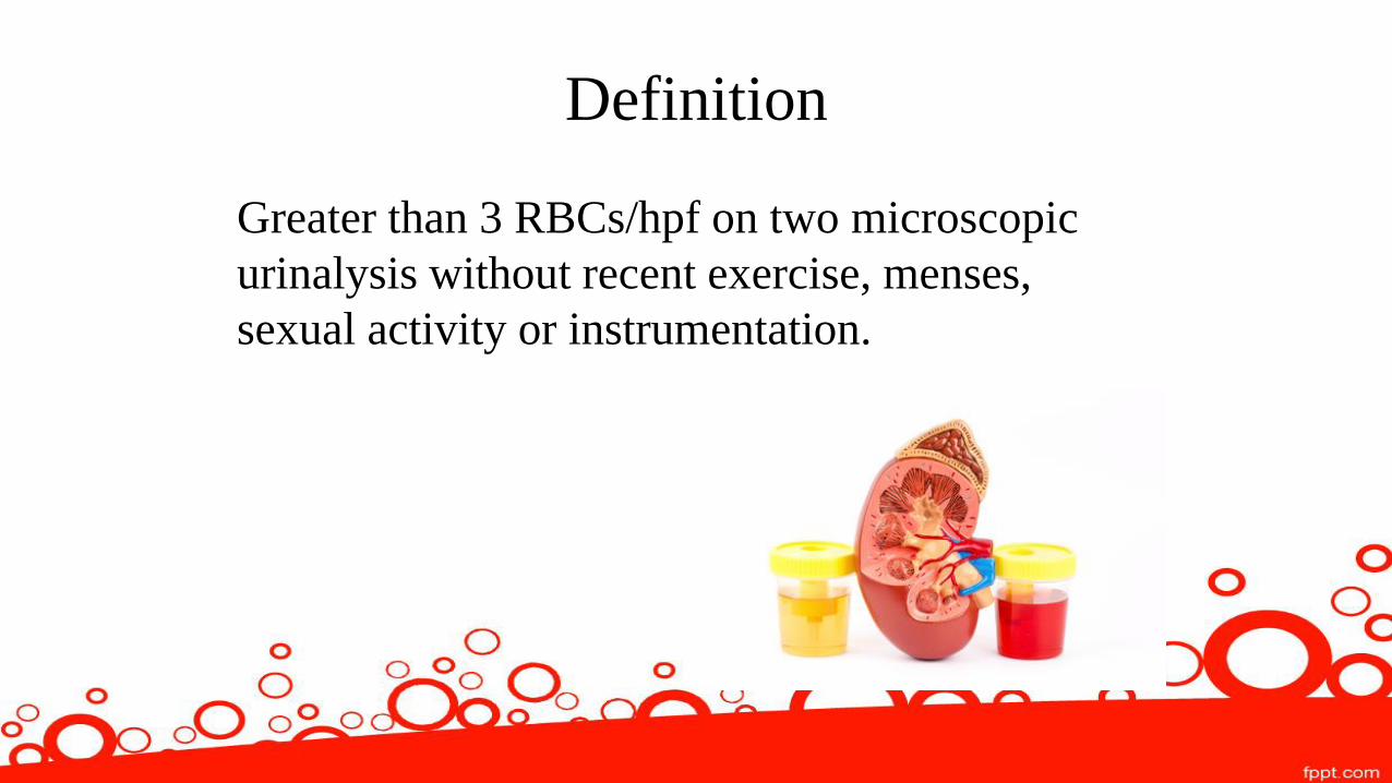
Definition
Greater than 3 RBCs/hpf on two microscopic
urinalysis without recent exercise, menses,
sexual activity or instrumentation.

2 or 3 urine analyses
should be conducted
to reduce rate of false
positive

History
• Age
• Duration: acute vs chronic
• Onset: transient, progressive, sudden, recurrent
• Characteristic: painless vs painful, gross vs
microscopic, constant vs intermittent

Associated Symptoms • Fever, back pain, urinary urgency/frequency, dysuria (UTI)
• Renal colic (renal calculi)
• Constitutional symptoms (bladder ca)
• Lower urinary tract symptoms (BPH)
• Recent infection history, edema or hypertension (glomerulonephritis)
• Recent back or abdominal injury, vigorous exercise (trauma)
• Recent bleeding from other sites, family history of bleeding disorders
(systemic coagulopathy)
• Cyclic hematuria in women (endometriosis)
• Sterile pyuria with hematuria (renal tuberculosis, analgesic
nephropathy, interstitial disease)

Pattern
• What colour is the urine?
• Diet?
• Blood at the beginning, middle or end of
stream?
• Recent surgery?

Family history • Sickle cell disease
• Deafness or ocular abnormalities
• Autoimmune disorders
• Malignancy history
• Birt Hogg Dube
• Von Hipple Lindau syndrome

Medications
• Anticoagulatants
• Cyclophosphamide
• Antibiotic: rifampin
• Analgesics

Physical Exam
• Vitals: temperature (UTI), BP (large RCC)
• Genital exam: – signs of bleeding at urethral meatus (both sexes)
–DRE for males
–Gyne/OB presentations with vaginal bleeding

Physical Exam • Inspection
– Rash, ecchymoses, or petechiae (coagulopathy)
–Hearing loss, lens abnormality (Alports Syndrome)
– Edema, sore throat
• Palpation - Renal colic pain radiating flank to groin (stone)
- Abdominal tenderness and mass (RCC)
- CVA tenderness (pyelonephritis)

Risk Factors for Malignancy • Male gender
• Age (> 40 years)
• Past or current smoking
• Occupational or other exposure to chemicals or dyes (benzenes or aromatic amines)
• Analgesic abuse
• History of gross hematuria
• History of urologic disorder or disease
• History of irritative voiding symptoms
• History of pelvic irradiation
• History of chronic urinary tract infection
• History of exposure to known carcinogenic agents or chemotherapy such as
alkylating agents
• History of chronic indwelling foreign body
Investigations
• Urine dip: nitrites, WBC, protein
• Urine microscopy: red cell casts, RBC count,
WBC count
• If red cell casts, protein or increased creatinine

Classification
Glomerular – Red cell casts, proteinuria and
dysmorphic red blood cells
Renal
- IgA nephropathy
- Alport syndrome
- Thin glomerular BM disease
- Post infectious
- MPGN
Multi-system
- SLE nephritis
- Wegener syndrome
- Goodpasture syndrome
- Sickle cell disease

Classification
Nonglomerular
• Upper tract nephroliathiasis, RCC, TCC, polycystic kidney,
pyelonephritis
• Lower tract cystitis, bladder ca, prostate ca, prostatitis

PROSTATITIS

UTI

RENAL ASSAULT

CANCER

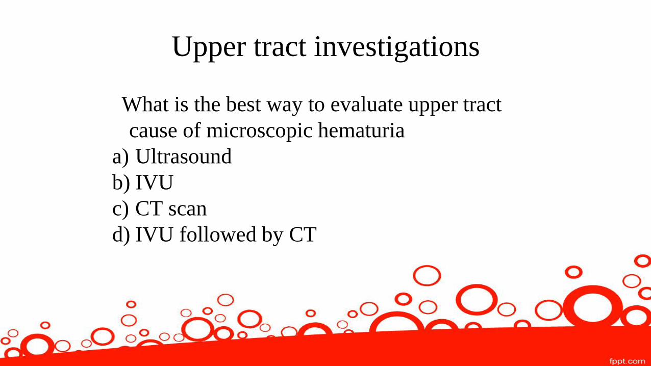
Upper tract investigations
What is the best way to evaluate upper tract
cause of microscopic hematuria
a) Ultrasound
b) IVU
c) CT scan
d) IVU followed by CT

Answer
a) Ultrasound

Investigations - UPPER
• U/S : some limitations in diagnosing
transitional cell carcinoma
- can miss very small tumours less than 0.5cm
• IVU (intravenous urography) :
–exposure to contrast
–less sensitive and specific to U/S
–at times hard to differentiate solid vs cystic

Investigations - UPPER
AUA guidelines:
•CT scan: enhanced multiphasic as gold standard
CUA:
• Ultrasound: as gold standard


Investigations - LOWER
• Cystoscopy – To be done in all patients above the
age of 35.
- In patients younger than 35, cystoscopy should be
done at the discretion of a physician Level C
Recommendation
• Cytology
–Sensitivity (34%); Specificity (81%)


Approach

DIAGNOSIS FREQUENCY (%)
Unknown 43 to 68
Urinary tract infection 4 to 22
Benign prostatic hyperplasia 10 to 13
Urinary calculi 4 to 5
Bladder cancer 2 to 4
Renal cystic disease 2 to 3
Renal disease 2 to 3
Kidney cancer < 1
Prostate cancer < 1
Urethral stricture disease < 1
Common Etiologies of Microscopic
Hematuria

Follow up
What is the appropriate follow up for reoccurrence of
microscopic hematuria in the setting of previous negative full
work up?
a) Referral to urology
b) Cytology and then refer to urology
c) Repeat cytology 3 times then refer to urology
d) Conduct serial urinalysis

Answer
d) serial urinalysis

Follow up
Urinalysis, cytology and BP check 6, 12, 24 and
36 months following initial presentation
– If negative, risk of future malignancy is less than 1%
– If positive and persistent then, repeat full evaluation in 3-
5years. Or if gross hematuria, positive cytology, and new
irritative symptoms in the setting of normal C&S then
earlier

Revisit Case
•49 year old female, otherwise healthy
•Urinalysis: 5 rbc/hpf
•C&S: negative
•No hx of gross hematuria
•No LUTS
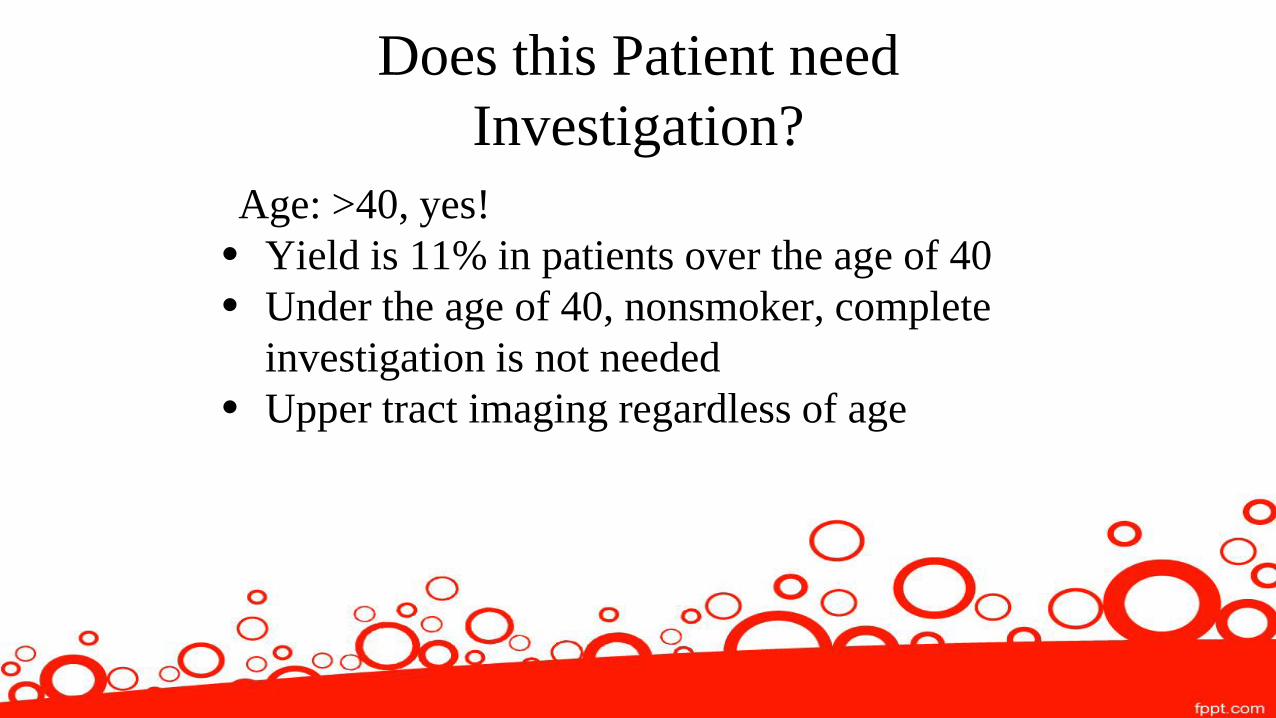
Does this Patient need
Investigation?
Age: >40, yes!
• Yield is 11% in patients over the age of 40
• Under the age of 40, nonsmoker, complete
investigation is not needed
• Upper tract imaging regardless of age

What is the appropriate approach?
a) Urinalysis, cytology, cystoscopy and
ultrasound
b) Urinalysis cytology, cystoscopy and CT scan
c) Urinalysis, cytology and ultrasound
d) Urinalysis, cytology and CT scan

Answer
a) Urinalysis, cytology, cystoscopy and
ultrasound

Upper tract
Ultrasound findings:
4cm mass on left kidney

CT Scan for better characterization

Lower Tract
Normal cystoscopy
Negative cytology

Case 2
•28 y.o M with 2 episodes of gross hematuria
•Nonsmoker
•6 months of ongoing LUTS
–Decreased stream
–Hesitancy of stream
–No dysuria

Does this warrant evaluation?
•5 fold increased risk of significant underlying
pathology in gross hematuria
•Age is not a factor in gross hematuria
YES!
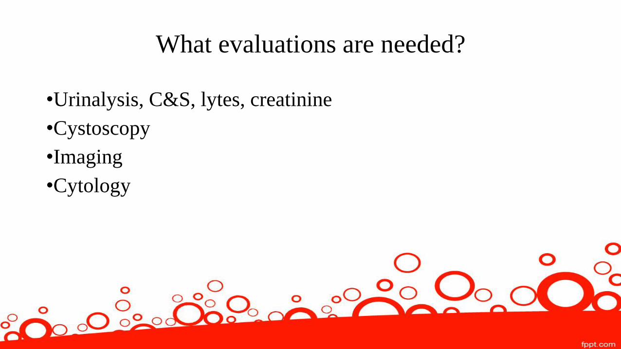
What evaluations are needed?
•Urinalysis, C&S, lytes, creatinine
•Cystoscopy
•Imaging
•Cytology

Results
•Urinalysis: 1-5 WBC, 5-10 RBC
•C&S: no growth
•Ultrasound: unremarkable
Questions

References 1. American Urological Association. (2012, Validity confirmed
2016). Diagnosis, Evaluation and Follow-up of Asymptomatic Microhematuria in Adults. Retrieved http://www.auanet.org/guidelines/asymptomatic-microhematuria-(2012-reviewed-and-validity-confirmed-2016)
2. Thaller, T. and Wang, L. (1999 Sep 15). Evaluation of asymptomatic microscopic hematuria in adults. Am Fam Physician. 1999 Sep 15;60(4):1143-1152. 3. Wolin, T., Laroche, B. and Psoosy, K. Canadian Urological Association. Canadian guidelines for the management of asymptomatic microscopic hematuria in adults. Retrieved https://www.cua.org/themes/web/assets/files/guidelines/en/amh_2008_e.pdf
4. Subak, L and Grady, D. Asymptomatic Microscopic Hematuria—Rethinking the Diagnostic Algorithm. JAMA Intern Med. 2017;177(6):808-809