evaluation of deep learning-based approaches for covid-19
TRANSCRIPT
Signal, Image and Video Processing (2021) 15:959–966https://doi.org/10.1007/s11760-020-01820-2
ORIG INAL PAPER
Evaluation of deep learning-based approaches for COVID-19classification based on chest X-ray images
Kamal KC1 · Zhendong Yin1 ·Mingyang Wu1 · Zhilu Wu1
Received: 4 July 2020 / Revised: 1 September 2020 / Accepted: 12 November 2020 / Published online: 7 January 2021© The Author(s) 2021
AbstractThe COVID-19, novel coronavirus or SARS-Cov-2, has claimed hundreds of thousands of lives and affected millions ofpeople all around the world with the number of deaths and infections growing exponentially. Deep convolutional neuralnetwork (DCNN) has been a huge milestone for image classification task including medical images. Transfer learning ofstate-of-the-art models have proven to be an efficient method of overcoming deficient data problem. In this paper, a thoroughevaluation of eight pre-trained models is presented. Training, validating, and testing of these models were performed on chestX-ray (CXR) images belonging to five distinct classes, containing a total of 760 images. Fine-tuned models, pre-trained inImageNet dataset, were computationally efficient and accurate. Fine-tuned DenseNet121 achieved a test accuracy of 98.69%and macro f1-score of 0.99 for four classes classification containing healthy, bacterial pneumonia, COVID-19, and viralpneumonia, and fine-tuned models achieved higher test accuracy for three-class classification containing healthy, COVID-19,and SARS images. The experimental results show that only 62% of total parameters were retrained to achieve such accuracy.
Keywords Deep convolution neural network · Transfer learning · Chest X-ray · COVID-19 · SARS
1 Introduction
COVID-19 , novel coronavirus or SARS-CoV-2, is a humancoronavirus within the virus family Coronaviridae, orderNidovirales,which aremulti-lipid, envelopednon-segmented,single-strained, positive-sense RNA viruses named aftercorona or crown-like surface projections [1]. In December2019, Wuhan, the capital of central China’s Hubei province,became ground zero for COVID-19 outbreak. As of June2020, more than 10 million cases of infections and 500thousands deaths have been reported from more than 213countries and territories. Severe acute respiratory syndrome(SARS) and Middle East respiratory syndrome (MERS),the predecessor to COVID-19, are viral respiratory diseases.SARS-Cov first reported in November 2002 in Guangdongprovince of southern China, spread to 29 countries withmore
The research presented in this article was supported by the NationalNatural Science Foundation of China (Grant Nos. 61471142,61601145, 61571167 and 61871157).
B Zhendong [email protected]
1 School of Electronics and Information Engineering, HarbinInstitute of Technology, Harbin 150001, China
than 8000 infected cases and 774 deaths. MERS-Cov wasfirst reported in Saudi Arabia in September 2012 and hasspread to 27 countries with more than 2500 infections and866 deaths. In general, respiratory virus infection can occurthroughdirect or indirect contact, droplet spray in short-rangetransmission and aerosol in long-range transmission [2].COVID-19 patients develop common symptoms like fever,dry cough, myalgia, fatigue, dyspnea, and anorexia at onsetof illness. In due course of time, these symptoms progressto acute respiratory distress syndrome (ARDS), arrhythmia,and shock [3]. X-rays are the most common, cheap, andwidely available diagnostic imaging technique used to detectand diagnosis tumors and other abnormal masses like lymphnodes, pneumonia, bone fractures, abscesses, etc. X-raysreveal abnormalities indicative of lung diseases. Early stagesof CXR radiograph of COVID-19 may read as normal, butwith progression in time, the readingsmay resemble of pneu-monia or ARDS. It is a difficult task to classify these X-rayimages between normal, bacterial pneumonia, viral pneumo-nia, SARS or COVID-19 irrespective of community acquiredpneumonia (CAP) or hospital-acquired pneumonia (HAP).Chest X-rays can be used as preliminary classification dueto its prevalent usage as primary diagnostic test.
123
960 Signal, Image and Video Processing (2021) 15:959–966
AI researchers have contributed a lot in the field of data pro-cessing. Advances in AI have the potential in transformingthe field of medicine since medical diagnostics and treat-ments are fundamentally a data problem. Deep learning, asub-field ofmachine learning, automatically extracts detailedfeatures from data which has proven to be highly efficient invarious data processing task. Deep learning has become aprominent solution for a wide variety of natural languageprocessing (NLP) or text processing, speech recognition andsynthesis, signal analysis, and computer vision tasks. CNNs,multilayered neural networks, play a pivotal role in variousdigital image processing tasks such as feature extraction,pattern recognition, segmentation, object recognition, classi-fication onvarious types of images includingmedical images.Deep learning demands plethora of data to achieve accept-able result and is resource hungry.Toovercome this difficulty,a machine learning technique, transfer learning, was intro-duced. Transfer learning re-purposes a model trained on onetask on a second related task. Pre-trained models are easilyre-trained on smaller data with lesser trainable parametersto achieve higher accuracy. Depending upon the data andmodel selected, pre-trained models are fine-tuned to attainfine result.
2 Related works
Severalmachine learning algorithms have been used for auto-matic classification of digitized medical images. Machinelearning recognizes patterns to compute image features suit-able for detection, diagnosis, or classification tasks. Machinelearning algorithms can be classified on the basis of trainingstyles: supervised, unsupervised, and reinforcement learn-ing. Supervised learning involves gaining experience byusing images that contain useful information and applyingthe gained expertise to predict on unseen new images (testdata). Examples of supervised learning algorithms includesupport vector machine (SVM), decision tree, linear regres-sion, logistic regression, naive Bayes, k-nearest neighbor(k-NN), random forest, AdaBoost, and neural network meth-ods. In unsupervised learning, data are processed with a goalof separating the images into groups. Since the algorithmdoes not have information regarding the groups, it automati-cally determines the number of groups and a way to separatethem. Examples of unsupervised learning algorithm systemsinclude K-means, hierarchical clustering, density-based spa-tial clustering of applicationswith noise (DBSCAN),Markovrandom fields (MRF), iterative self-organizing data (ISO-DATA), and fuzzy C-means (FCM) systems. Like supervisedlearning, reinforcement learning begins with a classifier thatwas built by using labelled data. However, the system is thengiven unlabelled data, and it tries to further improve theclassification by better characterizing these data similar to
unsupervised learning. Examples of reinforcement learningalgorithm systems includeMonte Carlo methods and tempo-ral difference (TD) algorithms [4]. Several machine learningor deep learning algorithms have been used for automaticclassification of medical images based on X-rays [5], CTscans [6], mammograms [7], MRI [8], PET scans [9], orRGB images [10] for detection of tumor, diabetic retinopa-thy (DR), cancer, Alzheimer’s disease, pneumonia, or a RGBimage for skin lesion classification.
2.1 Deep learning inmedical images
Deep learning is a new and powerful data oriented machinelearning method, which utilizes a range of neural networkarchitectures to perform various imaging tasks, like object(i.e., lesion or region of interest) detection, segmentationand classification. Deep learning methods differ from theconventional machine learning methods (i.e., support vectormachine (SVM), k-NN, naiveBayes, and random forest, etc.)in onemajor sense: The latter rely on feature extractionmeth-ods to train the algorithm, whereas deep learning methodsextract feature automatically to learn the image. Evaluated onthe 150 validation images from the ISIC 2017 classificationchallenge for skin lesion classification, a fusion of three pre-trained deep models, AlexNet, VGG16, and ResNet-18, asdeep feature generators yielded an area under receiver oper-ating characteristic (ROC) curve of 83.83% for melanomaclassification and of 97.55% for seborrheic keratosis classifi-cation [11]. Recurrent Attention DenseNet (RADnet) whichemploys original DenseNet architecture along with addingthe components of attention for slice level predictions andrecurrent neural network layer for incorporating 3D contextwas employed for classification of dataset composed of 185,67, and 77 brainCT scans for training, validation, and testing,respectively. RADnet achieved 81.82% hemorrhage predic-tion accuracy and 84.78% F1 score [12].
2.2 Convolutional neural network for pneumoniaand COVID-19 detection
A four convolutional layered CNN was employed for binaryclassification of presence of pneumonia based on a total of5856 X-ray images of anterior–posterior chests from ret-rospective pediatric patients of which 3722 images wereallocated to the training set and 2134 images to the val-idation set. The images were augmented using varioustechniques like rescale, rotation, shift, shear, zoom, horizon-tal ip, etc. The network achieved a validation accuracy of93.73% on input data of size 200 × 200 [13]. An ensem-ble model based on transfer learning reached an accuracyof 96.4% with a recall of 99.62% on unseen data from theGuangzhou Women and Children’s Medical Center datasetfor binary classification of healthy and pneumonia X-Ray
123
Signal, Image and Video Processing (2021) 15:959–966 961
images [14]. Classification of 50 images of healthy and 50images of COVID-19 patients using pre-trained ResNet50provided the classification performance of 98% accuracy[15]. Deep learning with X-ray imaging may extract sig-nificant biomarkers related to the COVID-19 disease wasexhibited by training and testing on an unbalanced dataset of1442 X-ray images including 224 images with confirmedCOVID-19 disease, 714 cases of both bacterial and viralpneumonia (400 bacteria and 314 viral), and 504 images ofnormal conditions. MobileNetV2 outperformed other mod-els and obtained 94.72% accuracy [16]. COVID-Net networkarchitecture which uses a lightweight residual projection-expansion projection-extension (PEPX) design pattern wasused on a dataset which comprised of 183 cases of COVID-19, 5538 of Pneumonia and 8066 of healthy subjects with atest set of 100 images of pneumonia and healthy lungs, and31 of COVID-19 with no patient overlap between the testand the training set. The model achieved a test accuracy of93.3% [17]. Covidx-Net used transfer learnedmodels to clas-sify healthy andCOVID-19 cases and concluded thatVGG19and Densenet201 achieved highest accuracy of 90% [18]. Atotal of 250 COVID-19 and 250 non-COVID-19 images fortraining, and an independent test set of 74 positive and 36negative samples were trainedwith an ensemble of ten CNNsand achieved an AUC of 0.81 for the classification task [19].
3 Materials andmethods
3.1 Proposed approach
In this work, we employ data-driven deep learning technique,convolutional neural network (CNN). Since its introductionby Lecun and due to its unrivalled performance in 2012 Ima-geNet challenge achieved by AlexNet, CNN has become aprominent technique in image processing tasks. Deep learn-ing or data-driven approaches, in general, require abundanceof data to generalize from training data and discover viablefeature sets and decision criteria, and finally extract mean-ingful results. Various state-of-the-art deep learning modelssuch as VGG-16, and VGG-19 [20], Inception [21], ResNet[22] , Xception [23], which performed well in ILSVRC [24]make use of CNN and its variant. We propose fine-tuning ofa transfer learned model trained on ImageNet dataset.
3.1.1 VGG
In 2014, Oxford’s Visual Geometry Group (VGG) developedand trained a 16 convolutional layered deep convolutionalnetwork for object recognition called VGGNet. Differentversions of VGGNet were introduced like VGG11, VGG11(LRN), VGG13, VGG16 (Conv1), VGG16„ and VGG19.
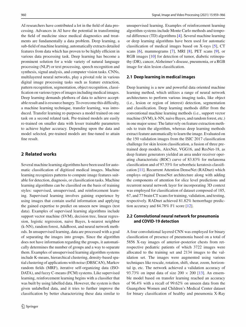
Fig. 1 Inception module: building block of inception model
VGG19 is 19 layers deep with each layer using a filter ofsize 3× 3 and has an image input size of 224× 224.
3.1.2 InceptionV3
Szegedy et al. [21] proposed inception module in GoogleNetor InceptionV1 that was designed to be efficient in terms ofparameters and computations, while achieving state-of-the-art performance. Different versions of Inception model wereintroduced by optimizing inceptionmodule using BatchNor-malization, factorized convolution and residual connections.InceptionV3 is a 42 layered deep network. Figure 1 showsthe inceptionmodule which is the building block of inceptionmodel.
3.1.3 ResNet
At the ILSVRC 2015, Residual Neural Network (ResNet) byHe et al. [22] introduced a novel architecture with residualconnections which greatly reduced the optimization difficul-ties and enabled the training of much deeper networks byintroducing skip connections between residual modules.
ResNet50 is a 50-layer deep neural network. Figure 2shows convolutional block and identity block which com-bine to form residual network.
3.1.4 MobileNet
The need for smaller model size suitable for real-timeanalysis in resource constrained mobile devices led to thedevelopment of MobileNet [25], which uses depthwise sep-arable convolutions as their core building blocks.MobileNetsare small, low-latency, low-power models parameterized tomeet the resource constraints of a variety of use cases byintroducing width and depth multiplier used for classifica-tion, detection, embeddings and segmentation.
3.1.5 DenseNet
Dense blocks were firstly proposed by Huang et al. inDenseNet [26] that introduced dense connectivity in whicheach layer receives signals from all its preceding layers
123

962 Signal, Image and Video Processing (2021) 15:959–966
Fig. 2 Convolutional block and identity block which combine to formresidual network
Fig. 3 Dense block: basic building blocks for DenseNet
combined by channel-wise concatenation resulting into lowinformation bottleneck.DenseNets integrate the properties ofidentity mappings, deep supervision, reduced feature redun-dancy and diversified depth allowing feature reuse makingit good feature extractors. DenseNets with different depths;DenseNet-121,169,209,264were introduced. Figure 3 showsdense block, the basic building block used in DenseNet.
3.1.6 NasNetMobile
Google Brain designed a search space, calledNASNet searchspace, which decouples the complexity of an architecturefrom the depth of the network, thus resulting in transferableand scalable network called Neural Architecture Search Net-work (NASNet). NASNet comprises of two types of cells:normal cell that returns a feature map of the same dimen-sion and reduced cell that returns a feature map where thefeature map height and width is reduced by a factor. NAS-NetMobile [27], also referred as NASNet-A(4 @ 1056) is a
reduced version of NASNetLarge, referred as NASNet-A(6@ 4032).
3.2 Transfer learning and fine-tuning
Transfer learning is a machine learning technique thatinvolves reusing an existing, trained neural network, devel-oped for one task, as the foundation for another task. Transferlearning removes the necessity of large training data for a taskas the basic features required to train a model are importedfrom previous task. In DNN, each layer generate a sequenceof representations of the data. For an image recognitionnetwork, the early layers in the CNN capture the simplestfeatures of the data that are universal such as the presenceof edges, contours, or gradients in color and can be reusedfor similar tasks, while later layers capture more complicatedwhich are highly data dependent and needs to be re-trained.The main challenge of transfer learning is to retain the exist-ing knowledge in the model while adapting the model to newtask which gives rise to the problem of the number of layersor parameters required to be re-trained to achieve optimalresult.Transfer learning involves firstly finding the suitable pre-trainedmodel, secondly, replacing the final layer of themodelaccording to the number of output layers for the new task andfinally, resume training the model with new data and fine-tuning the model till the accuracy converges towards highervalue.
4 Methodology
Deep neural network is a stack of layers where each hiddenlayer extractsmeaningful features automatically. This featureof DNNs is utilized in transfer learning. Transfer learning isemployed whenever the amount of training data is smaller innumber. Transfer learning is done either by training a modelfrom scratch on a suitable huge dataset and retraining themodel on desired smaller dataset or using a pre-trained state-of-the-art models. The former exploits lot of resources andtime as designing a model, creating a huge dataset and thenfinally training them is tiresome. However, using state-of-the-art model pre-trained on standard dataset reduces lots ofcomputational cost.Since collecting large amount of medical images is difficultdue to data privacy concerns, transfer learning is opted asa suitable solution. Transfer learning makes use of reusablefeatures by using the concept of layer freeze. Since eachlayer outputs some features in DNN, spotting the layer bestsuited for extracting the reusable feature is delicate. Freezinga certain portion of network that best provides the featureswhich can be used in new domain is the basic principle oftransfer learning. However, employing transfer learning on
123

Signal, Image and Video Processing (2021) 15:959–966 963
Table 1 Chest X-ray datasetsplit statistics
Class CXR A CXR B CXR CTrain Val Test Train Val Test Train Val Test
Bacterial Pneumonia 134 33 19 118 30 38 – – –
COVID-19 134 33 19 118 30 38 118 30 38
Normal 134 33 19 118 30 38 118 30 38
Viral Pneumonia 134 33 19 118 30 38 – – –
SARS – – – – – – 10 3 3
Fig. 4 Some samples from our dataset which includes an image fromeach class. Each row represents x-ray images from each class. From toprow to bottom: bacterial pneumonia, COVID-19, normal, SARS andviral pneumonia
models pre-trained in ImageNet demands the replacementof final output layer with the new layer suited for the newtask. The network can be either trained by freezing all thelayers except the final layers which is termed as featureextractor or freezing a portion of network for best resultswhich is termed as fine-tuning. We opted for fine-tuninginstead of feature extractor for higher accuracy in expenseof slightly higher computational cost [28]. Fine-tuning isconducted by using the concept of block freeze in whicha collection of fixed number of layers are bundled to form ablock. This collection of blocks are frozen instead of layers.Parameters corresponding to the frozen layers are referred as
non-trainable parameters, whereas the network trains withremaining trainable parameters. Freezing the layers does notaffect the weights corresponding to the layer. All the experi-ments were run for 1000 epochs.
5 Dataset
The dataset used is a collection of two subsets from two dif-ferent dataset, COVID-19 dataset [29] created by Dr. JosephCohen and Pneumonia dataset [30] which are Chext X-Raydataset. The dataset was divided in two ways. In the firstscenario, CXR A, the dataset was divided into training andtesting set with 90:10 split, and the training set was furthersplit into training and validation set with 80:20 split, respec-tively. In the second scenario, CXRB, the datasetwas dividedinto training and testing set with 80:20 split, and the trainingset was again split into training and validation set with 80:20split, respectively.
The chest X-Ray contains images collected from vari-ous patients all around the world. A total of 744 images,equally split over four classes (normal, bacterial pneumonia,viral pneumonia, and COVID-19), were used for classifi-cation. The CXR C contains a total of 388 X-ray imagesfrom three different classes (healthy, COVID-19, andSARS).Data are split similar to second scenario. While CXR A andB have balanced data, data in CXR C are highly imbal-anced. The imageset containsAP (anterior–posterior) and PA(posterior–anterior) CXR images. L (Lateral) X-ray imagesare discarded for the experiment. These x-rays were takeneither erect (standing or sitting) or supine position. Few ofthe X-rays contain images from patients with chest portinserted in them. While most of the chest X-ray imageswere taken from different patients all around the world,COVID-19 dataset contain someX-rays from a single patientover a course of time, i.e., x-rays from day 1, day 8 andday 13.
A detailed information about the images is shown inTable 1. Images are either in jpg, jpeg, or png format. Fig-ure 4 shows few samples of images used in the experiment.Images from each classes are shown in the figure.
123

964 Signal, Image and Video Processing (2021) 15:959–966
Table 2 Comparing validation and test accuracies (%) on fine-tunedmodels on chest X-ray dataset
Models CXR A CXR BValidation Test Validation Test
VGG19 100 97.37 100 94.74
InceptionV3 100 97.37 100 97.37
ResNet50 100 97.37 100 94.08
ResNet50V2 100 97.37 100 94.08
MobileNet 100 97.37 100 95.4
MobileNetV2 100 97.37 100 95.4
DenseNet121 100 97.37 100 98.69
NasNetMobile 100 97.37 98.44 97.37
Bold denotes the highest value for particular category
Fig. 5 Confusion matrix for CXR dataset. Confusion matrix on the topdepicts prediction result of CXR A dataset and the one in the bottomdepicts that of CXR B dataset
6 Results
Transfer learned eight different state-of-the-art models pre-trained on ImageNet achieved 97.37% test accuracy on CXRA dataset. Fine-tuned DenseNet121 outperformed othermodels by achieving 98.69% test accuracy onCXRBdataset.Table 2 shows the accuracies obtained while employing fine-tuned models on CXR dataset. All the models achieved avalidation accuracy of 100% except for NasNetMobile onCXR B dataset. Data split of 80:20 (train/test) with 80%further into 80:20 (train:validation) split performed bettercompared to 90:10 (train:test) with 90% further into 80:20(train/validation) split. The confusion matrix of CXR A andCXR B for test data is given in Fig. 5.
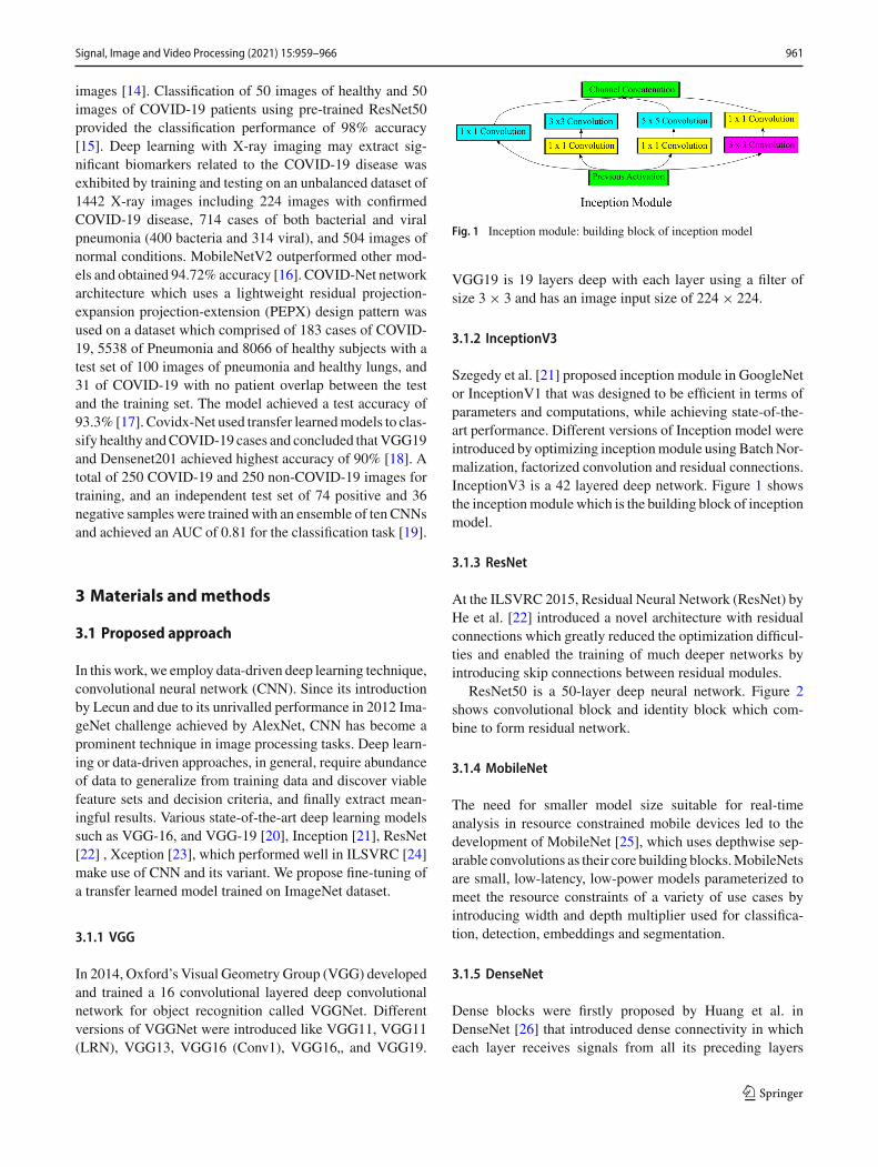
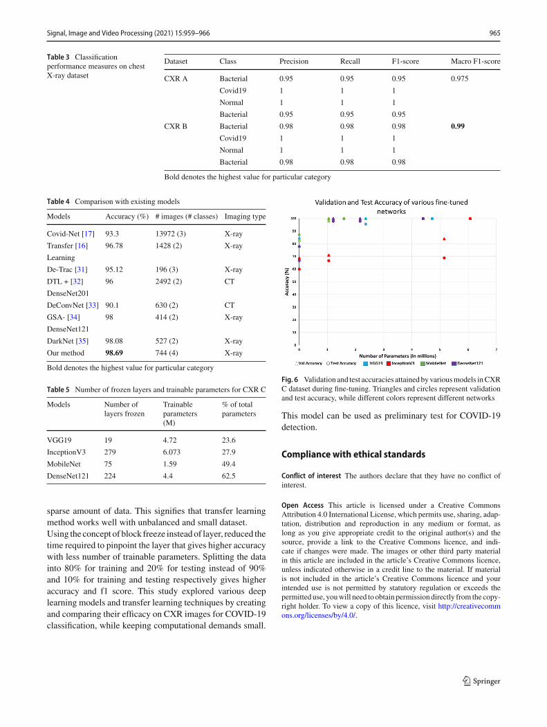
Table 2 shows the comparison of validation and testingaccuracy of CXR A and CXR B obtained after fine-tuningvarious standard pre-trained models. Table 3 shows the clas-sification performance measure of CXR datasets. CXR Bobtained the highest macro f1-score of 0.99 when fine-tunedusing DenseNet121. Table 4 compares accuracy of variousexisting models and our model. Table 5 shows the respec-tive number of layers frozen, trainable parameters, and thepercentage of total parameters trained by various fine-tunedmodels to obtain highest accuracy. The scatter plot in Fig. 6shows the validation and testing accuracy of various fine-tunedmodels.While employing transfer learning using blockfreeze, accuracies depend on the number of trainable param-eters. It can be seen that accuracy increases as number ofparameters increase.
7 Conclusion
In this work, various fine-tuned state-of-the-art deep learn-ing models pre-trained on ImageNet, were compared forefficient classification of chest X-ray images. These mod-els effectively classified healthy, bacterial, viral, COVID-19and SARS diseases based on CXR images. Fine-tuned mod-els achieved validation accuracy of 100%on all CXRdataset,100% test accuracy on CXR C, 97.37% of test accuracy onCXRAdataset and98.69%of test accuracyonCXRBdatasetby fine-tuned DenseNet121. DenseNet121 outperforms allmodels in terms of precision, recall, f1-score, and accuracy,thus, it is proven to be the most effective model for the spe-cific classification task, and the specific data sample.All the models were pre-trained on ImageNet dataset. Ima-geNet demonstrated to be a reliable source domain fortransferring learned features on CXR disease classification.The models performed well for classification of healthy anddisease X-ray images of COVID-19, SARS diseases. DatasetCXR A and CXR B had equal number of images per classwhile CXR C dataset had unbalanced data with one class
123
Signal, Image and Video Processing (2021) 15:959–966 965
Table 3 Classificationperformance measures on chestX-ray dataset
Dataset Class Precision Recall F1-score Macro F1-score
CXR A Bacterial 0.95 0.95 0.95 0.975
Covid19 1 1 1
Normal 1 1 1
Bacterial 0.95 0.95 0.95
CXR B Bacterial 0.98 0.98 0.98 0.99
Covid19 1 1 1
Normal 1 1 1
Bacterial 0.98 0.98 0.98
Bold denotes the highest value for particular category
Table 4 Comparison with existing models
Models Accuracy (%) # images (# classes) Imaging type
Covid-Net [17] 93.3 13972 (3) X-ray
Transfer [16] 96.78 1428 (2) X-ray
Learning
De-Trac [31] 95.12 196 (3) X-ray
DTL + [32] 96 2492 (2) CT
DenseNet201
DeConvNet [33] 90.1 630 (2) CT
GSA- [34] 98 414 (2) X-ray
DenseNet121
DarkNet [35] 98.08 527 (2) X-ray
Our method 98.69 744 (4) X-ray
Bold denotes the highest value for particular category
Table 5 Number of frozen layers and trainable parameters for CXR C
Models Number oflayers frozen
Trainableparameters(M)
% of totalparameters
VGG19 19 4.72 23.6
InceptionV3 279 6.073 27.9
MobileNet 75 1.59 49.4
DenseNet121 224 4.4 62.5
sparse amount of data. This signifies that transfer learningmethod works well with unbalanced and small dataset.Using the concept of block freeze insteadof layer, reduced thetime required to pinpoint the layer that gives higher accuracywith less number of trainable parameters. Splitting the datainto 80% for training and 20% for testing instead of 90%and 10% for training and testing respectively gives higheraccuracy and f1 score. This study explored various deeplearning models and transfer learning techniques by creatingand comparing their efficacy on CXR images for COVID-19classification, while keeping computational demands small.
Fig. 6 Validation and test accuracies attained byvariousmodels inCXRC dataset during fine-tuning. Triangles and circles represent validationand test accuracy, while different colors represent different networks
This model can be used as preliminary test for COVID-19detection.
Compliance with ethical standards
Conflict of interest The authors declare that they have no conflict ofinterest.
Open Access This article is licensed under a Creative CommonsAttribution 4.0 International License, which permits use, sharing, adap-tation, distribution and reproduction in any medium or format, aslong as you give appropriate credit to the original author(s) and thesource, provide a link to the Creative Commons licence, and indi-cate if changes were made. The images or other third party materialin this article are included in the article’s Creative Commons licence,unless indicated otherwise in a credit line to the material. If materialis not included in the article’s Creative Commons licence and yourintended use is not permitted by statutory regulation or exceeds thepermitted use, youwill need to obtain permission directly from the copy-right holder. To view a copy of this licence, visit http://creativecommons.org/licenses/by/4.0/.
123
966 Signal, Image and Video Processing (2021) 15:959–966
References
1. Poutanen, S.M.: Human Coronaviruses. Princ. Pract. Pediatr.Infect. Dis. 1148–1152.e3 (2018). https://doi.org/10.1016/B978-0-323-40181-4.00222-X
2. Moriyama, M., Hugentobler, W.J., Iwasaki, A.: Seasonality of res-piratory viral infections. Ann. Rev. Virol. 7, 83–101 (2020)
3. Wang, D., Hu, B., Hu, C., Zhu, F., Liu, X., Zhang, J., Wang, B.,Xiang, H., Cheng, Z., Xiong, Y., et al.: Clinical characteristicsof 138 hospitalized patients with 2019 novel coronavirus-infectedpneumonia in wuhan, china. JAMA 323(11), 1061 (2020)
4. Erickson, B.J., Korfiatis, P., Akkus, Z., Kline, T.L.: Machine learn-ing for medical imaging. Radiographics 37(2), 505 (2017)
5. Bar,Y.,Diamant, I.,Wolf, L., Lieberman, S.,Konen, E.,Greenspan,H.: Chest pathology detection using deep learning with non-medical training. In: 2015 IEEE 12th International SymposiumOn Biomedical Imaging (ISBI) (IEEE, 2015), pp. 294–297
6. Roth, H.R., Lu, L., Liu, J., Yao, J., Seff, A., Cherry, K., Kim, L.,Summers, R.M.: Improving computer-aided detection using con-volutional neural networks and random view aggregation. IEEETrans. Med. Imaging 35(5), 1170 (2015)
7. Arevalo, J., González, F.A., Ramos-Pollán, R., Oliveira, J.L.,Lopez, M.A.G.: Representation learning for mammography masslesion classification with convolutional neural networks. Comput.Methods Progr. Biomed. 127, 248 (2016)
8. Alfonse, M., Salem, A.B.M.: An automatic classification of braintumors through mri using support vector machine. Egy. Comp. Sci.J. 40(3), 1110–2586 (2016)
9. Liu,M., Cheng,D., Yan,W., Initiative, A.D.N., et al.: Classificationof alzheimers disease by combination of convolutional and recur-rent neural networks using fdg-pet images. Frontiers Neuroinf. 12,35 (2018)
10. Harangi, B.: Skin lesion classification with ensembles of deep con-volutional neural networks. J. Biomed. Inf. 86, 25 (2018)
11. Mahbod, A., Schaefer, G., Wang, C., Ecker, R., Ellinge, I.:Skin lesion classification using hybrid deep neural networks. In:ICASSP 2019–2019 IEEE International Conference on Acoustics,Speech and Signal Processing (ICASSP) (IEEE, 2019), pp. 1229–1233
12. Grewal, M., Srivastava, M.M., Kumar, P., Varadarajan, S.: Rad-net: Radiologist level accuracy using deep learning for hemorrhagedetection in ct scans. In 2018 IEEE 15th International Symposiumon Biomedical Imaging (ISBI 2018) (IEEE, 2018), pp. 281–284
13. Stephen, O., Sain, M., Maduh, U.J., Jeong, D.U.: An efficientdeep learning approach to pneumonia classification in health-care. J. Healthcare Eng. 2019 (2019). https://doi.org/10.1155/2019/4180949
14. Chouhan, V., Singh, S.K., Khamparia, A., Gupta, D., Tiwari, P.,Moreira, C., Damaševicius, R., De Albuquerque, V.H.C.: A noveltransfer learning based approach for pneumonia detection in chestx-ray images. Appl. Sci. 10(2), 559 (2020)
15. Narin, A., Kaya, C., Pamuk, Z.: Automatic detection of coronavirusdisease (covid-19) using x-ray images and deep convolutional neu-ral networks, arXiv preprint arXiv:2003.10849 (2020)
16. Apostolopoulos, I.D., Mpesiana, T.A.: Covid-19: automatic detec-tion from x-ray images utilizing transfer learning with convolu-tional neural networks. Phys. Eng. Sci. Med. 43, 635 (2020)
17. Wang, L.,Wong, A.: Covid-net: A tailored deep convolutional neu-ral network design for detection of covid-19 cases from chest x-rayimages, arXiv preprint arXiv:2003.09871 (2020)
18. Hemdan, E.E.D., Shouman, M.A., Karar, M.E.: Covidx-net: Aframework of deep learning classifiers to diagnose covid-19 in x-ray images, arXiv preprint arXiv:2003.11055 (2020)
19. Castiglioni, I., Ippolito, D., Interlenghi,M.,Monti, C.B., Salvatore,C., Schiaffino, S., Polidori, A., Gandola, D., Messa, C., Sardanelli,
F.: Artificial intelligence applied on chest x-ray can aid in the diag-nosis of covid-19 infection: a first experience from lombardy, italy,medRxiv (2020)
20. Simonyan, K., Zisserman, A.: Very deep convolutional networksfor large-scale image recognition, arXiv preprint arXiv:1409.1556(2014)
21. Szegedy, C., Liu, W., Jia, Y., Sermanet, P., Reed, S., Anguelov, D.,Erhan, D., Vanhoucke, V., Rabinovich, A.: Going deeper with con-volutions. In: Proceedings of the IEEE Conference on ComputerVision and Pattern Recognition, pp. 1–9 (2015)
22. He,K., Zhang,X., Ren, S., Sun, J.:Deep residual learning for imagerecognition. In: Proceedings of the IEEE Conference on ComputerVision and Pattern Recognition, pp. 770–778 (2016)
23. Chollet, F.: Xception: Deep learningwith depthwise separable con-volutions. In: Proceedings of the IEEE Conference on ComputerVision and Pattern Recognition, pp. 1251–1258 (2017)
24. Russakovsky, O., Deng, J., Su, H., Krause, J., Satheesh, S., Ma, S.,Huang, Z., Karpathy, A., Khosla, A., Bernstein,M., et al.: Imagenetlarge scale visual recognition challenge. Int. J. Comput.Vis.115(3),211 (2015)
25. Howard, A.G., Zhu, M., Chen, B., Kalenichenko, D., Wang, W.,Weyand, T., Andreetto, M., Adam, H.: Mobilenets: Efficient con-volutional neural networks for mobile vision applications, arXivpreprint arXiv:1704.04861 (2017)
26. Huang, G., Liu, Z., van der Maaten, L., Weinberger, K.Q.: Denselyconnected convolutional networks. In: The IEEE Conference onComputer Vision and Pattern Recognition (CVPR) (2017)
27. Zoph, B., Vasudevan, V., Shlens, J., Le, Q.V.: Learning transfer-able architectures for scalable image recognition. In: The IEEEConference on Computer Vision and Pattern Recognition (CVPR)(2018)
28. Kamal, K.C., Yin, Z., Li, B., Ma, B., Wu, M.: Transfer learning forfine-grained crop disease classification based on leaf images. In:2019 10th Workshop on Hyperspectral Imaging and Signal Pro-cessing: Evolution in Remote Sensing (WHISPERS) (2019), pp.1–5
29. Cohen, J.P., Morrison, P., Dao, L., Roth, K., Duong, T.Q., Ghas-semi, M.: Covid-19 image data collection: Prospective predictionsare the future. arXiv preprint arXiv:2006.11988 (2020)
30. Kermany, D., Zhang, K., Goldbaum, M.: Labeled optical coher-ence tomography (oct) and chest x-ray images for classification.Mendeley Data 2 (2018). https://doi.org/10.17632/rscbjbr9sj.2
31. Abbas, A.A., Abdelsamea, M.M., Gaber, M.M.: Detrac: transferlearning of class decomposedmedical images in convolutional neu-ral networks. IEEEAccess 8, 74901–74913 (2020). https://doi.org/10.1109/ACCESS.2020.2989273
32. Jaiswal, A., Gianchandani, N., Singh, D., Kumar, V., Kaur, M.:Classification of the covid-19 infected patients using densenet201based deep transfer learning. J. Biomol. Struct. Dyn. 1–8, (2020)
33. Zheng, C., Deng, X., Fu, Q., Zhou, Q., Feng, J., Ma, H., Liu, W.,Wang, X.: Deep learning-based detection for covid-19 from chestct using weak label, medRxiv (2020)
34. Ezzat, D., Ella H.A., et al., Gsa-densenet121-covid-19: a hybriddeep learning architecture for the diagnosis of covid-19 dis-ease based on gravitational search optimization algorithm, arXivpreprint arXiv:2004.05084 (2020)
35. Ozturk, T., Talo, M., Yildirim, E.A., Baloglu, U.B., Yildirim, O.,Acharya, U.R.: Automated detection of covid-19 cases using deepneural networks with x-ray images. In: Computers in Biology andMedicine pp. 103792 (2020)
Publisher’s Note Springer Nature remains neutral with regard to juris-dictional claims in published maps and institutional affiliations.
123