evaluation of dna damage in a her2+ cell line induced...
TRANSCRIPT

Evaluation of DNA damage in a Her2+ cell line induced by an Auger-emitting immunoconjugate
Behnaz Piroozfar, Behrouz Alirezapoor, Farahnaz Motamedi Sedeh, Amir Reza Jalilian, Mohammad Mirzaei, Gholamreza Raisali
Radiation Application Research School, Nuclear Science and Technology Research Institute,
Tehran, Iran
(Received 20 April 2016, Revised 31 May 2016, Accepted 2 June 2016)
ABSTRACT
Introduction: Auger electron based radioimmunotherapy (RIT) using 111In-DOTA-trastuzumab (111In-DOTA-antiHer2) feasibility was studied in vitro on a HER2/neu positive cell line, SkBr3. Methods: 111In-DOTA-antiHer2 was prepared according to the optimized conditions followed by quality control tests including radiochemical purity; immunoreactivity). SkBr3 as a HER2/neu positive cell line was used to determine the degree of internalization and cell viability of 111In-DOTA-antiHer2. For comet assay freshly cultivated SkBr3 cells incubated with 111In-DOTA-antiHer2 in 3.7 and 7.4 MBq doses at 37ºC for 24h. Results: 111In-DOTA-antiHer2 (>95% radiochemical purity; >79% immunoreactivity) demonstrated significant internalization in SKBr3 cells in 24 h. no significant cell viability difference observed for 111In-DOTA-antiHer2 and 111In cation treatments. Comet assay at 3.7 MBq demonstrated no significant DNA damage, while at 7.4 MBq dose DNA damage observed at least 16% more than control In-111 chloride after 24 h. Conclusion: Although the internalization of 111In-DOTA-antiHer2 was approved in this study, however, lack of cell death and slight DNA breakage for 111In-DOTA-antiHer2 treatment suggests absence of nucleus entry which is essential for demonstration of DNA damage according to Auger electron range at cellular level. Key words: Auger therapy; 111In-DOTA-antiHer2; SkBr3; Immunoreactivity; Internalization; Cell viability; Comet assay
Iran J Nucl Med 2016;24(2):107-114 Published: July, 2016 http://irjnm.tums.ac.ir
Corresponding author: Dr Amir Reza Jalilian, Radioisotope Products and Radiation Technology Section, Division of Physical and Chemical Sciences, Department of Nuclear Sciences and Applications, International Atomic Energy Agency, Vienna International Centre, PO Box 100, 1400 Vienna, Austria. E-mail: [email protected]
Orig
inal A
rticle

111In-DOTA-antiHer2 DNA damage in a Her2+ cell line Piroozfar et al.
Iran
J N
ucl M
ed 2
016,
Vol
24,
No
2 (S
eria
l No
46)
h
ttp:
//irjn
m.t
ums.
ac.ir
J
uly,
201
6
108
INTRODUCTION
In targeted therapy, the shorter the range of the particle emission, the better targetry and lower imposed dose to the surrounding cells, on condition that the radionuclide is close enough to target DNA molecules in nucleus and/or mitochondria in case of mammalian cells. -emitters now are the frontier radionuclides used in the development of therapeutic radiopharmaceuticals, however their relative particle long range often impose adverse effects to the surrounding cells [1]. On the other hand α-emitters offer better opportunities for targeted therapy applications in case they are internalized into target cells or even closer to the DNA molecules. Their low availability and high costs in production and distribution has limited application of these radionuclides for vast therapeutic applications [2]. Auger electrons are produced in cascades during electronic shell transitions followed by electronic capture and/or conversion in some radionuclides with a very low energy (from a few eV to a few keV). They are considered as high LET particles (from 4 to 26 keV/µm; energy <1 keV). Thus, their path length in biological matter is very short, and efforts have been done to bring Auger electrons in the cell nucleus to obtain the highest cytotoxicity. The clinical radioimmunotherapy (RIT) studies using Auger electrons such as I-125 labeled to monoclonal antibodies (mAbs) have shown no solid results [3, 4]. The possibility of iodinated antibody instability and long half-life limited the application I-125 agents for human use. In-111 is an interesting radionuclide used in the development various radiolabeled antibodies
for imaging studies during last 3 decades. This cyclotron-produced radionuclide produced usually via 112Cd(p,2n)111In, is available in no carrier added form and its 67 h half- life matches the biological half-life of full size mAbs (1-3 d) for ultimate use in radioimmunoscintigraphy and also allows enough shelf life for transport to rather distant centers from the production site. Interestingly In-111 is a source of Auger electrons demonstrating potentials for targeted therapy in case of reaching close to nucleus while carried by appropriate internalizing agents such as antibiotics [5], peptides [6] and antibodies [7] (Table 1). Not many reports on evaluation and application of In-111 therapeutic agents are available in the literature, however most of the studies have been focused on the development of mAbs and peptides as some have been presented in Table 2. Recently some In-111 labeled mAbs have been developed for human studies [9, 10] based on interesting chelating agent moiety DOTA, having potentials for radiolabeling of various theranostic radionuclides such as Y-90, Lu-177, Ga-68, etc., in one case 111In-DOTA-trastuzumab (111In-DOTA-antiHer2) production has been reported and in continuation of current Auger electron therapy we were interested in evaluation of its DNA breakage effect in vitro based on the comet assay (single-cell gel electrophoresis), a simple method for measuring deoxyribonucleic acid (DNA) strand breaks in eukaryotic cells as well as cell death.
Table 1: List of some important Auger-emitting radionuclides properties [8].
Radioisotope Half-life Average number of Auger and Coster-Kronig electrons Energies (keV) Dose from electrons
(i)e-rad.g.µ-1.h-1 99mTc 6.05 h 3 12 n.a. 111In 2.1 d 8 16 0.074 125I 60.5 d 20 24 0.041 123I 13.3 h 11 21 0.032
Table 2: Some selected studies on the therapeutic effects of In-111 radiolabeled biomolecules.
Agent Malignancy Study model Reference 111In-antiCD123 Acute myeloma Mice engrafted with primary human AML [11] 111In-dual-antiHER2/EGFR Human Breast Cancer Xenografts [12] 111In-[Lys40(Ahx-DTPA-111In)NH2]-exendin-4 Pancreatic neuroendocrine tumors Mouse model [13] 111In-DTPA-hEGF Human Breast Cancer 231-H2N xenografts [14] 111In-NLS-antiHER2 Human Breast Cancer MDA-MB-231 tumors [15] 111In-HuM195 anti-CD33 Acute myeloma HL-60 and HL-60-MX-1 cells [16] 111In-DTPA-hEGF Human breast cancer MDA-MB-468 [17] 111In-labeled N-myc antisense oligonucleotides. Human neuroblastoma cells Human neuroblastoma cells [18] 111In-labeled antibodies to HER-2 Human Carcinoma SK-OV-3.ip.1 cells [19] 111In-DTPA-octreotide Thyroid cancer Human subject [20, 21]

111In-DOTA-antiHer2 DNA damage in a Her2+ cell line Piroozfar et al.
Iran
J N
ucl M
ed 2
016,
Vol
24,
No
2 (S
eria
l No
46)
h
ttp:
//irjn
m.t
ums.
ac.ir
J
uly,
201
6
109
In comet assay method, cells embedded in agarose on a microscope slide are lysed to form nucleoids containing supercoiled loops of DNA linked to the nuclear matrix followed by electrophoresis at high pH. The tail of structures resembling comets, observed by fluorescence microscopy shows the intensity and the number of DNA breaks [22]. In this work we report brief preparation, cell binding, cell internalization, cell viability and comet assay results of 111In-DOTA-trastuzumab (111In-DOTA-antiHer2) on breast cancer SkBr3 cells as a Her2 positive line.
METHODS
NHS-DOTA was purchased from Macrocycles (NJ, USA). Trastuzumab (Herceptin) was a pharmaceutical sample purchased from Roche Co. Radiochromatography was performed by using a Bioscan AR-2000 radio thin layer chromatography (TLC) scanner instrument (Bioscan, Paris, France). A high purity germanium (HPGe) detector coupled with a Canberra™ (model GC1020-7500SL) multichannel analyzer and a dose calibrator ISOMED 1010 (Dresden, Germany) were used for radioactive measurements. Calculations were based on the 171 keV peak for 111In. All values were expressed as mean ± standard deviation (mean ± SD) and the data were compared using Student’s t-test. Statistical significance was defined as P < 0.05. Production and quality control of 111In-InCl3 sterile solution for in vitro studies Indium-111 chloride was prepared by 22 MeV proton bombardment of the enriched cadmium-112 target at a 30 MeV cyclotron, with a current of 100 µA for 48 min (80 µAh) according to the reported method [23]. Briefly, after the dissolution of the irradiated target by conc. HBr, the solution was passed through a cation exchange Dowex 50 × 8 resin, followed by washing with HBr conc. solution and DDH2O. Indium-111 was eluted with 1 N HCl as 111InCl3 for labeling use and quality control experiments. For cell studies the appropriate activity was evaporated under reduced pressure followed by the addition of pH.5.5 acetate buffer. Production and quality control of 111In-DOTA-antiHer2 Detailed production and extensive quality control of 111In-DOTA-antiHer2 has been reported [24]. Briefly, Trastuzumab (Roche) as a lyophilized powder was purified by Vivaspin-2 filters using ultrafiltration in 0.2 M Na2CO3 (pH 9.2) buffer solution followed by antibody concentration measurement and conjugated with DOTA‑NHS in bicarbonate buffer and
incubated at room temperature for 12 h. The mixture was then transferred on a Vivaspin 2 cut-off filter (30 kDa) and centrifuged and the conjugation step terminated using ammonium acetate buffer and the antibody concentration was re-measured for determination of the average chelate: antibody ratio the spectrophotometric method was used in presence of arsenazo yttrium(III) complex (2:1, 1 ml) [25]. For radiolabeling, 37 MBq of 111In-InCl3 (in 0.2M HCl) was added to a conical vial and dried under a flow of nitrogen followed by the addition of acetate buffer and vortexing for 10 min and incubating at 40°C for 90 min followed by radiochemical purity determination by RTLC and HPLC methods. Ethylene di-amine tetra acetic acid (ETDA) solution was added to the labeling mixture to scavenge the unlabeled In-111 cation followed by passing the mixture through the disposable PD10 De-salting column. The radioimmunoconjugate was analyzed for integrity by SDS-PAGE according to the method of Laemmli [26]. Cell culture A HER2/neu positive cell line, SkBr3, was obtained from Iranian Biological Resources Center (Tehran, Iran) and was grown in RPMI-1640 medium supplemented with 10 % heat-inactivated fetal bovine serum, 2 mM glutamine, 100 lg/ml streptomycin and 100 IU/ml penicillin. Immunoreactivity of 111In-DOTA-antiHer2 towards SkBr3 cell line Immunoreactivity of 111In-DOTA-antiHer2 towards SkBr3 cell line was determined using a double-inverse of the binding data which may be considered a modification of the Lineweaver-Burk plot as follows. Different numbers (5 × 10⁶, 2.5 × 10⁶, 1.25 × 10⁶, 0.62 × 10⁶, 0.31× 10⁶ and 0.15 × 10⁶) of SkBr3 cell were located in tubes; 30000 cpm of 111In-DOTA-antiHer2 was added and incubated for 3 h at 4°C. After incubation, the radioactivity of tubes was read by -counter. Then tubes were centrifuged in 3000 g and the radioactivity of cells (the pellet, not the supernatant) were read by -counter. The immunoreactivity was determined using a Lineweaver-Burk plot and data were analyzed by the Lindmo method [27] as known:
][][max
SKSVV
m
Internalization assays Internalization studies were performed using acid dissociation of 111In-DOTA-antiHer2 [28] SkBr3 cells were harvested and re-suspended at 1 × 10⁵ cells/200 µl in medium and cultured in 24 well plates.

111In-DOTA-antiHer2 DNA damage in a Her2+ cell line Piroozfar et al.
Iran
J N
ucl M
ed 2
016,
Vol
24,
No
2 (S
eria
l No
46)
h
ttp:
//irjn
m.t
ums.
ac.ir
J
uly,
201
6
110
After 24 h, 30000 cpm of 111In-DOTA-antiHer2 were added to each well and incubated at 37°C. At various times (4, 8, 12, 16, 24, 30 and 36 h), RPMI medium in wells were removed and the cells, half of wells, were then exposed to 1 ml solution of 0.1 M NaOH and half of wells were exposed to 1 ml solution of 0.1 M acetic acid pH 4.0. After 15 min, the radioactivity content of the supernatant was measured by -counter. NaOH solution destroyed the whole SkBr3 cells and this solution was considered as total activity (internalized, bound and dissociated 111In-DOTA-antiHer2). Acetic acid was used to remove the interaction between 111In-DOTA-antiHer2 and HER2/neu at the SkBr3 cell surface and the radioactivity of the solution was considered as bound and dissociated 111In-DOTA-antiHer2. Differences between the two activities were considered as internalized 111In-DOTA-antiHer2 fraction. Cell Treatment for comet assay SKBR-3 cell line was propagated by DMEM media and 10% fetal calf serum (FCS) as monolayer in 105 cells per well into 12-wells plate, after 48 h treated using of 111In-DOTA-antiHer2. Then 1 ml of 111In-DOTA-antiHer2 in two concentrations (3.7 and 7.4 MBq) was added to the wells and was incubated at 37 °C, after 1.5 h, the cells were washed by Phosphate buffered saline (PBS) for removing the free 111In, then added 1 ml of DMEM medium and incubated at 37 °C for 24 hours. The treated cells were harvested using a Teflon scraper, suspended in PBS and counted by a Neobar slide. The viability percent was tested by trypan blue dye and counted white and blue cells. Finally 50 µl of the treated SKBR-3 cells (with concentration: 10×106 cells per 1 ml PBS) was mixed with 50 µl of Low Melting Point agarose1% and coated on a pre-coated slide with agarose 1%. Calculations were based on Cell Viability (%) = total viable cells (unstained) ÷ total cells (stained and unstained) × 100 (n=5). Comet assay The double strand breaks of DNAs were determined using the neutral comet assay method with slight modifications [29, 30]. The treated SKBR-3 cells were diluted to a concentration of 10 × 106 cells/ml in PBS and 50 µl of the cell dilution was mixed at 37ºC with 50 µl of freshly prepared 1% low melting point agarose (LMP, Merck, Cat No: K39061202-835) in distilled water. An aliquot of 50 µl of the mixture was placed on a pre-coated slide with agarose 1% (Cinnagen, Cat No: MR7740), then covered with a coverslip and transferred to an ice-cold plate to promote fast gelling. As soon as the gel solidified, coverslips were smoothly removed and the slides were submerged in lysing solution (lysing solution; 2.5M NaCl, 100 mM EDTA, 1% Triton X-100, 10
mMTris, pH:10), overnight at 4 °C to lysis the cells. The slides were rinsed in TBE buffer (0.09 mol/l Tris–borate, 0.002 mol/l EDTA, pH 7.5) for 20 min to allow DNA unfolding, then transferred to an electrophoresis tank and immersed in fresh TBE electrophoresis buffer. Electrophoresis was performed at 20 V (6 V/cm), 12 mA for 40 min. The slides were washed in a neutralization buffer (0.4 mol/l Tris-HCl, pH 7.5) for 15 min and fixed with 10 min washes in absolute ethanol, air-dry for at least 2 h at room temperature. Finally, DNA was stained in two methods with SYBR Green I (Cinnagen, DNA safe stain, Cat No: PR381603) at a 1:10000 dilution in PBS and ethidium bromide (2 µg/ml in water) (Cinnagen, Cat No: MR7721C). Samples could be immediately analyzed or stored at room temperature in the dark until needed. The Comets could be assessed by visual scoring or digitalization and image processing. At least 10 cells were measured in each slid, the tail moment of DNA (%tail DNA × tail length) was evaluated by the fluorescence microscopy and comet-Score software.
RESULTS
Figure 1 shows the step-wise approach of 111In-DOTA-antiHer2 preparation. It has been reported that moderate increase of chelator to mAb ratio leads to a slight decrease in immunoreactivity which is likely due to non-specific conjugation of chelators to mAb, which possibly attaches a chelator in the region of the antigen-binding site [31]. In radiochemical purity determination, for 111In3+ detection, the best solvent mixture was 1 mM DTPA pH. 5 solution (data not shown), while for 111In-DOTA detection, mixture of 10% ammonium acetate: Methanol (1:1) can be used (Rf 0.3).
Fig 1. Detailed conjugation and radiolabeling steps of 111In-DOTA-antiHer2 at optimized conditions.
111In-DOTA-antiHer2 DNA damage in a Her2+ cell line Piroozfar et al.
Iran
J N
ucl M
ed 2
016,
Vol
24,
No
2 (S
eria
l No
46)
h
ttp:
//irjn
m.t
ums.
ac.ir
J
uly,
201
6
111
111In-DOTA-antiHer2 remains at the origin in all systems showing at least 95 ± 0.5% radiochemical purity (Figure 2).
Fig 2. Immunoreactivity of 111In-DOTA-antiHer2 using SkBr3 cell-binding assay (n = 5).
At the optimized conditions 111In-DOTA-antiHer2 was prepared (RCP >95 ± 0.5%, S.A. 5.3 µCi/µg) with the average number of chelators per antibody of 6:1 showing significant immune‑reactivity retention using ELISA. As shown in Figure 3, internalized activity for 111In-DOTA-antiHer2 reached to the maximum significantly in 8-12 h incubation at 37°C. After 36 h the internalized activity decreased to 3% in 36 hours as observed in other reports [32] possibly caused by proteolytic degradation in lysosomes, leading to metal trans-chelation to the internal metal containing biomolecules (Figure 3).
Fig 3. Internalization assay of 111In-DOTA-antiHer2 in different time on SkBr3 cell line (n = 5).
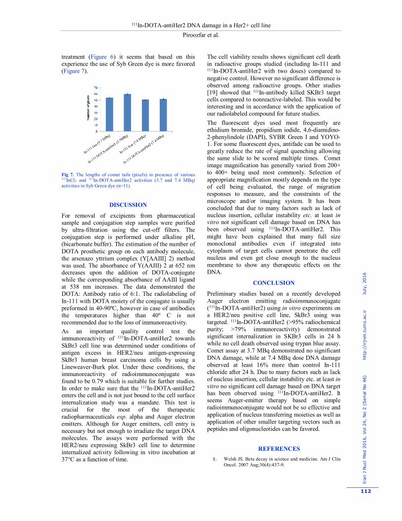
Cell viability The trypan blue exclusion test was used to determine the number of viable cells present in a cell suspension since live cells possess intact cell membranes that exclude certain dyes, such as trypan blue, Eosin, or propidium, whereas dead cells do not [33]. Figure 4 shows the viability of SKBr3 cells based on the percentage of undyed cells compared with dyed cells. Comet assay Figure 5 shows the comet assay of samples used in this study using ethidium bromide dye.
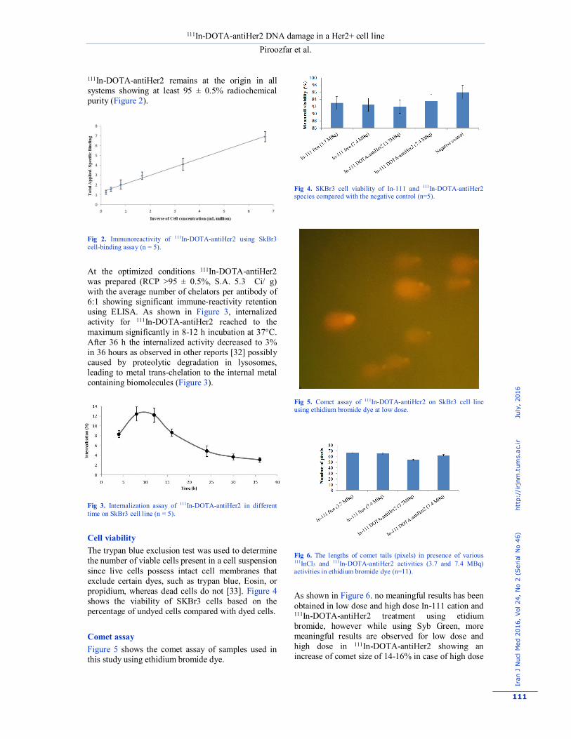
Fig 4. SKBr3 cell viability of In-111 and 111In-DOTA-antiHer2 species compared with the negative control (n=5).
Fig 5. Comet assay of 111In-DOTA-antiHer2 on SkBr3 cell line using ethidium bromide dye at low dose.
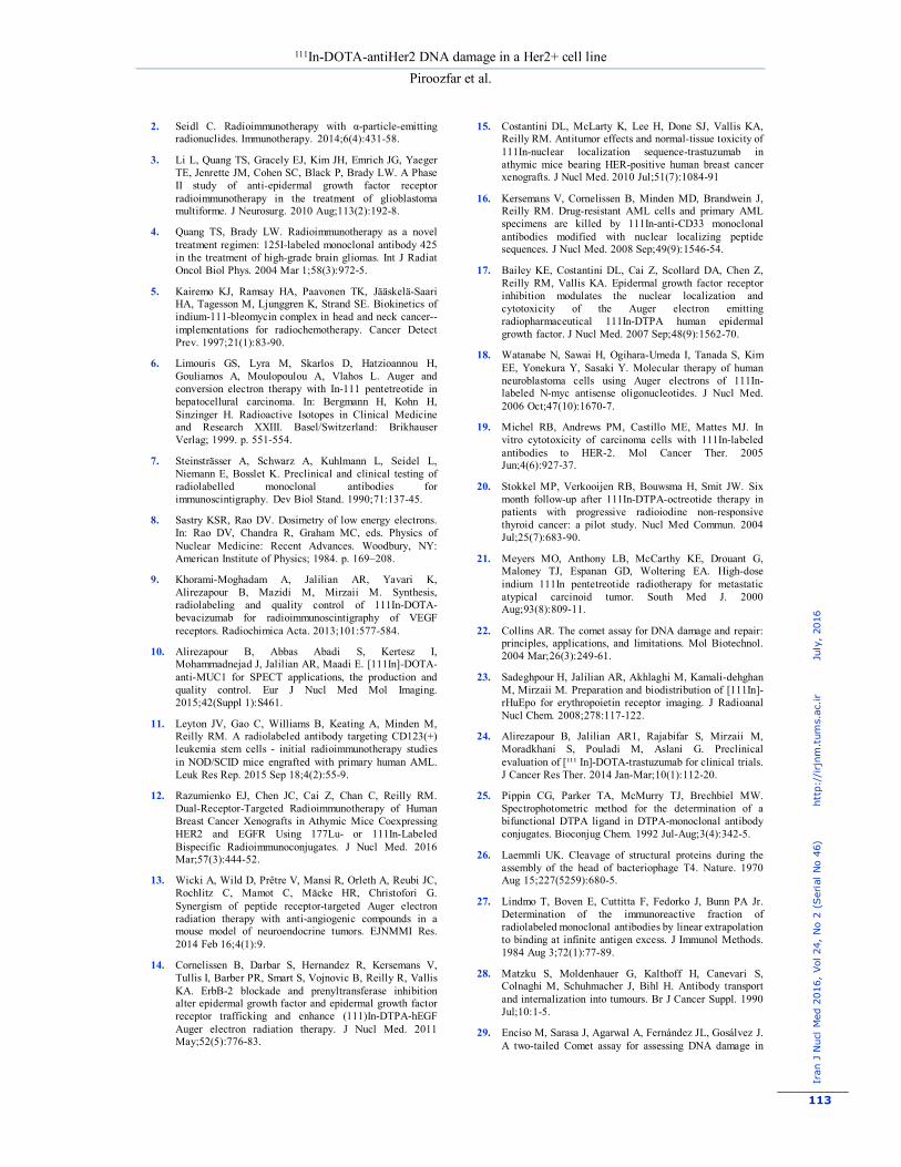
Fig 6. The lengths of comet tails (pixels) in presence of various 111InCl3 and 111In-DOTA-antiHer2 activities (3.7 and 7.4 MBq) activities in ethidium bromide dye (n=11).
As shown in Figure 6. no meaningful results has been obtained in low dose and high dose In-111 cation and 111In-DOTA-antiHer2 treatment using etidium bromide, however while using Syb Green, more meaningful results are observed for low dose and high dose in 111In-DOTA-antiHer2 showing an increase of comet size of 14-16% in case of high dose
111In-DOTA-antiHer2 DNA damage in a Her2+ cell line Piroozfar et al.
Iran
J N
ucl M
ed 2
016,
Vol
24,
No
2 (S
eria
l No
46)
h
ttp:
//irjn
m.t
ums.
ac.ir
J
uly,
201
6
112
treatment (Figure 6) it seems that based on this experience the use of Syb Green dye is more favored (Figure 7).
Fig 7. The lengths of comet tails (pixels) in presence of various 111InCl3 and 111In-DOTA-antiHer2 activities (3.7 and 7.4 MBq) activities in Syb Green dye (n=11).
DISCUSSION
For removal of excipients from pharmaceutical sample and conjugation step samples were purified by ultra-filtration using the cut-off filters. The conjugation step is performed under alkaline pH, (bicarbonate buffer). The estimation of the number of DOTA prosthetic group on each antibody molecule, the arsenazo yttrium complex (Y[AAIII] 2) method was used. The absorbance of Y(AAIII) 2 at 652 nm decreases upon the addition of DOTA-conjugate while the corresponding absorbance of AAIII ligand at 538 nm increases. The data demonstrated the DOTA: Antibody ratio of 6:1. The radiolabeling of In-111 with DOTA moiety of the conjugate is usually preformed in 40-90ºC, however in case of antibodies the temperatures higher than 40º C is not recommended due to the loss of immunoreactivity. As an important quality control test the immunoreactivity of 111In-DOTA-antiHer2 towards SkBr3 cell line was determined under conditions of antigen excess in HER2/neu antigen-expressing SkBr3 human breast carcinoma cells by using a Lineweaver-Burk plot. Under these conditions, the immunoreactivity of radioimmunoconjugate was found to be 0.79 which is suitable for further studies. In order to make sure that the 111In-DOTA-antiHer2 enters the cell and is not just bound to the cell surface internalization study was a mandate. This test is crucial for the most of the therapeutic radiopharmaceuticals esp. alpha and Auger electron emitters. Although for Auger emitters, cell entry is necessary but not enough to irradiate the target DNA molecules. The assays were performed with the HER2/neu expressing SkBr3 cell line to determine internalized activity following in vitro incubation at 37°C as a function of time.
The cell viability results shows significant cell death in radioactive groups studied (including In-111 and 111In-DOTA-antiHer2 with two doses) compared to negative control. However no significant difference is observed among radioactive groups. Other studies [19] showed that 111In-antibody killed SKBr3 target cells compared to nonreactive-labeled. This would be interesting and in accordance with the application of our radiolabeled compound for future studies. The fluorescent dyes used most frequently are ethidium bromide, propidium iodide, 4,6-diamidino-2-phenylindole (DAPI), SYBR Green I and YOYO-1. For some fluorescent dyes, antifade can be used to greatly reduce the rate of signal quenching allowing the same slide to be scored multiple times. Comet image magnification has generally varied from 200× to 400× being used most commonly. Selection of appropriate magnification mostly depends on the type of cell being evaluated, the range of migration responses to measure, and the constraints of the microscope and/or imaging system. It has been concluded that due to many factors such as lack of nucleus insertion, cellular instability etc. at least in vitro not significant cell damage based on DNA has been observed using 111In-DOTA-antiHer2. This might have been explained that many full size monoclonal antibodies even if integrated into cytoplasm of target cells cannot penetrate the cell nucleus and even get close enough to the nucleus membrane to show any therapeutic effects on the DNA.
CONCLUSION
Preliminary studies based on a recently developed Auger electron emitting radioimmunoconjugate (111In-DOTA-antiHer2) using in vitro experiments on a HER2/neu positive cell line, SkBr3 using was targeted. 111In-DOTA-antiHer2 (>95% radiochemical purity; >79% immunoreactivity) demonstrated significant internalization in SKBr3 cells in 24 h while no cell death observed using trypan blue assay. Comet assay at 3.7 MBq demonstrated no significant DNA damage, while at 7.4 MBq dose DNA damage observed at least 16% more than control In-111 chloride after 24 h. Due to many factors such as lack of nucleus insertion, cellular instability etc. at least in vitro no significant cell damage based on DNA target has been observed using 111In-DOTA-antiHer2. It seems Auger-emitter therapy based on simple radioimmunoconjugate would not be so effective and application of nucleus transferring moieties as well as application of other smaller targeting vectors such as peptides and oligonucleotides can be favored.
REFERENCES 1. Welsh JS. Beta decay in science and medicine. Am J Clin
Oncol. 2007 Aug;30(4):437-9.
111In-DOTA-antiHer2 DNA damage in a Her2+ cell line Piroozfar et al.
Iran
J N
ucl M
ed 2
016,
Vol
24,
No
2 (S
eria
l No
46)
h
ttp:
//irjn
m.t
ums.
ac.ir
J
uly,
201
6
113
2. Seidl C. Radioimmunotherapy with α-particle-emitting radionuclides. Immunotherapy. 2014;6(4):431-58.
3. Li L, Quang TS, Gracely EJ, Kim JH, Emrich JG, Yaeger TE, Jenrette JM, Cohen SC, Black P, Brady LW. A Phase II study of anti-epidermal growth factor receptor radioimmunotherapy in the treatment of glioblastoma multiforme. J Neurosurg. 2010 Aug;113(2):192-8.
4. Quang TS, Brady LW. Radioimmunotherapy as a novel treatment regimen: 125I-labeled monoclonal antibody 425 in the treatment of high-grade brain gliomas. Int J Radiat Oncol Biol Phys. 2004 Mar 1;58(3):972-5.
5. Kairemo KJ, Ramsay HA, Paavonen TK, Jääskelä-Saari HA, Tagesson M, Ljunggren K, Strand SE. Biokinetics of indium-111-bleomycin complex in head and neck cancer--implementations for radiochemotherapy. Cancer Detect Prev. 1997;21(1):83-90.
6. Limouris GS, Lyra M, Skarlos D, Hatzioannou H, Gouliamos A, Moulopoulou A, Vlahos L. Auger and conversion electron therapy with In-111 pentetreotide in hepatocellural carcinoma. In: Bergmann H, Kohn H, Sinzinger H. Radioactive Isotopes in Clinical Medicine and Research XXIII. Basel/Switzerland: Brikhauser Verlag; 1999. p. 551-554.
7. Steinsträsser A, Schwarz A, Kuhlmann L, Seidel L, Niemann E, Bosslet K. Preclinical and clinical testing of radiolabelled monoclonal antibodies for immunoscintigraphy. Dev Biol Stand. 1990;71:137-45.
8. Sastry KSR, Rao DV. Dosimetry of low energy electrons. In: Rao DV, Chandra R, Graham MC, eds. Physics of Nuclear Medicine: Recent Advances. Woodbury, NY: American Institute of Physics; 1984. p. 169–208.
9. Khorami-Moghadam A, Jalilian AR, Yavari K, Alirezapour B, Mazidi M, Mirzaii M. Synthesis, radiolabeling and quality control of 111In-DOTA-bevacizumab for radioimmunoscintigraphy of VEGF receptors. Radiochimica Acta. 2013;101:577-584.
10. Alirezapour B, Abbas Abadi S, Kertesz I, Mohammadnejad J, Jalilian AR, Maadi E. [111In]-DOTA-anti-MUC1 for SPECT applications, the production and quality control. Eur J Nucl Med Mol Imaging. 2015;42(Suppl 1):S461.
11. Leyton JV, Gao C, Williams B, Keating A, Minden M, Reilly RM. A radiolabeled antibody targeting CD123(+) leukemia stem cells - initial radioimmunotherapy studies in NOD/SCID mice engrafted with primary human AML. Leuk Res Rep. 2015 Sep 18;4(2):55-9.
12. Razumienko EJ, Chen JC, Cai Z, Chan C, Reilly RM. Dual-Receptor-Targeted Radioimmunotherapy of Human Breast Cancer Xenografts in Athymic Mice Coexpressing HER2 and EGFR Using 177Lu- or 111In-Labeled Bispecific Radioimmunoconjugates. J Nucl Med. 2016 Mar;57(3):444-52.
13. Wicki A, Wild D, Prêtre V, Mansi R, Orleth A, Reubi JC, Rochlitz C, Mamot C, Mäcke HR, Christofori G. Synergism of peptide receptor-targeted Auger electron radiation therapy with anti-angiogenic compounds in a mouse model of neuroendocrine tumors. EJNMMI Res. 2014 Feb 16;4(1):9.
14. Cornelissen B, Darbar S, Hernandez R, Kersemans V, Tullis I, Barber PR, Smart S, Vojnovic B, Reilly R, Vallis KA. ErbB-2 blockade and prenyltransferase inhibition alter epidermal growth factor and epidermal growth factor receptor trafficking and enhance (111)In-DTPA-hEGF Auger electron radiation therapy. J Nucl Med. 2011 May;52(5):776-83.
15. Costantini DL, McLarty K, Lee H, Done SJ, Vallis KA, Reilly RM. Antitumor effects and normal-tissue toxicity of 111In-nuclear localization sequence-trastuzumab in athymic mice bearing HER-positive human breast cancer xenografts. J Nucl Med. 2010 Jul;51(7):1084-91
16. Kersemans V, Cornelissen B, Minden MD, Brandwein J, Reilly RM. Drug-resistant AML cells and primary AML specimens are killed by 111In-anti-CD33 monoclonal antibodies modified with nuclear localizing peptide sequences. J Nucl Med. 2008 Sep;49(9):1546-54.
17. Bailey KE, Costantini DL, Cai Z, Scollard DA, Chen Z, Reilly RM, Vallis KA. Epidermal growth factor receptor inhibition modulates the nuclear localization and cytotoxicity of the Auger electron emitting radiopharmaceutical 111In-DTPA human epidermal growth factor. J Nucl Med. 2007 Sep;48(9):1562-70.
18. Watanabe N, Sawai H, Ogihara-Umeda I, Tanada S, Kim EE, Yonekura Y, Sasaki Y. Molecular therapy of human neuroblastoma cells using Auger electrons of 111In-labeled N-myc antisense oligonucleotides. J Nucl Med. 2006 Oct;47(10):1670-7.
19. Michel RB, Andrews PM, Castillo ME, Mattes MJ. In vitro cytotoxicity of carcinoma cells with 111In-labeled antibodies to HER-2. Mol Cancer Ther. 2005 Jun;4(6):927-37.
20. Stokkel MP, Verkooijen RB, Bouwsma H, Smit JW. Six month follow-up after 111In-DTPA-octreotide therapy in patients with progressive radioiodine non-responsive thyroid cancer: a pilot study. Nucl Med Commun. 2004 Jul;25(7):683-90.
21. Meyers MO, Anthony LB, McCarthy KE, Drouant G, Maloney TJ, Espanan GD, Woltering EA. High-dose indium 111In pentetreotide radiotherapy for metastatic atypical carcinoid tumor. South Med J. 2000 Aug;93(8):809-11.
22. Collins AR. The comet assay for DNA damage and repair: principles, applications, and limitations. Mol Biotechnol. 2004 Mar;26(3):249-61.
23. Sadeghpour H, Jalilian AR, Akhlaghi M, Kamali-dehghan M, Mirzaii M. Preparation and biodistribution of [111In]-rHuEpo for erythropoietin receptor imaging. J Radioanal Nucl Chem. 2008;278:117-122.
24. Alirezapour B, Jalilian AR1, Rajabifar S, Mirzaii M, Moradkhani S, Pouladi M, Aslani G. Preclinical evaluation of [¹¹¹ In]-DOTA-trastuzumab for clinical trials. J Cancer Res Ther. 2014 Jan-Mar;10(1):112-20.
25. Pippin CG, Parker TA, McMurry TJ, Brechbiel MW. Spectrophotometric method for the determination of a bifunctional DTPA ligand in DTPA-monoclonal antibody conjugates. Bioconjug Chem. 1992 Jul-Aug;3(4):342-5.
26. Laemmli UK. Cleavage of structural proteins during the assembly of the head of bacteriophage T4. Nature. 1970 Aug 15;227(5259):680-5.
27. Lindmo T, Boven E, Cuttitta F, Fedorko J, Bunn PA Jr. Determination of the immunoreactive fraction of radiolabeled monoclonal antibodies by linear extrapolation to binding at infinite antigen excess. J Immunol Methods. 1984 Aug 3;72(1):77-89.
28. Matzku S, Moldenhauer G, Kalthoff H, Canevari S, Colnaghi M, Schuhmacher J, Bihl H. Antibody transport and internalization into tumours. Br J Cancer Suppl. 1990 Jul;10:1-5.
29. Enciso M, Sarasa J, Agarwal A, Fernández JL, Gosálvez J. A two-tailed Comet assay for assessing DNA damage in

111In-DOTA-antiHer2 DNA damage in a Her2+ cell line Piroozfar et al.
Iran
J N
ucl M
ed 2
016,
Vol
24,
No
2 (S
eria
l No
46)
h
ttp:
//irjn
m.t
ums.
ac.ir
J
uly,
201
6
114
spermatozoa. Reprod Biomed Online. 2009 May;18(5):609-16.
30. Frenzilli G, Bernardeschi M, Barale R. Alkaline versus neutral version of comet assay in human leukocytes using 9 compounds. J Transl Toxicol. 2014;1(1):60-71.
31. Lewis MR, Boswell CA, Laforest R, Buettner TL, Ye D, Connett JM, Anderson CJ. Conjugation of monoclonal antibodies with TETA using activated esters: biological comparison of 64Cu-TETA-1A3 with 64Cu-BAT-2IT-1A3. Cancer Biother Radiopharm. 2001 Dec;16(6):483-94.
32. Costantini DL, Chan C, Cai Z, Vallis KA, Reilly RM. (111)In-labeled trastuzumab (Herceptin) modified with nuclear localization sequences (NLS): an Auger electron-emitting radiotherapeutic agent for HER2/neu-amplified breast cancer. J Nucl Med. 2007 Aug;48(8):1357-68.
33. Strober W. Trypan blue exclusion test of cell viability. Curr Protoc Immunol. 2001 May;Appendix 3:Appendix 3B.