evaluation of mediators associated with the inflammatory
TRANSCRIPT

Clinical StudyEvaluation of Mediators Associated with the InflammatoryResponse in Prostate Cancer Patients Undergoing Radiotherapy
Nice Bedini,1Alessandro Cicchetti,2,3 Federica Palorini,2 TizianaMagnani,2Valentina Zuco,4
Marzia Pennati,4 Elisa Campi,4 Paola Allavena,5 Samantha Pesce,5 Sergio Villa,1
Barbara Avuzzi,1 Sara Morlino,1 Maria Emanuela Visentin,1 Nadia Zaffaroni,4
Tiziana Rancati ,2 and Riccardo Valdagni1,2,6
1Radiation Oncology 1, Fondazione IRCCS Istituto Nazionale dei Tumori, Milan, Italy2Prostate Cancer Program, Fondazione IRCCS Istituto Nazionale dei Tumori, Milan, Italy3Department of Physics, Università degli Studi di Pavia, Pavia, Italy4Molecular Pharmacology, Fondazione IRCCS Istituto Nazionale dei Tumori, Milan, Italy5Laboratory of Cellular Immunology, Clinical and Research Institute Humanitas, Rozzano, Italy6Department of Oncology and Hemato-Oncology, Università degli Studi di Milano, Milan, Italy
Correspondence should be addressed to Tiziana Rancati; [email protected]
Received 25 August 2017; Accepted 16 November 2017; Published 25 February 2018
Academic Editor: Ira Skvortsova
Copyright © 2018 Nice Bedini et al. This is an open access article distributed under the Creative Commons Attribution License,which permits unrestricted use, distribution, and reproduction in any medium, provided the original work is properly cited.
A recent “hot topic” in prostate cancer radiotherapy is the observed association between acute/late rectal toxicity and the presenceof abdominal surgery before radiotherapy. The exact mechanism is unclear. Our working hypothesis was that a previous surgerymay influence plasma level of inflammatory molecules and this might result in enhanced radiosensitivity. We here presentresults on the feasibility of monitoring the expression of inflammatory molecules during radiotherapy. Plasma levels of a panelof soluble mediators associated with the inflammatory response were measured in prostate cancer patients undergoing radicalradiotherapy. We measured 3 cytokines (IL-1b, IL-6, and TNF alpha), 2 chemokines (CCL2 and CXCL8), and the longpentraxin PTX3. 20 patients were enrolled in this feasibility evaluation. All patients were treated with IMRT at 78Gy. 3/20patients reported grade 2 acute rectal toxicity, while 4/20 were scored as grade 2 late toxicity. CCL2 was the most interestingmarker showing significant increase during and after radiotherapy. CCL2 levels at radiotherapy end could be modelled usinglinear regression including basal CCL2, age, surgery, hypertension, and use of anticoagulants. The 4 patients with late toxicityhad CCL2 values at radiotherapy end above the median value. This trial is registered with ISRCTN64979094.
1. Introduction
Prostate cancer is one of the most common tumours in theWestern world. Radical prostatectomy and radiotherapyrepresent the two standard treatments. Both can be affectedby significant side effects that can adversely conditionpatients’ quality of life.
In the past decade, many advances have been made interms of treatment outcomes and reduction of the side effectsexperienced by prostate cancer survivors. In the field of
radiotherapy, this was primarily achieved through the intro-duction of sophisticated radiotherapy technologies. Theyallow the delivery of highly conformal doses to the tumortarget through intensity modulated beams (IMRT), volumet-ric arcs ((volumetric modulated arc therapy (VMAT)), andprecise image guidance (IGRT).
Nevertheless, a portion of the patient still suffers fromradiation-induced toxicity and the availability of toolspredicting unusual radiation toxicity could be crucial inimproving the potential of individualizing the treatment with
HindawiDisease MarkersVolume 2018, Article ID 9128128, 9 pageshttps://doi.org/10.1155/2018/9128128

respect to several aspects, concerning the choice of thetherapeutic strategy, dose prescription, fractionation, anduse of supportive therapies.
A recent “hot topic” in prostate cancer radiotherapy isthe observed association between acute/late rectal toxicityand the presence of abdominal surgery before radiotherapy[1, 2]. The reasons why surgery procedures not directlyinvolving the irradiated volume may be so strongly corre-lated to late intestinal toxicity are still unknown and onlysome hypothesis can be suggested. It has been speculatedthat previous surgery may act throughout a limitation inblood supply and/or in reducing bowel movements [2].
The working hypothesis, which guided the present study,was that a previous surgery may influence plasma level ofinflammatory molecules/cytokines, and this fact might resultin an enhanced radiosensitivity. Surgery could function as apotential precursor of inflammatory patterns that could leadto an increased sensitivity even far from the surgical injurythrough cytokine-mediated reactions.
Cytokines are small proteins released by cells that act viareceptors. The important role that cytokines play inmediatingradiation toxicity was first reported by Rubin [3]; later,Okunieff [4] elucidated the link between inflammation, fibro-sis, and tissue restitution. Some other investigators have evenshown that the levels of inflammatory cytokines in individualanimals of the same strain affects the severity of toxicityfrom animal to animal [5]. It is also well established thatpatients with intrinsically high inflammatory states (e.g.,collagen vascular disease and autoimmune disease) are atextremely high risk of severe fibrosis after pelvis radiotherapy[6] and thus, we could expect that the variability of thesecytokines among patients might explain the wide variabilityof clinical toxicity.
The role of these inflammatory molecules in the responseof tissues to irradiation has also been related to the abscopaleffect through adaptive immune responses [7].
The present analysis [8, 9] focused on three cytokines:interleukin 1 beta (IL-1b), interleukin 6 (IL-6), and tumornecrosis factor alpha (TNF alpha); two chemokines: chemo-kine ligand 2 (CCL2) and CXC chemokine ligand 8 (CXCL8);and the long pentatraxin, pentraxin 3 (PTX3) [10]. Theprimary aims were (a) to assess plasma levels of the selectedinflammatory molecules in prostate cancer patients under-going radical radiotherapy; (b) to study inflammatorymolecule kinetics as a function of radiation dose andfollow-up time; (c) to investigate the relationship betweenplasma levels of the selected inflammatory molecules andacute/late radiation-induced intestinal toxicity; and (d) toverify if abdominal surgery prior to radiotherapy influ-ences the absolute plasma levels of inflammatory mole-cules and/or their kinetics.
2. Materials and Methods
2.1. Study Population. Twenty patients with a diagnosis ofhistologically confirmed, locally confined, prostate adenocar-cinoma and receiving definitive intensity-modulated radia-tion therapy (IMRT) at 78Gy (2Gy/fraction) were enrolledin this pilot study. Six patients received neoadjuvant/
adjuvant hormone therapy. Detailed characteristics of thepatient population are given in Table 1. Patients wererecruited from March 2011 to June 2012. This study wasapproved by the Fondazione IRCCS Istituto Nazionale deiTumori Ethics Committee (INT 67/10), and writteninformed consent was obtained from all subjects prior tostudy enrolment.
2.2. Patient Blood Sampling, Processing, and Analysis. Tenmillilitres of EDTA blood samples were obtained beforeradiotherapy (baseline), after a dose of 8Gy, after 50Gy, atradiotherapy end and one month after treatment completion.Samples were centrifuged for 20minutes at 2200 r.c.f./4°Cand immediately stored at ≤−80°C until analysis.
All analyses were carried out blind to patient and therapyfactors. The amount of IL-1b, IL-6, CXCL8, TNF alpha,CCL2, and PTX3 was determined using commerciallyavailable ELISA kits (R&D Systems Inc., Minneapolis, MN,USA), according to the manufacturer’s protocols.
2.3. Grading Radiation-Induced Acute Toxicity. Patients wereexamined at the start of treatment, once weekly duringtreatment, at the end of RT, and every six months thereaftertill 5-year follow-up. Radio-induced was scored using a self-administered questionnaire. The questionnaire was previ-ously used and validated in a pilot study with a subset of 50patients enrolled within the retrospective study AIROPROS0101 [11]. It consists of 10 questions, the answers to whichare worded to be compatible with a 4-point categorical scale(1, not at all; 2, a little; 3, much; and 4, very much) whichcorrespond to the SOMA/LENT (subjective objective man-agement analytic/late effects on normal tissue) grading. Withthis questionnaire, four major types of rectal injury can beevaluated for rectal bleeding and mucosal loss, sphincter con-trol and continence, stool frequency, and pain and urgency.English version of the questionnaire is reported in [12].
Acute rectal symptoms were defined as the maximumgrade reached within one month after radiotherapy end. Late
Table 1: Details on study population characteristics.
Variable^Age 71 yrs (53–78 yrs)
PSA at diagnosis 7 ng/ml (2.5–14.8)
Clinical stage 13 cT1
5 cT2
2 cT3
Gleason pattern score 13 GPS = 3 + 3
7 GPS = 3 + 4
Neoadjuvant/adjuvant hormone therapy 6
BMI 25 (24–32)
Diabetes 1
Hypertension 13
Previous abdominal surgery 12
Use of anticoagulants 2
^Median value is reported together with range for continuous variable andprevalence of patients with the selected feature for dichotomic/categoricalvariables. PSA: prostate-specific antigen; BMI: body mass index; yrs: years.
2 Disease Markers
symptoms were determined as the maximum grade reachedbetween 6 months and 5 years after treatment completion.
2.4. Statistical Analysis. All analyses were done usingMedCalc (1993–2017 MedCalc Software bvba).
The Mann–Whitney U test was used to comparebaseline/end of treatment plasma levels of the selectedinflammatory molecules in patients with/without an abdom-inal surgery before radiotherapy.
Longitudinal evaluation of inflammatory moleculekinetics during radiotherapy and one month after treatmentcompletion was analyzed in the frame of one-way analysisof variance (ANOVA) for multiple measures, in order todiscover patterns of systematic variation with time.
3. Results
3.1. Results on Inflammatory Molecule Levels. Detaileddescriptive results on plasma levels of the selected
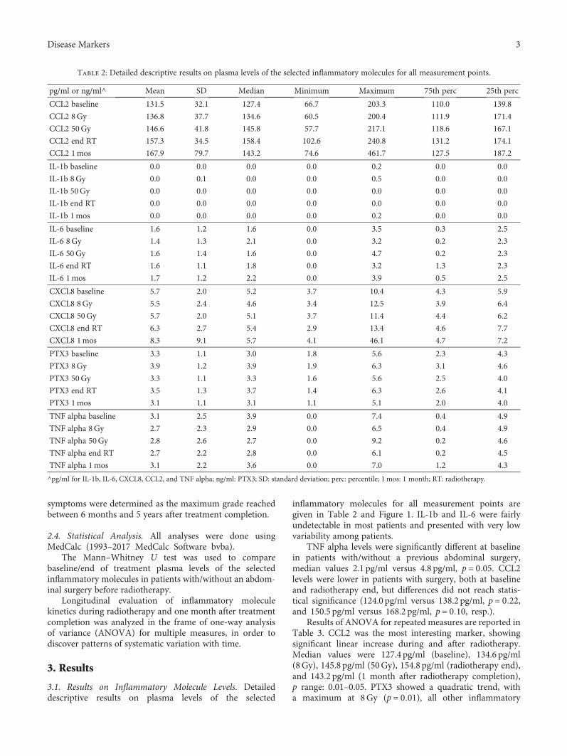
inflammatory molecules for all measurement points aregiven in Table 2 and Figure 1. IL-1b and IL-6 were fairlyundetectable in most patients and presented with very lowvariability among patients.
TNF alpha levels were significantly different at baselinein patients with/without a previous abdominal surgery,median values 2.1 pg/ml versus 4.8 pg/ml, p = 0 05. CCL2levels were lower in patients with surgery, both at baselineand radiotherapy end, but differences did not reach statis-tical significance (124.0 pg/ml versus 138.2 pg/ml, p = 0 22,and 150.5 pg/ml versus 168.2 pg/ml, p = 0 10, resp.).
Results of ANOVA for repeated measures are reported inTable 3. CCL2 was the most interesting marker, showingsignificant linear increase during and after radiotherapy.Median values were 127.4 pg/ml (baseline), 134.6 pg/ml(8Gy), 145.8 pg/ml (50Gy), 154.8 pg/ml (radiotherapy end),and 143.2 pg/ml (1 month after radiotherapy completion),p range: 0.01–0.05. PTX3 showed a quadratic trend, witha maximum at 8Gy (p = 0 01), all other inflammatory
Table 2: Detailed descriptive results on plasma levels of the selected inflammatory molecules for all measurement points.
pg/ml or ng/ml^ Mean SD Median Minimum Maximum 75th perc 25th perc
CCL2 baseline 131.5 32.1 127.4 66.7 203.3 110.0 139.8
CCL2 8Gy 136.8 37.7 134.6 60.5 200.4 111.9 171.4
CCL2 50Gy 146.6 41.8 145.8 57.7 217.1 118.6 167.1
CCL2 end RT 157.3 34.5 158.4 102.6 240.8 131.2 174.1
CCL2 1mos 167.9 79.7 143.2 74.6 461.7 127.5 187.2
IL-1b baseline 0.0 0.0 0.0 0.0 0.2 0.0 0.0
IL-1b 8Gy 0.0 0.1 0.0 0.0 0.5 0.0 0.0
IL-1b 50Gy 0.0 0.0 0.0 0.0 0.0 0.0 0.0
IL-1b end RT 0.0 0.0 0.0 0.0 0.0 0.0 0.0
IL-1b 1mos 0.0 0.0 0.0 0.0 0.2 0.0 0.0
IL-6 baseline 1.6 1.2 1.6 0.0 3.5 0.3 2.5
IL-6 8Gy 1.4 1.3 2.1 0.0 3.2 0.2 2.3
IL-6 50Gy 1.6 1.4 1.6 0.0 4.7 0.2 2.3
IL-6 end RT 1.6 1.1 1.8 0.0 3.2 1.3 2.3
IL-6 1mos 1.7 1.2 2.2 0.0 3.9 0.5 2.5
CXCL8 baseline 5.7 2.0 5.2 3.7 10.4 4.3 5.9
CXCL8 8Gy 5.5 2.4 4.6 3.4 12.5 3.9 6.4
CXCL8 50Gy 5.7 2.0 5.1 3.7 11.4 4.4 6.2
CXCL8 end RT 6.3 2.7 5.4 2.9 13.4 4.6 7.7
CXCL8 1mos 8.3 9.1 5.7 4.1 46.1 4.7 7.2
PTX3 baseline 3.3 1.1 3.0 1.8 5.6 2.3 4.3
PTX3 8Gy 3.9 1.2 3.9 1.9 6.3 3.1 4.6
PTX3 50Gy 3.3 1.1 3.3 1.6 5.6 2.5 4.0
PTX3 end RT 3.5 1.3 3.7 1.4 6.3 2.6 4.1
PTX3 1mos 3.1 1.1 3.1 1.1 5.1 2.0 4.0
TNF alpha baseline 3.1 2.5 3.9 0.0 7.4 0.4 4.9
TNF alpha 8Gy 2.7 2.3 2.9 0.0 6.5 0.4 4.9
TNF alpha 50Gy 2.8 2.6 2.7 0.0 9.2 0.2 4.6
TNF alpha end RT 2.7 2.2 2.8 0.0 6.1 0.2 4.5
TNF alpha 1mos 3.1 2.2 3.6 0.0 7.0 1.2 4.3
^pg/ml for IL-1b, IL-6, CXCL8, CCL2, and TNF alpha; ng/ml: PTX3; SD: standard deviation; perc: percentile; 1 mos: 1 month; RT: radiotherapy.
3Disease Markers

500450400350300250200150100
50
CCL2
_bas
CCL2
_8 G
y
CCL2
_50
Gy
CCL2
_end
RT
CCL2
_1 m
os(a)
−500
50100150200250300350
Delt
a_CC
L2_8
Gy
Delt
a_CC
L2_1
mos
Delt
a_CC
L2_e
nd R
T
Delt
a_CC
L2_1
mos
(b)
0.0
0.1
0.2
0.3
0.4
0.5
IL-1
b_ba
s
IL-1
b_8
Gy
IL-1
b_50
Gy
IL-1
b_en
d RT
IL-1
b_1
mos
(c)
−0.2
−0.1
0.0
0.1
0.2
0.3
0.4
0.5
Delt
a_IL
-1b_
8 G
y
Delt
a_IL
-1b_
50 G
y
Delt
a_IL
-1b_
end
RT
Delt
a_IL
-1b_
1 m
os
(d)
0
1
2
3
4
5
IL_6
_bas
IL-6
_8 G
y
IL-6
_50
Gy
IL-6
_end
RT
IL-6
_1 m
os
(e)
−1.5−1.0−0.5
0.00.51.01.52.02.53.0
Delt
a_IL
-6_8
Gy
Delt
a_IL
-6_5
0 G
y
Delt
a_IL
-6_e
nd R
T
Delt
a_IL
-6_1
mos
(f)
Figure 1: Continued.
4 Disease Markers

05
101520253035404550
CXCL
8_ba
s
CXCL
8_8
Gy
CXCL
8_50
Gy
CXCL
8_en
d RT
CXCL
8_1
mos
(g)
−10
0
10
20
30
40
50
Delt
a_CX
CL8_
8 G
y
Delt
a_ X
CL8_
50 G
y
Delt
a_CX
CL8_
end
RT
Delt
a_CX
CL8_
1 m
os
(h)
1
2
3
4
5
6
7
PTX3
_bas
PTX3
_8 G
y
PTX3
_50
Gy
PTX3
_end
RT
PTX3
_1 m
os
(i)
−2
−1
0
1
2
3
4
Delt
a_PT
X3_5
0 G
y
Delt
a_PT
X3_8
Gy
Delt
a_PT
X3_e
nd R
T
Delt
a_PT
X3_1
mos
(j)
0
2
4
6
8
10
TNFa
lfa_b
as
TNF
alph
a_8
Gy
TNF
alph
a_50
Gy
TNF
alph
a_en
d RT
TNF
alph
a_1
mos
(k)
−4−3−2−1
01234
Delt
a_TN
F al
pha_
8 G
y
Delt
a_TN
F al
pha_
50 G
y
Delt
a_TN
F al
pha_
end
RT
Delt
a_TN
F al
pha_
1 m
os
(l)
Figure 1: Plasma levels of the selected inflammatory molecules for all measurement points: absolute values (a, c, e, g, i, and k) and absolutevariations with respect to baseline (b, d, f, h, j, and l). 1mos: 1 month; RT: radiotherapy. Units of measure are pg/ml for IL-1b, IL-6, CXCL8,CCL2, and TNF alpha; ng/ml: PTX3.
5Disease Markers

markers did not exhibit systematic changes with radiother-apy dose/time.
CCL2 levels at radiotherapy end could be modelled usinglinear regression including the following variables: baselineCCL2 (coefficient = 1.15, p = 0 0001), age (coefficient =−3.26, p = 0 004), abdominal surgery (coefficient = 23.3,p = 0 09), hypertension (coefficient = 29.6, p = 0 02), and useof anticoagulants (coefficient = 41.0, p = 0 05) and multiplecorrelation coefficient = 0.89 (see plot). Significance level ofanalysis of variance for this linear regression was p = 0 002,multiple correlation coefficient was 0.87, and coefficient ofdetermination R2 was 0.75. Figure 2 shows the calibrationplot (observed CCL2 levels at radiotherapy end versusCCL2 values as predicted by the linear regression model).
3.2. Results on Radio-Induced Toxicity. Three out of twentypatients (15%) reported grade 2 acute rectal toxicity, while4/20 (20%) were scored as grade 2 late rectal toxicity in thefirst 36 months after radiotherapy completion. No grade3-4 event was observed. Details on incidence of acuteand late grade≥ 1 toxicity (as determined by questions inthe questionnaire) are reported in Table 4.
Multiple toxicity symptoms (>3) were experiencedby 56.3% and 43.8% of patients in the acute and latephase, respectively.
The 4 patients with late toxicity had CCL2 values atradiotherapy end above the median value.
Detailed comparison of plasma levels for selectedinflammatory markers for patients with/without multiplesymptoms of acute/late intestinal toxicity is reported inTable 5. t-test was statistically different only for baselineTNF alpha: 2.0 versus 4.7 ng/ml, p = 0 04.
Incidence of acute fecal incontinence and rectal bleedingwas slightly higher in the group of patients with previousabdominal surgery, 11% versus 0% and 33.3% versus 28.6%for incontinence and bleeding, respectively, differences werenot statistically significant. Late rectal bleeding was alsoslightly higher for patients with previous abdominal surgery,33.3% versus 28.6%, difference was not statistically signifi-cant. 67% of patients with abdominal surgery presented withat least three different acute intestinal toxicity symptomsversus 43% in the rest of the population; even in this case,difference was not statistically significant due to the verysmall size of the population of this pilot study.
4. Discussion
Preclinical and clinical studies have shown that radiotherapyinduces cytokine responses that could play a major role inmediating radiation toxicity [3–6].
In the present study, grade≥ 1 acute and late intestinaltoxicity (as defined by questions in the patient-reportedquestionnaire) were not found to be significantly associatedwith plasma levels of inflammatory markers, with the onlyexception of baseline TNF alpha level, which was higher inpatients experiencing multiple late intestinal toxicity symp-toms. The small size of the study population could highlightonly important differences, larger populations are required toinvestigate more modest variations.
When considering inflammatory molecule kineticsduring and immediately after radiotherapy, CCL2 was foundto significantly increase during IMRT. Though statisticalassociation between moderate/severe radio-induced (acuteand late) intestinal toxicity and CCL2 levels could not beinvestigated due to the low size of the study population,patients exhibiting late grade 2 toxicity were found to have
Table 3: Results of one-way analysis of variance for repeatedmeasures for all considered markers. Best trend for marker valuesas a function of time is reported.
Best trend p value
IL-1b Cubic 0.34
IL-6 Quadratic 0.16
CXCL8 Linear 0.19
CCL2 Linear 0.01∗
TNF alpha Quadratic 0.07
PTX3 Quadratic 0.06∗Statistically significant.
250
200
150
100
50
00 50 100 150 200 250
CCL2 RT-end predicted values (pg/ml)
CCL2
RT-
end
obse
ved
valu
es (p
g/m
l)
Figure 2: Calibration plot for the linear regression model predictingCCL2 levels at end of radiotherapy (observed CCL2 levels atradiotherapy end versus CCL2 values as predicted by the linearregression). Details are given in the text. Units of measure pg/ml.RT: radiotherapy.
Table 4: Details on incidence of acute and late grade≥ 1 toxicity(as determined by questions in the questionnaire).
Acute incidencegrade≥ 1
Late incidencegrade≥ 1
Stool frequency 50.0% 50.0%
Diarrhea 62.5% 31.3%
Tenesmus 43.8% 25.0%
Stool urgency 31.3% 43.8%
Fecal incontinence 6.3% 6.3%
Rectal pain 18.8% 25.0%
Rectal bleeding 31.3% 31.3%
6 Disease Markers

CCL2 levels at the end of radiotherapy above the medianvalue for the study cohort.
Interestingly, CCL2 levels at the end of radiotherapycould be modelled through linear regression including age,abdominal surgery, hypertension, and use of anticoagulants.All these features are known to be risk factors forincrease intestinal toxicity after prostate cancer radiother-apy [13–15], thus suggesting a first possible link betweenpatient clinical characteristics and his individual responsein terms of biomarkers.
CCL2 is a low molecular weight monomeric polypeptidewhose primary function was identified as promoting mono-cyte and macrophage migration to sites of inflammation[16]. For example, CCL2 is involved in monocyte infiltrationin inflammatory diseases such as rheumatoid arthritis as wellas in the inflammatory response against tumours.
There are limited data regarding the relationship betweenCCL2 and radiation exposure. Most results are related to theevidence that CCL2 overexpression in tumour is associatedwith macrophage infiltration and poor prognosis in humancancers and may play a pivotal role in creating the fertileenvironment in the bone for metastasis [17–19].
Connolly and coworkers [20] demonstrated that radio-therapy stimulates increased production CCL2 and CCL5 atthe tumour site, while Kalbasi and colleagues [21] found that
ablative radiotherapy for pancreatic ductal adenocarcinomaled to a significant increase in CCL2 production by tumourcells, with genetic deletion of CCL2 in pancreatic ductaladenocarcinoma cells improving radiotherapy efficacy.
When considering the association between CCL2 expres-sion and response of normal tissues to radiation, only twostudies are available in the literature and they reported inter-esting results. Lee et al. [22] showed that irradiation induces atransient nonclassical cytokine response with selective upreg-ulation of CCL2. Interestingly, absence of CCL2 signalling inthe hours after irradiation is sufficient to restore hippocam-pal neurogenesis and to decrease the risk of long-term defectsin neural stem cell function following cranial radiation inchildren. Holler and coworkers [23] demonstrated that prav-astatin has a mitigatory effect on radiation-induced vasculardysfunction in the skin in a mouse model. Remarkably, prav-astatin limits the radio-induced increase of blood CCL2expression and inflammatory cell migration in tissues.
When considering the other measured inflammatorymolecules, IL-1b and IL-6 were fairly undetectable in mostpatients and presented with very low variability amongpatients: for these reasons, they were considered of no inter-est for the purpose of the present study. TNF alpha levelswere found to be significantly different between patientswith/without a previous abdominal surgery, but it did not
Table 5: Comparison of plasma levels for the selected inflammatory molecules in subgroups of patients experiencing multiple symptoms (≥3)for intestinal acute/late toxicity. p values for test are reported.
Acute toxicity Late toxicityPatients with<3 symptoms
Patients with≥3 symptoms
t-testPatients with<3 symptoms
Patients with≥3 symptoms
t-test
Mean SD Mean SD Mean diff p Mean SD Mean SD Mean diff p
CCL2 baseline 139.4 34.1 134.2 30.4 −5.2 0.75 145.5 37.3 124.9 16.5 −20.7 0.18
CCL250Gy
155.7 33.9 159.7 40.9 4.1 0.83 162.4 38.5 152.3 36.7 −10.1 0.60
CCL2end RT
161.2 20.7 165.8 42.4 4.6 0.79 165.5 39.1 161.6 28.1 −3.9 0.82
CCL2 1mos 153.2 40.0 201.3 104.2 48.1 0.25 193.9 107.4 162.6 39.6 −31.3 0.46
IL-8 baseline 6.9 2.7 5.2 1.5 −1.7 0.15 6.4 2.6 5.4 1.6 −0.9 0.40
IL-850Gy
6.6 2.5 5.4 1.9 −1.2 0.29 6.2 2.4 5.6 2.0 −0.6 0.60
IL-8end RT
8.2 3.2 5.4 2.1 −2.8 0.06 7.6 3.1 5.4 2.3 −2.2 0.13
IL-81mos
7.1 2.2 10.6 13.4 3.4 0.50 11.6 13.0 5.8 2.1 −5.8 0.25
PTX3 baseline 3.1 1.2 3.2 1.1 0.2 0.75 2.9 1.1 3.5 1.0 0.5 0.37
PTX350Gy
3.3 1.5 3.1 1.1 −0.2 0.72 3.3 1.4 3.0 1.1 −0.3 0.69
PTX3end RT
3.2 1.4 3.4 1.4 0.2 0.80 3.6 1.7 3.0 0.8 −0.6 0.40
PTX3 1mos 3.2 1.3 3.0 1.2 −0.2 0.72 3.1 1.2 3.1 1.3 0.0 0.98
TNF alpha baseline 2.0 2.3 4.0 2.6 2.0 0.13 2.0 2.2 4.7 2.3 2.7 0.04
TNF alpha 50Gy 2.1 2.5 3.6 3.0 1.5 0.29 2.0 2.4 4.0 3.1 2.0 0.17
TNF alpha end RT 1.9 2.3 3.3 2.4 1.3 0.27 1.9 2.2 3.7 2.3 1.8 0.14
TNF alpha 1mos 2.3 2.3 3.8 2.3 1.5 0.23 2.5 2.6 4.1 1.8 1.6 0.17
^pg/ml for IL-1b, IL-6, CXCL8, CCL2, and TNF alpha; ng/ml: PTX3; SD: standard deviation; 1 mos: 1 month; RT: radiotherapy; diff: difference.
7Disease Markers

exhibit significant changes as a function of radiation dose anddid not result to be associated to acute/late toxicity. PTX3showed a quadratic trend with an early increase with dose(at 8Gy) with subsequent return to baseline levels by theend of treatment. Increase in PTX3 was not associated withpatient-reported morbidity.
To our knowledge, there is only one previously publishedstudy investigating cytokine expression during IMRT forprostate cancer and their relationship with acute toxicity[24]. Their study population consisted of 22 prostate patientstreated with exclusive IMRT (78Gy at 2Gy/fraction) and 20patients receiving radiotherapy after prostatectomy (70Gy at2Gy/fraction). They found IL-6 levels to be significantly ele-vated over baseline in the postprostatectomy group but nosignificant difference in the exclusive IMRT cohort. Increasesin IL-2 and IL-1 levels over baseline were significantly associ-ated with increased gastrointestinal and genitourinary toxic-ity, respectively, regardless of the radiotherapy regimen(exclusive IMRT versus postprostatectomy IMRT) regimen,while the analysis of IL-6 suggested that the increase of IL-6was associated with a higher risk for gastrointestinal toxicitybut it did not reach statistical significance.
Presence of previous abdominal surgery was not found tobe significantly associated with toxicity or to plasma levels ofinflammation markers in this pilot study. As already pointedout, interestingly, previous abdominal surgery was includedas a factor modulating CCL2 levels at radiotherapy end,together with other patient features known to be predictorsof intestinal toxicity. This modulating effect should be con-firmed on a wider population, in order to suggest a directeffect of factors associated with toxicity on CCL2 levels atradiotherapy end.
Besides interest in the comprehension of the biologicalrationale for the correlation of some clinical factors withmorbidity, investigation of association between inflamma-tory molecule levels and radio-induced toxicity is of interestbecause it could have the potential of being a biologicalmeasure of the individual patient radiosensitivity, thusprompting further optimization of radiotherapy treatmentfor more sensible patients or dose escalation on resistantpatients. Prophylactic treatment of toxicity symptoms couldalso be proposed in patients at higher risk of enhancedinflammation processes.
One important limitation of this study is related to thelimited sample size. Our pilot study was intended to beexploratory, to inspect the feasibility of multiple bloodsamples for biomarker investigation in the frame of clinicalpractice and to validate the study methodology. These resultsare expected to guide future, larger trials which could estab-lish the time course of plasma levels of inflammatory mole-cules after radiotherapy and how they are associated withnormal tissue radiation toxicity. Specific future researchtopics should include evaluation of a wider spectrum ofradio-induced symptoms (e.g., including urinary and hema-tologic toxicities) and of populations of patients treated withradiotherapy for different cancer types, such as head-and-neck patients or breast cancer patients. Interaction withconcomitant oncological treatment (such as chemotherapyor hormone therapy) should be considered.
5. Conclusions
This preliminary study identified a correlation between CCL2levels at the end of radiotherapy and basal CCL2, age, andsurgery, suggesting a different response to radiotherapy inolder patients and in patients with pretreatment abdominalsurgery. Interestingly, these clinical characteristics are thesame features included in predictive models for acute and laterectal toxicity. Larger accrual is needed to confirm these feasi-bility results and to study the association with radio-inducedacute and late toxicity.
Disclosure
An earlier version of this work was presented as posters at5th European Multidisciplinary Meeting on UrologicalCancers (EMUC 2013) [8] and as an oral presentation atthe International Conference on Translational Researchin Radio-Oncology/Physics for Health in Europe (ICTR-PHE 2014) [9].
Conflicts of Interest
The authors declare that there are no conflicts of interest.
Authors’ Contributions
Tiziana Rancati and Riccardo Valdagni contributed equallyto this work.
Acknowledgments
The study was supported by Fondazione ProADAMOOnlus.The authors thank for the generosity given by the Gheraduccifamily and by Fondazione Italo Monzino. AssociazioneItaliana per la Ricerca sul Cancro (AIRC) is acknowledged(Grant no. IG16087).
References
[1] G. Defraene, L. Van den Bergh, A. Al-Mamgani et al., “Thebenefits of including clinical factors in rectal normal tissuecomplication probability modeling after radiotherapy forprostate cancer,” International Journal of Radiation Oncology,Biology, Physics, vol. 82, no. 3, pp. 1233–1242, 2012.
[2] R. Valdagni, V. Vavassori, T. Rancati et al., “Increasing the riskof late rectal bleeding after high-dose radiotherapy for prostatecancer: the case of previous abdominal surgery. Results from aprospective trial,” Radiotherapy and Oncology, vol. 103, no. 2,pp. 252–255, 2012.
[3] P. Rubin, C. J. Johnston, J. P. Williams, S. McDonald, and J. N.Finkelstein, “A perpetual cascade of cytokines postirradiationleads to pulmonary fibrosis,” International Journal of Radia-tion Oncology, Biology, Physics, vol. 33, no. 1, pp. 99–109, 1995.
[4] P. Okunieff, Y. Chen, D. J. Maguire, and A. K. Huser,“Molecular markers of radiation-related normal tissue toxicity,”Cancer Metastasis Reviews, vol. 27, no. 3, pp. 363–374, 2008.
[5] L. Liang, D. Hu, W. Liu, J. P. Williams, P. Okunieff, andI. Ding, “Celecoxib reduces skin damage after radiation,”American Journal of Clinical Oncology, vol. 26, Supplement 2,pp. S114–S121, 2003.
8 Disease Markers

[6] C. G. Willett, C. J. Ooi, A. L. Zietman et al., “Acute and latetoxicity of patients with inflammatory bowel disease undergo-ing irradiation for abdominal and pelvic neoplasms,” Interna-tional Journal of Radiation Oncology, Biology, Physics, vol. 46,no. 4, pp. 995–998, 2000.
[7] B. Park, C. Yee, and K. M. Lee, “The effect of radiation onthe immune response to cancers,” International Journal ofMolecular Sciences, vol. 15, no. 1, pp. 927–943, 2014.
[8] N. Bedini, T. Rancati, N. Zaffaroni et al., “P064 evaluation ofmediators associated to the inflammatory response in prostatecancer patients undergoing radiotherapy: preliminary results,”European Urology, vol. 12, no. 6, p. 154, 2013.
[9] R. Valdagni, P. Allavena, B. Avuzzi et al., “202: mediatorsassociated to the inflammatory response in prostate cancerpatients undergoing RT: preliminary results,” Radiotherapyand Oncology, vol. 110, Supplement 1, article S99, 2014.
[10] B. Bottazzi, A. Doni, C. Garlanda, and A. Mantovani, “Anintegrated view of humoral innate immunity: pentraxins as aparadigm,” Annual Review of Immunology, vol. 28, no. 1,pp. 157–183, 2010.
[11] T. Rancati, C. Fiorino, G. Gagliardi et al., “Fitting late rectalbleeding data using different NTCP models: results from anItalian multi-centric study (AIROPROS0101),” Radiotherapyand Oncology, vol. 73, no. 1, pp. 21–32, 2004.
[12] V. Vavassori, C. Fiorino, T. Rancati et al., “Predictors for rectaland intestinal acute toxicities during prostate cancer high-dose3D-CRT: results of a prospective multicenter study,” Interna-tional Journal of Radiation Oncology, Biology, Physics,vol. 67, no. 5, pp. 1401–1410, 2007.
[13] C. Fiorino, R. Valdagni, T. Rancati, and G. Sanguineti,“Dose-volume effects for normal tissues in external radio-therapy: pelvis,” Radiotherapy and Oncology, vol. 93, no. 2,pp. 153–167, 2009.
[14] R. Valdagni and T. Rancati, “Reducing rectal injury duringexternal beam radiotherapy for prostate cancer,” NatureReviews Urology, vol. 10, no. 6, pp. 345–357, 2013.
[15] V. Landoni, C. Fiorino, C. Cozzarini, G. Sanguineti,R. Valdagni, and T. Rancati, “Predicting toxicity in radiother-apy for prostate cancer,” Physica Medica, vol. 32, no. 3,pp. 521–532, 2016.
[16] R. D. Loberg, L. L. Day, J. Harwood et al., “CCL2 is a potentregulator of prostate cancer cell migration and proliferation,”Neoplasia, vol. 8, no. 7, pp. 578–586, 2006.
[17] H. Roca, Z. S. Varsos, S. Sud, M. J. Craig, C. Ying, and K. J.Pienta, “CCL2 and interleukin-6 promote survival of humanCD11b+ peripheral blood mononuclear cells and induce M2-type macrophage polarization,” The Journal of BiologicalChemistry, vol. 284, no. 49, pp. 34342–34354, 2009.
[18] G. Soria and A. Ben-Baruch, “The inflammatory chemokinesCCL2 and CCL5 in breast cancer,” Cancer Letters, vol. 267,no. 2, pp. 271–285, 2008.
[19] J. Zhang, L. Patel, and K. J. Pienta, “CC chemokine ligand 2(CCL2) promotes prostate cancer tumorigenesis and metasta-sis,” Cytokine & Growth Factor Reviews, vol. 21, no. 1, pp. 41–48, 2010.
[20] K. A. Connolly, B. A. Belt, N. M. Figueroa et al., “Increasingthe efficacy of radiotherapy by modulating the CCR2/CCR5chemokine axes,” Oncotarget, vol. 7, no. 52, pp. 86522–86535, 2016.
[21] A. Kalbasi, C. Komar, G. M. Tooker et al., “Tumor-derivedCCL2 mediates resistance to radiotherapy in pancreatic ductal
adenocarcinoma,” Clinical Cancer Research, vol. 23, no. 1,pp. 137–148, 2017.
[22] S. W. Lee, U. Haditsch, B. J. Cord et al., “Absence of CCL2 issufficient to restore hippocampal neurogenesis followingcranial irradiation,” Brain, Behavior, and Immunity, vol. 30,pp. 33–44, 2013.
[23] V. Holler, V. Buard, M. H. Gaugler et al., “Pravastatin limitsradiation-induced vascular dysfunction in the skin,” TheJournal of Investigative Dermatology, vol. 129, no. 5,pp. 1280–1291, 2009.
[24] E. Christensen, M. Pintilie, K. R. Evans et al., “Longitudinalcytokine expression during IMRT for prostate cancer andacute treatment toxicity,” Clinical Cancer Research, vol. 15,no. 17, pp. 5576–5583, 2009.
9Disease Markers

Stem Cells International
Hindawiwww.hindawi.com Volume 2018
Hindawiwww.hindawi.com Volume 2018
MEDIATORSINFLAMMATION
of
EndocrinologyInternational Journal of
Hindawiwww.hindawi.com Volume 2018
Hindawiwww.hindawi.com Volume 2018
Disease Markers
Hindawiwww.hindawi.com Volume 2018
BioMed Research International
OncologyJournal of
Hindawiwww.hindawi.com Volume 2013
Hindawiwww.hindawi.com Volume 2018
Oxidative Medicine and Cellular Longevity
Hindawiwww.hindawi.com Volume 2018
PPAR Research
Hindawi Publishing Corporation http://www.hindawi.com Volume 2013Hindawiwww.hindawi.com
The Scientific World Journal
Volume 2018
Immunology ResearchHindawiwww.hindawi.com Volume 2018
Journal of
ObesityJournal of
Hindawiwww.hindawi.com Volume 2018
Hindawiwww.hindawi.com Volume 2018
Computational and Mathematical Methods in Medicine
Hindawiwww.hindawi.com Volume 2018
Behavioural Neurology
OphthalmologyJournal of
Hindawiwww.hindawi.com Volume 2018
Diabetes ResearchJournal of
Hindawiwww.hindawi.com Volume 2018
Hindawiwww.hindawi.com Volume 2018
Research and TreatmentAIDS
Hindawiwww.hindawi.com Volume 2018
Gastroenterology Research and Practice
Hindawiwww.hindawi.com Volume 2018
Parkinson’s Disease
Evidence-Based Complementary andAlternative Medicine
Volume 2018Hindawiwww.hindawi.com
Submit your manuscripts atwww.hindawi.com