evaluation of nguni bull semen extended in tris egg … · on egg yolk, soybean milk and coconut...
TRANSCRIPT

EVALUATION OF NGUNI BULL SEMEN EXTENDED IN TRIS EGG YOLK
EXTENDER, SOYBEAN MILK AND COCONUT WATER BASED EXTENDERS AND
STORED AT DIFFERENT TEMPERATURES
By
Mayombo Pie Veillard Kalonji
Student no: 15004155
A dissertation submitted in fulfilment of the requirements for the degree Master of Science in
Agriculture (Animal Science)
Centre of Excellence in Animal Assisted Reproduction
Department of Animal Science
School of Agriculture
University of Venda
South Africa
Supervisor : Prof. D.M. Barry (University of Venda, South Africa)
Co-supervisor: Prof. D.O. Owiny (Makerere University, Uganda)
2017

ii
DECLARATION
I, Pie Veillard Mayombo Kalonji, student number 15004155, the undersigned, hereby declare
that the contents of this dissertation submitted for the degree of Master of Science in Animal
Science (MScANS) at the University of Venda are my own original work and has not been
previously in whole or in part been submitted to any university for the award of other degree.
Student …………………….............. Date ………………………….
M.P.V. Kalonji
\

iii
DEDICATION
This work is dedicated to my family.
Thanks for always believing in me and for encouraging me to follow my dreams, whatever it
takes. Without your guidance, love and support, I would not have been able to achieve my
goals.
To my wife for always being there with your support through the entire time of this study, and
without your motivation and trust this would not have been possible.
To my parents for not giving up on me after so many years of education and not stopping to
cheer me up, you are the best parents in the world, and I am honoured to be called your son.
To my brothers and sisters, thank you for always keeping me in touch with reality, you all
mean a lot to me.
To my other brother, I really miss you but I know that you are happy and smiling down on me
where you are now “Sit tibi terra lewis”.
All my love,
Yopie
iv
AKNOWLEDGEMENTS
Firstly, I would like to thank God for walking with me through life.
My deepest thanks go to my supervisor, Prof. D.M. Barry for giving me the opportunity to do
this project and for guidance, critique and endless patience enabled this work to be completed.
By doing that taught me a lot about research and assisted reproductive technics which I will be
able to apply in my career in future.
I grateful to my co-supervisor Prof. D.O. Owiny for the training on semen handling and freezing
and on in vitro fertilisation (IVF) and he made a valuable contributions to this project.
To all my wonderful colleagues at the Centre of Excellence in Animal Assisted Reproduction
(CEAAR) for always being there and willing to assist and listen. Thank you for all your
encouragement and understanding.
Furthermore special thanks to Prof. D. Luseba for the support and playing an important
leadership role without which this study would never have started. He is like a father to me.
I also would like to thank Mr. R. Mashudu, Mr. F. Lubisi and my cousin Mss. N.N. Bukalo for
their valuable contributions regarding data analysis and interpretation of the results.
The support I received from Dr Delphin Somwe in the beginning of this project has not gone
unnoticed. Thank you for helping me as your young brother.
Finally I would like to acknowledge everyone who helped me with this project.

v
ABSTRACT
In order to realize many of the potential advantages of AI, storage of semen is
necessary. Semen storage is only possible using a system that decreases and/or halts the
metabolic processes of the spermatozoa, allowing no significant loss of fertility. Numerous
factors affect the success of spermatozoa storage. This study was designed to compare the
effects of egg yolk, soybean milk and coconut water in Tris extender using different storage
methods for Nguni bull spermatozoa storage. Bull semen was collected from two adult Nguni
bulls approximately four years old and kept under similar managerial conditions. Using electro-
ejaculator, semen was collected from each bull into a graduated semen collection tube.
Macroscopically evaluation of the sample was performed immediately after collection. Only the
semen free from contamination was processed. The kinetic properties namely: total
spermatozoa motility, and progressive spermatozoa motility were analysed using CASA.
Semen sample was stained and spermatozoa morphology and vitality also analysed using
CASA. The extended semen was then split into three groups. The first group was stored at
room temperature (25 °C). The second group was cooled to 4 °C and stored in the refrigerator.
The third group was also cooled to 4 °C for 2 h in the refrigerator, then held in LN2 vapour 5 cm
above the surface of LN2 at ~ -80 °C for 10 min and then plunged into LN2 for storage at -196
°C. Different colours of straws and plugging powder were used for identifying each extender.
After 3 days of storage at room temperature, in the refrigerator and in LN2, the extended
semen was split into three portions and assayed for kinetic properties using the first portion.
The second portion was assayed for spermatozoa morphology and the third portion for
spermatozoa vitality. The results from the fresh semen extended with all three extenders
(TEYE, SBME and COWE), and analysed immediately after dilution at room temperature (25
ºC), showed no significant difference (P > 0.05) in the mean values of the kinetic and
morphologic properties and viability, on spermatozoa TM, PM, AR, AT, CT; BT and LS. After
three days of storage, there was no significant difference (P > 0.05) in the kinetic morphologic
properties and viability of semen stored at room and refrigeration temperature regardless of
the extender in use. There were, however, significant differences (P < 0.05) in the TM, PM, AR
and DL of the frozen semen samples. For the short storage period of semen used for AI, from
this study, it is recommended that semen should be kept at room or refrigeration temperature
regardless of the three extenders used. However, for long storage of frozen semen TEYE is

vi
recommended. The egg yolk-based extender provided greater preservation of motility and bull
spermatozoa integrity during the freezing process than did SBME and COWE.
Keywords: Tris egg yolk, Soybean-milk, Coconut water, Spermatozoa, Nguni bull and storage
methods.

vii
TABLE OF CONTENTS
DECLARATION...................................................................................................................... ii
DEDICATION ........................................................................................................................ iii
AKNOWLEDGEMENTS ........................................................................................................ iv
ABSTRACT ............................................................................................................................ v
LIST OF TABLES ................................................................................................................... x
LIST OF FIGURES ................................................................................................................ xi
LIST OF ABREVIATIONS .................................................................................................... xii
CHAPTER 1 ........................................................................................................................... 1
RESEARCH THEME, HYPOTHESIS AND MOTIVATION ...................................................... 1
CHAPTER 2 ........................................................................................................................... 3
LITERATURE REVIEW .......................................................................................................... 3
2.1 Introduction ...................................................................................................................... 3
2.2. History of AI and semen cryopreservation in cattle .......................................................... 3
2.3. Advantages and disadvantages of AI and the cryopreservation of semen ....................... 5
2.4. Economic implication of AI in cattle ................................................................................. 6
2.5. Structure and functional characteristics of spermatozoon ................................................ 7
2.5.1. Structure of spermatozoon ........................................................................................... 7
2.5.2. Composition of spermatozoa plasma membrane .......................................................... 8
2.5.3. Role of plasma membrane in spermatozoa function ..................................................... 9
2.6. Spermatozoa plasma membrane changes during cryopreservation .............................. 11
2.7. Principles in semen cryopreservation ............................................................................ 12
2.7.1. Semen temperature .................................................................................................... 12
2.7.2. Energy source ............................................................................................................ 13
2.7.3. Osmotic pressure and electrolyte balance .................................................................. 13
2.7.4. Buffering and pH ........................................................................................................ 13
2.7.5. Proper gas phase ....................................................................................................... 13

viii
2.7.6. Inhibition of microbial growth ...................................................................................... 14
2.7.7. Exclusion of toxic substances ..................................................................................... 14
2.7.8. Semen extension and dilution effect ........................................................................... 14
2.7.9. Protection of spermatozoa against cold shock ............................................................ 15
2.8. Extenders used in this study .......................................................................................... 15
2.8.1. Egg yolk and egg yolk extenders ................................................................................ 15
2.8.2. Soybean milk and soybean milk extenders ................................................................. 16
2.8.3. Coconut water and coconut water extenders .............................................................. 16
2.9. Fresh and frozen semen................................................................................................ 16
2.10. Effect of cryopreservation on spermatozoa.................................................................. 17
2.11. Protection of spermatozoa against cold shock ............................................................. 19
2.12. Pre and post-freezing evaluation of bovine semen ...................................................... 20
2.12.1. Spermatozoon morphology ...................................................................................... 21
2.12.2. Spermatozoon motility and motion............................................................................ 22
CHAPTER 3 ......................................................................................................................... 23
MATERIALS AND METHODS .............................................................................................. 23
3.1. Study Area .................................................................................................................... 23
3.2. Nutrition and housing of the bulls .................................................................................. 23
3.3. Preparation of Extenders ............................................................................................... 23
3.4. Semen collection ........................................................................................................... 25
3.5. Semen extension .......................................................................................................... 26
3.6. Semen evaluation.......................................................................................................... 27
3.6.1. Macroscopic evaluation of semen .............................................................................. 27
3.6.2. Microscopic evaluation ............................................................................................... 27
3.7. Experimental design, packing, freezing and storage of semen ...................................... 30
3.8. Thawing and post-freezing evaluation of semen ............................................................ 31
3.7. Statistical analysis ......................................................................................................... 31
ix
CHAPTER 4 ......................................................................................................................... 32
RESULT AND DISCUSSION ............................................................................................... 32
CHAPTER 5 ......................................................................................................................... 40
CONCLUSION AND RECOMMENDATION ......................................................................... 40
REFERENCES .................................................................................................................... 41
APPENDICES ...................................................................................................................... 52
x
LIST OF TABLES
Table 3.1.: Table illustrating the constituents of the different extenders used.
Table 4.1.: Table presenting the parameters and values of Initial (macroscopic)
characteristics of freshly collected bull semen.
Table 4.2.: Table showing the effect of egg yolk, soybean milk and coconut water based
extenders on kinetic and morphologic properties on fresh bull semen.
Table 4.3.: Illustration of the effect of egg yolk, soybean milk and coconut water based
extenders on bull spermatozoa kinetic; morphologic properties and viability
after three days of storage.
Table 6.1.: ANOVA for kinetic and morphologic properties and viability of fresh bull semen
on egg yolk, soybean milk and coconut water based extenders.
Table 6.2.: ANOVA for kinetic and morphologic properties and viability of fresh extended
semen. Effect of egg yolk, soybean milk and coconut water based extenders
on bull spermatozoa kinetic; morphologic properties and viability after three
days of storage.
xi
LIST OF FIGURES
Figure 2.1. Structure of Spermatozoon
Figure 3.1.: The electro-ejaculator used for semen collection.
Figure 3.2.: Styrofoam box, straw holding rack and semen straws.
Figure 3.3.: The Sperm Class Analyzer® (SCA) used for microscopic analysis of
spermatozoa.
Figure 4.1.: Fresh extended semen characterization immediately after dilution.
Figure 4.2.: Semen characterization after three days of storage at room temperature (25 ºC).
Figure 4.3.: Semen characterization after three days of refrigeration (4 ºC).
Figure 4.4.: Semen characterization after three days of storage in the LN2 (-196 ºC).

xii
LIST OF ABREVIATIONS
AI : Artificial insemination
ATP : Adenosine triphosphate
AV : Artificial vagina
BCF : Beat cross frequency
BIOX : Bioxcell
CASA : Computer aided sperm analysis
CE : Conventional extender
CED : Cauda epididymal dissection
oC : Degree Celsius
COWE : Coconut water based extender
DMSO : Dimethyl sulfoxide
EE : Electro-ejaculator
ET : Embryo transfer
EYC : Egg-yolk citrate
g : Gram
h : Hour
IU : International unit
IVF : In vitro fertilisation
LN2 : Liquid nitrogen
mg : Milligram
min : Minute
ml : Millilitre
mm : Millimetre
P : Probability
xiii
PSM : Progressive spermatozoa motility
SBME : Soybean-milk based extender
sec : Second
SM : Spermatozoa motility
STR : Straightness
TEYE : Tris-citric egg-yolk
TM : Total motility
Tris : Tetra-hydroxymethylaminomethane
v/v : Volume per volume

1
CHAPTER 1
RESEARCH THEME, HYPOTHESIS AND MOTIVATION
Artificial insemination (AI) had a major impact on animal breeding (Arzondo, et al.
2012). By means of AI, a single ejaculate of a bull is used for the insemination of various
females, which could not have been possible without sperm extension and/or cryopreservation
(Jones, 1976; Rauch, 2013).
Although there has been extensive research done on the subject of bovine semen
cryopreservation, most of these studies were completed decades ago (Almquist, 1951; Dunn
et al., 1953; Graham et al., 1956; Rauch, 2013). There has been significant progress in the
components used for semen extenders, and also the preparation of these extenders which
plays a role in determining the optimum equilibration periods for bovine semen
cryopreservation (Almquist, 1954; Bean et al., 1963; Berndtson & Foote, 1976; Kumaresan et
al., 2011). However, bull semen has been cryopreserved for AI for over half a century, many of
the protocols used are still empirical, with the number of spermatozoa that do not survive the
freezing process being considerable and those that do survive being affected structurally or
functionally after thawing. In general, many of the strategies tested to achieve successful
cryopreservation do not involve the spermatozoon itself, but the medium in which it is stored
(Jones, 1976; El-Harairy et al., 2011).
The aim of this study was to improve the cryopreservation and storage protocols of
Nguni bull semen for artificial insemination purposes by determining the effect of egg yolk,
soybean-milk and coconut water as extenders on kinetic, morphology properties and viability
of spermatozoa during different storage methods. The null hypothesis for both experiments
was that egg yolk, soybean milk and coconut water based extenders will not have a significant
effect on protecting bull spermatozoa during different storage methods. The alternative
hypothesis for both experiments was that there will be significant differences on kinetic and
morphology properties of spermatozoa during different storage methods and between egg
yolk, soybean milk and coconut water based extenders. The motivation of this study was to
define and improve the cryopreservation and storage process of semen for cattle indigenous
to Africa, in order to improve the pre-insemination motility of spermatozoa and in turn the
conception rates of cows inseminated with the good quality extended semen. This in turn may

2
lead to farmers using artificial insemination in their herds, for accelerated genetic improvement
of these indigenous cattle breeds.
3
CHAPTER 2
LITERATURE REVIEW
2.1 Introduction
Throughout history man and animals have always competed for nutritional resources.
Due to the demands of growing and evolving human population, livestock producers are
constantly under pressure to supply more products, from even more limited resources.
Livestock producers are therefore continuously searching for ways to improve the production
efficiency of their livestock. This includes selecting for animals with better feed conversion
ratios, disease resistance and improved reproductive efficiency. These factors can all be
improved by genetic selection, in other words selecting and breeding only superior animals.
This is known as genetic improvement (Salisbury et al., 1978; Bailey et al., 2000; Vishwanath,
2003; Bozkurt et al., 2011). For genetic improvement to take place in any population there has
to be an increase in both the gene frequency and gene combinations of the desired genes.
This is achieved by selecting individuals possessing the desired traits and then manipulating
their breeding. Artificial insemination has contributed enormously to the genetic improvement
of beef and dairy cattle in the last 50 years. Frozen semen in 0.5 ml or 0.25 ml straws has
become the universally accepted unit of storage and transfer of bovine genetics to cattle
producers (Baracaldo et al., 2007).
Cryopreservation of semen would be to minimize damage resulting from the freezing
and thawing procedures and to maximize recovery of motile and viable spermatozoa used for
AI. Many scientific studies have been carried out to improve the quality of frozen-thawed
semen. For this purpose, different cryoprotective agents, freezing and thawing protocols, and
various extenders have been used (England, 1993; Silva et al., 2002; 2003; Walters et al.,
2009).
2.2. History of AI and semen cryopreservation in cattle
Research on artificial insemination (AI) dates back several centuries. The first scientific
step in AI was the discovery by Leeuwenhoek in 1677 used a simple microscope to view the
small motile cells which he named “animalcules”. This discovery was the cornerstone of any
further research or discoveries that led to the development of current assisted reproductive

4
technologies. The next notable event was the first documented case of AI which occurred
more than 100 years later in 1784, when Lazzaro Spallanzani performed the first successful
AI. He inseminated a confined bitch in heat by depositing semen in the uterus with a syringe.
This resulted in the birth of three pups 62 days later that resembled the dam as well as the
semen donor. Spallanzani later also found that when semen is filtered, fertilization only
occurred when the residue (sperm) left on the filter was used and not when the filtrate (seminal
fluids etc.) was used. He also observed that when equine semen was cooled the sperm
weren’t killed, but only remained motionless until the temperature was increased. Another
century passed before the use of AI was also reported by Walter Heape (1897) and several
other researchers in independent studies on horses, dogs and rabbits. Walter Heape was also
credited for establishing the basics for the relationship between reproduction and seasonality
(Foote, 1999; 2002; Walters et al., 2009).
Later in 1899, Ivanoff was credited with initial attempted to establish AI as a breeding
practice. By 1907 he had applied AI in the breeding management programmes for rabbits,
dogs, foxes, poultry and domestic farm animals. As part of his research he trained technicians
as inseminators and developed semen extenders to prolong the life of sperm. Ivanoff’s work
also stimulate interest in AI outside Russia. In 1912, Dr. Ishikawa, a scientist who studied with
Ivanoff, started an AI programme in Japan with horses. Later similar programmes followed for
poultry, swine, goats, sheep and cattle. Milovanov later succeeded Ivanoff and established
projects for the breeding of cattle and sheep in Russia. Milovanov also designed and made
practical versions of the artificial vagina, specifically for these species as well as other practical
equipment for semen collection. Many apparatus currently used are still based on these
designs (Foote, 2002). The artificial vagina used in AI today was first devised by Amantea in
1914. It was originally meant for the collection of dog semen, but was quickly adapted for use
in bulls, stallions and rams. Gunn (1936) experimented with using the electro-ejaculator to
collect semen from male animals. This is an alternative method used when animals are not
trained for semen collection using the artificial vagina (Salisbury et al., 1978; Foote, 1999; El-
Sisy et al., 2016).
Although the discovery of AI had occurred much earlier, it was not until the mid-1930’s
that all the necessary ingredients for the successful commercial applications thereof, were
available. There ingredients include an economic need to improve milk yield in dairy cows, as
well as a measurement system for assessing the phenotypic expression of milk-producing
capacity of cows and several other factors (Salisbury et al., 1978; Akhter et al., 2011). During
5
the period of the 1940’s and 1950’s, there were various revolutionary discoveries regarding the
semen cryopreservation process, as well as the storage environment for cryopreserved
semen. This, together with new methods of sire selection, resulted in the application of AI to
herds in remote regions that previously did not have access to viable collected semen
(Vishwanath, 2003). When the first calf emanating from AI with cryopreserved semen was
born in 1951, the technique was already starting to be incorporated into commercial breeding
management programmes, and it established itself rapidly thereafter (Curry, 2000). With time,
the process of AI became more accessible, and by the next decade, the majority of
replacement stock was the result of applying AI (Vishwanath, 2003; Walters et al., 2009).
Several discoveries and developments played crucial roles in establishing the basic
principles for the freezing of bovine semen. Phillips developed a phosphate-buffered egg yolk
based extender in 1940, and a year later Salisbury et al. (1941) improved on this by using a
citrate buffer that resulted in a clearer medium. This allowed for more accurate and critical
sperm evaluation (Foote, 1999). A few years later, two independent reports by Foote and
Almquist (1948) described the remarkable results achieved when adding antibiotics to the
semen extender. When Polge et al. (1949) discovered the cryopreservative properties of
glycerol; it was the beginning of a new era in cryopreservation of semen. Semen could now be
frozen and stored for long periods, while still yielding acceptable fertility results.
The discovery of protective agents within egg yolk and glycerol was a major milestone
in sperm cryopreservation. These agents protected bovine sperm during cooling and freezing
procedures and resulted in increased survival rates (Medeiros et al., 2002). Over the last few
decades research has concentrated on the cryopreservation of bovine sperm, but this did very
little to advance the preservation of any other body cells. This is partly due to the slow
realization that bovine sperm cells are unique in its composition, and a lack of methods to
measure damage caused by cryopreservation to sub-cellular compartment (Hammerstedt et
al., 1990; Walters et al., 2009).
2.3. Advantages and disadvantages of AI and the cryopreservation of semen
Some factors affecting the success rate of AI generally include herd management
(including the nutritional status of the females), seasonal effects on reproduction and the
quality of semen used (Kathiravan et al., 2011). These factors should be managed and
controlled in such a way that the advantages of AI application within the herd will be fully
utilised (Haugan et al., 2005; Van Staden, 2010). The most obvious advantages of applying AI
6
in a herd include genetic improvement, increase of productivity, control of venereal diseases,
as well as reduced cost (Vishwanath, 2003). When compared to other reproductive
technologies, it becomes apparent that AI is very simple, yet successful and economical
method that can be applied to introduce new genes in a population (Vishwanath, 2003;
Kathiravan et al., 2011). The presence of lethal genes can also be reduced and genetic
improvement of milk production traits by the selection of bulls used in AI programs can be
seen as benefits on their own. However they also collectively lead to overall economic benefits
for the dairy industry (Foote, 1999). Sperm cryopreservation is then also used to build fertility
reserves for endangered species (Ehmcke & Schlatt, 2008).
The ability to control the transmission of venereal diseases possibly had a large effect
on the initial decision to apply AI commercially than the genetic advantages. However careful
and continuous monitoring of health status of donor sires, and regular disease testing of their
semen is of critical importance. If this is not done efficiently, AI can become a very effective
tool for transmitting diseases to herds worldwide (DeJarnette et al., 2004). Much of the labour
and money saved by not having to keep bulls on site may have to be spent in the detection of
cows in oestrus and the restraint on these cows for Ai. As dairy cows are confined for milking
each day, some of these costs are more easily justified in a dairy, compared to a beef farm
(Salisbury et al., 1978). Another limitation of semen cryopreservation is that only matured
spermatozoa can be preserved, and thus sperm from pre-pubertal males cannot be preserved
with any degree of success (Ehmcke & Schlatt, 2008).
2.4. Economic implication of AI in cattle
Effects resulting from the commercial implementation of AI, are most prominent in the
dairy cattle industry. The overall result of implementing AI is accelerated genetic progress,
resulting in improved production capabilities of the animals (Medeiros et al., 2002). Improving
the production efficiency of dairy herds has the benefit of satisfying both producer and
consumer. The increased demand for inexpensive product of acceptable quality is satisfied,
and a higher profit potential for producers is generated. (DeJarnette at al., 2004; Kathiravan et
al., 2011). Depending on the reason why AI or semen cryopreservation is implemented, the
economic implications of these procedures will differ. The various reasons for preservation of
male fertility may lead to different approaches, depending on the discipline of interest (Ehmcke
& Schlatt, 2008).
7
2.5. Structure and functional characteristics of spermatozoon
Spermatozoa have certain characteristics that give them the distinction of being
“terminal cells”. They are very susceptible to injury and are usually the preferred cells used in
research into optimal cryopreservation procedures (Suarez, 2007). Other characteristics of
spermatozoon include being haploid and containing very little cytoplasm and other organelles
are the chromosomes within the nucleus are very condensed, which prevents protein
transcription taking place (Hu, 2010). The minimal quantity of endoplasmic reticulum present
on the Golgi apparatus is not sufficient to maintain membrane molecule to supply building
blocks to maintain cell membranes and undergo maturational changes (Medeiros et al., 2002).
2.5.1. Structure of spermatozoon
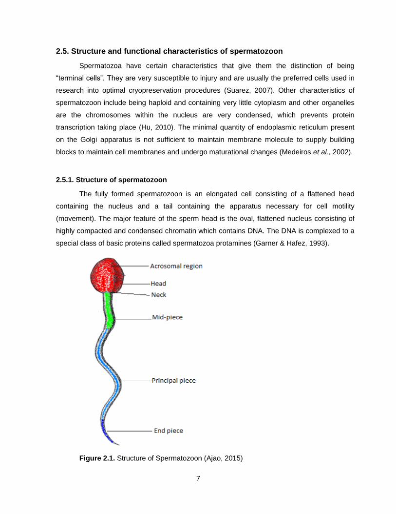
The fully formed spermatozoon is an elongated cell consisting of a flattened head
containing the nucleus and a tail containing the apparatus necessary for cell motility
(movement). The major feature of the sperm head is the oval, flattened nucleus consisting of
highly compacted and condensed chromatin which contains DNA. The DNA is complexed to a
special class of basic proteins called spermatozoa protamines (Garner & Hafez, 1993).
Figure 2.1. Structure of Spermatozoon (Ajao, 2015)

8
A spermatozoon is entirely covered by a plasma membrane, identical to somatic cells.
The study of the spermatozoon membrane function status is of particular importance since an
intact and functionally active membrane is required for metabolism, capacitation, acrosome
reaction, attachment and penetration of the oocyte (Tardif et al., 1998). The spermatozoon can
be divided in several membrane domains and subdomains, depending on the function. The
domains of the spermatozoa head include the acrosomal and the postacrosomal region (Anzar
et al., 2011). The plasma membrane can be divided into an acrosomal cap and an equatorial
subdomain. The latter is separated by the posterior ring from the neck region of the midpiece.
Besides the specialization in function of the different domains of plasma membranes, the lipids
and proteins of the plasma membrane vary between different parts of the spermatozoon
(Correa et al., 1997; Rauch, 2013).
2.5.2. Composition of spermatozoa plasma membrane
The spermatozoon plasma membrane consists of a phospholipid bilayer with
cholesterol, complex carbohydrates and proteins, typical for plasma membranes. The
carbohydrate structures are bound to proteins or specific lipids on the outside of the plasma
membrane (glycocalyx) (Tiwari et al., 2008). The phospholipids in the spermatozoa plasma
membrane vary between mammalian species, but generally include phosphatidylethanolamine
(PE), phosphatidylinositol (PI), phosphatidylserine (PS), phosphatidylcholine (PC),
sphingomyelin, lysophosphatidylcholine and cardiolipin (Anzar et al., 2011). In contrast to
other species, spermatozoa from bull have a high ratio of PC to PE. The proteins constitute
about 50 % of the total membrane weight and can be either peripheral to or integrated in the
plasma membrane (Gürler et al., 2015).
The cell membrane is described as a mosaic of different degrees of localized fluid
areas that are also called lipid domains. These domains consist of certain lipids (mainly
phospholipid and sterols) with certain functions. The lipids and proteins are mobile and able to
move laterally in the plane of the membrane (Tiwari et al., 2008). At room temperature, the
membrane lipids are generally in a fluid (liquid crystalline) phase, but some domains contain
lipids in the gel-phase. In the gel-phase, the lipids are more clustered and less mobile (Anzar
& Graham, 1996). More recent reports claimed that the proteins in plasma membrane are
predominant, cause less fluidity and therefore there is less lateral diffusion of the lipids (Gürler
et al., 2015). Further, the degree of fluidity depends on the type and amount of the present
lipids. For example, long chain polyunsaturated fatty acids in the phospholipids as well as a

9
smaller amount of cholesterol increase the fluidity of the membrane at room temperature
(Rauch, 2013).
2.5.3. Role of plasma membrane in spermatozoa function
The assessment of spermatozoa membrane functional status appears to be a
significant marker for the fertilizing capacity of spermatozoa (Correa et al., 1997). Concurrent
with the acrosomal membrane, the spermatozoa plasma membrane has an impact on the
spermatozoa shape and volume, motility, energy production, penetration, capacitation and
acrosome reaction, and interaction with the oocyte. The spermatozoon membrane changes its
lipid composition and location of the lipid domains during the physiological events before
fertilization occurs (Correa et al., 1997; Kumaresan et al., 2011).
2.5.3.1. Spermatozoa motility
Spermatozoa motility requires adenosine triphosphate (ATP) which is produced by
mitochondria (10 %) and by anaerobic glycolysis in the spermatozoon tail (90 %). Thus, an
intact transport mechanism of monosaccharides from the extracellular environment into the
spermatozoon is essential for flagellar movement (Kumaresan et al., 2011). Specific plasma
membrane proteins enable the transport of glucose and fructose into the spermatozoa. These
transporter systems require an intact plasma membrane and a specific chemical gradient of
ions and soluble components to function properly (Singh et al., 2013).
2.5.3.2. Changes during capacitation
Ejaculated spermatozoa of mammalian species are not ready to fertilize an oocyte
immediately, and have to be in the female reproductive tract for a period of time. This period is
species-specific, and is regulated by female’s hormone status in order to prevent gamete
ageing (Medeiros et al., 2002). In 1951, Austin and Chang independently discovered that
spermatozoon has to undergo a process called capacitation within the female reproductive
tract, before it could fertilize an oocyte. It was determined that the acrosome reaction could
only take place if capacitation had already occurred, and it became standard practice to use
the acrosomal reaction as an endpoint in determining whether spermatozoa were fully
capacitated (Curry, 2000; Medeiros et al., 2002; Jones et al., 2007).
10
The process of capacitation is still not clearly defined, but it is accomplished by
acquisition of the ability of the spermatozoon to fertilize an oocyte. The initial event of
capacitation includes a rise in intracellular calcium, bicarbonate, and hydrogen peroxide
(Medeiros et al., 2002). This triggers the production of cyclic adenosine monophosphate
(AMP; cAMP) which in turn causes the tyrosine phosphorylation of proteins in the plasma
membrane and in the cytoplasm of the spermatozoa (Curry, 2000). Besides a change in
conformation and dimerization of membrane proteins, tyrosine phosphorylation is linked to
increased zona pelucida affinity, spermatozoa hyperactivity and the induction of acrosome
reaction. Hyperactivation of the spermatozoa occurs in vitro at some point during capacitation,
but the processes leading to hyperactivation and acrosomal responsiveness can happen
independently (Kumaresan et al., 2011). In addition, so-called decapacitating factors
(substances coating the spermatozoa surface) are removed from the plasma membrane
during capacitation. The coating factors originating from the seminal plasma are also known as
bovine seminal plasma proteins (BSPP) or phospholipid binding proteins, or binder of sperm
proteins (Jones et al., 2007). Their removal of BSPP is essential for fertilization. Another key
event of capacitation is the efflux of cholesterol from the plasma membrane. This leads to
reorganization of lipids within the plasma membrane and in an increase in membrane fluidity
(Jones et al., 2007; Kumaresan et al., 2011).
2.5.3.3. Changes during acrosome reaction and fertilization
The acrosome is located underneath the spermatozoa plasma membrane in the
anterior head region and consists of a protein matrix that is enclosed by an inner and outer
acrosomal membrane (Kumaresan et al., 2011). The conformational changes in tyrosine
phosphorylated transmembrane proteins enable the binding of the spermatozoa to the zona
pelucida (ZP3 proteins) of the oocyte. As a result of the phosphorylation, receptors aggregate
in the spermatozoa plasma membrane and induce a calcium ion flux into the spermatozoon
(Muin˜o et al., 2008). The proteins involved form bridges between the outer acrosomal
membrane and the apical spermatozoa plasma membrane (Muin˜o et al., 2008). The
acrosomal matrix swells and both the plasma membrane and the outer plasma membrane are
dissolved by vesiculation and fusion (Mogielnicka-Brzozowska & Kordan, 2011). This fusion is
accomplished by the destabilization of the plasma membrane during capacitation.
Subsequently, hydrolytic enzymes from the acrosomal matrix are released which dissolve the

11
cumulus cells and the zona pelucida, and enable the penetration of the sperm into the
perivitelline space of the oocyte (Kumaresan et al., 2011).
2.6. Spermatozoa plasma membrane changes during cryopreservation
The spermatozoa plasma membrane is drastically altered by cryopreservation. There
are several mechanisms proposed on how cryopreservation leads to spermatozoa damage.
One mechanism involves lateral lipid rearrangement which destabilizes the membrane (known
as phase transition during cold shock (Muin˜o et al., 2008). The lipid components (mainly
phospholipids and sterols) of the plasma membrane undergo reorganization during the cooling
process. In detail, the lipids in the bilayer start aggregating in different lipid domains, which
results in new associations between proteins and lipids in the membrane (Jones et al., 2007).
Therefore, the fluidity of the plasma membrane decreases during cryopreservation which
results in transition from liquid crystalline to gel phase. The temperature at which the phase
transition occurs is specific for each particular lipid (Eidan, 2016). The shorter the fatty acyl
chain and the more cis-unsaturated carbon bonds, the lower the phase transition temperature.
Similarly, cholesterol decreases the phase transition temperature (Rauch, 2013; Eidan, 2016).
Freezing can further promote clustering of the lipids. Rewarming of the spermatozoa
does not initially return lipids in their pre-cooling state. It was hypothesized that over time the
original assembly could be achieved by lipid diffusion (Cheema et al., 2016). The
reorganization of lipids also affects the proteins, whereas integral proteins become clustered
and subsequently can lose their functionality. Another phenomenon related to
cryopreservation is the change in membrane lipid composition. In boar sperm, sphingomyelin
(a phospholipid) and the saturated fatty acids content of the phospholipids decreased during
freezing, whereas the content of cholesterol did not change (Eidan, 2016).
Another mechanism that influences the plasma membrane constitution is the
peroxidation of membrane lipids as a result of formation of reactive oxygen species. The high
content of polyunsaturated fatty acids in the spermatozoa membrane increases the
susceptibility of spermatozoa to peroxidation if oxygen is present (Divyaswetha et al., 2008).
The consequences of membrane peroxidation on spermatozoa include the irreversible loss of
motility, impaired metabolism, damage to the plasma membrane, leakage of intracellular
enzymes, and decrease in oocyte penetration and fertilizing capacity (Divyaswetha et al.,
2008). The consequences of the changes in lipid fluidity, lipid composition and lipid damage
12
lead to membrane destabilization and the spermatozoon becomes more susceptible to
premature acrosome reactions (Curry, 2000). Further, functional proteins like ion channel
proteins are affected by the changes during cooling, which is linked to a general leakiness of
the spermatozoa membrane. Consequently, the decrease of spermatozoa motility and
metabolism after cooling could be attributed to the loss of cations and enzymes (Singh et al.,
2013).
Since calcium channels are affected, the intracellular calcium level increases and
results in a decline in spermatozoa motility and spermatozoa necrosis. The increased calcium
level and the reorganization of the plasma membrane during cooling also occur during the
physiologic process of capacitation (Correa et al., 1997). Further, capacitation and changes
during cooling and cryopreservation of spermatozoa have the production of reactive oxygen
species and the phosphorylation of proteins in common. Due to these similarities, the
damages during cryopreservation are often referred to as “capacitation-like changes” (Rauch,
2013). These induced alterations render the spermatozoa to a partially capacitated state and
decrease their life span (Eidan, 2016).
2.7. Principles in semen cryopreservation
There are certain principal factors that should be kept in mind during the process of
extending and cooling of semen. These factors will affect sperm independently, but
interactions between these factors also affect the sperm. Many of these interactions have
been studied, which have led to combining different procedures. The result was the
development of “systems” for semen preservation (Saacke, 1983). Some of these factors
involved ambient temperature, energy source, osmotic pressure and electrolyte balance,
buffering and pH, proper gas phase, inhibition of microorganism growth, exclusion of toxic
materials, the dilution effect and protection of against the cold (Perry et al., 2017).
2.7.1. Semen temperature
As the metabolic rate of cells are generally proportional to the absolute temperature,
semen is kept at lowered temperature (usually 5 ºC) during storage, as this slows down the
semen to 5 ºC must be controlled to prevent the occurrence of cold shock (Bearden et al.,
2004). The metabolic rate is not the only factor affected by the lower temperature, as the
temperature decreases, but both the internal and external environments of cells undergo
changes (Correa et al., 1997). So for instance gas solubility for environmental gases increase
13
as the temperatures decreases. As decreased temperatures as a large part of the total
metabolic activities are due to the oxidative metabolism, because of the presence of the air
and increased solubility of gases at these temperatures (Cheema et al., 2016).
2.7.2. Energy source
Spermatozoa do not only require energy for motility, but also for cell maintenance.
Spermatozoa can utilize energy sources through both aerobic and anaerobic metabolism, as
the energy source naturally available in seminal plasma is fructose, diluted greatly during
semen extension. Thus a supplementary source of energy such as glucose or fructose should
be added to the extender. It is known that egg yolk contains several compounds suitable for
use as energy sources by bovine sperm (Bearden et al., 2004).
2.7.3. Osmotic pressure and electrolyte balance
Spermatozoa are capable of adapting in size according to the tonicity of different
media. Cell permeability also differs for various substances. Although spermatozoa are
tolerant towards the tonicity of media, if this deviates too much it could result in spermatozoa
having bent tails, swimming in circles and ultimately dying (Salisbury et al., 1978). Solutions
with compositions similar to that of body fluids, like egg yolk and milk, seem to be the most
compatible with spermatozoa (Salisbury et al., 1978; Bearden et al., 2004).
2.7.4. Buffering and pH
Spermatozoa need to be protected from auto-toxication from acids that build up as a
product of the metabolism. This effect is more pronounced if semen is not kept at a lower
temperature, which decreases the rate of metabolism (Mogielnicka-Brzozowska & Kordan,
2011). Extenders containing egg yolk or milk usually have a pH of approximately 7, but may
even be beneficial to reduce the pH to 6.5 (Bearden et al., 2004).
2.7.5. Proper gas phase
As the cells’ permeability for gases increase with decreased temperature, the
proportion of gas liquid phase, as well as the composition of the gas phase should be
controlled to maintain the desired gas conditions within the extender (Almquist, 1948). Care
must also be taken to limit exposure to light, as such exposure in the presence of oxygen
14
damages the spermatozoa. Gassing with nitrogen will be beneficial to the cell (Salisbury et al.,
1978; Mogielnicka-Brzozowska & Kordan, 2011).
2.7.6. Inhibition of microbial growth
The AI industry relies upon the use of antibiotics in semen extenders to control
contaminating organisms in bull semen (Almquist, 1948; Contri et al., 2014). While there are
many positive aspect for the use of natural products such as egg yolk and milk as additives,
there are also downsides to their use, as part of the extension media. These products provide
a fertile breeding ground for microorganisms, which produce many products harmful to
spermatozoa, or that have the potential to infect the cows. This is one of the reason why it is
standard procedure for antibiotics to be included in any medium used for the extension of
bovine semen (Salisbury et al., 1978). Although many of these organisms are not pathogenic,
they compete with spermatozoa for nutrients and their by-products are toxic to spermatozoa.
This reduces the livability of spermatozoa and reduces fertility (Foote & Berndtson, 1976;
Contri et al., 2014).
2.7.7. Exclusion of toxic substances
Any extender should be prepared to be free of any and all harmful substances like
heavy metals. These substances should also prevent the formation of any toxic products
during storage (Urata et al., 2001). Even though care is taken to minimize exposure to toxic
products, some products, such as glycerol or dead spermatozoa, cannot be totally eliminated.
Glycerol, a cryoprotective agent that protects cells during freezing, can be toxic to sperm at
high levels and at room temperature (Amann & DeJarnette, 2012). Dead spermatozoa also
provide amino acid oxidase, which produces hydrogen peroxide (H2O2), a toxic substance to
sperm (Salisbury et al., 1978; Amann & DeJarnette, 2012).
2.7.8. Semen extension and dilution effect
Semen should be extended in such a way that the initial spermatozoa concentration is
reduced to a number that yields optimum result (Tirpak et al., 2015). The dilution (or
extension) lowers the motility of spermatozoa, but that the addition of certain amino acids and
macromolecules, such as egg albumin or casein, minimizes this effect (Vishwanath, 2003).
Rottensten et al. (1960) found that if egg yolk or milk was included in the extender, the effect
of spermatozoa motility was almost prevented.

15
2.7.9. Protection of spermatozoa against cold shock
Spermatozoa are subjected to cold shock when they are cooled down to 5 ºC, and this
causes intracellular enzymes and other material of the cell to leak out, as well as decreased
motility (Rego et al., 2015). Cold shock also causes the flagella of spermatozoa to bend. It is
not known what the exact mechanisms occur at different rates both internally and on the
surface of the cell, that results in both physical and chemical damage (Rego et al., 2015).
Lecithin, proteins, lipoproteins, milk and egg yolk have proved to be the most effective agents
in preventing damages of the spermatozoa caused by cold shock (Salisbury et al., 1978;
Rauch, 2013). The application of consistent, stringent quality control standards is essential for
supply the best possible semen to the breeders, thus individual batches of semen should
always be frozen using the best known cryopreservation procedures (Foote, 1975; Tirpak et
al., 2015).
2.8. Extenders used in this study
2.8.1. Egg yolk and egg yolk extenders
The use of egg yolk in extenders dates back to 1939, when Phillips discovered its
protective effect on cooled bovine semen (Rego et al., 2015). Van Denmark et al. (1957) and
Foote (1975) contributed to modern egg yolk extender recipes by finding that 16%, 20% and
24% chicken egg yolk in the freezing extender is favorable. Dried chicken egg yolk consists of
63% lipids and 33% proteins. The fresh egg yolk can be fractionated into 78% plasma and
22% granules. The granules contain 16% high density lipoproteins (HDL), 4% phosvitin and
2% low-density lipoproteins (LDL) (Rego et al., 2015). In the yolk plasma, the main component
is LDL (66%), followed by livetins (10%). Phosvitin is a highly phosphorylated protein with
bactericidal and antioxidant properties. An egg yolk extender provides protection for cooled
and cryopreserved bovine spermatozoa. It is now the most commonly used semen extender
worldwide for bulls (Akhter et al., 2011).
It is widely accepted that the protective agent in the egg yolk is a phospholipid moiety
of the low-density lipoprotein fraction (El-Sisy et al., 2016). Thus, there are several
mechanisms proposed on how the LDL fraction decreases damage to the spermatozoa during
cooling and cryopreservation. One possibility is that the LDLs, particularly the phospholipids,
associate with the spermatozoa membrane and thereby provide stabilization (Kathiravan et al.,
2011). Another possibility is that the phospholipids lost during spermatozoa cryopreservation
16
are replaced by phospholipids of the egg yolk (El-Sisy et al., 2016). However, Quinn et al.
(1980) and Ricker et al., (2006) did not observe that the added phospholipids were integrated
into the spermatozoa membrane. The most recent hypothesis is that the LDLs bind detrimental
BSP proteins that are responsible for the efflux of cholesterol and phospholipids from the
spermatozoa membrane (El-Sisy et al., 2016).
2.8.2. Soybean milk based extenders
The use of vegetal extenders for preservation of live cells has been reported for a long
time. Different types of vegetable products such as tylose, sodium alginate, agar-agar, semi-
hydrolyzed starch, dextrin, flax and quince seed, as well as soybean milk were some of the
first vegetable liquids used as semen extenders reported as good alternatives extenders for
bovine semen (Vale et al., 1997; El-Sisy et al., 2016). Fukui et al. (2008) found that egg-yolk
can be replaced by soybean-milk in semen extenders, without any deleterious effects of
spermatozoa during cryopreservation.
2.8.3. Coconut water based extenders
In 1987, coconut water was first used as semen extender during refrigerated storage of
goat semen. During the last decade, researchers developed a technology to dehydrate
coconut water and produce a stable and standardized powder (ACP®) (Viveiros et al., 2008).
Some vegetables derivate like coconut water and tomato juice seem to have some properties
to protect bovine and caprine spermatozoa (Vale et al., 1997). Coconut water has shown to be
an excellent alternative for conservation of bovine and swine semen as well as caprine semen
due the presence of phytohormones (Nunes et aI., 1996). This substance protects the semen
of this species against the deleterious effect of the enzyme phospholipase A, secreted by the
Cowper’s glands and present in the ejaculated semen (Nunes et aI., 1996; Boonsorn et al.,
2010). Moreover, the indole-3-acetic appeared to be the main spermatozoa protective
substance present in the coconut water (Vale et al., 1997; Boonsorn et al., 2010).
2.9. Fresh and frozen semen
Successful cryopreservation of animal semen have the potential to enhance the
advantages of applying AI. However, in order to achieve this, the result have to be comparable
to that of fresh semen (Bailey et al., 2000; England & Millar, 2008). In order to achieve
success when applying AI with frozen semen, the damaging effects on the cells caused by

17
freezing procedures must be reduced to a minimum. This is generally a complex problem, as
these effects can occur during any of the procedures of semen extension, equilibration and
freezing (Fleisch et al., 2017). Therefore, all procedures should be kept as simple and
straightforward as possible. Cryopreservation of bovine semen is good example of the
potential success that can be achieved with cryopreserved semen (Curry, 2000; Amirat et al.,
2005; Fleisch et al., 2017). Even though there has been considerable progress in semen
cryopreservation; both the viability and fertility of semen still deteriorated during the
cryopreservation process. This deterioration is a result of numerous cellular injuries occurring
throughout the process. These injuries have to be minimised, in order to improve the survival
rate of spermatozoa, which could thus result in higher fertility rates obtained (Medeiros et al.,
2002; Fleisch et al., 2017).
Many researchers have reported a post thaw motility of 50 % of a fresh semen sample
motility to be acceptable. This has been achieved in number of species. Regardless, the
number of spermatozoa needed per successful insemination is still variable between species.
In some species, the fertility rates of cryopreserved semen can be comparable to that of the
fresh semen, if the number of spermatozoa per insemination is high enough (Curry, 2000;
Medeiros et al., 2002; Chaveiro et al., 2006). In 1995 Shannon and Vishwanath reported that
to achieve fertilization rates in cattle comparable to that of fresh semen, the number of
cryopreserved spermatozoa needed per insemination was 8 times more than the required
number for fresh semen AI (Bailey et al., 2000; Akhter et al., 2016). When Shannon (1978)
determined the number of fresh and frozen bovine spermatozoa needed per insemination to
yield comparable fertility rates in the 1970’s, it was clearly demonstrated that 10 times more
frozen spermatozoa were required to yield the same result as a dose of fresh spermatozoa.
Although the total number of spermatozoa required per fresh and frozen insemination dose
has decreased from 2.5 million and 25 million in 1978 to 1 – 1.5 million and 10 – 15 million,
respectively (Vishwanath, 2003). The fresh: frozen spermatozoa ratio still remained 1 to 10
(Holt, 1997). Matter et al. (1969) reported a poor in vivo survival rate of spermatozoa that were
not irreversibly damaged by the cryopreservation process, as a possible reason for the vast
difference in spermatozoa numbers required for fresh and frozen semen AI.
2.10. Effect of cryopreservation on spermatozoa
It is a well-known fact that freezing of spermatozoa causes structural damage. This is a
major reason why progress in developing methods for the cryopreservation of sperm was slow

18
(Medeiros et al., 2002). Reduced fertility of frozen semen is attributed largely to altered
membrane structure and function during cooling, freezing and thawing. The nature of this
damage remains unclear, but it has been suggested that membranes are compromised due to
reordering of membrane lipids during cooling and rewarming, thereby disturbing the lipid-lipid
and lipid-protein associations required for normal membrane function (Poulos et al, 1973;
Hammerstedt et al., 1990; Campanholi et al., 2017).
Cryopreservation of spermatozoa is associated with an oxidative stress (Mazur et al.,
2000). Superoxide dismutase activity, a scavenger of the superoxide radical, is decreased in
frozen/thawed spermatozoa (Bilodeau et al., 2000; Cheema et al., 2016). However, in 1979
Jones and Stewart found the cooling of spermatozoa to 5 ºC cause the acrosomal head to
swell. The freezing and thawing of spermatozoa also caused the acrosome to rupture as well
as causing damage to the middle piece in a large number of spermatozoa. Cryopreserved
sperm display signs of being in an advanced stage of capacitation, prior to the freezing
process (Medeiros et al., 2002). Nishizono et al. (2004) reported that cryopreservation induces
the morphological abnormalities including mitochondria with an increased relative area of the
matrix, thickening of the outer membrane and swelling with loss of cristae.
The biggest problem encountered when freezing any cellular structure is the phase
changes that all membranes and cell contents undergo. As the temperature lowers, eventually
the lipid phase changes into solid phase and this causes structural changes (Januskauskas &
Zilinskas, 2002). The lipids transform into solids, ice or crystals, which can cut through
membrane. As the external solvents change a liquid to solid phase, the concentrations of other
solutes increase dramatically in the remaining liquid. The entire cell must then respond to all
these changes in the limited time allowed by the protocol used (Campanholi et al., 2017).
During the thawing process, the cell again has to go through a reverse process, where the
solid phase changes into a liquid phase. All of the above mentioned changes underline the
importance of identifying a rate of temperature change that allows water and cryoprotectant
movement without causing intracellular crystal formation or any irreversible membrane
changes (Januskauskas & Zilinskas, 2002; Heise, 2012).
Many factors that seem to play a role in the success of cryopreservation of semen of
different species have been studied. The species with lower cholesterol levels in the cell
membranes (including bovine and ovine) were more susceptible to membrane damage during
cryopreservation (Akhter et al., 2016). White (1993) also found that spermatozoa from species

19
with a higher ratio of unsaturated fatty acids (bull and ram), are more sensitive to lower
temperatures. Another possible factor is the reactive oxygen species (ROS), which is thought
to play role in the initiation of the capacitation process (Medeiros et al., 2002; Griffin, 2004;
Fleisch et al., 2017).
As spermatozoa are not adapted to endure extreme temperatures, many of these
spermatozoa are injured during the cryopreservation process. There are different mechanisms
during each of the phases, resulting in different injuries to the cells. The spermatozoa injuries
can be divided into two main groups: spermatozoa injuries occurring during cooling and due to
cold shock, and cellular injuries inflicted during freezing (Hammerstedt et al., 1990). The whole
practice of bovine semen processing can essentially be divided into 5 steps. Step 1 is the
extension of semen and cooling to 5 ºC. Step 2 includes the addition and equilibration period
of glycerol, as well as the packaging. Step 3 is the freezing. Step 4 the semen storage and
step 5 the thawing process (Campanholi et al., 2017). Each of these steps has specific effects
on the spermatozoon membrane and metabolism. Step 1 causes changes in the physical
properties of the cell membrane due to temperature changes. Step 2 causes a large change in
cell volume to which cells have to adapt, while step 3 causes further modifications to both the
spermatozoa volume and membrane structures in a very limited period of time. Step 4
represents a dormancy period of the cell and step 5 requires the spermatozoa membranes to
recover from changes caused by previous steps to expand back to normal (Hammerstedt et
al., 1990; Medeiros et al., 2002).
Between the ejaculation of semen and fertilization of an oocyte there are however
several phases where man can influence the ability of spermatozoon to successfully fertilize
an oocyte. These phases include the evaluation of initial semen quality, extension, storage
and thawing (Foote, 1975; Campanholi et al., 2017).
2.11. Protection of spermatozoa against cold shock
The cold shock phenomenal could be explained as the irreparable damage to several
areas of an individual spermatozoon and it can be observed as a permanent loss of motility in
the thawed spermatozoa. Cold shock usually takes place when the semen temperature rapidly
decreases from approximately body temperature of the bull (37 ºC), to the freezing
temperature of water (0 ºC) (Bailey et al., 2000; Medeiros et al., 2002; Akhter et al., 2016).
Cold shock injury is damage to cell structure and function resulting from a sudden reduction in

20
temperature. Spermatozoa incorrectly cooled to 5°C are subject to cold shock, which results in
structural and biochemical damage. In particular, membranes lose their selective permeability
with the result that many cellular components are released including lipids, proteins and ions
(Salisbury et al., 1978; Cheema et al., 2016). Additionally, sodium and calcium gain access to
the interior of the cell and consequently, metabolic activities are diminished and further
changes ensue. Cold shock can be prevented by cooling semen slowly in the presence of
protective agents (Moussa et al., 2002; Pena et al., 2009).
2.12. Pre- and post-freezing evaluation of bovine semen
By improving AI and semen cryopreservation techniques, the minimum number of
spermatozoa needed per dose can be decreased, while still yielding optimal fertility results.
Methods of semen evaluation become essential in order to evaluate any potential
improvements in these techniques (Saacke, 1983; Eidan, 2016). A successful commercial
evaluation method must be fast, easy and relatively inexpensive (Januskauskas & Zilinskas,
2002; Fleisch et al., 2017). Evaluating the viability of spermatozoa before, during and after
cryopreservation and storage is the foundation of any research relating to semen preservation;
because of this, accurate and dependable methods are critical. Many of these methods have
been questioned during earlier research studies related to correlation between evaluated
factors and fertility which seemed to be inconsistent (Pena et al., 2009).
In 1961 Salisbury and VanDemark suggested an asymptotic model as explanation for
these apparent inconsistencies. This model explained the relationship between fertility and
semen viability to be as follows: as the number of spermatozoa increase towards a threshold
value, which may differ between species, as well as individuals of the same species, fertility
will also increase (Saacke, 1983). At this threshold value, maximum fertility is reached, which
means that further increases in spermatozoon numbers will not result in increased fertility. This
implies that if semen samples used in a study to observe the correlation between fertility and a
specific trait surpass the threshold value for that specific quality trait, no correlation between
fertility and the specific trait exists (Pena et al., 2009). However, if the semen samples
contained spermatozoa below the threshold value, the conclusion would be that a high
correlation exists between the trait and fertility. Several studies done by researchers have
supported this model (Saacke, 1983; Cheema et al., 2016).

21
2.12.1. Spermatozoon morphology
Spermatozoon morphology is generally dependent on spermatogenesis or events that
occur after spermiation (Ahmad et al., 2014). Poor handling techniques or problems during
cooling and freezing could also damage the acrosome or cause reflection of the spermatozoon
tail (Fleisch et al., 2017). Disturbances during spermatogenesis in the testis or during
epididymal transit that affect spermatozoa morphology can be classified in a variety of ways,
including the spermiogram, a differential count of spermatozoa morphology (Ahmad et al.,
2014). This system generates a frequency distribution of all defects. Frequent bovine defects
affecting the spermatozoa head include knobbed acrosomes, nuclear vacuoles, pyriform and
detached heads. Defects that affect the mid-piece such as the distal mid-piece reflexes,
segmental aplasia of the mitochondrial sheath (gaps), fractures, proximal droplets and
teratoids are common (Ahmad et al., 2014).
Another system classifies spermatozoon abnormalities based on their presumptive
origin: primary defects occur during spermatogenesis and secondary defects are caused by
abnormal function of the epididymis or from semen handling (Ahmad et al., 2014). A third
classification system divides abnormal spermatozoa based on their relationship to male
fertility: Major spermatozoon defects were considered to be more likely to affect male fertility
and minor defects may have a minor effect on male fertility (Ahmad et al., 2014). A fourth
system was based on whether the defect was compensable versus uncompensable. It
distinguishes between the spermatozoa defects that can be compensated by inseminating
high numbers of spermatozoa and spermatozoon defects that result in fertilization failure
regardless of the sperm concentration inseminated (Fleisch et al., 2017).
Morphological evaluation can be performed visually or with computer created images.
A visual subjective microscopic evaluation of spermatozoon morphology can be conducted on
unstained wet samples using differential interference phase contrast of fixed sperm or stained
dried samples under oil immersion using eosin nigrosin (Ahmad et al., 2014). The
spermatozoa are judged based on the percentage of normal cells and the nature of the defects
as mentioned above. Generally, 70% morphologically normal spermatozoa and not more than
20% head defects are necessary to reach the highest possible fertility. Computer-aided
spermatozoa head morphometric analysis uses the head length, width, and area to calculate
spermatozoon specific permutations and perimeters. Digital images can also be analyzed

22
using Fourier functions which describe the shape of the spermatozoon head (Ahmad et al.,
2014; Eidan, 2016).
2.12.2. Spermatozoon motility and motion
Spermatozoa motility is the most commonly evaluated trait for semen quality. A manual
microscopic evaluation can be performed on an unstained semen sample which is evaluated
for its percentage of total and/or progressively motile spermatozoa. It is an easy and fast
method which does not need expensive equipment (Pena et al., 2009). However, the visual
assessment is subjected to human bias and the repeatability within a laboratory and especially
across laboratories may be of limited value. Alternatively to the subjective visual motility
evaluation, photographic analysis or computer assisted semen analysis (CASA) can be used.
Computer assisted semen analysis allows the analysis of spermatozoa concentration,
spermatozoa motility, spermatozoa motion, and to certain extent spermatozoa head
morphology (Akhter et al., 2016).
Besides the total and progressive motility, several spermatozoa motion characteristics
can be determined (Ahmad et al., 2014). The use of CASA reduces human bias and allows for
a more objective semen evaluation. Similar to the manual assessment of spermatozoa motility,
there are limitations in comparing the spermatozoa parameters across laboratories (Fleisch et
al., 2017). There are differences in the optics and software between machines that are on the
market. The settings and parameters entered for the spermatozoa vary along with the
chamber type for the spermatozoa and the expertise and training have an effect on the
reliability, accuracy and precision of CASA. In addition, the concentration, the extender used
and the temperature at which the spermatozoa are examined has an impact on the motion
parameters (Eidan, 2016).
23
CHAPTER 3
MATERIALS AND METHODS
3.1. Study Area
The study was conducted at the University of Venda located in Thohoyandou, in the
Limpopo Province of South Africa. Thohoyandou 22° 57' 0" South, 30° 29' 0" East, falls under
Thulamela municipality in Vhembe District. The Vhembe region extends northwards to the
Limpopo River which forms the boundary between South Africa and Zimbabwe (Rix et al.,
1989). Thohoyandou is located within a subtropical climate region with high temperatures and
humidity in summer and mild winters. This study was carried out in strict accordance with the
recommendations in the guide for the care and use of animals under the guidelines of the
University of Venda Animal Ethics Committee.
3.2. Nutrition and housing of the bulls
Both bulls were fed a standard daily diet consisting of 2 Kg of 12 % protein concentrate
pellets and 10 Kg of dried Lucerne (Medicago sativa), Eragrostis (Eragrostis curvula) hay
bales and minerals. Fresh water was available ad libitum. The bulls were housed at the feedlot
of the experimental farm of the University of Venda and kept under similar managerial
conditions.
3.3. Preparation of extenders
The different constituents of the extenders (tris, citric acid, fructose etc.) were dissolved
in distilled water and split into the graduated tubes. Egg yolk, soybean milk and coconut water
were added according to the formula given in Table 3.1.

24
Table 3.1. Illustration of the constituents of different extenders
Ingredients TEYE SBME COWE
Tris (g) 2.442 2.442 2.442
Citric acid (g) 1.3 1.3 1.3
Monohydrate glucose (g) 1 1 1
Gentamycin sulphate (g) 0.1 0.1 0.1
Egg yolk (% v/v) 20 _ _
Soybean milk (% v/v) _ 2.5 _
Coconut water (% v/v) _ _ 35
Total volume (ml) 100 100 100
Distilled water was filed up to 100 ml
TEYE: Tris egg-yolk extender
SBME: Tris extender based on soybean-milk
COWE: Tris extender based on coconut water
These extenders were stored in the refrigerator and only used for a maximum of 7 days.
The pH was measured with pH meter (Metter-Toledo AG, Analytical, Sonnenbergstrasse 74,
Schwerzenbach) and adjusted to 6.8 using acetic acid and sodium hydroxide.
3.3.1. Preparation of egg yolk
The chicken eggs used as a source of egg yolk were collected from the experimental
farm of the University of Venda. Fresh eggs were collected the day before the extenders were
prepared. All eggs were washed with clean water and wiped with a paper towel. Eggs were
then sprayed with 70 % alcohol and again wiped with clean paper towel. This was done in
order to ensure that no dirt or micro-organisms were present that could contaminate the
extenders. The egg shell was broken by lightly tapping the egg against the side of an egg
divider. Egg yolks were obtained by using a sterile egg divider and then transferred to a clean
filter paper so that the remaining albumin could be absorbed. A sterile needle was used to
carefully puncture the yolk membrane and aspirated into a 10 ml syringe. After it was
aspirated, the egg yolk was then poured into a graduated tube.

25
3.3.2.. Preparation of soybean milk
Soybean grains were purchased from the local market. Ten grams of soybean were
washed and soaked in 100 ml distilled water and boiled for 30 minutes. After boiling, the water
was discarded and the whole grains were washed again and finally cooled down with 50 ml
distilled water containing 0.25 % NaHCO3. The grains were then grounded for 5 minutes and
the slurry cooled. Soybean milk was extracted by filtration through a clean cotton cloth,
centrifuged and then boiled again for 10 minutes. The slurry was then allowed to cool down.
After that, the soybean milk (SBM) was ready for use (El-Keraby et al,. 2010).
3.3.3. Preparation of coconut water
The coconuts (Cocos nucifera) were purchased from the local market. Coconut was
brushed during washing in clean water in order to thoroughly remove dirt and debris. Washed
coconut was then cut horizontally at one end while the coconut was in a vertical position. The
liquid was filtered to remove undesirable particles. The coconut water was then ready to be
used.
3.4. Semen collection
Semen samples were collected from two adult Nguni bulls of approximately the same
age (four years) using the Pulsator IV, Auto. AjustTM, 15 A. 125 V electro-ejaculator (Lane
manufacturing INC Denver Colorado, USA) was used for semen collection. Semen samples
were collected twice weekly for 6 weeks. The electro-ejaculation was performed in the
morning, with the bull restrained in the crush-pen in a standing position. The prepuce hair were
trimmed to about 0.5 mm. The prepuce was cleaned externally with 0.9 % normal saline and a
sterilized paper towel to remove the dirt. The collection tubes were kept in thermos containing
warm water at 35 °C, to avoid cold shock. The cone and graduated semen collection tubes
were covered with an insulation jacket. Contamination of semen by water or other harmful
substances was avoided.
The bulls were restrained in the crush-pen and the electro-ejaculator with the probe
connected to the Pulsator IV with the battery charged was set for use. The rectum was
emptied of faeces using an examination clove. The probe was lubricated with cooking oil and
inserted into the rectum with the electrodes facing ventrally (downwards). The voltage was

26
increased (automatically) in small increments until semen was ejaculated. The clear pre-
seminal fraction that started to flow from the protruded penis was not collected. As soon as the
cloudy sperm rich fraction begains to flow from the penis, the collection cone with the
graduated semen collection tube attached was placed over the penis and the sample was
collected. After collecting a suitable sample, the stimulation was stopped and the rectal probe
removed.
Figure 3.1. The electro-ejaculator used for semen collection: Carrying bag (1), Pulsator IV charger (2),
Probe (3), Probe cord (4), Semen collection handle (5), Semen collection cone (6), Power supply cords
(7), Scrotal tape (8).
3.5. Semen extension
The semen in the graduated semen collection tubes were placed in the thermos and
transported to the laboratory to avoid semen temperature changes. Semen was evaluated
macroscopically, and after pooling the semen from the two bulls, the semen sample was then
split into three different tubes and each extended with equal fractions for the Tris Egg Yolk
Extender (TEYE), SoyBean Milk based Extender (SBME) and Coconut Water based Extender
(COWE) (Table 3.1. above). The spermatozoa concentration was determined by CASA
[Sperm Class Analyzer® (SCA) 5.3.Microptic Barcelona, Spain] and semen was extended to
approximately 120 x 106 /ml, a concentration that yields the optimal results.
27
3.6. Semen evaluation
3.6.1. Macroscopic evaluation of semen
Immediately after the semen was collected and taken to the lab, an initial evaluation of
the sample was performed macroscopically. This included the assessment of general
appearance, pH, volume and viscosity. Ejaculated semen volume was measured using the
calibrations on the collection tubes and then recorded. The semen pH was determined using a
pH meter (Mettler-Teledo AG, Switzerland) calibrated to measure semen pH for bulls. Before
and after use, to avoid any contamination, the electrode of the pH meter was rinsed with sterile
water and wiped with clean paper towel. Colour was determined by visual assessment. Bull
semen sample should be uniformly near-white, but could be light yellow in colour due to higher
riboflavin content in some bulls (Ahmad et al., 2014). The smell and the viscosity were also
determined subjectively. This included the assessment of the presence of blood, pus and dirt.
Only semen free from any contamination was further processed.
3.6.2. Microscopic evaluation
The semen samples were first diluted with the three extenders (TEYE, SBME and
COWE) at 37 ºC to a final concentration of 20 × 106 spermatozoa per ml. Immediately after
dilution, microscopic evaluation was done in order to characterize the semen sample. Each
extended semen sample was then split into three portions, the first kept at room temperature,
the second kept in the refrigerator and the third frozen and stored in the Liquid Nitrogen (LN2)
for three days.
3.6.2.1. Spermatozoa kinetic properties evaluation
The characteristics relating to the kinetic (motion or movement) properties; namely total
spermatozoa motility (TM), progressive spermatozoa motility (PM) were analysed through the
computer aided sperm analysis (CASA) system (Sperm Class Analyzer® [SCA] 5.3, Microptic,
Barcelona, Spain). Two micro-litres from each of the three extended semen samples were
placed in one chamber of the eight chambered Leja® slides on the warm glass stage at 37 ºC
of the CASA microscope for evaluation. Five fields were captured for each analysis and the
software automatically calculated and recorded the motility parameters of sperm in the
sample.

28
3.6.2.2. Spermatozoa morphology properties evaluation
Spermac® stain (Stain Enterprises, Wellington 7654, South Africa) was used to
determine spermatozoa acrosome integrity/acrosome reacted (AR), coiled (CT) or bent tail
(BT) and absent tail (AT). Spermatozoa smears were prepared at room temperature (25 ºC) by
pipetting a 15 µL drop of extended semen on end of a clear glass slide. The edge of the
second slide was then placed on the front side of the drop horizontally at an angle of 20º. The
upper slide was then pushed forward to pull the semen sample across the slide to make a
smear. The spermatozoa smears were then air dried at the room temperature before staining.
The dried smears were placed horizontally down on absorbent paper inside a staining tray and
1 ml of Spermac® fixative was then dropped on the smear using a plastic disposable pipette.
After 10 min the slides were placed vertically on their end on absorbent paper to drain off
excess fixative. The slides were then washed by slowly dipping seven times (about 1 second
each dip) into distilled water. Excess water was drained by touching the long end of slide onto
absorbent paper.
One millilitre of stain A was then dropped onto the slide for 2 min and after that washed
as above. The slides were washed for the second time in fresh distilled water to remove the
excess of stain A. The smears were stained with B the same way as stain A, and then washed
by dipping seven times into fresh distilled water. The stain C was used to stain the smears for
another 2 min and washed as above. The slides were then left in an upright position (at about
70º angle) to let fluid drain until air dry. Thereafter, the stained slides were then placed on the
CASA microscope stage at room temperature. A drop of immersion oil was placed on the
stained slide and covered with the cover slip before evaluation of spermatozoa morphology.
The CASA morphology program was used at × 60 magnification to count 100 spermatozoa per
stained slide and the results were recorded. Under the microscope the post-acrosomal region
and the nuclear portion of the head appears red, while the acrosome, midpiece and tail
appears green.

29
Figure 3.2. The Sperm Class Analyzer® (SCA) system
3.6.2.3. Viability (live/dead) test
Nigrosin-eosin stain was used to determine sperm viability. To stain, two clean
microscope slides were warmed to 37 ºC. Six micro-litres of a semen sample and 20 µL of
eosin stain were placed on the end of the slide and mixed using a pipette tip on a warm glass
stage at 37 ºC. Twenty micro-litres of nigrosine stain was then added onto the mixture on the
slide and mixed using the same pipette tip. The edge of the second slide pre-warmed at 37 ºC
was then placed on the mixture at an angle of 20º from the horizontal plane and pushed
forward to pull the smear across the slide. The smeared slide was immediately placed on a hot
Bluehler® slide warmer (Bluehler Ltd., 41 Waukegan road, lake Buff, Illinois, USA) at 120 ºC to
allow drying.
The dried stained slides were then placed on the CASA microscope stage at room
temperature. A drop of immersion oil was placed on the stained slide and covered with a cover
slip before evaluation. The CASA vitality program was then used at × 60 magnification to count
the number of live and dead spermatozoa. A total of 200 sperm were counted per stained slide
and the results were recorded. Under the microscope, the live spermatozoa (LS) heads
appeared white, while dead spermatozoa (DS) appeared pinkish in colour.
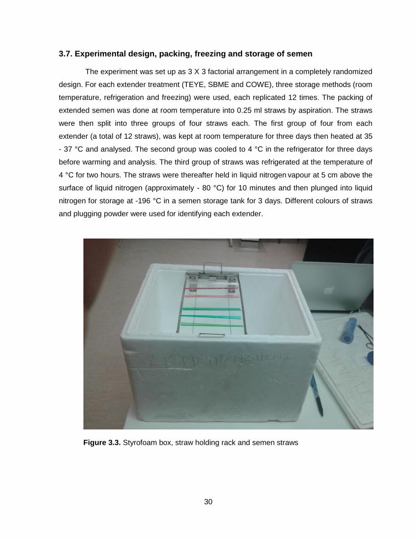
30
3.7. Experimental design, packing, freezing and storage of semen
The experiment was set up as 3 X 3 factorial arrangement in a completely randomized
design. For each extender treatment (TEYE, SBME and COWE), three storage methods (room
temperature, refrigeration and freezing) were used, each replicated 12 times. The packing of
extended semen was done at room temperature into 0.25 ml straws by aspiration. The straws
were then split into three groups of four straws each. The first group of four from each
extender (a total of 12 straws), was kept at room temperature for three days then heated at 35
- 37 °C and analysed. The second group was cooled to 4 °C in the refrigerator for three days
before warming and analysis. The third group of straws was refrigerated at the temperature of
4 °C for two hours. The straws were thereafter held in liquid nitrogen vapour at 5 cm above the
surface of liquid nitrogen (approximately - 80 °C) for 10 minutes and then plunged into liquid
nitrogen for storage at -196 °C in a semen storage tank for 3 days. Different colours of straws
and plugging powder were used for identifying each extender.
Figure 3.3. Styrofoam box, straw holding rack and semen straws

31
3.8. Thawing and post-freezing evaluation of semen
Refrigerated and frozen groups of semen straws were warmed for 30 seconds in a
water bath at 35 - 37 °C. One semen straw from each group was wiped with the paper towel
and split into three portions. The first portion was assayed for the characteristics relating to the
kinetic properties. The second portion was evaluated for live-dead counts. The last portion was
assayed for morphology properties.
3.7. Statistical analysis
Analysis of variance (ANOVA) on quality of fresh extended semen data (Model I) and
extended semen stored with different methods data (Model II) was performed at P <0.01 and P
<0.05 according to Steel and Torrie (1980) using general linear model procedures of Minitab
Statistical package version 17 (Minitab, 2014). Where significant differences between the
treatment groups was detected, means were separated using the Tukey’s test (Tukey, 1953).
The models used were as follows:
Yij= µ + Ei + Ԑij Model I
Where, Yij = the observation – TM, PM, AR, AT, CT, BT and DL;
µ = overall mean common to all observations;
Ei = effect of ith extenders, i = 1, 2 or 3; and
Ԑij = random residual error.
Yijk= µ + Ei + Sj + (ES)ij + Ԑijk Model II
Where, Yijk = the observation – TM, PM, AR, AT, CT, BT and DL;
µ = overall mean common to all observations;
Ei = effect of ith extenders, i = 1, 2 or 3; and
Sj = effect of jth storage temperatures, j = 1, 2 or 3;
(ES)ij= interaction between ith extenders and jth storage methods; and
Ԑijk = random residual error.

32
CHAPTER 4
RESULT AND DISCUSSION
EXPERIMENT 1: Effect of different extenders on spermatozoa motility, morphology
and viability
In the laboratory, before the semen was processed, an accurate macroscopic
evaluation of the samples was performed. Detection of the quality of semen before use
ensures achieving optimum reproductive efficiency. Therefore, physical properties, particularly
volume of ejaculate, pH and semen color were investigated. The results are summarized
below in Table 4.1.
Table 4.1. : Initial (macroscopic) characteristics of the collected bull semen
Parameters Values
Volume (ml) 7.62 ± 0.12
pH 6.46 ± 0.08
Semen color Ivory
The mean volume of Nguni bull semen was 7.62 ± 0.12 ml and the pH was 6.46 ± 0.08 cm.
The colour of the semen was ivory which could be considered normal according to Hossain et
al (2012).
The current study also investigated the behaviour of freshly collected Nguni bull semen
following dilution with Tris Egg Yolk Extender (TEYE), Soy Bean Milk based Extender (SBME)
and Coconut Water based Extender (COWE). The results are presented below.
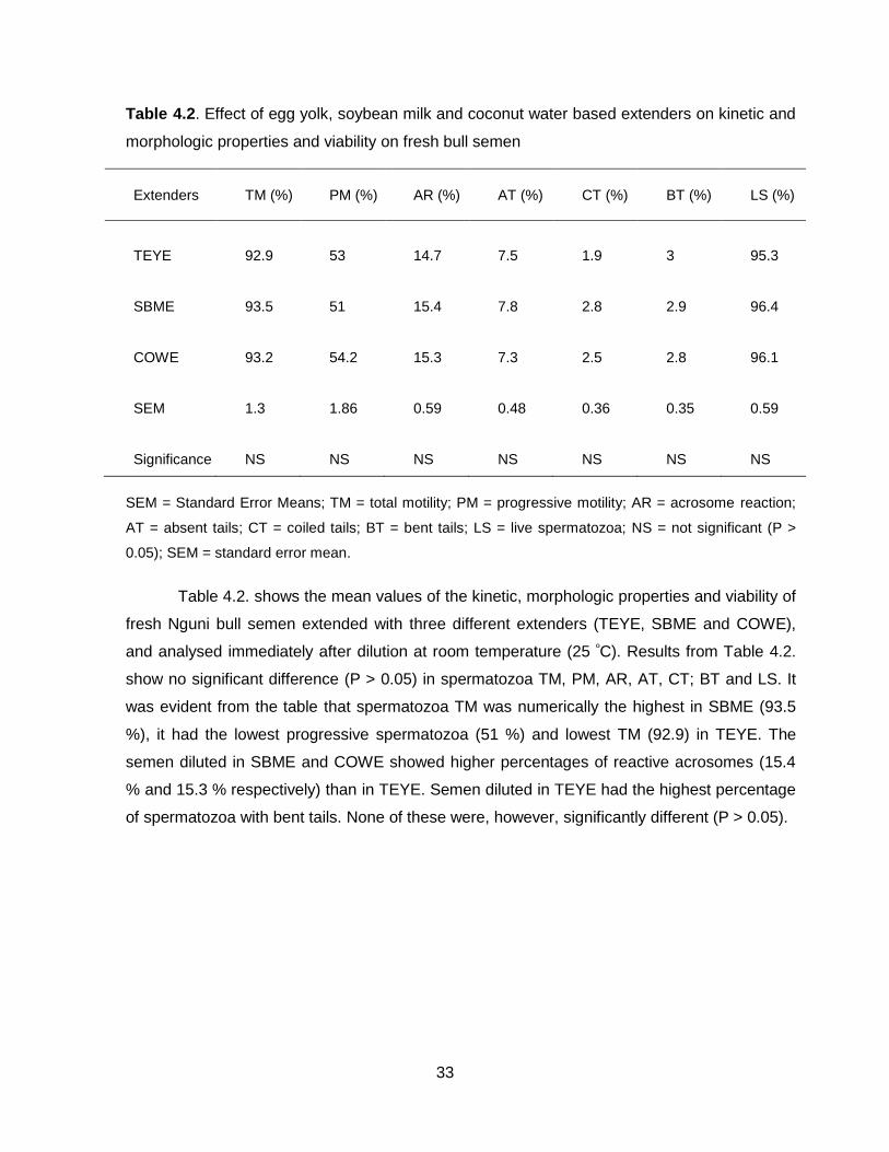
33
Table 4.2. Effect of egg yolk, soybean milk and coconut water based extenders on kinetic and
morphologic properties and viability on fresh bull semen
Extenders TM (%) PM (%) AR (%) AT (%) CT (%) BT (%) LS (%)
TEYE 92.9 53 14.7 7.5 1.9 3 95.3
SBME 93.5 51 15.4 7.8 2.8 2.9 96.4
COWE 93.2 54.2 15.3 7.3 2.5 2.8 96.1
SEM 1.3 1.86 0.59 0.48 0.36 0.35 0.59
Significance NS NS NS NS NS NS NS
SEM = Standard Error Means; TM = total motility; PM = progressive motility; AR = acrosome reaction;
AT = absent tails; CT = coiled tails; BT = bent tails; LS = live spermatozoa; NS = not significant (P >
0.05); SEM = standard error mean.
Table 4.2. shows the mean values of the kinetic, morphologic properties and viability of
fresh Nguni bull semen extended with three different extenders (TEYE, SBME and COWE),
and analysed immediately after dilution at room temperature (25 ºC). Results from Table 4.2.
show no significant difference (P > 0.05) in spermatozoa TM, PM, AR, AT, CT; BT and LS. It
was evident from the table that spermatozoa TM was numerically the highest in SBME (93.5
%), it had the lowest progressive spermatozoa (51 %) and lowest TM (92.9) in TEYE. The
semen diluted in SBME and COWE showed higher percentages of reactive acrosomes (15.4
% and 15.3 % respectively) than in TEYE. Semen diluted in TEYE had the highest percentage
of spermatozoa with bent tails. None of these were, however, significantly different (P > 0.05).
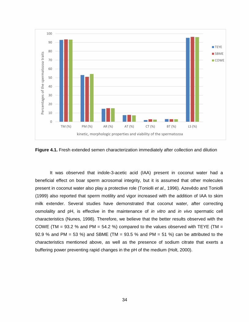
34
Figure 4.1. Fresh extended semen characterization immediately after collection and dilution
It was observed that indole-3-acetic acid (IAA) present in coconut water had a
beneficial effect on boar sperm acrosomal integrity, but it is assumed that other molecules
present in coconut water also play a protective role (Toniolli et al., 1996). Azevêdo and Toniolli
(1999) also reported that sperm motility and vigor increased with the addition of IAA to skim
milk extender. Several studies have demonstrated that coconut water, after correcting
osmolality and pH, is effective in the maintenance of in vitro and in vivo spermatic cell
characteristics (Nunes, 1998). Therefore, we believe that the better results observed with the
COWE (TM = 93.2 % and PM = 54.2 %) compared to the values observed with TEYE (TM =
92.9 % and PM = 53 %) and SBME (TM = 93.5 % and PM = 51 %) can be attributed to the
characteristics mentioned above, as well as the presence of sodium citrate that exerts a
buffering power preventing rapid changes in the pH of the medium (Holt, 2000).
0
10
20
30
40
50
60
70
80
90
100
TM (%) PM (%) AR (%) AT (%) CT (%) BT (%) LS (%)
Per
cen
tage
s o
f th
e sp
erm
ato
zoa
trai
ts
kinetic, morphologic properties and viability of the spermatozoa
TEYE
SBME
COWE

35
Experiment 2: Effect of extenders on bull spermatozoa kinetic, morphologic
properties and viability after three days of storage
Nguni bull semen samples were diluted with TEYE, SBME and COWE extenders and
were stored for 72 hours at room temperature (25 ºC), refrigerator temperature (4 ºC) and
cryopreserved in LN2 (-196 ºC). The results of analyses are presented in Table 4.3. below.
Table 4.3. Effect of egg yolk, soybean milk and coconut water based extenders on bull
spermatozoa kinetic; morphologic properties and viability after three days of storage
Treatment SM TM (%) PM (%) AR (%) AT (%) CT (%) BT (%) LS (%)
TEYE RT 72.6a 32.2a 23.5c 7.7ab 4.5b 5.1bc 59.1a
RF 72.7a 32.4a 22.3c 7.1ab 4.7b 6.0bc 61.2a
FR 32.7b 14.8b 35.2b 8.3ab 6.3ab 6.9b 36.0b
SBME RT 73.6a 31.2a 21.3c 7.6ab 4.5b 4.2c 59.1a
RF 71.1a 32.9a 22.8c 6.5 5.3b 5.1bc 61.0a
FR 28.8b 13.1b 37.8b 9.1a 6.3ab 6.9b 25.0c
COWE RT 73.9a 34.6a 21.6c 7.9ab 4.8b 5.3bc 57.3a
RF 73.6a 33.1a 23.1c 6.8ab 4.7b 4.8c 60.3a
FR 16.1c 6.7c 54.8a 8.3ab 8.0a 9.9a 13.7d
SEM
1.05 1.15 0.88 0.5 0.41 0.44 1.1
EXT Means
TEYE
59.3a 26.4 27.0b 7.7 5.2 6.0ab 52.1a
SBME
57.8a 25.7 27.3b 7.7 5.4 5.4b 48.4b
COWE
54.5b 24.8 33.1a 7.7 5.8 6.7a 43.8c
SEM
0.61 0.67 0.51 0.29 0.24 0.25 0.65
SM Means
RT
73.4a 32.7a 22.1b 7.7ab 4.6b 4.8b 58.5b
RF
72.4a 32.8a 22.7b 6.8b 4.9b 5.3b 60.8a
FR
25.9b 11.5b 42.6a 8.6a 9.6a 7.9a 24.9c
SEM
0.61 0.67 0.51 0.29 0.24 0.25 0.65
Significance
EXT (E)
** NS ** NS NS * **
SM (S)
** ** ** ** ** ** **
E × S
** ** ** NS NS ** **
Mean values that do not share superscript letters in the same column, are significantly different. SEM =
Standard Error Means; TM = total motility; PM = progressive motility; AR = acrosome reacted; AT =
absence tails; CT = coiled tails; BT = bent tails; LS = live spermatozoa; RT = room temperature; RF =
refrigeration; FR = freezing; NS = not significant (P > 0.05); * = significant (P < 0.05); ** = highly
significant (P < 0.01); SM = storage methods; EXT = extenders.
36
Results from Table 4.3. above show that although, there is no significant difference (P >
0.05) in the kinetic morphologic properties and viability of semen stored at room temperature
and refrigerator temperature regardless of the extender in use, they were significantly
difference (P < 0.05) in the TM, PM, AR and DL to that of frozen semen. While the percentage
of sperm with reactive acrosome was highest (P < 0.01) in the cryopreserved sample diluted
with COWE, there were no significant difference (P > 0.05) between the percentages of total
acrosomes that reacted as shown with Spermac® stain.
The percentage of absent tails showed no significant difference (P > 0.05) in any of the
storage methods in TEYE and COWE, but it was significantly different (P < 0.05) in the means
of all the tested storage methods. Table 4.3. indicated that there was no significant difference
(P > 0.05) between the percentages of sperm with coiled tails in semen samples diluted with
all three extenders and stored at room temperatures and, they were significantly different (P <
0.05) from the cryopreserved sample in each case of the extenders. The semen sample
diluted with COWE and cryopreserved had the highest percentage (P < 0.01) of spermatozoa
with bent tails; while the sample diluted with TEYE and SBME and stored at room and
refrigerator temperature were not significantly different (P > 0.05). The percentages of live
spermatozoa stored at room and refrigeration temperature (25 ºC and 4 ºC respectively) in all
three extenders showed no significant difference (P > 0.05) and they are significantly different
(P < 0.05) from cryopreserved samples in all extenders.
In this study it was observed that better post thaw MT, MP, AR, AT, CT, BT, and LS
values were associated with the use of TEYE, followed by SBME and COWE in the frozen
semen. However, at room and refrigeration temperature, the values were similar regardless of
the extenders. This result is in line with the observation of Celeghini et al. (2008) that greater
post-thawing TM, PM, AR, AT, CT, BT and LS values were associated with the use of Botu-
Bov® (BB) containing 20 % egg yolk compared to Bioxcell® medium composed of soy lecithin.
It has been verified that BB gives greater sperm protection than does Tris medium when used
for refrigeration, as was previously observed for bull semen cryopreservation.

37
Figure 4.2. Semen characterization after three days of storage at room temperature (25 ºC)
Figure 4.3. Semen characterization after three days of refrigeration (4 ºC)
0
10
20
30
40
50
60
70
80
90
100
TM PM AR AT CT BT LS
Per
cen
tage
s o
f th
e sp
erm
ato
zoa
trai
ts
Spermatozoa kinetic and morphologic properties and viability
TEYE
SBME
COWE
0
10
20
30
40
50
60
70
80
90
100
TM PM AR AT CT BT LS
Per
cen
tage
s o
f th
e sp
erm
ato
zoa
trai
ts
Spermatozoa kinetic and morphologic properties and viability
TEYE
SBME
COWE

38
Figure 4.4. Semen characterization after three days of storage in the LN2 (-196 ºC)
It can be argued that storage of semen at room temperature would provoke
degenerative changes leading to cell death (Aalseth & Saacke, 1985). Barros and Toniolli
(2011) reviewed the potential effects of coconut water on semen technology and reported that
its utilization as an extender is important, because it is an easily prepared and cheap
alternative for semen transport over small distances and for use in artificial insemination
programs in several species. Initially, it was reported that isotonic sodium chloride solution was
the first extender used to dilute semen from domestic animals to evaluate spermatozoa
metabolic activity (Salisbury and Nakabayashi, 1957).
In the present study, superior numeric results have also been observed with COWE
(TM = 73.9 % and PM = 34.6 %) compared to TEYE (TM = 72.6 % and PM = 32.2 %) and
SBME (TM = 73.6 % and PM = 31.2 %). The temperature exerts a significant influence on
spermatozoa survival throughout the refrigeration period (Batellier et al., 2001). All variables
related to bull semen motility (total and progressive motility) decreased significantly over the
72 h of refrigeration period regardless of the extender in use, consistent with the results
obtained by Verberckmoes et al. (2005). Even though the decrease in temperature causes a
significant decrease in sperm metabolism, reducing the rates of fructolysis and oxygen
consumption (Blackshawet et al., 1957), spermatozoa quality decreases throughout the
refrigeration period regardless of the extender, dilution rate or storage conditions (O’Hara et
al., 2010).
0
10
20
30
40
50
60
70
80
90
100
TM PM AR AT CT BT LS
Per
cen
tage
s o
f th
e sp
erm
ato
zoa
trai
ts
Spermatozoa kinetic and morphologic properties and viability
TEYE
SBME
COWE

39
The present study shows that COWE can be used to refrigerate spermatozoa, since
similar results in terms of TM and PM were observed compared to TEYE and SBME. The
differences observed in the TM and PM variables among the different extenders for the
freezing storage method were not similar to those observed in previous studies evaluating the
extenders containing 20 % of egg yolk [Tris (Tris-R) and Botu-Bov® (BB)] and another
composed of 1 % soy lecithin [Botu-Bov® Lecithin (BB-L)] for semen cryopreservation.
Crespilho et al. (2012) noted more TM and PM for cryopreserved bull spermatozoa
stored in Botu-Bov® compared to spermatozoa processed in Tris egg yolk fructose extender. In
the previous study it was concluded that the lipid particles found on egg yolk based extenders
could also have a deleterious role on spermatozoa motility, acting as a physical barrier for
spermatozoa. For this reason, the lesser viscosity of the Botu-Bov® medium (clarified by
centrifugation) allowed greater preservation of post-thaw spermatozoa movement. However, in
the present study, better results from frozen semen, were observed with TEYE (TM = 32.7 %
and PM = 14.8 %) as compared to SBME (TM = 28.8 % and 13.1 %) and COWE (TM = 16.1
% and PM = 6.7 %).

40
CHAPTER 5
CONCLUSION AND RECOMMENDATION
5.1. Conclusions
The success of spermatozoa used for AI and its ability to fertilize, is affected at
different level during its course from its origin until it reaches the ovum.
Cryopreservation continues to be the most frequently employed technique for use in
modern animal production. Commercial AI will inevitably use this technique for long storage
and transport semen over a wider area around the world. However, even with the most up to
date procedures, cryopreservation still causes severe detrimental effect to spermatozoa and
their function. To ensure that semen used for AI is of a relatively high quality, artificial breeding
organizations should also consider the different storage methods of semen based on the
period between the collection and the AI.
5.2. Recommendations
For the short storage period of semen used for AI, it is recommended that semen
should be kept at room or refrigeration temperature regardless of the three extenders used.
However, for long storage of frozen semen TEYE is recommended. The egg yolk-based
extender provided greater preservation of motility and bull spermatozoa integrity during the
freezing process than did SBME and COWE.

41
REFERENCES
Aalseth, E.P., Saacke, R.G. (1985). Morphological change of the acrosome on motile bovine
spermatozoa due to storage at 4 %. J. Reprod. Fertil. 74, 473-478.
Ahmad, M., Tadros, N.T., Polackwich, A.S., Sharma, R., Agarwal, A., Sabanegh, E. (2014).
Vasectomy reversal semen analysis: new reference ranges predict pregnancy. Fertil. and
Steril., 107, 905.
Ajao, O.A. (2015). Evaluation of the effectiveness of different extenders of goat buck semen under
refrigerated conditions. Dissertation.
Akhter, S., Ansari, S., Rakha, B.A., Ullah, N., Andrabi, S.M.H., Khalid, M. (2011). In vitro
evaluation of liquid-stored buffalo semen at 5 °C diluted in soya lecithin based extender
(Bioxcell®, tris-citric egg yolk, skim milk and egg yolk-citrate extenders). Reprod. Dom.
Anim. 46, 45–49.
Akhter, S., Husna, A.U.I., Ejaz, R., Qadeer, S., Azam, A., AllahRakha, B., Ansari, M.S., Shahzad,
Q., Moazzam Javed, M., Mónica H. Vazquez-Levin, M.H. (2016). A comparative analysis of
sperm selection procedures prior to cryopreservation for Nili-Ravi buffalo bull (Bubalus
bubalis) semen: Assessment of its impact on post-thaw sperm functional quality. J. Anim.
Reprod. Sci. 174, 29-36.
Almquist, J.O. (1948). The effect of penicillin upon the fertility of semen from relatively infertile
bulls. J. Dairy. Sci. 31, 681-682.
Almquist, J.O. (1951). The fertility of bovine semen diluters containing varying amounts of egg yolk.
J. Dairy Sci. 34:8, 763-766.
Almquist, J.O. (1954). Diluters for bovine semen. V. A comparison of heated milk and egg yolk-
citrate as diluters for semen from bulls of high and low fertility. J. Dairy Sci. 37, 1308–1315.
Amann, R.P., DeJarnette, J.M. (2012). Impact of genomic selection of AI dairy sires on their likely
utilization and methods to estimate fertility: a paradigm shift. Theriogenology 77, 795–817.

42
Amirat, L., Anton, M., Tainturier, D., Chatagnon, G., Battut, I., Courtens, J.L. (2005). Modifications
of bull spermatozoa induced by three extenders: Biociphos, low density lipoprotein and
Triladyl, before, during and after freezing and thawing. Reprod. 129, 535-543.
Anzar, M., Graham, E.F. (1996). Role of sperm motility and acrosome integrity in the filtration of
bovine semen. Theriogenology 45, 513–520.
Anzar, M., Kroetsch, T., Boswall, L. (2011). Cryopreservation of bull semen shipped overnight and
its effect on post-thaw sperm motility, plasma membrane integrity, mitochondrial membrane
potential and normal acrosomes. Anim. Reprod. Sci. 126, 23–31.
Arzondo, M.M., Caballero, J.N., Marín-Briggiler, C.I., Dalvit, G., Cetica, P.D.,Vazquez-Levin, M.H.,
(2012). Glass wool filtration of bull cryopreserved semen: a rapid and effective method to
obtain a high percentage of functional sperm. Theriogenology 78, 201–209.
Austin, J.W., Chang, E.W., (1951). Comparison of quality of bull semen collected in the artificial
vagina and by electro-ejaculation. J. Dairy Sci. 44, 2292–2297.
Azevedo, D.M.M.R., Toniolli, R. (1999). Água de coco estabiliz ada suplementada com antibióticos
e ácido 3-indol acético na conservação de sêmen de caprinos marota. Ciênc. Anim. 9, 37-
42.
Bailey, J.L., Bilodeau, J.F., Cormier, N. (2000). Semen cryopreservation in domestic animals: A
damaging and capacitating phenomenon. J. Androl. 21, 1-7.
Baracaldo, M.I., Barth, A.D., Bertrand, W. (2007). Steps for freezing bovine semen: from semen
collection to the liquid nitrogen tank. Vet. Record. 30, 105-107.
Batellier, F., Vidament, M., Fauquant, J., Duchamp, G., Arnaud, G., Yvon, L.M., Magistrini, M.
(2001). Advances in cooled semen technology. Anim. Reprod. Sci. 68, 181-190.
Bean, B.H., Pickett, B.W., Martig, R.C. (1963). Influence or freezing method, extenders and
storage, temperatures on motility and pH of frozen bovine semen. J. Dairy Sci. 46:2, 145-
149.
Bearden, H.J., Fuguay, J.W., Willard, S.T. (2004). Applied animal reproduction. Sixth Edition.
Pearson Prentice Hall, New Jersey.

43
Berndtson, W.E., Foote, R.H. (1976). Survival and fertility of antibiotic-treated bovine spermatozoa.
J. Dairy Sci. 59:12, 2130-2133.
Bilodeau, J.F., Chatterjee, S., Sirard, M.A., Gagnon, C. (2000). Levels of antioxidant defenses are
decreased in bovine spermatozoa after a cycle of freezing and thawing. Mol. Reprod. Dev.
5, 282-288.
Blackshaw, A.W., Salisbury, G.W. (1957). Factors influencing metabolic activity of bull
spermatozoa II. Cold-shock and its prevention. J. Dairy Sci. 40:9, 1099-1106.
Blackshaw, A.W., Salisbury, G.W. (1957). Factors influencing metabolic activity of bull
spermatozoa II. Cold-shock and its prevention. J. Dairy Sci. 40:9, 1099-1106.
Boonsorn, T., Kongbuntad, W., Narkkong, N., Aengwanich, W. (2010). Effect of catechin addition
to extender on semen quality and lipid peroxidation in boar semen. J. Agric. Environ. Sci. 7,
283 – 288.
Campanholi, S.P., Monteiro F.M., Dias, E.A.R., Mercadante, M.E.Z., De Paz, C.C.P., Dell’Aqua
Junior, J.A., Papa, F.O., Dell’Aqua, J.O., Vantini, R., Garcia, J.M. (2017). Effect of seminal
plasma removal before cryopreservation of bovine semen obtained by electroejaculation on
semen quality and in vitro fertility. Theriogenology 89, 114-121.
Celeghini, E.C., De Arruda, R.P., De Andrade, A.F., Nascimento, J., Raphael, C.F., Rodrigues,
P.H. (2008). Effects that bovine sperm cryopreservation using two different extenders has
on sperm membranes and chromatin. Anim Reprod Sci. 104(2-4), 119-131.
Chaveiro, A., Machado, L., Frijters, A., Engel, B., Woelders, H. (2006). Improvement of parameters
of freezing medium and freezing protocol for bull sperm using two osmotic supports.
Theriogenology 65, 1875–1890.
Cheema, R.S., Patel, M.K., Bansal, A.K., Gandotra, V.K. (2016). A 31-kDa seminal plasma
heparin-binding protein reduces cold shock stress during cryopreservation of cross-bred
cattle bull semen. Theriogenology 86, 1599-1606.
Contri, A., Gloria, A., Wegher, L., Vignola, G., Dellamaria, D., Carluccio, A. (2014). The effects of
antibiotic additions to extenders on fresh and frozen-thawed bull semen. J. Anim. Reprod.
Sci. 150, 15-23.

44
Correa, J.A., Heersch, G., Zavos, P.M. (1997). Sperm membrane functional integrity and response
of frozen-thawed bovine spermatozoa during the swelling test incubation at varying
temperatures. Theriogenology. 47, 715-729.
Curry M.R. (2000). Cryopreservation of semen from domestic livestock. Rev. Reprod., 5, 46–52.
DeJarnette, J.M., Marshall, C.E., Lenz, R.W., Monke, D.R., Ayars, W.H., Sattler, C.G. (2004).
Sustaining the fertility of artificially inseminated dairy cattle: The role of the artificial
insemination industry. J. Dairy Sci. 87 (E), E93-E104.
Divyaswetha, P., Bindu, N., Abdullah, K., Jean, M.F., Shane, C.B., Erdogan, M. (2008).
Comprehensive proteomic analysis of bovine spermatozoa of varying fertility rates and
identification of biomarkers associated with fertility. Syst. Biol. 2, 1–14.
Dunn, H.O., Larson, G.L., Willet, E.L. (1953). The effects of freezing bovine spermatozoa in
extenders containing antibacterial agents. J. Dairy Sci. 36:7, 728-732.
Ehmcke, J. and Schlatt, S. (2008). Animal models for fertility preservation in the male. Reprod.
136, 717-732.
Eidan, S.M. (2016). Effect on post-cryopreserved semen characteristics of Holstein bulls of adding
combinations of vitamin C and either catalase or reduced glutathione to Tris extender. J.
anim. Reprod. Sci. 167, 1-7.
El-Harairy, M.A., Laila, N., Zeidan, A.E.B., El-Salaam, A.M.A., El-Kishk, M.A.M. (2011). Quality and
fertility of the frozen-thawed bull semen as affected by the different cryoprotectants and
glutathione levels. J. Amin. Reprod. Sci. 7:5, 792-797.
El-Keraby, F.E., Osman, K.T., Ganah, H.B., El-Siefy, E.M. (2010). Soymilk- based Extender for
Cryopreservation of bovine semen. J. Anim. And poultry prod. 1 (2), 61-69.
El-Sisy, G.A., El-Nattat, W.S., El-Sheshtawy, R.I., El-Maaty, A.M.A. (2016). Substitution of egg yolk
with different concentrations of soybean lecithin in Tris-based extender during bulls’ semen
preservability. Asian Pac. J. Anim. Reprod. Sci. ■ 1-5.
England, G.C.W. (1993). Cryopreservation of dog semen: a review. J. Reprod. Fertil. Suppl. 47,
243–255.
45
England, G.C.W., Millar, K.M. (2008). The Ethics and Role of AI with Fresh and Frozen Semen in
Dogs. Reprod. Dom. Anim. 43 (2), 165–171.
Fleisch, A., Malama, E., Witschi, U., Leiding, C., Siuda, M. (2017). Effects of an extension of the
equilibration period up to 96 hours on the characteristics of cryopreserved bull semen.
Theriogenology 89, 255-262.
Foote, R.H. (1975). Sperm quality from the bull to the freezer: An assessment. Theriogenology 3:6,
219-235.
Foote, R.H. (1999). Artificial insemination from the origins up today. Proc. of the Spallanzani Int.
system. Reggio Emilia. Italy 12, 94-104.
Foote, R.H. (2002). The history of artificial insemination: Selected notes and notables. J. Anim. Sci.
80, 1-10.
Foote, R.H., Almquist, J.O. (1948). The fertility of bovine semen cooledwith and without the
addition of citrate sulfanilamide yolk extender. J. Dairy Sci. 32, 856–861.
Foote, R.H., Berndtson, W.E. (1976). Antibacterial agents for bull semen; do they help? Paper
presented at: Proc Sixth Tech Conf. Art. Insem. Reprod. 6, 23-30.
Fukui, Y., Kohno, H., Togari, T., Hiwasa, M., Okabe, K. (2008). Fertility after artificial insemination
using soybean based extenders in sheep. J. Reprod. Dev, Vol. 54, 44-52.
Garner, D.L., Hafez, E.S.E. (1993). Spermatozoa and seminal plasma. In: Reproduction in Farm
Animals, (6th Eds.). Philadelphia. 165-187.
Graham, E.F., Erickson, W.E., Bayley, N.D. (1956). Effect of glycerol equilibration on fronfen
bovine spermatozoa. J. Dairy Sci. 40:5, 510-515.
Griffin, E.R. (2004). Development of an extender protocol to enhance the viability of frozen-thawed
bovine spermatozoa. MSc Thesis. Texas A&M University. USA.
Gürler, H., Calisici, O., Bollwein, H. (2015). Inter- and intra-individual variability of total antioxidant
capacity of bovine seminal plasma and relationships with sperm quality before and after
cryopreservation. Anim. Reprod. Sci. 155, 99–105.

46
Hammerstedt R.H., Graham, J.K., Nolan, J.P. (1990). Cryopreservation of mammalian sperm:
What we ask them to survive. J. Androl. 11, 73-88.
Haugan, T., Reksen, O., Grohn, Y.T., Kommisrud, E., Ropstard, E., Sehested, E. (2005). Seasonal
effects of semen collection and artificial insemination on dairy cow conception. Anim.
Reprod. Sci. 90, 57-71.
Heise, A., (2012). Artificial insemination in veterinary science, a bird’s-eye view of veterinary
medicine, Dr. Carlos C. Perez-Marin (ed). ISBN: 978 – 953 – 51 – 0031 – 7. In Tech.
http://www.intechope.com/books/a-bird-s-eye-view-of-veterinary-medicine/artificial-
insemination-in-veterinaryscience.
Holt, W.V. (1997). Alternative strategies for long-term preservation. Reproduction, Fertility and
Development. 9, 309–319.
Holt, W.V. (2000). Basic aspects of frozen storage of semen. Anim. Reprod. Sci. 62, 3-22.
Hossain, M.E., Khatun, M.M., Islam, M.M., Miazi, O.F. (2012). Semen characteristics of breeding
bulls at the Central Cattle Breeding and Dairy Farm of Bangladesh. Bang. J. Anim. Sci. 41
(1), 1-5.
Hu, J.H., Tlan, W.Q., Zhao, X.L., Zan, L.S., Wang, H., Li, Q.W., Xin, Y.P., (2010). The
cryoprotective effects of ascorbic acid supplementation onbovine semen quality. Anim.
Reprod. Sci. 121, 72–77.
Januskauskas, A., Zilinskas, H. (2002). Bull semen evaluation post-thaw and relation of semen
characteristics to bull’s fertility. Vet. Ir. Zootech. 17, 39-49.
Jones, R., James, P.S., Howes, L., Bruckbauer, A., Klenerman, D. (2007). Supramolecular
organization of the sperm plasma membrane during maturation and capacitation. Asian J.
Androl. 9 (4), 438–444.
Jones, R.C. (1976). The nature of ultrastructural changes indusced by exposure of spermatozoa to
lysolecithin. Theriogenology 6, 656-667.
Jones, R.C. and Steward, D.M. (1979). The effects of cooling to 5 C and freezing and thawing on
the ultrastructure of bull spermatozoa. J. Reprod. Fert. 56, 233-238.
47
Kathiravan, P., Kalatharan, J., Karthikeya, G., Rengarajan, K., Kadirvel, G. (2011). Objective sperm
motion analisis to assess dairy bull fertility using computer-aided system: a review. Reprod.
Dom. Anim. 46, 165 – 172.
Kumaresan, A., Siqueira, A.P., Hossain, M.S., Bergqvist, A.S., (2011). Cryopreservation induced
alterations in protein tyrosinephosphorylation of spermatozoa from different portions of the
boarejaculate. Cryobiology. 63, 137–144.
Mazur, P., Katkov, I., Katkova, N., Cristar, J. (2000). The enhancement of the ability of mouse
sperm to survive freezing anf thawing by the use of high concentrations of glycerol and the
presence of an Escherichia coli membrane preparation (oxyrase) to lower the oxygen
concentration. Cryobiology. 40,187-209.
Medeiros, C.M.O., Forell, F., Oliviera, A.T.D., Rodriguez, J.L. (2002). Current status of sperm
cryopreservation: why isn’t it better? Theriogenology 57, 327-344.
Minitab. (2014) Minitab 17 statistical software. Minitab Inc., State College, Pennsylvania, USA.
Mogielnicka-Brzozowska, M., Kordan, W. (2011). Characteristics of selected seminal plasma
proteins and their application in the improvement of the reproductive processes in
mammals. Polish J. Vet. Sci. 14, 489–499.
Moussa, M., Martinet, V., Trimeche, A., Tainturier, D., Anton, M. (2002). Low density lipoproteins
extracted from hen egg yolk by an easy method: cryoprotective effect on frozen-thawed bull
semen. Theriogenology. 57, 1695-1706.
Muin˜o, R., Tamargo, C., Hidalgo, C.O., Pen˜a, A.I. (2008). Identification of sperm subpopulations
with defined motility characteristics in ejaculates from Holstein bulls: effects of
cryopreservation and between-bull variation. Anim. Reprod. Sci. 109, 27–39.
Nishizono, H., Shioda, M., Takeo, T., Irie, T., Nakagata, N. (2004). Decrease of fertilizing ability of
mouse spermatozoa after freezing and thawing is related to cellular injury. Biol. Reprod. 71,
973-978
Nunes, J.F. (1998). Utilização da água de coco como diluidor do sêmen de animais domésticos e
do homem. Rev. Bras. Reprod. Anim. 22, 109-112.

48
Nunes, J.F., Cornhamous, Y., Oliveira, L.F., Teixeira, M.D. (1996). Utilização da água-de-coco e
suas frações ativas na conservação in vitro e avaliação in vivo do sêmen na espécie
caorina. Rev. Ciência Anim. 2(1), 32.
O’Hara, L., Richardson, L., Hanrahan, J.P., Donovan, A., Fair, S., O'Sullivan, M., Carrington, S.D.,
Lonergan, P., Evans, A.C.O. (2010). Ewe breed differences in fertility after cervical AI with
frozen–thawed semen and associated differences in sperm penetration and
physicochemical properties of cervical mucus. Anim. Reprod. Sci. 129, 37–43.
Pena, A.I., Muino, R., Rodriguez, A., Tamargo, C., Hidalgo, C.O. (2009). Effects of
cryopreservation on the motile sperm subpopulations in semen from asturiana de los Valles
bulls. Theriogenology 72, 860-868.
Perry, G.A., Kruse, S.G., Bridges, G.A., Funnell, B.J., Bird, S.L., Lake, S.L., Arias, R.P.,
Amundson, O.L., Larimore, E.L., Keisler, D.H. (2017). Influence of post-insemination
nutrition on embryonic development in beef heifers. Theriogenology, 90, 185-190.
Phillips P.H. (1939). Preservation of bull semen. J. Biol. Chem. 130, 415.
Polge, C., Smith, A.U., Parkes A.S. (1949). Revival of/ Bovine / Spermatozoa after vitrification and
dehydration at low temperature. Nature 164, 666-669.
Poulos, A., Darin-Bennett, A., White, I.G. (1973). The phospholipid-bound fatty acids and
aldehydes of mammalian spermatozoa. Comp. Biochem. Physiol. 46B, 541-549.
Quinn, P.J., Chow, P.Y.W., White, I.G. (1980). Evidence that phospholipid protects ram
spermatozoa from cold shock at a plasma membrane site. J Reprod Fertil. 60, 403-407
Rauch, A. (2013). Cryopreservation of bovine semen in egg yolk based extenders. Dissertation.
Rego, J.P.A., Moura, A.A., Nouwens, A.S., Mcgowan, M.R., Boe-hansen, G.B. (2015). Seminal
plasma protein profiles of ejaculates obtained by internal artificial vagina and
electroejaculation in Brahman bulls. Anim. Reprod. Sci. 160, 126–37.
Ricker, J.V., Linfor, J.J., Delfino W.J., Kysar, P., Scholtz. E.L,, Tablin, F. (2006). Equine sperm
membrane phase behavior: The effects of lipid-based cryoprotectants. Biol Reprod 74,
359-365.
49
Rix, D., Earle, J.L., Keats, G., Beavon, K.S.O., Powell, E., Van Schalkwyk, P.L. (1989).Geography
in action, Juta and Co, LTD Cape town and Johannesburg.
Rottensten, K., Koefoed-Johnsen, H.H., Andersen, H. (1960). The effect of spermatozoal extracts
on the viability of bovine spermatozoa. Acta. Agric. Scand. 10, 280.
Saacke, R.G. (1983).Semen quality in relation to semen preservation.J. Dairy Sci. 66, 2635-2644
Salisbury, G.W., Nakabayashi, N.T. (1957). Factors influencing the metabolic activity of bovine
spermatozoa. J. Dairy Sci. 42, 1806-1814.
Salisbury, G.W., Van Demark, N.L., Lodge, J.R. (1978). Extenders and extension of unfrozen
semen. In: Physiology of reproduction and artificial insemination in cattle. Freeman and
Company, San Francisco, 294–554.
Salisbury, G.W., Van Dermark, N.L. (1961). Physiology of Reproduction and Artificial Insemination
of Cattle. Freeman and San Francisco. 412-435.
Shannon, P. (1978). Factors affecting semen preservation and conception rates in cattle. J
Reprod. Fertil. 54, 519-527.
Shannon, P., and Vishwanath, R. (1995). The effect of optimal and suboptimal concentrations of
sperm on the fertility of fresh and frozen bovine semen and a theoretical model to explain
the fertility differences. Anim. Reprod. Sci. 39, 1-10.
Silva, A.R., Cardoso, R.C.S., Uchoa, D.C., Silva, L.D.M. (2002). Effect of Tris-buffer, egg yolk and
glycerol on canine semen freezing. Vet. J. 164, 244-246.
Silva, A.R., Cardoso, R.C.S., Uchoa, D.C., Silva, L.D.M. (2003). Quality of canine semen submitted
to single or fractioned glycerol addition during the freezing process. Theriogenology 59,
821-829.
Singh, V.K., Singh, A.K., Kumar, R., Atreja, S.K., (2013). Development of soya milk extender for
semen cryopreservation of Karan Fries (cross breed cattle). Cryo. Letters 34 (1), 52–61.
Steel, R.G.D. and Torrie, J.H. (1980). Principles and procedures of statistics, Second Edition, New
York: McGra0w-Hill Book Co.

50
Suarez, S.S. (2007). Sperm interaction with the female tract; inspirationfor assisted reproduction.
Reprod. Fertil. Dev. 19, 104–110.
Tardif, A.L., Farrell, P.B., Trouern-Trend, V., Simkin, M.E., Foote, R.H. (1998). Use of Hoechst
33342 stain to evaluate live fresh and frozen bull sperm by computer-assisted analysis. J.
Androl. 19, 201–206.
Tirpak, F., Slanina, T., Ofukany, M., Lukac, N., Massanyi, P. (2015). Effect of taurine on bovine
spermatozoa motility parameters following cryopreservation. Slovak J. Anim. Sci., 48, 49-
56.
Tiwari, A., Singh, D., Kumar, O.S., Sharma, M.K. (2008). Expression ofcytochrome P450
aromatase transcripts in buffalo (Bubalus bubalis) ejaculated spermatozoa and its
relationship with sperm motility. Domest. Anim. Endocrinol. 34, 238–249.
Toniolli, R. (2011). Pouvoir fecondant des spermatozoïdes de verrat: amèlioration des conditions
de conservation. Dissertation.
Toniolli, R., Courot, M., Combarnous, Y., Bussière, J., Magistrini, M. (1996). Effect of índole-3-
acetic (plant auxin) on the preservation at 15°C of boar sêmen for artificial insemination.
Reprod. Nutr. Dev. 36, 503-511.
Tukey, J.W. (1953). The problem of multiple comparisons. Mimeographed monograph. Princeton.
Dissertation.
Urata, K., Narahara, H., Tanaka, Y., Egashira, T., Takayama, F., Miyakawa, I. (2001). Effect of
endotoxin-induced reactive oxygen species on sperm motility. Fertil Steril, 76, 163–166.
Vale, W.G., Silva, A.O.A., Sousa, J.S., Ribeiro, H. F.L., Souza, H.E.M., Nahum, B.S., Ohashi, O.M.
(1997). Preliminary report on the use of coconut water (Cocos nucifera) as a diluter of
buffalo semen. J. Anim. Repro. 836-839.
Van Demark, N.L., Miller, W.J., Kinney, Jr, W.C., Rodriguez, C., Friedman, M.E. (1957).
Preservation of bull semen at sub-zero temperatures. Urbana: Univ. Illinois Agric. Exp. Stn.
Van Staden, J. (2010). Ensuring quality in herbal medicines: Toxic phthalates in plastic-packaged
commercial herbal products. J. of Botany 82, 60–66.

51
Verberckmoes, S., Soom, A.V., Dewulf, J., De Kruif, A. (2005). Corrigendum to “comparison of
three diluents for the storage of fresh bovine semen”. Theriogenology 63, 912-922.
Vishwanath, R. (2003). Artificial insemination: The state of the art. Theriogenology 59(2), 571-584.
Viveiros, A.T.M., Orf˜ao, L.H., Maria, A.N., Allaman, I.B. (2008). A simple, inexpensive and
successful freezing method for curimba Prochilodus lineatus (Characiformes) semen. Anim.
Reprod. Sci. 112, 293–300.
Walters, E.M., Benson, J.D., Woods, E.J., Crister, J.K. (2009). The history of sperm
cryopreservation. Theory and practice. J. Tomlinson. Cambridge University Press. FAO.
White, I.G. (1993). Lipids and calcium uptake of sperm in relation to cold shock and preservation: a
review. Reprod. Fertil. Dev. 5, 639-658.
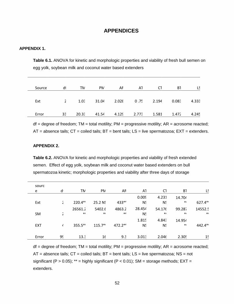
52
APPENDICES
APPENDIX 1.
Table 6.1. ANOVA for kinetic and morphologic properties and viability of fresh bull semen on
egg yolk, soybean milk and coconut water based extenders
Source df TM PM AR AT CT BT LS
Ext 2 1.03 31.04 2.028 0 .75 2.194 0.083 4.333
Error 33 20.33 41.54 4.129 2.773 1.581 1.472 4.245
df = degree of freedom; TM = total motility; PM = progressive motility; AR = acrosome reacted;
AT = absence tails; CT = coiled tails; BT = bent tails; LS = live spermatozoa; EXT = extenders.
APPENDIX 2.
Table 6.2. ANOVA for kinetic and morphologic properties and viability of fresh extended
semen. Effect of egg yolk, soybean milk and coconut water based extenders on bull
spermatozoa kinetic; morphologic properties and viability after three days of storage
source df TM PM AR AT CT BT LS
Ext 2 220.4** 25.2 NS 433** 0.009
NS 4.231
NS 14.704
** 627.4**
SM 2 26561.2
** 5402.6
** 4863.2
**
28.454 NS
54.176**
99.287**
14552.5**
EXT 4 355.5** 115.7** 472.2** 1.815
NS 4.843
NS 14.954
** 442.4**
Error 99 13.3 16 9.3 3.013 2.046 2.305 15
df = degree of freedom; TM = total motility; PM = progressive motility; AR = acrosome reacted;
AT = absence tails; CT = coiled tails; BT = bent tails; LS = live spermatozoa; NS = not
significant (P > 0.05); ** = highly significant (P < 0.01); SM = storage methods; EXT =
extenders.