evaluation of stiffness feedback for hard nodule...
TRANSCRIPT

RESEARCH ARTICLE
Evaluation of stiffness feedback for hard
nodule identification on a phantom silicone
model
Min Li1,2*, Jelizaveta Konstantinova3, Guanghua Xu1,2*, Bo He1, Vahid Aminzadeh4,
Jun Xie1,2, Helge Wurdemann5, Kaspar Althoefer6
1 School of Mechanical Engineering, Xi’an Jiaotong University, Xi’an, Shaanxi, China, 2 State Key
Laboratory for Manufacturing Systems Engineering, Xi’an Jiaotong University, Xi’an, Shaanxi, China,
3 School of Engineering and Materials Science, Queen Mary University of London, London, United Kingdom,
4 De Beers Technologies, Maidenhead, United Kingdom, 5 Department of Mechanical Engineering,
University College London, London, United Kingdom, 6 Faculty of Science & Engineering, Queen Mary
University of London, London, United Kingdom
* [email protected] (ML); [email protected] (GX)
Abstract
Haptic information in robotic surgery can significantly improve clinical outcomes and help
detect hard soft-tissue inclusions that indicate potential abnormalities. Visual representation
of tissue stiffness information is a cost-effective technique. Meanwhile, direct force feed-
back, although considerably more expensive than visual representation, is an intuitive
method of conveying information regarding tissue stiffness to surgeons. In this study, real-
time visual stiffness feedback by sliding indentation palpation is proposed, validated, and
compared with force feedback involving human subjects. In an experimental tele-manipula-
tion environment, a dynamically updated color map depicting the stiffness of probed soft
tissue is presented via a graphical interface. The force feedback is provided, aided by a
master haptic device. The haptic device uses data acquired from an F/T sensor attached to
the end-effector of a tele-manipulated robot. Hard nodule detection performance is evalu-
ated for 2 modes (force feedback and visual stiffness feedback) of stiffness feedback on an
artificial organ containing buried stiff nodules. From this artificial organ, a virtual-environ-
ment tissue model is generated based on sliding indentation measurements. Employing this
virtual-environment tissue model, we compare the performance of human participants in dis-
tinguishing differently sized hard nodules by force feedback and visual stiffness feedback.
Results indicate that the proposed distributed visual representation of tissue stiffness can
be used effectively for hard nodule identification. The representation can also be used as a
sufficient substitute for force feedback in tissue palpation.
Introduction
Soft-tissue tumors are often localized by conducting intra-operative manual palpation during
open surgery. Manual palpation is performed to identify hard nodules by direct tactile sensation.
PLOS ONE | DOI:10.1371/journal.pone.0172703 March 1, 2017 1 / 18
a1111111111
a1111111111
a1111111111
a1111111111
a1111111111
OPENACCESS
Citation: Li M, Konstantinova J, Xu G, He B,
Aminzadeh V, Xie J, et al. (2017) Evaluation of
stiffness feedback for hard nodule identification on
a phantom silicone model. PLoS ONE 12(3):
e0172703. doi:10.1371/journal.pone.0172703
Editor: Jose Manuel Garcia Aznar, University of
Zaragoza, SPAIN
Received: November 3, 2016
Accepted: February 8, 2017
Published: March 1, 2017
Copyright: © 2017 Li et al. This is an open access
article distributed under the terms of the Creative
Commons Attribution License, which permits
unrestricted use, distribution, and reproduction in
any medium, provided the original author and
source are credited.
Data Availability Statement: All relevant data are
within the paper and its Supporting Information
files.
Funding: The research leading to these results has
received funding from the National Natural Science
Foundation of China (Received by GX, Approval No.
91420301, http://www.nsfc.gov.cn) and the China
Postdoctoral Science Foundation Grant (Received
by ML, Grant No. 2015M570821, http://res.
chinapostdoctor.org.cn/BshWeb/index.shtml).
Competing interests: The commercial affiliation
(De Beers Technologies) of the author (VA) does

The procedure enables surgeons to gather information regarding the reaction force and help
them understand the material properties, particularly soft-tissue stiffness. A stiffer tissue typically
indicates the location of tumors [1]. Force and tactile sensing technologies allow the detection of
tumors that are invisible on the surface of the soft tissue. These methods also help determine an
adequate resection margin. Robot-assisted minimally invasive surgery (RMIS) has been widely
studied in recent years [2–6]. However, the lack of haptic feedback presents a significant draw-
back of current surgical tele-manipulators [1].
In conventional minimally invasive surgery (MIS) in which laparoscopic tools are used,
remote palpation can be indirectly conducted using surgical tools (i.e., instrument palpation)
[7]. Instrument palpation can also be employed if the RMIS system is equipped with direct
force feedback. Note that remote palpation, here, refers to palpation conducted over the dis-
tance between the soft tissue and the surgeon introduced by MIS or RMIS. However, slight
delays and errors in the system may cause undesired oscillations on the master and slave sides
during surgery; such an occurrence may be unsafe for both the surgeon and the patient [8].
Moreover, the trade-off between system transparency—i.e., matching the level of feedback
forces and forces applied at the tip of the tool—and system stability is a limitation of direct
force feedback [9]. Consequently, feedback methods that do not use direct force feedback have
recently drawn significant attention [7,8,10–12]. Miller et al. [7] proposed to display a colored
stiffness map on top of the tissue image based on tissue probe; a tactile sensor mounted on the
underside of the probe head was used. Yamamoto et al. [13] described a similar approach
using a tissue probe with a force sensor. Visual representation of information on tissue stiffness
distribution is cost-effective, and force feedback remains an intuitive method of relaying infor-
mation on tissue stiffness. Visual representation of soft-tissue stiffness distribution has not
been compared with force feedback in terms of hard nodule detection performance in a tele-
manipulation environment [9,11,14].
As shown in [11,15], if an intra-operative tissue model for palpation based on patient data
(by imaging modalities and intra-operative palpation) can be generated using estimated soft-
tissue parameters, users can explore the stiffness distribution in a virtual environment by using
a haptic device. The technique avoids control issues inherent to systems that provide direct
force feedback. Thus, this method represents another approach to intra-operative tumor iden-
tification. In our previous study, a virtual tissue model was created based on the (i) surface of
an artificial soft tissue reconstructed using a Kinect depth sensor and (ii) organ stiffness distri-
bution achieved during sliding indentation measurements. We preliminary demonstrated the
feasibility of this method [16]. With this virtual tissue model, both force feedback and visual
stiffness feedback can be provided.
In the current study, we proposed a novel visual stiffness feedback method and evaluated
this technique in (1) a tele-manipulation environment and (2) a virtual environment. Palpa-
tion by sliding indentation, which can obtain faster the stiffness distribution, replaces the
point-to-point uniaxial indentation behavior, as presented in [7,13]. In our proposed system, a
graphical interface was employed to display a dynamically updated color map depicting the
stiffness of the probed soft tissue. Experiments on hard nodule detection were conducted to
validate the usefulness of visual stiffness feedback in a tele-manipulation environment. For the
second environment (the virtual environment), a tissue model was generated based on inden-
tation depth/reaction force pairs from indentation measurements that were derived by sliding
over a tissue phantom. With this tissue model, we evaluated the performance of human partici-
pants in distinguishing the different sizes of hard nodules that are integrated into soft tissue by
using our visual stiffness feedback method. This study presents the following contributions:
1. Presentation of color-coded tissue stiffness information based on sliding indentation;
Soft-tissue stiffness feedback
PLOS ONE | DOI:10.1371/journal.pone.0172703 March 1, 2017 2 / 18
not alter our adherence to PLOS ONE policies on
sharing data and materials.

2. Generation of a tissue model that can visually and haptically represent the tissue stiffness
distribution of the examined soft tissue;
3. Comparison between the visual representation of soft-tissue stiffness distribution and force
feedback for hard nodule detection and hard nodule size discrimination.
This paper is organized as follows: Section 2 elaborates on soft-tissue stiffness feedback for
palpation; Section 3 shows the experimental study involving humans; Section 4 presents
conclusions.
Soft-tissue stiffness feedback
Overview
A visual stiffness feedback method is proposed and evaluated in both tele-manipulation and
virtual environments. Fig 1(A) depicts the procedure flow of tissue palpation in a tele-manipu-
lation environment. Tele-manipulation, here, refers to manipulation overcoming the distance
between the soft tissue and the surgeon introduced by RMIS. A master-slave tele-manipulation
configuration is created and utilized to simulate the tele-manipulation environment of RMIS.
A user applies position control to a robot arm (FANUC robot arm, M-6iB, FANUC Corpora-
tion) aided by an input device (Geomagic Touch, 3DS Inc.). A sliding force sensing probe
[12,17,18] scans an artificial organ. A virtual model of the artificial organ and the position of
Fig 1. Method flowcharts. (a) Palpation in a tele-manipulation environment; (b) Palpation in a data-driven virtual tissue model.
Haptic device Geomagic Touch is used to control the position; Fanuc robot follows the trajectory; force sensing probe scans the
silicone phantom; virtual tissue displays on a computer monitor; probe position is displayed in real time; stiffness distribution color
map is generated using force and indentation depth data.
doi:10.1371/journal.pone.0172703.g001
Soft-tissue stiffness feedback
PLOS ONE | DOI:10.1371/journal.pone.0172703 March 1, 2017 3 / 18

the robotic end effector—that is, the tip of the force sensing probe—are displayed in a virtual
environment on a computer monitor. A color map representing the stiffness distribution of
the artificial organ is generated using the reaction force and indentation depth data. A camera
view of the palpation site is also provided on the same computer monitor. In addition, force
feedback can be provided via the haptic feedback mechanism of the input device. This experi-
mental platform, designed and created as part of the current study, consisted of the following
main components: tele-manipulators (a slave robot and a master robot), a vision system, and
a visual stiffness display. Fig 2 depicts the system design of the experimental platform. The
software was classified by function into 3 main parts: camera image viewer, reconstruction of
virtual soft-tissue surface and stiffness visualization, as well as tele-manipulation. Sensor mea-
surements, including force and position were transmitted from the slave side to enable the
virtual soft-tissue surface reconstruction, visual stiffness feedback, and force feedback on the
master side. The methodology are presented in detail in subsequent sections.
As shown in Fig 1(B), a virtual soft-tissue model is created based on mechanical characteri-
zation of a silicone artificial organ; therefore, our proposed method allows exploring the stiff-
ness distribution via either force feedback or visual stiffness feedback independently of the
artificial silicone organ. Sliding indentation [12,17,18] is conducted with controlled indenta-
tion depths by using a robot arm to obtain the indentation depth and reaction force pairs of
the tissue phantom. During the process, the reaction force data are recorded. A virtual soft-tis-
sue model is then established based on the probing data obtained from the tissue phantom.
With the generated tissue model, the user can explore the stiffness distribution apart from the
Fig 2. Schematic of the experimental haptic manipulator. The right column shows the slave-side hardware: a camera, a
robot arm, a sliding indentation probe, and a silicone phantom tissue. The left column shows the master-side configuration:
a live camera image, a visual stiffness display, and force feedback via a haptic device. The middle column lists the software.
doi:10.1371/journal.pone.0172703.g002
Soft-tissue stiffness feedback
PLOS ONE | DOI:10.1371/journal.pone.0172703 March 1, 2017 4 / 18

real tissue. Palpation is conducted on this virtual model by force feedback or visual stiffness
feedback.
Tele-manipulator
A block diagram of the tele-operation architecture is presented in Fig 3. A TCP/IP communi-
cation link was used between the master side and the slave side. Both main loops of the master
and slave sides were synchronized at a frequency of 21.3 Hz. The position of the master robot
end-effector was transmitted to the slave side as an input of the slave robot control loop. Simul-
taneously, the position information of the slave robot was transmitted to the master side to dis-
play the indenter. Haptic and graphic rendering was performed concurrently in separate
threads. The haptic device servo thread ran at a frequency of 1000 Hz to provide a satisfactory
kinesthetic sense of the physical interaction. Graphic rendering and force data acquisition had
the same frequency as that of the main loop of the master side.
Fig 3. Tele-operation architecture.
doi:10.1371/journal.pone.0172703.g003
Soft-tissue stiffness feedback
PLOS ONE | DOI:10.1371/journal.pone.0172703 March 1, 2017 5 / 18

Our FANUC M-6iB robot arm was equipped with an R-J3iC controller consisting of an
embedded kinematic and dynamic controller optimized for the particular robot. The sequence
of the positions provided by the master was passed directly to the trajectory generator of the
robot; however, the trajectory generator was set to work in a linear interpolation mode to
avoid discontinuity between points.
In the linear interpolation mode, the trajectory generated followed the Hermite curve (see
Fig 4) with the following set of equations:
pðtÞ ¼ pcið1 � 3ðti � tcÞ2t2 þ 2ðti � tcÞ
3t3Þ þ peðiÞð3ðti � tcÞ2t2 � 2ðti � tcÞ
3t3Þ
þaðpeði� 1Þ � pciÞðt � 2ðti � tcÞ2t2 þ ðti � tcÞ
3t3Þ
þaðpci � peði� 1ÞÞððti � tcÞ2t2 þ ðti � tcÞ
3t3Þ;
ð1Þ
where pei and pe(i-1) are 2 consecutive points passed to the trajectory generator, pci is the cur-
rent location of the robot upon receiving an update on the position pei; α is a scalar, which
determines how strongly the motion of the robot should align with the intermediate point. By
adjusting the scalar parameter α to a high value, the motion trajectory of the robot from pciisparallel to the vector pe(i-1)- pci and ends parallel to the vector pei- pe(i-1). This method allows
online and continuous mirroring of the motion of the master–slave manipulator. The choice
of parameter α is a trade-off between the vibration caused by the robot at higher values and the
positioning error at smaller values. In addition, the parameter was chosen empirically based
on the trajectories generated; here: α = 3.5.
The position response during the random motion was evaluated to determine the perfor-
mance of the controller. The master and slave position responses were recorded when the mas-
ter robot was manipulated in a random motion with and without force feedback. Fig 5 shows
the experimental results of the recorded master and slave trajectories of the random motion,
with and without force feedback. The mean delay of the trajectories without force feedback was
0.25 s with a standard deviation of 0.03 s; meanwhile, the mean delay of the trajectories with
force feedback was 0.25 s with a standard deviation of 0.04 s. The slave trajectories matched the
master trajectories. No significant difference was indicated between the trajectory tracking with
force feedback (mean position error of 0.48 mm) and the trajectory tracking without force feed-
back (mean position error of 0.53 mm). The encountered delay in position response could have
been caused by the data communication between the master and slave robots.
Fig 4. Hermite curve interpolation trajectory generation.
doi:10.1371/journal.pone.0172703.g004
Soft-tissue stiffness feedback
PLOS ONE | DOI:10.1371/journal.pone.0172703 March 1, 2017 6 / 18

Force feedback
In the proposed design, force feedback was provided to help the operator form an instinctive
impression of the magnitude of tissue stiffness. Instead of using force sensors, bilateral tele-
operation controllers and patient-side arm actuator electric currents were used to estimate
force [11,14,19]. In [14], estimated forces and measured forces are compared using a force sen-
sor. The result indicates that using estimated forces a tele-operated system can achieve trans-
parency that is sub-optimal relative to what employing a force sensor can achieve. To match
the feedback forces presented to the user on the master side with the forces applied on the
slave side is particularly challenging for tele-manipulators without force sensors [14]. In the
current study, an ATI Nano 17 force sensor (SI-12-0.12, resolution 0.003N with 16-bit data
acquisition card) was used to measure force. The separate point (point-to-point) uniaxial
indentation method may encounter difficulties for rapid soft-tissue scanning over a large tissue
area [12], Consequently, rolling indentation using a force-sensitive wheeled probe for localiza-
tion were proposed in [12,17,18]. Rolling indentation can rapidly acquire stiffness distribution
from trajectories along soft-tissue surfaces. However, this earlier probe with its wheeled
indenter needed to be rotated when the rolling direction changes. Thus, an enhanced sliding
indentation probe was proposed in [20]. A round-shaped end effector (6 mm in diameter),
which was fixed inside the tip of the probe, replaced the indentation wheel of the rolling inden-
tation probe. The tissue surface was lubricated for to reduce the friction while sliding over the
tissue surface. A force distribution matrix could be obtained by scanning the soft-tissue surface
with the sliding indentation probe. This matrix showed the elastic modulus of the tissue at a
given indentation depth with the assumption that the investigated tissue was linear elastic, iso-
tropic, homogeneous, and incompressible [21]. Liu et al. [21] validated the linear elastic
assumption in the experimental study. They concluded that this assumption allowed accurate
estimate of the elastic modulus when the indentation depth din is small (din< 3.5 mm). Like-
wise, the indentation speed had no significant impact on the estimated elastic modulus. There-
fore, Liu et al. used the equation below to calculate the elastic modulus E in their indentation
experiments [21]. The same equation was used in the current study.
E ¼3f ð1þ nÞ
8dinffiffiffiffiffiffiffirdin
p ; ð2Þ
where f is the interaction force normal to tissue surface, r is the radius of the sphere, din is the
Fig 5. Position response. (a) when no force feedback is applied and (b) when force feedback is applied.
doi:10.1371/journal.pone.0172703.g005
Soft-tissue stiffness feedback
PLOS ONE | DOI:10.1371/journal.pone.0172703 March 1, 2017 7 / 18

indentation depth and ν is the Poisson’s ratio [22]. Soft tissue is assumed to be incompressible.
For incompressible material, Poisson’s ratio is 0.5.
For palpation using the tele-manipulation environment, force feedback was applied via a
haptic device according to the force sensor measurements on the slave side. For palpation on
the data-driven virtual tissue model, the current cursor position and the previous cursor posi-
tion were first read by the program. In case of a contradiction between the cursor and the orig-
inal contour of the soft tissue, the indentation depth was calculated. On the basis of the
calculated indentation depth, the reaction force was acquired by a look-up table method line-
arly interpolating between the elements of the table containing pairs of premeasured indenta-
tion depth and reaction force from sliding indentation. The maximum executable force in the
nominal (orthogonal arms) position of the used 3-DOF of the force feedback device Geomagic
Touch was 3.3 N. The force data consisted of 3 components: fx, fy, and fz. The normal reaction
force was generated from the value of fz; the horizontal force was the resultant of fx and fy; and
the force direction was calculated based on the difference between the previously updated posi-
tion Pl and the current position Pc.
Vh � PlPc��!
; ð3Þ
where Pl is the previous position (xl, yl), Pc is the current position (xc, yc). The horizontal com-
ponent vector of the force direction was then transformed to a unit vector with the same direc-
tion. The tangent force was generated in the direction of the tangent unit vector
bVh ¼Vh
jVhj; ð4Þ
Where bVh is the unit vector of the force direction. If the force value exceeded the maximum
output (3.3 N), the value was retained.
Visual stiffness feedback
To help an operator acquire a clear view of the stiffness distribution, the surface of a deform-
able virtual soft tissue with a real-time updated stiffness map was displayed through a graphical
interface. A live camera image of the palpation site and a depiction of a separate virtual soft-tis-
sue surface were rendered on a graphical interface in the proposed system. In this study, a
phantom tissue with a flat surface was used. The tissue surface height was assumed to be con-
stant. For the uneven tissue surface, a contour scan was needed. This tissue contour scan was
discussed in detail in [16].
Deformation of the virtual soft tissue during palpation was displayed in real time by using a
geometric deformable soft-tissue model with consideration of the influence of the indenter
diameter based on predefined FE modeling; the details of this model were discussed in [23].
The indentation depth was calculated using the distance between the indenter position (P0)and the nearest triangle planar to the original contour (vertices: P1, P2, P3). The normal vector
of the planar n was obtained from the cross product (P2-P1) × (P3-P1). This distance was calcu-
lated using the absolute value of the dot product v�n, where v is the vector from P0 to P1. With
the indentation depth and reaction force, soft-tissue stiffness was derived using Eq (2).The cal-
culated Young’s modulus E was stored at its palpated location. This value was then converted
to an RGB value by using the minimum and maximum stiffness values in the current storage
space (see Fig 6(A)). These pairs of RGB value and palpated location were used to dynamically
update the stiffness map. Blue was used to denote the minimum stiffness, whereas red was
used to represent the maximum stiffness (see Fig 6(B)).
Soft-tissue stiffness feedback
PLOS ONE | DOI:10.1371/journal.pone.0172703 March 1, 2017 8 / 18

Evaluation of the proposed visual stiffness feedback
Experiment I: Stiffness map generation test in a tele-manipulation
environment
Prostate tissue was chosen as the organ interest. This experiment aimed to locate the position
of a stiff nodule buried under the flat surface of the silicone phantom tissue. The phantom,
measuring 120 mm×120 mm×30 mm, contained 3 embedded spherical nodules (A, B, C) (see
Fig 7(A)). The silicone block had a flat surface. Identifying T1 stage tumors (up to 20 mm
along its largest extension [24]) is highly significant to increase survival rates. In the present
study, T1 stage tumors were simulated using artificial tumor models in silicone phantom tis-
sues. Cancerous formations are typically stiffer compared with healthy soft tissues [25]. There-
fore, within the scope of the current study, tumors are assumed to be homogeneous and stiffer
than the surrounding healthy soft tissues [25]. The phantom was fabricated using RTV6166
Fig 6. (a) Mapping stiffness data to RGB value and (b) stiffness map generation.
doi:10.1371/journal.pone.0172703.g006
Soft-tissue stiffness feedback
PLOS ONE | DOI:10.1371/journal.pone.0172703 March 1, 2017 9 / 18

(TECHSIL Limited, UK) (A: B = 4: 6, Young’s modulus 7.63 kPa [26]). The nodules were
made from STAEDTLER Mars plastic 526 50 (47–50 Shore A, Young’s modulus of about 1.59
MPa). The spherical nodules were 10, 8, and 6 mm in diameter. All nodules were buried at a
depth of 6 mm, measured from the top of the nodules to the silicone surface.
To evaluate the robustness of the proposed real-time stiffness calculation and visual feed-
back generation, the master robot was repeatedly operated in such a way as to palpate the
phantom silicone organ along one straight trajectory that covers nodules A and B, using vari-
able velocities. For trials 1 to 7, we used different levels of velocities from the slowest to the fast-
est achievable speeds. Position and force data were recorded. Fig 7(B) displays the trajectory of
motion, which moved across Nodules A and B during this test. Fig 8 shows the stiffness map
calculated from the perpendicular reaction force along the same trajectory, in multiple trials of
remote palpation, with increasing velocity from trials 1 to 7. Although palpation velocity and
indentation depth were not constant in different trials, the stiffness maps produced were simi-
lar. The Young’s moduli of the artificial organ are comparable to prostate tissue elasticity prop-
erties shown in [27]. This result proves the robustness of the presented method in calculating
real-time stiffness. Liu et al. [21] found that the indentation speed exhibited no significant
impact on the estimated elastic modulus in their rolling indentation experiments. In the cur-
rent study, the stiffness map generation test proved that the calculated stiffness is similar for
different palpation velocities, verifying the findings by Liu et al. [21]. The accuracy of the con-
tour of soft tissue could affect the estimation of the indentation depth and further influence
the accuracy of the stiffness. Thus, a tissue silicone phantom with a flat surface was used in the
present study. For uneven tissue surfaces, as shown in our previous study [16], we propose to
provide 3D reconstruction of the soft tissue using a Kinect sensor.
Experiment II: User study on nodule detection
One left-handed and 9 right-handed subjects aged 23 to 42 years were asked to perform a pal-
pation task with the tele-operation system by the 2 feedback modes described earlier. Since the
proposed method could possibly affect the way palpation is performed and experienced sur-
geons typically have special techniques for palpation, we wanted to use subjects that were not
familiar with palpation. Three of the subjects had previous experience with the system, and the
Fig 7. Silicone soft-tissue phantom. (a) the locations of the 3 embedded nodules are highlighted (A, B, C);
(b) an operator remotely palpated the phantom tissue by using the same trajectory, which covers nodules A
and B, guided by the two black tags.
doi:10.1371/journal.pone.0172703.g007
Soft-tissue stiffness feedback
PLOS ONE | DOI:10.1371/journal.pone.0172703 March 1, 2017 10 / 18

remaining subjects had little or no experience with haptic feedback devices. None had palpa-
tion experience or a medical background. Information on the participants is presented in
Table 1. We would like to conduct experiments with more participants with different levels of
palpation expertise in our future study. This work was approved by the King’s College London
Biomedical Sciences, Dentistry, Medicine and Natural & Mathematical Sciences Research Eth-
ics Subcommittee (BDM/10/11-95). Participants signed a written consent form before the
experiment.
All trials were performed by participants controlling the slave robot to palpate the silicone
phantom tissue with the use of the Geomagic Touch stylus. To prevent damage to the phantom
tissue during palpation and maintain the palpation force within a safe range, a limit on the
indentation depth (6 mm) was set. The participants viewed the environment on a graphical
interface via a computer monitor. The surface of the phantom tissue was covered by a trans-
parent plastic film and lubricated to reduce friction and dragging forces (Boots Pharmaceuti-
cals Intrasound Gel). The surface of the phantom was palpated continuously to allow fast
scanning and stiffness representation. The palpation trials were continuously conducted using
the sliding indentation probe.
Fig 8. Stiffness map estimated from the perpendicular reaction force along the same trajectory in
multiple trials of remote palpation with increased velocity from trials 1 to 7. Nodules A and B are
presented in red or orange, whereas other areas are presented in blue or cyan.
doi:10.1371/journal.pone.0172703.g008
Table 1. Overview of participant demographics and experience in the palpation experiment with the
tele-operation system (Experiment II).
Detail
Age range 23–42
Average age 27.8
Gender ♀: 3; ♂: 7
Handedness R: 9; L: 1
Tele-manipulator 3
Palpation experience 0
doi:10.1371/journal.pone.0172703.t001
Soft-tissue stiffness feedback
PLOS ONE | DOI:10.1371/journal.pone.0172703 March 1, 2017 11 / 18

The first participant was asked to choose either of the 2 feedback modes (force feedback
and visual stiffness feedback) to start the experiment. The second participant started with the
other feedback mode. The visual stiffness feedback consisted of a representation of a deform-
able virtual soft-tissue surface. This tissue surface was overlaid with a dynamically updated
color stiffness map. Prior to the first trial, the participants were allowed a trial on a different
phantom tissue. Force data and time elapsed during each trial were recorded for each partici-
pant. The orientation of the phantom tissue was changed for each trial.
Fig 9 shows 2 stiffness maps obtained during a palpation trial by visual stiffness feedback.
As indicated by the color stiffness maps, the 3 nodules were detected at their accurate loca-
tions. The largest nodule, nodule A, is marked in red color; the middle-sized nodule, nodule B,
in orange; and the smallest nodule, nodule C, in yellow.
The magnitude of the palpation force recorded by the system ranged from 0 to 3.24 N. The
average palpation force of the participants was 2.27 N. Sensitivity Se [28], which relates to the
ability of the test to identify positive results, was defined as the sum over all the n trials of the
true positives TP divided by the sum of false negatives FN and TP. Fig 10(A) presents the nod-
ule detection sensitivities obtained using the different feedback methods. Wilson score inter-
vals [29], which exhibit good properties even for a small number of trials (less than 30), were
calculated at a 95% confidence level. In the current study, since the confidence level of 95%,
the error αwas 5%. The sample size was 30 (3 nodules × 10 participants). The nodule detection
Se values were as follows: 66.7% (95% confidence interval: 48.8%–80.8%) for visual feedback,
76.7% (95% confidence interval: 59.1%–88.2%) for force feedback, and 73.3% (95% confidence
interval: 55.5%–85.8%) visual + force feedback. Fig 10(B) presents the nodule detection sensitivi-
ties of nodules A, B, and C. The middle-sized nodule B seems more easily detected by visual feed-
back than force feedback (visual vs. force: 90.0% vs. 60.0%), whereas the largest and smallest
nodules (visual/force: 80.0% vs. 100%, 30.0% vs. 70.0%) were more easily detected by force feed-
back than visual feedback. The significance of the difference in sensitivity Se between paired tests
was examined by comparing the observed probabilities (p1 and p2) with a combined interval CI[30]. If |p1 –p2|>CI, then a significant difference between the two tests was present. However, in
Fig 9. Two stiffness maps generated during 2 trials by visual stiffness feedback. The upper maps show
the orientations of the artificial organ, whereas the lower maps show the corresponding stiffness.
doi:10.1371/journal.pone.0172703.g009
Soft-tissue stiffness feedback
PLOS ONE | DOI:10.1371/journal.pone.0172703 March 1, 2017 12 / 18

the present study, CI was 0.225 and |p1 –p2| was 0.100; given that |p1 –p2|<CI, we conclude that
no significant difference in nodule detection Sewas indicated between these two methods.
Fig 10(C) shows the time elapsed for nodule detection. In general, the proposed tele-manip-
ulator was time-efficient for tumor identification, with 107.6 s as the average time for all trials
(As shown in [23], participants needed 106.2 s to palpate a similar artificial silicone organ).
Wilcoxon matched-pairs signed-rank test [31,32]was used to compare the time elapsed by
each pair of feedback method modes. Using this test, one can determine whether the sample
size distributions are identical without checking the normal distribution [33]. The significance
level of 0.05 was checked. In the present study, nr = 10, W= 12, and Wcritical = 8. Given
W>Wcritical., no significant difference in the time required to complete nodule detection was
indicated between the tests.
Fig 10. Nodule detection results. (a) Nodule detection sensitivities of visual stiffness feedback and force feedback
and Wilson score intervals at a 95% confidence level; (b) Detection sensitivities of each nodule and Wilson score
intervals at a 95% confidence level; (c) Time elapsed to determine nodule locations by visual stiffness feedback and
force feedback in a tele-manipulation environment: data were averaged over all 10 subjects, and standard error bars
are shown (The standard error of the mean is the standard deviation of the sampling distribution of a statistic [34]; it is
an indicator of result precision).
doi:10.1371/journal.pone.0172703.g010
Soft-tissue stiffness feedback
PLOS ONE | DOI:10.1371/journal.pone.0172703 March 1, 2017 13 / 18

The proposed haptic tele-manipulator provides more flexibility for tissue stiffness feedback
modes. The experiments showed no significant difference in nodule detection rates among the
methods used. Thus, in cases when direct force feedback could not be achieved, visual stiffness
feedback can be used to provide tissue property information to surgeons. An example of these
cases is when haptic feedback devices cannot be integrated in the surgical tele-operator.
The experimental results in [14] show that surgeons with extended da Vinci experience per-
formed better when force feedback was used in tele-operated palpation. This finding support
previous findings, which show that the benefits of force feedback depends on the RMIS experi-
ence of the surgeon [35,36]. Most participants of the current study had no experience with
haptic feedback or tele-manipulators, suggesting that performance can be further improved
with more practice.
Experiment III: User study on nodule size discrimination
We also evaluated the performance of human participants in distinguishing different sizes of
hard nodules that were integrated into the soft tissue to further investigate the effectiveness of
the stiffness feedback. The palpation experiments were conducted on a data-driven virtual tis-
sue model that was created based on indentation depth and reaction force pairs of the artificial
silicone organ. For each trial, the buried nodules (A>B>C) were marked on the surface using
black hollow squares (see Fig 11). A total of 22 participants were asked to perform a palpation
task. Details on the participants are presented in Table 2. This study was approved by the insti-
tutional review board of Xi’an Jiaotong University. Participants signed a written consent form
before the experiment. During the user study, participants were asked to compare the stiffness
of the 3 locations and rank the stiffness levels. The order of the stiffness levels of the 3 hard
nodules and the elapsed time were then recorded. The first participant was asked to choose
either force feedback or visual stiffness feedback to start the experiment, the second participant
started with the other feedback mode, and so on. The orientation of the phantom tissue was
changed for each trial.
As shown in Fig 12, when visual stiffness feedback was applied, 77.3% (95% confidence
interval: 67.5%–84.7%) of the participants could correctly discriminate nodules A and B, and
100% (95% confidence interval: 95.9%–100%) could correctly discriminate nodules A and C as
well as B and C. For force feedback, the ratios were 54.5% (95% confidence interval: 44.2%–
64.5%) for nodules A and B, 90.9% (95% confidence interval: 83.1%–95.3%) for nodules A
and C, and 86.4% (95% confidence interval: 77.7%–90.0%) for B and C. The overall size
Fig 11. User interfaces of the nodule size discrimination experiment. The buried nodule is marked using black hollow squares; the
orientation of the phantom tissue is changed for each trial.
doi:10.1371/journal.pone.0172703.g011
Soft-tissue stiffness feedback
PLOS ONE | DOI:10.1371/journal.pone.0172703 March 1, 2017 14 / 18
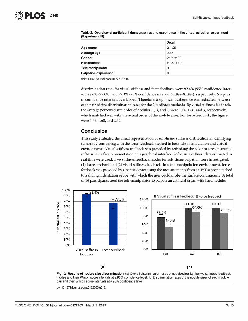
discrimination rates for visual stiffness and force feedback were 92.4% (95% confidence inter-
val: 88.6%–95.0%) and 77.3% (95% confidence interval: 71.9%–81.9%), respectively. No pairs
of confidence intervals overlapped. Therefore, a significant difference was indicated between
each pair of size discrimination rates for the 2 feedback methods. By visual stiffness feedback,
the average perceived size order of nodules A, B, and C were 1.14, 1.86, and 3, respectively,
which matched well with the actual order of the nodule sizes. For force feedback, the figures
were 1.55, 1.68, and 2.77.
Conclusion
This study evaluated the visual representation of soft-tissue stiffness distribution in identifying
tumors by comparing with the force feedback method in both tele-manipulation and virtual
environments. Visual stiffness feedback was provided by refreshing the color of a reconstructed
soft-tissue surface representation on a graphical interface. Soft-tissue stiffness data estimated in
real time were used. Two stiffness feedback modes for soft-tissue palpation were investigated:
(1) force feedback and (2) visual stiffness feedback. In a tele-manipulation environment, force
feedback was provided by a haptic device using the measurements from an F/T sensor attached
to a sliding indentation probe with which the user could probe the surface continuously. A total
of 10 participants used the tele-manipulator to palpate an artificial organ with hard nodules
Table 2. Overview of participant demographics and experience in the virtual palpation experiment
(Experiment III).
Detail
Age range 21–25
Average age 22.8
Gender ♀: 2; ♂: 20
Handedness R: 20; L: 2
Tele-manipulator 0
Palpation experience 0
doi:10.1371/journal.pone.0172703.t002
Fig 12. Results of nodule size discrimination. (a) Overall discrimination rates of nodule sizes by the two stiffness feedback
modes and their Wilson score intervals at a 95% confidence level; (b) Discrimination rates of the nodule sizes of each nodule
pair and their Wilson score intervals at a 95% confidence level.
doi:10.1371/journal.pone.0172703.g012
Soft-tissue stiffness feedback
PLOS ONE | DOI:10.1371/journal.pone.0172703 March 1, 2017 15 / 18

embedded. We also evaluated the performance of 22 human participants in distinguishing hard
nodules of different sizes. We used a virtual tissue model that was based on a mechanical char-
acterization of the artificial organ. Results indicated that subjects could localize nodules by both
feedback modes; no significant difference in nodule detection sensitivity and time elapsed for
hard nodule detection was found between these two feedback modes; compared with force feed-
back, visual stiffness feedback showed a significantly higher nodule size discrimination rates.
Stiffness feedback was confirmed to be an effective method. To conclude, our proposed visual
stiffness feedback method can be used for tissue palpation if the force feedback method is not
available.
Supporting information
S1 Table. Nodule detection results.
(DOCX)
S2 Table. Nodule size orders recognized by participants.
(DOCX)
Acknowledgments
The authors would like to thank participants in these experiments and anonymous reviewers
for their helpful comments.
Author Contributions
Conceptualization: ML.
Data curation: ML.
Formal analysis: ML JK HW JX.
Funding acquisition: GX ML.
Investigation: ML BH JK.
Methodology: ML VA.
Resources: KA GX.
Software: ML VA.
Supervision: KA.
Writing – original draft: ML.
Writing – review & editing: ML JK KA.
References1. Gwilliam JC, Pezzementi Z, Jantho E, Okamura AM, Hsiao S. Human vs. robotic tactile sensing: Detect-
ing lumps in soft tissue. In: 2010 IEEE Haptics Symposium [Internet]. Ieee; 2010. p. 21–8. Available
from: http://ieeexplore.ieee.org/lpdocs/epic03/wrapper.htm?arnumber=5444685
2. Wilson T, Torrey R. Open versus robotic-assisted radical prostatectomy: which is better? Curr Opin
Urol. 2011; 21(3):200–5. doi: 10.1097/MOU.0b013e32834493b3 PMID: 21427586
3. Ji W, Zhao Z, Dong J, Wang H, Lu F, Lu H. One-stage robotic-assisted laparoscopic cholecystectomy
and common bile duct exploration with primary closure in 5 patients. Surg Laparosc Endosc Percutane-
ous Tech [Internet]. 2011; 21(2):123–6. Available from: http://www.embase.com/search/results?
subaction=viewrecord&from=export&id=L361640602
Soft-tissue stiffness feedback
PLOS ONE | DOI:10.1371/journal.pone.0172703 March 1, 2017 16 / 18

4. Zehnder P, Gill IS. Cost-effectiveness of open versus laparoscopic versus robotic-assisted laparoscopic
cystectomy and urinary diversion. Curr Opin Urol [Internet]. 2011; 21(5):415–9. doi: 10.1097/MOU.
0b013e3283490582 PMID: 21814054
5. Currie ME, Romsa J, Fox SA, Vezina WC, Akincioglu C, Warrington JC, et al. Long-term angiographic
follow-up of robotic-assisted coronary artery revascularization. Ann Thorac Surg [Internet]. Elsevier
Inc.; 2012; 93(5):1426–31. Available from: http://www.ncbi.nlm.nih.gov/pubmed/22342992 doi: 10.
1016/j.athoracsur.2011.11.031 PMID: 22342992
6. Masroor S, Plambeck C, Dahnert M. Complex repair of a Barlow’s valve using the Da Vinci robotic surgi-
cal system. J Heart Valve Dis [Internet]. 2010; 19(5):593–5. Available from: http://www.ncbi.nlm.nih.
gov/entrez/query.fcgi?cmd=Retrieve&db=PubMed&dopt=Citation&list_uids=21053737 PMID:
21053737
7. Miller AP, Peine WJ, Son JS, Hammoud MDZT. Tactile imaging system for localizing Lung nodules dur-
ing video assisted thoracoscopic surgery. In: Proceedings 2007 IEEE International Conference on
Robotics and Automation [Internet]. Ieee; 2007. p. 2996–3001. Available from: http://ieeexplore.ieee.
org/lpdocs/epic03/wrapper.htm?arnumber=4209545
8. Meli L, Member S, Pacchierotti C, Member S. Sensory subtraction in robot-assisted surgery: fingertip
skin deformation feedback to ensure safety and improve transparency in bimanual haptic interaction.
2014; 61(4):1318–27. doi: 10.1109/TBME.2014.2303052 PMID: 24658255
9. Okamura AM. Haptic feedback in robot-assisted minimally invasive surgery. Curr Opin Urol [Internet].
NIH Public Access; 2009 [cited 2011 Dec 6]; 19(1):102. Available from: http://www.ncbi.nlm.nih.gov/
pmc/articles/PMC2701448/ doi: 10.1097/MOU.0b013e32831a478c PMID: 19057225
10. Prattichizzo D, Pacchierotti C, Member S, Rosati G. Cutaneous Force Feedback as a Sensory Subtrac-
tion Technique in Haptics. 2012; 5(4):289–300.
11. Mahvash M, Gwilliam J, Agarwal R, Vagvolgyi B, Su L-M, Yuh DD, et al. Force-feedback surgical teleo-
perator: controller design and palpation experiments. In: 2008 Symposium on Haptic Interfaces for Vir-
tual Environment and Teleoperator Systems [Internet]. Ieee; 2008. p. 465–71. Available from: http://
ieeexplore.ieee.org/lpdocs/epic03/wrapper.htm?arnumber=4479994
12. Liu H, Noonan DP, Challacombe BJ, Dasgupta P, Seneviratne LD, Althoefer K. Rolling mechanical
imaging for tissue abnormality localization during minimally invasive surgery. IEEE Trans Biomed Eng
[Internet]. 2010 Feb; 57(2):404–14. Available from: http://www.ncbi.nlm.nih.gov/pubmed/19789104 doi:
10.1109/TBME.2009.2032164 PMID: 19789104
13. Yamamoto T, Abolhassani N. Augmented reality and haptic interfaces for robot-assisted surgery. Int J
Med Robot Comput Assist Surg [Internet]. 2012 [cited 2013 Jul 25]; 8(November 2011):45–56.
14. Gwilliam JC, Mahvash M, Vagvolgyi B, Vacharat A, Yuh DD, Okamura AM. Effects of haptic and graphi-
cal force feedback on teleoperated palpation. 2009 IEEE Int Conf Robot Autom [Internet]. Ieee; 2009
May;677–82. Available from: http://ieeexplore.ieee.org/lpdocs/epic03/wrapper.htm?arnumber=
5152705
15. Khaled W, Reichling S, Bruhns OT, Boese H, Baumann M, Monkman G, et al. Palpation imaging using
a haptic system for virtual reality applications in medicine. Stud Health Technol Inform [Internet]. 2004
Jan; 98:147–53. Available from: http://www.ncbi.nlm.nih.gov/pubmed/15544261 PMID: 15544261
16. Li M, Faragasso A, Konstantinova J, Aminzadeh V, Seneviratne LD, Dasgupta P, et al. A Novel Tumor
Localization Method using Haptic Palpation Based on Soft Tissue Probing Data. In: 2014 IEEE Interna-
tional Conference on Robotics and Automation (ICRA). 2014. p. 4188–93.
17. Liu H, Li J, Poon Q, Seneviratne LD, Althoefer K. Miniaturized force-indentation depth sensor for tissue
abnormality identification during laparoscopic surgery. In: International Conference on Robotics and
Automation (ICRA) [Internet]. 2010 [cited 2013 Jan 22]. p. 3654–9. Available from: http://ieeexplore.
ieee.org/xpls/abs_all.jsp?arnumber=5509440
18. Noonan DP, Liu H, Zweiri YH, Althoefer KA, Seneviratne LD. A dual-function wheeled probe for tissue
viscoelastic property identification during minimally invasive surgery. In: IEEE International Conference
on Robotics and Automation [Internet]. Institute of Electrical and Electronics Engineers Inc.; 2007.
p. 2629–34.
19. Mahvash M, Okamura AM. Enhancing Transparency of a Position-Exchange Teleoperator. In: Second
Joint EuroHaptics Conference and Symposium on Haptic Interfaces for Virtual Environment and Teleo-
perator Systems (WHC’07) [Internet]. Ieee; 2007. p. 470–5. Available from: http://ieeexplore.ieee.org/
lpdocs/epic03/wrapper.htm?arnumber=4145219
20. Zirjakova J. Prostate post-surgical 3D imaging and data analysis. King’s College London; 2011.
21. Liu H, Li J, Song X, Seneviratne LD, Althoefer K. Rolling indentation probe for tissue abnormality identifi-
cation during minimally invasive surgery. IEEE Trans Robot [Internet]. IEEE; 2011; 27(3):450–60. Avail-
able from: http://ieeexplore.ieee.org/lpdocs/epic03/wrapper.htm?arnumber=5746537
Soft-tissue stiffness feedback
PLOS ONE | DOI:10.1371/journal.pone.0172703 March 1, 2017 17 / 18

22. Lee EH, Radok JRM. The contact problem for viscoelastic bodies. J Appl Mech [Internet]. 1960; 27
(65):438–44. Available from: http://link.aip.org/link/?JAMCAV/33/395/1
23. Li M, Konstantinova J, Secco EL, Jiang A, Liu H, Nanayakkara T, et al. Using visual cues to enhance
haptic feedback for palpation on virtual model of soft tissue. Med Biol Eng Comput. 2015; 53(11):1177–
86. doi: 10.1007/s11517-015-1309-4 PMID: 26018755
24. Woodward W a, Strom E a, Tucker SL, McNeese MD, Perkins GH, Schechter NR, et al. Changes in the
2003 American Joint Committee on Cancer staging for breast cancer dramatically affect stage-specific
survival. J Clin Oncol Off J Am Soc Clin Oncol [Internet]. 2003 Sep 1 [cited 2013 Aug 22]; 21(17):3244–
8. Available from: http://www.ncbi.nlm.nih.gov/pubmed/12947058
25. Wellman P, Howe R. Breast tissue stiffness in compression is correlated to histological diagnosis [Inter-
net]. Harvard BioRobotics . . .. 1999 [cited 2013 Aug 22]. Available from: https://biorobotics.harvard.edu/
pubs/1999/mechprops.pdf
26. Ottensmeyer MP. In vivo measurement of solid organ visco-elastic properties. Stud Health Technol
Inform [Internet]. 2002; 85:328–33. Available from: http://www.scopus.com/scopus/inward/record.url?
eid=2-s2.0-0005702074&partnerID=40&rel=R8.0.0 PMID: 15458110
27. Hoyt K, Castaneda B, Zhang M, Nigwekar P, di Sant’Agnese PA, Joseph J V, et al. Tissue elasticity
properties as biomarkers for prostate cancer. Cancer Biomark. 2008; 4(4–5):213–25. PMID: 18957712
28. Altman D, Bland J. Diagnostic test 1: Sensitivity and specificity. BMJ. 1994; 308:1552. PMID: 8019315
29. Wilson EB. Probable inference, the law of succession, and statistical inference. J Am Stat Assoc. 1927;
22:209–12.
30. Wallis S. Binomial confidence intervals and contingency tests: mathematical fundamentals and the
evaluation of alternative methods. J Quant Linguist [Internet]. 2013 Aug; 20(3):178–208.
31. Wilcoxon F. Individual comparisons of grouped data by ranking methods. J Econ Entomol. 1946;
39:269. PMID: 20983181
32. Yau C. R tutorial eBook [Internet]. r-tutor.com. 2009. Available from: http://www.r-tutor.com/
elementary-statistics/non-parametric-methods/wilcoxon-signed-rank-test
33. Conover WJ. Practical nonparametric statistics. 2nd Editio. John Wiley & Sons; 1980. 225–226 p.
34. Everitt BS. The cambridge dictionary of statistics. 2003.
35. Zhou M, Perreault J, Schwaitzberg SD, Cao CGL. Effects of experience on force perception threshold
in minimally invasive surgery. Surg Endosc [Internet]. 2008; 22(2):510–5. Available from: http://www.
ncbi.nlm.nih.gov/pubmed/18649102 doi: 10.1007/s00464-007-9499-y PMID: 17704870
36. Wagner CR, Howe RD. Force Feedback Benefit Depends on Experience in Multiple Degree of Freedom
Robotic Surgery Task [Internet]. IEEE Transactions on Robotics. 2007. p. 1235–40. Available from:
http://ieeexplore.ieee.org/xpl/freeabs_all.jsp?arnumber=4359258.
Soft-tissue stiffness feedback
PLOS ONE | DOI:10.1371/journal.pone.0172703 March 1, 2017 18 / 18