evaluation of the cytotoxicity activity of gypsophila
TRANSCRIPT
*Corresponding author: [email protected]
© 2020. Open access. This article is distributed under the terms of the Creative Commons Attribution 4.0
International License (https://creativecommons.org/licenses/by-nc/4.0/)
Research Journal of Pharmacognosy (RJP) 7(4), 2020: 75-82
Received: 17 June 2020
Accepted: 14 Sep 2020
Published online: 16 Sep 2020 DOI: 10.22127/rjp.2020.234735.1606 Original article
Evaluation of the Cytotoxicity Activity of Gypsophila ruscifolia by Bioassay-
Guided Fractionation
Marzie Kamali1
, Mahmoud Mosaddegh1,2*
, Mohammad-Reza Delnavazi3,
Roksana
Shahrestani2, Maryam Malek Mohammadi
4, Maryam Hamzeloo-Moghadam
5
1Department of Pharmacognosy, Faculty of Pharmacy, Shahid Beheshti University of Medical Sciences,
Tehran, Iran. 2Traditional Medicine and Materia Medica Research Center, Shahid Beheshti University of Medical
Sciences, Tehran, Iran. 3Department of Pharmacognosy, Faculty of Pharmacy, Tehran University of Medical Sciences, Tehran,
Iran. 4Department of Plant Sciences, School of Biology, College of Sciences, University of Tehran, Tehran,
Iran. 5Department of Traditional Pharmacy and Traditional Medicine and Materia Medica Research Center,
School of Traditional Medicine, Shahid Beheshti University of Medical Sciences, Tehran, Iran.
Abstract Background and objectives: The second cause of death in the world and the third cause in Iran is
cancer which requires special attention for treatment. The previous reports of Gypsophila genus show
significant toxic effects on different cancer cell lines. In this study, bioassay-guided fractionation was
applied to determine the cytotoxic activity of root and aerial parts extracts and fractions of Gypsophila
ruscifolia Methods: n-Hexane, chloroform, and methanol: H2O (8:2) extracts of root and aerial parts
were prepared. Inhibition of cell growth determined by MTT assay in MCF-7, HT-29, A-549, and
AGO-1522 cell lines. The most effective extract was fractionated by column chromatography. The
cytotoxic effect of fractions was evaluated by MTT assay and apoptotic property of the cytotoxic
fraction was determined by annexin V/PI assay in MCF-7 cell line. Results: The chloroform root
extract of G. ruscifolia showed cytotoxicity in MCF-7 cells with IC50 value of 111.6 ±11.78 μg/mL.
MTT assay of five of fractions demonstrated that F3 and F4 with IC50 values of 73.09 ±14.22 and
67.98 ±15.31 μg/mL on MCF-7 cell line, respectively showed cytotoxic effects. Also, F4
demonstrated apoptotic potential in MCF-7 cell line. Conclusion: Considering the results of
cytotoxicity and apoptosis studies, isolation and identification of responsible compounds in the
chloroform root extract of Gypsophila ruscifolia can be useful in cancer research studies.
Keywords: apoptosis; cell line; cell survival; Gypsophila ruscifolia; medicine plant
Citation: Kamali M, Mosaddegh M, Delnavazi MR, Shahrestani R, Malek Mohammadi M, Hamzeloo-Moghadam
M. Evaluation of the cytotoxicity activity of Gypsophila ruscifolia by bioassay-guided fractionation. Res J
Pharmacogn. 2020; 7(4): 75-82.
Introduction The uncontrolled cell division that is influenced
by environmental factors and genetic disorders
result in cancer [1]. There are many types of
cancer; among them, cancer of lung, stomach,
liver, rectum, and breast have the highest
mortality rates. According to the World Health
Organization reports, 70% of cancer deaths
worldwide are in Africa, Asia, Central, and South

Kamali M. et al.
76 Res J Pharmacogn 7(4): 75-82
America [2]. In Iran, due to the changing of
people’s lifestyles, cancer is a third cause of
death. Based on 2017 reports, age-standardized
mortality rate was 81.9 per 100000 that will
increase each year [3]. After isolation of
morphine with painkilling and hypnotic
properties from Papaver somniferum L in 1803,
natural compounds were widely used for
different diseases [4]. About 50% of anticancer
drugs are comprised of isolated compounds of
natural origin and semisynthetic or synthetic
compounds with templates of natural products.
Taxol, camptothecin, Vinca alkaloids, and
podophyllotoxins are effective anticancer
examples of natural origin [2]. The
Caryophyllaceae family with 85 genera and 2,630
species exhibits various biological activities. A
large number of plants in this family have shown
anticancer properties [5]. Gypsophila is one of
the largest genera of this family with around 150
species, 35 of which are found in Iran [6].
Triterpenoid saponins are characteristic
compounds in the roots of Gypsophila [7];
besides, sterols, flavonoids, phenolic acids,
oligosaccharides, cyclic peptides, fatty acids, and
alkaloids have been also reported from this
genus. Studies have demonstrated
hypocholesterolemic, anti-inflammatory,
antiviral, alpha-glucosidase inhibitory,
immunomodulatory, spermicidal and cytotoxic
activities as well as being used as adjuvants in
vaccines [8-10]. Considering the availability and
distribution as well as the previous cytotoxic
reports about the genus, Gypsophila ruscifolia
Boiss. a perennial herbaceous plant growing in
East and West Azarbaijan provinces of Iran [11],
was selected for cytotoxicity and induction
apoptosis evaluations.
Material and Methods Ethical considerations
The Ethics Committee of Shahid Beheshti
University of Medical Sciences approved this
research with the code of
IR.SBMU.REC.1398.037 on 2019-07-24.
Chemicals
Dulbecco’s Modified Eagle Medium (DMEM),
Fetal Bovine Serum (FBS), (Gibco, New
Zealand), RPMI 1640 medium, Penicillin-
Streptomycin, MTT (3- (4,5-dimethylthiazol-2-
yl) -2,5-diphenyltetrazolium bromide),
Phosphate-buffered saline (PBS), 5-Fluorouracil
and DMSO (Dimethyl Sulfoxide) (Merck,
Germany), (Sigma, USA), methanol, hexane,
chloroform, ethyl acetate (Dr.Mojallali, Iran)
were used in this study. Invitrogen Bioscience
annexin V Apoptosis Detection Kit FITC was
used to flow cytometry.
Plant material
Gypsophila ruscifolia was collected from Yam
village (East Azarbaijan province, Iran) in June
2018 and identified by Dr. Maryam
Malekmohammadi, botanist at the Traditional
Medicine and Materia Medica Research Center
(TMRC), Shahid Beheshti University of Medical
sciences, Tehran, Iran. A voucher the specimen
was deposited at the Herbarium of TMRC
(TMRC-4479).
Extraction
Two hundred g of the air-dried aerial parts and
roots of G. ruscifolia were separately powdered
and extracted by maceration method with hexane,
chloroform, and methanol: water (8:2). The
extracts were concentrated and dried.
Fractionation The most effective extract was subjected to column
chromatography (230-400 mesh, 20×700 mm) with
a gradient system of hexane-ethyl acetate (100:0
→0:100). The fractions evaluated by thin-layer
chromatography (TLC) and similar fractions were
combined. Finally, five fractions obtained.
Cell lines
MCF-7 (human breast adenocarcinoma), HT-29
(human colon adenocarcinoma), A-549 (non-
small cell lung carcinoma), and AGO-1522
(human dermal fibroblast) were provided from
National Cell Bank of Iran (Pasteur Institute,
Tehran, Iran).
Cytotoxicity assay One of the most common assays to monitor viable
cells in multi-well plates is the MTT assay [12].
MTT assay was performed according to the method
by Mosmann [13]. In brief, the cells in the
exponentially growing phase were seeded into 96
well microplates (5 -9) ×103 cells/well. After 24 h,
they were exposed to variant concentrations (12.5,
25, 50, 100, 200, 300 μg/mL) of extracts and
fractions. Finally, MTT solution (0.5 mg/mL in
PBS) was added to each well and incubated for 4 h.
The formed formazan crystals were dissolved in
200 µL of DMSO and the absorbance was

Evaluation of the cytotoxicity activity of Gypsophila ruscifolia by bioassay-guided fractionation
77
measured at 570 nm by a microplate reader. 5-
Fluorouracil was applied as the positive control.
Annexin V/PI assay Flow cytometry is a useful tool for the concurrent
evaluation of necrosis and apoptosis in population
of cells [14]. MCF-7 cells were cultured into the
six-well plate 1×10⁵ cells/well. The cells were
treated with fraction 4, the most cytotoxic fraction
of chloroform root extract of G.rusifolia, with the
concentrations of 25, 50 and 100 μg/mL. After 48h,
cells were washed with tissue culture medium and 5
μg/mL of the annexin V conjugate was added. After
45 min at room temperature, again cells were
washed and 1 μg/mL of PI was added [15]. DMSO
1% was used as the negative control.
Statistical analyses
Cytotoxic activity of extracts and fractions were
evaluated by the concentration-response curve
(Graph Pad Prism 7.0) and expressed as IC50
value ± SD. The significant difference between
each group and the negative control group
(DMSO) was analyzed by Student’s t-test and ANOVA; p value<0.05.
Results and Discussion MTT assay was applied to measure the cytotoxic
activity of n-hexane, chloroform, methanol:water
8:2 extracts of Gypsophila ruscifolia against
MCF-7, HT-29, A-549, and AGO-1522 cell lines.
The n-hexane extract did not dissolve in DMSO;
even with adding 10% PG. The methanol extracts
of the root and aerial part did not show toxic
effects at tested concentration. There was
significant difference between cytotoxic effects
of the chloroform root and aerial extract
(p<0.05). The cytotoxic behavior of the
chloroform root extract against MCF-7, HT-29,
A-549, and AGO-1522 cell lines has been
exhibited in figure 1. Treatment with chloroform
root extract (50, 100, 200 and 300 μg/mL)
compared to the control, showed significantly
decrease in cell viability for the MCF-7 cell line
(p< 0.0001). However, cell viability showed no
significant change at concentrations below 50
μg/mL. For A-549 cell line at 25 μg/mL and
higher concentrations, the extract demonstrated
toxicity (p<0.05). As shown in figure 1,
chloroform root extract at 100, 200 and 300
μg/mL concentrations on HT-29 a human colon
cell line and AGO-1522 as a normal cell line
displayed significant decrease in cell viability.
The IC50 value of the chloroform root extract was
calculated as 111.6 ± 11.78, 179.2 ± 9.59, 141.7
± 11.02, and 165.4 ± 14.29 for MCF-7, HT-29,
A-549, and AGO-1522 cell lines, respectively.
Regarding more considerable cytotoxic effects of
this extract on three human cell lines, the
chloroform extract of root was selected to be
fractionated. The IC50 of the extracts and positive
control have been demonstrated in table 1.
MC
F-7
HT
-29
A-5
4 9
AG
O-1
5 2 2
0
5 0
1 0 0
C e ll l in e s
Via
bil
ity
(%)
c o n tro l
1 2 .5 g /m L
2 5 g /m L
5 0 g /m L
1 0 0 g /m L
2 0 0 g /m L
3 0 0 g /m L
****
******
****
***
**
***
***
μ μ
μ μ
μ
μ
Figure 1. Concentration-dependent manner of chloroform root extract cytotoxicity on MCF-7 (human breast adenocarcinoma),
HT-29 (human colon adenocarcinoma), A-549 (non-small cell lung carcinoma), and AGO-1522 (human dermal fibroblast) cell
lines. Experiments were performed in triplicate. Significant difference between each concentration and control group (*p < 0.05,
**p<0.01, ***p<0.001, ****p< 0.0001).

Kamali M. et al.
78 Res J Pharmacogn 7(4): 75-82
Table 1. Cytotoxicity of extracts and chloroform fractions of Gypsophila ruscifolia extract
Samples IC50 (μg/mL)
MCF-7 HT-29 A-549 AGO-1522
Aerial parts: n-hexane extract * * * *
Roots: n-hexane extract * * * *
Aerial parts: chloroform extract 194.3 ± 11.32 222.2 ± 10.27 ** 195.7 ± 18.90
Roots: chloroform extract 111.6 ± 11.41 179.2 ± 9.59 141.7± 11.02 165.4 ± 14.29
Aerial parts: 80%methanolic extract ** ** ** **
Roots: 80%methanolic extract ** ** ** **
Fraction 1 **
** ** **
Fraction 2 ** ** ** **
Fraction 3 73.09 ± 14.22 ** ** 103.7 ± 10.71
Fraction 4 67.98 ± 15.31 ** 184.4 ± 7.80 80.3 ± 11.44
Fraction 5 195.3 ± 8.25 ** 108.3 ± 14.7 87.56 ± 12.32
Positive control 5-Fu 3.1 ± 1.02 3.99 ± 1.01 3.1 ± 1.03 14.2 ± 1.02
*in the investigated concentrations, the extracts did dissolve in DMSO.
**in the investigated concentrations, the IC50 could not be determined.
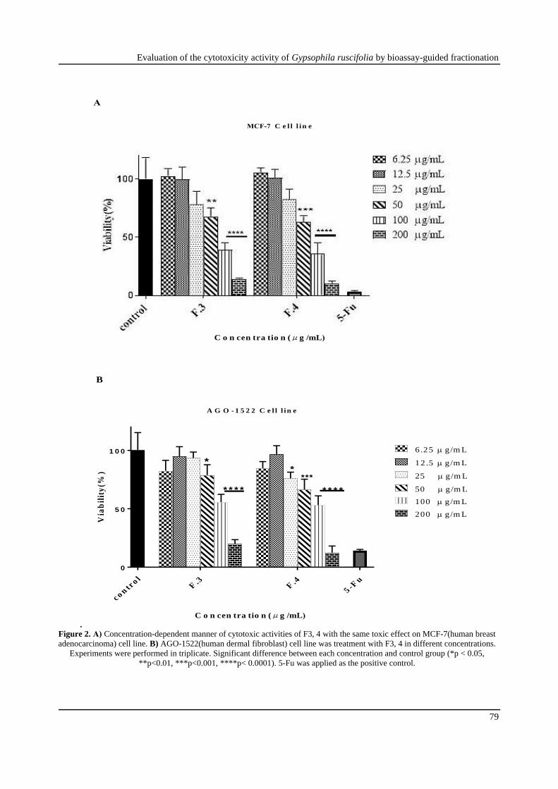
Further investigation was applied to detect the
cytotoxicity compounds of the chloroform roots
extract. So, five fractions of chloroform extract
(6.25, 12.5, 25, 50, 100, 200 μg/mL) were tested
again with MTT assay. IC50 values of F 1-5 in
table 1 showed that MCF-7 cell line was the most
susceptible cell line treated with with F3 and 4;
(IC50 of 73.09 ± 14.22 and 67.98 ± 15.31 μg/mL,
respectively). F5 exhibited significant inhibition
of cell growth on the A-549 cell line; 108.3 ±
14.7 μg/mL (p value<0.001). None of the
fractions displayed effects on HT-29 cell line and
there was no significant difference between F 3,
4, 5 on normal cell (AGO-1522). Based on NCI
protocols [16], F3, 4, 5 of G. ruscifolia, fractions
obtained from column chromatography,
displayed moderate cytotoxicity on MCF-7 and
A-549 cell lines. As shown in figure 2, F3 and
F4 at concentrations 50, 100 and 200μg/mL
exhibited inhibition in cell growth in MCF-7 cell
line; however, AGO-1522, which is a normal
cell, treated with F3 at 50; 100; 200 μg/mL and
F4 at 25; 50; 100; 200 μg/mL indicated
significant decline in cell viability compared to
control.
To detect the underlying basis for the toxic effect
of the most effective fraction in breast cancer
cell, the percentage of cell apoptosis occurred by
fraction 4 was analyzed in MCF-7 cells. The
results reveal that F4 in concentrations of 25, 50
and 100 μg/mL against MCF-7 cell line showed
increase induced apoptosis in a concentration-
dependent manner compared with negative
control after 48 h. As shown in figure 3,
percentage of cell apoptosis of F4 in
concentrations of 25, 50, 100 μg/mL was
reported 20.94%, 28.87% and 52.44%,
respectively. Also, there was significant
difference between F4 and the control group
(p<0.05).
Earlier evaluation about the cytotoxic effects of
G.ruscifolia in TMRC supported the result of this
study, as the aerial parts of the chloroform extract
of this species demonstrated cytotoxic effect
against MCF-7 cell line, whereas methanolic
extract was not toxic [17,18]. Gypsophila is
mainly known as a triterpenoid saponins source
with considerable cytotoxic effects. These group
of compounds are mostly present in polar the
fractions such as n- butanol or methanol fractions
[7,19-21]. However, bioassay-guided
fractionation of G. oldhamiana resulted in
isolation and identification of triterpenoids from
the EtOAc fraction that showed antiangiogenic
activities and notable cytotoxicity against lung
cancer (H460 cell line) [8]. In a previous
research, inhibition of cell growth by the
methanol extract of several species of Gypsophila
against A-549, HT-29, HepG-2, MCF-7, and
MDBK cell lines revealed that up to
concentration 100 μg/mL they did not effect on
MCF-7, A-549, and HT-29 cell lines, only G.
bicolar showed cytotoxic effect on MDBK cell
line with IC50= 7.82 μg/mL [22]. On the other
hand, IC50 values of methanol extract of G.
spheaerocephala against A-549, HT-29 and
MCF-7 cell lines were obtained to be than 700
μg/mL [23]. The result of the present study
revealed that chloroform root extract and
fractions demonstrated toxic effect against MCF-
7 cell lines. Mixtures of compounds in botanical
extracts can enhance the activity of each other
(synergism) or decrease the effects (antagonism)
[24].
Evaluation of the cytotoxicity activity of Gypsophila ruscifolia by bioassay-guided fractionation
79
.
c o ntr
o lF
.3F
.4
5 -Fu
0
5 0
1 0 0
A G O - 1 5 2 2 C e l l l in e
C o n cen tra tio n (μg /mL)
Via
bil
ity
(%)
6 .25 g/m L
12 .5 g/m L
25 g/m L
50 g/m L
100 g/m L
200 g/m L
*
* * * *
*
***
* * * *
B
Figure 2. A) Concentration-dependent manner of cytotoxic activities of F3, 4 with the same toxic effect on MCF-7(human breast
adenocarcinoma) cell line. B) AGO-1522(human dermal fibroblast) cell line was treatment with F3, 4 in different concentrations.
Experiments were performed in triplicate. Significant difference between each concentration and control group (*p < 0.05,
**p<0.01, ***p<0.001, ****p< 0.0001). 5-Fu was applied as the positive control.
C o n cen tra tio n (μg /mL)
MCF-7 C e l l l in e
A

Kamali M. et al.
80 Res J Pharmacogn 7(4): 75-82
Control 25 μg/mL 50 μg/mL 100 μg/mL
c o ntr
o l 2 5
5 01 0 0
0
2 0
4 0
6 0
To
tal
Ap
op
tosi
s(%
)
* * *
* *
* * *
B
Figure 3. A) AnnexinV/PI assay with F4 at 25, 50, 100 µg/mL concentration on MCF-7 (human breast adenocarcinoma) cell
line. DMSO was used as the negative control. B) Comparison of induced apoptosis of each concentration with negative control.
Experiments were performed in triplicate. Significant difference between each concentration and control group: (**p<0.01,
***p<0.001).
Gypsophila trichotoma methanol extract at 100
μg/mL concentration was non-active on NR8383
macrophage [25], however new triterpenoid
saponin isolated from the species showed
cytotoxicity activity below 100 μg/mL of on
leukaemic (NB-4, EOL-1) cell lines [19]. As
cytotoxic compound can operate various
pathways of cell death, it is essential to find how
toxic compound act to determine the side effects
of this [14]. Analysis of flow cytometry
displayed that fraction 4 might induce apoptosis
in MCF-7 cells. Previous reports of G.
oldhamiana root extract showed apoptotic effects
in human hepatoma compared to normal human
hepatic cell [26] and G. trichotoma root extract
induced apoptosis in a macrophage cell line [27].
Considering the inhibitory cell growth results of
the chloroform root extract and its fractions on
cancer cells, additional phytochemistry
investigations should be applied to identify
responsible compounds. Also, the evaluation of
the molecular mechanisms of anticancer effects is
needed.
Acknowledgments This research was supported financially by
School of Traditional Medicine, Shahid Beheshti
University of Medical Sciences (grant No. 228).
Authors would like to thank Ms. Ara and Ms.
Keramatian, the technical assistance of laboratory
experts in phytochemistry.
Author contributions Mahmoud Mosaddegh and Maryam Hamzeloo-
Moghadam supervised the study; Mohammad
Reza Delnavazi desigened phytochemistry part;
Maryam Malekmohammadi collected and
identified the specimen; Roksana Shahrestani
A
C o n cen tra tio n (μg /mL)

Evaluation of the cytotoxicity activity of Gypsophila ruscifolia by bioassay-guided fractionation
81
was involved in cell culture experiments;
Marzieh Kamali preformed practical experiments;
Maryam Hamzeloo-Moghadam and Marzieh
Kamali analyzed the data and prepared the
manuscript.
Declaration of interest The authors declare that there is no conflict of
interest. The authors alone are responsible for the
accuracy and integrity of the paper content.
References [1] Parsa N. Molecular and cellular basis of
human cancer. J Cell Tiss. 2012; 2(4): 365-
376.
[2] Moraes DFC, De Mesquita LSS, Do Amaral
FMM, De Sousa Ribeiro MN. Anticancer
drugs from plants. In: Malik S, Ed.
Biotechnology and production of anti-cancer
compounds. Cham: Springer, 2017.
[3] Mohamadian M, Salehiniya H,
Mohammadian Hafshejani A. Some facts on
incidence and mortality of cancer in Iran.
Iran J Public Health. 2017; 46(10): 1446-
1447.
[4] Bernardini S, Tiezzi A, Laghezza Masci V,
Ovidi E. Natural products for human health:
an historical overview of the drug discovery
approaches. Nat Prod Res. 32(16): 1926-
1950.
[5] Chandra S, Rawat DS. Medicinal plants of
the family Caryophyllaceae: a review of
ethno-medicinal uses and pharmacological
properties. Integr Med Res. 2015; 4(3): 123-
131.
[6] Amini E, Zarre S, Assadi M. Seed micro-
morphology and its systematic significance
in Gypsophila (Caryophyllaceae) and allied
genera. Nord J Bot. 2011; 29(6): 660-669.
[7] Pertuit D, Avunduk S, Mitaine-Offer AC,
Miyamoto T, Tanaka C, Paululat T,
Delemasure S, Dutartre P, Lacaille-Dubois
MA. Triterpenoid saponins from the roots of
two Gypsophila species. Phytochemistry.
2014; 102: 182-188.
[8] Xie LX, Zhang HC, Wang HY, Wang Y,
Wang FL, Sun JY. Two new triterpenoids
from Gypsophila oldhamiana. Nat Prod Res.
2016; 30(9): 1068-1074.
[9] Emirdag-Ozturk S, Karayildirim T, Capci-
Karagoz A, Alankus-Caliskan O, Ozmen A,
Poyrazoglu-Coban E. Synthesis,
antimicrobial and cytotoxic activities, and
structure-activity relationships of gypsogenin
derivatives against human cancer cells. Eur J
Med Chem. 2014; 82: 565-573.
[10] Gevrenova R, Voutquenne-Nazabadioko L,
Harakat D, Prost E, Henry M. Complete 1H-
and 13C NMR assignments of saponins from
roots of Gypsophila trichotoma Wend. Magn
Reson Chem. 2006; 44(7): 686-691.
[11] Rechinger KH. Caryophyllaceae. In:
Rechinger KH, Ed. Flora des Iranische
Hochlandes und der umrahmenden. Gebirge:
Lieferung, 1988.
[12] Riss TL, Moravec RA, Niles AL, Duellman
S, Benink HA, Worzella TJ, Minor L. Cell
viability assays. In: Markossian S,
Sittampalam GS, Grossman A, Eds. Assay
Guidance Manual. Bethesda: Eli Lilly &
Company and the National Center for
Advancing Translational Sciences, 2004.
[13] Mosmann T. Rapid colorimetric assay for
cellular growth and survival: application to
proliferation and cytotoxicity assays. J
Immunol Methods. 1983; 65(1-2): 55-63.
[14] Cummings BS, Schnellmann RG.
Measurement of cell death in mammalian
cells. Curr Protoc Pharmacol. 2012; 56(1):
1-24.
[15] Mattes MJ. Apoptosis assays with
lymphoma cell lines: problems and pitfalls.
Br J Cancer. 2007; 96(6): 928-936.
[16] Geran RI, Greenberg NH, MacDonald MM,
Schumaker AM, Abbott BJ. Protocols for
screening chemical agents and natural
products against animal tumors and other
biological systems. Cancer Chemother Rep 3.
1972; 3(2): 59-61.
[17] Rad AS, Esmaeili S, Motamed SM,
Hamzeloo-Moghadam M. Cytotoxicity of
two Gypsophila species to human breast
adenocarcinoma (MCF-7). Int Pharm Acta.
2018; 1(1): 82.
[18] Irani M, Shahrestani R, Bahmani B,
Esmaeili S. Cytotoxic activity of plants from
East Azarbaijan province, Iran. Res J
Pharmacogn. 2017; 4(S): 1.
[19] Yotova M, Krasteva I, Jenett-Siems K,
Zdraveva P, Nikolov S. Triterpenoids in
Gypsophila trichotoma Wend. Phytochem
Lett. 2012; 5(4): 752-755.
[20] Arslan I, Celik A, Chol JH. A cytotoxic
triterpenoid saponin from under-ground parts
of Gypsophila pilulifera Boiss.& Heldr.
Fitoterapia. 2012; 83(4): 699-703.

Kamali M. et al.
82 Res J Pharmacogn 7(4): 75-82
[21] Jia ZKK, Sahu NP, Nikido T. Triterpenoid saponins from Caryophyllaceae family. In: Atta-ur-Rahman, Ed. Studies in natural products chemistry. Amsterdam: Elsevier Science B.V, 2002.
[22] Naghibi F, Irani M, Hassanpour A, Pirani A, Hamzeloo-Moghadam M. Cytotoxic effects of selective species of Caryophyllaceae in Iran. Res J Pharmacogn. 2014; 1(2): 29-32.
[23] Altay ADS, Korkmaz M, Cankaya M, Koksal E. In vitro evaluation of antioxidant and anti-proliferative activities of Gypsophila sphaerocephala (Caryophyllaceae) extracts together with their phenolic profile. J Food Meas Charact. 2018; 12(4): 2936-2945.
[24] Caesar LK, Cech NB. Synergy and antagonism in natural product extracts: when 1+ 1 does not equal 2. Nat Prod Rep. 2019; 36(6): 869-888.
[25] Voutquenne-Nazabadioko L, Gevrenova R, Borie N, Harakat D, Sayagh C, Weng
A, Thakur M, Zaharieva M, Henry
M. Triterpenoid saponins from the roots
of Gypsophila trichotoma
Wender. Phytochemistry. 2013; 90: 114-127.
[26] Zhang W, Luo JG, Zhang C, Kong LY.
Different apoptotic effects of triterpenoid
saponin-rich Gypsophila oldhamiana root
extract on human hepatoma SMMC-7721
and normal human hepatic L02 cells. Biol
Pharm Bull. 2013; 36(7): 1080-1087.
[27] Gevrenova R, Joubert O, Mandova T, Zaiou
M, Chapleur Y, Henry M. Cytotoxic effects
of four Caryophyllaceae species extracts on
macrophage cell lines. Pharm Biol. 2014;
52(7): 919-925.
Abbreviations MCF-7: human breast adenocarcinoma; HT-29:
human colon adenocarcinoma; A-549: non-small
cell lung carcinoma; AGO-1522: human dermal
fibroblast; TLC: thin-layer chromatography; IC50:
50% inhibitory concentration; MTT: 3- (4,5-
dimethylthiazol-2-yl)-2,5-diphenyl tetrazolium
bromide; PG: propylene glycol; NCI: National
Cancer Institute