evaluation of the effects of photobiomodulation on ... · in two rat models of experimental...
TRANSCRIPT

ORIGINAL ARTICLE
Evaluation of the effects of photobiomodulation on vertebrasin two rat models of experimental osteoporosis
Mohammadjavad Fredoni1 & Mahdi Ghatrehsamani2 &
Mohammad-amin Abdollahifar3 & Sahar Bayat4 & Mohammad Bayat5
Received: 15 July 2016 /Accepted: 29 June 2017 /Published online: 19 July 2017# Springer-Verlag London Ltd. 2017
Abstract The aim of this study was to evaluate the ef-fects of photobiomodulation (PBM) on cancellous bonein rat models of ovariectomized induced osteoporosis(OVX-D) and glucocorticoid-induced osteoporosis(GIOP). The experiment comprised of nine groups. Agroup of healthy rats was used for baseline evaluations.The OVX-D rats were further divided into groups as fol-lows: control rats with osteoporosis, OVX-D rats thatreceived alendronate (1 mg/kg 60 days), OVX-D ratstreated with pulsed wave laser (890 nm, 80 Hz, 900 s,0.0061 W/cm2, 5.5 J/cm2, three times a week, 60 days),and OVX-D rats treated with alendronate + pulsed laser.Dexamethasone was administered to the remaining ratsthat were split into four groups: control, alendronate-treated rats, laser-treated rats, and GIOP rats treated withalendronate + laser. T12, L1, L2, and L3 vertebrae weresubjected to laser. Results of the current study demon-strated that OVX-D and GIOP significantly decreasedsome stereological parameters, and type 1 collagen geneexpression compared to the healthy group. There was asignificant increase in osteoclast number in both OVX-D
and glucocorticoid administration compared to thehealthy group. However, the detrimental effect of theOVX-D procedure on bone was more serious than gluco-corticoid administration. Results showed that laser alonehad a detrimental effect on trabecular bone volume, andcortical bone volume in groups GIOP and OVX-D com-pared to those in the healthy group. Alendronate signifi-cantly improved total vertebral bone volume, trabecularbone volume, and cortical bone volume, in GIOP andOVX-D groups compared to the laser-treated groups.Furthermore, the alendronate + laser in OVX-D rats andGIOP rats produced significantly increased osteoblastnumber and type 1 collagen gene expression and causeda significant decrease in osteoclast number compared tothe controls.
Keywords Osteoporosis . Photobiomodulation . Low-levellaser therapy . Glucocorticoid-induced osteoporosis .
Ovariectomized induced osteoporosis .Vertebra . Stereology .
Real-time polymerase chain reaction . Rat
* Mohammad [email protected]; [email protected]
Mohammadjavad [email protected]
Mahdi [email protected]
Mohammad-amin [email protected]
Sahar [email protected]
1 Department of Anatomy, School of Medicine, Zanjan University ofMedical Sciences, Zanjan, Iran
2 Cellular and Molecular Biology Research Center, ShahrekordUniversity of Medical Sciences, Shahrekord, Iran
3 Department of Biology and Anatomical Sciences, School ofMedicine, Shahid Beheshti University of Medical Sciences,Tehran, Iran
4 No. 1 Farzanegan High School, Tehran, Iran5 Cellular and Molecular Biology Research Center, Department of
Anatomical Sciences and Biology, School of Medicine, ShahidBeheshti University of Medical Sciences, 19395/4719,Tehran 1985717443, Iran
Lasers Med Sci (2017) 32:1545–1560DOI 10.1007/s10103-017-2278-7

AbbreviationsOP OsteoporosisGC GlucocorticoidGIOP Glucocorticoid (GC)-induced OPECM Extracellular matrixTGF-β Transforming growth factor betaIGF-I Insulin-like growth factor-IBMPs Bone morphogenetic proteinsrhPTH Recombinant human parathyroid hormonePBM PhotobiomodulationPW Pulse waveALP Alkaline phosphataseLED Light-emitting diodeRANKL Receptor activator of nuclear factor kappa-B
ligandCW Continuous waveOPG OsteoprotegerinGaAlAs Gallium aluminum arsenideOVX-D Ovariectomized induced OPRT-PCR Real-time polymerase chain reactionH HealthyH&E Hematoxylin and eosinANOVA One-way analysis of varianceBMD Bone mineral densityDXA Dual-energy X-ray absorptiometryGTP Guanosine triphosphatePPARγ Peroxisome proliferator-activated receptor-γRUNX Runt-related transcription factor
Introduction
Bone tissue formation, maintenance, and repair depend onfinely tuned interaction of osteoclasts and osteoblasts.Bone tissue is in a constant process of remodeling thatinvolves regeneration of its matrix; osteoblasts have a rolein new bone matrix deposits, while osteoclasts are in-volved in degrading the old matrices. In a post-menopausal skeleton, regenerative bone formation in thearm ultimately declines, the process is known as primaryosteoporosis (OP) [1]. Glucocorticoid (GC)-induced OP(GIOP) is the most common cause of secondary OP [2].OP is a skeletal disorder distinguished by low bone massand micro-architectural deterioration of the bone tissuethat makes bones vulnerable and susceptible to fracture[3]. Estimates show that among adults aged 50 yearsand over in the USA (2010), 10.3% or 10.2 million peo-ple suffered from OP in some region of the body such asin the femoral neck or the lumbar spine [4]. OP can leadto fracture even in cases of mild trauma, so it is veryimportant to maintain and treat patients with osteoporosisin order to prevent fracture [5]. Osteoporotic fracture has
become a common condition but no satisfactory treatmenthas been developed to date [6].
There are many cytokines and growth factors that gov-ern bone cell activity during normal bone remodeling. Theextracellular matrix (ECM) releases substances such astransforming growth factor beta (TGF-β) and insulin-like growth factor-I (IGF-I) during bone resorption.These factors recruit and activate osteoblasts to begin col-lagen synthesis. Collagen type 1 is involved in skeletaldevelopment and has a regulatory effect on bone cells[7]. The process of osteoblast maturation is affected byactivity of various endocrine, paracrine, and autocrineprocesses. These factors include a number of bone mor-phogenetic proteins (BMPs) and IGF-I. The recombinanthuman BMP-2 is a medical administration used to facili-tate spinal fusion and is approved for OP therapy [8].
Prevention of OP and its complications is an essential so-cioeconomic priority [9].
Drug agents for the treatment of OP are defined as eitherantiresorptive (catabolic) or anabolic. Antiresorptive agentssuch as bisphosphonate work by reducing osteoclast activityand thus declining bone resorption. Teriparatide (recombinanthuman parathyroid hormone (rPTH)1–34) is the only anabolicagent currently available in the USA. PTH stimulates osteo-blast function and bone formation of vertebral fracture [10].Treatment with anabolic agents such as photobiomodulation(PBM) is a strong stimulant for bone turnover and seems to bea superior treatment compared with alendronate [11]. Someinvestigations have demonstrated that laser irradiation had apositive effect on osteoblasts in vitro. Ueda and Shimizu re-ported that pulsed wave (PW) laser significantly stimulatedcellular proliferation, bone nodule formation, alkaline phos-phatase (ALP) activity, and ALP gene expression, comparedwith a non-irradiation group [12, 13]. Sohn et al. report on aninvestigation on the impact of 635-nm irradiation dose from alight-emitting diode (LED) on osteoclastogenesis in vitro. Itwas reported that LED irradiation significantly inhibited re-ceptor activator of nuclear factor kappa-B ligand (RANKL)-mediated osteoclast differentiation from bone marrow-derivedmacrophages. Sohn et al. present LED irradiation/laser as analternative, conservative approach to OP management [14].Kiyosaki et al. reported the effects of continuous wave (CW)laser on osteoblasts via insulin-like growth factor I (IGF-I)signal transduction. The report suggests that laser escalatesin vitro mineralization by increasing IGF-I and BMP produc-tion [15]. Xu et al. reported on the effect of laser (650 nm,2 mW, 2.28 J/cm2, 6000 Hz) on messenger RNA (mRNA)expression of RANKL and osteoprotegerin (OPG) in ratcalvarial cells. Xu et al. suggested that laser may have pro-moted osteoblast proliferation and differentiation directly, andindirectly inhibited of osteoclast differentiation by downregu-lating the RANKL:OPG mRNA ratio in osteoblasts. Thus,other research has shown that laser was beneficial for treating
1546 Lasers Med Sci (2017) 32:1545–1560

bone diseases such as OP [16]. In vivo studies have suggestedthat both CW laser (632.8 nm, 10 mW, 382.2 J/cm2) and laser(890 nm, 80 Hz, 0.972 J/cm2) improved biomechanical prop-erties of tibia and vertebra compared to healthy bones in rats[17–19]. Consistent with other reports, it has been confirmedthat laser (830-nm gallium aluminum arsenide (GaAlAs) laserdiode, 5 and 15 J/cm2) not only made changes in thickness ofthe epiphyseal cartilage but also increased the number of thechondrocytes [20].
However, research has some controversy over the ben-efits of PBM for treatment of OP. Several studies havereported a positive effect of PBM on ovariectomized in-duced OP (OVX-D) rats, and orthodontic tooth movementon post-menopausal women [21–24], whereas others havereported no improvement in OVX-D and GIOP OPmodels subjected to PBM [25–30]. To the best of ourknowledge, no study has yet examined the effects of laseron gene expression and stereological analyses of histolog-ical parameters in the vertebrae of OVX-D and GIOP rats.Paraclinical information such as bone mineral density(BMD), biochemical markers, histological parameters,and gene expression findings can assist clinicians in de-termining treatment for patients with OP [31]. The aim ofthis study was to assess the effects of PBM on trabecularbone in two rat models of OP. A stereological analysis ofhistological parameters was done to make determinationsfor total volume of vertebral bone, cortical bone volume,trabecular bone volume, and total bone marrow volumeand by evaluating numbers of osteocyte, osteoblast, andosteoclast. TGF-β, IGF-I, BMP-2, and type 1 collagengene expressions were measured using the real-time poly-merase chain reaction (RT-PCR) method.
Materials and methods
Animals and study design
Fifty-four adult male and female Wistar rats, aged18 weeks, were housed in standard rat cages in a 12-hlight/dark environment. Rats received water ad libitum.The procedure was approved by the Medical EthicsCommittee of Shahid Beheshti University of MedicalSciences, Tehran, Iran (protocol no. 1391-1-115-1092).Rat body weight was measured weekly, and volumes ofdrug application were calculated according to the mostrecent body weight.
In this study, second and third vertebrae (L2 and L3) ofboth OVX-D and GIOP rats received pulsed laser andalendronate. L2 and L3 subsequently underwent a stereolog-ical analysis of histological parameters and RT PCR geneexpression evaluation methods, respectively. The healthy (H)
group (group 5) comprised six normal rats used as thebaseline.
Sampling of OVX-D and GIOP groups
Fourty-eight rats were randomly divided into eight groups(six rats per group) and treated as follows: there were fourgroups of OVX-D rats and four groups of dexamethasone.OVX was made on rats under sterile conditions via twoparavertebral skin incisions while rats were under generalanesthesia. Uterine tubes were ligated, and incisions wereclosed after ovary removal. Rats received antibiotic ther-apy with ceftriaxone (50 mg/kg) immediately before and24 and 48 h after surgery [26]. All animals were kept incages for 14 weeks after surgery to allow development ofOP [26]. At the end of this period, rats were subjected tothe following treatments: group 1 (OC) comprised controlrats with OP, group 2 (OA) were OVX-D rats treatedsubcutaneously with 1 mg/kg alendronate (Alborz DarouCo., Tehran, Iran), group 3 (OL) consisted of OVX-D ratsthat received laser three times weekly (Table 1), andgroup 4 (OAL) were OVX-D rats treated with laser andconcomitant administration of alendronate (1 mg/kg). Theremaining rats received a daily intramuscular (i.m.) doseof dexamethasone (1 mg/kg; Alborz Darou Co., Tehran,Iran), administered 6 days per week for 5 weeks [26].After 5 weeks, dexamethasone-treated rats weresubdivided into four groups: group 6 (control) consistedof OP rats treated with i.m. injection of vehicle (distilledwater (DC)), group 7 (DA) or GIOP rats receivedalendronate, group 8 (DL) consisted of GIOP rats treatedwith laser (Table 1), and group 9 (DAL) was made up ofGIOP rats treated with laser and alendronate.
Laser and alendronate administration
Hashmi et al., in their review article, reported that PWlaser may be superior to CW light [32]. We observed thebeneficial effects of the current PW laser protocol in pre-vious studies [18, 19]. In these studies, PW laser had aremarkable effect on bone tissue at lower energy densitycompared to that of CW laser. A gallium–arsenide (Ga-As) laser (Mustang 2000; Technical Co., Moscow, Russia)was used. All the laser radiation information were calcu-lated and measured and shown in Table 2. Below, powerdensity calculation was reported. T12, L1, L2, and L3vertebrae were subjected to laser, three times per weekfor 2 months. The probe was fixed vertically with a por-table holder at a distance of 0.5 cm above the skin (ver-tebras). Subcutaneous injections of alendronate at a doseof 1 mg/kg (Alborz Darou Co., Tehran, Iran) [26] wereperformed daily for 30 days.
Lasers Med Sci (2017) 32:1545–1560 1547

Light source and method of power density determination
Power density ¼ energy density=duration of exposurefor each point
Power density ¼ 5:5=900Power density ¼ 0:0061 W=cm2
In the laser-treated groups, T12, L1, L2, and L3 vertebraswere radiated with the laser probe that was held vertically tothe bone at the distance of less than 10 mm for 2 months.During PW laser, animals were sedated by administration of1/2 of the anesthetic drug dose. Rats received daily 30 mg/kgalendronate for 30 days. At 2 months, after the beginning ofthe treatment, all rats were euthanized by administration of anoverdose of anesthesia. The L2 and L3 vertebrae were extract-ed carefully. L2 vertebral body used for histological examina-tion and the L3 were frozen at −80 °C for RT-PCR geneexpression analysis.
Histological and stereological examinations
The bones were fixed in formalin saline and decalcified inEDTA for 56 days. Primary volume V (primary) of vertebraewas measured using the immersion method [25]. Then, verte-brae were embedded in paraffin blocks and cut vertically into5- and 25-μm-thick sections with a microtome. Slides werestained using hematoxylin and eosin (H&E) dye for micro-scopic descriptive analysis of each group. All measurementswere taken using a magnifying objective (×4, ×40).
Stereological study
Measurement of bone volumes
Live images of each section of vertebra were evaluated using aprojecting microscope. Following this procedure, eight to tensections were obtained and analyzed for each vertebra.Volumes (V) were calculated using the Cavalieri method.
Table 2 Primers of the selectedgenes for quantitative real-timePCR
Gene Gene ID Primer sequences
TGF-beta1 NM_021578.2 Forward 5′ TAGCAACAATTCCTGGCGTTAC
Reverse 5′ CCTGTATTCCGTCTCCTTGGTTC
IGF-I NM_001082478.1 Forward 5′ GGAACATAAGGCACGCTGAAC
Reverse 5′TGAGGAAGCAGGTAGATGGTGA
BMP-2 NM_017178.1 Forward 5′ AGAAGCCAGGTGTCTCCAAGA
Reverse 5′ CCACATCACTGAAGTCCACATACA
Type1 collagen NM_053304.1 Forward 5′ GGAGCAGCAAGAGCAAGGAG
Reverse 5′ ACAGCAGGCGTAGGAAGGTC
Table 1 Specifications of thelaser used Parameters Dose and unit
Peak power output at surface of probe and distance of 0.5 cm 69 and 59.5 W
Average power 0.001 mW
Power density 0.001 W/cm2
Wavelength 890 nm
Pulse frequency 80 Hz
Spot shape and size of laser beam at the source Oval, 0.05 cm2
Spot shape and size of laser beam at distance of 0.5 cm Oval, 0.14 cm2
Pulsed duration 180 ns
Duration of exposure for each point 900 s
Energy densities at the surface of probe and at distance of 0.5 17.8 and 5.5 J/cm2
Divergence angle: horizontal × vertical angle 25 × 10°
The frequency of treatment Three times per week
Number of shootings per session 4
The cumulative dose (ED) given in each session 4 × 5.5 = 22 J/cm2
The cumulative dose (ED) given totally (23 sessions) 23 × 22 = 506 J/cm2
1548 Lasers Med Sci (2017) 32:1545–1560

Stereological software was used for grid points superimposedon the images. The total vertebral volume of L2 was estimatedby the following formula:
V boneð Þ ¼ Σp� a=p� t
whereΣp is the total of points hitting a vertebral bone section,a/p is the area associated with each point, and t is the distancebetween the sampled sections [25].
Estimation of number of bone cells
The dissector method was used to estimate numerical densityand total number of bone cells. Specimens were evaluated at×40 magnification with high numerical aperture. The capturedimages were sent to the image analysis computer. The focusplane was set at the surface of each specimen. Then, a set ofthree unbiased measurement frames was superimposed on thelive image. At the same time, the microcator measuring theoptical distance through the specimen in the z axis was zeroed.By gently moving the focus down through the specimen, anapproximately 0.5-mm-thin focal plane made objects comeinto focus and then disappear. Bone cells that fell in the per-mitted area of the measurement frame were counted as theycame into focus until the microcator showed that the focalplane had traveled 10 μm through the specimen. Numericaldensity of cells was obtained by
Nv ¼ ΣQ−= h� a=ƒ� Σpð Þ�� t=BAð Þ
whereΣQ− is the number of cells counted in all the dissectors,h is the height of the optical dissector, a/ƒ is the area of thecounting frame,Σp is the total number of counted frames, BAis the microtome block advance to cut the block (25 μm), and tis the mean of the final section thickness (20 μm). The fol-lowing formula was used to estimate the total number of bonecells: N (bone cell) = Nv × V (final) [25].
RNA extraction
Frozen bones crushed by Chinese Haven were homogenizedin TRIzol (Invitrogen, USA) using a tissue laser instrument(Qiagen, GmbH). The total RNAwas purified. Of the chloro-form (Merck, Germany), 200 μL was added to the resultantTRIzol mixture; samples were then shaken intensely and in-cubated at room temperature for 5 min. Samples were centri-fuged at 10,000 rpm for 20 min at 4 °C. The separated, color-less aqueous layer was mixed with 0.5 mL of isopropanol.Samples were centrifuged again at 10,000×g or 10,000 rpmfor 20 min at 4 °C. The pellet was suspended in 1 mL ethanoland centrifuged at 12,000 rpm for 20 min at 4 °C. The resul-tant pellet was dried for 10 min at room temperature.Nuclease-free deionized water (50 μL) was added, and the
RNA concentration was estimated by UV spectrophotometryat a 260-nm wavelength. Purity was assessed by ratios ofA260/A280 and A260/A230.
Prior to reverse transcription, each sample was treated withRNase-free DNase I (TaKaRa Bio Inc., Japan) to remove anycontaminating DNA. For RT-PCR, 1 μg total RNA was re-versely transcribed to complementary DNA (cDNA) witholigo(dT) and random hexamer primers using 1 u/mL M-MLV reverse transcriptase (Invitrogen, CA, USA) at 95 °Cfor 15 min and 42 °C for 45 min [25].
Real-time PCR
Expression of the candidate gene development was analyzedusing quantitative RT-PCR with the SYBR®Green Real-timePCR Master Mix (TaKaRa Bio, Inc.). Table 2 shows the oli-gonucleotide primers.
For each sample, three replicates were produced for eachtarget gene in a final volume of 20 μL that contained 10 μL of2× SYBR®Green PCRMaster Mix, 1 μL cDNA, 2 μL of theforward and reverse primers, and 7 μL of water.Thermocycling conditions were as follows: an initial denatur-ation at 95 °C for 2 min, followed by 35–40 cycles of 92 °Cfor 15 s, 60 °C annealing for 30 s, and a 72 °C extension for45 s. Specificity of the PCR amplification procedure waschecked with a melting curve. Quantification of the relativechanges in gene expression was performed using the 2−ΔΔCt
method [25]. The mRNA level of candidate genes was nor-malized to the signal for 18S ribosomal RNA (rRNA).
For each sample, expression was performed in a final vol-ume of 20μL that contained 10 μL of 2× SYBR®Green PCRMaster Mix, 1 μL cDNA, 2 μL of the forward and reverseprimers, and 7 μL of water. Thermocycling conditions were asfollows: an initial denaturation at 95 °C for 2 min, followed by35–40 cycles of 92 °C for 15 s, 60 °C annealing for 30 s, and a72 °C extension for 45 s. Melting curve was used to checkspecificity for each PCR amplification procedure.Quantification of the relative changes in gene expressionwas performed using the 2−ΔΔCt method [25]. The mRNAlevels of candidate genes were normalized to the signal for18S rRNA [25].
Statistical analysis
All data were expressed as mean ± standard errors of mean(SEMs). Normal distribution of data was analyzed using theShapiro test. Parametric and non-parametric statisticalmethods were used. One-way analysis of variance(ANOVA) was used to compare changes among groups withnormal distribution of data, and the least significant difference(LSD) test was used to identify differences. A p value of <0.05was considered statistically significant. Non-parametricmethods were used for statistical analysis of the other groups.
Lasers Med Sci (2017) 32:1545–1560 1549

These data were analyzed using the Kruskal-Wallis andMann-Whitney U tests. Difference was significant ifp < 0.005 for analyses between groups 1 and 9. Differencewas also regarded as significant if p ≤ 0.01 for analyses be-tween groups 1 and 5 and groups 5 and 9.
Results
General observations
Evaluations for blood glucose level and body weight of rats inthe studied groups were reported in our previous study [21].L2 weight in the studied groups are shown in Fig. 1.According to the LSD test, the vertebral weight of the DLgroup significantly decreased compared to DA (p = 0.000),DC (p = 0.001), DAL, and healthy (both p = 0.003) groups.OVX significantly decreased the vertebral weight of OA(LSD test, p = 0.012), OL (p = 0.022), and OC (p = 0.07)groups in comparison to the healthy group. All results arereported in Fig. 1.
Stereological analyses
Light micrographs of studied groups are shown in Fig. 2.Results of stereological analyses are shown in Figs. 3, 4, 5,6, 7, 8, and 9.
In terms of total vertebral bone volume (mm2), results ofthe LSD test demonstrated a significant decrease in total ver-tebral bone volume in the DL group compared to the DA(p = 0.000), DC and H (both p = 0.001), and DAL(p = 0.003) groups. OVX significantly decreased total verte-bral bone volume of OA (p = 0.012), OL (p = 0.22), and OC(p = 0.027) groups compared to the healthy group. All resultsare shown in Fig. 3.
Results showed a significant decrease in trabecular bonevolume (mm2) in DC (LSD test, p = 0.001), DL (p = 0.002),and DAL (p = 0.003) compared to the healthy group. Resultsshowed a significant decrease in trabecular bone volume ofDL compared to DA (p = 0.000), DC (p = 0.002), and DAL(p = 0.003). There was a significant decrease in trabecularbone volume of OC, OA, OL, and OAL (all p = 0.000) com-pared to H group. All results are shown in Fig. 4.
Results showed a significant increase in cortical bone vol-ume (mm2) in DA (LSD test p = 0.000) and DC (p = 0.001),DAL (p = 0.002), and healthy (p = 0.027) groups compared tothe DL group. A significant increase was observed in corticalbone volume in the DA group compared to H and DC groups(p = 0.000 and 0.007, respectively). All results are shown inFig. 5.
Results for total bone marrow volume (mm2) in the DLgroup showed significant decrease in total bone marrow vol-ume compared to the DA (LSD test, p = 0.000), DAL(p = 0.002), and H (p = 0.003) groups. Significant decrease
Fig. 1 Mean ± SEM of L2 vertebral weight (mg) of the studied groupscompared by the LSD test; *p < 0.05, **p < 0.01, ***p < 0.001.Ovariectomized (OVX-D) control rats (OC), OVX-D rats treated withalendronate (OA), OVX-D rats treated with laser (OL), OVX-D ratstreated with laser and alendronate (OAL), healthy rats (H),dexamethasone-treated control rats (DC), dexamethasone-treated rats
that received alendronate (DA), dexamethasone-treated rats thatreceived laser (DL), and dexamethasone-treated rats that receivedalendronate and laser (DAL). According to the LSD test, the vertebralweight of the DL group significantly decreased compared to DA, DC,DAL, and healthy groups. OVX significantly decreased the vertebralweight of OA, OL, and OC groups in comparison to healthy group
1550 Lasers Med Sci (2017) 32:1545–1560

was observed in total bonemarrow volume of OA (p = 0.012),OL (p = 0.022), and OC (p = 0.027) groups compared tohealthy group. All results are shown in Fig. 6.
ANOVA test showed no significant differences in osteo-cyte numbers (106) of dexamethasone-treated groups.According to the LSD test, significant decreases were detectedin osteocyte numbers in the OL and OC (both groups,
p = 0.000), OA (p = 0.005), and OAL (p = 0.006) groupscompared to H group. All results are shown in Fig. 7.
According to the Mann-Whitney test, there was non-significant difference in osteoblast number (106) in thedexamethasone-treated rats. According to the LSD test, sig-nificant decrease was recognized in osteoblast number of OCand OL groups (both p = 0.000) and OA (p = 0.013) group
Fig. 2 Light micrographs of studied groups (H&E). BM bone marrow, TR trabecula, oc osteocyte, ob osteoblast
Fig. 3 Mean ± SEM of the totalvertebral volume (mm2) of thestudied. Groups compared byLSD test; *p < 0.05, **p < 0.01,***p < 0.001. LSD test showed asignificant decrease in totalvertebral bone volume in the DLgroup compared to DA, DC andH, and DAL groups. OVXsignificantly decreased totalvertebral volume of OA, OL, andOC groups
Lasers Med Sci (2017) 32:1545–1560 1551

compared to the healthy group. Significant decrease in osteo-blast number of OL compared to OAL (p = 0.005) and OA(p = 0.02) as well as significant increase in osteoblast numberof OA andOAL (both groups, p = 0.003) compared to OC. Allresults are shown in Fig. 8.
According to the LSD test, significant increase wasdiscovered in osteoclast number in the DC (106) groupcompared to healthy, DAL, and DA groups (allp = 0.000). Significant increase existed in osteoclast num-ber in the DL group in comparison to DAL and DAgroups (p = 0.004 and p = 0.005, respectively).According to the LSD test, significant increases were de-termined in osteoclast number in the OC group comparedto OA (p = 0.002), OAL (p = 0.004), H (p = 0.006), andOL (p = 0.015) groups. All results are shown in Fig. 9.
Real-time PCR analysis
The results of RT-PCR analysis are shown in Figs. 10, 11, 12,and 13.
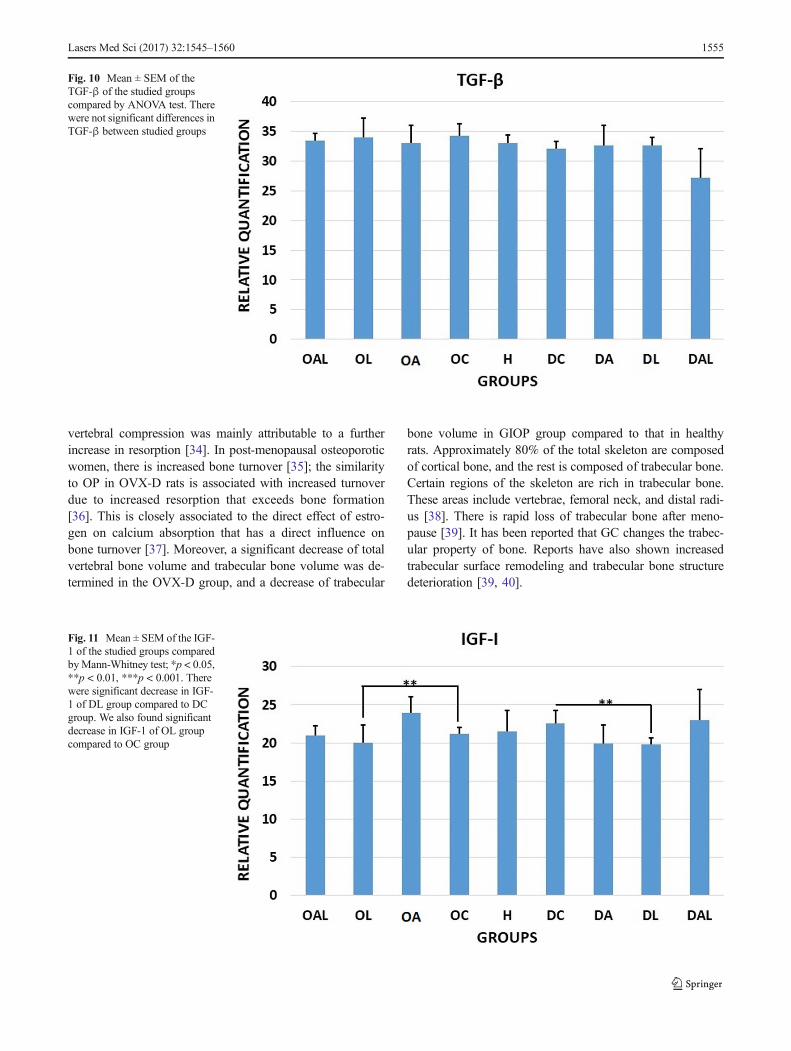
According to the ANOVA test, there was non-significantdifference in TGF-β1 between the studied groups. All resultsare demonstrated in Fig. 10.
According to the Mann-Whitney test, there was significantdecrease in IGF-1 of DL group compared to DC group(p = 0.004). Results showed a significant decrease in IGF-1of OL group compared to OC group (p = 0.005). All resultsare shown in Fig. 11.
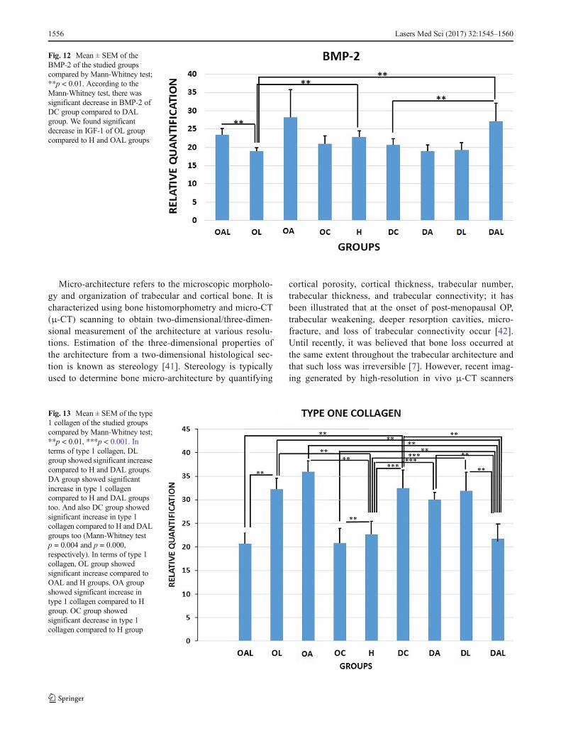
According to the Mann-Whitney test, there was signif-icant decrease in BMP-2 of DC group compared to DALgroup (p = 0.009). Significant decrease was determined in
Fig. 4 Mean ± SEM of thetrabecular bone volume (mm2) ofthe studied groups compared byLSD test; **p < 0.01,***p < 0.001. There weresignificant differences intrabecular bone volume (mm2) inDC, DL, and DAL compared tohealthy group. We foundsignificant decrease in trabecularbone volume of DL compared toDA, DC, and DAL. Significantdecrease existed in trabecularbone volume of OC, OA, OA,and OAL compared to H group
Fig. 5 Mean ± SEM of thecortical bone volume (mm2) ofthe studied groups compared byLSD test; *p < 0.05, **p < 0.01,***p < 0.001. There weresignificant increases that existedin cortical bone volume (mm2) inDA and DC, DAL, and healthygroups compared to the DLgroup. We observed a significantincrease in cortical bone volumein the DA group compared to theDC and H groups
1552 Lasers Med Sci (2017) 32:1545–1560

IGF-1 of OL group compared to H and OAL groups (bothp = 0.004). All results are shown in Fig. 12. In terms oftype 1 collagen, the DL group showed significant increasecompared to H and DAL groups (Mann-Whitney testp = 0.000 and p = 0.004, respectively). DA group indi-cated significant increase in type 1 collagen compared toH and DAL groups (Mann-Whitney test p = 0.001 andp = 0.000, respectively). And the DC group also showedsignificant enhancement in type 1 collagen compared to H
and DAL groups (Mann-Whitney test p = 0.004 andp = 0.000, respectively). OL group showed significantincrease in type 1 collagen compared to OAL and Hgroups (Mann-Whitney test p = 0.004 and p = 0.008, re-spectively). OA group indicated significant increase intype 1 collagen compared to H group (Mann-Whitney testp = 0.008). OC group showed significant decrease in type1 collagen compared to H group (Mann-Whitney testp = 0.002). All results are illustrated in Fig. 13.
Fig. 6 Mean ± SEM of the bonemarrow volume (mm2) of thestudied groups compared by LSDtest; *p < 0.05, **p < 0.01,***p < 0.001. DL group showedthe significant decrease in totalbone marrow volume comparedto the DA, DAL, and H groups.We observed significant decreasein total bone marrow volume ofOA and OC groups compared tohealthy group. All results areshown in Fig. 5
Fig. 7 Mean ± SEM of theosteocyte numbers (106) of thestudied groups compared byANOVA test; *p < 0.05,**p < 0.01, ***p < 0.001.ANOVA test showed nosignificant differences inosteocyte numbers (106) ofdexamethasone-treated groups.According to the LSD test, therewere significant differences inosteocyte numbers in the OL andOC, OA, and OAL groupscompared to H group
Lasers Med Sci (2017) 32:1545–1560 1553

Discussion
Results of the current study demonstrate that OVX-D signif-icantly decreased evaluations of vertebral weight, total ver-tebral bone volume, trabecular bone volume, total bone mar-row volume, osteocyte number, osteoblast number, and type1 collagen gene expression in comparison to test results inthe healthy group. Results for GIOP showed a significantdecline in trabecular bone volume compared to the healthygroup. Results also showed a significant increase of osteo-clast number of groups of OVX-D and glucocorticoid
adminis t ra t ion compared to the heal thy group.Additionally, the detrimental effect of the OVX-D procedureon bone was more serious than that of glucocorticoid admin-istration. Furthermore, results indicated decreased trabecularbone volume in both tested models. Rapid bone loss hasbeen reported during the first few years after menopause,especially in the trabecular compartment that results in tra-becular perforation followed by entire loss of trabeculae[33]. Nordin et al. suggested that the main determinant ofage-related bone loss in women was increased bone resorp-tion and that further trabecular bone loss associated with
Fig. 8 Mean ± SEMof the osteoblast numbers (106) of the studied groups;**p < 0.01, ***p < 0.001. Mann-Whitney test showed that there were nosignificant differences in osteoblast numbers (106) in the dexamethasone-treated rats. According to the LSD test, we observed significant decreases
in osteoblast numbers of OC and OL groups, and OA group compared tohealthy group. We observed significant decrease in osteoblast numbers ofOL compared to OAL and OA .We also found significant increase inosteoblast numbers of OA and OAL compared to OC
Fig. 9 Mean ± SEM of theosteoclast numbers (106) of thestudied groups compared by LSDtest; *p < 0.05, **p < 0.01,***p < 0.001. There weresignificant increases in osteoclastnumbers in the DC (106) groupcompared to healthy, DAL, andDA groups. Significant increaseexisted in osteoclast numbers inthe DL group compared to DALand DA groups. According to theLSD test, we observed significantincreases in osteoclast numbers inthe OC group compared to OA,OAL, H, and OL groups
1554 Lasers Med Sci (2017) 32:1545–1560
vertebral compression was mainly attributable to a furtherincrease in resorption [34]. In post-menopausal osteoporoticwomen, there is increased bone turnover [35]; the similarityto OP in OVX-D rats is associated with increased turnoverdue to increased resorption that exceeds bone formation[36]. This is closely associated to the direct effect of estro-gen on calcium absorption that has a direct influence onbone turnover [37]. Moreover, a significant decrease of totalvertebral bone volume and trabecular bone volume was de-termined in the OVX-D group, and a decrease of trabecular
bone volume in GIOP group compared to that in healthyrats. Approximately 80% of the total skeleton are composedof cortical bone, and the rest is composed of trabecular bone.Certain regions of the skeleton are rich in trabecular bone.These areas include vertebrae, femoral neck, and distal radi-us [38]. There is rapid loss of trabecular bone after meno-pause [39]. It has been reported that GC changes the trabec-ular property of bone. Reports have also shown increasedtrabecular surface remodeling and trabecular bone structuredeterioration [39, 40].
Fig. 10 Mean ± SEM of theTGF-β of the studied groupscompared by ANOVA test. Therewere not significant differences inTGF-β between studied groups
Fig. 11 Mean ± SEM of the IGF-1 of the studied groups comparedbyMann-Whitney test; *p < 0.05,**p < 0.01, ***p < 0.001. Therewere significant decrease in IGF-1 of DL group compared to DCgroup. We also found significantdecrease in IGF-1 of OL groupcompared to OC group
Lasers Med Sci (2017) 32:1545–1560 1555
Micro-architecture refers to the microscopic morpholo-gy and organization of trabecular and cortical bone. It ischaracterized using bone histomorphometry and micro-CT(μ-CT) scanning to obtain two-dimensional/three-dimen-sional measurement of the architecture at various resolu-tions. Estimation of the three-dimensional properties ofthe architecture from a two-dimensional histological sec-tion is known as stereology [41]. Stereology is typicallyused to determine bone micro-architecture by quantifying
cortical porosity, cortical thickness, trabecular number,trabecular thickness, and trabecular connectivity; it hasbeen illustrated that at the onset of post-menopausal OP,trabecular weakening, deeper resorption cavities, micro-fracture, and loss of trabecular connectivity occur [42].Until recently, it was believed that bone loss occurred atthe same extent throughout the trabecular architecture andthat such loss was irreversible [7]. However, recent imag-ing generated by high-resolution in vivo μ-CT scanners
Fig. 12 Mean ± SEM of theBMP-2 of the studied groupscompared by Mann-Whitney test;**p < 0.01. According to theMann-Whitney test, there wassignificant decrease in BMP-2 ofDC group compared to DALgroup. We found significantdecrease in IGF-1 of OL groupcompared to H and OAL groups
Fig. 13 Mean ± SEM of the type1 collagen of the studied groupscompared by Mann-Whitney test;**p < 0.01, ***p < 0.001. Interms of type 1 collagen, DLgroup showed significant increasecompared to H and DAL groups.DA group showed significantincrease in type 1 collagencompared to H and DAL groupstoo. And also DC group showedsignificant increase in type 1collagen compared to H and DALgroups too (Mann-Whitney testp = 0.004 and p = 0.000,respectively). In terms of type 1collagen, OL group showedsignificant increase compared toOAL and H groups. OA groupshowed significant increase intype 1 collagen compared to Hgroup. OC group showedsignificant decrease in type 1collagen compared to H group
1556 Lasers Med Sci (2017) 32:1545–1560
has produced new findings that question this assumption.Waarsing et al. report that following initial trabecularthinning in OVX-D rats, the few remaining trabecularsubsequently recovered slowly and increased in thickness[43, 44]. Stereological results showed that laser signifi-cantly decreased total vertebral bone volume, trabecularbone volume, cortical bone volume, and bone marrowvolume in glucocorticoid-treated rats compared to thosein the healthy group; laser also significantly decreasedtotal vertebral bone volume, bone marrow volume, osteo-cyte number, and osteoblast number in OVX-D rats.
Bone is a complex tissue. Its fundamental function is toresist mechanical injury and absorb pressure. Bone strengthdepends on quantity of bone tissue and its quality, which isoutlined by the geometry and shape of the bone, the micro-architecture of the trabecular bone morphology, corticalthickness, and porosity; it is also characterized by intrinsicproperties of bony tissue such as turnover, mineral, andcollagen. These determinants of bone quality are interrelat-ed, especially mineral and collagen, and analysis of theirspecific roles in bone strength is difficult to identify. Themajor complication of some bone diseases such as osteopo-rosis, i.e., fracture, is due to weak bone strength. Therefore,any treatment of OP relies on improving bone strength.Bone strength is indirectly measured by BMD using dual-energy X-ray absorptiometry (DXA). Since DXA-measuredBMD represents 60–70% of the variation in bone strength,some important factors are not determined by DXA, interms of the effects of antiosteoporotic treatment and theprogression of OP. In addition to BMD, geometry and tra-becular micro-architecture also need consideration. Thus,evaluation of the intrinsic mechanical quality of boneshould provide a better understanding of the role of tissuequality in determining bone strength [38, 45, 46].Consistent with results of our recent study, it is reportedthat laser significantly improved important biomechanicalparameters of the vertebrae in glucocorticoid-induced OPand OVX-D tests compared to results of healthy and controlgroups, respectively [21].
Osteoblast cells synthesize collagen during the initial phaseof bone formation when unmineralized bone (osteoid) ismade. The collagen molecules are assembled extracellularlyand immature ketomine and aldimine cross-links are formed;these contribute to the formation of mature pyridinium or pyr-role cross-links. Together, these cross-links result in fibril for-mation that act as a scaffold for bone minerals and providestrength [7, 46, 47]. The collagen in bone is primarily type Icollagen, but types III, IV, and VI are also present [48]. At theonset of post-menopausal OP, diverse changes in the compo-sitional properties of the collagen have been reported; a recentstudy indicates that synthesis of type I collagen enhancedduring OP, while earlier studies reported reduced amounts oftype VI collagen and type III collagen [7, 49].
It was determined that evaluations of vertebral weight, totalvertebral bone volume, trabecular bone volume, cortical bonevolume, and total bone marrow volume of the DA group weresignificantly higher in comparison to results from the DLgroup and that number of osteoclasts in the DA group wassignificantly lower than that of the DL group (p = 0.005). Itwas also found that osteoblast number of the OA group wassignificantly higher compared to the OL group.
Bisphosphonates are currently used as a first-line therapeu-tic agent for OP. Bisphosphonates inhibit bone resorption byselective adsorption to a mineral surface and subsequent inter-nalization by bone-resorbing osteoclasts where they interferewith various biochemical processes. Alendronate inhibits akey enzyme, farnesyl pyrophosphate synthase, in themevalonate pathway that prevents biosynthesis of isoprenoidcompounds that are considered essential for post-translationalmodification of small guanosine triphosphate (GTP)-bindingproteins. Both protein prenylation and function of these regu-latory proteins are impaired, leading to osteoclast dysfunction[48]. Results of the current study demonstrated thatalendronate alone was statistically more effective than laseralone on the above-mentioned stereological parameters.
In the present study, the effects of laser irradiation wereexamined on IGF-I and BMP-2, which are considered as themost important factors in bone formation in healthy animals[49]. There is evidence to show that expression of moleculesthat modulate osteoblast activity may be altered during post-menopausal osteoporosis. During physiologic bone resorp-tion, growth factors such as IGF-I and TGF-β are releasedfrom the extracellular matrix, which recruit and activate oste-oblasts to begin collagen synthesis [7]. Osteogenic andadipogenic processes are modulated by the BMP-2. This cy-tokine regulates expression of the nuclear receptor peroxisomeproliferator-activated receptor-γ (PPARγ) and Runt-relatedtranscription factor 2 (RUNX2) [50]. Laser can promote boneformation and inhibit bone resorption, thus facilitating boneremodeling [12–15]. However, it was demonstrated that lasersignificantly decreased IGF-I and BMP-2 in both testedmodels of OP.
Interestingly, it was found that laser treated in both testmodels of OP showed significant increase of type 1 collagengene expression compared to those in the healthy group.Results for laser in OVX-D rats and GIOP rats in combinationshowed significantly increased osteoblast number and type 1collagen gene expression, and significantly decreased osteo-clast number compared to control groups. These results (sig-nificant increase of type 1 collagen gene expression) may bein agreement with increasing biomechanical properties of theosteoporotic vertebrae in the laser-treated rats as recently re-ported in Bayat et al. [21]. Bayat et al. report on an evaluationof the effects of laser on cancellous bone strength in OVX-Dand GIOP test model of OP. There were four OVX-D groupsand four dexamethasone-treated groups. Laser (890 nm,
Lasers Med Sci (2017) 32:1545–1560 1557

80 Hz, 5.5 J/cm2) was carried out on the spinal processes ofthe vertebrae T12, L1, L2, and L3. Biomechanical test find-ings indicate a positive influence of laser and alendronateadministration on increasing bending stiffness and maximumforce of the osteoporotic bones in comparison to the healthygroup. Furthermore, laser treatment of OVX-D rats signifi-cantly escalated stress high load compared to OVX-D controlrats. Bayat et al. concluded that laser conserved cancellous(trabecular) bone of vertebrae against the detrimental influ-ence of OVX-D osteoporosis on bone strength in rats in com-parison to the control.
These results demonstrate that laser alone had a detrimentaleffect on many stereological parameters such as total vertebralbone volume, trabecular bone volume, cortical bone volume,and bone marrow volume in both glucocorticoid-treated ratsand OVX-D rats compared to those in the healthy group.Furthermore, results showed that the combination ofalendronate and laser in OVX-D rats and GIOP rats signifi-cantly increased osteoblast number and type 1 collagen geneexpression and significantly decreased osteoclast numbercompared to that of the controls. The results of this study werein agreement with result of other studies. Diniz et al. report onevaluation on the effect of laser in combination withalendronate sodium on osteopenic bone. Laser (GaAlAs;830 nm, 50 mW, and 4 J/cm2) on the femoral neck and verte-bral segments were performed. In the osteopenic +alendronate sodium group, the trabecular bone volume in L2was significantly higher than that in the controls. Notably, inthe combination between laser and alendronate sodium, thetrabecular bone volume in osteoporotic animals was signifi-cantly higher in vertebrae L2 and T13 than osteoporotic boneand was similar to that in the sham-operation group. Dinizet al. demonstrated that treatment with laser together withbisphosphonate therapy was the most effective on reversingvertebral osteopenia due to the ovariectomy [28].
Mohsenifar et al. reported an investigation of the effect oflaser on the cortical bone of osteoporotic rat tibia in two testmodels, GIOP rats and OVX-D rats. GIOP rats and OVX-Drats treated with alendronate (1 mg/kg) and laser (890 nm,80 Hz, 0.972 J/cm2). Rats were given alendronate for 30 daysand subjected to laser three times per week for 8 weeks. Testsdemonstrated a significant rise in cortical bone volume in allstudy groups compared to healthy rats. Results showed signif-icant reduction in trabecular bone volume in all study groupscompared to healthy rats. The control rats with OP showedsignificantly increased osteoclast number. Alendronate signifi-cantly decreased osteoclast number in osteoporotic rats. In thelaser + alendronate group, the same effect was observed onosteoporotic bone [25]. Fridoni et al. report on the effects oflaser on cortical bone in OVX-D and GIOP test models of OPin rats. The osteoporotic rats received alendronate for 30 days.Laser (890 nm, 80 Hz, 0.972 J/cm2) was performed on the tibiathree times per week for 2 months. After 2 months, tibia were
extracted and submitted to a three-point bending test. Laser didnot enhance the biomechanical parameters of osteoporoticbones compared to controls and healthy rats. Laser withalendronate treatment significantly increased stress high loadin OVX-D rats compared to those in the healthy group [26].
The physical parameters used in PW laser such as wave-length, power (peak power, average power, and powerdensity),pulse frequency, pulse duration (width), duration ofexposure, and energy density all impact the biological effectsof laser irradiation [32, 51]. PW laser at 5.5-J/cm2 energydensity and other delivery parameters used in the current studyfailed to cause any beneficial effects in the histological andgene expression parameters of osteoporotic bones. A probablecause is the relatively lower energy density of laser employedin this study. The applied dose was selected in accordancewith two studies that demonstrated increased bone density inrats from an 890-nm infrared laser at 0.972 J/cm2 [19, 32].
Recently, Muniz Renno et al. studied the effects of PBM(830 nm, 100 W/cm2, 120 J/cm2) on femurs of exercised os-teoporotic rats. The exercise program and the laser radiationwere performed 48 h over an 8-week period. The exerciseprogram consisted of jumps. Femurs were submitted to aphysical and geometrical property evaluation, a biomechani-cal test, and calcium and phosphorus evaluation. MunizRenno et al. concluded that exercised animals showed higherbone strength and physical property values. However, thePBM did not improve the biostimulatory effects of the exer-cise on the osteoporotic rats [30].
It was concluded that PBM alone had a detrimental effecton many stereological parameters of vertebral bone such astotal vertebral bone volume, trabecular bone volume, corticalbone volume, and bonemarrow volume in both GIOP rats andOVX-D rats compared the healthy group. Alendronate signif-icantly improved many stereological parameters such as totalvertebral bone volume, trabecular bone volume, cortical bonevolume, and bone marrow volume in both GIOP rats andOVX-D rats compared to those in laser groups. Furthermore,the combination of alendronate and laser in OVX-D rats andGIOP rats significantly increased osteoblast number and type1 collagen gene expression. Since the PBM had osteogeniceffects, different laser parameters should be examined to ver-ify whether an appropriate laser protocol alone or administra-tion with a proper antiosteoporotic agent might reverse thedetrimental effects of OP. We suggest measuring Ca++ levelson blood in further studies.
Compliance with ethical standards
Role of funding source This article is financially supported by “theResearch Department” at Shahid Beheshti University of MedicalSciences, Tehran, Iran (Grant No. 10397-1391-1-115-1092).
Conflict of interest The authors declare that they have no conflict ofinterest.
1558 Lasers Med Sci (2017) 32:1545–1560

Ethical approval The procedure was approved by the Medical EthicsCommittee of Shahid Beheshti University of Medical Sciences, Tehran,Iran (protocol no. 1391-1-115-1092).
Informed consent This study was performed on animal model, so itwas not included of “Informed Consent.”
References
1. Bidwell JP, AlvarezMB, HoodM Jr, Childress P (2013) Functionalimpairment of bone formation in the pathogenesis of osteoporosis:the bone marrow regenerative competence. Curr Osteoporos Rep 1:117–125. doi:10.1007/s11914-013-0139-2
2. Briot K, Roux C (2015) Glucocorticoid-induced osteoporosis.RMD Open 1:e000014. doi:10.1136/rmdopen-2014-000014
3. Bouillon R, Burckhardt P, Christiansen C, Fleisch H, Fujita T,Gennari C et al (1991) Consensus development conference: pro-phylaxis and treatment of osteoporosis. Am J Med 90:107–110
4. Wright NC, Looker AC, Saag KG, Curtis JR, Delzell ES, Randall Set al (2014) The recent prevalence of osteoporosis and low bonemass in the United States based on bone mineral density at thefemoral neck or lumbar spine. J Bone Miner Res 29:2520–2526.doi:10.1002/jbmr.2269
5. Mansjur KQ, Kuroda S, Izawa T, Maeda Y, Sato M, Watanabe K,et al (2016) The effectiveness of human parathyroid hormone andlow-intensity pulsed ultrasound on the fracture healing in osteopo-rotic bones. Ann Biomed Eng 1–9. doi:10.1007/s10439-015-1533-y
6. Izzah Ibrahim N, Mohamad S, Mohamed N, Nazrun SA (2013)Experimental fracture protocols in assessments of potential agentsfor osteoporotic fracture healing using rodent models. Curr DrugTargets 14:1642–1650. doi:10.2174/1389450114666131216224003
7. McNamara L (2010) Perspective on post-menopausal osteoporosis:establishing an interdisciplinary understanding of the sequence ofevents from the molecular level to whole bone fractures. J R SocInterface 7:353–372. doi:10.1098/rsif.2009.0282
8. Khosla S, Westendorf JJ, Oursler MJ (2008) Building bone to re-verse osteoporosis and repair fractures. J Clin Invest 118:421–428.doi:10.1172/JCI33612
9. Cashman KD (2007) Diet, nutrition, and bone health. J Nutr 137:2507S–2512S
10. Tee S-I, Yosipovitch G, Chan YC, Chua SH, Koh ET, Chan YH,Tan SS, Tsou IY, Tan SH (2012) Prevention of glucocorticoid-induced osteoporosis in immunobullous diseases with alendronate:a randomized, double-blind, placebo-controlled study. ArchDermatol 148:307–314. doi:10.1001/archdermatol.2011.354
11. da Silva JP, da Silva MA, Almeida APF, Junior IL, Matos AP(2010) Laser therapy in the tissue repair process: a literature review.Photomed Laser Surg 28:17–21. doi:10.1089/pho.2008.2372
12. UedaY, ShimizuN (2003) Effects of pulse frequency of low-level lasertherapy (LLLT) on bone nodule formation in rat calvarial cells. J ClinLaser Med Surg 21:271–277. doi:10.1089/104454703322564479
13. Ueda Y, Shimizu N (2001) Pulse irradiation of low-power laserstimulates bone nodule formation. J Oral Sci 43:55–60. doi:10.2334/josnusd.43.55
14. Sohn H, Ko Y, Park M, Kim D, Moon YL, Jeong YJ, Lee H, MoonY, Jeong BC, Kim O, LimW (2015) Effects of light-emitting diodeirradiation on RANKL-induced osteoclastogenesis. Lasers SurgMed 47:745–755. doi:10.1002/lsm.22413
15. Kiyosaki T, Mitsui N, Suzuki N, Shimizu N (2010) Low-level lasertherapy stimulates mineralization via increased Runx2 expressionand ERK phosphorylation in osteoblasts. Photomed Laser Surg28(Suppl 1):S167–S172. doi:10.1089/pho.2009.2693
16. Xu M, Deng T, Mo F, Deng B, Lam W, Deng P, Zhang X, Liu S(2009) Low-intensity pulsed laser irradiation affects RANKL andOPG mRNA expression in rat calvarial cells. Photomed Laser Surg27:309–315. doi:10.1089/pho.2008.2283
17. BayatM,Abdi S, Javadieh F,Mohsenifar Z, RashidMR (2009) Theeffects of low-level laser therapy on bone in diabetic and nondia-betic rats. Photomed Laser Surg 27:703–708. doi:10.1089/pho.2008.2351
18. Freidouni M, Nejati H, Salimi M, Bayat M, Amini A, Noruzian M,Asgharie MA, Rezaian M (2015) Evaluating glucocorticoid admin-istration on biomechanical properties of rats’ tibial diaphysis. IranRed Crescent Med J 17(3):e19389. doi:10.5812/ircmj.19389eCollection 2015
19. Najar A, Fridoni M, Rezaei F, Bayat S, Bayat M (2015)Supraphysiologic glucocorticoid administration increased biome-chanical bone strength of rats’ vertebral body. Lab Anim Res 31:180–187. doi:10.5625/lar.2015.31.4.180
20. Cressoni MD, Giusti HH, Piao AC, de Paiva Carvalho RL,Anaruma CA, Casarotto RA (2010) Effect of GaAlAs laser irradi-ation on the epiphyseal cartilage of rats. Photomed Laser Surg 28:527–532. doi:10.1089/pho.2009.2572
21. Bayat M, Fridoni M, Nejati H, Mostafavinia A, Salimi M,Ghatrehsamani M et al (2016) An evaluation of the effect of pulsedwave low-level laser therapy on the biomechanical properties of thevertebral body in two experimental osteoporosis rat models. LasersMed Sci 31:305-14. doi:10.1007/s10103-015-1842-2
22. Ko CY, Kang H, Ryu Y, Jung B, Kim H, Jeong D, Shin HI, Lim D,Kim HS (2013) The effects of minimally invasive laser needlesystem on suppression of trabecular bone loss induced by skeletalunloading. Lasers Med Sci 28:1495–1502. doi:10.1007/s10103-013-
23. Kang H, Ko CY, Ryu Y, Seo DH, Kim HS, Jung B (2012)Development of a minimally invasive laser needle system: effectson cortical bone of osteoporotic mice. Lasers Med Sci 27:965–969.doi:10.1007/s10103-011-1014-y
24. Chen Y, Cao Z, Zhang L, Xu X, Chen Y, Chen Y (2011) Low levellaser can be a novel adjuvant method for orthodontic tooth move-ment on postmenopausal women. Med Hypotheses 76:479–481.doi:10.1016/j.mehy.2010.11.025
25. Mohsenifar Z, Fridoni M, Ghatrehsamani M, Abdollahifar MA,Abbaszadeh H, Mostafavinia A, Fallahnezhad S, Asghari M,Bayat S, Bayat M (2016) Evaluation of the effects of pulsed waveLLLT on tibial diaphysis in two rat models of experimental osteo-porosis, as examined by stereological and real-time PCR gene ex-pression analyses. Lasers Med Sci 31:721–732. doi:10.1007/s10103-016-1916-9
26. Fridoni M, Masteri Farahani R, Nejati H, Salimi M, Gharavi SM,Bayat M, Amini A, Torkman G, Bayat S (2015) Evaluation of theeffects of LLLT on biomechanical properties of tibial diaphysis intwo rat models of experimental osteoporosis by a three point bend-ing test. Lasers Med Sci 30:1117–1125. doi:10.1007/s10103-014-1706-1
27. Medalha CC, Amorim BO, Ferreira JM, Oliveira P, Pereira RM,Tim C, Lirani-Galvão AP, da Silva OL, Renno AC (2010)Comparison of the effects of electrical field stimulation and low-level laser therapy on bone loss in spinal cord-injured rats.Photomed Laser Surg 28:669–674. doi:10.1089/pho.2009.2691
28. Diniz JS, Nicolau RA, de Melo ON, do Carmo Magalhães F, deOliveira Pereira RD, Serakides R (2009) Effect of low-powergallium-aluminum-arsenium laser therapy (830 nm) in combinationwith bisphosphonate treatment on osteopenic bone structure: anexperimental animal study. Lasers Med Sci 24:347–352. doi:10.1007/s10103-008-0568-9
29. Renno AC, de Moura FM, dos Santos NS, Tirico RP, Bossini PS,Parizotto NA (2006) Effects of 830-nm laser light on preventing
Lasers Med Sci (2017) 32:1545–1560 1559

bone loss after ovariectomy. Photomed Laser Surg 24:642–645.doi:10.1089/pho.2006.24.642
30. Muniz Renno AC, de Moura FM, dos Santos NS, Tirico RP,Bossini PS, Parizotto NA (2006) The effects of infrared-830 nmlaser on exercised osteopenic rats. LasersMed Sci 21:202–207. doi:10.1007/s10103-006-0396-8
31. Kanis J, McCloskey E, Johansson H, Cooper C, Rizzoli R,Reginster J-Y (2013) European guidance for the diagnosis andmanagement of osteoporosis in postmenopausal women.Osteoporos Int 24:23–57. doi:10.1007/s00198-012-2074-y
32. Hashmi JT, Huang YY, Sharma SK, Kurup DB, De Taboada L,Carroll JD, Hamblin MR (2010) Effect of pulsing in low-level lighttherapy. Lasers Surg Med 42:450–466. doi:10.1002/lsm.20950
33. Je A, Nb M, Sagreiya K (1987) The microanatomy of trabecularbone loss in normal aging men and women. Clin Orthop Relat Res215:260–271 3802645
34. Nordin B, Speed R, Aaron J, Crilly R (1981) Bone formation andresorption as the determinants of trabecular bone volume in post-menopausal osteoporosis. Lancet 318:277–279 6114324
35. Van Brussel M, Bultink I, Lems W (2009) Prevention ofglucocorticoid-induced osteoporosis. Expert Opin Drug Deliv 10:997–1005. doi:10.1517/14656560902868225
36. Yeh J, Chen M-M, Aloia J (1996) Ovariectomy-induced high turn-over in cortical bone is dependent on pituitary hormone in rats.Bone 18:443–450
37. Jagtap VR, Ganu JV, Nagane NS (2011) BMD and serum intactosteocalcin in postmenopausal osteoporosis women. Indian J ClinBiochem 26:70–73
38. Green D, Wallace H (2003) Late effects of childhood cancer. CRCPress
39. Parfitt AM (1987) Trabecular bone architecture in the pathogenesisand prevention of fracture. Am J Med 82:68–72
40. Lane NE, Yao W, Balooch M, Nalla RK, Balooch G, Habelitz Set al (2006) Glucocorticoid-treated mice have localized changes intrabecular bone material properties and osteocyte lacunar size thatare not observed in placebo-treated or estrogen-deficient mice. JBone Miner Res 21:466–476. doi:10.1359/JBMR.051103
41. Russ JC, Dehoff RT (2012) Practical stereology. Springer Science& Business Media
42. Lane NE, Thompson JM, Haupt D, Kimmel DB, Modin G, KinneyJH (1998) Acute changes in trabecular bone connectivity and oste-oclast activity in the ovariectomized rat in vivo. J Bone Miner Res13:229–236. doi:10.1359/jbmr
43. Waarsing J, Day J, Van der Linden J, Ederveen A, Spanjers C, DeClerck N, Sasov A, Verhaar JA, Weinans H (2004) Detecting andtracking local changes in the tibiae of individual rats: a novel meth-od to analyse longitudinal in vivomicro-CT data. Bone 34:163–169
44. Waarsing JH, Day JS, Verhaar JA, EderveenAG,Weinans H (2006)Bone loss dynamics result in trabecular alignment in aging andovariectomized rats. J Orthop Res 24:926–935. doi:10.1002/jor.20063
45. Ammann P (2002) Determining factors of bone mechanical resis-tance. Therapie 58:403–407. doi:10.2515/Therapie:2003065
46. Viguet-Carrin S, Garnero P, Delmas P (2006) The role of collagenin bone strength. Osteoporos Int 17:319–336. doi:10.1007/s00198-005-2035-9
47. Eyre DR, Dickson I, Van Ness K (1988) Collagen cross-linking inhuman bone and articular cartilage. Age-related changes in the con-tent of mature hydroxypyridinium residues. Biochem J 252:495–500
48. Bailey A, Wotton S, Sims T, Thompson P (1993) Biochemicalchanges in the collagen of human osteoporotic bone matrix.Connect Tissue Res 29:119–132
49. Fujimoto K, Kiyosaki T, Mitsui N, Mayahara K, Omasa S, SuzukiN, ShimizuN (2010) Low-intensity laser irradiation stimulates min-eralization via increased BMPs in MC3T3-E1 cells. Lasers SurgMed 42:519–526. doi:10.1002/lsm.20880/full
50. Donoso O, Pino AM, Seitz G, Osses N, Rodríguez JP (2015)Osteoporosis-associated alteration in the signaling status of BMP-2 in human MSCs under adipogenic conditions. J Cell Biochem116:1267–1277. doi:10.1002/jcb.25082
51. Bayat M (2014) The necessity for increased attention to pulsed low-level laser therapy. Photomed Laser Surg 32(8):427–428. doi:10.1089/pho.2014.9858
1560 Lasers Med Sci (2017) 32:1545–1560