everflex- self-expanding peripheral stent system · everflex-self-expanding peripheral stent system...
TRANSCRIPT
EverFlex-Self-Expanding Peripheral Stent System
Instructions for Use
DEVICE DESCRIPTION
The EverFlexN Self-Expanding Peripheral Stent System is a self-expanding Nitinol stent system intended for permanentimplantation. The self-expanding stent is made of a nickel titanium alloy (Nitinol) and comes pre-mounted on a 6F, 0.035" over-the-wire delivery system. The stent is cut from a Nitinol tube in an open lattice design, and has tantalum radiopaque markers at theproximal and distal ends of the stent. Upon deployment, the stent achieves its predetermined diameter and exerts a constant, gentleoutward force to establish patency.
The Delivery System, as shown in Figures 1, la, and 1b, includes an inner subassembly (1) and outer subassembly (2), which arelocked together with a safety lock (3). The inner subassembly terminates distally in a flexible catheter tip (4) and originatesproximally at the hub (5).
The distal portion of the Delivery System for the 20 - 150 mm stents, as shown in Figure 1 a, is comprised of two radiopaquemarkers, one marker distal (6) and one marker/retainer proximal (7) to the stent, on the inner subassembly.
The distal portion of the Delivery System for the 200 mm stents, as shown in Figure 1 b, includes the same components as those inFigure Ia except for the radiopaque markers: one marker/retainer distal (13) and one marker/holder proximal (14) to the stent, onthe inner subassembly.
The outer sheath connects proximally to the manifold subassembly (8). The self-expanding stent is constrained within the spacebetween the inner and outer subassemblies. This space is flushed prior to the procedure through the stopcock (9). The outersubassembly has a radiopaque marker at its distal end (10).
The stent is positioned at the target lesion using the two radiopaque markers on the inner subassembly and the radiopaque markerson the stent.
For stent deployment, turn the safety lock counterclockwise to unlock the outer subassembly. The outer subassembly retracts bypulling the distal grip (11) toward the proximal grip (12). Stent deployment is complete when the radiopaque marker on the outersubassembly passes the proximal radiopaque marker on the inner subassembly.
February 7, 2012 Page 1 of 13 501102-001
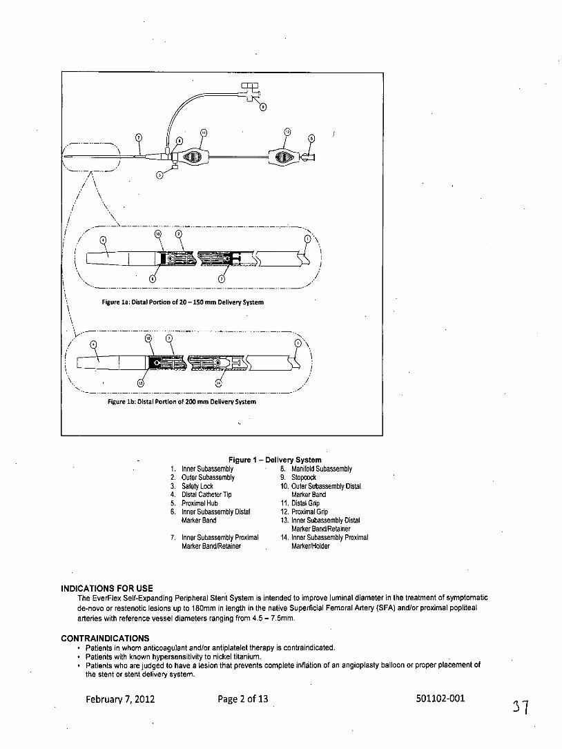
Figure 1a: DistalI Portion of 20 -150 mm Delivery System
Figure 1b: Distal Portion of 200 mot Delivery System
Figure 1 - Delivery System1. Inner Subassembly 8. Manifold Subassembly
2. Outer Subassembly 9. Stopcock3. Safety Lock 10. Outer Subassembly Distal4. Distal Catheter Tip Marker Band5. Proximal Hub 11. Distal Grip6. Inner Subassembly Distal 12. Proximal Grip
Marker Band 13. Inner Subassembly DistalMarker Band/Retainer
7. Inner Subassembly Proximal 14. Inner Subassembly ProximalMarker Band/Retainer Marker/Holder
INDICATIONS FOR USEThe EverFlex Self-Expanding Peripheral Stent System .is intended to improve luminal diameter in the treatment of symptomaticde-novo or restenotic lesions up to 180mm in length in the native Superficial Femoral Artery (SFA) and/or proximal poplitealarteries with reference vessel diameters ranging from 4.5- 7.5mm.
CONTRAINDICATIONS* Patients in whom anticoagulant and/or antiplatelet therapy is contraindicated.* Patients with known hypersensitivity to nickel titanium.* Patients who are judged to have a lesion that prevents complete inflation of an angioplasty balloon or proper placement of
the stent or stent delivery system.
February 7, 2012 Page 2 of 13 501102-001

WARNINGS* The device is provided STERILE for single use only. Do not reprocess or resterilize. Reprocessing and resterilizing could
increase the risk of patient infection and risk of compromised device performance.* If resistance is encountered at any time during the insertion procedure, do not force passage. Resistance may cause
damage to stent or vessel. Carefully withdraw the stent system without deploying the stent.* If resistance is felt when initially pulling back on the distal grip, do not force deployment. Carefully withdraw the stent system
without deploying the stent.* If resistance is met during delivery system withdrawal, advance the outer sheath until the outer sheath marker contacts the
catheter tip and withdraw the system as one unit.
PRECAUTIONS* Carefully inspect the sterile package and device prior to use to verify that no damage occurred during shipment.* Do not exceed 300 psi / 20 ATM while flushing the delivery system.* Do not use if the stent is partially deployed upon removal from the package, or before starting the deployment procedure.* Support from a sheath is necessary to minimize lengthening or shortening during stent deployment.* Always use a sheath during the implant procedure to protect both the vessel and puncture site.* Failure to pre-dilate the lesion may impair the ability to remove the stent system after stent deployment.* The stent system is not designed for recapturing or repositioning after establishing vessel apposition.* Failure to hold the proximal grip in a fixed position may result in partial deployment, foreshortening, lengthening or increased
deployment force.* The stent is not designed to be lengthened or shortened past its nominal length. Excessive stent lengthening or shortening
may increase the risk of stent fracture.* Use caution when crossing a deployed stent with any adjunct device.* Stent should not be expanded past its nominal diameter.
ADVERSE EVENTSThe EverFlex Self-Expanding Stent System was evaluated in a study titled the US StuDy for EvalUating EndovasculaR TreAtmentsof Lesions in the Superficial Femoral Artery and Proximal Popliteal By using the EverfLex Nitlnol STent SYstem II (DURABILITY II).A total of 287 subjects were enrolled. The primary objective was to evaluate the safety and effectiveness of primary stenting usingthe EverFlex Self-Expanding Stent System compared to percutaneous transluminal angioplasty (PTA) performance goals for thetreatment of stenotic, restenotic or occluded lesions (non-stented) of the native superficial femoral artery or the superficial femoraland proximal popliteal arteries.
Table I provides a summary of the adverse events documented in the DURABILITY 11 study. The data are presented as apercentage of subjects experiencing an AE followed by the total number of events in brackets.
Table 1: Summary of Adverse Events
Adverse Event Events at 5 30 days Events at 5 1 Year Total Events
Total Subjects with AEs' 45.3% (129/285) [210) 86.1% (242/281) [756] 87.8% (252/287) [1111]
Allergic reaction 1.4% (4/285) [4] 1.8% (5/281) [5] 1.7% (5/287) [5]
Amputation 0.4% (1/285) [1] 0.7% (2/281) [2] 1.4% (4/287) 14]
Angina 0.4% (1/285) [1] 4.3% (12/281) [13] 7.0% (20/287) [22]
Arrhythmia 0.7% (2/285) [2] 2.8% (8/281)[91 3.8% (11/287) 12]
Arterial dissection/perforation 14.0% (40/285) [42] 15.3% (43/281) [49] 15.0% (43/287) [51)
Bleeding disorders (including GI, lymphatic) 1.8% (5/285) (5] 5.0% (14/281) [15] 6.6% (19/287) (22]
Cerebrovascular accident 1.8% (5/281) [5] 2.8% (8/287) [8]
Death** 0.7% (2/281) [2] 1.4% (4/287) [4]
Edema 1.8% (5/285) [5] 5.0% (14/281) [14] 6.6% (19/287) (22]
GI bleeding 0.4% (11285) [11 1.4% (4/281) [4] 2.1% (6/287) [6]
Hematoma at vascular access site 3.9% (11/285) [11] 3.9% (11/281) [11] 3.8% (11/287) [11]
Hypertension/hypotension 2.1% (6/285) [6] 4.3% (12/281) [12] 4.9% (14/287) [16]
Infection, local or systemic including bacteremia or 0.4% (1/285) [1] 3.6% (10/281) [11) . 5.6% (16/287) [22]septicemia
Myocardial infarction 1.1% (3/281) [3] 2.1% (6/287) [6
February 7, 2012 Page 3 of 13 501102-001

Adverse Event Events at 5 30 days Events at 5 1 Year Total Events
Other Cardiac Disorders 0.7% (2/285) [2] 8.5% (24/281) [26] 9.8% (28/287) [34
Other GU Disorders 0.7% (2/285) [2] 3.2% (9/281) [10] 4.9% (14/287) [17)
Other Gastrointestinal Disorders 3.2% (9/285) [11] 12.8% (36/281) [52] 13.9% (40/287) [69]
Other Musculoskeletal disorders 4.6% (13/285) [15] 14.9% (42/281) [52] 20.2% (58/287) [81]
Other Respiratory Issues 0.4% (1/285) [1] 10.7% (30/281) [34] 14.6% (42/287) [54)
Other Vascular Disorders 5.3% (15/285) [16] 21.7% (61/281) (81] 32.1% (921287) [133]
Percutaneous revascularization 0.4% (1/285) [1) 4.3% (12/281) [14] 4.5% (13/287) [17]
Pseudoaneurysm 1.4% (4/285) [4] 1.4% (4/281) [41 1.4% (4/287) (4]
Renal Insufficiency/Failure 1.1% (3/281) [3] 1.4% (4/287) [4]
Restenosis 1.4% (4/285) [4] 21.7% (61/281) [66] 32.8% (94/287) [113]
Slow/no flow during procedure 0.7% (2/285) [2] 0.7% (2/281) [2] 0.7% (2/287) [2]
Stentessel thrombosis 0.4% (1/285) [1] 3.6% (10/281) [11] 4.2% (12/287) [13]
Vessel spasm 0.4% (1/285) [1] 0.4% (1/281) [1) 0.3% (1/287) [1]
Other 18.9% (54/285) [71] 46.3% (130/281) [245] 51.6% (148/287) [358]
* Note:The denominators in each column represent the number of subjects with adverse events for the reported time period (285 subjects at 30 days; 281subjects at 51 year;287 total subjects).*Note Count of AEs labeled "death" is less than total number of study deaths since death may be attributable to other AEs.
POTENTIAL ADVERSE EVENTSThe potential adverse effects (e.g., complications) that may occur and/or require intervention with the use of this device include, butare not limited to:
* Abrupt or sub-acute closure * Infection* Allergic reaction to device materials or procedure Inflammation
medications* Allergic reaction to Nitinol * Intraluminal thrombus* Amputation * Myocardial infarction* Aneurysm * Pain* Angina * Partial stent deployment* Arrhythmia * Pseudoaneurysm* Arterio-venous fistula * Renal failure requiring dialysis* Artery perforation or rupture * Renal insufficiency (new or worsening)* Bleeding requiring transfusion * Restenosis* Bruising * Sepsis* Contrast medium reaction/renal failure * Shock* Death * Stent collapse or fracture* Device breakage * Stent migration* Dissection or intimal flap * Stent misplacement* Edema * Stroke* Embolism * Surgical or endovascular intervention* Failure to deploy stent * Thrombosis/occlusion of the stent* Fever * Transient ischemic attack* Gastrointestinal bleeding due to anticoagulation * Venous thromboembolism* Hematoma * Vessel spasm* Hypertension/hypotension
February 7, 2012 Page 4 of 13 501102-001 31
CLINICAL STUDIESThe US StuDy for EvalUating EndovasculaR TreAtments of Lesions in the Superficial Femoral Artery and Proximal Popliteal ByusIng the EverfLex Nitinol STent SYstem II (DURABILITY II) study was a prospective, multi-center, non-randomized, single armstudy. DURABILITY II compared percutaneous transluminal angioplasty (PTA) and primary stenting with the EverFlexm stent toperformance goals of PTA alone in the treatment of atherosclerotic lesions of the native superficial femoral artery (SFA) or thesuperficial femoral and proximal popliteal arteries. The safety and effectiveness performance goals were based on an aggregate ofpublished trial data as described by VIVA physicians Inc. (VPI). DURABILITY 11 was conducted at 40 US and four Europeaninvestigational sites. A total of 287 subjects were enrolled. Eligible subjects either had stenotic, restenotic (non-stented) oroccludedlesions. The reference vessel diameter of the treated subjects was to be 4.5-7.5 mm and the lesion length from 4-18 cm long.Subjects had to have Rutherford Clinical Categories of 2-4. Subject follow-up occurred at 30 days, 6 months, 1, 2 and 3 years post-procedure. The primary safety endpoint for the study was major adverse event (MAE) rate at 30 days and the primary effectivenessendpoint was primary stent patency rate at 1 year.
Subject Eligibility CriteriaSubjects with stenosis of the native superficial femoral artery or the superficial femoral and proximal popliteal arteries, whoconsented to participate, were eligible for inclusion in the DURABILITY 11 study. To be included, they had to be at least 18 yearsold.
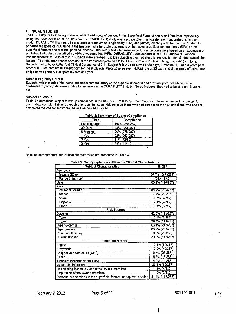
Subject Follow-upTable 2 summarizes subject follow-up compliance in the DURABILITY II study. Percentages are based on subjects expected foreach follow-up visit. Subjects expected for each follow-up visit included those who had completed the visit and those who had notcompleted the visit but for whom the visit window had closed.
Table 2: Summary of Subject ComplianceTime Compliance
Pre-discharge 100% (287/287)30 Days 98% (280/287)6 Months 96% (275/287)1 Year 92% (263/287)2 Year 81%(72/89)3 Year 79%(11/14)
Baseline demographics and clinical characteristics are presented in Table 3.
Table 3: Demographics and Baseline Clinical CharacteristicsSubject Characteristics N=287
Age (yrs.)
Mean ± SD (N) 67.7 ± 10.7 (287)Range (min, max) (39.4, 93.3)
Male 66.2% (190/287)Race
White/Caucasian 88.9% (255/287)African 7.7% (22/287)Asian 0.7% (2/287)Hispanic 2.4% (7/287)Other 0.3% (1/287)
Risk Factors
Diabetes 42.5% (122/287)Type I 3.1% (9/287)Type II 39.4% (113/287)
Hyperlipidemia 86.1% (247/287)Hypertension 88.2% (253/287)Renal Insufficiency 9.8% (28/287)Current smoker 39.0% (112/287)
Medical HistoryAngina 17.4% (50/287)Arrhythmia 13.9% (40/287)Congestive heart failure (CHF) 9.4% (27/287)Stroke 6.3% (18/287)Transient ischemic attack (TIA) 4.9% (14/287)Myocardial infarction 20.9% (60/287)Non-healing schemic ulcer in the lower extremities 1.4% (4/287)Amputation of the lower extremities 1.0% (3/287)Previous interventions in the superficial femoral or popliteal arteries 41.1% (118/287)
February 7, 2012 Page 5 of 13 501102-001
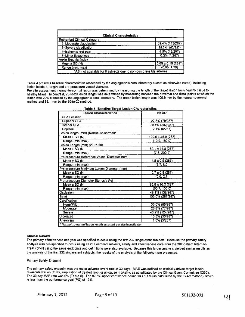
Clinical Characteristics
Rutherford Clinical Category2=Moderate claudication 39.4% (113/287)3=Severe claudication 55.7% (160/287)4=lschemic rest pain 4.5% (13/287)5=Minor tissue loss 0.3% (1/287)
Ankle Brachial IndexMean ± SD (N) 0.69 ± 0.19 (281*)Range (min, max) (0.06, 1.38)
*ABI not available for 6 subjects due to non-compressible arteries
Table 4 presents baseline characteristics (assessed by the angiographic core laboratory except as otherwise noted), includinglesion location, length and pre-procedure vessel diameter.Per site assessment, normal-to-normal lesion was determined by measuring the length of the target lesion from healthy tissue tohealthy tissue. In contrast, 20-to-20 lesion length was determined by measuring between the proximal and distal points at which thelesion was 20% stenosed by the angiographic core laboratory. The mean lesion length was 109.6 mm by the normal-to-normalmethod and 89.1 mm by the 20-to-20 method.
Table 4: Baseline Target Lesion CharacteristicsLesion Characteristics N=287
SFA Location
Superior SFA 27.5% (79/287)Inferior SFA 70.4% (202/287)Popliteal 2.1% (6/287)
Lesion length (mm) (Normal-to-normal)*Mean ± SD (N) 109.6 ± 45.0 (287)Range (min, max) (10.0, 180.0)
Lesion Length (mm) (20-to-20)Mean ± SD (N) 89.1 ± 44.8 (287)Range (min, max) (7.3, 200.9)
Pre-procedure Reference Vessel Diameter (mm)Mean ± SD (N) 4.8 ± 0.9 (287)Range (min, max) (2.7, 8.0)
Pre-procedure Minimum Lumen Diameter (mm)Mean ± SD (N) 0.7 ± 0.8 (287)Range (min, max) (0.0, 2.7)
Pre-procedure Diameter Stenosis (%)Mean ± SD (N) 85.8 ± 16.2 (287)Range (min, max) (50.7, 100.0)
Occlusion 48.1% (138/287)Bend 100.0% (287/287)Calcification
None/Mild 30.0% (86/287)Moderate 26.8% (77/287)Severe 43.2% (124/287)
Ulcerated 10.5% (30/287)Aneurysm 1.0% (3/287)* Normal-to-normal lesion length assessed per site investigator
Clinical ResultsThe primary effectiveness analysis was specified to occur using the first 232 single-stent subjects. Because the primary safetyanalysis was pre-specified to occur using all 287 enrolled subjects, safety and effectiveness data from the 287-patient Intent-to-Treat cohort using the same endpoints and definitions were also available. Because this larger analysis yielded similar results asthe analysis of the first 232 single-stent subjects, the results of the analysis of the full cohort are presented.
Primary Safety Endpoint
The primary safety endpoint was the major adverse event rate at 30 days. MAE was defined as clinically-driven target lesionrevascularization (TLR), amputation of treated limb, or all-cause mortality, as adjudicated by the Clinical Event Committee (CEC).The 30 day MAE rate was 0% (Table 5). The 97.5% upper confidence bound was 1.1% (as calculated by the Exact method), whichis less than the performance goal (PG) of 12%.
February 7, 2012 Page 6 of 13 501102-001
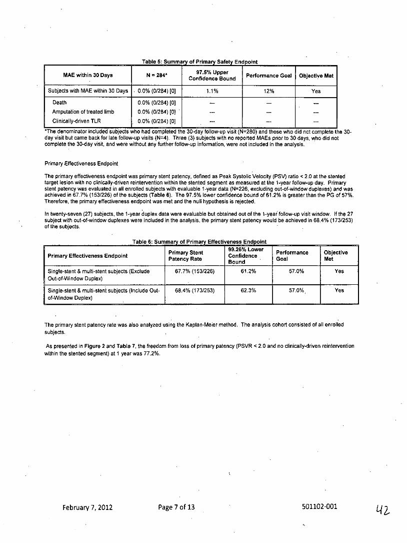
Table 5: Summary of Primary Safety Endpoint
MAE within 30 Days N = 284* 97.5% UpperConfidence Bound Performance Goal Objective Met
Subjects with MAE within 30 Days 0.0% (0/284) [0] 1.1% 12% Yes
Death 0.0% (0/284) [0] -- -
Amputation of treated limb 0.0% (0/284) [0) -- -- --
Clinically-driven TLR 0.0% (0/284) [0]
*The denominator included subjects who had completed the 30-day follow-up visit (N=280) and those who did not complete the 30-day visit but came back for late follow-up visits (N=4). Three (3) subjects with no reported MAEs prior to 30 days, who did notcomplete the 30-day visit, and were without any further follow-up information, were not included in the analysis.
Primary Effectiveness Endpoint
The primary effectiveness endpoint was primary stent patency, defined as Peak Systolic Velocity (PSV) ratio < 2.0 at the stentedtarget lesion with no clinically-driven reintervention within the stented segment as measured at the 1-year follow-up day. Primarystent patency was evaluated in all enrolled subjects with evaluable 1-year data (N=226, excluding out-of-window duplexes) and wasachieved in 67.7% (153/226) of the subjects (Table 6). The 97.5% lower confidence bound of 61.2% is greater than the PG of 57%.Therefore, the primary effectiveness endpoint was met and the null hypothesis is rejected.
In twenty-seven (27) subjects, the 1-year duplex data were evaluable but obtained out of the 1-year follow-up visit window. If the 27subject with out-of-window duplexes were included in the analysis, the primary stent patency would be achieved in 68.4% (173/253)of the subjects.
Table 6: Summary of Primary Effectiveness Endpoint
Primary Stent Lower Performance ObjectivePrimary Effectiveness Endpoint Patency Rate Confidence Goal MetBound
Single-stent & multi-stent subjects (Exclude 67.7% (153/226) 61.2% 57.0% YesOut-of-Window Duplex)
Single-stent & multi-stent subjects (Include Out- 68.4% (173/253) 62.3% 57.0% Yesof-Window Duplex)
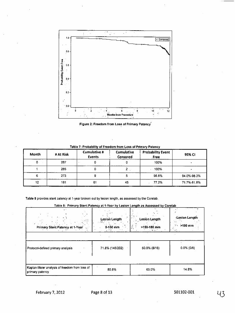
The primary stent patency rate was also analyzed using the Kaplan-Meier method. The analysis cohort consisted of all enrolledsubjects.
As presented in Figure 2 and Table 7, the freedom from loss of primary patency (PSVR <2.0 and no clinically-driven reinterventionwithin the stented segment) at 1 year was 77.2%.
February 7, 2012 Page 7 of 13 501102-001
0.6-
06
0.2-
0 2 .4 6 8 10 12Months from Procedure-
Figure 2: Freedom from Loss of Primary Patency
Table 7: Probability of Freedom from Loss of Primary Patency
Month #f At Risk Cumulative # Cumulative Probability Event 95% CI
Events Censored . Free0 287 0 0 100%
1 285 0 2. 100%
6 273 9 5 96.8% 94.0%-98.3%
12 181 61 45 . 77.2% 71.7%-81.8%
Table 8 provides stent patency at 1-year broken out by lesion length, as assessed by the Corelab.
Table 8: Primary Stent Patency at 1-Year by Lesion Length as Assessed by Corelab
Lesion Length Lesion Length Lesion Length
Primary Stent Patency at 1-Year .O-1 mm >150-180 mm >180 mm
Protocol-defined primary analysis 71.8% (145/202) 50.0% (8/16) 0.0% (0/8)
Kaplan-Meier analysis of freedom from loss of 808% 65.0% 14.8%primary patency
February 7, 2012 Page 8 of 13 501102-001
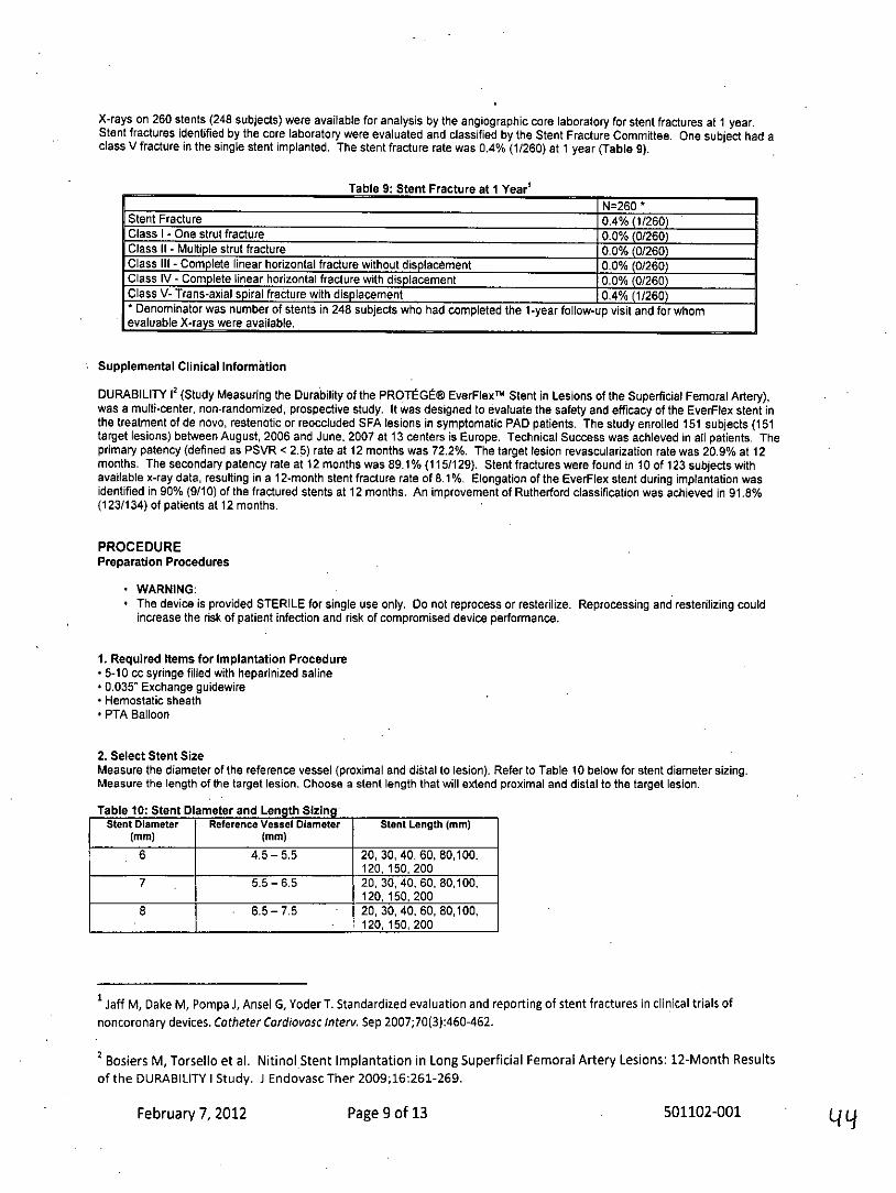
X-rays on 260 stents (248 subjects) were available for analysis by the angiographic core laboratory for stent fractures at 1 year.Stent fractures identified by the core laboratory were evaluated and classified by the Stent Fracture Committee. One subject had aclass V fracture in the single stent implanted. The stent fracture rate was 0.4% (1/260) at 1 year (Table 9).
Table 9: Stent Fracture at 1 Year'N=260 *
Stent Fracture 0.4% (1/260)Class I - One strut fracture 0.0% (0/260)Class II - Multiple strut fracture 0.0% (0/260)Class III - Complete linear horizontal fracture without displacement 0.0% (0/260)Class IV - Complete linear horizontal fracture with displacement 0.0% (0/260)Class V- Trans-axial spiral fracture with displacement 0.4% (1/260)* Denominator was number of stents in 248 subjects who had completed the I-year follow-up visit and for whomevaluable X-rays were available.
Supplemental Clinical Information
DURABILITY 12 (Study Measuring the Durability of the PROTEGE® EverFlexN Stent in Lesions of the Superficial Femoral Artery),was a multi-center, non-randomized, prospective study. It was designed to evaluate the safety and efficacy of the EverFlex stent inthe treatment of de novo, restenotic or reoccluded SFA lesions in symptomatic PAD patients. The study enrolled 151 subjects (151target lesions) between August, 2006 and June, 2007 at 13 centers is Europe. Technical Success was achieved in all patients. Theprimary patency (defined as PSVR < 2.5) rate at 12 months was 72.2%. The target lesion revascularization rate was 20.9% at 12months. The secondary patency rate at 12 months was 89.1% (115/129). Stent fractures were found in 10 of 123 subjects withavailable x-ray data, resulting in a 12-month stent fracture rate of 8.1%. Elongation of the EverFlex stent during implantation wasidentified in 90% (9/10) of the fractured stents at 12 months. An improvement of Rutherford classification was achieved in 91.8%(123/134) of patients at 12 months.
PROCEDUREPreparation Procedures
* WARNING:* The device is provided STERILE for single use only. Do not reprocess or resterilize. Reprocessing and resterilizing could
increase the risk of patient infection and risk of compromised device performance.
1. Required Items for Implantation Procedure* 5-10 cc syringe filled with heparinized saline* 0.035" Exchange guidewire* Hemostatic sheath* PTA Balloon
2. Select Stent SizeMeasure the diameter of the reference vessel (proximal and distal to lesion). Refer to Table 10 below for stent diameter sizing.Measure the length of the target lesion. Choose a stent length that will extend proximal and distal to the target lesion.
Table 10: Stent Diameter and Length SizingStent Diameter Reference Vessel Diameter Stent Length (mm)
(mm) (mm)6 4.5-5.5 20, 30, 40, 60, 80,100,
120,150,200
7 5.5-6.5 20, 30, 40, 60, 80,100,120,150,200
8 . 6.5-7.5 20, 30, 40, 60, 80,100,120, 150, 200
Jaff M, Dake M, Pompa J, Ansel G, Yoder T. Standardized evaluation and reporting of stent fractures in clinical trials ofnoncoronary devices. Catheter Cardiovasc Interv. Sep 2007;70(3):460-462.
2 Bosiers M, Torsello et al. Nitinol Stent Implantation in Long Superficial Femoral Artery Lesions: 12-Month Resultsof the DURABILITY I Study. J Endovasc Ther 2009;16:261-269.
February 7, 2012 Page 9 of 13 501102-001
3. Preparation of Stent Delivery Systema. Open the shelf box to reveal the pouch containing the stent and delivery catheter.b. After careful inspection of the pouch, looking for damage to the sterile barrier, carefully peel open the outer pouch and extract thetray with contents.c. Set the tray on a flat surface. Carefully pull the lid off the tray and remove the stent/delivery system.CAUTION: Carefully inspect the sterile package and device prior to use to verify that no damage occurred during shipment.
d. Verify the device is locked by tightening the safety lock clockwise.
CAUTION: Do not exceed 300 psi / 20 ATM while flushing the delivery system.
e. Attach a 5-10 cc syringe filled with heparinized saline to the stopcock on the manifold. Open the stopcock and vigorously injectsaline into the annular space between the shafts until it comes out the outer sheath.f. Attach a 5-10 cc syringe filled with heparinized saline to the proximal luer lock injection hub. Inject the saline solution through theguidewire lumen until it comes out the catheter tip.g. Examine the distal end of the catheter to ensure the stent is flush with the outer subassembly. If a gap exists between thecatheter tip and outer subassembly, open the safety lock and gently pull the inner shaft in a proximal direction until the gap isclosed. Lock the safety lock after the adjustment by turning the knob clockwise.
CAUTION: Do not use if the stent is partially deployed upon removal from the package, or before starting the deploymentprocedure.
Stent Deployment Procedure
1. Insertion of Sheath and Guidewirea. Gain femoral access using a sheath with a hemostatic valve that is compatible with a 6F delivery system. The sheath should be ofadequate length to provide support of stent delivery system beyond the aortoiliac arch.
CAUTION: Support from a sheath is necessary to minimize lengthening or shortening during stent deployment.
b. Insert a guidewire of appropriate length across the target lesion via the sheath.
CAUTION: Always use a sheath during the implant procedure to protect both the vessel and puncture site.
2. Dilation of LesionPre-dilate the lesion using standard PTA techniques. Remove the PTA balloon from the patient while maintaining lesion access withthe guidewire.
CAUTION: Failure to pre-dilate the lesion may impair the ability to remove the stent system after stent deployment
3. Introduction of Stent Delivery SystemAdvance the device over the guidewire through the hemostatic valve and sheath.
WARNING: If resistance is encountered at any time during the insertion procedure, do not force passage. Resistance may causedamage to stent or vessel. Carefully withdraw the stent system without deploying the stent.
4. Stent Deploymenta. Advance the delivery system until the distal (leading) radiopaque inner subassembly marker is distal to the target lesion.
NOTE: For the 200 mm stent delivery system, the stent will move back approximately 5 mm from the distal retainer upon initialrelease.
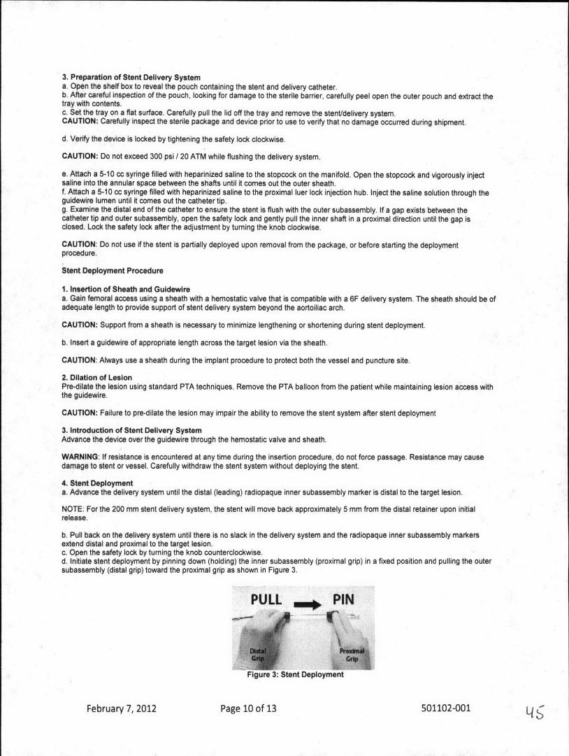
b. Pull back on the delivery system until there is no slack in the delivery system and the radiopaque inner subassembly markersextend distal and proximal to the target lesion.c. Open the safety lock by turning the knob counterclockwise.d. Initiate stent deployment by pinning down (holding) the inner subassembly (proximal grip) in a fixed position and pulling the outersubassembly (distal grip) toward the proximal grip as shown in Figure 3.
PULL PIN
Figure 3: Stent Deployment
February 7, 2012 Page 10 of 13 501102-001
e. Once initial deployment is visible on fluoroscopy and prior to achieving vessel apposition, reposition stent as needed usingradiopaque markers.
NOTE: It is recommended to lock the safety lock in order to ensure that there is no relative movement between the grips duringrepositioning.
CAUTION: The stent system is not designed for recapturing or repositioning after establishing vessel apposition.
f. During deployment of the stent, the whole length of the flexible deployment system should be kept as straight as possible. In orderto ensure that no slack is introduced into the delivery system, hold the proximal grip stationary and fixed. Deployment is completewhen the outer subassembly marker passes the proximal inner shaft stent marker and the stent is released.
WARNING: If resistance is felt when initially pulling back on the distal grip, do not force deployment. Carefully withdraw the stentsystem without deploying the stent.
CAUTION: Failure to hold the proximal grip in a fixed position may result in partial deployment, foreshortening, lengthening orincreased deployment force.
CAUTION: The stent is not designed to be lengthened or shortened past its nominal length. Excessive stent lengthening orshortening may increase the risk of stent fracture.NOTE: If a second stent is needed, place the more distal stent first. If overlap of sequential stents is necessary, the amount ofoverlap should be kept to a minimum.
5. Post Stent Deploymenta. While using fluoroscopy following stent deployment, withdraw the entire delivery system as one unit, over the guidewire, into thecatheter sheath and out of the body. Remove the delivery system from the guidewire.
WARNING: If resistance is met during delivery system withdrawal, advance the outer subassembly until the outer subassemblymarker contacts the catheter tip and withdraw the system as one unit.
b. Using fluoroscopy, visualize the stent to verify full deployment.c. If incomplete expansion exists within the stent at any point along the lesion, post deployment balloon dilation may be performed.
CAUTION: Use caution when crossing a deployed stent with any adjunct device.
CAUTION: Stent should not be expanded past its nominal diameter.
d. To dilate the stent, select an appropriate size PTA balloon catheter and dilate with conventional technique. The inflation diameterof the PTA balloon should approximate the diameter of the reference vessel.e. Confirm full stent expansion is complete, then remove the PTA balloon from the patient.f. Remove the guidewire and sheath from the body.g. Close entry wound as appropriate.h. Discard the delivery system, guidewire and sheath.
MRI INFORMATION
MR ConditionalNon-clinical testing demonstrated that the EverFlex Self-Expanding Peripheral Stent System stent is MR Conditional. A patient maybe scanned safely, immediately after stent placement under the following conditions:
* Static magnetic field of 3-Tesla or 1.5T.* Maximum spatial gradient magnetic field of 720-Gauss/cm or less.* Normal operating mode (maximum WBA-SAR of 2.0 W/kg) for 15 minutes of scanning.
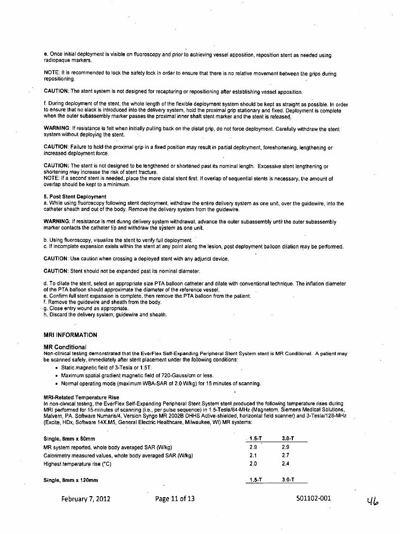
MRI-Related Temperature RiseIn non-clinical testing, the EverFlex Self-Expanding Peripheral Stent System stent produced the following temperature rises duringMRI performed for 15-minutes of scanning (i.e., per pulse sequence) in 1.5-Tesla/64-MHz (Magnetom, Siemens Medical Solutions,Malvern, PA. Software Numaris/4, Version Syngo MR 2002B DHHS Active-shielded, horizontal field scanner) and 3-Tesla/128-MHz(Excite, HDx, Software 14X.M5, General Electric Healthcare, Milwaukee, WI) MR systems:
Single, 8mm x 80mm . 1.5-T 3.0-T
MR system reported, whole body averaged SAR (V/kg) 2.9 2.9Calorimetry measured values, whole body averaged SAR (W/kg) 2.1 2.7Highest temperature rise (*C) 2.0 2.4
Single, 8mm x 120mm 1.5-T 3.0-T
February 7, 2012 Page 11 of 13 501102-001 L&

MR system reported, whole body averaged SAR (W/kg) 2.9 2.9Calorimetry measured values, whole body averaged SAR (W/kg) 2.1 2.7Highest temperature rise ('C) 2.9 3.7
Single, 8mm x 200mm 1.5-T 3.0-TMR system reported, whole body averaged SAR (/kg) 2.9 2.9Calorimetry measured values, whole body averaged SAR (W/kg) 2.1 2.7Highest temperature rise ('C) 3.9 4.5
These temperature changes will not pose a hazard to a patient under the conditions indicated above. It is recommended thatpatients register conditions under which the implant may be scanned safely with the MedicAlert Foundation (www.medicalert.org) orequivalent organization.
Artifact InformationMR image quality may be compromised if the area of interest is in the exact same area or relatively close to the position of the stent.Therefore, optimization of MR imaging parameters to compensate for the presence of this device may be necessary.
SYMBOLLEGENDManufacturer
Consult instructions for use
STERiLE EQ Sterilized using ethylene oxide
Catalogue number
Batch code
A Keep dry
Keep away from sunlight
Use by
Do not reuse
Do not use if package is damaged
Telephone
Facsimile
Rx Only For prescription use only
February 7, 2012 Page 12 of 13 501102-001

WARRANTY DISCLAIMERAlthough this product has been manufactured under carefully controlled conditions, ev3 Inc. has no control over the conditionsunder which this product is used. ev3 Inc. therefore disclaims all warranties, both express and implied, with respect to the productincluding, but not limited to, any implied warranty of merchantability or fitness for a particular purpose. ev3 Inc. shall not be liable toany person or entity for any medical expenses or any direct, incidental or consequential damages caused by any use, defect, failureor malfunction of the product, whether a claim for such damages is based upon warranty, contract, tort or otherwise. No person hasany authority to bind ev3 Inc. to any representation or warranty with respect to the product. The exclusions and limitations set outabove are not intended to, and should not be construed so as to contravene mandatory provisions of applicable law. If any part orterm of this Disclaimer of Warranty is held to be illegal, unenforceable or in conflict with applicable law by a court of competentjurisdiction, the validity of the remaining portions of this Disclaimer of Warranty shall not be affected, and all rights and obligationsshall be construed and enforced as if this Disclaimer of Warranty did not contain the particular part or term held to be invalid,
February 7, 2012 Page 13 of 13 501102-001
w'4
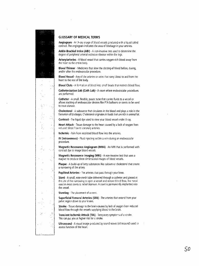
GLOSSARY OF MEDICAL TERMS
Angiogram -An X-ray image of blood vessels produced with a liquid calledcontrast. The angiogram indicates the area of blockage in your arteries.
Ankle-Brachial Index (ABI) -A non-invasive test used to determine thedegree of peripheral arterial occlusive disease within the legs.
Arterylarteries -A blood vessel that carries oxygen-rich blood away fromthe heart to the entire body.
Blood Thinner - Medicines that slow the clotting of blood before, during,and/or after the endovascular procedure.
Blood Vessel -Any of the arteries or veins that carry blood to and from theheart to the rest of the body.
Blood Clots -A formation of blood into small beads that restricts blood flow.
Catheterization Lab (Cath Lab) -A room where endovascular proceduresare performed.Catheter -A small, flexible, plastic tube that carries fluids to a vessel orallows tracking of endovascular devices like PTA balloons or stents to be usedto treat disease.
Cholesterol - A substance that circulates in the blood and plays a role in theformation of blockages. Cholesterol originates in foods that are rich in animal fat.
Contrast -The liquid dye used to view your blood vessel under X-ray.
Heart Attack -Tissue damage to the heart caused by a lack of oxygen fromreduced blood flow to coronary arteries.
Ischemic - Pain from restricted blood flow into the arteries.
IV (Intravenous) - Fluid injecting within a vein during an endovascularprocedure.Magnetic Resonance Angiogram (MRA) -An MRI that is performed withcontrast dye to image blood vessels.
Magnetic Resonance Imaging (MRI) -A non-invasive test that uses amagnet to produce three dimensional images of blood vessels.
Plaque -A build-up of fatty substances like calcium or cholesterol that createa narrowing of the artery.
Popliteal Arteries -The arteries that pass through your knee.
Stent -A small, wire-mesh tube delivered through a catheter and placed atthe site of the narrowing to open a vessel and restore blood flow. The metalused in most stents is nickel titanium. A stent is permanently implanted intothe vessel.
Stenting The placement of a stent.
Superficial Femoral Arteries (SFA) -The arteries that extend from yourpelvic region down to your knees.
Stroke -Tissue damage to the brain caused by lack of oxygen from reducedblood flow through the vessels supplying blood to the brain.
Transient Ischemic Attack (TIA) - Temporary symptoms.of a stroke.This can put you at higher risk for a stroke.
Ultrasound -A visual image produced by sound waves (ultrasound) used toassess function of the heart.
What is PAD? 2
How is PAD treated? ........ 3. 3Lifestyle M odifications......... .. ........ .. . 3M ed icatio n ............. .. . . .. .. 3 .... .. . 3Superficial Femoral Artery Balloon Angioplasty and Stenting .. .... 3Superficial Femoral Artery Bypass Surgery........ .. . ... .3A therectom y .. ... .......... .. . .. ... 3
EverFlex Self-Expanding Peripheral Stent System 4. . ...... ... .. ... 4Device Description. ..... . .... .. . 4When Should the Device Not Be Used (Contraindications).. 4
Stent Implant Procedure Risks and Benefits - , ., . 5What are the risks?... . . 5How will you benefit? 5
How is PAD diagnosed?. . .. 7
Talk to Your Doctor... . 7
Peripheral Stent Procedure .8Before Your Procedure . .... .8During Your Procedure . . .... . . 9
After Your Procedure .......... . .... 10Your Recovery .. .. , -. . ..... . .11
Your Stent Implant Card ...... 11
1S
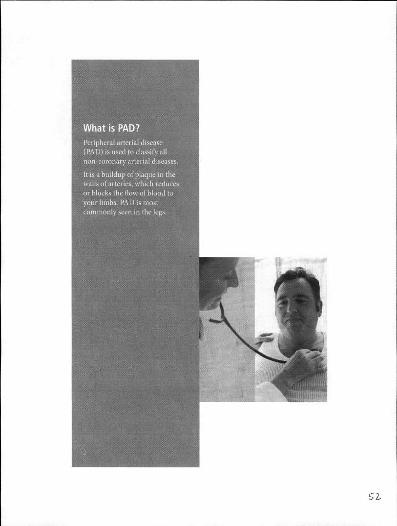
What is PAD?Peripheral arterial disease(PAD) is used to dassify allnon-coronary arterial diseases,
It is a buildup of plaque in theivalls of arteries, which reducesor blocks tht flow of blood toyour linibs. 1,M) is mostcoluniOnly seen in the legs.
52,
How is PAD treated?Your doctor may recommend one or more options for treating your PAD. Treatmentoptions for PAD include lifestyle modifications, medications, surgery, and less-invasiveprocedures, such as placing a stent in the narrowed artery.
LIFESTYLE MODIFICATIONSLifestyle changes your doctor may recommend are:
* Quitting smoking and refraining from use of * Achieving and maintaining a desirabletobacco products weight, including regular exercise
* Controlling high blood pressure and diabetes * Properly controlling other physical* Having regular check-ups with your doctor ailments, such as atrial fibrillation and* Maintaining a diet of foods low in saturated heart disease
fats and cholesterol* Monitoring and controlling your lipids
(good versus bad cholesterol levels)
MEDICATIONYour doctor may also prescribe blood thinner medications. Common drugs used includeaspirin, Plavix", Coumadin" (also known as warfarin), or Ticlid". These drugs lower yourrisk for blood clots. In addition, your doctor may prescribe medications to lower yourblood pressure or cholesterol.
SUPERFICIAL FEMORAL ARTERY BALLOON ANGIOPLASTY AND STENTINGAn angioplasty procedure uses a small tube (catheter) with a small balloon on the endto open the narrowed superficial feioral artery or popliteal artery by compressing theplaque against the vessel wall. This process is designed to expand the narrowed area sothat it no longer restricts the flow of blood through the limb. The balloon is deflated andremoved from the artery.A stent is a metallic tube made of wire mesh that is placed in the opened artery. Whenexpanded, the stent acts as a support that keeps the artery open and therefore restoresnortmal blood flow. Over time, the artery wall heals around the stent, which continues tosupport the vessel.
SUPERFICIAL FEMORAL ARTERY BYPASS SURGERYA man-made graft or one of your veins can be used as a detour (bypass) that actuallycreates a new pathway to carry blood to and through the legs.
ATHERECTOMYIn this minimall[' invasive procedure, a small catheter is inserted into the artery to removeplaque. This helps to restore blood flow through the artery without damaging the arterialwall or leaving anything behind.
3
S3
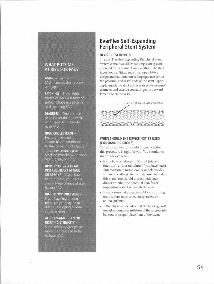
EverFlex Self-ExpandingPeripheral Stent SystemDEVICE DESCRIPTIONThe EverFlex Self-Expanding Peripheral Stent
System contains a self-expanding stent systemT RS intended for permanent implantation. The stent
is cut from a Nitinol tube in an open lattice
design and has tantalum radiopaque markers at
the proximal and distal ends of the stent. Upondeployment, the stent achieves its predetermineddiameter and exerts a constant, gentle outwardforce to open the vessel.
smokng hve gicterrisk Ni'lex Selt-Expandirg Peripheral Stent
WHEN SHOUL.D THE DEVICE NOT BE USED(CONTRAI NDICATIONS)ofu and your doctor should discuss whetherthis procedure is right for you. You should notuse this device when:
* If you have an allergy to Nitinol (nickel,titanium), and/or tantalum. If you have had askin reaction to metal jewelry or belt buckles
you may be allergic to the metal used to makethis stent. You should discuss with yourdoctor whether the potential benefits ofimplanting a slle ouiweigh the risks.
* If you cannot take aspirin or blood-thinningmedications (also called antiplatelets oranticoagulan ts).
* If the physician decides that the blockage willnot allow complete inflation of the angioplastyballoon or proper placement of the stent.
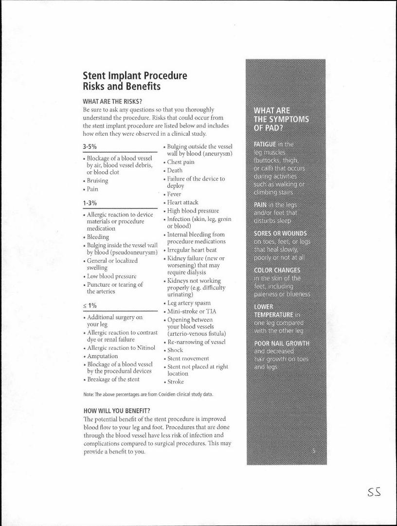
Stent Implant ProcedureRisks and BenefitsWHAT ARE THE RISKS?Be sure to ask any questions so that you thoroughlyunderstand the procedure. Risks that could occur fromthe stent implant procedure are listed below and includeshow often they were observed in a clinical study.
3-5% * Bulging outside the vesselwal by blood (aneurysm) lemucs
* Blockage of a blood vesselby air, blood vessel debris, * Chest painor blood clot Death
* Bruising Failure of the device to
" Pain deployFever
1-3% * Heart attack
* Allergic reaction to device High blood pressurematerials or procedure Infection (skin, leg, groinmedication or blood)
* Bleeding Internal bleeding from *
* Bulging inside the vessel wall procedure medicationsby blood (pseudoaneurysm) * Irregular heart beat
* General or localized * Kidney failure (new or
swelling worsening) that may* Low blood pressure require dialysis
Puncture or tearing of' Kidneys not working*t e rte properly (e.g. difficulty
the arteries uiaigurinating)
<1% Leg artery spasmA Mini-stroke or TIA
* Additional surgery on * Opening betweenyour leg vour blood vessels
* Allergic reaction to contrast (arterio-venous fistula)dye or renal failure 'Re-narrowing of vessel
* Allergic reaction to Nitinol * Shock* Amputation * Stent movement* Blockage of a blood vessel * Stent not placed at right
by the procedural devices location* Breakage of the stent * Stroke
Note: The above percentages are from Covidien clinical study data.
HOW WILL YOU BENEFIT?The potential benefit of the stent procedure is improvedblood flow to your leg and foot. Procedures that are donethrough the blood vessel have less risk of infection andcomplications compared to surgical procedures. This may
provide a benefit to you.
SS
6
How is PAD diagnosed?Whether you see a family physician, internist, physicians assistant, ornurse practitioner, the first step is to ask about your risk for PAD. Yourprovider will take a medical and family history, perform a physical, andconduct diagnostic tests.
PHYSICAL EXAMDuring the physical exam, your health care provider may check:* Pulse in your legs and feet to determine if there is sufficient blood flow* Color, temperature, and appearance of your legs and feet. Signs of poor wound healing on the legs and feet
DIAGNOSTIC TESTSWhen checking for PAD, your health care provider may perform asimple non-invasive test called an ankle-brachial index (AB). Painlessand quick, the ABI compares the blood pressure readings in your ankleswith the blood pressure readings in your arms.
Talk to your DoctorYour doctor is in the best position to advise you of your diagnosisand treatment options. Early diagnosis and treatment can preventcomplications associated with the progression of PAD, If your doctorsuspects that you have PAD, your doctor may use one or more of thefollowing tests to help diagnose your condition:
* Ankle-Brachial Index: The Ankle-Brachial Index (ABI) is a testdone by measuring blood pressure at the ankle and the arm while aperson is at rest. Measurements are usually repeated at the ankle andthe arm after 5 minutes of walking on a treadmill. The result of theABI test is used to predict the severity of PAD. A slight drop in yourABI with exercise means that you probably have PAD. This drop maybe important because PAD can be linked to a higher risk of heartattack or stroke.
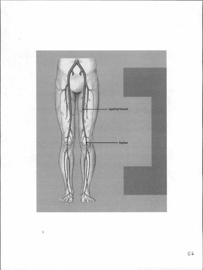
* Superficial femoral artery ultrasound: A sound-wave test thatprojects an image of the superficial femoral artery onto a screen.This test allows the size of the vessel to be measured and the flowof blood to the legs to be tracked. This can be helpful in identifyingnarrowing in the superficial femoral artery. This test is painless anddoes not require the use of needles, dye, or X-ravs.
* Fluoroscopy/Angiogran: An X-ray based image obtained byinjecting contrast dye through a small tube (catheter) inserted into anartery in the groin or arm. This procedure shows exactly where thenarrowing is located and will help to guide further treatments.
7
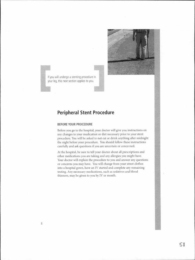
If you will undergo a stenting procedure inyour leg, this next section applies to you.
Peripheral Stent Procedure
BEFORE YOUR PROCEDURE
Before you go to the hospital, your doctor will give you instructions onany changes to your medication or diet necessary prior to your steatprocedure. You will be asked to not eat or drink anything after midnightthe night before your procedure. You should follow these instructionscarefully and ask questions if you are uncertain or concerned.
At the hospital, be sure to tell your doctor about all prescriptions andother medications you are taking and any allergies you might have.Your doctor will explain the procedure to you and answer any questionsor concerns you may have. You will change from your street clothesinto a hospital gown, have an IV started and complete any remainingtesting. Any necessary medications, such as sedatives and bloodthinners, may be given to you by IV or mouth.
8
DURING YOUR PROCEDURE
1. Your procedure will occur in a catheterization laboratory. Physicians,nurses, and technologists will place you onto the procedure table andattach monitoring equipment to you for the stenting procedure.
2. You may be given additional medication to help you relax.
3. During the procedure, the room will be darkened and a large camera placedover you. The camera will rotate and move throughout the procedure.
4. Just prior to placing any catheters into an artery most likely in your groin,you will feel a small stinging sensation as your doctor numbs the area,
i. The doctor will use various wires and catheters to gain access to yoursuperficial femoral or popliteal artery. These catheters will enter yourbody through the small incision in your groin. The catheters will travelthrough your body via your arteries until they reach the area to betreated. Because there are no nerve endings in your arteries, you will notbe able to feel these wires and catheters moving inside you.
6. As your doctor treats you, he or she will take many X-ray pictures of youusing the X-ray camera and contrast dye. This contrast dye may brieflymake you feel warm as it is injected.
7. If the diseased area in your artery has caused the vessel to become verynarrow, your doctor may use a small balloon to open it slightly. This willhelp your doctor reach the area where your stent will be placed. You mayfeel some pressure in your leg when the balloon is inflated.
8. Your doctor will use a special catheter to place the stent in the diseasedarea of your superficial femoral artery or popliteal artery. This cathetercontains the stent in a compressed state. Your doctor will carefully alignthe catheter and deploy the stent.
9. After the EverFlex Self-Expanding Peripheral Stent is deployed, yourdoctor may use another balloon to ensure that your artery is fully openedand any plaque is pushed against the vessel wall. You may feel a smallamount of pressure in your leg as the balloon is inflated. The stent willstay in place permanently
10. When your doctor is satisfied that your artery has been treated properly,he or she will remove the catheter device from your body. The stent staysin your body.
9
After Your ProcedureOnce your procedure is complete, you will beplaced on bed rest in the hospital. The amountof time before you are allowed to stand ormove freely will depend on how the incisionfrom the catheter insertion is closed and whatmedication you have been given. During thistime, the doctors and nurses will monitor youcarefully. As always, you should let your doctorknow immediately if you experience anyunusual sensations such as pain, numbness,tingling, dizziness, or difficulty seeing,hearing, speaking, or swallowing.
YOUR RECOVERYBefore you leave the hospital, your doctor will give you guidelines for activities,diet, and medications. Because medications will be an important part of yourtreatment, your doctor will prescribe drugs that you should take at home tohelp prevent clots from forming. Always follow your doctor's instructionsvery carefully and ask questions if there is anything you do not understand.It is also important to keep all follow-up appointments that are scheduled soyour doctor can follow your progress closely He or she may give you tests suchas an ECG, ultrasound, and/or blood work as part of the follow-up process.These tests are designed to detect any problems that may arise and will helpyour doctor to ensure your complete recovery.
You may need to have an MRI or MRA to look at your arteries some timeafter your stent implant. You can have an MRI or MRA at any time after yourstent is implanted: the EverFlex'" Self-Expanding Peripheral Stent is MRIcompatible. Before having an MRI, make sure you let the people operating theMRI equipment know that you have a stent. Please keep your Stent ImplantCard with you and present it to the people running the MRI equipment so
they know what type of equipment to use. The majority of patients who gohome after a successful stent implantation have no further problems. If youdo experience any discomfort or bleeding from your puncture site contactyour doctor immediately. If your doctor is unavailable, contact your localemergency service and have them take you to the nearest hospital.
YOUR STENT IMPLANT CARDTell any dentist or doctor who treats you for any condition that you havea stent implant in your leg, and keep your Stent Implant Card with youat all times. Your Stent Implant Card identifies the doctor who implantedyour stent and how to reach him or her, the hospital where the proceduretook place, and the location in which the stent was placed. It also identifiesimportant information about your stent, such as the size of the stent andthe date the stent was manufactured. The card gives your doctor valuableinformation that is necessary if you need an MRI or MRA. There are alsophone nunbers on the card that your dentist or doctor can call if he or shehas any questions. It is recommended that patients register conditions underwhich the implant maybe scanned safely with the MedicAlert Foundation(wwwrmedicalert.org) or equivalent organization.
11

NOTES
tooJ

CCVVEtJ COVIDIEN w&it logo alid (Cmidien logo are US.and internationally regtstered tradorarks of Coeidien AG.
COVIIENTM- Trademrk of its aesectifea .,,er.C12V2IDIENiopoatw reults joe lift'I S703-OWrItA) jAN'1 2
(0