evidence based management third molars - aaoms · evidence_based_management_third_molars.ppt...
TRANSCRIPT
Evidence-‐Based Management of Third Molar Teeth

Disclaimer The informa,on depicted in this slide presenta,on is intended for educa,onal purposes only to help health care professionals be6er understand the evidence-‐based management and treatment of third molar teeth. It is not meant as professional advice as all therapeu,c decisions remain the responsibility of individual prac,,oners. The opinions expressed and statements made on this Evidence-‐based Third Molar PowerPoint Presenta,on are those of the presenters and do not imply endorsements by or official policy of the AAOMS unless specifically stated otherwise. AAOMS makes no representa,on or warranty with respect to the material contained within. As with all clinical materials, these slides reflect the science at the ,me of their development. They should be used with the clear understanding that con,nued research and prac,ce may result in new knowledge or recommenda,ons. They are not intended to set any standards of care, and they cannot be subs,tuted for individual professional judgment brought to each clinical situa,on. For assistance concerning the use of this presenta,on and development of any personal disclaimers to accompany this presenta,on, fellows and members need to consult their own a6orneys locally.
Impacted third molars
Wisdom teeth, or third molars, are located at the back of the mouth. They are the last adult teeth to erupt, or enter the mouth. Most people have four wisdom teeth, two on the top, two on the bo6om.
Third molars are considered to be “impacted” when they don't have enough room to emerge or grow normally.

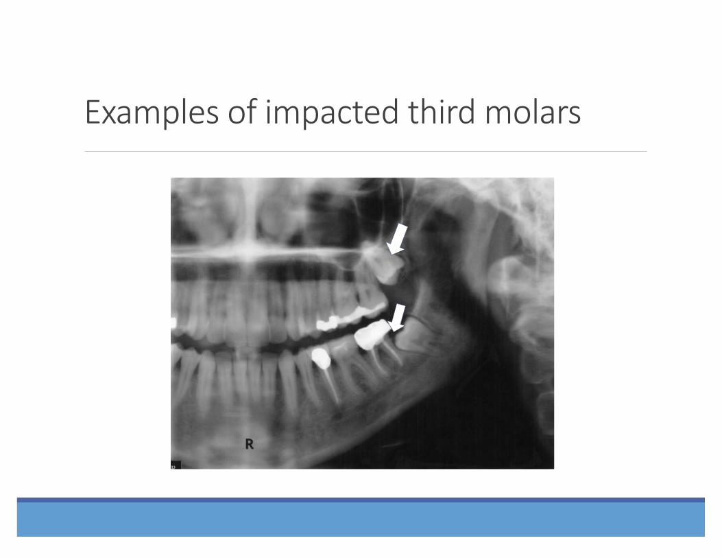
Examples of impacted third molars
⇑ ! ⇑ !
Examples of impacted third molars
⇑ !
⇑ !

Examples of impacted third molars
⇑ ! ⇑
!

Examples of impacted third molars
⇑ ! ⇑
!
⇑ ! ⇑ !

16-‐year-‐old female with 4 asymptoma,c and unerupted M3s.
Organization Name!
⇒ !
⇒ !⇒ !
Examples of a common dilemma
⇒ !
⇒ !⇒ !
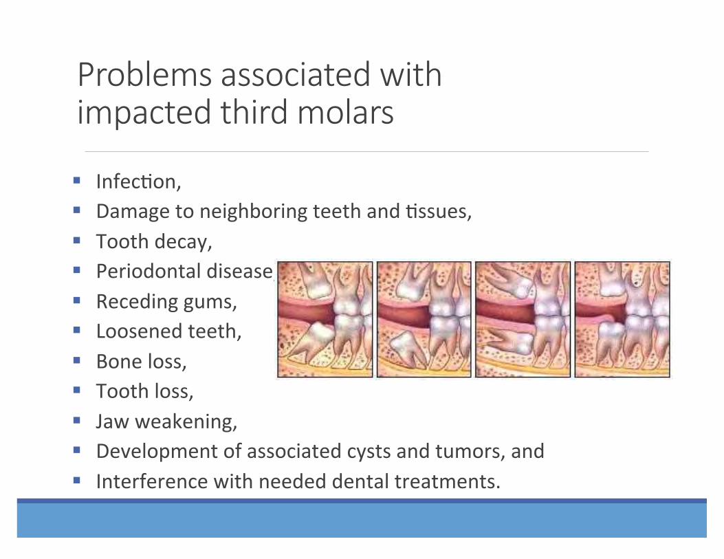
Problems associated with impacted third molars
§ Infec,on, § Damage to neighboring teeth and ,ssues, § Tooth decay, § Periodontal disease, § Receding gums, § Loosened teeth, § Bone loss, § Tooth loss, § Jaw weakening, § Development of associated cysts and tumors, and § Interference with needed dental treatments.

Problems associated with malposi?oned third molars
Ectopic posi,oning due to supra-‐erup,on:
⇐ !⇐ !
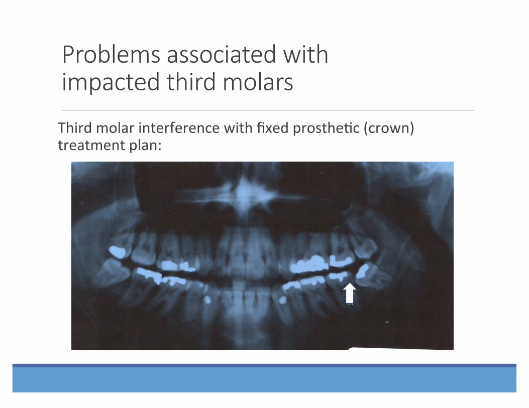
Problems associated with impacted third molars
Third molar interference with fixed prosthe,c (crown) treatment plan:
⇑ !⇑ !

Retained third molar tooth interference with removable prosthe,c treatment plans:
Problems associated with impacted third molars
⇒ !

Impacted third molar interference with erup,on of adjacent second molar and orthodon,c tooth movement:
Problems associated with impacted third molars

Problems associated with impacted third molars
Impacted third molar in the line of fracture:

Cysts and Tumors
Bilateral Mandibular Den,gerous Cyst
Problems associated with impacted third molars

“Asymptoma?c” versus “Disease-‐Free”
§ “Asymptoma,c” does not mean “Disease Free” § Even third molars that have erupted into the mouth in a normal, upright posi,on may not be problem-‐free.
§ Their loca,on in the back of the mouth makes them extremely difficult to keep clean.
§ Bacteria that cause periodontal disease may exist in and around asymptoma,c third molars, leading to damage before symptoms appear.
§ Pathology is always present before symptoms appear. § Once damage has occurred, it is not always
treatable.

Addi?onal risks
§ Bacteria may contribute to systemic health problems, including: § diabetes, § heart disease, § kidney disease, and § other health problems.
§ Studies have found that periodontal disease in expectant mothers may be associated with a greater likelihood of preterm and low birthweight babies.

Research has also shown a rela?onship between the presence of wisdom teeth and the progression of periodontal disease.
§ White RP, Offenbacher S, et.al. Chronic oral inflamma,on and the progression of periodontal pathology in the third molar region. J Oral Maxillofac Surg. 64:880, 2006

25% of wisdom teeth pa?ents who perceive themselves as asymptoma?c actually already have inflammatory periodontal disease.
§ Blakey GH, Marciani RD, Haug RH, et.al: Periodontal pathology associated with asymptoma,c third molars; Journal of Oral and Maxillofacial Surgery. 2001;60:1227-‐1233
The role of age in third molar surgery
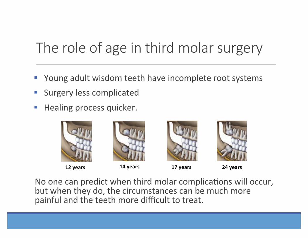
§ Young adult wisdom teeth have incomplete root systems § Surgery less complicated § Healing process quicker.
No one can predict when third molar complica,ons will occur, but when they do, the circumstances can be much more painful and the teeth more difficult to treat.
12 years 14 years 17 years 24 years

Morbidity associated with surgical management of third molar teeth, as well as the risk of complica=ons, has been shown to increase with age.
§ Bui CH, Selodin EB, Dodson TB. Types, frequencies and risk factors for complica,ons aeer third molar extrac,on. J Oral Maxillofac Surg . 61: 1379, 2003
§ Bouloux GF, Steed MB, and Perciaccante VJ. Complica,ons of third molar surgery. Oral and Maxillofacial Surgery Clinics of North America. 19:117-‐128, 2007
§ Marciani RD. Third molar removal: An overview of indica,ons, imaging, evalua,on, and assessment of risk.. Oral and Maxillofacial Surgery Clinics of North America. 19:1-‐13, 2007
The risk of future disease requiring removal of retained wisdom teeth in asymptoma=c pa=ents who retain their wisdom teeth, exceeds 70% aEer 18 years of follow-‐up.
§ Venta I, Ylipaavalniemi P, Turtola L: Clinical outcome of third molars in adults followed during 18 years. J Oral Maxillofac Surg. 62:182, 2004

20 years aEer UK adopts the “Na=onal Ins=tute of Clinical Excellence” (NICE) guidelines, volume of third molar surgeries decrease, with a corresponding increase in mean age for surgical admissions and an increase in “caries” and “pericoroni=s” as e=ologic factors.
§ Renton T, Al-‐Haboubi M, Pau A, Shepherd J, Gallagher JE: What has been the United Kingdom’s experience with reten,on of third molars? J Oral Maxillofac Surg. 70:48-‐57, 2012, Suppl 1

Reten=on of third molars is associated with increased risk of second molar pathology in middle-‐aged and older adult men.
§ Nunn, ME, et al. Retained Asymptoma,c Third Molars and Risk for Second Molar Pathology. Nunn et al. J DENT RES published online 16 October 2013.

Evidence-‐based management of third molars
Every den,st and dental specialist should understand the importance of evidence-‐based management of third molar teeth and office-‐based anesthesia in the dental office.
What is evidence-‐based management?
A systema,c approach to clinical problem solving which allows the integra,on of the best available SCIENTIFIC evidence with clinical exper,se and pa,ent values.
(Sacke6 DL, Strauss SE, Richardson WS,et al. Evidence-‐based medicine: how to prac,ce and teach EBM. London: Churchill-‐Livingstone, 2000)

Evidence-‐Based Clinical Decision-‐Making (EBCD)
§ Apply per,nent data to each individual case § Cri,cally weigh the treatment op,ons
§ Choose the best course of ac,on for the pa,ent
Evalua?ng third molars for surgery
§ Every pa,ent and every case is unique. § Decisions regarding surgery must consider: § Careful examina,on of your mouth; § Radiographic examina,on involving x-‐rays or a CT-‐scan; and
§ Consulta,on between pa,ent, den,st and oral and maxillofacial surgeon with a balanced discussion of the benefits and risks of reten,on versus benefits and risks of opera,ve management.

Available op?ons
§ Depending on the results of the dental examina,on, the OMS may: § Extract the complete tooth; § Par,ally remove the tooth; or § Ac,ve Surveillance -‐ Observe the tooth over ,me for changes in condi,on.

What is “par?al removal?”
Par,al removal of third molars, or “Coronectomy,” is a surgical procedure that removes the crown of the tooth and leaves the root and associated nerve complex.
§ May be performed when removing the en,re tooth might damage the nerve, poten,ally causing facial numbness.
Pogrel, MA, Lee, JS, Muff, DF: Coronectomy: A Technique to Protect the Inferior Alveolar Nerve. J Oral Maxillofac Surg. 1447-‐1452, 2004

When must third molars be surgically removed?
§ In general, dental professionals agree that third molars should be removed whenever there is evidence that predicts: § Periodontal disease, § Cavi,es that cannot be restored, § Infec,ons, § Cysts or tumors, and/or § Damage to neighboring teeth.

Removal of wisdom teeth is always appropriate “where there is evidence of pathological changes such as infec,on, non-‐restorable carious lesions, cysts, tumors, and damage to adjacent teeth.” (American Public Health Associa,on (APHA) Statement on Wisdom Teeth, 2008)

Under what circumstances may third molars be retained?
Third molars may not require surgery if they are:
§ Completely erupted and func,onal,
§ Painless, § Free of cavi,es, § Disease-‐free, and § In a posi,on that can be kept clean and healthy.

AAOMS Third Molar Posi?on Statement
§ All pa,ents with impacted teeth should be carefully evaluated by a qualified den,st. Predicated on the best evidence-‐based data, impacted teeth that demonstrate pathology, or are at high risk of developing pathology, should be surgically managed. In the absence of pathology or significant risk of pathology, ac,ve clinical and radiographic surveillance is indicated.
§ All third molar teeth should be managed deliberately using an evidence-‐based approach. Appropriate management of third molar teeth may include removal, par,al removal or reten,on, followed by ac,ve clinical and radiographic surveillance to make sure that pathology does not develop.
§ The absence of symptoms does not necessarily mean the absence of disease.
§ All third molar teeth should be managed by a qualified den,st. Oral and maxillofacial surgeons surgically manage acute, chronic and poten,al pathological condi,ons of third molar teeth.
§ Third molar therapy is an evidence-‐based treatment paradigm. It includes radiographic surveillance to assess tooth posi,on, pathology and possible need for removal. All third molars that are retained require periodic follow-‐up.
AAOMS Third Molar Posi?on Statement

Deliberate management of third molar teeth
The Oral and Maxillofacial Surgeon has the most extensive educa,on and training for appropriate third molar management. This includes:
§ Removal
§ Par,al removal
§ Reten,on with ac,ve surveillance

Case Reports

A 32 year-‐old physician presented with bilateral jaw pain that started one week previously. Prior to this episode, he was completely asymptoma,c. Examina,on revealed malposed and impacted third molar teeth with gross caries involving teeth #s 17, 18, 31 and 32.

Carious mandibular second and third molar teeth
⇑ ! ⇑
!

A 39 year-‐old female pa,ent is evaluated prior to crown replacement at the lower lee second molar tooth #18.

This 39 year old female pa,ent is scheduled for replacement of crown #18. The impacted third molar teeth should first be appropriately managed.
⇑ !

An otherwise healthy 63 year-‐old male pa,ent (also a physician) presented to the office with complaints of pain and swelling associated with the area of the mandibular lee third molar tooth #17. Clinical examina,on revealed recurrent caries, pericoroni,s and a mesial periodontal probing depth of 8mm.

Periodontal bone loss and pericoroni?s
⇑ !

An otherwise healthy 25 year-‐old male pa,ent presented to the hospital emergency department with complaints of severe pain and swelling of the lee face and neck, trismus, and difficulty swallowing, speaking and breathing. Clinical examina,on and a CT scan showed an abscessed lower lee wisdom tooth with infec,on of the lee submandibular, masseteric, medial pterygoid and sublingual spaces. A shie of the airway to the right was beginning on the CT scan. Laboratory values were consistent with acute infec,on.

Abscessed mandibular leS third molar tooth

Coronal CT scan of odontogenic infec?on
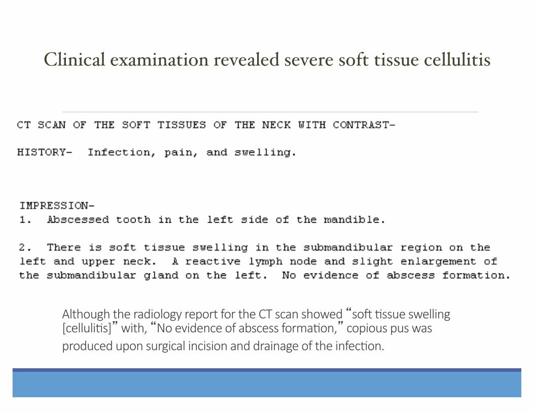
Although the radiology report for the CT scan showed “soS ?ssue swelling [celluli?s]” with, “No evidence of abscess forma?on,” copious pus was produced upon surgical incision and drainage of the infec?on.
Clinical examination revealed severe soft tissue cellulitis!


An 80 year-‐old female seen at a local hospital with a right facial infec,on underwent emergent incision and drainage by an ENT surgeon who placed her on an,bio,cs and referred her to the OMS. When first seen by the OMS, she was infected and in severe pain. Clinical and radiographic evalua,on revealed a severely atrophic mandible with a grossly carious impacted mandibular right third molar tooth with an associated radiolucency (odontogenic cyst).

Atrophic mandible with carious impacted right third molar tooth #32 and cyst

Postopera?ve radiograph following removal of tooth #32 and associated cyst

A 42 year-‐old male experienced pain and swelling associated with his right mandibular wisdom tooth that was consistent with pericoroni,s. His symptoma,c lee maxillary and mandibular wisdom teeth were unevennully removed when he was 18 years-‐old. Clinical and radiographic evalua,on revealed probing depths >7mm and a pericoronal radiolucency (den,gerous cyst) at tooth #32.

Mandibular right third molar tooth #32 with pericoroni?s and den?gerous cyst

Periopera?ve boney defect right posterior mandible

Odontogenic cyst
A 66 year-‐old female experienced mild intermi6ent pain associated with her previously asymptoma,c lee mandibular “wisdom” tooth. Clinical and radiographic evalua,on revealed a large pericoronal radiolucency (Den,gerous Cyst) at impacted tooth #17.

LeS mandibular den?gerous cyst

Bilateral odontogenic cysts
A 47 year-‐old male experienced pain and swelling associated with his lee mandibular wisdom tooth. Clinical and radiographic evalua,on revealed a large pericoronal radiolucency (den,gerous cyst) at tooth #17, a smaller pericoronal radiolucency (den,gerous cyst) at tooth #32, and carious mandibular second molar teeth.

Bilateral mandibular den?gerous cysts

A 27 year-‐old pa,ent was referred by his general den,st for evalua,on and treatment of a lesion of the lee mandible. He had undergone removal of his asymptoma,c “wisdom teeth” with another den,st seven years previously. The panoramic radiograph revealed a unilocular radiolucency consistent with a large residual cyst. However, clinical examina,on revealed boney expansion sugges,ve of a more aggressive process. A Cone Beam CT scan showed erosion through the buccal and lingual plates. Surgical excision of the lesion combined with aggressive manual and rotary cure6age was followed by applica,on of Carnoy’s solu,on. Pathologic diagnosis revealed a Keratocys,c Odontogenic Tumor (KCOT) of the lee mandible.
Odontogenic tumor/KCOT
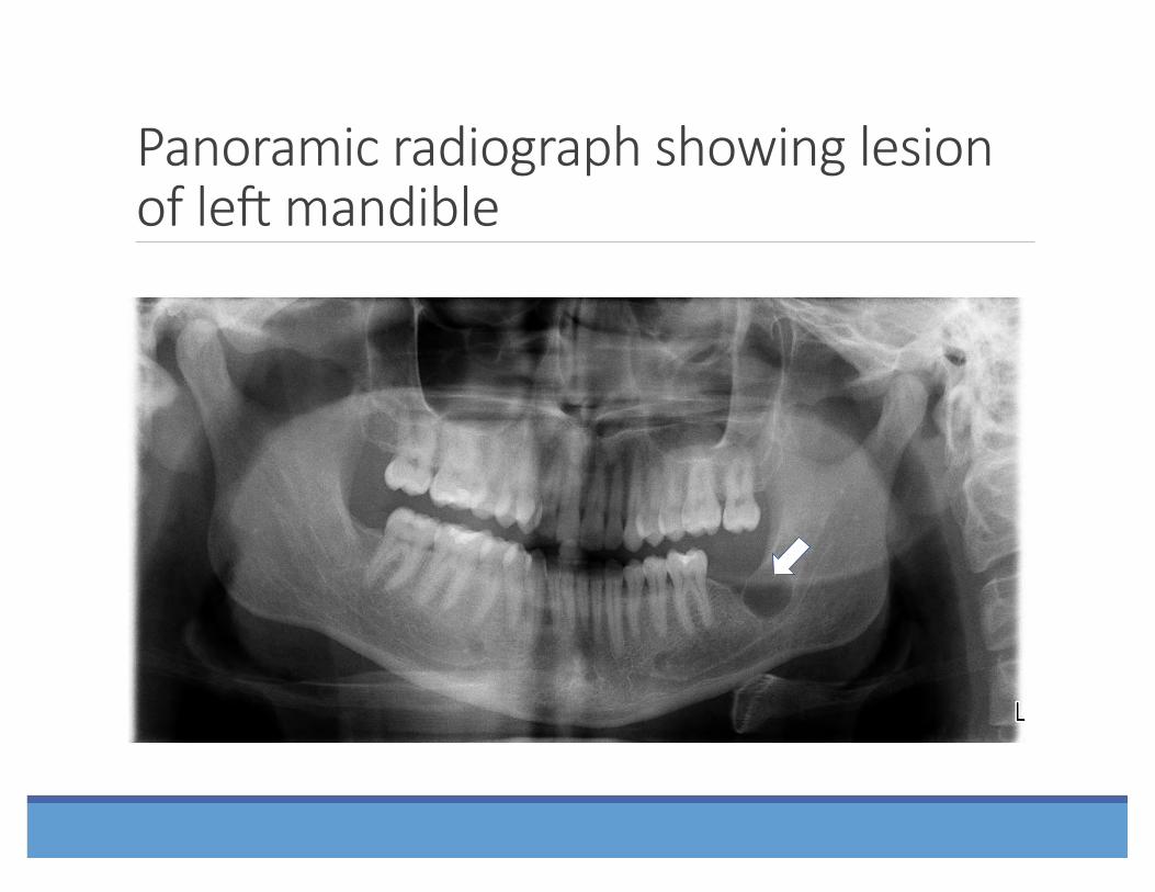
Panoramic radiograph showing lesion of leS mandible

Cone beam CT in panoramic view showing KCOT of leS mandible

Cone beam CT in coronal view showing KCOT of leS mandible
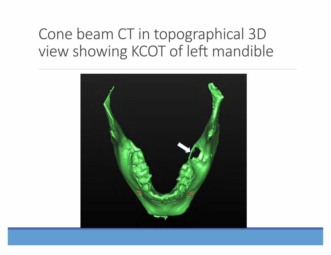
Cone beam CT in topographical 3D view showing KCOT of leS mandible

Cone beam CT in topographical 3D view showing KCOT of leS mandible

⇓ !
Cone beam CT in topographical 3D view showing KCOT of leS mandible
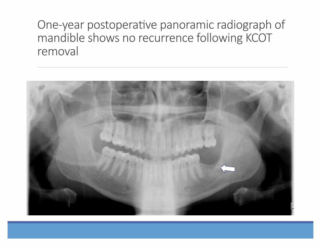
One-‐year postopera?ve panoramic radiograph of mandible shows no recurrence following KCOT removal
⇒ !

Odontogenic tumor
A completely asymptoma,c 17 year-‐old pa,ent was referred by her orthodon,st for removal of her third molar teeth and evalua,on of a mixed radiopacity/radiolucency of her lee posterior maxilla (odontoma with unicys,c amelloblastoma).

Cone beam CT scan of odontoma and ameloblastoma of leS maxilla in panoramic view
⇑ !

Cone beam CT scan of odontoma and ameloblastoma of leS maxilla in coronal view

Cone beam CT scan of odontoma and ameloblastoma of leS maxilla 3D view from below

⇑ !
Cone beam CT scan of odontoma and ameloblastoma of leS maxilla 3D view from above
⇑ !

Odontoma with unicys?c ameloblastoma

For more informa?on, please see the AAOMS Advocacy White Paper on Evidence-‐Based Third Molar Surgery