evidence-based topics for today class ii treatment levels of · pdf file ·...
TRANSCRIPT
4/04/2014
1
Evidence-based
Class II treatment
Peter Miles Virginia - 2014
Levels of Evidence – Cochrane Reviews
Very early treatment (5-8) – should we?
Early treatment (9-12)
Class II – Sliding extraction mechanics
Anchorage – TPA‟s, TAD‟s
Class II – non-extraction timing of treatment
Class II – non-extraction treatment options
Topics for today
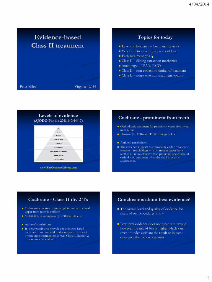
Levels of evidence(AJODO Pandis 2011;140:446-7)
www.TheCochraneLibrary.com
Cochrane - prominent front teeth
Orthodontic treatment for prominent upper front teeth in children
Harrison JE, O'Brien KD, Worthington HV
Authors' conclusions
The evidence suggests that providing early orthodontic treatment for children with prominent upper front teeth is no more effective than providing one course of orthodontic treatment when the child is in early adolescence.
Cochrane - Class II div 2 Tx
Orthodontic treatment for deep bite and retroclinedupper front teeth in children
Millett DT, Cunningham SJ, O'Brien KD et al.
Authors' conclusions
It is not possible to provide any evidence-based guidance to recommend or discourage any type of orthodontic treatment to correct Class II division 2 malocclusion in children.
Conclusions about best evidence?
The overall level and quality of evidence for
many of our procedures is low
Low level evidence does not mean it is „wrong‟
however the risk of bias is higher which can
over or under-estimate the result or in some
cases give the incorrect answer
4/04/2014
2
Evidence-Based Clinical
Orthodontics
Eds: Peter Miles, Dan &
Don Rinchuse
Tizziano Baccetti
William Brantley
Ali Darendeliler, Lam Cheng
Ted Eliades, Nick Pandis
Sanjivan Kandasamy
Eric Liou, James Noble
Peter Ngan, Tim Tremont
Jack Sheridan
Quintessence
Sept 2012 Very early treatment
(ages 5 – 8)
Eruption Guidance Appliances
Occlus-o-Guide
Nite-Guide
MRC Pre-orthodontic Trainer
T4K – soft
Myobrace
Eruption Guidance Appliance
Methenitou S et al. J Pedod 1990; 14:219-30.
43 subjects, avg age 6.17 years,
Nightly wear over 13 mths
Overbite reduced 2 mm
Overjet reduced 1.6 mm
Trainer appliance (MRC)
Usumez et al. Angle 2004;74:605-609
20 patients vs. 20 controls
Age 9.6 yrs, Tx over 13.1 mths
Lower incisors tipped forward 4º, upper incisors
tipped back 2.7º, no skeletal effect
Eruption Guidance Appliance
Keski-Nisula et al. AJODO 2008;133:254-60.
167 subjects of 243 (selection bias)
31% were not included
Age= 5.1yo, 2-3 appliances used in each patient
4/04/2014
3
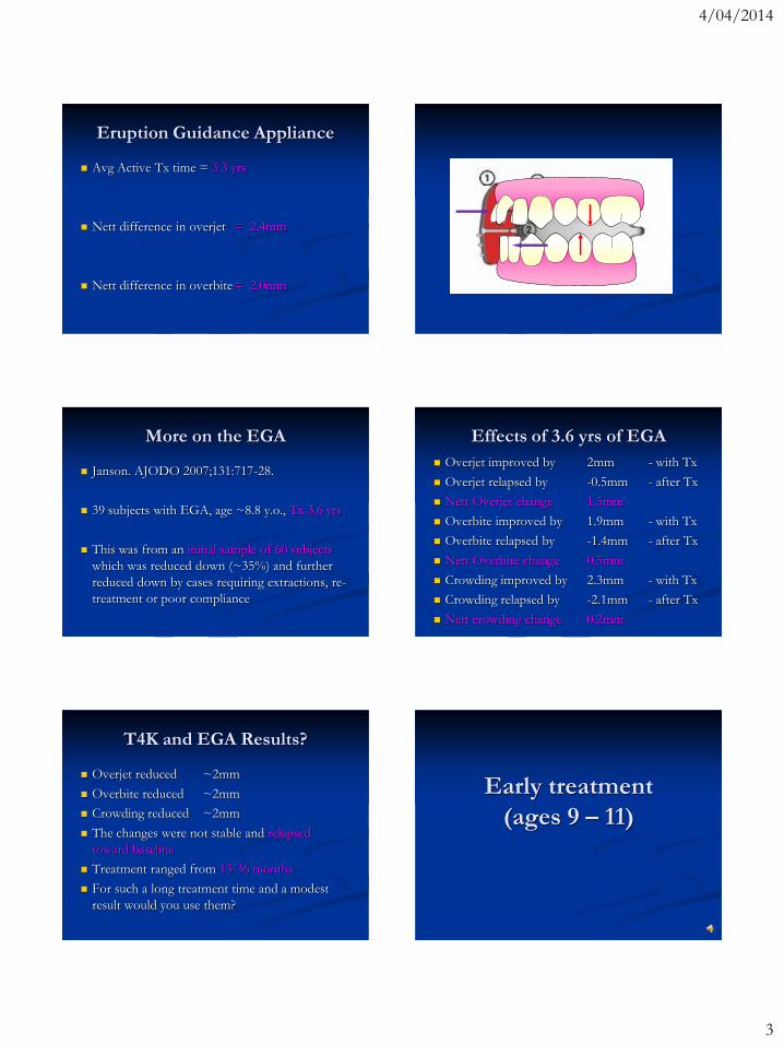
Eruption Guidance Appliance
Avg Active Tx time = 3.3 yrs
Nett difference in overjet = 2.4mm
Nett difference in overbite= 2.0mm
More on the EGA
Janson. AJODO 2007;131:717-28.
39 subjects with EGA, age ~8.8 y.o., Tx 3.6 yrs
This was from an initial sample of 60 subjects
which was reduced down (~35%) and further
reduced down by cases requiring extractions, re-
treatment or poor compliance
Effects of 3.6 yrs of EGA
Overjet improved by 2mm - with Tx
Overjet relapsed by -0.5mm - after Tx
Nett Overjet change 1.5mm
Overbite improved by 1.9mm - with Tx
Overbite relapsed by -1.4mm - after Tx
Nett Overbite change 0.5mm
Crowding improved by 2.3mm - with Tx
Crowding relapsed by -2.1mm - after Tx
Nett crowding change 0.2mm
T4K and EGA Results?
Overjet reduced ~2mm
Overbite reduced ~2mm
Crowding reduced ~2mm
The changes were not stable and relapsed
toward baseline
Treatment ranged from 13-36 months
For such a long treatment time and a modest
result would you use them?
Early treatment
(ages 9 – 11)

4/04/2014
4
Cochrane - prominent front teeth
Orthodontic treatment for prominent upper front teeth in children
Harrison JE, O'Brien KD, Worthington HV
Authors' conclusions
The evidence suggests that providing early orthodontic treatment for children with prominent upper front teeth is no more effective than providing one course of orthodontic treatment when the child is in early adolescence.
Timing of Class II treatment
If UK and UNC studies find no difference
between early and late adolescence then why
treat early?
They were designed to evaluate if early
treatment was beneficial or advantageous over
later treatment for patient‟s with overjet >7 mm
Patient/parent demand (informed)
Psychosocial benefit
Trauma benefit
Psychosocial benefit
Factor Twin Block Control P
Appearance 9.3 8.0 0.005
Anxiety 10.8 9.3 0.005
Popularity 9.9 8.3 0.005
Happiness 8.9 8.2 0.005
Total 63.6 59.0 0.005
Results of UK study - Trauma
Half the trauma had occurred before
orthodontic treatment was started
Treatment did not have an effect on the
incidence of trauma to the incisors (p=0.36)
8% early Tx vs. 14% late Tx
Results of UNC study - Trauma
New incisor trauma in Phase 1
Headgear 4 in 50 = 8%
Functional 3 in 52 = 6%
Control 9 in 61 = 15%
New incisor trauma in Phase 2
Headgear 4 in 46 = 9%
Functional 5 in 42 = 12%
Control 12 in 51 = 24%
Results of UNC study - Trauma
Should we treat very early (~age 7) to reduce
incisor trauma?
The estimated Cost vs. Risk of trauma was
calculated and higher in the control group BUT
Most injuries were minor
The expected extra cost of early treatment in a 2-
phase strategy is significantly more
Choice will be affected by a family‟s values and
the relative risk they are willing to bear
4/04/2014
5
Univ of Florida study
After the 1st phase the incidence of trauma was
7-8% higher than the early treatment group
(p=0.13)
Consistent finding amongst all 3 trials suggesting
it is a real difference (more power)
Higher incidence in boys
Florida study – incisor trauma
Effect of early Class II treatment on the
incidence of incisor trauma.
Chen et al. AJODO 2011;140:e155-e160.
3 groups
Headgear/biteplane
Bionator
Control, age ~9.6 yo
80% were enamel only, 19% involved dentine,
1% had pulpal involvement
Relative risk of trauma
If you delay treatment to age 12-13 vs. treat at 9-
10, what is the relative increased risk of trauma?
UK and UNC studies show 7-8% elevated risk
Florida study shows 19% dentine, 1% pulpal
Therefore:
80% of 8% = ~6.4% risk of enamel chip
19% of 8% = ~1.5% risk of dentine involvement
1% of 8% = ~0.08% risk of pulpal involvement
Informed choice and risk assessment
Expansion stimulates jaw growth
Expansion stimulates growth?
Guest, McNamara et al. AJO 2010;138:582-91
50 Class II subjects with RME. Some also had
partial braces or a lower Schwarz expander.
Compared with literature control group
“The protocol ... can help to improve the Class
II malocclusion as a side-effect.”
“The results of this study show that the
improvements are far more pervasive than
anticipated.”
Expansion stimulates growth?
Molar 6/6
Co-Gn
Overjet
1.7mm
1.3mm
1mm

4/04/2014
6
Expansion stimulates growth?
Historical control so less valid comparison
No blinding so risk of bias
If the changes are real, do they hold up over time?
Systematic review
Lagravere et al. Angle 2005;75:1046–1052
No significant alterations in A-P were found in
any of the studies reviewed.
After the posttreatment and postretention, the
maxilla and mandible of the treated groups
presented no statistical or clinical significance.
Expansion in Class II
Angle Orthod. 2011;81:298–303
Farronato et al.
In Class II patients ... The ANB decreased,
statistically improving the skeletal classification.
8.8 yo expansion – observed 6mths? unclear?
Avg 1.81° decrease in ANB
Occlusal change was Not measured
Expansion stimulates growth?
Volk et al. AJODO 2010;137:310-5
Small retrospective study of 13 Class II subjects
who underwent expansion and then observation
7 of the 13 subjects underwent improvement
5 of the remaining subjects actually got worse
The authors concluded their results do not
support the „foot in the shoe‟ theory and that
maxillary expansion does not predictably
improve Class II dental relationships.
Plates vs. Fixed expanders
Expansion of maxillary aches with crossbite:
Systematic review of RCT‟s in the last 12 years.
Zuccati et al. Eur J Orth 2013:35:29-37.
Plates were unsuccessful in 30% of patients due
to poor cooperation.
Expansion arches were as good as quad-helix
Plates vs. Fixed expanders
Early correction of posterior crossbites – a cost
minimisation analysis
Petren et al. Eur J Orth 2013:35:14-21
Quad-helix was the most cost effective
compared with a removable plate mainly due to
poor compliance with plates.
Petren et al. AJODO 2003;133:790.e7-e13

4/04/2014
7
RPE with anterior arms Early treatment - should we?
Do the findings of the UNC and UK studies
indicate we should not do early treatment?
Their purpose was to assess whether early Tx in
OJ > 7 mm resulted in enhanced mandibular
growth and simplified future treatment
This is not to say that early treatment is not
indicated for some individuals – but which ones?
UK study 2 phase
Early Tx for Cl2 Div 1 malocclusion with the
Twin-block appliance: ......a multi-center RCT
O‟Brien et al. AJODO 2009;135:573-579
~15% of the early Tx group did not proceed
with a 2nd phase. This could be due to
A successful outcome or close-enough and no other
Tx needed
Burnt out and did not want further Tx
But.. Cost is not a factor as provided free of charge
Twin-block RCT
Incremental versus maximum bite advancement
during Twin-block therapy: A randomized
controlled clinical trial
Banks, ..., O‟Brien. AJODO 2004;126:583-8
203 subjects aged 10-14 randomly assigned to
full advancement vs. Incremental advancement
Twin-block RCT
70 – 81% compliance rate in this study
UK Twin-block study had 84% compliance
Harradine. Clin Orthod Res 2000;3:202-9. had
83% compliance rate with TB
Patients aged ≤ 12.3 years were 3 times more
likely to complete treatment
Operator influenced a) compliance and b)
treatment duration
Florida study
Effectiveness of early treatment of Class II
malocclusion
Wheeler et al. AJODO 2002;121:9-17.
Percentage of treatment goal achieved:
Bionator = 83%
Headgear = 100%
Control = 14%
Slightly more relapse in the headgear group

4/04/2014
8
Measure compliance/progress
Maximum protrusion OverjetAdolescent treatment
(ages 12 – 16)
Class II extraction treatment Survey of extractions
JCO 2008 - USA
Australian Orthodontic Journal 2013
23%
Class II – extraction vs. non
Class II treatment efficiency in maxillary
premolar extraction and nonextraction protocols
G Janson et al. AJODFO 2007;132:490-498
The 2-maxillary-premolar-extraction protocol
has greater treatment efficiency than the
nonextraction protocol of complete Class II
malocclusion.
Hold space in Class 2 treatment
Orthodontic treatment time in 2- and 4-
premolar-extraction protocols
G Janson, et al. AJODFO 2006;129:666-671
Treatment time will be shorter and the occlusal
results more predictable with a 2-premolar-
extraction protocol compared with 4 premolar
extractions.
4/04/2014
9
Lower incisor stability?
Alignment stability in Class II malocclusion
treated with 2- and 4-premolar extraction
protocols
G Janson et al. AJODFO 2006;130:189-195
Treatment of Class II malocclusion with
extraction of either 2 maxillary premolars or 4
premolars provides the same mandibular
anterior-tooth alignment stability.
Serial extraction?
Efficiency of serial extraction and late premolar
extraction cases treated with fixed appliances
O'Shaughnessy et al. AJODO 2011;139:510-16
Retrospective chart review identified 51 SE
patients and 49 LPE patients treated with fixed
appliances. Number of appointments, length of
time, and estimated total chair time were
determined prior to the placement of fixed
appliances and during fixed appliance treatment.
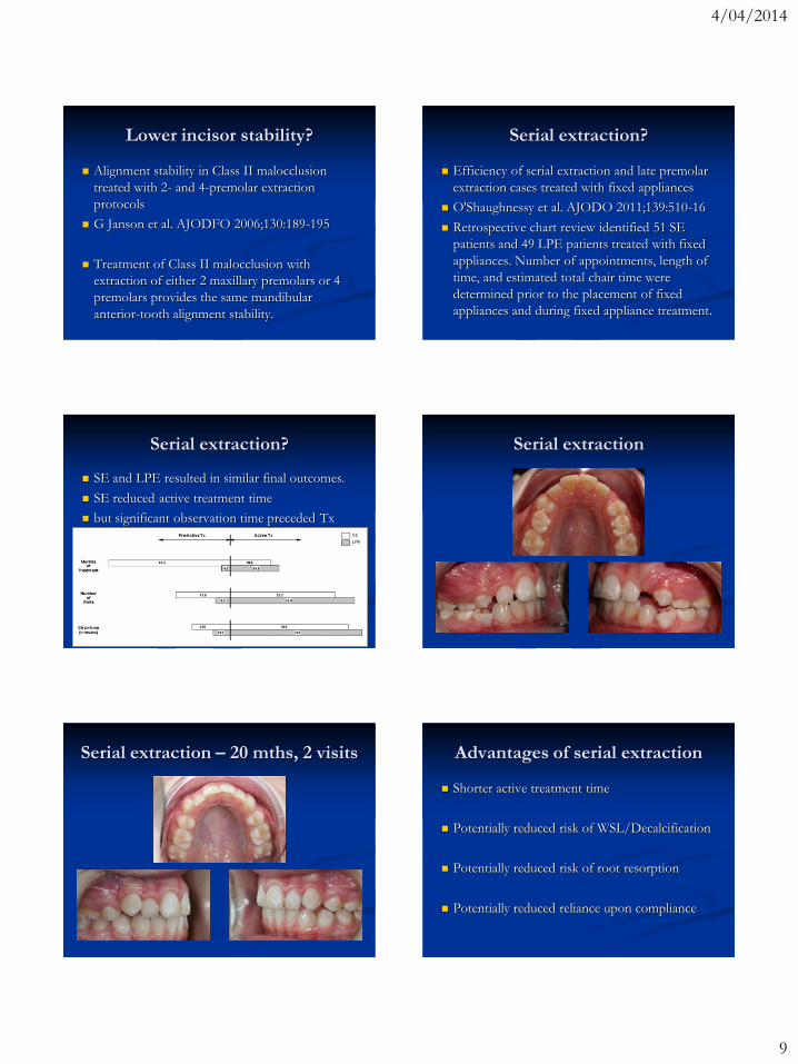
Serial extraction?
SE and LPE resulted in similar final outcomes.
SE reduced active treatment time
but significant observation time preceded Tx
Serial extraction
Serial extraction – 20 mths, 2 visits Advantages of serial extraction
Shorter active treatment time
Potentially reduced risk of WSL/Decalcification
Potentially reduced risk of root resorption
Potentially reduced reliance upon compliance

4/04/2014
10
Closing extraction spaces
Start 37 weeks/8.5 mths
Lacebacks
Consistency of lacebacks
Magnitude and reproducibility of forces
generated by clinicians during laceback
placement.
Khambay, McHugh, Millett.
J Orthod. 2006;33:270-275.
In vitro, there was a large inter-operator
variation in the forces produced. The forces by
clinicians ranged from 0 to 11.1 N (1.1kg).
Upper canine lacebacks
A randomized clinical trial to compare the effectiveness of canine lacebacks with reference to canine tip.
Usmani, O‟Brien et al. J Orthod 2002;29:281–286
The effect of canine lacebacks on preventing an increase in upper incisor proclination at the start of treatment is in the order of 1 mm and their effect on mesial molar movement is insignificant.
Canines lacebacks are similarly effective for patients with mesially inclined, upright or distally angulated upper canines.
Lower canine lacebacks
The effectiveness of laceback ligatures: A randomized controlled clinical trial
R. Irvine, S. Power, and F. McDonald. J Orthod. 2004; 31: 303 - 311.
The use of laceback ligatures conveys no difference in the antero-posterior or vertical position of the lower labial segment.
The use of laceback ligatures creates a statistically and clinically significant increase in the loss of posterior anchorage (0.83mm).
Laceback systematic review
The effectiveness of laceback ligatures during
initial orthodontic alignment: a systematic
review and meta-analysis
Fleming, Johal, Pandis.
According to the GRADE assessment, the
overall quality of evidence was high. There is no
evidence to support the use of lacebacks for the
control of the sagittal position of the incisors
during initial orthodontic alignment.
4/04/2014
11
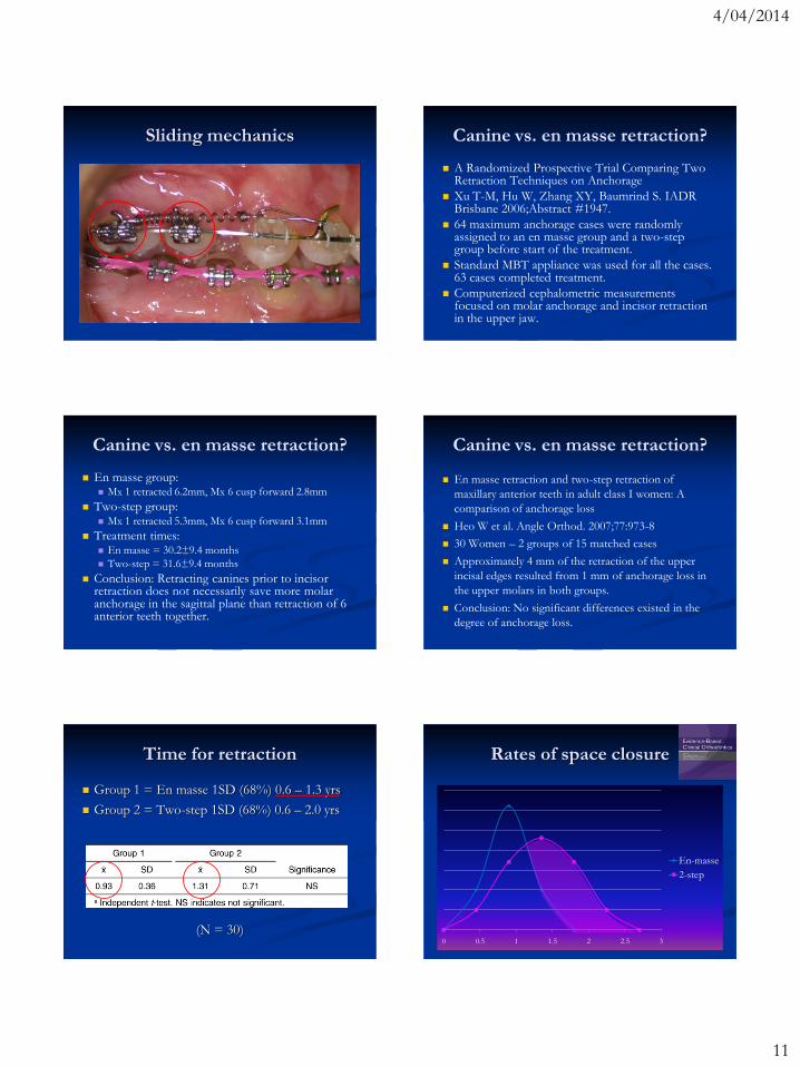
Sliding mechanics Canine vs. en masse retraction?
A Randomized Prospective Trial Comparing Two Retraction Techniques on Anchorage
Xu T-M, Hu W, Zhang XY, Baumrind S. IADR Brisbane 2006;Abstract #1947.
64 maximum anchorage cases were randomly assigned to an en masse group and a two-step group before start of the treatment.
Standard MBT appliance was used for all the cases. 63 cases completed treatment.
Computerized cephalometric measurements focused on molar anchorage and incisor retraction in the upper jaw.
Canine vs. en masse retraction?
En masse group: Mx 1 retracted 6.2mm, Mx 6 cusp forward 2.8mm
Two-step group: Mx 1 retracted 5.3mm, Mx 6 cusp forward 3.1mm
Treatment times: En masse = 30.2±9.4 months
Two-step = 31.6±9.4 months
Conclusion: Retracting canines prior to incisor retraction does not necessarily save more molar anchorage in the sagittal plane than retraction of 6 anterior teeth together.
Canine vs. en masse retraction?
En masse retraction and two-step retraction of
maxillary anterior teeth in adult class I women: A
comparison of anchorage loss
Heo W et al. Angle Orthod. 2007;77:973-8
30 Women – 2 groups of 15 matched cases
Approximately 4 mm of the retraction of the upper
incisal edges resulted from 1 mm of anchorage loss in
the upper molars in both groups.
Conclusion: No significant differences existed in the
degree of anchorage loss.
Time for retraction
Group 1 = En masse 1SD (68%) 0.6 – 1.3 yrs
Group 2 = Two-step 1SD (68%) 0.6 – 2.0 yrs
(N = 30)0 0.5 1 1.5 2 2.5 3
En-masse
2-step
Rates of space closure
4/04/2014
12
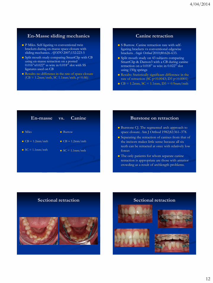
En-Masse sliding mechanics
P Miles. Self-ligating vs conventional twin brackets during en-masse space closure with sliding mechanics. AJODO 2007;132:223-5
Split mouth study comparing SmartClip with CB using en-masse retraction on a posted 0.016”x0.022” ss wire in 0.018” slot with SS ligatures used on CB
Results: no difference in the rate of space closure (CB = 1.2mm/mth, SC 1.1mm/mth, p=0.86)
Canine retraction
S Burrow. Canine retraction rate with self-ligating brackets vs conventional edgewise brackets. Angle Orthod 2010;80:626-633.
Split mouth study on 43 subjects comparing SmartClip & Damon3 with a CB during canine retraction on a 0.018” ss wire in 0.022” slot using 150g springs
Results: Statistically significant difference in the rate of retraction (SC p<0.0043; D3 p<0.0001)
CB = 1.2mm, SC = 1.1mm, D3 = 0.9mm/mth
En-masse vs. Canine
Miles
CB = 1.2mm/mth
SC = 1.1mm/mth
Burrow
CB = 1.2mm/mth
SC = 1.1mm/mth
Burstone on retraction
Burstone CJ. The segmented arch approach to
space closure. Am J Orthod 1982;82:361–378.
Separating the retraction of canines from that of
the incisors makes little sense because all six
teeth can be retracted at once with relatively low
forces
The only patients for whom separate canine
retraction is appropriate are those with anterior
crowding as a result of archlength problems.
Sectional retraction Sectional retraction

4/04/2014
13
Tipping vs. bodily retraction
Duration & anchorage management of canine
retraction with bodily versus tipping mechanics
Shpack N et al. Angle Orthod 2008;78:95-100
14 subjects, 22 slot, split mouth, xtn Mx 4‟s
Bodily retraction was faster than tipping due to
less time root uprighting.
Anchorage loss was similar for both groups (17-
20% or 1.2-1.4mm)
Nance did not provide absolute anchorage
Fastest way to close spaces?
Samuels RHA, Rudge SJ, Mair LH. A comparison of the rate of space closure using a nickel-titanium spring and an elastic module: a clinical study. Am J Orthod Dentofac Orthop. 1993;103:464-467
Samuels RHA, Rudge SJ, Mair LH. A clinical study of space closure with nickel-titanium closed coil springs and an elastic module. Am J Orthod Dentofac Orthop. 1998;114:73-79
C. Nightingale, SP. Jones. A clinical investigation of force delivery systems for orthodontic space closure. J Orthod 2003;30:229-36
Space closure
A randomized clinical trial to compare three
methods of orthodontic space closure
Dixon V, Read MJF, O'Brien KD, Worthington
HV, Mandall NA. J Orthod. 2002;29:31-36.
Ligatures vs. Chain vs. NiTi springs
Ligatures, chain replaced every visit
Lig = 0.35mm/mth*, PC = 0.58, NiTi = 0.81*
What is clinically significant?
Consider closing a 6mm space
Lig at 0.35mm/mth = ~17.1 mths
Chain at 0.58mm/mth = ~10.3 mths
Spring at 0.81mm/mth = ~7.4 mths
What do you consider clinically significant?
If using extended appointment intervals to avoid
reactivations which could be relied upon more?
Canine retraction rate
Rate reported by A. Bagden (unpublished) using
0.018” with 16ss
Damon bkts 1.8mm/mth
Alexander bkts 1.4mm/mth
Rate reported by Bokas & Woods (Aust Orth J,
2006;22:39-46) using conventional 0.018” twin
brackets with 16x16ss, reactivated @ 4 wks
1.9mm/mth with springs
1.7mm/mth with chain
Alexander/Lang bracket

4/04/2014
14
Canine retraction
S Burrow. Canine retraction rate with self-ligating brackets versus conventional edgewise brackets. Angle 2009: Accepted
Split mouth study on 43 subjects comparing SmartClip & Damon3 with a CB during canine retraction on a 0.018” ss wire in 0.022” slot using 150g springs
Results: Statistically significant difference in the rate of retraction (SC p<0.0043; D3 p<0.0001)
CB = 1.2mm, SC = 1.1mm, D3 = 0.9mm/mth
Bracket width and binding
Moment Arm
Contact Angle
Siamese/Twin
bracket
Alexander/Lang
bracket
Ligation distal to extractions? Ligatures, modules, SL brackets?
Wong et al. J Orth 2013;40:155-162.
Does the bracket-ligature combination affect the
amount of orthodontic space closure..... RCT
45 subjects with 1st Bi‟s xtn – 0.022” slot
Conventional elastomeric modules
SuperSlick „low-friction‟ elastomeric ligatures
Damon 3MX®
No difference in rate of closure (p=0.72)
1mm per 28 days but a lot of variation
Anchorage TPA’s enhance anchorage?
Effects of transpalatal arch on molar movement
produced by mesial force: A finite element
simulation.
Y Kojima, H Fukui. AJODFO 2008;134:335-6.
The TPA had no effect on the initial movement
and almost no effect preserving anchorage. The
TPA merely prevented rotational and transverse
movements of the anchor teeth.
4/04/2014
15
More on TPAs in xtn cases
Effect of the transpalatal arch during extraction
treatment.
Zablocki HL, McNamara JA, Franchi L, Baccetti
T. AJODFO. 2008;133:852-60.
No significant effect on either the AP or vertical
position of the maxillary 1st molars during xtn
Tx.
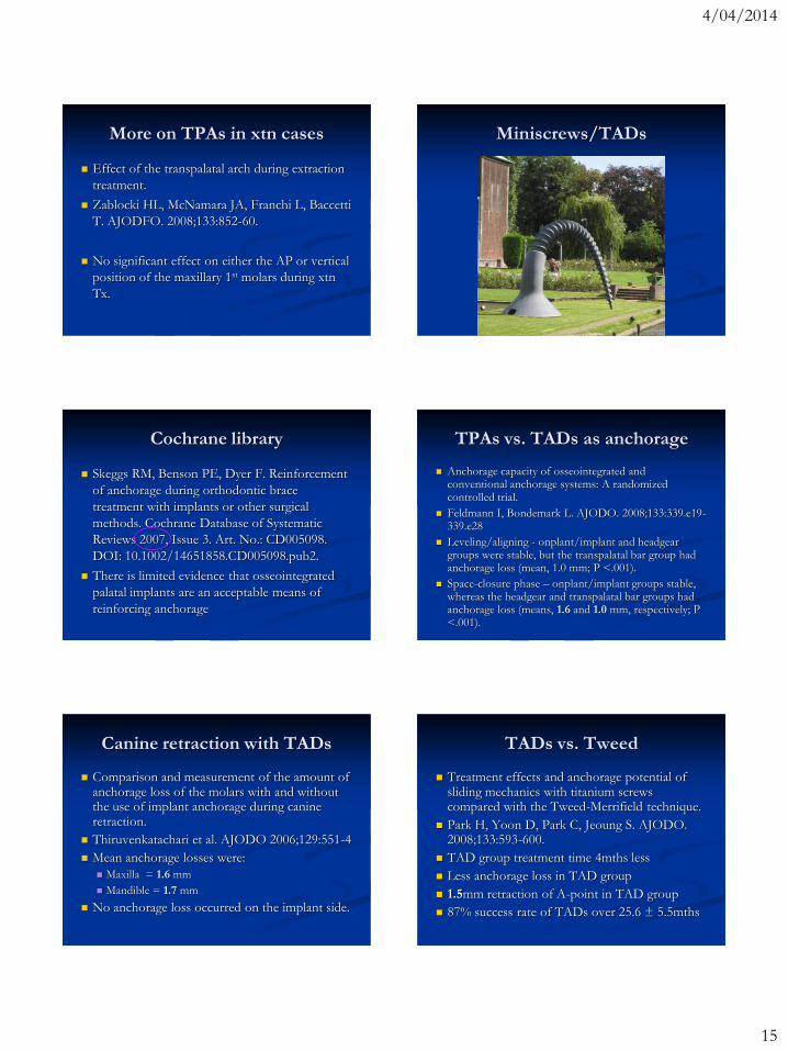
Miniscrews/TADs
Cochrane library
Skeggs RM, Benson PE, Dyer F. Reinforcement
of anchorage during orthodontic brace
treatment with implants or other surgical
methods. Cochrane Database of Systematic
Reviews 2007, Issue 3. Art. No.: CD005098.
DOI: 10.1002/14651858.CD005098.pub2.
There is limited evidence that osseointegrated
palatal implants are an acceptable means of
reinforcing anchorage
TPAs vs. TADs as anchorage
Anchorage capacity of osseointegrated and conventional anchorage systems: A randomized controlled trial.
Feldmann I, Bondemark L. AJODO. 2008;133:339.e19-339.e28
Leveling/aligning - onplant/implant and headgear groups were stable, but the transpalatal bar group had anchorage loss (mean, 1.0 mm; P <.001).
Space-closure phase – onplant/implant groups stable, whereas the headgear and transpalatal bar groups had anchorage loss (means, 1.6 and 1.0 mm, respectively; P <.001).
Canine retraction with TADs
Comparison and measurement of the amount of anchorage loss of the molars with and without the use of implant anchorage during canine retraction.
Thiruvenkatachari et al. AJODO 2006;129:551-4
Mean anchorage losses were:
Maxilla = 1.6 mm
Mandible = 1.7 mm
No anchorage loss occurred on the implant side.
TADs vs. Tweed
Treatment effects and anchorage potential of sliding mechanics with titanium screws compared with the Tweed-Merrifield technique.
Park H, Yoon D, Park C, Jeoung S. AJODO. 2008;133:593-600.
TAD group treatment time 4mths less
Less anchorage loss in TAD group
1.5mm retraction of A-point in TAD group
87% success rate of TADs over 25.6 ± 5.5mths

4/04/2014
16
Headgear vs. TADs
Comparison of treatment outcomes between skeletal anchorage and extraoral anchorage in adults with maxillary dentoalveolar protrusion
Yao C et al. AJODO 2008;134:615-624
The skeletal anchorage group had greater anterior tooth retraction (8.17 vs 6.73 mm) and less maxillary molar mesialization (0.88 vs 2.07 mm) than did the headgear group, with a shorter treatment duration (29.81 vs 32.29 months).
Difference = 1.42 mm greater retraction
TADs vs. other anchorage
Treatment effects of mini-implants for en-masse
retraction of anterior teeth in bialveolar dental
protrusion patients: A RCT.
Upadhyay M et al. AJODO 2008;134:18-29
TADs achieved absolute anchorage and greater
levels of skeletal and dental change than
conventional anchorage (eg. HG, TPA, 2nd
molars, differential moments)
93% success rate of TADs
Headgear vs. TADs
Palatal implants are a good alternative to
headgear: A randomized trial.
Sandler et al. AJODO. 2008;133:51-7
The use of palatal implants to reinforce
anchorage was as effective as extraoral
anchorage with headgear.
Hawthorne effect?
Headgear vs. TADs
Comparison of the zygoma anchorage system
with cervical headgear in buccal segment
distalization
Kaya et al. Eur JO. 2009; 31:417–424
The use of zygomatic plate anchorage was as
effective as extraoral anchorage with headgear.
Both groups ~5mm molar correction in 9 mths
Invisalign Accelerated Tx
Miles, Smith, Weyant, Rinchuse.
Aust Orth J 2012;28:213-8.
The effects of a vibrational appliance on tooth
movement and patient discomfort: a prospective
randomised clinical trial.
66 non-extraction subjects randomly assigned to
Tooth Masseuse vs. No appliance
4/04/2014
17
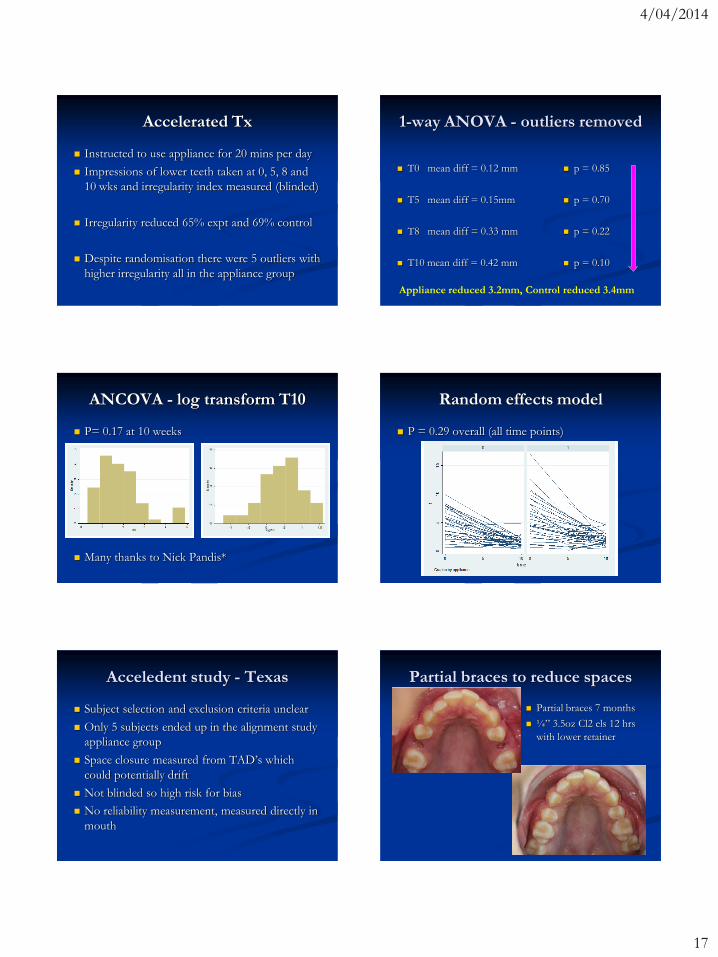
Accelerated Tx
Instructed to use appliance for 20 mins per day
Impressions of lower teeth taken at 0, 5, 8 and
10 wks and irregularity index measured (blinded)
Irregularity reduced 65% expt and 69% control
Despite randomisation there were 5 outliers with
higher irregularity all in the appliance group
1-way ANOVA - outliers removed
T0 mean diff = 0.12 mm
T5 mean diff = 0.15mm
T8 mean diff = 0.33 mm
T10 mean diff = 0.42 mm
p = 0.85
p = 0.70
p = 0.22
p = 0.10
Appliance reduced 3.2mm, Control reduced 3.4mm
ANCOVA - log transform T10
P= 0.17 at 10 weeks
Many thanks to Nick Pandis*
Random effects model
P = 0.29 overall (all time points)
Acceledent study - Texas
Subject selection and exclusion criteria unclear
Only 5 subjects ended up in the alignment study
appliance group
Space closure measured from TAD‟s which
could potentially drift
Not blinded so high risk for bias
No reliability measurement, measured directly in
mouth
Partial braces to reduce spaces
Partial braces 7 months
¼” 3.5oz Cl2 els 12 hrs
with lower retainer

4/04/2014
18
Invisalign study intruding laterals Maxillary 2’s intrusion
Aim: To determine if the use of an ovoid
auxiliary on maxillary 2‟s prevents unwanted
intrusion during Invisalign treatment
Method: 30 subjects randomly assigned to
having ovoid attachments or not. If the Align
technician had placed auxiliaries on the 2‟s then
the patient was excluded.
Assess clinically whether buttons or refinement
involving extrusions was required or not
Maxillary 2’s intrusion
Results: 28 subjects completed the study (14 in
each group)
4 subjects required buttons for extrusion
All 4 in the NO attachment group (4/14 = 29%)
Wilcoxon test p= 0.02
Class II non-extraction Tx
Kevin’s Blog
Kevinobrienorthoblog.com
http://orthoartmeetsscience.blogspot.com.au
Cochrane - Class II div 2 Tx
Orthodontic treatment for deep bite and retroclinedupper front teeth in children
Millett DT, Cunningham SJ, O'Brien KD et al.
Authors' conclusions
It is not possible to provide any evidence-based guidance to recommend or discourage any type of orthodontic treatment to correct Class II division 2 malocclusion in children.
4/04/2014
19
Cochrane - prominent front teeth
Orthodontic treatment for prominent upper front teeth in children
Harrison JE, O'Brien KD, Worthington HV
Authors' conclusions
The evidence suggests that providing early orthodontic treatment for children with prominent upper front teeth is no more effective than providing one course of orthodontic treatment when the child is in early adolescence.
Review of distalisation
Development And Use Of A Taxonomy To Carry Out A Systematic Review Of The Literature On Methods Described To Effect Distal Movement Of Maxillary Molars.
G.J. Atherton, A.M. Glenny, K. O‟Brien
J Orthod 2002;29:211-216
The authors concluded that no more than 2-2.5mm of distal maxillary molar movement could be achieved and the quality of evidence for any method of moving maxillary molars distally was not high
Therefore only ~1/3 unit Class II cases
Molar distalisation
Upper removable appliance or Jones Jig for distalizing first molars? A randomized clinical trial
Paul LD, O'Brien KD, Mandall NA. OrthodCraniofac Res 2002;5:238-242
There were no statistically significant differences between the two treatment methods for any of the outcome measures (P > 0.05).
Distal movement by both was 1.2-1.3 mm.
Headgear vs. Nance/coils
Extraoral vs Intraoral Appliance for Distal Movement of Maxillary First Molars: A RCT.
Bondemark & Karlsson. Angle 2005;75:699–706
Mean distal molar movement:
Nance/coil = 3.0 mm
Headgear = 1.7 mm
Mean incisor flaring
Nance/coil = 0.9 mm
Headgear = -0.9 mm
Headgear vs. Nance/coils
Final result = molar movement – anchorage loss
Nance/coil = 3.0 - 0.9 = 2.1mm
Headgear = 1.7 - -0.9 = 2.6mm
Pendulum vs. headgear
Comparison of the effects produced by headgear and pendulum appliances followed by fixed orthodontic treatment
Angelieri F, de Almeida RR, Janson G et al.
Eur J Orthod 2008;30:572-9
Retrospective study but no significant pre-Txdifferences between the groups
Greater restriction 1.5mm of maxillary growth in the CHG group (P<0.05) and 3° more flaring of lower incisors in the Pendulum group (P<0.05)

4/04/2014
20
Pendulum vs. extractions
Comparative efficiency of Class II malocclusion treatment with the pendulum appliance or two maxillary premolar extractions and edgewireappliances
Pinzan-Vercelino CRM, Janson G et al.
Eur J Orthod 2009;31:333-340
Retrospective study with Pendulum appliances in less severe Cl II and extractions in full Cl II
Pendulum Tx time = 45.7 mths
Extraction Tx time = 23.0 mths
Cochrane review of distalisers
Jambi S, Thiruvenkatachari B, O'Brien KD,
Walsh T.
Orthodontic treatment for distalising upper first
molars in children and adolescents.
Cochrane Database of Systematic Reviews 2013
Meta-analysis – Molars distalise Meta-analysis – Incisors distalise
Meta-analysis – Overjet TAD anchored sectional
LOMAS or Quattro screw

4/04/2014
21
TAD anchored distalisers?
Likely gain additional anchorage of 1.5-2mm
Sectional reduces time in full fixed so less
hygiene and OIRR risk and less impact on QOL
Additional time to distalise prior to placement of
full fixed appliances
If we have to distalise that far, extract 2 x Bi‟s?
Beneslider
2 mth then 4 mths 6 mths then 8 mths
Fixed Functional
Appliances
“Orthodontic Functional Appliances”
Ed: Prof R. Lee and P. Fleming
Timing of treatment
Initially many felt that functional appliance
therapy should be initiated at ~9-10 yo
UK and UNC studies showed early treatment
made no difference in the final outcome
Others have suggested timing to peek growth
spurt for the greatest skeletal effect (~12-13)
4/04/2014
22
Timing of treatment
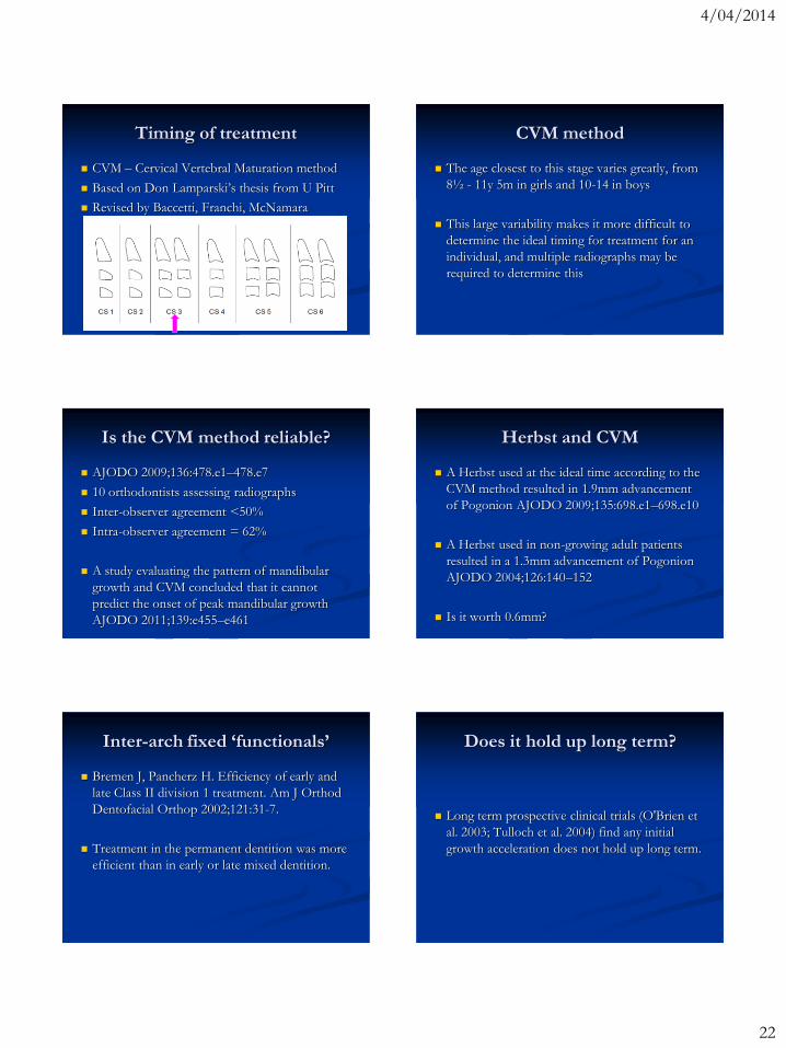
CVM – Cervical Vertebral Maturation method
Based on Don Lamparski‟s thesis from U Pitt
Revised by Baccetti, Franchi, McNamara
CVM method
The age closest to this stage varies greatly, from
8½ - 11y 5m in girls and 10-14 in boys
This large variability makes it more difficult to
determine the ideal timing for treatment for an
individual, and multiple radiographs may be
required to determine this
Is the CVM method reliable?
AJODO 2009;136:478.e1–478.e7
10 orthodontists assessing radiographs
Inter-observer agreement <50%
Intra-observer agreement = 62%
A study evaluating the pattern of mandibular
growth and CVM concluded that it cannot
predict the onset of peak mandibular growth
AJODO 2011;139:e455–e461
Herbst and CVM
A Herbst used at the ideal time according to the
CVM method resulted in 1.9mm advancement
of Pogonion AJODO 2009;135:698.e1–698.e10
A Herbst used in non-growing adult patients
resulted in a 1.3mm advancement of Pogonion
AJODO 2004;126:140–152
Is it worth 0.6mm?
Inter-arch fixed ‘functionals’
Bremen J, Pancherz H. Efficiency of early and
late Class II division 1 treatment. Am J Orthod
Dentofacial Orthop 2002;121:31-7.
Treatment in the permanent dentition was more
efficient than in early or late mixed dentition.
Does it hold up long term?
Long term prospective clinical trials (O'Brien et
al. 2003; Tulloch et al. 2004) find any initial
growth acceleration does not hold up long term.
4/04/2014
23
Herbst vs. elastics
Class II correction in patients treated with Cl2
elastics and with fixed functional appliances: ....
Nelson et al. AJODO 2000;118:142-9.
18 Begg/elastics for 1.3 years
18 Herbst only for 0.5 years
Skeletal improvement in Herbst 2mm better
OJ improvement in Begg was 2mm better
Skeletal contribution 4% in Begg, 51% in Herbst
Herbst vs. Elastics – long term?
A long-term follow-up study of Class II
malocclusion correction after treatment with Cl2
elastics or fixed functional appliances
Nelson et al. AJODO 2007;132:499-503.
15 from each group returned ~6-8 years later
During the total observation period many of the
changes reversed and the differences did not last
The final outcome may be similar regardless
What is a functional appliance?
“One that engages both dental arches and acts
principally by holding the mandible away from
its normal resting position” (Isaacson et al.
1990)
“An appliance aimed at modifying growth”
(Proffit 2007)
Fixed functional appliances
The more appropriate description is fixed Class
II correctors but the current convention is FFA
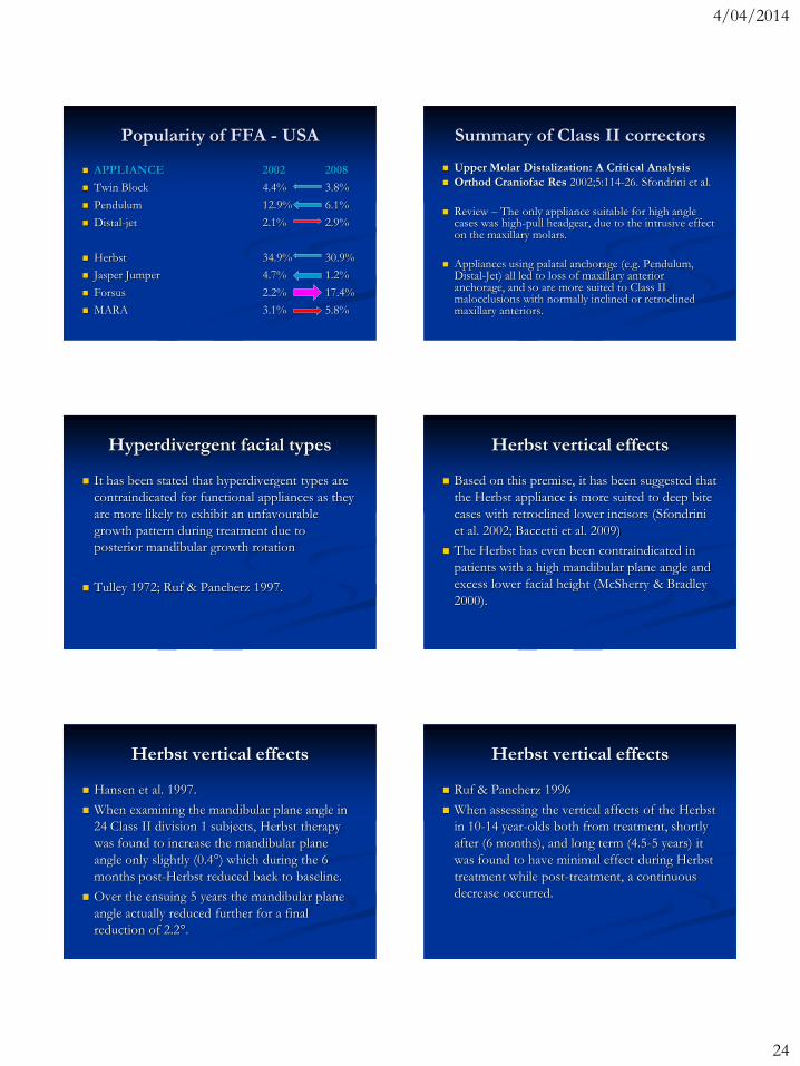
Popularity of FFA - USA Popularity of FFA - Australia
APPLIANCE
Twin Block
Pendulum, Distal-jet
Herbst
Forsus, Jasper Jumper
MARA
2013
70%
11%
33%
61%
0%
4/04/2014
24
Popularity of FFA - USA
APPLIANCE
Twin Block
Pendulum
Distal-jet
Herbst
Jasper Jumper
Forsus
MARA
2002 2008
4.4% 3.8%
12.9% 6.1%
2.1% 2.9%
34.9% 30.9%
4.7% 1.2%
2.2% 17.4%
3.1% 5.8%
Summary of Class II correctors
Upper Molar Distalization: A Critical Analysis
Orthod Craniofac Res 2002;5:114-26. Sfondrini et al.
Review – The only appliance suitable for high angle cases was high-pull headgear, due to the intrusive effect on the maxillary molars.
Appliances using palatal anchorage (e.g. Pendulum, Distal-Jet) all led to loss of maxillary anterior anchorage, and so are more suited to Class II malocclusions with normally inclined or retroclined maxillary anteriors.
Hyperdivergent facial types
It has been stated that hyperdivergent types are
contraindicated for functional appliances as they
are more likely to exhibit an unfavourable
growth pattern during treatment due to
posterior mandibular growth rotation
Tulley 1972; Ruf & Pancherz 1997.
Herbst vertical effects
Based on this premise, it has been suggested that
the Herbst appliance is more suited to deep bite
cases with retroclined lower incisors (Sfondrini
et al. 2002; Baccetti et al. 2009)
The Herbst has even been contraindicated in
patients with a high mandibular plane angle and
excess lower facial height (McSherry & Bradley
2000).
Herbst vertical effects
Hansen et al. 1997.
When examining the mandibular plane angle in
24 Class II division 1 subjects, Herbst therapy
was found to increase the mandibular plane
angle only slightly (0.4°) which during the 6
months post-Herbst reduced back to baseline.
Over the ensuing 5 years the mandibular plane
angle actually reduced further for a final
reduction of 2.2°.
Herbst vertical effects
Ruf & Pancherz 1996
When assessing the vertical affects of the Herbst
in 10-14 year-olds both from treatment, shortly
after (6 months), and long term (4.5-5 years) it
was found to have minimal effect during Herbst
treatment while post-treatment, a continuous
decrease occurred.
4/04/2014
25
Herbst vertical effects
No statistically significant differences were
found between hypodivergent, normodivergent,
and hyperdivergent subjects although a large
amount of variation was noted.
Herbst vertical effects
Ruf & Pancherz 1997.
When examining Hyper- and hypo-divergent
Class II subjects aged 11 to 14 years, skeletal and
dental changes were independent upon the
vertical facial type.
Herbst vertical effects
Windmiller 1993
When using an acrylic splint Herbst, although
the lower anterior face height increased 2.4 mm,
both the Y-axis and mandibular plane angle
remained essentially the same.
Thus, the overall vertical skeletal pattern did not
change which is in agreement with others
(Martin & Pancherz 2009).
Herbst vertical effects conclusion
It therefore seems that functional appliance
therapy with the Herbst appliance is equally
effective and vertically neutral regardless of the
pre-treatment vertical jaw base relationship.
Risk of recession?
Does orthodontic proclination of lower incisors in children and adolescents cause gingival recession?
Ruf S, Hansen K, Pancherz H. AJODO 1998; 114:100–106.
Only 3% of mandibular incisors developed recession after the use of Herbst appliance.
They concluded that no interrelationship was found between the amount of incisor proclination and gingival recession.
Risk of recession?
Correlation between mandibular central incisor proclination and gingival recession during fixed appliance therapy
Djeu G et al. Angle Orthod 2002; 72:238-245
Previous reports on irreversible gingival recession during fixed therapy = 1.3-10%
This study found 12 % with gingival recession.
No correlation between mandibular incisor proclination during treatment and gingival recession or change in clinical crown length.

4/04/2014
26
Risk of recession?
Melsen & Allais AJODO 2005;127:552-561
No orthodontic variable was associated with
recession.
They concluded that thin gingival biotype, visual
plaque, and inflammation are useful predictors
of gingival recession.
What is biotype?
Claffey & Shanley (J Clin Periodontol 1986)
Thin tissue biotype gingival thickness ≤ 1.5mm
Kan et al. J Periodontol 2003;74:557-562)
The outline of a periodontal probe (PCP-UNC
15) can be seen through the gingival margin then
it is a Thin Biotype
Risk of recession?
Yared et al. AJODO 2006;130:6.e1-6.e8
In adult patients greater proclination >95° and
especially free gingival margin thickness
(biotype) had the greatest association with the
risk of recession.
Systematic review of recession
Aziz, Flores-Mir. Aust Orth J 2011;27:33-39
No association between labial movement of
mandibular incisors and gingival recession.
Important factors that may predispose to
gingival recession were
reduced thickness of the gingival margin (biotype)
narrow mandibular symphysis
inadequate plaque control
aggressive tooth brushing.
Risk of recession?
Handelman. Angle 1996;66:95-109.
Thinner labial cortical bone demonstrated in
untreated hyperdivergent facial types.
Enhos et al. Angle 2012;82:868-874.
Slightly higher occurrence of dehiscences in
hyperdivergent facial types.
Putting it all together
There is therefore a potentially elevated risk of
recession when advancing lower incisors in
hyperdivergent facial types.
Care should be exercised in maintaining
excellent oral hygiene and particular care and
monitoring in those with a thin tissue biotype
and/or a hyperdivergent facial type.

4/04/2014
27
Jasper Jumper vs. Activator/HG
Comparative Evaluation of a New Removable Jasper Jumper Functional Appliance vs. an Activator-Headgear Combination.
Sari et al. Angle Orthod 2003;73:286–293
Increase in total facial height was greater in the activator group than in the JJ group. Vertical growth inhibition of lower incisors was greater in the JJ group.
JJ seemed more suitable for high-angle cases, particularly with maxillary excess and some mandibular deficiency.
Fixed functional vectors
Consider the desired response
Intrude/Limit eruption Allow eruption
Forsus and Jasper Jumper
Forsus Nitinol Flat Spring and Jasper Jumper
Corrections of Class II division 1 Malocclusions.
Karacay S et al. Angle Orthod 2006;76:666–672.
Both the appliances were effective in the
treatment of Class II malocclusion
Both appliances cause significant incisor and
molar movements, and these dentoalveolar
changes are more effective than the skeletal
changes in attaining Class I molar relationship.
Forsus success
Effectiveness of comprehensive fixed appliance
Tx used with the Forsus FRD in Cl2 patients
Franchi, Alvetro et al. Angle 2011;81:678-683
32 subjects compared with matched control
87.5% success rate with Tx over 2.4 yrs (± 0.4)
Overjet reduced ~5.5mm, molar relationship
3.4mm, lower incisors flared ~5º
Forsus FRD vs. elastics
Class II Non-Extraction Patients Treated with
the Forsus Fatigue Resistant Device Versus
Intermaxillary Elastics.
Jones G et al. Angle Orthod 2008;78:332–338.
With the exception of lower molar mesial
movements and total molar correction, which
were significantly (P < .05) greater in the Forsus
group, there were no statistically significant
group differences in the treatment changes.
(retro/matched)
4/04/2014
28
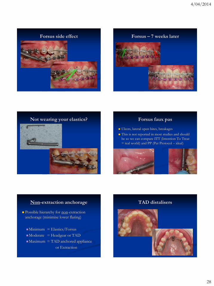
Forsus side effect Forsus – 7 weeks later
Not wearing your elastics? Forsus faux pas
Ulcers, lateral open bites, breakages
This is not reported in most studies and should
be so we can compare ITT (Intention To Treat
= real world) and PP (Per Protocol – ideal)
Non-extraction anchorage
Possible hierarchy for non-extraction
anchorage (minimise lower flaring)
Minimum = Elastics/Forsus
Moderate = Headgear or TAD
Maximum = TAD anchored appliance
or Extraction
TAD distalisers

4/04/2014
29
TAD anchored FFA
Angle Orthod. 2014;84:76–87. Aslan et al.
16 patients (13.7 yo) were treated with FRDMS,
whereas 17 subjects (14.6 yo) were treated with
only FRD
Tx times avg FRDMS 6.5 mths & FRD 5.5 mths
No skeletal effect but labial tipping of
mandibular incisors was significantly greater in
the FRD group than in the FRDMS group
TAD Forsus
Not randomised so a risk of selection bias
Not blinded when tracing so a risk of bias
FRD group 10-12 mths older so potential for
less growth
FRD group 1 mth less possibly due to greater
dental movement - we correct until we achieve
what we want
If less dental movement then we have to wait
longer to achieve the desired change
TAD Forsus
Skeletal changes tend to relapse long term in e.g.
Herbst studies
VanLaecken et al. 2006
Pancherz & Anehus-Pancherz 1993
Wieslander 1993
Need long term follow-up study to evaluate the
final extent of the changes and any differences
Incisor movements
1.1mm vs. 2.3mm labial movement
L1 to MP 3.6° vs. 9.3°
1.2mm labial movement and 6° tipping – could
be related to tracing error/bias?
Most TAD studies find ~1.5mm difference to
other forms of anchorage
Perhaps tie ligature to premolars to reduce risk
of „screwing it up‟
TAD tied with ligature
4/04/2014
30
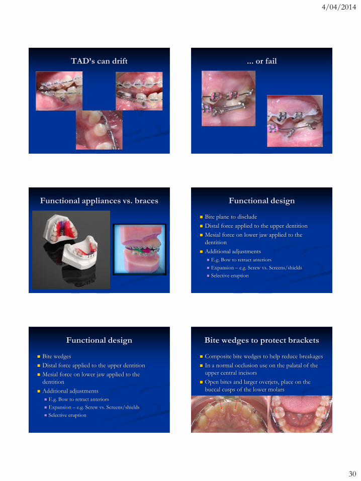
TAD’s can drift ... or fail
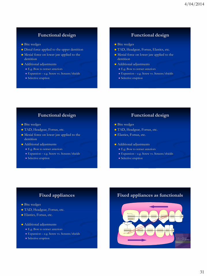
Functional appliances vs. braces Functional design
Bite plane to disclude
Distal force applied to the upper dentition
Mesial force on lower jaw applied to the
dentition
Additional adjustments
E.g. Bow to retract anteriors
Expansion – e.g. Screw vs. Screens/shields
Selective eruption
Functional design
Bite wedges
Distal force applied to the upper dentition
Mesial force on lower jaw applied to the
dentition
Additional adjustments
E.g. Bow to retract anteriors
Expansion – e.g. Screw vs. Screens/shields
Selective eruption
Bite wedges to protect brackets
Composite bite wedges to help reduce breakages
In a normal occlusion use on the palatal of the
upper central incisors
Open bites and larger overjets, place on the
buccal cusps of the lower molars
4/04/2014
31
Functional design
Bite wedges
Distal force applied to the upper dentition
Mesial force on lower jaw applied to the
dentition
Additional adjustments
E.g. Bow to retract anteriors
Expansion – e.g. Screw vs. Screens/shields
Selective eruption
Functional design
Bite wedges
TAD, Headgear, Forsus, Elastics, etc.
Mesial force on lower jaw applied to the
dentition
Additional adjustments
E.g. Bow to retract anteriors
Expansion – e.g. Screw vs. Screens/shields
Selective eruption
Functional design
Bite wedges
TAD, Headgear, Forsus, etc.
Mesial force on lower jaw applied to the
dentition
Additional adjustments
E.g. Bow to retract anteriors
Expansion – e.g. Screw vs. Screens/shields
Selective eruption
Functional design
Bite wedges
TAD, Headgear, Forsus, etc.
Elastics, Forsus, etc.
Additional adjustments
E.g. Bow to retract anteriors
Expansion – e.g. Screw vs. Screens/shields
Selective eruption
Fixed appliances
Bite wedges
TAD, Headgear, Forsus, etc.
Elastics, Forsus, etc.
Additional adjustments
E.g. Bow to retract anteriors
Expansion – e.g. Screw vs. Screens/shields
Selective eruption
Fixed appliances as functionals

4/04/2014
32
Upcoming book on functionals
Orthodontic Functional Appliances
Editors: Prof. Bob Lee & Padhraig Fleming
Wiley/Blackwell
Peter Miles: Fixed Functional Appliances
Efficiency (Molar mm/year)
0.00
0.20
0.40
0.60
0.80
1.00
1.20
1.40
1.60
1.80
2.00
Herbst MARA Jasper Jumper Forsus TwinForce
Summation
Treatment times and molar effects are similar
However, this does not factor in the number of
appointments, appointment duration, or
appliance cost which influence the cost
effectiveness
Ideally this would be the subject of future high
quality RCT‟s
Compare with removable FA
The overall molar change, treatment time and
efficiency (mm/yr) was no more or less effective
using a continuous 2-stage approach with a
Herbst (Franchi et al. 1999, Schaefer et al. 2004)
or MARA (Azizollahi 2012) followed
immediately by preadjusted edgewise appliances
than using a single phase with a Jasper Jumper,
Forsus FRD, Herbst (Al-Jewair et al. 2012) or
MARA (Al-Jewair et al. 2012, Ghislanzoni et al.
2013).
Compare with removable FA
This contrasts with the UNC clinical trial
(Tulloch et al. 2004) of removable functional
appliances and headgear which found the overall
treatment time to be significantly longer with the
2-phase approach.
Possible explanations?
The efficiency in terms of mm/month change of
the Bionator has been suggested to be less than
the Herbst (Cozza et al. 2006).
Another study comparing the MARA, Bionator,
Herbst and Twin-block followed by preadjusted
edgewise appliances also found treatment
involving the Bionator to be slower by ~7
months or 17% (Siara-Olds et al. 2010).
4/04/2014
33
Possible explanations?
The delay between phases in the UNC trial
allows some possible loss of the molar
correction which then needs to be recovered?
Shorter treatment times using fixed functionals
(Herbst, MARA) compared with the Bionator
used in the UNC trial suggest the Bionator may
be less efficient.
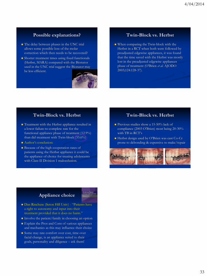
Twin-Block vs. Herbst
When comparing the Twin-block with the
Herbst in a RCT when both were followed by
preadjusted edgewise appliances, it was found
that the time saved with the Herbst was mostly
lost in the preadjusted edgewise appliances
phase of treatment (O'Brien et al. AJODO
2003;124:128-37).
Twin-Block vs. Herbst
Treatment with the Herbst appliance resulted in
a lower failure-to-complete rate for the
functional appliance phase of treatment (12.9%)
than did treatment with Twin-block (33.6%)
Author‟s conclusion:
Because of the high cooperation rates of
patients using the Herbst appliance it could be
the appliance of choice for treating adolescents
with Class II Division 1 malocclusion.
Twin-Block vs. Herbst
Previous studies show a 15-50% lack of
compliance (2003 O‟Brien) most being 20-30%
with TB in RCT‟s
Herbst design used by O‟Brien was cast Co-Cr
prone to debonding & expensive to make/repair
Appliance choice
Dan Rinchuse (Seton Hill Univ) - “Patients have
a right to autonomy and input into their
treatment provided that it does no harm.”
Involve the patient/family in choosing an option
Explain the Pros and Cons of various appliances
and mechanics as this may influence their choice
Some may rate comfort over cost, time over
facial change, is an appliance suited to their
goals, personality and diligence – ask them!