evidence- based wound care interventions arterial leg … leg ulcer... · evidence- based wound...
TRANSCRIPT
Waterloo Wellington Integrated Wound Care Program: Evidence-Based Care for Arterial Leg Ulcers Final May 5, 2016 1
Waterloo Wellington Integrated Wound Care Program Evidence- Based Wound Care Interventions
Arterial Leg Ulcers Prevention and Management Recommendations
Content:
1. Objectives
2. Background
a. Best Practices for Assessment, Prevention and Treatment of Venous Leg Ulcers Registered Nurses Association of Ontario (RNAO)
b. Clinical Best Practice Guidelines Strategies to Support Self-Management in Chronic Conditions: Collaboration with Clients
c. Wound Bed Preparation Paradigm
3. Address Patient-Centered Concerns
a. Assess Psychosocial Needs /Pain and Quality of Life (QOL) b. Socioeconomic Determinates of Health c. Chronic Disease Self-management
4. Identify and Treat the Cause
4.1 Assessment
a. Risk Factors and Etiology of Arterial Leg Ulcers b. Odds Ratio of Arterial Leg Ulcers NOT healing in 24 weeks c. Common Signs and Symptoms of Arterial Ischemic Leg and Foot Ulcers
4.2 Obtain a Comprehensive Patient History and Perform a Physical Assessment
a. Obtain a comprehensive patient history b. Complete a comprehensive physical examination c. Lower Leg Assessment d. Assess Wound and Peri-wound e. Wound Measurement f. Comparison of Venous versus Arterial versus Mixed Venous/Arterial Leg Ulcers g. Ankle Brachial Pressure Index (ABPI) / Toe Brachial Pressure Index (TBPI ) and Further
Investigations h. Determine if the wound is “Healable, Maintenance or Non-Healable” i. Nutritional Assessment
4.3 Optimize Medical Therapy

Waterloo Wellington Integrated Wound Care Program: Evidence-Based Care for Arterial Leg Ulcers Final May 5, 2016 2
4.4 Pharmacological Treatment a. Pentoxyfilline (Trental) b. Nifedipine
4.5 Surgical and Medical Interventions a. Endovascular Perfusion Strategies b. Surgical Perfusion Strategies c. Oxygenation Strategies
5. Provide Local Wound Care
a. Intervention Algorithm b. Signs and Symptoms of Wound Infection c. Signs and symptoms of Lower Leg Cellulitis d. Management of Lower Leg Cellulitis e. Venous Dermatitis: Signs, Symptoms, Prevention and Treatment f. Determining Goals for Local Treatment for Arterial Leg Ulcers g. Utilize Product Picker from Canadian Association of Wound Care (CAWC) h. South West Region Wound Care Program: Wound Cleansing Table and Dressing Selection & Cleansing Enablers i. Patient Education on Skin Care j. Adjunctive Therapies
6. Provide Organizational Support
a. Multi-disciplinary Referral Criteria b. Patient Teaching and Learning Resources c. Discharge or Transfer Planning and Communications d. Waterloo Wellington Integrated Wound Care Program Evidence-Based Wound Care Arterial
Clinical Pathway
7. Arterial Leg Ulcer Toolkit

Waterloo Wellington Integrated Wound Care Program: Evidence-Based Care for Arterial Leg Ulcers Final May 5, 2016 3
RNAO’s Assessment and Management of Venous Leg Ulcers 1, 2 Levels of Evidence
A
Evidence obtained from at least one randomized controlled trial or meta-analysis of randomized controlled trials
B
Evidence from well-designed clinical studies but no randomized controlled trials
C
Evidence from expert committee reports or opinion and/or clinical experience or respected authorities. Indicates absence of directly applicable studies of good quality
RNAO’s Strategies to Support Self-Management in Chronic Conditions: Collaboration with Clients 12
Levels of Evidence
la
Evidence obtained from meta-analysis or systematic review of randomized controlled trial
lb
Evidence obtained from at least one randomized controlled trial
lla
Evidence obtained from at least one well-designed controlled study without randomization
llb
Evidence obtained from at least one other type of well-designed quasi- experimental study, without randomization
ll
Evidence obtained from well-designed non-experimental descriptive studies, such as comparative studies, correlation studies and case studies
lV
Evidence obtained from expert committee reports or opinions and/or clinical experiences of respected authorities
RNAO’s Integrating Smoking Cessation into Daily Nursing Practice 14
Levels of Evidence
A
Requires at least two randomized controlled trials as part of the body of literature of overall quality and consistency addressing the specific recommendations
B
Requires availability of well conducted clinical studies, but no randomized controlled trials on the topic of recommendations.
C
Requires evidence from expert committee reports or opinions and/or clinical experience of respected authorities. Indicates absence of directly applicable studies of good quality.

Waterloo Wellington Integrated Wound Care Program: Evidence-Based Care for Arterial Leg Ulcers Final May 5, 2016 4
1. Objectives
The objectives of the development and implementation of these resources is to help Health Care Providers to:
Find practical, evidence-based resources to use when caring for individuals that have or who are at risk of developing, arterial leg ulcers
Perform a comprehensive patient assessment including assessing for indicators of arterial leg ulcers that will not heal in the inpatient and outpatient care settings (Acute Care, Long Term Care and Community Care Settings)
Identify the correct etiology of lower leg ulcers Arrange for a holistic Lower Leg Assessment (LLA) including ABPIs in order to identify patient’s
ability to heal or need for referral to vascular surgeon. If patient is a diabetic, toe pressures should also be obtained.
Perform accurate wound assessment including progress towards healing Recognize signs & symptoms of infection and identify treatment interventions Increase the use and implementation of evidence-based arterial leg ulcer treatment plans
including pain management using pharmacological and non-pharmacological interventions Identify and implement appropriate topical wound care Improve the coordination and communication between care providers/care institutions
regarding the transfer/discharge plan for patients with arterial leg ulcers
2. Background
Peripheral vascular disease (PVD) is any disease that affects the blood flow through veins and arteries distal to the heart. Peripheral arterial disease affects the arteries only.32 Peripheral arterial disease (PAD) is a life-threatening condition which results from the narrowing of the blood vessels.” 3
Patients with arterial disease often have a history of cerebral vascular accident (stroke), coronary artery disease or diabetes.4, 5, 6 Cigarette smokers, the elderly and those with high blood pressure are at particular risk for developing PAD. Arterial leg ulcers usually result from trauma, pressure, thermal extremes, chemicals, blood clot/embolus or infection in this susceptible population.3 Ischemia is a restriction of blood supply caused by stenosis (narrowing) or occlusion (blockage) of the blood vessel leading to damage of the surrounding tissue. Hypoxia occurs when tissue is deprived of oxygen. Both ischemia and hypoxia can cause critical limb ischemia which can lead to the need for limb amputation and/or death.3
It is estimated that between 14-20 % of the adult population will develop peripheral arterial disease in their lifetime.7 Nearly 10% of all patients that present with leg or foot ulcers have ‘pure arterial insufficiency’.8 Critical limb ischemia occurs in approximately 3% of patients that develop PAD. 9, 10 The ten year mortality rate of those with PAD is 60%. The five year mortality rate of those patients that experience critical limb ischemia is 50 to 70%.9, 10
Inadequate tissue perfusion can result in formation of arterial ulcers due to partial or complete blocking of arterial flow. These ulcers, which are usually found on the lower legs and feet, are very often painful and difficult to heal.33

Waterloo Wellington Integrated Wound Care Program: Evidence-Based Care for Arterial Leg Ulcers Final May 5, 2016 5
Patients with arterial ulcers often suffer from a significant decreased level of quality of life. Pain, which can be described as ‘worst pain possible’, can make mobility and sleep difficult or nearly impossible. Independence can be affected, often rendering caregivers to become care receivers. Physical limitations can cause issues with performing housework, employment requirements and other activities of daily living.33 Difficulty working can have a financial impact on patients’ lives, affecting job security and possibly causing the need to retire earlier than planned. Problems sleeping can create a negative state of well-being, anxiety and depression. Personal hygiene can be a challenge for patients with arterial ulcers. Limited mobility and fear of further injury may lead to a decrease in personal hygiene. Wound odour may leave the patient with a sense of uncleanliness. Many feel that their sex life is negatively affected by having an arterial ulcer. 34, 35
The diagnosis and prognosis of arterial disease can cause patients to become very fearful of the possibility of amputation, discrimination and social rejection. Self-image can be affected as concerns of tissue decay and body weakness are known to cause feelings of discomfort, shame and fear. Patients often go out less frequently, reducing interaction time with family and friends. Social isolation can result in feelings of embarrassment, sadness, anger and decreased self-esteem. These patients often develop avoidance strategies to prevent further injury or pain. They may avoid crowds, having children on their laps, interaction with pets, taking vacations, gardening and other leisure activities. 33, 35
From April 2013 until March 2014, arterial leg ulcer nursing care in Waterloo Wellington cost home and community care over one hundred thousand dollars. A significant number of nursing visits were required for over 56 patients with arterial leg ulcers at an average cost per client of $1944. The average length of stay requiring community wound care for patients with arterial leg ulcers in Waterloo Wellington was 129 days. 11
Best Practices for Assessment, Prevention, and Treatment of Arterial Leg Ulcers
Evidence-based practice can be defined as a “process for making informed clinical decisions. Research evidence is integrated with clinical experience, patient values, preferences and circumstances.”29 This process allows for professional judgement to become professional standards of practice.
Although the Registered Nurses Association of Ontario (RNAO) does not have best practice guidelines specific for arterial leg ulcers, some very useful information regarding best practice recommendations for arterial leg ulcers can be found in the RNAO Assessment and Management of Venous Leg Ulcers guidelines and the 2007 supplement.1,2
Recently in 2014, Sibbald et al published two peer-reviewed articles in Advances in Skin and Wound Care addressing clinical diagnosis, investigation and treatment of arterial disease ulcers. Information gleaned from these two papers can be found throughout these recommendations.3, 20
1. RNAO BPG Venous Leg
2. RNAO BPG VLU Supplement
3. Arterial Disease Ulcers Part 1Ulcer Sibbald et al
4. Arterial Disease Ulcers Part 2Ulcer Sibbald et al
5. RNAO BPG Self-Management
Link to: 1.
http://rnao.ca/sites/rnao-
ca/files/Assessment_and_Managemen
t_of_Venous_Leg_Ulcers.pdf
2.
http://rnao.ca/sites/rnao-
ca/files/storage/related/2469_RNAO_
Venous_Leg_Ulcer_Supplement.pdf
CAWC
3.
http://www.researchgate.net/publicati
on/264902358_Arterial_disease_ulcer
s_part_1_clinical_diagnosis_and_inve
stigation
4.
http://journals.lww.com/aswcjournal/
Fulltext/2014/10000/Arterial_Disease
_Ulcers,_Part_2__Treatment.9.aspx
5.
http://rnao.ca/sites/rnao-
ca/files/Strategies_to_Support_Self-
Management_in_Chronic_Conditions
_-_Collaboration_with_Patients.pdf

Waterloo Wellington Integrated Wound Care Program: Evidence-Based Care for Arterial Leg Ulcers Final May 5, 2016 6
Wound Bed Preparation Paradigm
The wound bed preparation (WBP) 17 paradigm is used to assess, diagnosis, and treat wounds while considering patient concerns. It links evidence-based literature, expert opinion, and clinical experiences of respected wound care specialists. The framework is beneficial because the components are interrelated and can be re-evaluated if the wound deviates from the care plan. Furthermore, the interprofessional team is able to collaborate together through shared discussion to classify a healable, maintenance, and non-healable wound.
Person with a Arterial Leg Ulcer
Debridement
After Corrective Surgery and Ulcer Deemed
Healable by Vascular Surgeon
Careful Sharp Surgical, Mechanical, Enzymatic
or Autolytic
Inflammation and Infection
Control
Rule Out/Treat
Moisture Balance
Dry Gangrene or Eschar Should be Kept Dry to
Prevent Infection
Edge of Wound
Adjunctive Therapies
Treat the Cause
Vascular Flow
Local Wound Care Patient-Centred Concerns
Adherence to Plan of Care
Quality of Life Issues Related to Lifestyle Changes
Pain
Figure 1: Adapted from: a. Sibbald R.G, Orstead H.L, Coutts P.M, Keats D.H. Best Practice Recommendations for Preparing the
Wound Bed: Update 2006. Wound Care Canada. Volume 4 Number 1. 2006 b. Weir G.R, Hiske S, Marle J.V, Cronje F.J, Sibbald R.G. Arterial Disease Ulcers, Part 2: Treatment.
Advances in Skin and Wound Care: September 2014

Waterloo Wellington Integrated Wound Care Program: Evidence-Based Care for Arterial Leg Ulcers Final May 5, 2016 7
3. Address Patient-Centered Concerns 1,2,12,13 (see Toolkit Item #6 for worksheet) (Level B, C: RNAO’s Assessment and Management of Venous Leg Ulcers)
(Level la, lb, lll: RNAO’s Strategies to Support Self-Management in Chronic Conditions: Collaboration with Clients)
a Assess Psychosocial Needs /Pain and Quality of Life (QOL)
Communicate with patients, their caregivers and significant others to identify patient-centered goals to determine realistic expectations for healing or non-healing outcomes.
Assess pain and in collaboration with patient and caregivers, create a pain relief plan6
Assess quality of life (QOL) (see Toolkit Item #10a and #10b for assessment forms) and screen for mental health concerns (i.e. depression see Toolkit Item #11 for assessment forms)
Encourage and provide ongoing support for smoking cessation if applicable (see Toolkit Item #7a for Smoking, Chronic Wound Healing, and Implications for Evidence-Based Practice – McDaniel and Browning, Toolkit Item #7b for Checklist to readiness to quit smoking, see Toolkit Item #7c for Applying 5 A’s to smoking cessation, see Toolkit Item #7d for WHY test, see Toolkit Item #7e for smoking cessation medication comparison chart and see Toolkit Item #7f for Strategies to avoid relapse).14
b Socioeconomic Determinates of Health (see Toolkit Item #5 for Canadian Nurses Association Social Determinants of Health and Nursing: A Summary of Issues)
Provide education to patients, caregivers and significant others for care and the management of arterial disease.
Assess for the presence or absence of social support system for treatment and preventions of arterial leg ulcers.
Health is a resource for everyday life and is influenced by the determinants of health: income, social status, support networks, education, employment and working conditions, health services, healthy child development, physical environment, gender, culture, genetics, and personal health practices. 15 Unemployment, lack of sick benefits, job insecurity, low income, and homelessness can deter healing and cause more stress. For example, money is needed to purchase adequate food that is vital for wound healing. Patient may need a referral for a social worker to assist with finances. The following questions could assist in assessing your patient’s financial concerns:
1. RNAO BPG Assessment and
Management of Pain
4. RNAO BPG Integrating Smoking Cessation into
Daily Nursing Practice 3. RNAO BPG Woman Abuse: Screening,
Identification and Initial Response
2. RNAO BPG Assessment and Care of Adults at
Risk of Suicide Ideation and Behaviour

Waterloo Wellington Integrated Wound Care Program: Evidence-Based Care for Arterial Leg Ulcers Final May 5, 2016 8
Do you have benefits from any other sources to cover cost of compression stockings, medical drugs, parking fees, food allowance (e.g. work place or private Insurance, Veterans Affairs Canada, Aboriginal Affairs, Workers Safety and Insurance Board (WSIB), Trillium Drug Plan, Ontario Disability Support Program (ODSP))
Are you the sole bread-winner in your family?
How often have you used the food bank or soup kitchen this month?
Do you have sick-time benefits or unemployment insurance?
Would you like a referral to Meals on Wheels or information on food bank/soup kitchen?
Social Supports There is evidence to suggest that strong supportive networks improve health and healing.15
Patients who have limited social support are more at risk for depression, greater risk for complications, decreased well-being, poor mental health and physical health. Furthermore, patients who are disabled, migrants from other countries, ethnic minorities and refugees are vulnerable to racism, discrimination and hostility that may harm their health. Patients who have stigmatizing conditions such as mental health, addictions (street drug use, methadone patients and cigarette smokers), and diseases such as HIV/AIDS suffer from higher rates of poverty and limited supports. The following questions could assist in assessing your patient’s support system:
Do you have someone to help you? Friend, family, neighbor, church member?
Does patient seem depressed or suicidal?
Do you have transportation to receive medical follow-up and to obtain groceries?
Do you have someone to help you with your personal care such as showering?
Do you have someone to get your groceries, housekeeping and other necessities?
Are you afraid of your partner or family member?
Would you like a referral to a social worker or case worker?
c Chronic Disease Self-management
Assess level of patient’s self-management skills
Chronic Disease Self-management Self-management promotes and strengthens the confidence (self-efficacy) of the patient to be able to care for their chronic disease.5 The focus of self-management is to allow the patient to self-identify concerns and to address these concerns collaboratively with nurses and health professionals. Fostering and promoting independence is strongly encouraged but the patient and caregiver will need to be assessed by health professional for cognitive and physical ability.
1. Canadian Nurses Association Social Determinants of Health and Nursing: A Summary of Issues

Waterloo Wellington Integrated Wound Care Program: Evidence-Based Care for Arterial Leg Ulcers Final May 5, 2016 9
The Self-management Initiative, through the Ontario Ministry of Health and Long-Term Care (MOHLTC), is an integrated, comprehensive strategy aimed at preventing and improving management of chronic conditions in Ontario. The goal of this cost-free program is to provide education and skills training workshops to both health care providers and patients with chronic conditions. For more information, please call 1-866-337-3318 or www.wwselfmanagement.ca.
2. Self-Management Initiative Link
for Health Care Providers
1. Self-Management Initiative Link for
Patients with Chronic Conditions

Waterloo Wellington Integrated Wound Care Program: Evidence-Based Care for Arterial Leg Ulcers Final May 5, 2016 10
The 5 A’s of Behavioural Change
These activities are not necessarily linear with each step following the other sequentially. The goal of the 5 A’s, in the context of self-management support, is to develop a personalized, collaborative action plan that includes specific behavioural goals and a specific plan for overcoming barriers and reaching those goals. The 5 A’s are elements that are interrelated and are designed to be used in combination to achieve the best results especially when working with patients in complex health and life situations.
Assess
Beliefs, Behavior and Knowledge
Advise
Provide specific information about health
risks and benefits of change
Agree
Collaboratively set goals based on patient's interest
and confidence in their ability to change the
behaviour
Assist
Identify personal barriers, strategies, problem- solving
techniques and social/environmental
support
Arrange
Specify plan for follow-up (e.g. visits, phone
calls, mailed reminders)
Personal Action Plan List specific goals in behavioral terms List barriers and strategies to address them Specify follow-up plan Share plan with practice team and patient’s social support
Figure 2: RNAO Clinical Best Practice Guideline: Strategies to Support Self-Management in Chronic Conditions: Collaboration with Clients 12

Waterloo Wellington Integrated Wound Care Program: Evidence-Based Care for Arterial Leg Ulcers Final May 5, 2016 11
1. ASSESS Beliefs, Behavior and Knowledge
Establish rapport with patients and families
Screen for depression on initial assessment, at regular intervals and advocate for follow-up treatment of depression
Establish a written agenda for appointments in collaboration with the patient and family, which may include: a) Reviewing clinical data b) Discussing patient’s experiences with self-management c) Medication administration d) Barriers/stressors e) Creating action plans f) Patient education including assessing learning style
Consistently assess patient’s readiness for change to help determine strategies to assist patient’s readiness for change to help determine strategies to assist patient with specific behaviours
Identify patient specific goals
2. ADVISE Provide specific information about health risks and benefits of change
Combine effective behavioural, psychosocial strategies and self-management education processes as part of delivering self-management support
Utilize the “ask-tell-ask” (also known as Elicit-Provide-Elicit) communication technique to ensure the patient receives the information required or requested
Use the communication technique “Closing the Loop” (also known as “ teach back”) to assess a patient’s understanding of information
Assist patients in using information from self-monitoring techniques (e.g., glucose monitoring, home blood pressure monitoring) to manage their condition
Encourage patients to use monitoring methods (e.g., diaries, logs, personal health records) to monitor and track their health condition
Identify community resources for self-management (e.g., support groups) 3. AGREE
Collaboratively set goals based on patient’s interest and confidence in their ability to change the behaviour
Collaborate with patients to: a) Establish goals b) Develop action plans that enable achievement of SMART goals (see below) c) Establish target dates for success of goals and reassessment d) Monitor progress towards goals

Waterloo Wellington Integrated Wound Care Program: Evidence-Based Care for Arterial Leg Ulcers Final May 5, 2016 12
SMART Goals Specific
A specific goal is detailed, focused and clearly stated. Everyone reading the goal should know exactly what you want to learn.
Measurable A measurable goal is quantifiable, meaning you can see the results.
Attainable An attainable goal can be achieved based on your skill, resources and area of practice.
Relevant
A relevant goal applies to your current role and is clearly linked to your key role responsibilities.
Time-limited A time-limited goal has specific timelines and a deadline. This will help motivate you to move toward your goal and to evaluate your progress
4. ASSIST
Identify personal barriers, strategies, problem-solving techniques and social/environmental support
Use motivational interviewing with patients to allow them to fully participate in identifying their desired behavioural changes
Teach and assist patients to use problem-solving techniques
Be aware of community self-management programs in a variety of settings, and link patients to these programs through the provision of accurate information and relevant resources
5. ARRANGE Specify plan for follow-up (e.g., visits, phone calls, mailed reminders)
Arrange regular and sustained follow-up for patients based on the patient’s preference and availability (e.g., telephone, email, regular appointments). Nurses and patients discuss and agree on the data/information that will be reviewed at each appointment and share with other interdisciplinary team members involved
Use a variety of innovative, creative and flexible modalities with patients when providing self-management support such as:
a) Electronic support systems b) Printed materials c) Telephone contact d) Face-to-face interaction
College of Nurses SMART Goals Link

Waterloo Wellington Integrated Wound Care Program: Evidence-Based Care for Arterial Leg Ulcers Final May 5, 2016 13
e) New and emerging modalities
Tailor the delivery of self-management support strategies to the patients’ culture, social and economic context across settings
Facilitate a collaborative practice team approach for effective self-management support
Share with caregiver/family members/circle of care
Stages of Change Model
Stage in Transtheoretical Model
of Change
Patient Stage
Pre-contemplation
Not thinking about change May be resigned Feeling of no control Denial: does not believe it applies to self Believes consequences are not serious
Contemplation
Weighing benefits and costs of behavior, proposed change
Preparation
Experimenting with small changes
Action
Taking a definitive action to change
Maintenance
Maintaining new behavior over time
Relapse
Experiencing normal part of process of change Usually feels demoralized
There are 3 self-management strategies that health professionals can use to
promote self-management in patients with arterial leg ulcers 12
Table 1: RNAO Clinical Best Practice Guideline: Strategies to Support Self-Management in Chronic Conditions: Collaboration with Clients 12

Waterloo Wellington Integrated Wound Care Program: Evidence-Based Care for Arterial Leg Ulcers Final May 5, 2016 14
1. Motivational Interviewing (assess patient-centered concerns) (see Toolkit Item #6 for worksheet)
The following questions could assist in assessing your patient’s concerns:
What is your most important problem or concern? (It may not be related to the disease)
Do you have a history of depression? Are you depressed now?
What has worked in the past and what did not work?
Why do you want to change and how hard are you willing to work?
Are you willing to make the changes in your lifestyle to improve your health?
What might prevent you from working hard on this (e.g., barriers that are present)
Choose the one area that you would like to work on:
Improve physical activity
Perform wound care
Practice leg exercises
Purchasing, wearing and caring for my prescribed compression stockings
Donning and doffing prescribed compression stockings using aids
Nutrition
Leg elevations
Skin care of my legs
Control weight
Stop smoking
Prevention of new ulcers
Managing co-morbidities
Alternative therapy modalities
Work modifications
Meet new people
How willing are you to set goals and make changes in lifestyle on a scale of 1-10? What is it that you find most difficult about living with arterial disease and how can I help you? 2. Goal Setting
Provide specific health information and health risks requested from patient and family. Here is a sample of topics to discuss: ABPI, prescribed compression bandaging, stockings for life, wound treatment, managing pain, nutrition, smoking cessation, vascular consult, benefits of walking, ankle/leg exercises.
Collaboratively develop a Personal Action Plani (see below)
Set SMART Goals (specific, measureable, achievable, relevant and timely) Try to make goals small enough to achieve success or patient may not try again if she/he fails
Personal Action Plan

Waterloo Wellington Integrated Wound Care Program: Evidence-Based Care for Arterial Leg Ulcers Final May 5, 2016 15
1. List specific goals in behavioral terms 2. List barriers and strategies to address them 3. Specify Follow-up Plan 4. Share plan with practice team and client's social support
3. Problem Solving
Assist with problem solving to help identify barriers and enlist family/social support
Ascertain financial barriers
Arrange for follow-up visits to review goals and discuss challenges
Encourage healthy coping such as yoga, music, counselling, friends, and family support
4. Identify and Treat the Cause
(Level C: RNAO’s Assessment and Management of Venous Leg Ulcers 1, 2)
4.1 Assessment Should be undertaken by healthcare professional(s) trained and experienced in leg ulcer management
a. Identify Risk Factors and Etiology of Arterial Leg Ulcers (ALUs) 3, 20
History of:
Smoking
Diabetes mellitus
Hyperlipidemia
Hypertension
Poor nutrition
Low hemoglobin
Obesity
Decreased thyroid function
History of vascular surgery or deep vein thrombosis
Bleeding disorders
History of cerebral vascular accident (CVA)
Autoimmune diseases
Chronic renal disease
Congestive heart failure
Impaired liver function
Coronary artery disease (CAD)
Psoriasis
Use of systemic steroids, immunosuppressives and chemotherapy
RNAO BPG Self- Management Link College of Nurses SMART Goals Link

Waterloo Wellington Integrated Wound Care Program: Evidence-Based Care for Arterial Leg Ulcers Final May 5, 2016 16
Family history of arterial disease
>70 years of age
Age 50-69 years with history of diabetes or smoking
< 50 years with diabetes and one other atherosclerotic factor
b. Odds Ratio of Arterial Leg Ulcer NOT Healing in 24 weeks1,2 Research demonstrates that several factors will influence whether the ulcer is going to heal, which include the initial size of the ulcer and the length of time that the ulcer has been present. These ulcers often do not follow the trajectory of venous ulcers (venous ulcers should be 30% smaller at week 4 and should be closed by week 12). Further consultation with a wound care specialist and/or vascular surgeon should be considered if healing is not improving with conservative treatment in 4 to 6 weeks. 1, 2
Factors that may affect healing potential Local
Presence of necrosis, foreign body and/or infection
Disruption of microvascular supply
Cytotoxic (toxic to cells) agents Host
Co-morbidities (i.e. inflammatory conditions, nutritional insufficiencies, peripheral vascular or coronary artery disease)
Adherence to plan of care by patient and caregivers
Cultural and personal belief systems Environment
Access to care
Family support
Healthcare sector
Geographic
Socioeconomic status Predictors of delayed healing
ABPI < 0.8
Fixed ankle joint
Wound base has more than 50% yellow fibrin
Wound has been present longer than 6 months
Wound is larger than 5cm2 (L x W=>5cm2)
Patient had previous hip or knee surgery
Patient has history of vein ligation or stripping
c. Common Signs and Symptoms of Peripheral Arterial Disease (PAD)3

Waterloo Wellington Integrated Wound Care Program: Evidence-Based Care for Arterial Leg Ulcers Final May 5, 2016 17
Peripheral vascular disease (PVD) is any disease that affects the blood flow through veins and arteries distal to the heart. Peripheral arterial disease affects the arteries only.32 Peripheral arterial disease (PAD) is a life-threatening condition which results from the narrowing of the blood vessels. 3
The following are common signs and symptoms of PAD
• Pain or cramping with elevation of lower limbs usually described as gnawing, aching, throbbing or tenderness (nociceptive pain)
• Rest pain (pain present without exercise) is indicative of advanced PAD (90% occlusion) • Nocturnal Pain: Sleep in a recumbent position (legs at same level as the heart) causes
the blood pressure in the legs to drop, therefore perfusion to the extremities in decreased causing ischemic neuritis (patients often dangle affected leg over side of bed or sleep sitting up in chair)
• Intermittent Claudication: the patient has enough blood flow to meet needs at rest, but exercise causes an increase in metabolic demands and the calf muscle becomes ischemic. Patients may complain of pain or cramping in legs when walking with early disease. This indicates mild to moderate PAD
• Pain may not be experienced or may be described as burning, stabbing, stinging or shooting (neuropathic pain) if extensive sensory neuropathy is present
• Intense hyperesthesia (cannot tolerate light touch) • Limb muscle may appear wasted from ischemic atrophy • Pulselessness (weak or absent) • Delayed capillary refill (normal refill time is less than 3 seconds) • Temperature difference between legs • Dependent rubor (redness) in lower legs and feet • Pallor in feet on elevation • Thick, yellow or flaking toenails (onychogryphosis) • Dry, shiny skin on lower legs • Hairless lower legs and feet • Edema subsequent to leg being dependent • Distal gangrene of toes with palpable pulse and/or adequate circulation may indicate
microemboli from proximal atheromatous plaques (small pieces of debris or lipids on the innermost portion of an artery)
• Erectile dysfunction in men • Non-healing wound
Arterial Leg Ulcers occur due to insufficient arterial blood supply (APBI<0.8 or TBPI <0.7) resulting in the following:
A lack of oxygenated blood reaching the tissue especially in the lower limbs
Tissue ischemia and necrosis
Need increased blood supply for healing to occur
Diagnostic studies are needed to identify the cause
“Time is Tissue”20
Acute arterial occlusion is a life and limb-threatening situation which requires immediate emergency intervention
Signs and symptoms include sudden pain in the leg or foot that may become severe associated with the following:
Pale or blue skin

Waterloo Wellington Integrated Wound Care Program: Evidence-Based Care for Arterial Leg Ulcers Final May 5, 2016 18
Signs and Symptoms of Peripheral Arterial Disease (PAD)20
Examples
Hairless
Little or no hair on the lower legs or feet
http://www.medetec.co.uk/slide%20scans/leg-ulcer-images/target66.html
Thin skin
Skin appears thin and fragile and shiny on legs and feet
May be pale in colour unless dependent rubor is present
http://www.medetec.co.uk/slide%20scans/leg-ulcer-images-2/target32.html
Dependent rubor
Occurs in the presence of arterial compromise and can mimic cellulitis
Disappears when the foot is elevated, which would not happen with cellulitis
Can be bilateral
**
Blanching on elevation
Occurs in the presence of arterial compromise and represents decrease in arterial flow without the gravitational effect of having the foot below the level of the heart
Can be bilateral **

Waterloo Wellington Integrated Wound Care Program: Evidence-Based Care for Arterial Leg Ulcers Final May 5, 2016 19
Feet cool/cold/blue:
Often just involving one leg or foot in comparison to the other, but both can be involved to some degree
Toes cool/cold/blue:
Blue toes may be caused by mechanical obstruction (secondary to emboli or atherosclerosis) or mechanical damage to blood vessels.
*** Lower temperature in one leg compared to other
One leg feels cooler than the corresponding area on the other leg – this generally suggests the presence of PAD in the cooler leg, but can also be from increased temperature in a leg with infection or cellulitis.
****
Capillary refill time: > 3 seconds
Delayed capillary refill time (CFT) is suggestive of peripheral arterial disease
Normal CFT is less than 3 seconds.
Ulcer located on foot
Heels and malleoli
Tips of toes
Between the toes where the toes rub against one another
Any area where bony prominences rub against bed sheets, socks or shoes
Toes where the toenail cuts into the skin
Aggressive toe nail trimming/removal of an ingrown toenail
http://www.medetec.co.uk/sli
de%20scans/foot-ulcers/target21.html
Ulcer located on leg
usually associated with trauma (fall, car door, shopping cart, wheelchair)
http://www.medetec.co.uk/sli
de%20scans/leg-ulcer-images/target45.html
Ulcer base pale and dry &/or contains eschar
Yellow, brown, grey, pale pink or black color
Usually does not bleed
Minimal exudate unless edema and infection are present
May initially have grey or purplish tissue that bleeds very little and will turn to eschar if allowed to dry out
http://www.medetec.co.uk/slide%20scans/leg-ulcer-images-
2/target5.html

Waterloo Wellington Integrated Wound Care Program: Evidence-Based Care for Arterial Leg Ulcers Final May 5, 2016 20
Ulcer round and punched out in appearance
Do not usually have irregular edges and the edges do not slope gently down to the wound bed
“punched out” appearance with straight sides to the wound
If irritation or infection are present, there may or may not be swelling and redness of the periwound skin
http://www.medetec.co.uk/slide%20scans/leg-ulcer-images/target19.html
Gangrene dry/wet
Dry gangrene (ischemia) may start out red in colour and cool to touch, then turn blue or brownish and then becomes black and will dessicate if allowed to dry
Wet gangrene (infection causing ischemia) starts out with swelling and putrifies, may have foul smelling exudate, fever
http://www.medetec.co.uk/slide%20scans/toe
s/target8.html
http://www.medetec.co.uk/slide%20scans/toes/target4.html
Photographs/Graphics References * http://www.medetec.co.uk
** Used with permission from Mavis Hicknell
*** http://www.angiologist.com(need permission)
**** Used with permission from Laura Rowbotham
Table 2: Adapted from SWRWCF Toolkit: Section B.2.1 Purpose and Instructions for the Lower leg Assessment Tool. Revised March 2014. Used with permission.

Waterloo Wellington Integrated Wound Care Program: Evidence-Based Care for Arterial Leg Ulcers Final May 5, 2016 21
4.2 Obtain a Comprehensive Patient History and Perform a Physical Assessment (Level C: RNAO’s Assessment and Management of Venous Leg Ulcers 1, 2) Information obtained should be documented in a structured format assessment form) for a patient presenting with either their first or recurrent leg ulcer and should be ongoing thereafter
a. Complete a comprehensive patient history including:
Medical history including history of arterial/venous insufficiency
Family history of venous, arterial or mixed ulcers
History of deep vein thrombosis (DVT) and/or lower leg injury
History of episodes of chest pain, hemoptysis or pulmonary embolus
History of heart disease, stroke or transient ischemic attack (TIA)
Comorbidities (diabetes, peripheral vascular disease, intermittent claudication, rheumatoid arthritis or Ischemic rest pain)
Pain (in calves, buttocks or thighs especially with walking and/or with elevation of leg above level of heart)
Where patient sleeps at night (e.g. if patient sleeps upright in chair at night, could indicate pain if leg elevated in bed)
Smoking history
History of ulcer and past treatments
Current and past medications
Nutritional status
Allergies
Psychosocial status including quality of life
Functional, cognitive, emotional status and ability for self-care
Lifestyle (activity level, interests, employment, dependents, support system)
b. Complete a comprehensive physical examination including:
Blood Pressure, height, weight, pulses in foot and ankle
Review bloodwork that should include the following:
Protein-Calorie Malnutrition
Pre-albumin if available (low scores indicate risk for malnutrition)
Serum albumin level (<30g/l will delay healing; <20g/l will be non-healable)
C-reactive Protein (CRP)
Check for anemia
CBC (including RBC, Hct, Hgb, MCV, Platelets etc.)
If anemic, proceed to checking →
Serum Iron
Total Iron Binding
Ferritin
Transferrin
B12
Red blood cell folate level Kidney function (To check hydration)
BUN
Creatinine
Potassium
Link to Patient History and Physical Form

Waterloo Wellington Integrated Wound Care Program: Evidence-Based Care for Arterial Leg Ulcers Final May 5, 2016 22
c. Lower Leg Assessment (Level A: RNAO’s Assessment and Management of Venous Leg Ulcers 1, 2) Perform a BILATERAL lower leg assessment including ABPI/TPBI “All clinicians involved in the management of patients with lower limb ulcers should have direct access to an 8 MHz hand held Doppler device. This should not be considered a special investigation limited to vascular laboratory”3 Assess for the following:
ABPI/TPBI completed within last 3 mths and results documented
If unable to obtain ABPI/TPBI, referral to vascular surgeon is recommended
Assess pulses (popliteal – behind knee , dorsalis pedis – top of foot , posterior tibial – medial ankle)
Measurement of edema
Assess capillary refill (normal less than 3 seconds)
Leg measurements (foot, ankle, calf, thigh)
Ankle range of motion (ROM)
Foot deformities
Ankle flare
Skin temperature (compare both legs)
Skin colour (dependent and on elevation)
Presence of pain
Nail changes
Presence of hair on lower leg, feet and toes
Presence of varicosities (varicose veins)
Dermatological changes due to impaired blood flow
Repeat ABPI/TPBI assessment every 3 months if healing is not progressing
Link to Lower Leg Assessment Form
Acute arterial occlusion is a life and limb-threatening situation which requires immediate emergency intervention
Signs and symptoms that may become severe may be associated with the following:
Pale or blue skin
Skin cold to the touch
Sudden decrease in mobility
No pulse where one was present prior to this
Sudden and severe pain
Link to Video for Performing ABPI &
Waveform Identification

Waterloo Wellington Integrated Wound Care Program: Evidence-Based Care for Arterial Leg Ulcers Final May 5, 2016 23
d. Assess the Wound and Peri-wound
Wound and Peri-wound Assessment is best performed using a validated and reliable wound assessment tool. (See Toolkit item #8a for Bates-Jensen Wound Assessment Tool and #8b Leg Ulcer Measurement Tool (LUMT) ) A comprehensive wound assessment should include observation and documentation of the following: 1, 2
1. Location: Arterial leg ulcers are usually situated on the lateral malleolus, mid
tibia, phalangeal heads, toe tips or web spaces 2. Odour 3. Sinus Tracts (including undermining and tunneling): Measurement can be
obtained by gently inserting small probe into sinus tract, marking probe with end of finger and measuring length from end of probe to finger end
4. Exudate: Comment on amount and colour of exudate present. Arterial wounds usually have low to no exudate
5. Pain: Usually more painful than expected 6. Wound bed appearance: colour and type of tissue present (fibrin, granulation or
epithelial tissue) and presence of eschar or slough. Arterial ulcers generally have a pale wound base and a ‘punched-out’ appearance
7. Condition of peri-wound (surrounding skin) and wound edges 8. Document percentage of healing since last visit 9. Obtain photos following best practice
1. Link to Bates-Jensen Wound Assessment Form
2. Link to Leg Ulcer Measurement Tool (LUMT)
Link to Suggested Reading for Obtaining Photos
4 P’s of Arterial Ulcers
Pale wound base
Punched-out appearance
Painful
Parched (low to no exudate)
Waterloo Wellington Integrated Wound Care Program: Evidence-Based Care for Arterial Leg Ulcers Final May 5, 2016 24
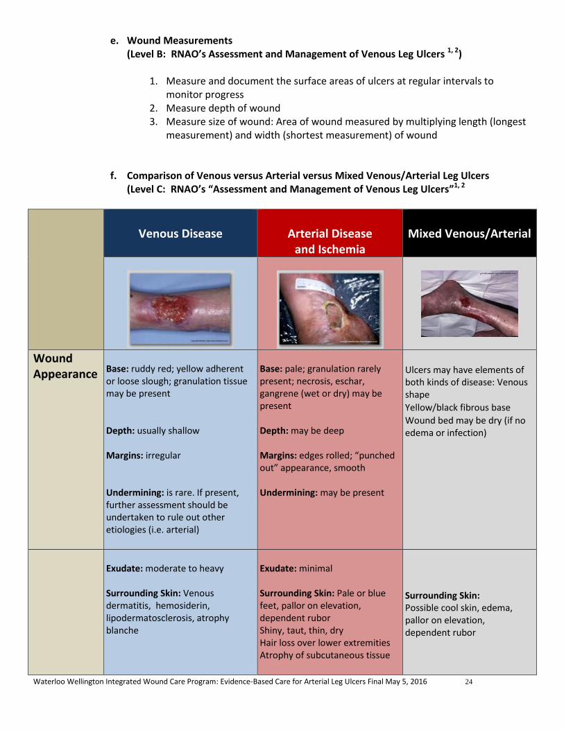
e. Wound Measurements (Level B: RNAO’s Assessment and Management of Venous Leg Ulcers 1, 2)
1. Measure and document the surface areas of ulcers at regular intervals to
monitor progress 2. Measure depth of wound 3. Measure size of wound: Area of wound measured by multiplying length (longest
measurement) and width (shortest measurement) of wound
f. Comparison of Venous versus Arterial versus Mixed Venous/Arterial Leg Ulcers (Level C: RNAO’s “Assessment and Management of Venous Leg Ulcers”1, 2
Venous Disease
Arterial Disease
and Ischemia
Mixed Venous/Arterial
Wound Appearance
Base: ruddy red; yellow adherent or loose slough; granulation tissue may be present Depth: usually shallow Margins: irregular Undermining: is rare. If present, further assessment should be undertaken to rule out other etiologies (i.e. arterial)
Base: pale; granulation rarely present; necrosis, eschar, gangrene (wet or dry) may be present Depth: may be deep Margins: edges rolled; “punched out” appearance, smooth Undermining: may be present
Ulcers may have elements of both kinds of disease: Venous shape
Yellow/black fibrous base
Wound bed may be dry (if no edema or infection)
Exudate: moderate to heavy Surrounding Skin: Venous dermatitis, hemosiderin, lipodermatosclerosis, atrophy blanche
Exudate: minimal Surrounding Skin: Pale or blue feet, pallor on elevation, dependent rubor Shiny, taut, thin, dry Hair loss over lower extremities Atrophy of subcutaneous tissue
Surrounding Skin: Possible cool skin, edema, pallor on elevation, dependent rubor

Waterloo Wellington Integrated Wound Care Program: Evidence-Based Care for Arterial Leg Ulcers Final May 5, 2016 25
Edema: pitting or non-pitting; may worsen with prolonged standing or sitting from legs being in a dependent position
Edema: atypical
Edema: variable
Scarring: from previous ulcers, ankle flare, tinea pedis (athlete’s foot) Nails: Usually normal unless infection present Temperature: normal; warm to touch
Nails: Dystrophic Temperature: decreased/cold
Nails: Thickened toenails
Infection: less common but chronic venous ulcers are prone to biofilms, induration, cellulitis, inflamed, tender blisters
Infection: frequent (signs may be subtle) Cellulitis, necrosis, eschar, gangrene may be present
Infection: can have signs and symptoms of both venous and arterial disease
Location Ulceration is usually on the medial lower leg superior to malleolus in gaiter region but can be on lateral aspect as well or may encircle the entire ankle or leg Ulcers occurring above the mid-calf or on the foot likely have other origins, but may be caused by trauma in a leg with existing venous insufficiency
Areas exposed to pressure or repetitive trauma, or rubbing of footwear Lateral malleolus Mid tibial Phalangeal heads Toe tips or web spaces
Same as venous or ulcer may be circumferential
Pain Described as throbbing, sharp, itchy, sore, tender, heaviness Worsens with prolonged dependency. Some relief on elevation of limb.
Pain is increased with elevation of limb. Pain may also be incurred with walking. This is usually due to the presence of intermittent claudication which will be relieved with 10 minutes of rest
Pain with elevation Intermittent claudication (early) Night time rest pain (late disease)
g. Ankle Brachial Pressure Index (ABPI) / Toe Brachial Pressure Index (TBPI )
Table 3: Adapted from WOCN Clinical Fact Sheet Quick Assessment of Leg Ulcers (November 2009) by CarePartners (2014), Registered Nurses Association of Ontario. Nursing Best Practice Guideline: Assessment and Management of Venous Leg Ulcers. March 2004 and Registered Nurses Association of Ontario. Nursing Best Practice Guideline Supplement: Assessment and Management of Venous Leg Ulcers. March 2007
Photos courtesy of:http://www.medetec.co.uk

Waterloo Wellington Integrated Wound Care Program: Evidence-Based Care for Arterial Leg Ulcers Final May 5, 2016 26
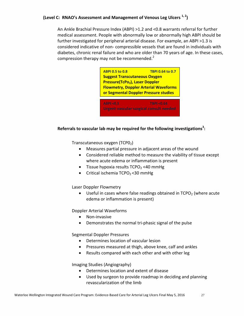
(Level B: RNAO’s Assessment and Management of Venous Leg Ulcers 1, 2)
Perform ABPI/TBPI to rule out the arterial disease. If patient is a diabetic, toe pressures should be obtained
An Ankle Brachial Pressure Index (ABPI) measurement should be performed by a trained practitioner to rule out the presence of peripheral arterial disease, particularly prior to the application of compression therapy. ABPI measurement offers valuable information as a screening tool for lower extremity peripheral arterial disease.16
Further Investigation Required
Ankle Brachial Pressure Index (ABPI) / Toe Brachial Pressure Index (TBPI ) Interpretations 17,18
ABPI > 0.9-1.2 ....Normal (1.2 or > could indicate calcification, seen in diabetes, patients that smoke, hypertension,
rheumatoid arthritis, systemic vasculitis or advanced age ) 0.80-0.9 ......Mild ischemia (inflow disease may be present) 0.50-0.79 ....Moderate ischemia (Would benefit from vascular surgeon consult to expedite wound healing) 0.35-0.49 ....Moderately severe ischemia (Urgent vascular surgery consult recommended) 0.20-0.34 ....Severe ischemia (Urgent vascular surgery consult recommended) <0.20 ..........Likely critical ischemia, but absolute pressure and clinical picture must be considered
(Urgent vascular surgery consult recommended) TBPI:
> 0.7 …………Normal > 0.7 0.64 - 0.7…..Borderline < 0.64………. Abnormal indicating arterial disease (Urgent vascular surgery consult recommended)
Lower Leg Vascular Assessment RNAO recommends a 3 month complete reassessment if no evidence of healing and a 6 month reassessment for resolving and healing (but not yet healed) wounds
1,2
If ulceration does not heal or show improvement after 3 months and patient has an Ankle Brachial Pressure Index (ABPI) of > 0.8 to 1.3, a referral to a vascular surgeon to review potential surgical interventions is recommended
Right Left
ABPI: TBPI: ABPI: TBPI:
Waterloo Wellington Integrated Wound Care Program: Evidence-Based Care for Arterial Leg Ulcers Final May 5, 2016 27
(Level C: RNAO’s Assessment and Management of Venous Leg Ulcers 1, 2) An Ankle Brachial Pressure Index (ABPI) >1.2 and <0.8 warrants referral for further medical assessment. People with abnormally low or abnormally high ABPI should be further investigated for peripheral arterial disease. For example, an ABPI >1.3 is considered indicative of non- compressible vessels that are found in individuals with diabetes, chronic renal failure and who are older than 70 years of age. In these cases, compression therapy may not be recommended.2
Referrals to vascular lab may be required for the following investigations3:
Transcutaneous oxygen (TCP02)
Measures partial pressure in adjacent areas of the wound
Considered reliable method to measure the viability of tissue except where acute edema or inflammation is present
Tissue hypoxia results TCPO2 <40 mmHg
Critical ischemia TCPO2 <30 mmHg
Laser Doppler Flowmetry
Useful in cases where false readings obtained in TCPO2 (where acute edema or inflammation is present)
Doppler Arterial Waveforms
Non-invasive
Demonstrates the normal tri-phasic signal of the pulse
Segmental Doppler Pressures
Determines location of vascular lesion
Pressures measured at thigh, above knee, calf and ankles
Results compared with each other and with other leg Imaging Studies (Angiography)
Determines location and extent of disease
Used by surgeon to provide roadmap in deciding and planning revascularization of the limb
ABPI 0.5 to 0.8 TBPI 0.64 to 0.7
Suggest Transcutaneous Oxygen Pressure(TcPo2), Laser Doppler Flowmetry, Doppler Arterial Waveforms or Segmental Doppler Pressure studies
ABPI <0.5 TBPI <0.64
Urgent vascular surgical consult needed

Waterloo Wellington Integrated Wound Care Program: Evidence-Based Care for Arterial Leg Ulcers Final May 5, 2016 28
h. Determine if the wound is “Healable, Maintenance or Non-Healable”
Healable: Have sufficient vascular supply, underlying cause can be corrected, & health can be optimized Maintenance: have healing potential, but various patient factors are compromising wound healing at this time Non-healable/Palliative wound: has no ability to heal due to untreatable causes such as terminal disease or end-of-life19
i. Nutritional Assessment
(Level B: RNAO’s Assessment and Management of Venous Leg Ulcers 1, 2) The following assessments and blood work should be considered when investigating nutritional status of a person with a wound:
Protein-Calorie Malnutrition
Pre-albumin if available (low scores indicate risk for malnutrition)
Serum albumin level (<30g/l will delay healing; <20g/l will be non-healable)
C-reactive Protein (CRP)
Check for anemia
CBC (including RBC, Hct, Hgb, MCV, Platelets etc.)
If anemic, proceed to checking →
Serum Iron
Total Iron Binding
Ferritin
Transferrin
B12
Red blood cell folate level Kidney function (To check hydration)
BUN
Creatinine
Potassium
In addition to inquiring about recent weight loss, signs of dehydration, and assessing the Braden Scale Nutritional sub-scale, which helps to capture protein intake, there are several signs of micronutrient deficiencies that are easy to detect when you know what to look for.
Signs of micronutrient deficiencies:
Reddish tongue with a smooth surface (Vitamin B deficiency)
Magenta flank-steak appearing tongue with cracks at corners of the mouth (called angular stomatitis) (Vitamin B2 deficiency )
Dementia, diarrhea, dermatitis (pellagra)—crepe paper skin with wrinkles in the skin and flat surfaces between the wrinkles –also associated with bullous pemphigoid and gramuloma annulare (Vitamin B3 deficiency)
Prominent “snowflake” exfoliation of the epidermis of the lower legs (Essential Fatty Acid deficiency)
Skin and capillary fragility with purpura, skin tears, increase risk of pressure ulcers, severe collagen deficiency so that the skin is like plastic wrap, and extensor tendons and venous plexus is easily seen through the transparent epidermis (Chronic Scurvy/Vitamin C deficiency)

Waterloo Wellington Integrated Wound Care Program: Evidence-Based Care for Arterial Leg Ulcers Final May 5, 2016 29
Reddish, scaly, itchy skin lesions (Vitamin A, E, and K deficiency)
Seborrheic-like rash that is red, flaky seen along the lateral eyebrows, nasal labial folds and chin (Zinc deficiency)
Prolonged tenting of the skin in the presence of adequate fluid intake
If the presence of any of these signs of micronutrient deficiencies is noted, a referral should be made to a Registered Dietitian who can work with the primary care provider for screening of dietary deficiencies and treatment.
The Nestle Mini-Nutritional Assessment (MNA) ( Toolkit item #9) is a screening and assessment tool that identifies individuals age 65 and above who are malnourished or at risk of malnutrition, allowing for earlier intervention to provide adequate nutritional support. It has not been validated for use with younger individuals. The screening tool consists of 6 questions.
Complete the screen by filling in the boxes with the appropriate numbers.
Total the numbers for the screening score. The screening score (max 14 points):
12- 14 points = normal nutritional status 8-11 points = at risk of malnutrition 0 -7 points = malnourished
Nutritional Supplementation
Nutritional supplementation should be provided to a patient only after a thorough nutritional assessment has been completed and the reason for malnutrition has been identified.30
Macronutrients Macronutrients such as carbohydrates, proteins and lipids (fats) are required in adequate amounts to provide the body with total energy needs. Caloric intake of 30-35 kcal/kg of body weight is recommended for patients with chronic wounds. Patients that are underweight may require a caloric intake of 35-40% kcal/kg of body weight.30 These macronutrients should be consumed daily in the following amounts:
Carbohydrates 45-60%
Fat 25-30%
Protein 15-20% (1.25-1.5 g/kg of body weight) 30
Protein needs are increased in order for healing to occur. Diets that include inadequate amounts of protein can be blamed for “increased skin fragility, decreased immune function, poorer healing and longer recuperation after illness”. 30 Caution should be taken when administering protein to patients with liver or kidney failure. Consultation with a Registered Dietician is recommended with this patient population.
Link to Mini-Nutritional Assessment Form

Waterloo Wellington Integrated Wound Care Program: Evidence-Based Care for Arterial Leg Ulcers Final May 5, 2016 30
Arginine and Glutamine are amino acids that are needed in the production of collagen. Collagen is required for healing to occur. Although supplementation of Glutamine is controversial, it is believed to be helpful in those patients where malnutrition and chronic wound healing are being addressed. Arginine is required by the body when under metabolic stress. Supplementation of Arginine has been shown to improve healing. It is important to note that both Arginine and Glutamine require adequate protein intake to be of any value.30 Fats are an integral part of a healthy diet required for healing to occur. Omega 3 fatty acids are antithrombotic, vasodilators and anti-inflammatory. Omega 6 fatty acids are responsible for platelet aggregation, inflammation and vasoconstrictors. Further research is required before supplementation of Omega 3 or Omega 6 should be recommended. 30 Micronutrients 30 Zinc
Should only be supplemented if deficiency is determined
Recommended dose: 40mg of elemental zinc/day (176 mg zinc sulfate) for up to 10 days to enhance wound healing
Asorbic Acid (Vitamin C)
Recommended dose: 500 to 1000 mg daily in divided doses
Vitamin A
Recommended in patients taking corticosteroids
Recommended dose: 10,000-25,000 IU daily for 10-14 days
Use with caution in patients with protein deficiencies or liver failure
4.3 Optimize Medical Therapy 9, 10,20,27,28 The two strategies of caring for patients with arterial wounds are to improve circulation and improve oxygenation
Smoking and nicotine cessation
Barriers to cessation should be addressed at each patient visit
Educational, pharmacological and behavioral techniques should be utilized
See Toolkit Item #7a for Smoking, Chronic Wound Healing, and Implications for Evidence-Based Practice – McDaniel and Browning, Toolkit Item #7b for Checklist to readiness to quit smoking, see Toolkit Item #7c for Applying 5 A’s to smoking cessation, see Toolkit Item #7d for WHY test, see Toolkit Item #7e for smoking cessation medication comparison chart and see Toolkit Item #7f for Strategies to avoid relapse. 14
1. Nutrition and Chronic Wounds article 2. Role of Nutrition in Tissue Viability article

Waterloo Wellington Integrated Wound Care Program: Evidence-Based Care for Arterial Leg Ulcers Final May 5, 2016 31
Suggest initiating statin therapy
Control hypertension
Control blood sugar if diabetic
Suggest antiplatelet therapy (caution when used with Trental – increased chance of bleeding)
Avoid extremes that can impair blood flow and oxygen delivery
Avoid very hot or very cold contact
Avoid very tight or very loose fitting clothing and footwear
Encourage exercise
Minimum of 30 minutes/3 times week is recommended
Address dehydration
Can impair blood flow and oxygen delivery
Control Pain The most effective pain control with arterial ulcers is to improve the blood supply
Encourage use of analgesics (pain medication) at regular intervals (eg. Every 3-6 hours) instead of taking only as needed
Recommendations for nociceptive pain (described as sharp, aching or throbbing) Non-Opioids – eg. ASA or Acetaminophen Mild Opioids – eg. Codeine Strong Opioids – eg. Morphine or Oxycodone Recommendations for neuropathic pain (described as burning, stinging, shooting, stabbing or hyperesthesia – sensitivity to touch)
Second generation tricyclic agents – eg. Nortriptyline or Desipramine If pain is not relieved try using Gabapentin or Pregabalin Other recommendations for pain control include:
Positioning of leg in a dependent position may increase arterial blood flow
Calm patient’s fears (anxioytic may be required)

Waterloo Wellington Integrated Wound Care Program: Evidence-Based Care for Arterial Leg Ulcers Final May 5, 2016 32
Patient may benefit from the head of their bed being elevated. Caution: protect from pressure
Spinal cord stimulation may reduce pain
Prostanoids may help with rest pain
Amputation may have to be considered for pain control and quality of life issues
4.4 Pharmacological Treatment:
a. Pentoxyfilline (Trental),20,21
It is a haemorheological agent, thought to increase red and white cell filterability by altering the shape and flexibility and therefore the flow of cells, and decrease whole blood viscosity, platelet aggregation and fibrinogen levels 18
Influences microcirculatory blood flow and oxygenation of ischaemic tissues
May increase blood flow for patients with vasospastic disorders such as Raynaud phenomenon
Would have very little effect in advanced peripheral arterial disease
The full product monograph should be consulted re: precautions when using with anticoagulants such as Plavix, as Trental may increase the risk of bleeding
In a Cochrane review of 11 randomised trials comparing Pentoxifylline with placebo or other therapy in the presence or absence of compression, in people with venous leg ulcers, Pentoxifylline was seen to be an effective adjunct to compression bandaging for treating arterial ulcers and may be effective in the absence of compression 19
The majority of adverse effects were gastrointestinal disturbances
If woody fibrosis and induration are present in the peri-wound area or in the leg, Pentoxyfilline (Trental) 400mg TID helps to soften fibrosis and allows the wound to heal.
Start with a BID dosage and increase to TID as tolerated, with appropriate precautions with individuals with known history of indigestion or GERDs.
Be aware that it may take two months before benefit can be seen b. Nifedipine 20
May increase blood flow for patients with vasospastic disorders such as Raynaud phenomenon
4.4 Surgical and Medical Intervention Strategies 20
Interventions are aimed at reversing ischemia
a. Endovascular Perfusion Strategies (catheter-based revascularization) Angioplasty
Least invasive
Cannula with guidewire inserted into artery guided by fluoroscopy
Diseases section of artery is dilated using an angioplasty balloon

Waterloo Wellington Integrated Wound Care Program: Evidence-Based Care for Arterial Leg Ulcers Final May 5, 2016 33
Stent MAY be used to maintain the lumen Potential Complications include:
Hemorrhage
Thrombosis
Restenosis
Occlusion
Catheter-directed Thrombolytic Therapy
Thrombus dissolved using catheter
b. Surgical Perfusion Strategies Endarterectomy
Open surgical procedure
Lesions are removed from the artery
Can be performed under local anesthetic
Appropriate for higher risk surgical patients
Arterial Bypass
Used for extensive vascular pathology
Creates autogenous (natural) or prosthetic (artificial) vein that bypass obstruction
Restores circulation to ischemic tissue
Autogenous vein is usually preferred (saphenous vein)
Higher risk of infection if prosthetic graft used
Successful in 85-89% of patients with critical limb ischemia
c. Oxygenation Strategies Used for patients with incomplete arterial obstruction to increase dissolved blood oxygen
Normobaric Oxygen (can increase plasma-dissolved oxygen by 40%)

Waterloo Wellington Integrated Wound Care Program: Evidence-Based Care for Arterial Leg Ulcers Final May 5, 2016 34
Venturi Mask
Delivers 24 to 60 % oxygen
Nasal Cannulas
Delivers 24 to 40% oxygen Requires nasal breathing
Nonrebreather Mask
Delivers 80 to 90% oxygen
Use should be limited to 24-36 hours
Hyperbaric Oxygen Therapy (Level of Evidence 2)
Can increase dissolved blood oxygen by up to 6 mL/dL
Can benefit angiogenesis, fibroblast growth and collagen production
Enhances removal of carbon monoxide from hemoglobin
Decreases neutrophil adherence to vessel walls
Reduces edema
Should be considered in patients with non-reconstructable anatomy or where ulcer is not healing despite revascularization
5. Provide Local Wound Care
a. Intervention Algorithm Figure 3

Waterloo Wellington Integrated Wound Care Program: Evidence-Based Care for Arterial Leg Ulcers Final May 5, 2016 35
b. Signs and Symptoms of Wound Infection 22,23 (Level A, B and C: RNAO’s Assessment and Management of Venous Leg Ulcers 1, 2)
Intervention Algorithm Place Holder

Waterloo Wellington Integrated Wound Care Program: Evidence-Based Care for Arterial Leg Ulcers Final May 5, 2016 36
Arterial ulcers, like most chronic wounds, can become infected with superficial or spreading bacteria. The validated mnemonics “NERDS” and “STONEES” classify the signs and symptoms of localized infection (NERDS) and spreading infection (STONEES). Increased localized pain is a significant predictor of deep compartment infection.
Presence of Superficial Bacteria
N- Non-healing wound
E- Exudate increased
R- Red friable (fragile tissue that bleeds easily)
D- Debris (presence of necrotic tissue (eschar/slough) in wound
S- Smell
Presence of Spreading Bacteria (< 3 low bacteria count, >3 high bacteria count)
S- Size increasing
T- Temperature increased (> 3 degrees F difference)
O- Os (probes to bone or bone is increased)
N- New areas of breakdown
E- Exudate present
E- Erythema and/or Edema
S- Smell
In addition to recognizing the signs and symptoms of infection in arterial leg ulcers, it may be helpful to obtain a culture and sensitivity (C&S) using a validated method of sampling to quantify bacteria in wounds. Tissue biopsies are considered the gold standard but unfortunately are not practical in many settings. Fortunately, a linear relationship between quantitative tissue biopsy and swab for C&S taken using the Levine method of sampling (see below) has been validated and is recommended for assessing any open wound. Swabs for C&S are important in determining the type of bacteria and the appropriate antibiotics, but are not necessary to confirm the presence or absence of infection. The C&S results may not reflect the presence or absence of biofilm.
Levine Method for obtaining C&S laboratory swab 23
1. Cleanse wound thoroughly 2. Place swab on granulation tissue (must be granulation tissue only –if none
present, tissue aspiration or biopsy may be required) 3. Apply enough pressure to extract fluid 4. Turn swab 360 degrees on fluid (avoid slough or debris) 5. Place swab in transport medium
Recommendations for the use of antiseptics and antiseptic dressings

Waterloo Wellington Integrated Wound Care Program: Evidence-Based Care for Arterial Leg Ulcers Final May 5, 2016 37
An international consensus panel studied use of silver in healable wounds. This panel recommended that silver be used for a two week period if infections is suspected and then be reassessed. It is the opinion of Dr. David Keast, a leading wound care specialist that these recommendations can be extended to the use of all antiseptics and antiseptic dressings (eg. iodine and PHMB).
Signs and symptoms of Lower Leg Cellulitis 24
Choices for after initial two weeks using antiseptics or antiseptic dressings
Healable wounds Bacterial burden has been reduced and the wound is progressing to healing
Discontinue use of antiseptics and antiseptic dressings
Bacterial burden has been reduced the wound is progressing but there are patient risk factors that put them at risk of re-infection
Continue to use and monitor
Suggest: Low adherent knitted viscose fabric impregnated with a polyethylene glycol (PEG) base containing 10% Povidone Iodine
Bacterial burden is controlled but the location of the wound is such that it is at risk of recontamination e.g. perianal, or exit sites for g-tubes etc
Continue to use as an antimicrobial barrier.
No effect Discontinue and change strategy such as systemic antibiotics or a change of the topical antiseptic or better debridement. As always factors such as adequate plantar pressure redistribution in neuropathic foot ulcers or compression therapy for venous disease must be in place.
Slough/Eschar
No slough or obvious biofilm present
Suggest: Iodine gel
Slough is present Topical antiseptic to remove biofilm needed
Suggest: Periodic debridement provided arterial blood supply is adequate
Maintenance or Non-healable
Wounds
Eschar to be kept dry No real limit to use Use as long as required
Suggest: Povidone iodine is best as an antiseptic with drying properties. Use it as long as required to keep dry As per Dr. Stephan Landis and Dr. David Keast (Leading Wound Care Specialists) Aug.2015

Waterloo Wellington Integrated Wound Care Program: Evidence-Based Care for Arterial Leg Ulcers Final May 5, 2016 38
Cellulitis is a spreading bacterial infection of the dermis and subcutaneous tissues, where the edge of the erythema may be well-defined or more diffuse and typically spreads rapidly
Systemic upset with fever and malaise occurs in most cases, and may be present before the localising signs such as the local symptoms seen with STONEES23
Lower leg cellulitis can be extremely serious with long-term morbidity, including lower leg edema. It requires prompt recognition by health care providers and appropriate interventions
Note that lower leg cellulitis usually affects only one leg, not both. If both legs are affected, it is likely venous dermatitis or allergic contact dermatitis, but this does not mean that it could never be cellulitis in both legs 25
Signs and Symptoms of Cellulitis of Lower Legs
Risk Factors
Waterloo Wellington Integrated Wound Care Program: Evidence-Based Care for Arterial Leg Ulcers Final May 5, 2016 39
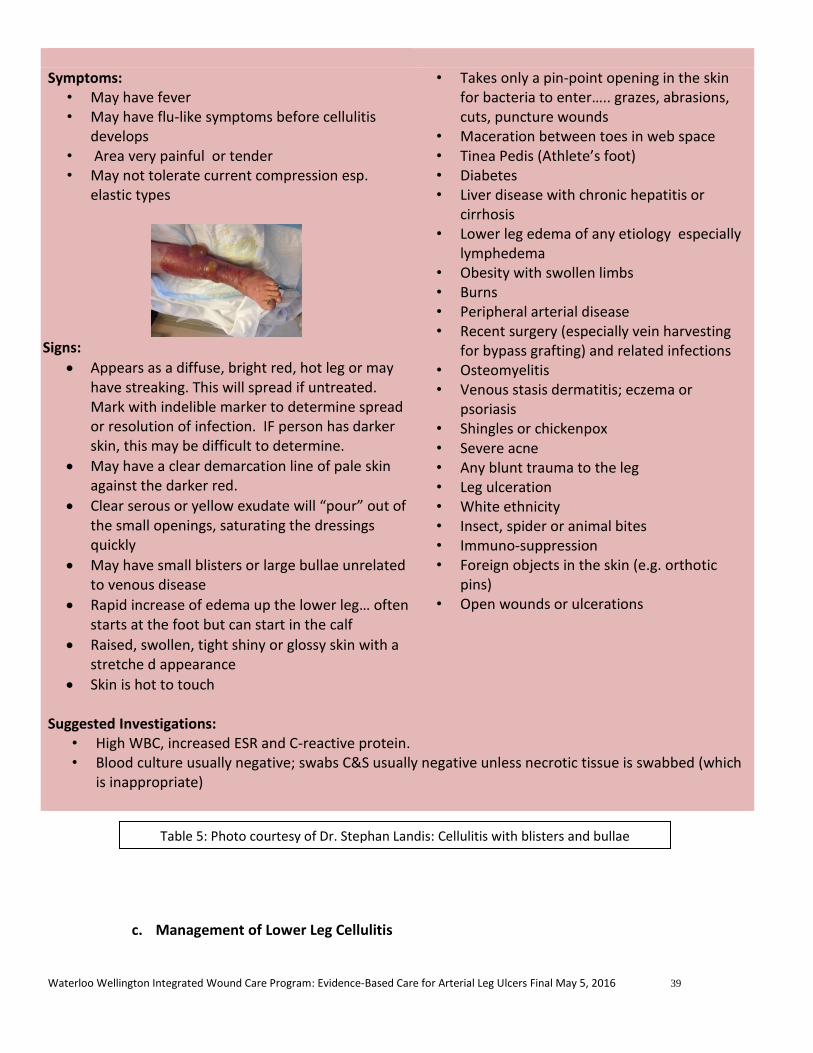
Symptoms:
• May have fever • May have flu-like symptoms before cellulitis
develops • Area very painful or tender • May not tolerate current compression esp.
elastic types
Signs:
Appears as a diffuse, bright red, hot leg or may have streaking. This will spread if untreated. Mark with indelible marker to determine spread or resolution of infection. IF person has darker skin, this may be difficult to determine.
May have a clear demarcation line of pale skin against the darker red.
Clear serous or yellow exudate will “pour” out of the small openings, saturating the dressings quickly
May have small blisters or large bullae unrelated to venous disease
Rapid increase of edema up the lower leg… often starts at the foot but can start in the calf
Raised, swollen, tight shiny or glossy skin with a stretche d appearance
Skin is hot to touch
• Takes only a pin-point opening in the skin for bacteria to enter….. grazes, abrasions, cuts, puncture wounds
• Maceration between toes in web space • Tinea Pedis (Athlete’s foot) • Diabetes • Liver disease with chronic hepatitis or
cirrhosis • Lower leg edema of any etiology especially
lymphedema • Obesity with swollen limbs • Burns • Peripheral arterial disease • Recent surgery (especially vein harvesting
for bypass grafting) and related infections • Osteomyelitis • Venous stasis dermatitis; eczema or
psoriasis • Shingles or chickenpox • Severe acne • Any blunt trauma to the leg • Leg ulceration • White ethnicity • Insect, spider or animal bites • Immuno-suppression • Foreign objects in the skin (e.g. orthotic
pins) • Open wounds or ulcerations
Suggested Investigations:
• High WBC, increased ESR and C-reactive protein. • Blood culture usually negative; swabs C&S usually negative unless necrotic tissue is swabbed (which
is inappropriate)
c. Management of Lower Leg Cellulitis
Table 5: Photo courtesy of Dr. Stephan Landis: Cellulitis with blisters and bullae

Waterloo Wellington Integrated Wound Care Program: Evidence-Based Care for Arterial Leg Ulcers Final May 5, 2016 40
Swabs for c&s not usually helpful if cellulitis is dry; if wet then should be done using LEVINE semi-quantitatitve method
Levine Method for obtaining C&S laboratory swab 23
1. Cleanse wound thoroughly 2. Place swab on granulation tissue (must be granulation tissue only –if none
present, tissue aspiration or biopsy may be required) 3. Apply enough pressure to extract fluid 4. Turn swab 360 degrees on fluid (avoid slough or debris) 5. Place swab in transport medium
Mark line of demarcation on leg distally and proximally with soft-tip indelible marker (not pen) which helps caregivers and patient to visualize if the infection spreads beyond the point of first assessment
Compression, especially elastic systems, may be too painful to tolerate until the infection starts to respond to the antibiotic therapy. Do not stop compression entirely, because the edema will increase as a result of the cellulitis. May use appropriate lower mmHg compression such as two layers of tubular support bandage (e.g. Tubigrip)
Treat any co-existing conditions such as venous ulcer, venous dermatitis or tinea pedis in addition to the systemic antibiotics
In some individuals, discomfort can be soothed using a compress of Burosol solution or Burrow’s solution x 15-20 minutes available from some compounding pharmacies
Polyhexamethylene Biguanide (PHMB –e.g. AMD) antimicrobial kerlix loose- woven (11.4 cm x 3.7 m) may be used. Wrap the affected leg from the base of the toe to below the knee, overlapping each turn by 50%. If exudate amount is large, cover with absorptive secondary dressing and kling wrap, covered by appropriate lower mmHg compression such as two layers of tubular support bandage (e.g. Tubigrip)
Another option if there is dermatitis along with the cellulitis and the individual is not allergic to sulpha or silver, is to obtain a prescription for Silver Sulfadiazine applied 3-5 mm thick. Care should be taken to prevent the spread of the cream onto non-ulcerated areas. The cream should be followed by an absorbent pad or gauze dressing, with further application of pressure bandaging as appropriate for the ulcer. The dressing should be changed every 2 or 3 days, with cleaning and debriding being performed before application of silver sulfadiazine. It is not recommended that silver sulfadiazine cream be used in leg ulcers that are very exudative.36
Combination systemic antibiotic therapy is needed for cellulitis (see table 6)
Situation
Suggested antibiotics
If allergic to penicillin
Comments

Waterloo Wellington Integrated Wound Care Program: Evidence-Based Care for Arterial Leg Ulcers Final May 5, 2016 41
Non-purulent Skin/Soft Tissue Infection (i.e. erysipelas, cellulitis,
necrotizing infections)
MILD: Oral treatment Penicillin VK Amoxicillin Cephalexin Cloxacillin Clindamycin MODERATE: IV treatment Penicillin G Cefazolin or ceftriaxone Clindamycin SEVERE: Surgical vs. empiric treatment Surgical Vancomycin + Piperacilin/tazobactam
Clindamycin Or Vancomycin
Treat for about 10 to 14 days or until signs of inflammation have resolved
Purulent Skin/Soft Tissue Infection (i.e.iImpetigo, ecthyma, furuncle, carbuncle,
abscess)
MILD: Incision and drainage MODERATE: Incision & drainage and culture & sensitivity , plus empiric or defined treatment Trimethoprim/Sulfamethoxa
zole Doxycycline Cephalexin Cloxacillin SEVERE: Incision & drainage and culture & sensitivity, plus empiric or defined treatment Vancomycin Linezolid Trimethoprim/Sulfamethoxazole Cefazolin Clindamycin
Clindamycin Or Vancomycin Or Linezolid
d. Venous Dermatitis: Signs, Symptoms, Prevention and Treatment
Table 6: Per Dr. Stephan Landis Guelph 2015

Waterloo Wellington Integrated Wound Care Program: Evidence-Based Care for Arterial Leg Ulcers Final May 5, 2016 42
Venous Stasis Dermatitis: Signs, Symptoms, Prevention and Treatment Table 7 24
Description
Treatment
Venous Stasis dermatitis (also known as “Venous dermatitis”, “Gravitational dermatitis” or “Venous, stasis eczema” describes the red, itchy rash on the lower legs which can be dry and scaly or can weep and form crusts commonly seen in people with chronic venous insufficiency. The skin may appear brown or purple in colour and the lower legs become increasingly edematous. It may be associated with acute contact dermatitis, which appears as itching, burning red areas on the lower leg corresponding to an area where a topical product has been used.
Avoid the use of known sensitizers in individuals with venous disease (perfume, latex, dyes, lanolin or wool alcohols, balsam of peru, cetylsterol alcohol, parabens, colophony propylene glycol, neomycin, rubber, some adhesives, framycetin or gentamycin) (Sibbald et al. 2007)
Limit baths and showers to 15 minutes in warm not hot water
Avoid harsh soaps
Avoid vigorous use of a washcloth or towel. Blot or pat areas dry so there is still some moisture left on the skin
Use moisturizers immediately after bathing such as Glaxal Base (ask pharmacist if not on shelf), Cliniderm, Eucerin or Moisturel lotions (not cream) or plain Vaseline petrolatum ointment to keep the skin healthy and free of dry scales.
Any products containing petrolatum or alcohol should be stopped if severely dry scaly skin develops
For severely dry, scaly skin (Xerosis) use products containing Urea such as Uremol 20% or Attractain (contains 10% urea and 4% AHA), Eucerin 10% Urea Lotion, Lac-Hydrin 12% Urea works by enhancing the water-binding capacity of the stratum corneum. Long-term treatment with urea has been demonstrated to decrease transepidermal water loss. Urea also is a potent skin humidifier and descaling agent, particularly in 10% concentration Lactic acid (in the form of an alpha hydroxy acid) can accelerate softening of the skin, dissolving or peeling the outer layer of the skin to help maintain its capability to hold moisture. Lactic acid in concentrations of 2.5% to 12% is the most common alpha hydroxy acid used for moderate to severe xerosis.
Use creams and lotions as directed, and stop if any signs of dermatitis occur.
Only use topical corticosterioid preparations for two weeks at a time (if being applied more frequently than 2 x/ week) because they cause skin to break down or develop a rebound dermatitis
If dermatitis occurs and patient is using compression stockings, there is a risk that the lotions or creams will cause accelerated deterioration of the stocking material. In this case, it is best to only apply the topical products at bedtime when the stockings are removed
If the dermatitis is severe, there may be a need to switch to compression bandaging with a medicated wrap containing zinc or other products. Zinc products without preservatives are available if a reaction to zinc with preservative occurs (e.g. Zipzoc)
Systemic antibiotic therapy is not needed for acute contact dermatitis, unless cellulitis has developed
Referral to dermatologist for allergy patch testing is indicated if dermatitis does not respond to treatment
Dressing Choices for Venous Stasis Dermatitis (Eczema) 24
Photo Courtesy of Dr. Stephan Landis

Waterloo Wellington Integrated Wound Care Program: Evidence-Based Care for Arterial Leg Ulcers Final May 5, 2016 43
Itching and burning can be soothed using a compress of Burosol solution or Burrow’s solution x 15-20 minutes (product is no longer available over the counter (OTC) but can be obtained in powdered sachets from some compounding pharmacies
Apply prescribed steroidal cream to all affected areas- with added Menthol ¼ % to ½ % will aid in soothing and anti-itch effect, and cream can be kept in refrigerator
Apply Unna’s boot using a medicated zinc paste bandage* (e.g. Viscopaste) wrapped in a spiral wrap using fan-fold pleats to prevent constriction
e. Determining Goals for Local Treatment for Arterial Leg Ulcers 1,2
(Level A, B and C: RNAO’s Assessment and Management of Venous Leg Ulcers)
Healable Wounds: Have sufficient vascular supply, underlying cause can be corrected, & health can be optimized Goal: Principles of wound bed preparation and moist wound healing: debridement, bacterial balance, exudate control, protect peri-wound skin Maintenance Wounds: have healing potential, but various patient factors are compromising wound healing at this time
Goal: Principles of wound bed preparation and moist wound healing: debridement, bacterial balance, exudate control and protect periwound skin. Avoid higher cost advanced wound treatments until factors compromising wound healing are resolved. Focus on quality of life issues, exudate and odour management
Non-healable/Palliative wounds: has no ability to heal due to untreatable causes such as insufficient vascular supply, terminal disease or end-of-life Goal: Avoid higher cost advanced wound treatment and focus on exudate and odour management, quality of life issues. Calculating Current Percentage of Healing Since Admission
Treatment Plan20
Surface Area (admission) – Surface Area (current) X 100 = _______% reduction Surface Area (admission)
*Surface area = length x width
Arterial ulcers do not follow the expected trajectory that estimates a wound should be 30% smaller at week 4 and healed by week 12. Further intervention should be considered if conservative treatment
does not improve ulcer healing in 4-6 weeks.

Waterloo Wellington Integrated Wound Care Program: Evidence-Based Care for Arterial Leg Ulcers Final May 5, 2016 44
If arterial supply is uncertain, dressings should be based on a non-healable program with moisture and bacterial reduction until further assessments can be performed to provide objective evidence of ‘healability’. Healing Treatment Plan
Plan determined by vascular testing
Need increased blood supply for healing to occur
Surgery will likely be required to progress to healing
Correction of the underlying disease process if possible
Mutual agreement between the physician, nurses, team, and the client regarding setting goals about the “healability” of the wound
Irrigation of wound should be avoided
Surgical debridement should only be considered when there is objective evidence that the wound is healable (circulation and oxygenation issues have been treated and/or has been deemed to be sufficient for healing and cleared by vascular surgeon)
Debridement can lead to wound enlargement, spread infection or lead to further necrosis
If there is objective evidence that wound is healable, careful sharp, surgical, mechanical, enzymatic or autolytic debridement is recommended
Recommended non-adherent dressings include: alginates, hydrogels or hydrocolloids
Avoid ‘tourniquet affect’ when securing dressings
Avoid nicotine and caffeine use
Use of correct fitting footwear
Avoid use of heating pads and ice packs due to decreased sensation
Elevate head of bed on 4-6” blocks to keep heart above feet for ischemic pain
Maintenance/Non-healing Plan (no possibility of revascularization surgery)
NO DEBRIDEMENT to be performed
Minimize risk of infection with use of providone-iodine or chlorhexidine
Health teaching regarding signs and symptoms of an infection to client and caregiver
Moist wound healing is not recommended for arterial leg ulcer that cannot be re-vascularized. Dry gangrene or eschar should be left dry. Adding moisture to devitalized tissue would increase bacterial growth
Use non-adherent dressings. Soft silicones are recommended to prevent further trauma
Caution: USE DRY WOUND HEALING 1. Keep eschar dry 2. No occlusive dressings 3. Do NOT debride 4. Avoid tourniquet effect when securing
dressings with wraps (kling, tubigrip etc). 5. If eschar becomes wet/boggy – URGENT
referral to advanced wound care specialist is recommended

Waterloo Wellington Integrated Wound Care Program: Evidence-Based Care for Arterial Leg Ulcers Final May 5, 2016 45
Care should be used when removing tape to prevent trauma
Avoid ‘tourniquet affect’ when securing dressings
Pain Control
Elevate head of bed on 4-6” blocks to keep heart above feet for ischemic pain Compression Use
Only to be used if there is objective evidence that arterial supply is sufficient for healing
Used only under close supervision of very experienced wound care specialists for mixed (venous and arterial) etiologies
Mild compression may be used after by-pass surgery to prevent edema (only with vascular surgeon’s order)
Should be removed immediately if pain develops
f. Utilize Product Picker from Canadian Association of Wound Care (CAWC)
Product Picker for Classification of Dressing Products Each organization may use the PDF Fillable CAWC Product Picker to list the products available within their organization (see Toolkit Item #12)
g. South West Regional Wound Care Program’s Wound Cleansing Table: 8 (see Toolkit Item #13
for reference chart)
Link to Product Picker
Compression is typically contraindicated in the presence of peripheral arterial disease. In some circumstances light compression may be beneficial. In such cases, compression should be ordered by an advanced wound care physician, vascular surgeon or nurse practitioner only! See algorithm in guidelines.

Waterloo Wellington Integrated Wound Care Program: Evidence-Based Care for Arterial Leg Ulcers Final May 5, 2016 46
Wound Assessment
Clean Epithelializing
Wound
Clean Granulating Wound, Decreasing in Surface Area 20-30% in 3-4 Weeks*
Clean Granulating Wound NOT
Decreasing in Size 20-30% in 3-4
Necrotic Healable Wound
(Debridement is Appropriate)
Necrotic Non-Healable Wound (Debridement is
NOT Appropriate)
Irrigate with < 7 PSI
pressure, or pour
solution over the
wound bed.
Use at least 100cc’s
of solution, at room
or body temperature.
Cleanse the
periwound skin of
debris, exudates.
No antimicrobial
solutions.
Irrigate with < 7 PSI
pressure, or pour
solution over the
wound bed.
Use at least 100cc’s
of solution, at room
or body temperature.
Cleanse the
periwound skin of
debris, exudates.
No antimicrobial
solutions.
Irrigate with 7-15 PSI
pressure.
Use at least 150cc’s of
solution, at room or
body temperature.
Cleanse the
periwound skin of
debris, exudates.
*Granulating wounds
not decreasing in size
may have a localized
infection.
Irrigate with 7-15 PSI
pressure.
Use at least 150cc’s
of solution, at room
or body temperature.
Cleanse the
periwound skin of
debris, exudates.
Do not irrigate or
cleanse the
wound itself (the
intent is to allow
the necrotic tissue
to dry out and
stabilize).
If there is exudate
present on the
periwound skin,
gently cleanse it
and pat dry.
Topical
application of
proviodine-iodine
solution or
Chlorhexadine to
the wound surface
is appropriate, i.e.
paint with
Proviodine.
Malignant Wounds
Wound with Debris
or Contamination/
Superficial & Partial
Wound with Debris
or Contamination/
Superficial & Partial
* Localized And/Or
Spreading Infection
Maintenance
Wounds

Waterloo Wellington Integrated Wound Care Program: Evidence-Based Care for Arterial Leg Ulcers Final May 5, 2016 47
Thickness Burn Thickness Burn
Irrigate with 7-15 PSI
pressure, if tolerated.
Reduce pressure as
needed to minimize
pain and damage to
friable tumor tissue.
Use at least 150cc’s
of solution, at room
or body temperature.
Cleanse the
periwound skin of
debris, exudates.
Foul odor indicates
presence of
anaerobes - use an
antimicrobial
solution, and/or
topical
Metronidazole.
Irrigate with 7-15 PSI
pressure.
Use at least 150cc’s
of solution, at room
or body temperature.
Cleanse the
periwound skin of
debris, exudates.
May cleanse small
burns with lukewarm
tap water and mild
soap.
Irrigate into
tunneled/undermined
area using a 5Fr
catheter or “soft-
cath” with a 30cc
syringe.
Use at least 150cc’s of
solution, at room or
body temperature.
Irrigate until returns
are clear.
Gently palpate over
undermined or
tunneled areas to
express any irrigation
solution that is
retained.
Do not force irrigation
when resistance is
detected.
Cleanse the
periwound skin of
debris, exudates.
Irrigate with 7-15 PSI
pressure.
Use at least 150cc’s
of antimicrobial
solution, at room or
body temperature.
Cleanse the
periwound skin of
debris, exudates.
Two week challenge:
May use a 10 – 14
day cleansing regime
with an antimicrobial
solution to address
bacterial burden.
Cleansing will be
dependent on
characteristics of
wound bed and
goal of treatment.
If goal is to
prevent wound
from
deteriorating,
cleanse as per a
Necrotic Non-
Healable Wound.
NOTE: Normal saline and sterile water do NOT contain preservatives and must be discarded 48 hours after opening Table courtesy of South West Regional Wound Care Program 2015

Waterloo Wellington Integrated Wound Care Program: Evidence-Based Care for Arterial Leg Ulcers Final May 5, 2016 48
Wound Appearance
Description Eschar Primarily Slough or Mixed
Granulating/Slough Wound Tissue
Fibrin Granulating **** Epithelializing **** Open Surgical Incision (small
dehiscence along otherwise intact
incision)
Closed Wounds and/or Skin at Risk
Exudate Level None → Moderate Small → Large Small→ Large Small → Moderate Small→ Moderate Small Moderate None Depth Unknown Full Partial
Thickness Full → Partial
Thickness Full Thickness →
Superficial Superficial Partial Thickness
Superficial Closed
Treatment Objective
Debride (unless the eschar is stable
and on a heel. If stable eschar is on
a heel use the maintenance
enabler)*
Cleanse, protect, moist wound healing, fill
dead space, debride
Cleanse, protect, moist wound healing,
fill dead space, debride
Cleanse, protect, moist wound healing,
fill dead space
Cleanse, protect, moist wound
healing
Cleanse, protect, moist wound
healing, fill dead space
Protect
Cleansing (min. 100 mL of
room/body temperature solution) **
7-15 PSI irrigation NS or sterile water
or commercial wound cleanser
7-15 PSI irrigation NS or sterile water or
commercial wound cleanser
7-15 PSI irrigation NS or sterile water or
commercial wound cleanser
POUR NS or sterile water solution
POUR NS or sterile water solution
POUR NS or sterile water solution
Tap water
Suggested Dressing Products
and Rate Changes***
Have ET/WCS cross-hatch hard eschar
first! Hypertonic dressing
[1+] (daily) Or
Hydrogel [0] (max 3 days)
Or Hypertonic dressing [1+] buttered with
Hypertonic dressing [1+] (daily)
Or Hydrogel [0] (max 3
days) Or
Hypertonic dressing [1+] buttered with a Hydrogel [0] (daily)
Or Cadexomer Iodine [1+]
Hypertonic dressing [1+] (daily)
Or Hydrogel [0] (max 3
days) Or
Hypertonic dressing [1+] buttered with a Hydrogel [0] (daily)
Or Cadexomer Iodine
Hydrogel [0] (max 3 days)
Or Calcium Alginate [2+]
(max 7 days) Or
Hydrophilic Fiber [2+] (max 7 days)
Cover Choices: Hydrocolloid [1-2+] +/- Film [1+] (max 7
Hydrogel [0] (max 3 days)
+/- Non-Adherent Synthetic [0] (max 7-
14 days) Or
Calcium Alginate [2+]
+/- Non-Adherent Synthetic [0] (max 7
days)
Hydrogel [0] (max 3 days)
+/- Non-Adherent Synthetic [0] (max
7-14 days) Or
Calcium Alginate [2+]
+/- Non-Adherent Synthetic [0] (max 7
days)
Film [1+] Or
Hydrocolloid [1-2+] (max 7 days)
Or Clear Acrylic [2+] (max
21 days)
South West Regional Wound Care Program’s Dressing Selection and Cleansing Enabler – HEALABLE WOUNDS

Waterloo Wellington Integrated Wound Care Program: Evidence-Based Care for Arterial Leg Ulcers Final May 5, 2016 49
* Only debride healable wounds. ** If antimicrobial effect is required, consider topical antiseptic cleansers, i.e. chlorhexadine 2% or 4% (for pseudomonas – must soak x 5 min. minimum), povidone-iodine, or ¼ strength acetic acid (for pseudomonas only – must soak x 5 min. minimum). *** If antimicrobial effect is required, consider topical antimicrobial dressings, i.e. silver compounds, iodine compounds, chlorhexadine derivatives, honey, or gentian violet and methylene blue. **** For stalled granulating or epithelializing wounds consider cadexomer iodine or pocidone iodine to initiate acute inflammation or calcium alginate or protease inhibitor dressings to address chronic inflammation.
a Hydrogel [0] (daily)
Cover Choices: Hydrocolloid [1-2+] +/- Film [1+] (max 7
days) Or
Foam [1-3+] +/- Film [1+] (max 7
days) Or
Composite [2-3+] +/- Film [1+] (max 7
days) Or
Gauze sealed by Film [1+] (for daily
changes ONLY)
buttered on Calcium Alginate [2+] or
Hydrophilic Fiber [2+] (max 3 days)
Cover Choices: Hydrocolloid [1-2+] +/- Film [1+] (max 7
days) Or
Foam [1-3+] (max 7 days)
Or Composite [2-3+] (max
7 days) Or
Gauze +/- Film seal [1+] (for daily changes
ONLY)
[1+] buttered on Calcium Alginate [2+] or Hydrophilic Fiber
[2+] (max 3 days) Cover Choices:
Hydrocolloid [1-2+] +/- Film [1+] (max 7
days) Or
Foam [1-3+] (max 7 days)
Or Composite [2-3+]
(max 7 days) Or
Gauze +/- Film seal [1+] (for daily
changes ONLY)
days – do not use on plantar foot wounds)
Or Clear Acrylic [2+]
(max 21 days) Or
Foam [1-3+] (max 7 days)
Or Composite [2-3+]
(max 7 days)
Or Hydrophilic Fiber
[2+] +/- Non-Adherent
Synthetic [0] (max 7 days)
Cover Choices: Hydrocolloid [1-2+] +/- Film [1+] (max 7 days – do not use on
plantar foot wounds)
Or Clear Acrylic [2+]
(max 21 days) Or
Foam [1-3+] (max 7 days)
Or Composite [2-3+]
(max 7 days)
Or Hydrophilic Fiber
[2+] +/- Non-Adherent
Synthetic [0] (max 7 days)
Cover Choices: Hydrocolloid [1-2+] +/- Film [1+] (max 7
days) Or
Clear Acrylic [2+] (max 21 days)
Or Foam [1-3+] (max 7
days) Or
Composite [2-3+] (max 7 days)
If the wound is superficial you may consider having your secondary dressing be
your primary contact layer as well.
Exudate capacity of dressings: [0] = none
[1+] = small [2+] = moderate
[3+] = large
Dressing wear times are found in brackets ( )

Waterloo Wellington Integrated Wound Care Program: Evidence-Based Care for Arterial Leg Ulcers Final May 5, 2016 50
ALSO, may consider pain controlling dressings for painful exudating wounds, biologic dressings for stalled granulating +/- epithelializing wounds in the absence of infection or large drainage, charcoal dressings for odor control (once the cause of the odor has been investigated and treated if able, and adjunctive therapies as indicated. Disclaimer: The information herein is for general informational purposes only and is not intended, nor should it be considered, as a substitute for professional medical advice. Always seek the advice of the attending physician or other qualified healthcare provider regarding a medical condition or treatment. Dressing selection cannot take place in isolation – a holistic patient assessment is MANDATORY. Table courtesy of South West Regional Wound Care Program 2015

Waterloo Wellington Integrated Wound Care Program: Evidence-Based Care for Arterial Leg Ulcers Final May 5, 2016 51
Wound
Appearance
Description
Eschar Primarily Slough, Mixed Granulation/Slough, or
Fibrin Wounds
Granulation Malignant Wound Localized or Spreading Infection (in an otherwise healable wound)
Exudate Level
None → Moderate Small → Large Small Large Small Copious Small Copious
Depth
Unknown Partial → Full Thickness Partial Full Thickness Partial Full Thickness Superficial Full Thickness
Treatment Objective
Stabilize/dry necrotic tissue, prevent
extension/infection, manage odor**/pain,
protect
Absorb/dry, fill dead space, prevent
extension/infection, manage odor**/pain,
protect
Absorb/dry, fill dead space, prevent extension/infection,
manage odor**/pain, protect
Absorb/dry, fill dead space, prevent infection, manage odor**/bleeding/pain***,
protect
Absorb/dry, fill dead space, reduce bacterial burden, manage
odor**/bleeding/pain, protect
Cleansing (min. 100 mL of room/body
temperature solution) **
Cleanse exudate from periwound skin.
Paint wound with Proviodine or
Chlorhexadine 2%. Pat or air dry.
POUR Proviodine or Chlorhexadine 2% solution
(min. 100cc). Pat or air dry.
POUR Proviodine or Chlorhexadine 2% solution
(min. 100cc). Pat or air dry.
POUR Proviodine or Chlorhexadine 2% solution (min.
100cc). Soak with solution if pouring is too painful.
Pat or air dry.
7-15 PSI irrigation Proviodine or Chlorhexadine 2% solution (min.
100cc). Pat or air dry.
South West Regional Wound Care Program’s Dressing Selection and Cleansing Enabler – Maintenance/Non-Healing

Waterloo Wellington Integrated Wound Care Program: Evidence-Based Care for Arterial Leg Ulcers Final May 5, 2016 52
Suggested Dressing Products and Rate
Changes
(Dressing change
frequency depends on the wear time of the primary dressing and the ability of the dressing components to keep the wound dry)
DO NOT DEBRIDE* Proviodine or
Chlorhexadine 2% soaked non-woven gauze [1+] +/- Non-Adherent Synthetic
(daily) Or
PHMB gauzed based dressings [0-1+] +/-
Non-Adherent Synthetic (max 3-7 days)
Or Antimicrobial Non-
Adherent Dressing [0] (max 7 days)
Cover Choices: Non-woven gauze [1+]
Or Ultra-Absorbent [2-4+]
OR After painting the
eschar, leave it open to air if it is non-draining!
DO NOT DEBRIDE* Proviodine or
Chlorhexadine 2% soaked non-woven gauze [1+] +/- Non-Adherent Synthetic
(daily) Or
PHMB gauzed based dressings [0-1+] +/- Non-
Adherent Synthetic (max 3-7 days)
Antimicrobial Non-Adherent Dressing [0] (max
7 days) Or
Nanocrystalline Silver Dressings [1+] (max 7 days)
Cover Choices: Non-woven gauze [1+]
Or Ultra-Absorbent [2-4+]
Proviodine or Chlorhexadine 2% soaked non-woven gauze
[1+] +/- Non-Adherent Synthetic (daily)
Or PHMB gauzed based dressings
[0-1+] +/- Non-Adherent Synthetic (max 3-7 days)
Or Antimicrobial Non-Adherent
Dressing [0] (max 7 days) Or
Nanocrystalline Silver Dressings [1+] (max 7 days)
Cover Choices: Non-woven gauze [1+]
Or Ultra-Absorbent [2-4+]
DO NOT DEBRIDE* Proviodine or Chlorhexadine 2%
soaked non-woven gauze [1+] +/- Non-Adherent Synthetic (daily)
Or PHMB gauzed based dressings [0-
1+] (max 3-7 days) Or
Non-Adherent Synthetic +/- antimicrobial component [0]
(max 7-14 days) Or
Calcium Alginate +/- antimicrobial component [2+]
+/- Non-Adherent Synthetic [0] (max 7 days)
Or Hydrophilic Fiber +/-
antimicrobial component [2+] +/- Non-Adherent Synthetic [0]
(max 7 days) Cover Choices:
Foam [1-3+] (max 7 days) Or
Composite [2-3+] (max 7 days) Or
Ultra-Absorbent [2-4+]
Consider conservative sharp debridement of NON-VIABLE tissue
only, by an ET/WCS, to reduce bacterial burden
PHMB gauze based dressings [0-1+] (max 3-7 days)
Or Antimicrobial Non-Adherent
Synthetic [0] (max 7 days) Or
Antimicrobial Calcium Alginate [2+] (max 7 days)
Or Antimicrobial Hydrophilic Fiber [2+]
(max 7 days) Or
Cadexomer Iodine [1+] buttered on Calcium Alginate [2+] or
Hydrophilic Fiber [2+] (max 3 days) Or
Nanocrystalline Silver Dressings [1+] (max 7 days)
Or Antimicrobial Charcoal [0-2+] (max
7 days) Or
Antimicrobial Foam [1-3+] (max 7 days – acts as secondary dressing
too) Cover Choices:
Foam [1-3+] (max 7 days) Or
Composite [2-3+] (max 7 days) Or
Ultra-Absorbent [2-4+]
Exudate capacity of dressings: [0] = none
[1+] = small [2+] = moderate
[3+] = large
[4+] = copious
Dressing wear times are found in brackets ( )

Waterloo Wellington Integrated Wound Care Program: Evidence-Based Care for Arterial Leg Ulcers Final May 5, 2016 53
* Only debride healable wounds. ** Consider charcoal dressings or topical Metronidazole for odor control once the underlying cause has been determined and managed if possible. *** Consider pain control foam dressing for painful, exudating wounds. Dressing must be in direct contact with wound bed. **** Consider ¼ strength acetic acid or Chlorhexadine 4% soaks (x 5 minutes) for pseudomonas treatment. Disclaimer: The information herein is for general informational purposes only and is not intended, nor should it be considered, as a substitute for professional medical advice. Always seek the advice of the attending physician or other qualified healthcare provider regarding a medical condition or treatment. Dressing selection cannot take place in isolation – a holistic patient assessment is MANDATORY. Table courtesy of South West Regional Wound Care Program 2015 Note
Link to: Canadian Association for Enterostomal Therapy’s ‘Evidence-Based Recommendations for Conservative Sharp Wound Debridement’

Waterloo Wellington Integrated Wound Care Program: Evidence-Based Clinical Interventions for Arterial Leg Ulcers May 5, 2016 54
h. Patient Education on Skin Care 26 Skin care is a vital element to promote wound healing and prevent recurrence of arterial leg ulcers. The following information is provided to clients as recommended practices:
Avoid harsh soaps or highly perfumed soaps.
Soothe any local skin irritation with a moisturizing cream.
Avoid creams with perfumes and lanolin, as these products increase the risk of dermatitis.
Monitor skin for potential reactions, and if present, contact your care provider.
Discuss long-term use of steroids with your care provider.
Avoid the use of adhesive products due to increased sensitivity of people with arterial
disease
i. Adjunctive Therapies 1,2,20
Consider Multi-disciplinary referrals for adjunctive therapy. Adjunctive therapy refers to additional treatment used together with the primary treatment to achieve the outcome of the primary treatment. These should be limited to healable wounds.
Autografts and Allografts
Can accelerate wound closure after adequate blood flow has been restored
Spinal Cord Stimulation
Improves limb salvage and rest pain (requires physiotherapy referral)
Intermittent Pneumatic Leg Compression
Increases blood flow
May be used prior to or after revascularization Hyperbaric Oxygen
Should be considered when patient is not a surgical candidate or when vascular surgery does not result in wound healing

Waterloo Wellington Integrated Wound Care Program: Evidence-Based Clinical Interventions for Arterial Leg Ulcers May 5, 2016 55
Insufficient evidence to use the following with arterial ulcers
Use of biomaterials
Ultrasound
Electrical Stimulation
Negative Pressure Wound Therapy (NPWT)
Nitropatch
Note: NPWT may be used with caution with patients that have chronic limb ischemia when all other modalities have failed. If TCPO2 is less than 40 mmHg, NPWT is contraindicated
6. Provide Organizational Support 1, 2
a. Multi-disciplinary Team Intervention Referral Criteria Checklist
Identify multi-disciplinary team referrals that need to be arranged
Primary Care Physician
Advanced Wound Specialist
Nurse Practitioner
Infectious Disease Specialist
Vascular Surgeon
Dermatologist
Plastic surgeon
Internist/Endocrinologist
Mental Health Specialist
Psychologists
Social work
Registered Dietitian
Pharmacist
Occupational Therapist
Physiotherapy
Chiropodist
Certified Pedorothist
Certified Orthotist
Certified Prosthetist
Podiatrist
Lymphatic Massage
Compression Stocking Fitter
iFUN Criteria guidelines for referral to an advanced wound specialist
i Intervention If an intervention is required (i.e. ABPI, toe pressures, debridement)
F Frequency If the frequency of dressing changes is not less than 3 x a week within 4 weeks of treatment
U Unknown If the cause of the wound or the cause of the failure to heal is unknown
N Number If the size of the wound has not decreased by 20-30% in 3-4 weeks of treatment

Waterloo Wellington Integrated Wound Care Program: Evidence-Based Clinical Interventions for Arterial Leg Ulcers May 5, 2016 56
CRITERIA SUGGESTIONS FOR REFERRAL
Presence of fixed ankle joint or impaired calf muscle pump in the presence of edema
Refer to physiotherapy for ankle/calf-muscle pump training and controlled exercise. (Level A: RNAO’s Assessment and Management of Venous Leg Ulcers 1,2)
If ulcer >5cm² &/or > 6 months duration on admission, or not healed (100%) at 3 months.
Refer to physiotherapy or other qualified health professional for spinal cord stimulation or Intermittent Pneumatic Leg Compression20
Patient requiring assistance to quit smoking Refer patient to smoking cessation program, pharmacist, social worker, physician and/or counsellor (Level A: RNAO’s Integrating Smoking Cessation into Daily Practice14)
Medical management may include appropriate systemic antibiotic therapy for patients with bacteremia, sepsis, advancing cellulitis or osteomyelitis.
Refer to family physician or Infectious Diseases Specialist for antibiotic treatment. (Level C: RNAO’s Assessment and Management of Venous Leg Ulcers 1,2)
Prevent or manage pain Refer to family physician, pain and symptom management team or pharmacist as needed (Level C: RNAO’s Assessment and Management of Venous Leg Ulcers 1,2)
Patient is not a surgical candidate or wound does not heal despite revascularization surgery
Refer to Hyperbaric Oxygen Therapist (Level 2: Cochrane Database Review31)
Mini Nutritional Assessment (MNA) < 24 Unable to afford or have access to nutritional food
Refer to Registered Dietitian Refer to Social Work (Level B: RNAO’s Assessment and Management of Venous Leg Ulcers 1,2)
Contact dermatitis due to suspected sensitivity to allergens
Refer to dermatologist for patch testing (Level B: RNAO’s Assessment and Management of Venous Leg Ulcers 1.2)
Mobility Issues Refer to physiotherapy or occupational therapist for mobility or gait aids

Waterloo Wellington Integrated Wound Care Program: Evidence-Based Clinical Interventions for Arterial Leg Ulcers May 5, 2016 57
(Level B: RNAO’s Assessment and Management of Venous Leg Ulcers 1,2)
Need for spinal stimulation Refer to physiotherapy for this adjunctive therapy20
Unable to complete lower leg assessment and/or ABPI Refer to wound care specialist, trained nurse or vascular lab (Level B: RNAO’s Assessment and Management of Venous Leg Ulcers 1,2)
ABPI 0.5 to 0.8 TBPI 0.64 to 0.7
Suggest Transcutaneous Oxygen Pressure(TcPo2), Laser Doppler Flowmetry, Doppler Arterial Waveforms or Segmental Doppler Pressure studies be performed and vascular surgeon consult obtained
b. Patient, Caregiver and Healthcare Provider Teaching and Learning Resources
Patient Journal
c. Discharge or Transfer Planning and Communications
Regardless of the method of providing the information (e.g. Care Connect, photocopy or Discharge Summary), it is agreed that the following information is critical in providing seamless care when individuals who have arterial leg ulcers are being discharged or transferred to a different care setting:
Lower leg assessment
Need to reassess ABPI/TBPI in 6 months
Recent vascular study results (eg. ABPI, TPBI, Transcutaneous Oxygen Pressure(TcPo2), Laser Doppler Flowmetry, Doppler Arterial Waveforms or Segmental Doppler Pressure studies)
Relevant consultation notes
Diagnostic results
Current treatment plan
d. Waterloo Wellington Integrated Wound Care Program Evidence-Based Wound Care Arterial Clinical Pathway Placeholder for Pathway

Waterloo Wellington Integrated Wound Care Program: Evidence-Based Clinical Interventions for Arterial Leg Ulcers May 5, 2016 58
Arterial Leg Ulcer Toolkit
Content Item #
1. Arterial Leg Ulcer Pathway
2. CAWC Best Practice Enabler
3. CAWC Quick Reference Guide
4. Brief Pain Inventory Short Form
5. Canadian Nurses Association Social Determinants of Health and Nursing: A Summary of Issues
6. Assessing Patient-Centered Concerns Worksheet
7. Smoking Cessation
a. Smoking Cessation Smoking, Chronic Wound Healing and Implications for Evidence-Based Practice (Article by: McDaniel and Browning 2014)
b. Readiness to Quit Smoking Checklist c. Applying 5A’s to Smoking Cessation d. WHY test e. Smoking Cessation Medication Comparison chart f. Strategies to Avoid Relapse
8. Patient Medical History and Physical Assessment Form
9. Lower Leg Assessment Form
10. Wound Assessment Forms
a. Bates-Jensen Wound Assessment b. Lower Leg Assessment Tool (LUMT)
11. Mini Nutritional Assessment Form (MNA)
12. Quality of Life Assessments
a. Cardiff Wound Impact Questionnaire b. World Health Organization QOL
13. Depression Screening Tools
a. Geriatric Depression Screen
14. Dressing ‘Product Picker’
15. South West Region Wound Care Program: Wound Cleansing Table and Dressing Selection & Cleansing Enablers

Waterloo Wellington Integrated Wound Care Program: Evidence-Based Clinical Interventions for Arterial Leg Ulcers May 5, 2016 59
16. Registered Nurses Association of Ontario Learning Package: Assessment and Management of Venous Leg Ulcers
17. Patient Diary
18. Arterial Treatment Algorithm

Waterloo Wellington Integrated Wound Care Program: Evidence-Based Clinical Interventions for Arterial Leg Ulcers May 5, 2016 60
References 1. Registered Nurses Association of Ontario. Nursing Best Practice Guideline: Assessment and Management of
Venous Leg Ulcers. March 2004. 2. Registered Nurses Association of Ontario. Nursing Best Practice Guideline Supplement: Assessment and
Management of Venous Leg Ulcers. March 2007. 3. Weir G.R, Hiske S, Marle J.V, Cronje F.J. Arterial Disease Ulcers, Part 1: Clinical Diagnosis and Investigation.
Advances in Skin and Wound Care: September 2014. 4. Zaidi Z, Lanigan SW. Leg Ulcers. In: Zaidi Z, Lanigan SW, eds. Dermatology in Clinical Practice. New York, NY:
Springer, 2010:622. 5. Criqui MH. Peripheral arterial diseaseVepidemiological aspects. Vasc Med 2001;6:3-7. 6. Federman DG, Kravetz JD. Peripheral arterial disease: diagnosis, treatment, and systemic implications. Clin
Dermatol 2007;25:93-100. 7. Smith FCT, Sharma P, Kyriakides C. Natural history and medical management of chronic lower extremity
ischemia. In: Hallett JW, Mill JL, Earnshaw JJ, Reekers JA, Rooke TW, eds. 8. Young JR. Differential diagnosis of leg ulcers. Cardiovasc Clin 1983;13:171-93. 9. Norgren L, Hiatt WR, Dormandy JA, Nehler MR, Harris KA, Fowkes FG. Inter-Society Consensus for the
Management of Peripheral Arterial Disease (TASC II). J Vasc Surg 2007;45 Suppl S:S5-67. 10. Hirsch AT, Haskal ZJ, Hertzer NR, et al. ACC/AHA 2005 guidelines for the management of patients with peripheral
arterial disease (lower extremity, renal, mesenteric, and abdominal aortic): executive summary a collaborative report from the American Association for Vascular Surgery/Society for Vascular Surgery, Society for Cardiovascular Angiography and Interventions, Society for Vascular Medicine and Biology, Society of Interventional Radiology, and the ACC/AHA Task Force on Practice Guidelines (Writing Committee to Develop Guidelines for the Management of Patients with Peripheral Arterial Disease) endorsed by the American Association of Cardiovascular and Pulmonary Rehabilitation; National Heart, Lung, and Blood Institute; Society for Vascular Nursing; TransAtlantic Inter-Society Consensus; and Vascular Disease Foundation. J Am Coll Cardiol 2006;47:1239-312.
11. Waterloo Wellington Community Care Access Centre. Current-State Assessment. February 2014. 12. Registered Nurses Association of Ontario. Nursing Best Practice Guideline: Strategies to Support Self-
Management in Chronic Conditions: Collaboration with Clients. September 2010. 13. Registered Nurses Association of Ontario. Nursing Best Practice Guideline: Assessment and Management of Pain
3rd Edition. December 2013. 14. Registered Nurses Association of Ontario. Nursing Best Practice Guideline: Integrating Smoking Cessation into
Daily Nursing Practice. Revised March 2007. 15. Health Canada. Population Health Approach. 2000. Retrieved on October 29, 2014 from http//www.hc-
sc.gc.ca/hppb/phdd/approach/index.html. 16. Hirsch AT, Criqui MH, et al. Peripheral Arterial Disease Detection Awareness and Treatment in Primary Care.
JAMA, 2001. September 19:286 (11):1317-24. 17. Burrows C, Miller R, et al. Best Practice Recommendations for the Prevention and Treatment of Venous Leg
Ulcers: Update 2006. Wound Care Canada. 2006;4(1). 18. Jahromi Afshin, Vascular Surgeon, Guelph General Hospital 2015. 19. Despatis,M.,Shapera,L.,Parslow,N.Woo,K.(2008) Complex Wounds Wound Care Canada 8(2):24‐25. 20. Weir G.R, Hiske S, Marle J.V, Cronje F.J, Sibbald R.G. Arterial Disease Ulcers, Part 2: Treatment. Advances in Skin
and Wound Care: September 2014. 21. Sanofi Aventis, www.sanofi.ca/products/en/trental.pdf. 22. Krasner DL, Rodeheaver GT, Sibbald RG. Chronic Wound Care: A Clinical Source Book for Healthcare
Professionals. 4th Ed. Malvern, PA. HMP Communications; 2007. 23. Sibbald R.G, Woo K, Ayello E. Increased bacterial burden and infection: The story of NERDS and STONES. Adv
Skin Wound Care 2006; 19 (8): 447-461. 24. Wellington Waterloo CarePartners, Kitchener Ontario. 25. Eagle M. Understanding cellulitis of the lower limb. Wound Essentials. 2007. 2:34-44.

Waterloo Wellington Integrated Wound Care Program: Evidence-Based Clinical Interventions for Arterial Leg Ulcers May 5, 2016 61
26. Registered Nurses Association of Ontario. Learning Package: Assessment and Management of Venous Leg Ulcers. June 2006.
27. World Health Organization. Cancer pain relief. With a guide to opioid availability. 1996. http://whqlibdoc.who.int/publications/9241544821.pdf.
28. World Health Organization. Cancer pain relief. With a guide to opioid availability. 1996. http://whqlibdoc.who.int/publications/9241544821.pdf.
29. Woodbury M.G, Kuhnke J.L. Evidence-based Practice vs. Evidence-informed Practice. Wound Care Canada. Volume 12, Number 1. Spring 2014.
30. Molnar J.A, Underdown M.J, Clark W.A. Nutrition and Chronic Wounds. Advances in Wound Care. Volume 3 Number 11. 2014.
31. Kranke P, Bennett M.H, Martyn-St James M, Schnabel A, Debus S.E. Hyperbaric Oxygen Therapy for Chronic Wounds. Cochrane Database Syst Review 2012;(4):CD004123.
32. Miller-Keane. Encyclopedia and Dictionary of Medicine, Nursing and Allied Health – Seventh edition. Saunders Publishing 2003.
33. Soares P.P.B, Ferreira L.A, Gonfalves J.R. L, Zuffi F.B. Impact of Arterial Ulcers in the Quality of Life Through the Perception of Patients. Journal of Nursing UFPE On Line 7(8):5225-31. August 2013.
34. Budgen V. Evaluating the Impact on Patients Living with a Leg Ulcer. Nursing Times Publication. February 17 2004.
35. Herber O.R, Schnepp W, Rieger M.A, A Systematic review on the impact of leg ulceration on patients’ quality of life. Health and Quality of Life. July 2007.
36. Smith and Nephew Canada. Accessed September 22, 2015 http://www.smith-nephew.com/canada/products/advanced-wound-management/flamazine1/