evidence-basedregenerative injectiontherapy ... - proloterapia€¦ · loading conditions, that...
TRANSCRIPT
CH AP T E R(50Evidence-Based RegenerativeInjection Therapy (Prolotherapy)in SportsMedicine
K. Dean Reeves, MD; Bradley D. Fullerton, MD, FAAPMR;and Gaston Topol, MD
KEY POINTS
. The treatment of sports injuries to the point of restoration of fullsports performance is an obvious goal in sports medicine.However, healing is the preferred goal because returningconnective tissue to normal strength allows for a durable returnto full sports performance.
. Regenerative injection therapy (prolotherapy) is the injection ofgrowth factors or growth factor production stimulants topromote the regeneration of normal cells and tissue.Inflammation is not required, and scarring is not the result.
. Open-label clinical trials have been uniformly positive inoutcome,but double-blindclinical trials havebeenhamperedbyaneedling control that does not appear to be aplacebo.Recentstudies are making use of a noninjection control.
. Making use of consecutive patient data from athletes withcareer-threatening injuries (i.e., chronic groin strain in soccer orrugby players) that are not responsive to other treatments is arecommended study approach to assess regenerativeinjection therapy’s ability to reverse otherwise permanentconditions.This is an avenue for the critical assessment ofregenerative injection therapy’s potential.
. Serial high-resolution ultrasound images are limited somewhatby uniformity of technique, but they offer a way to follow healingfrom regenerative injections.
INTRODUCTION
The treatment of sports injuries to the point of restoration of full sportsperformance is an obvious goal in sports medicine. Healing, how-ever, is the preferred goal because returning connective tissue tonormal strength allows for a durable return to full sports performance.
Given the advancements in the knowledge of the degenerativenature of chronic sprain or strain and the ability of high-definitionultrasound to demonstrate the objective healing of soft tissue, the
use of prolotherapy, which is also called regenerative injection ther-apy (RIT), is expected to greatly accelerate in the next decade. Thischapter will cover the pathology of injury; the current treatment meth-ods and their limitations; and the rationale, basic science, and clinicalstudies of prolotherapy/RIT. In the latter section, it will also introducetwo areas of particularly pertinent research approaches in sports med-icine: the treatment of connective-tissue–based, career-threateninginjuries and the useof high-resolution ultrasound to document healing.
PATHOLOGYOF INJURY
During sports participations, tendons are subjected to unpredict-able mechanical loads as they transmit forces to bone. Ligamentsare likewise unpredictably stressed as they attempt to hold bonystructures together at a fixed length. These mechanical loads,when excessive, lead to unhealthy changes in tendon or ligamentstructures. Numerous terms have been used to describe theseunhealthy changes. Tendinitis implies inflammation, and tendino-sis implies degeneration. Because inflammation and degenerationcan only be confirmed via biopsy, the generic term tendinopathy isproposed as perhaps the best descriptive term.1
Mechanical testing of tendon specimens has provided a stress-strain curve, and this curve demonstrates that collagen fibersuncrimp by 2% stretch of a tendon and microscopically rupturebeginning at 4% to 8% stretch. Beyond 8% stretch, macroscopictears are noted, and, beyond 12%, complete rupture is likely.1
Repetitive submaximal loading can cause microscopic injuriesthat, through the failure of individual collagen fibers, reduce theeffective cross-sectional area of the tendon or ligament and thusmake it more susceptible to failure.2
CURRENT TREATMENT METHODS AND THEIRLIMITATIONS
Although the structure, composition, and mechanical properties ofthe tendon can change favorably in response to altered mechanical
loading conditions, that response is not consistently favorable,even in animal models. For example, although the strength ofthe insertion site may increase after long-term training,3 the max-imum stress of failure of the tendon may still decrease.4
Although appropriate training or exercise produces positiveeffects on tendons, long-term repetitive loading often producesinflammatory mediators such as prostaglandin E2 and degradativeenzymes such as matrix metalloproteinase 1 and 3, even whenloads are within the strength limits of the tendon.5 Other factorssuch as vascular supply, age, and genetics can also contributeto tendinopathy, which helps explain how it can occur in seden-tary people.6
It has been observed that rest is limited in its efficacy for bring-ing about healing in tendons in part because tendon metabolicactivity is only 13% of muscle; this leads to an extended healingperiod that is not practical for the athlete.7 Eccentric exerciseappears to offer benefit in tendinopathy, and it has been usedsince the 1980s.8 Mechanical loading with certain magnitudesand frequencies may enhance tendon repair and remodeling viafibroblast stimulation.9
The major goal of clinicians when treating acute musculoske-letal injuries is to return athletes to their preinjury level of function,ideally in the shortest time possible and without compromisingtissue-level healing.10 Inflammation can lead to the degradationof intact collagen and to viable cell death, thus potentially increas-ing the functional deficit and recovery period. Nonsteroidalanti-inflammatory drugs (NSAIDs) are the most frequently usedpharmacologic substances for the treatment of tendinopathy.11
It was logical years ago to assume, without rigorous clinicalstudy or sufficient basic science backing, that inflammationmight be harmful during healing, and thus treatment with anti-inflammatory medications or the injection of such should behelpful. However, it has been shown in animal studies thatmerely limiting neutrophil and leukocyte numbers after injurydoes not necessarily improve tendon function or strength.12
A key issue is that many cellular and subcellular events thatoccur during the inflammatory response lead to the productionand release of a plethora of growth factors that trigger the healingphase.13 During the late 1990s, basic science evidence beganaccumulating about the negative effects of NSAIDs on fibroblastgrowth.14 In 2001, Elder and colleagues published a sentinel arti-cle showing that a COX-2 inhibitor impaired the repair of themedial collateral ligament in rats after induced injury.15 NSAIDslikely vary in their degree of inhibition of fibroblast growth, asRiley and colleagues showed with human patella and flexortendon cells.16 There is currently no randomized, controlled trialevidence of the tissue-level effects of cyclooxygenase inhibitors onacute musculoskeletal injuries.10 Further questions regarding theuse of these agents have been raised given the links betweenNSAIDs and adverse cardiovascular events.10 It is fair to statethat care needs to be taken before presumptively interferingwith the natural processes of the healing cascade. It is nowaccepted that, when fracture healing or spine fusion is desired,NSAIDs should be avoided.17 Current recommendations are tobegin limiting the use of certain NSAIDs in soft-tissue injuries,18
and, as nonselective NSAIDs are further investigated, these recom-mendations may expand. Cohen and colleagues’ recent publica-tion showing that both traditional and COX-specific NSAIDssignificantly inhibited tendon-to-bone healing in a study ofrotator cuff repair in rats is particularly sobering.19 Given the ques-tionable effects of oral anti-inflammatory drugs on soft tissue, it isunderstandable that the anti-inflammatory effects on criticalgrowth factors are particularly profound if an anti-inflammatorysolution is injected. Thus, the intratendinous injection ofcorticosteroids leads to negative rather than positive mechani-cal effects, such as reduced tensile strength and a loss ofviscoelasticity in tendons.20,21
CURRENT DEFINITION AND POPULARNON�CONNECTIVE TISSUE USES OFPROLOTHERAPY/RIT
Since 1995, the definition of prolotherapy has changed.22 Theprior definition of prolotherapy concentrated on the injectionof inflammatory solutions to induce growth. However, as ourunderstanding of the direct use of growth factors and multi-ple ways to stimulate them has expanded, the definition ofprolotherapy is best described simply as RIT, or, more specifically,as ‘‘the injection of growth factors or growth factor productionstimulants to promote regeneration of normal cells and tissue.’’23
The most widespread form of RIT is the injection of erythrocytegrowth factor (erythropoietin) to cause red cell proliferation inpatients with chronic anemia and, more recently, in preparationfor an acute loss of blood such as occurs during surgicalprocedures.24
At this point, the question has become more complicated:Although virtually all physicians are ordering the injection ofgrowth factors for non�soft-tissue applications, what is the evi-dence for injection of growth factor or growth factor productionstimulators in sports medicine conditions such as degeneration intendons, ligaments, or cartilage?
Growth stimulation through singlegrowth-factor injectionWang and colleagues describe the ‘‘application of growth factorsthat stimulate cell proliferation and extracellular matrix synthesis intendinopathy,’’25 and they cited Molley and colleagues regardingthis description. 26
To confirm its practical usefulness, growth-factor injectionshould cause a microscopic or macroscopic change in structure,a measurable mechanical improvement in the local structure,and an improved functionality of the animal or human. Allthree of these have not been studied systematically for anysingle growth factor. However, primary publication findings doshow the following:
Microscopic or macroscopic change in structure fromsingle growth-factor injection1. Improved collagen structure from the injection of insulin-like
growth factor (IGF-1) in injured or degenerated animaltendons27
2. Increase in the amount of tendon callus in transected ratAchilles tendon via the injection of bone morphogenetic pro-teins 13 and 1428
3. Increase in cell proliferation and gene expression of procolla-gen types I and III when bone morphogenetic protein 12 isadded to human patellar tendon fibroblast cultures29
Measurable mechanical improvement in thelocal structure due to a single growth-factorinjection1. Improved tensile strength in transected tendons via the injec-
tion of cartilage-derived morphogenetic protein 230
2. Increase in failure load of transected and repaired Achillestendon by a single injection of transforming growth factor b31
Improved pain or function of the animal or humanvia single growth-factor injection1. Improved walking pattern after the injection of IGF-1 in simu-
lated Achilles-equivalent injury in rat tendon32
Chapter 50 . Evidence-based regenerative injection therapy (prolotherapy) in sports medicine(612

Summary of single growth-factor injectionSingle growth factor use has been studied at the animal level, butno single growth factor has been studied enough to demonstrateall key elements of macroscopic or macroscopic change in struc-ture, improved mechanics, or improved pain or function in eitheranimals or humans.
Providing multiple simultaneous growthfactors by injection: Emphasis onthrombin-stimulated plateletaggregatesThe most important complexity thus far discovered about growthfactors is that they work in coordination and cooperation with eachother. For example, IGF-1 primarily stimulates fibroblast migrationand proliferation and increased collagen production; transforminggrowth factor b regulates cell migration and the binding tendenciesof collagen; vascular endothelial growth factor is heavily related toangiogenesis; platelet-derived growth factor stimulates IGF-1 pro-duction and has a role in tissue remodeling; and basic fibroblastgrowth factor stimulates angiogenesis and regulates cell migrationand proliferation. In addition, increasing the breaking energy of ahealing tendon is a verifiable effect of several growth factors (IGF-1, transforming growth factor b and platelet-derived growthfactor).26 Tsubone and colleagues demonstrated that all majorgrowth factors are expressed within 10 days after tendoninjury but by different cell types and in different locations (i.e.,some in tendon cells [platelet-derived growth factor, vascularendothelial growth factor] and some in inflammatory cells only[epidermal growth factor, IGF, basic fibroblast growth factor]).33
Intervention with a growth-factor injection will ideally be donewith an awareness of this healing timeline when each factor isexpressed.
Injecting multiple growth factors simultaneously may be donewith combinations of artificially produced (recombinant) growthfactors. For example, Thomopoulos and colleagues demonstratedthat platelet-derived growth factor BB and basic fibroblast growthfactor in combination led to more proliferation effect than eitherfactor demonstrated individually.34 Another method of injectingmultiple growth factors simultaneously is by injecting thrombin-activated platelet concentrates (platelet-rich plasma), which con-tain the chief growth factors for connective tissue. Platelet-richplasma, when activated by thrombin, can also serve to stimulatefurther growth-factor production by cells that are exposed to thesolution.35 The results from the injection of thrombin-activatedplatelet concentrates are as follows.
Microscopic or macroscopic changes in structurefrom the injection of multiple growth factors throughthrombin-activated platelet aggregates1. Human tendon fibroblasts exposed to activated platelet con-
centration react by proliferation.35
Measurable mechanical improvement in thelocal structure from the injection of multiplegrowth factors via thrombin-activated plateletaggregates1. After transection repair and the injection of platelet concen-
trate in postsurgical hematoma, the Achilles tendon equivalentin rats improved 30% more in strength and stiffness than did thecontrol group.36
2. The normal patellar tendon of the rabbit, when injected directlywith autologous blood, improved significantly in strength ascompared with noninjected control tendon; it also maintainednormal morphology.37
Improved pain or function of the animal or humanfrom the injection of multiple growth factors viathrombin-activated platelet aggregates1. In patients with refractory tennis elbow symptoms, autolo-
gous blood injections eliminated pain even during strenu-ous activity in 22 out of 28 subjects (LOE: D).38
Summary of multiple growth-factor injection usingthrombin-activated platelet aggregatesThe provision of multiple growth factors more closely simulatesnatural healing and is attainable via thrombin-activated plateletconcentrate. Microscopic evidence of proliferation, measurablemechanical improvement in animals, and improved function in ahuman application (tennis elbow) have been described in recentstudies but require repetition to confirm the results.
Providing multiple simultaneous growthfactors by stimulating their production:Emphasis on noninflammatory dextroseDiabetic research into the effects of elevated glucose levels onhuman fibroblasts and other cells has provided much of the invitro basic science for such an alternative.
A normal human cell contains only 0.1% dextrose. Normalhuman cells, when exposed to an extracellular d-glucose (dex-trose) concentration of as little as 0.5%, begin to produceplatelet-derived growth factor,39 transforming grown factor b,40,41
epidermal growth factor,42 basic fibroblast growth factor,43 IGF,44
and connective tissue growth factor.41 Note that these growth fac-tors are pertinent to the growth of tendon, ligament, and cartilagebut not to bone.45 Dextrose from 0.5% to 10% continues to benoninflammatory in nature. This is evidenced by the peripheralvein tolerance of hypertonic dextrose up to 10%. Ten percent dex-trose has been studied sparingly because the standard concentra-tion in clinical use for many years has been 12.5%, and it hasgenerally been accepted (but not proven) that 12.5% dextrose isthe minimum concentration that will stimulate the inflammatorycascade for a more vigorous growth effect. However, it is impor-tant to demonstrate that something as simple and ubiquitous in thebody as dextrose, when concentrated, can create a stimulation ofgrowth by noninflammatory means. In short, we truly have a pro-totype for noninflammatory, inexpensive growth stimulation. Whatwe know about noninflammatory, dextrose growth is summarizedby the following:
Microscopic ormacroscopic changes in structure due tononinflammatory dextrose exposure1. Cell proliferation and collagen synthesis increase has been
demonstrated in human renal cortical fibroblasts (0.6%dextrose).46
Measurable mechanical improvement in thelocal structure by the injection of noninflammatorydextrose1. In a pilot study, consecutive patients with anterior cruciate lig-
ament laxity as measured by mechanical arthrometer (KT-1000)were injected with 9 mL of simple 10% dextrose at 0, 2, and4 months. Subsequently, they were injected as needed if theywere symptomatic at 6, 8, and 10 months (LOE: C).47 Sixteenpatients were included in this trial, and 14 of 16 had moderate tosevere osteoarthritis as demonstrated by osteophyte formationand minimal (<3 mm) residual cartilage. Despite this, at 1 year,the difference in KT-1000-measured anterior displacement
Current definition and popular non�connective tissue uses of prolotherapy/RIT(613

from side to side improved 54%, and 9 out of 16 patientsno longer tested as having laxity using standard KT-1000criteria.
Improved pain or function of the animal or humanfrom the injection of noninflammatory dextrose1. In the previously described study involving patients with
anterior cruciate ligament laxity and concomitant kneeosteoarthritis, patients were followed for 3 years usingintention-to-treat criteria without data dropout. Walkingpain improvement at 1 year was 40%, subjective swellingimproved 52%, and range of motion improved by 14.1degrees.
2. A double-blind, placebo-controlled study was conducted onpatients with knee osteoarthritis (LOE: A).48 One hundredeleven knees were injected with 9 mL of 10% dextrose at 0,2, and 4 months. Knee pain had been present for an average ofmore than 8 years, an average of less than 3 mm of cartilageremained, and 35 out of 111 knees were bone on bone inat least one compartment. Walking pain reduced 35%, subjec-tive swelling reduced by 45%, knee buckling episodesreduced by 67%, and range-of-motion improvement was 13.2degrees with three injections of dextrose solution. Control solu-tion injection led to improvements as well, but multivariateanalysis demonstrated that the dextrose solution was superior(P = 0.028).
3. A double-blind, placebo-controlled study of patients withfinger osteoarthritis was also conducted (LOE: A).49 Subjectswere patients with finger osteoarthritis as determined by stan-dard radiographic criteria and who had had pain for more than5 years. In this study, symptomatic finger joints were injectedwith 0.25 to 0.5 mL of 10% dextrose on both sides of eachjoint at 0, 2, and 4 months; and this resulted in a 42%improvement in grip pain and 8 degrees of improvement inthe flexion range of motion. The study demonstrated the supe-rior results of dextrose as compared with placebo with regardto pain (P = 0.027) and flexibility of joints (P = 0.003) at6 months.
Summary of basic science and clinical researchon the injection of noninflammatory dextroseDextrose elevation to as little at 0.6% in vitro stimulates humancells to produce key growth factors, and it has been demonstratedto cause cell proliferation in renal fibroblasts. In addition, it hasbeen shown in pilot studies to tighten loose anterior cruciateligaments and to be safe and probably effective therapeuticallyby two double-blind studies in patients with osteoarthritis.More basic science data and the repetition of double-blindstudies are recommended. If simple dextrose stimulates theproduction of all key growth factors for ligament, tendon, andcartilage, it would be an inexpensive method of noninflammatorygrowth stimulation that may prove to be cost-effective for thelong term.
Providing multiple simultaneous growthfactors by stimulating their production:Emphasis on the use of brief inflammatorycascade activationAlthough the stimulation of growth without inflammation hassome advantages, the most cost-effective approach to RITmay involve the use of the natural inflammatory route ofgrowth factor stimulation. This inflammatory cascade is alsobriefly stimulated after a significant injury, but smaller (overuse)sports injuries create damage and do not stimulate the healing
cascade at all.25 Thus, growth-factor production is either timelimited or does not occur at all in many sports-related injuries.When the inflammatory cascade is stimulated by injury, celldeath and tissue stretch need to be corrected. However,growth-factor stimulation by brief inflammation does not requiresignificant damage to the tissue in question, and, thus, positivechanges in structure and function can occur without having tocorrect the negative effects of injury. The primary solutions inclinical use for inflammatory cascade initiation have been dex-trose 12.5% to 25% (which becomes inflammatory at thoselevels), phenol from 0.5% to 1.25%, and sodium morrhuate0.1% to 1%. Research in the area of inflammation inductionfor repair has been hampered by limited research funding asa result of the inexpensive solutions being used; differences intechnique among investigators sometimes leading to incorrectinjection methods, which can be counterproductive (LOE:A)50; and the lack of a placebo control because the traumaof needling and microbleeding have led to significant benefitin a number of cases (LOE: B).51
Microscopic or macroscopic changes in structureafter injection to briefly activate an inflammatorycascade1. After the injection of Sylnasol into the rabbit Achilles equiva-
lent, 40% macroscopic thickening as compared with the oppo-site leg control at 9 months postinjection was seen.52
2. Macroscopic increase in the size of the attachment of rabbitAchilles tendon equivalent to bone was found 9 months afterthe injection of Sylnasol as compared with the opposite controlleg.52
3. An increase in ligament fibril diameter of rabbit medialcollateral ligament was demonstrated after injectionwith sodium morrhuate as compared with saline-injectedcontrol.53
4. An increase in the number of cells in rabbit patellar and Achillestendons occurs when they are injected with sodium morrhuateas compared with saline-injected control.54
Measurable mechanical improvement in localstructure after injection to briefly activate aninflammatory cascade1. Increases in thickness of 28%, in mass of 47%, and in ligament-
to-bone-junction strength of 27% were seen in rabbit medialcollateral ligament that was injected with sodium morrhuateas compared with saline-injected control.53
2. Increases in the diameter of rabbit patellar and Achilles tendonswere seen when they were injected with sodium morrhuate ascompared with saline-injected control.54
3. An increase in the strength of the rabbit patellar ligament of36% was seen when it was injected once with sodiummorrhuate 5% as compared with saline control.55
4. Injection of knees with phenol 1.25%, dextrose (glucose)12.5%, and glycerin 12.5% (P2G)56 resulted in a highly signifi-cant decrease in laxity, as measured by AP drawer testing withthe Genucom knee apparatus.
Improved pain or function of the animal orhuman after injection to briefly activatean inflammatory cascadeMany studies have been conducted, but only those with 25 ormore patients, the name of the solution used, the percentageof improvement, and the percentage of patients with painresolved or pain measured with a visual analog scale are summar-ized here.
Chapter 50 . Evidence-based regenerative injection therapy (prolotherapy) in sports medicine(614
1. Older case series in chronic back pain patients (not clearlystated as consecutive patients):a. A subjective average pain improvement of more than 50%
with Sylnasol injection was seen among 100 adults with lowback pain and sacroiliac laxity (LOE: D).57
b. Complete pain relief was seen in 48% of 42 adultswith low back pain who were injected with Sylnasol(LOE: D).58
c. The resolution of pain was seen in 82% of 267 adult patientswith low back pain who were injected with Sylnasol/ponto-caine or zinc/phenol (LOE: D).59
d. Among 136 adults with low back pain who were injectedwith P2G, 45% experienced pain relief of more than 75%(LOE: D).60
e. Of 43 adults with low back pain who were injected withsodium morrhuate, more than 75% pain relief was experi-ence by 72% of patients (LOE: D).61
2. Older case series in chronic neck or head pain patients (notclearly stated as consecutive patients):a. Eighty-two patients with chronic neck sprain with pain were
injected with P2G, and good to excellent pain reduction wasseen in 82% of them (LOE: D).62
b. Three hundred twenty-two patients with posttraumaticheadache with pain that had lasted an average of 4 yearswere injected with Sylnasol, phenol/dextrose/glycerine, orzinc sulfate. Good to excellent pain elimination was seenamong 59% of these patients (LOE: D).63
3. Recent double-blind studies with clear methods in low backpain patients:a. Eighty-one patients with chronic back pain were treated
with P2G in lidocaine or with saline. Pain improvement of60% as compared with 23% in control was seen at 6 months(P < 0.001) (LOE: A).64
b. Chronic back pain in 81 patients was treated with P2G inlidocaine or saline with lidocaine. Pain improvement of 53%as compared with 38.5% in controls was seen at 6 months(P = 0.056) (LOE: A).65
c. Chronic back pain in 74 patients was treated with P2G inlidocaine or 0.5% lidocaine in saline. Incorrect injection sitesusing inflammatory solution led to worse results in the activegroup (5% improvement in pain) and less than a placeboresult in the control group (15% improvement in pain)(LOE: A).50
d. One hundred ten patients with chronic back pain wereinjected with dextrose 20% in 0.2% lidocaine or 0.2% lido-caine. Incomplete injection method with deep sacroiliac lig-ament not treated for four sessions and inferior sacroiliacand sacrospinous/sacrotuberous ligaments not treated.A more than 50% reduction in pain was noted among 46%of glucose patients as compared with 36% of controlpatients. This difference was not significant, but resultswere durable at 2 years in both groups, thus indicatingstrongly that needling has a therapeutic effect even withoutproliferant included in the solution (LOE: A).66
Summary of basic science and clinical researchon the injection of inflammatory proliferantsRIT using an inflammatory solution has received considerable clin-ical research attention for many years. Animal studies regardingmicroscopic and macroscopic changes are missing for dextroseand P2G, but they have been performed with sodium morrhuate.Mechanical changes in thickness, mass, and the strength of theligament have been studied only with sodium morrhuate,53 buttightening of knee laxity by an arthrometric measure has beendemonstrated in a pilot study using P2G.56 Case reports overmany years demonstrate the safety of inflammatory solution
injection for both low back and neck pain, and they suggest effi-cacy.67 However, double-blind studies with P2G or dextrose forback pain have been hampered by design flaws, including treat-ments simultaneous to injection,64,65 incomplete injection techni-que,66 improper patient selection leading to incorrect areainjection,50 a control that is not a placebo,50,64-66 and the inclusionof patients who are receiving compensation for disability.50
Nevertheless, treatment in each study resulted in considerableand sustainable improvement in pain and function. Similar to acu-puncture and manipulation, true placebo controls for studies inRIT are difficult to design and expensive for investigators withoutusual funding sources for research.
Using regenerative injection therapy forthe treatment of connective-tissue^based,career-threatening injuries in sports medicine(example of inflammatory dextrose use)Conditions that are critically blocking full performance in the ath-lete and that are not amenable to surgery or that would requirelong periods of sports cessation are suitable for consecutive patientstudy using noninflammatory or inflammatory proliferant solu-tions. An example is a study by Topol and colleagues of 24 con-secutive elite athletes (22 rugby and 2 soccer) with career-threatening or, potentially, career-ending chronic groin pain pre-venting full sports participation that was nonresponsive to therapywith graded sports reintroduction.23 Patients received monthlyinjection of 12.5% dextrose and 0.5% lidocaine in adductor andabdominal insertions and the symphysis pubis, depending on pal-pation tenderness. Injections were given until complete resolutionor lack of improvement for two consecutive treatments occurred.A mean of 2.8 treatments were given. A reduction in the visualanalog pain scale score for pain with sports was from a mean of 6.3to 1.0 (P < 0.0001), and the reduction in the Nirschl pain phasescale score was from 5.25 to 0.79 (P < 0.0001). Twenty out of 24patients had no pain in the groin at an average follow-up time of17 months, and 22 out of 24 patients were no longer restricted withregard to sports participation, with a success rate of return to elitesports of 92% (LOE: D).
Further such studies are forthcoming and will likely involve theuse of brief inflammatory cascade stimulation; this appears to benot only economical and safe, but it also has been the best studiedin both animals and humans.
Use of high-resolution ultrasound to documentchanges after proliferant injectionCase1: Complete Achilles tendon ruptureA sectional study was recently published by Lazzara using radio-graphic imaging (magnetic resonance imaging and high-resolutionultrasound) to document healing (LOE: E).68 The subject was a26-year-old former European national soccer player who, during asoccer tournament, ruptured her Achilles tendon with a 1.1-cm gap;this was treated with casting in plantarflexion and no weight bearingfor 60 days. The player refused surgery against medical advice, andshe opted for proliferant injection. Strict avoidance of weight bear-ing was continued, and RIT was performed approximately every10 days for 8 treatments over 3 months using 15% dextroseand 3.75% sodium morrhuate. Palpable filling in of the gap wasnoted by the second treatment visit, and, by 6 weeks (after threetreatments), high-resolution ultrasound demonstrated newly formedtendon bridging the gap. Magnetic resonance imaging obtained atthe tenth week after treatment onset showed an intact Achillestendon. The athlete was jogging and aggressively stretching herAchilles tendon by 4 months. Clearly this was an instance in whichsurgery was the preferred alternative for treatment, and yet it servesto illustrate the potential for radiographic confirmation of soft-tissue
Current definition and popular non�connective tissue uses of prolotherapy/RIT(615
healing by brief inflammatory cascade stimulation. Radiographicfindings are found in the original source manuscript, but the follow-ing cases have ultrasound images available.
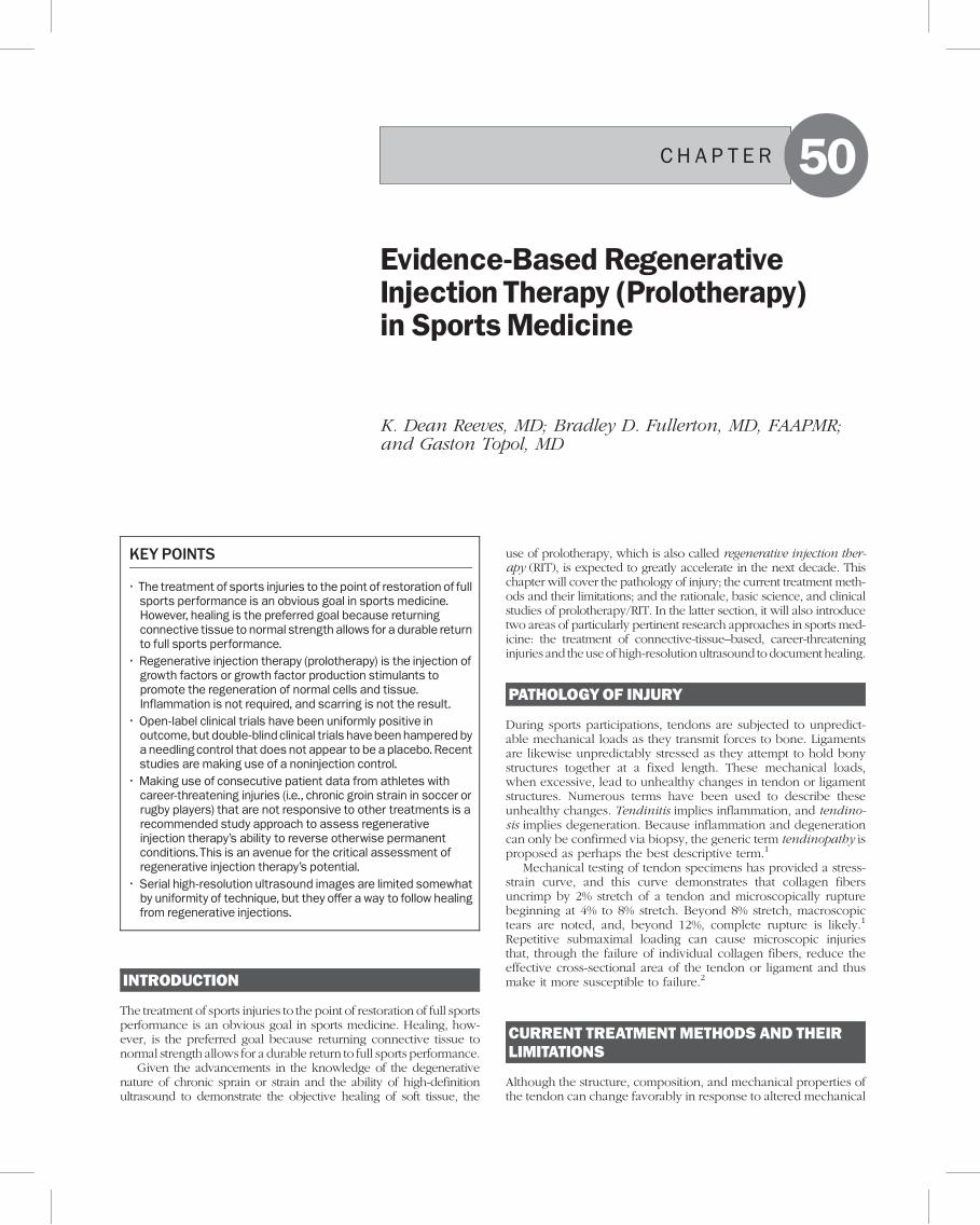
Case 2: A 61-year-oldmale golfer with extensortendinosisThis patient had 3 years of left lateral elbow pain and 2 years ofextension deficit in his elbow range, and he had received 3 steroidinjections. His chief complaint was difficulty playing golf. On exam-ination, he had a firm end feel to extension at�10 degrees, and therewas pain over the common extensor insertion and the radial head.Magnetic resonance imaging was diagnostic for common extensortendinosis. Figure 50.1 shows a high-resolution ultrasound of theelbow in pronation at three different points in time. The images onthe left and right are identical, but the images on the right are labeledanatomically: A is the radial head; the line labeled B is the bonynarrowing between the lateral epicondyle and the capitulum of thehumerus (the rounded portion of the end of the humerus that articu-lates with the radial head); C represents movement up the bonetoward the lateral epicondyle; and E, which is only seen clearly inthe bottom right view, is along the side of the capitulum of thehumerus, which is better seen after proliferant injection. This patientreceived 9 injection sessions beginning on November 29, 2004.Several treatments were with dextrose 15%, and two included 0.5%sodium morrhuate. Common extensor entheses, annular ligament,radial collateral ligament, and capsular entheses were injected. Theclinical result by August 15, 2005, was anextension range gain to�2 to�3 degrees, no pain on palpation, and no functional limitations. Theserial ultrasounds demonstrate hypoechoic (dark) areas of tissue sep-aration or insufficiency and edema (D is the common extensortendon). By the time of the ultrasound on August 15, the entireregion above the bones was more densely populated with organizedconnective tissue fibers. It is interesting to note that the capitulum(although it is not seen well on the first two ultrasounds) appears tomove closer to the radial head, and dynamic ultrasound showed thatradial head subluxation ceased as treatment progressed. This appearsto correlate with the range-of-motion loss at treatment onset that alsoresolved with treatment. Note also that, although bony growth factorsare not stimulated by injection, the typical effects after treatment withproliferant include a periosteal reaction that allows for the better visu-alization of contours of bone and an increased echogenicity of the softtissue as edema resolves and tissue becomes more tightly packed.
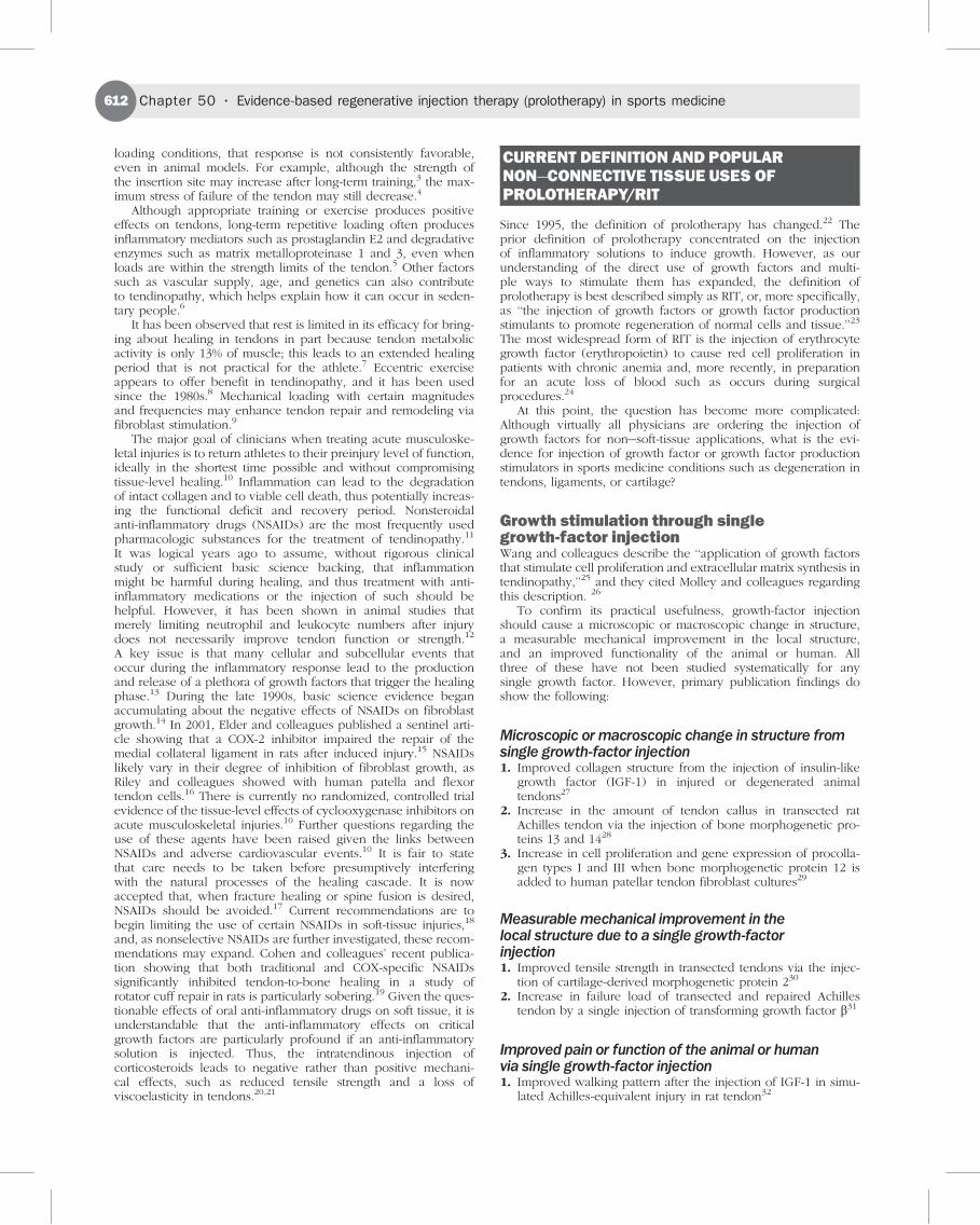
Case 3: A cyclist with patellar tendinosisA 40-year-old male competitive cyclist was first seen in November2004 because he could not run or handle rough terrain or sustainedhills as a result of knee pain. He had history of remote distal realign-ments (patellofemoral tracking type surgery). Pain was at the inferiorpatella and over the tibial tuberosity. He was treated on February 17,2005, and April 21, 2005, with an injection of 15% dextrose over thepatellar tendon origin on the inferior patella pole and its insertionover the tibial tuberosity. Complete symptom resolution occurredwith the last follow-up evaluation on January 23, 2006, at whichpoint the patient was training for the racing season. Figure 50.2shows a high-resolution ultrasound at the time of the first two ses-sions and at 6-month follow-up on October 19, 2005. On the rightside of the figure are the same images but with red outlining thepatellar tendon to depict its thickness. In addition, the yellow circlesurrounds an area of hypoechogenicity. From February 17, 2005,through October 19, 2005, an increase in the echogenicity of thetendon is demonstrated.
Case 4: An 85-year-oldmale patient with bicipitaltendinosisAlthough this patient was not an athlete in the competitive sense,he was quite active for 85 years of age. This patient had chronic,worsening anterior shoulder pain. The initial examination on May16, 2005, showed that the bicipital tendon and the surroundingregion were painful to palpation. The patient received three treat-ments consisting of the injection of 15% dextrose around the bici-pital region on May 27, June 17, and July 8, 2005, without regardfor whether the injections were precisely extratendinous or intra-tendinous because the injections are always given on bone insuccessive rows. Figure 50.3 shows a longitudinal ultrasoundthrough the bicipital tendon at the time of the first evaluationand at follow-up on August 18, 2005. On the right side are thelabeled images. Deltoid muscle thickness is represented by A inthe figure, and the degree of decrease in the swelling in the deltoidis easily seen by the decrease in thickness by August 18. The longhead of the bicipital tendon is outlined in yellow on the right, and,
1 1
Figure 50.2 Patellar tendinosis changes with regenerative injectiontherapy.
Figure 50.1 Extensor tendinosis changes with regenerative injectiontherapy.
Chapter 50 . Evidence-based regenerative injection therapy (prolotherapy) in sports medicine(616
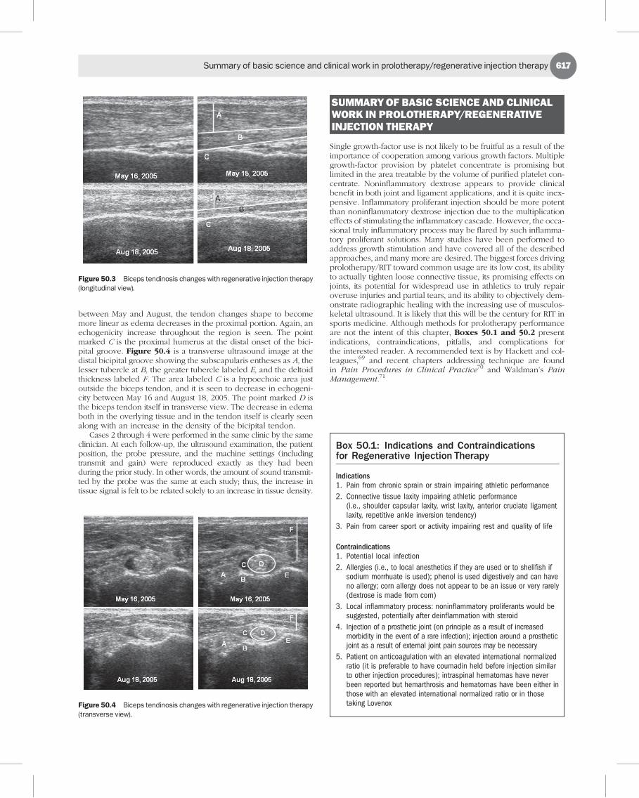
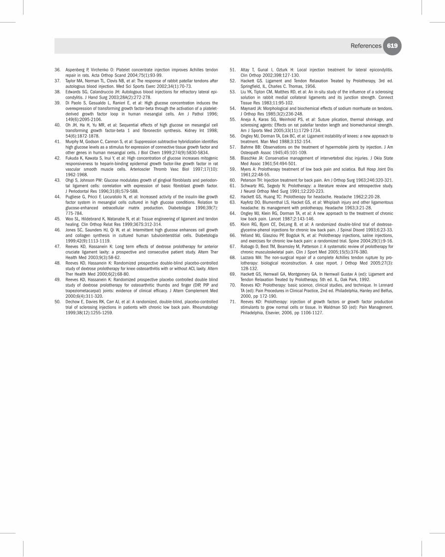
between May and August, the tendon changes shape to becomemore linear as edema decreases in the proximal portion. Again, anechogenicity increase throughout the region is seen. The pointmarked C is the proximal humerus at the distal onset of the bici-pital groove. Figure 50.4 is a transverse ultrasound image at thedistal bicipital groove showing the subscapularis entheses as A, thelesser tubercle at B, the greater tubercle labeled E, and the deltoidthickness labeled F. The area labeled C is a hypoechoic area justoutside the biceps tendon, and it is seen to decrease in echogeni-city between May 16 and August 18, 2005. The point marked D isthe biceps tendon itself in transverse view. The decrease in edemaboth in the overlying tissue and in the tendon itself is clearly seenalong with an increase in the density of the bicipital tendon.
Cases 2 through 4 were performed in the same clinic by the sameclinician. At each follow-up, the ultrasound examination, the patientposition, the probe pressure, and the machine settings (includingtransmit and gain) were reproduced exactly as they had beenduring the prior study. In other words, the amount of sound transmit-ted by the probe was the same at each study; thus, the increase intissue signal is felt to be related solely to an increase in tissue density.
SUMMARYOF BASIC SCIENCE AND CLINICALWORK IN PROLOTHERAPY/REGENERATIVEINJECTION THERAPY
Single growth-factor use is not likely to be fruitful as a result of theimportance of cooperation among various growth factors. Multiplegrowth-factor provision by platelet concentrate is promising butlimited in the area treatable by the volume of purified platelet con-centrate. Noninflammatory dextrose appears to provide clinicalbenefit in both joint and ligament applications, and it is quite inex-pensive. Inflammatory proliferant injection should be more potentthan noninflammatory dextrose injection due to the multiplicationeffects of stimulating the inflammatory cascade. However, the occa-sional truly inflammatory process may be flared by such inflamma-tory proliferant solutions. Many studies have been performed toaddress growth stimulation and have covered all of the describedapproaches, and many more are desired. The biggest forces drivingprolotherapy/RIT toward common usage are its low cost, its abilityto actually tighten loose connective tissue, its promising effects onjoints, its potential for widespread use in athletics to truly repairoveruse injuries and partial tears, and its ability to objectively dem-onstrate radiographic healing with the increasing use of musculos-keletal ultrasound. It is likely that this will be the century for RIT insports medicine. Although methods for prolotherapy performanceare not the intent of this chapter, Boxes 50.1 and 50.2 presentindications, contraindications, pitfalls, and complications forthe interested reader. A recommended text is by Hackett and col-leagues,69 and recent chapters addressing technique are foundin Pain Procedures in Clinical Practice70 and Waldman’s PainManagement.71
Figure 50.3 Biceps tendinosis changes with regenerative injection therapy(longitudinal view).
Figure 50.4 Biceps tendinosis changes with regenerative injection therapy(transverse view).
Box 50.1: Indications and Contraindicationsfor Regenerative Injection Therapy
Indications1. Pain from chronic sprain or strain impairing athletic performance
2. Connective tissue laxity impairing athletic performance(i.e., shoulder capsular laxity, wrist laxity, anterior cruciate ligamentlaxity, repetitive ankle inversion tendency)
3. Pain from career sport or activity impairing rest and quality of life
Contraindications1. Potential local infection
2. Allergies (i.e., to local anesthetics if they are used or to shellfish ifsodium morrhuate is used); phenol is used digestively and can haveno allergy; corn allergy does not appear to be an issue or very rarely(dextrose is made from corn)
3. Local inflammatory process: noninflammatory proliferants would besuggested, potentially after deinflammation with steroid
4. Injection of a prosthetic joint (on principle as a result of increasedmorbidity in the event of a rare infection); injection around a prostheticjoint as a result of external joint pain sources may be necessary
5. Patient on anticoagulation with an elevated international normalizedratio (it is preferable to have coumadin held before injection similarto other injection procedures); intraspinal hematomas have neverbeen reported but hemarthrosis and hematomas have been either inthose with an elevated international normalized ratio or in thosetaking Lovenox
Summary of basic science and clinical work in prolotherapy/regenerative injection therapy(617

CONCLUSION
Suboptimal healing may lead to elimination of symptoms andreturn to full sport. However, suboptimal tissue leaves the athletewith a decrease in tensile strength of the damaged tissue or relativelaxity with stretch of fixed length nerve endings. These effectsincrease susceptibility of the athlete to repetitive injury or rupture,can reflexly inhibit full performance, and create a regional stiff-ness, even without associated pain. All these are threats to a fulland enjoyable carrer for the elite athlete and can increase thepotential for chronic pain after retirement. Much remains to bediscovered about stimulating regeneration and blocking degenera-tion after acute or chronic sports injuries. However, current agentsappear capable of restoring connective tissue organization, as seenby ultrasonographic confirmation. The choice of agents willdepend on such factors as speed of healing needed, cost efficacy,and the stage of the season.
Education on the basic science of connective tissue injuries andtraining on how to choose and apply the most cost-effectivemethod of regenerative injection therapy will best be achievedin the context of routine physical medicine and rehabilitationtraining. Ultrasonographic documentation of lesion reversal mayultimately be used to monitor healing efficacy in this age ofevidence-based medicine.
REFERENCES
1. Wang JH: Mechanobiology of tendon. J Biomech 2006;39:1563-1582.2. Kirkendall DT, Garrett WE: Function and biomechanics of tendons. Scand J Med Sci
Sports 1997;7:62-66.
3. Woo SL, Gomez MA, Amiel D, et al: The effects of exercise on the biomechanicaland biochemical properties of swine digital flexor tendons. J Biomech Eng1981;103:51-56.
4. Soslowsky LJ, Thomopoulos S, Tun S, et al: Overuse activity injures the supraspinatustendon in an animal model: a histologic and biomechanical study. J Shoulder ElbowSurg 2000;9:79-84.
5. Kjaer M: Role of extracellular matrix in adaptation of tendon and skeletal muscle tomechanical loading. Physiol Rev 2004;84:649-698.
6. Young JS, Kumta SM, Maffulli N: Achilles tendon rupture and tendinopathy: manage-ment of complications. Foot Ankle Clin 2005;10:371-382.
7. Zernicke RF, Garhammer J, Jobe FW: Human patellar-tendon rupture. J Bone Joint SurgAm 1977;59:179-183.
8. Stanish WD, Rubinovich RM, Curwin S: Eccentric exercise in chronic tendinitis. ClinOrthop Relat Res 1986;208:65-68.
9. Kannus P, Jozsa L, Natri A, Jarvinen M: Effects of training, immobilization and remo-bilization on tendons. Scand J Med Sci Sports 1997;7:67-71.
10. Warden SJ: Cyclo-oxygenase-2 inhibitors: beneficial or detrimental for athletes withacute musculoskeletal injuries? Sports Med 2005;35(4):271-283.
11. Saltzman CL, Tearse DS: Achilles tendon injuries. J Am Acad Ortho Surg 1998;6:316-325.
12. Marsolais D, Cote CH, Frenette J: Nonsteroidal anti-inflammatory drug reduces neu-trophil and macrophage accumulation but does not improve tendon regeneration. LabInvest 2003;83(7):991-999.
13. Marsolais D, Frenette J: Inflammation and tendon healing. Med Sci 2005;21(2):181-186.
14. Sun R, Gimbel HV, Liu S, et al: Effect of diclofenac sodium and dexamethasone oncultured human Tenon’s capsule fibroblasts. Ophthalmic Surg Lasers 1999;30(5):382-388.
15. Elder CL, Dahners LE, Weinhold PS: A cyclooxygenase-inhibitor impairs ligamenthealing in the rat. Am J Sports Med 2001;29(6):801-810.
16. Riley GP, Cox M, Harrall RL, et al: Inhibition of tendon cell proliferation and matrixglycosaminoglycan synthesis by non-steroidal anti-inflammatory drugs in vitro. J HandSurg 2001;26(3):224-228.
17. Dahners LE, Mullis BH: Effects of nonsteroidal anti-inflammatory drugs on boneformation and soft-tissue healing. J Am Acad Orthop Surg 2004;12(3):139-143.
18. Paoloni JA, Orchard JW: The use of therapeutic medications for soft-tissue injuries insports medicine. Med J Aust 2005;183(7):384-388.
19. Cohen DB, Kawamura S, Ehteshami J, et al: Indomethacin and celecoxib impairrotator cuff tendon-to-bone healing. Am J Sports Med 2006;34(3):362-369.
20. Kennedy J, Willis RB: The effects of local steroid injections on tendons: a biomecha-nical and microscopic correlative study. Am J Sports Med 1976;4:11-21.
21. Nirschl RP: Elbow tendinosis/tennis elbow. Clin Sports Med 1992;11(4):851-870.22. Reeves KD: Technique of prolotherapy. In Lennard TA (ed): Physiatric Procedures in
Clinical Practice. Philadelphia, Hanley and Belfus, 1995, pp 57-70.23. Topol GA, Reeves KD, Hassanein K: Efficacy of dextrose prolotherapy in elite male
kicking-sport athletes with chronic groin pain. Arch Phys Med Rehabil 2005;86(4):697-702.
24. Price S, Pepper JR, Jaggar SI: Recombinant human erythropoietin use in a critically illJehovah’s witness after cardiac surgery. Anesth Analg 2005;101(2):325-327.
25. Wang JH, Losifidis MI, Fu FH: Biomechanical basis for tendinopathy. Clin Orthop RelatRes 2006;443:320-322.
26. Molloy T, Wang Y, Murrell G: The roles of growth factors in tendon and ligamenthealing. Sports Med 2003;33:381-394.
27. Dahlgren LA, van der Meulen MC, Bertram JE, et al: Insulin-like growth factor-1improves cellular and molecular aspects of healing in a collagenase-inducedmodel of flexor tendinitis. J Orthop Res 2003;20:910-919.
28. Aspenberg P, Forslund C: Bone morphogenetic proteins and tendon repair. Scand JMed Sci Sports 2000;10:372-375.
29. Fu SC, Wong YP, Chan BP, et al: The roles of bone morphogenetic protein (BMP) 12 instimulating the proliferation and matrix production of human patellar tendon fibro-blasts. Life Sci 2003;72:2965-2974.
30. Forslund C, Aspenberg P: Improved healing of transected rabbit Achilles tendon aftera single injection of cartilage-derived morphogenetic protein-2. Am J Sports Med2003;31:555-559.
31. Kashiwagi K, Mochizuki Y, Yasunaga Y, et al: Effects of transforming growth factor-beta1 on the early stages of healing of the Achilles tendon in a rat model. Scand J PlastReconstr Surg Hand Surg 2004;38(4):193-197.
32. Kurtz CA, Loebig TG, Anderson DD, et al: Insulin-like growth factor I acceleratesfunctional recovery from Achilles tendon injury in a rat model. Am J Sports Med1999;27:363-369.
33. Tsubone T, Moran SL, Amadio PC, et al: Expression of growth factors in canine flexortendon after laceration in vivo. Ann Plast Surg 2004;53(4):393-397.
34. Thomopoulos S, Harwood FL, Silva MJ, et al: Effect of several growth factors on canineflexor tendon fibroblast proliferation and collagen synthesis in vitro. J Hand Surg2005;30(3):441-447.
35. Anitua E, Andia I, Sanchez M, et al: Autologous preparations rich in growth factorspromote proliferation and induce VEGF and HGF production by human tendon cells inculture. J Orthop Res 2005;23(2):281-286.
Box 50.2: Pitfalls and Complicationsof Regenerative Injection Therapy
Needling Risks (Depending on the Portion of the Body Being Treated)1. Septic joint: This appears to be similar to steroid injection risk at a
rate of 1 in 10,000 to 1 in 40,000 for intra-articular injections.
2. Spinal headache: This is estimated to occur in 1 in 300 low backprocedures with lumbosacral junction treatment.
3. Peripheral nerve contact: As a result of the ubiquitous presence ofsmall nerve branches and the occasional contact of larger nervebranches, nerve irritation will occur rarely with treatment. Burningpain reactions are typical, lasting from days to several months.
4. Pneumothorax: This is estimated to occur in about 1 in 20,000needle insertions in the thoracic region.
Solution Risks1. Stiffness and soreness after treatment: Typically this will last from
1 to 3 days but will occasionally last longer. It is important to havepatient contact the physician if flare lasts for more than 10 daysbecause this can be counterproductive in patients with chronic pain.
2. Allergy: Any physician performing injection should prepare for sucha reaction and check, in particular, for shellfish allergy beforeadministering sodium morrhuate.
3. Chemical arachnoiditis: This appears to occur with midline injectiononly of stronger proliferants such as phenol and when boluses inthe midline exceed 0.5% or the concentration of phenol exceeds1.25%. However, this is rare and almost always temporary.Nevertheless, paraspinal injections anywhere near the neuralforamina should be with a lesser solution or with small doses anda careful emphasis on concentration limits.
Chapter 50 . Evidence-based regenerative injection therapy (prolotherapy) in sports medicine(618
36. Aspenberg P, Virchenko O: Platelet concentrate injection improves Achilles tendonrepair in rats. Acta Orthop Scand 2004;75(1):93-99.
37. Taylor MA, Norman TL, Clovis NB, et al: The response of rabbit patellar tendons afterautologous blood injection. Med Sci Sports Exerc 2002;34(1):70-73.
38. Edwards SG, Calandruccio JH: Autologous blood injections for refractory lateral epi-condylitis. J Hand Surg 2003;28A(2):272-278.
39. Di Paolo S, Gesualdo L, Ranieri E, et al: High glucose concentration induces theoverexpression of transforming growth factor-beta through the activation of a platelet-derived growth factor loop in human mesangial cells. Am J Pathol 1996;149(6):2095-2106.
40. Oh JH, Ha H, Yu MR, et al: Sequential effects of high glucose on mesangial celltransforming growth factor-beta 1 and fibronectin synthesis. Kidney Int 1998;54(6):1872-1878.
41. Murphy M, Godson C, Cannon S, et al: Suppression subtractive hybridization identifieshigh glucose levels as a stimulus for expression of connective tissue growth factor andother genes in human mesangial cells. J Biol Chem 1999;274(9):5830-5834.
42. Fukuda K, Kawata S, Inui Y, et al: High concentration of glucose increases mitogenicresponsiveness to heparin-binding epidermal growth factor-like growth factor in ratvascular smooth muscle cells. Arterioscler Thromb Vasc Biol 1997;17(10):1962-1968.
43. Ohgi S, Johnson PW: Glucose modulates growth of gingival fibroblasts and periodon-tal ligament cells: correlation with expression of basic fibroblast growth factor.J Periodontal Res 1996;31(8):579-588.
44. Pugliese G, Pricci F, Locuratolo N, et al: Increased activity of the insulin-like growthfactor system in mesangial cells cultured in high glucose conditions. Relation toglucose-enhanced extracellular matrix production. Diabetologia 1996;39(7):775-784.
45. Woo SL, Hildebrand K, Watanabe N, et al: Tissue engineering of ligament and tendonhealing. Clin Orthop Relat Res 1999;367S:312-314.
46. Jones SC, Saunders HJ, Qi W, et al: Intermittent high glucose enhances cell growthand collagen synthesis in cultured human tubulointerstitial cells. Diabetologia1999;42(9):1113-1119.
47. Reeves KD, Hassanein K: Long term effects of dextrose prolotherapy for anteriorcruciate ligament laxity: a prospective and consecutive patient study. Altern TherHealth Med 2003;9(3):58-62.
48. Reeves KD, Hassanein K: Randomized prospective double-blind placebo-controlledstudy of dextrose prolotherapy for knee osteoarthritis with or without ACL laxity. AlternTher Health Med 2000;6(2):68-80.
49. Reeves KD, Hassanein K: Randomized prospective placebo controlled double blindstudy of dextrose prolotherapy for osteoarthritic thumbs and finger (DIP, PIP andtrapeziometacarpal) joints: evidence of clinical efficacy. J Altern Complement Med2000;6(4):311-320.
50. Dechow E, Davies RK, Carr AJ, et al: A randomized, double-blind, placebo-controlledtrial of sclerosing injections in patients with chronic low back pain. Rheumatology1999;38(12):1255-1259.
51. Altay T, Gunal I, Ozturk H: Local injection treatment for lateral epicondylitis.Clin Orthop 2002;398:127-130.
52. Hackett GS. Ligament and Tendon Relaxation Treated by Prolotherapy, 3rd ed.Springfield, IL, Charles C. Thomas, 1956.
53. Liu YK, Tipton CM, Matthes RD, et al: An in-situ study of the influence of a sclerosingsolution in rabbit medial collateral ligaments and its junction strength. ConnectTissue Res 1983;11:95-102.
54. Maynard JA: Morphological and biochemical effects of sodium morrhuate on tendons.J Orthop Res 1985;3(2):236-248.
55. Aneja A, Karas SG, Weinhold PS, et al: Suture plication, thermal shrinkage, andsclerosing agents: Effects on rat patellar tendon length and biomechanical strength.Am J Sports Med 2005;33(11):1729-1734.
56. Ongley MJ, Dorman TA, Eek BC, et al: Ligament instability of knees: a new approach totreatment. Man Med 1988;3:152-154.
57. Bahme BB: Observations on the treatment of hypermobile joints by injection. J AmOsteopath Assoc 1945;45:101-109.
58. Blaschke JA: Conservative management of intervertebral disc injuries. J Okla StateMed Assoc 1961;54:494-501.
59. Myers A: Prolotherapy treatment of low back pain and sciatica. Bull Hosp Joint Dis1961;22:48-55.
60. Peterson TH: Injection treatment for back pain. Am J Orthop Surg 1963;246:320-321.61. Schwartz RG, Segedy N: Prolotherapy: a literature review and retrospective study.
J Neurol Orthop Med Surg 1991;12:220-223.62. Hackett GS, Huang TC: Prolotherapy for headache. Headache 1962;2:20-28.63. Kayfetz DO, Blumenthal LS, Hacket GS, et al: Whiplash injury and other ligamentous
headache: its management with prolotherapy. Headache 1963;3:21-28.64. Ongley MJ, Klein RG, Dorman TA, et al: A new approach to the treatment of chronic
low back pain. Lancet 1987;2:143-146.65. Klein RG, Bjorn CE, DeLong B, et al: A randomized double-blind trial of dextrose-
glycerine-phenol injections for chronic low back pain. J Spinal Disord 1993;6:23-33.66. Yelland MJ, Glasziou PP, Bogduk N, et al: Prolotherapy injections, saline injections,
and exercises for chronic low-back pain: a randomized trial. Spine 2004;29(1):9-16.67. Rabago D, Best TM, Beamsley M, Patterson J: A systematic review of prolotherapy for
chronic musculoskeletal pain. Clin J Sport Med 2005;15(5):376-380.68. Lazzara MA: The non-surgical repair of a complete Achilles tendon rupture by pro-
lotherapy: biological reconstruction. A case report. J Orthop Med 2005;27(3):128-132.
69. Hackett GS, Hemwall GA, Montgomery GA. In Hemwall Gustav A (ed): Ligament andTendon Relaxation Treated by Prolotherapy, 5th ed. IL, Oak Park, 1992.
70. Reeves KD: Prolotherapy: basic science, clinical studies, and technique. In LennardTA (ed): Pain Procedures in Clinical Practice, 2nd ed. Philadelphia, Hanley and Belfus,2000, pp 172-190.
71. Reeves KD: Prolotherapy: injection of growth factors or growth factor productionstimulants to grow normal cells or tissue. In Waldman SD (ed): Pain Management.Philadelphia, Elsevier, 2006, pp 1106-1127.
References(619