evidence brief: coronary computed tomography angiography
TRANSCRIPT

June 2021
Prepared for:
Department of Veterans Affairs Veterans Health Administration Health Services Research & Development Service Washington, DC 20420
Prepared by:
Evidence Synthesis Program (ESP) Coordinating Center Portland VA Health Care System Portland, OR Mark Helfand, MD, MPH, MS, Director
Evidence Brief: Coronary Computed Tomography Angiography with Fractional Flow Reserve in Noninvasive Diagnosis of Coronary Artery Disease Supplemental Materials
Authors:
Johanna Anderson, MPH Sarah Young, MPH Mark Helfand, MD, MPH, MS
Evidence Synthesis Program

Evidence Brief: FFRCT for Diagnosis of CAD Evidence Synthesis Program
i
TABLE OF CONTENTS Appendix A. Search Strategies ....................................................................................................... 1
Appendix B. List of Excluded Studies ............................................................................................ 5
Appendix C. Evidence Tables ....................................................................................................... 17
Data Abstraction of Included Systematic Reviews ................................................................... 17
Data Abstraction of Included Primary Studies .......................................................................... 17
Data Abstraction of Primary Studies Evaluating Diagnostic Accuracy of HeartFlow FFRCT ................................................................................................................... 17
Data Abstraction of Primary Studies Evaluating Clinical or Therapeutic Outcomes ........... 18
Quality Assessment of Included Studies ................................................................................... 29
Quality Assessment of Systematic Reviews using ROBIS-SR ............................................. 29
Quality Assessment of Diagnostic Accuracy Studies Using QUADAS-2 ............................ 30
Quality Assessment of Cohort Studies Using ROBINS-I ..................................................... 31
Quality Assessment of Case Series Using Murad et al. ........................................................ 37
Strength of Evidence of Included Studies ................................................................................. 39
Appendix D. Ongoing HeartFlow FFRCT Studies ........................................................................ 41
Appendix E. Disposition of Peer Reviewer Comments ................................................................ 43
References ..................................................................................................................................... 45

Evidence Brief: FFRCT for Diagnosis of CAD Evidence Synthesis Program
1
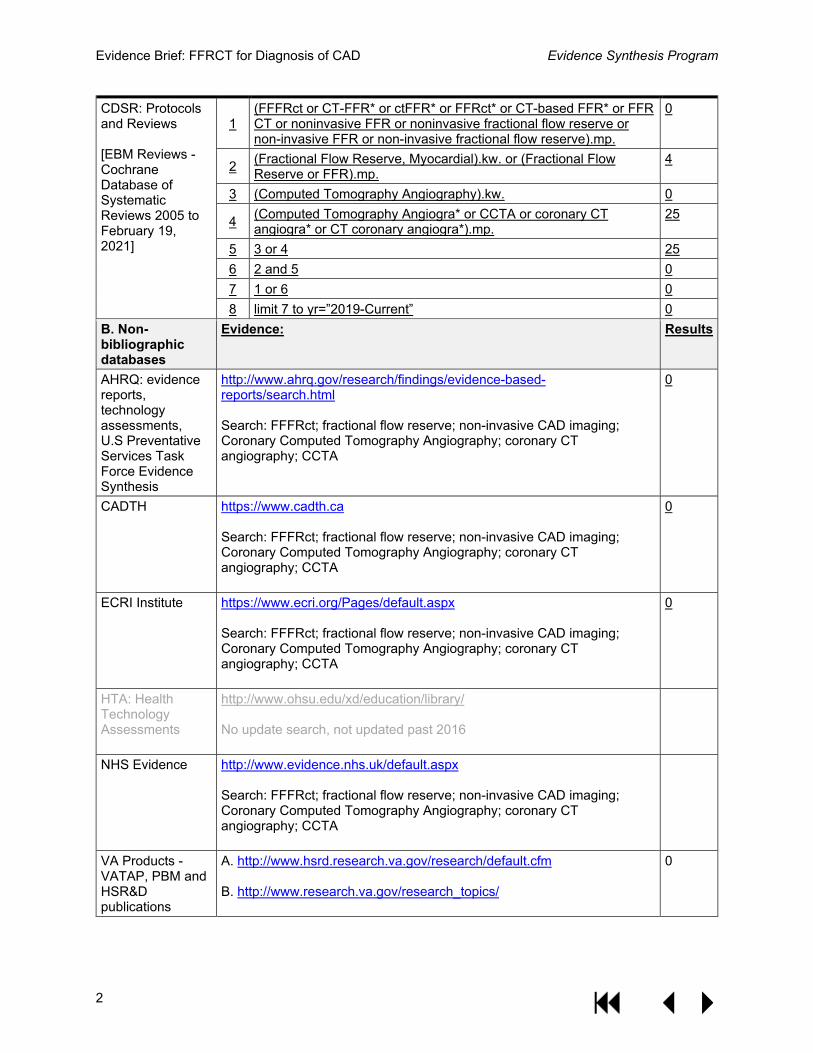
APPENDIX A. SEARCH STRATEGIES 1. Search for current systematic reviews (limited to last 7 years) Date Searched: 2-23-21 A. Bibliographic databases
# Search Statement Results
MEDLINE: Systematic Reviews [Ovid MEDLINE(R) ALL 1946 to February 22, 2021]
1 (FFFRct or CT-FFR* or ctFFR* or FFRct* or CT-based FFR* or FFR CT or noninvasive FFR or noninvasive fractional flow reserve or non-invasive FFR or non-invasive fractional flow reserve).mp.
376
2 exp Fractional Flow Reserve, Myocardial/ or (Fractional Flow Reserve or FFR).mp.
4878
3 exp Computed Tomography Angiography/ 11297
4 (Computed Tomography Angiogra* or CCTA or coronary CT angiogra* or CT coronary angiogra*).mp.
21196
5 3 or 4 21196 6 2 and 5 663 7 1 or 6 733
8
(systematic review.ti. or meta-analysis.pt. or meta-analysis.ti. or systematic literature review.ti. or this systematic review.tw. or pooling project.tw. or (systematic review.ti,ab. and review.pt.) or meta synthesis.ti. or meta-analy*.ti. or integrative review.tw. or integrative research review.tw. or rapid review.tw. or umbrella review.tw. or consensus development conference.pt. or practice guideline.pt. or drug class reviews.ti. or cochrane database syst rev.jn. or acp journal club.jn. or health technol assess.jn. or evid rep technol assess summ.jn. or jbi database system rev implement rep.jn. or (clinical guideline and management).tw. or ((evidence based.ti. or evidence-based medicine/ or best practice*.ti. or evidence synthesis.ti,ab.) and (((review.pt. or diseases category/ or behavior.mp.) and behavior mechanisms/) or therapeutics/ or evaluation studies.pt. or validation studies.pt. or guideline.pt. or pmcbook.mp.)) or (((systematic or systematically).tw. or critical.ti,ab. or study selection.tw. or ((predetermined or inclusion) and criteri*).tw. or exclusion criteri*.tw. or main outcome measures.tw. or standard of care.tw. or standards of care.tw.) and ((survey or surveys).ti,ab. or overview*.tw. or review.ti,ab. or reviews.ti,ab. or search*.tw. or handsearch.tw. or analysis.ti. or critique.ti,ab. or appraisal.tw. or (reduction.tw. and (risk/ or risk.tw.) and (death or recurrence).mp.)) and ((literature or articles or publications or publication or bibliography or bibliographies or published).ti,ab. or pooled data.tw. or unpublished.tw. or citation.tw. or citations.tw. or database.ti,ab. or internet.ti,ab. or textbooks.ti,ab. or references.tw. or scales.tw. or papers.tw. or datasets.tw. or trials.ti,ab. or meta-analy*.tw. or (clinical and studies).ti,ab. or treatment outcome/ or treatment outcome.tw. or pmcbook.mp.))) not (letter or newspaper article).pt.
438670
9 7 and 8 31 10 Limit 9 to English language only 31 11 Limit 10 to yr=”2019-Current” 9
Evidence Brief: FFRCT for Diagnosis of CAD Evidence Synthesis Program
2
CDSR: Protocols and Reviews [EBM Reviews - Cochrane Database of Systematic Reviews 2005 to February 19, 2021]
1 (FFFRct or CT-FFR* or ctFFR* or FFRct* or CT-based FFR* or FFR CT or noninvasive FFR or noninvasive fractional flow reserve or non-invasive FFR or non-invasive fractional flow reserve).mp.
0
2 (Fractional Flow Reserve, Myocardial).kw. or (Fractional Flow Reserve or FFR).mp.
4
3 (Computed Tomography Angiography).kw. 0
4 (Computed Tomography Angiogra* or CCTA or coronary CT angiogra* or CT coronary angiogra*).mp.
25
5 3 or 4 25 6 2 and 5 0 7 1 or 6 0 8 limit 7 to yr=”2019-Current” 0
B. Non-bibliographic databases
Evidence: Results
AHRQ: evidence reports, technology assessments, U.S Preventative Services Task Force Evidence Synthesis
http://www.ahrq.gov/research/findings/evidence-based-reports/search.html Search: FFFRct; fractional flow reserve; non-invasive CAD imaging; Coronary Computed Tomography Angiography; coronary CT angiography; CCTA
0
CADTH https://www.cadth.ca Search: FFFRct; fractional flow reserve; non-invasive CAD imaging; Coronary Computed Tomography Angiography; coronary CT angiography; CCTA
0
ECRI Institute https://www.ecri.org/Pages/default.aspx Search: FFFRct; fractional flow reserve; non-invasive CAD imaging; Coronary Computed Tomography Angiography; coronary CT angiography; CCTA
0
HTA: Health Technology Assessments
http://www.ohsu.edu/xd/education/library/ No update search, not updated past 2016
NHS Evidence http://www.evidence.nhs.uk/default.aspx Search: FFFRct; fractional flow reserve; non-invasive CAD imaging; Coronary Computed Tomography Angiography; coronary CT angiography; CCTA
VA Products - VATAP, PBM and HSR&D publications
A. http://www.hsrd.research.va.gov/research/default.cfm B. http://www.research.va.gov/research_topics/
0
Evidence Brief: FFRCT for Diagnosis of CAD Evidence Synthesis Program
3
Search: FFFRct; fractional flow reserve; non-invasive CAD imaging; Coronary Computed Tomography Angiography; coronary CT angiography; CCTA
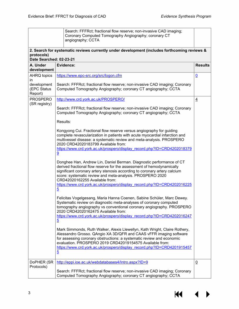
2. Search for systematic reviews currently under development (includes forthcoming reviews & protocols) Date Searched: 02-23-21 A. Under development
Evidence: Results
AHRQ topics in development (EPC Status Report)
https://www.epc-src.org/src/logon.cfm Search: FFFRct; fractional flow reserve; non-invasive CAD imaging; Coronary Computed Tomography Angiography; coronary CT angiography; CCTA
0
PROSPERO (SR registry)
http://www.crd.york.ac.uk/PROSPERO/ Search: FFFRct; fractional flow reserve; non-invasive CAD imaging; Coronary Computed Tomography Angiography; coronary CT angiography; CCTA Results: Kongyong Cui. Fractional flow reserve versus angiography for guiding complete revascularization in patients with acute myocardial infarction and multivessel disease: a systematic review and meta-analysis. PROSPERO 2020 CRD42020183799 Available from: https://www.crd.york.ac.uk/prospero/display_record.php?ID=CRD42020183799 Donghee Han, Andrew Lin, Daniel Berman. Diagnostic performance of CT derived fractional flow reserve for the assessment of hemodynamically significant coronary artery stenosis according to coronary artery calcium score: systematic review and meta-analysis. PROSPERO 2020 CRD42020162255 Available from: https://www.crd.york.ac.uk/prospero/display_record.php?ID=CRD42020162255 Felicitas Vogelgesang, Maria Hanna Coenen, Sabine Schüler, Marc Dewey. Systematic review on diagnostic meta-analyses of coronary computed tomography angiography vs conventional coronary angiography. PROSPERO 2020 CRD42020162475 Available from: https://www.crd.york.ac.uk/prospero/display_record.php?ID=CRD42020162475 Mark Simmonds, Ruth Walker, Alexis Llewellyn, Kath Wright, Claire Rothery, Alessandro Grosso. QAngio XA 3D/QFR and CAAS vFFR imaging software for assessing coronary obstructions: a systematic review and economic evaluation. PROSPERO 2019 CRD42019154575 Available from: https://www.crd.york.ac.uk/prospero/display_record.php?ID=CRD42019154575
4
DoPHER (SR Protocols)
http://eppi.ioe.ac.uk/webdatabases4/Intro.aspx?ID=9 Search: FFFRct; fractional flow reserve; non-invasive CAD imaging; Coronary Computed Tomography Angiography; coronary CT angiography; CCTA
0

Evidence Brief: FFRCT for Diagnosis of CAD Evidence Synthesis Program
4
Cochrane Database of Systematic Reviews: Protocols
http://www.ohsu.edu/xd/education/library/ Search: See strategy above
0
Search for primary literature Date searched: 02-23-21 MEDLINE [Ovid MEDLINE(R) ALL 1946 to February 22, 2021]
# Search Statement Results
1 (FFFRct or CT-FFR* or ctFFR* or FFRct* or CT-based FFR* or FFR CT or noninvasive FFR or noninvasive fractional flow reserve or non-invasive FFR or non-invasive fractional flow reserve).mp.
376
2 exp Fractional Flow Reserve, Myocardial/ or (Fractional Flow Reserve or FFR).mp. 4878 3 exp Computed Tomography Angiography/ 11297
4 (Computed Tomography Angiogra* or CCTA or coronary CT angiogra* or CT coronary angiogra*).mp.
21196
5 3 or 4 21196 6 2 and 5 663 7 1 or 6 733 8 Limit 7 to english language 718 9 Limit 8 to yr=”2019-Current” 288 CCRCT [EBM Reviews - Cochrane Central Register of Controlled Trials January 2021]
# Search Statement Results
1 (FFFRct or CT-FFR* or ctFFR* or FFRct* or CT-based FFR* or FFR CT or noninvasive FFR or noninvasive fractional flow reserve or non-invasive FFR or non-invasive fractional flow reserve).mp.
48
2 exp Fractional Flow Reserve, Myocardial/ or (Fractional Flow Reserve or FFR).mp. 701 3 exp Computed Tomography Angiography/ 0
4 (Computed Tomography Angiogra* or CCTA or coronary CT angiogra* or CT coronary angiogra*).mp. 1308
5 3 or 4 1308 6 2 and 5 61 7 1 or 6 79 8 Limit 7 to english language 60 9 Limit 8 to yr=”2019-Current” 17

Evidence Brief: FFRCT for Diagnosis of CAD Evidence Synthesis Program
5
APPENDIX B. LIST OF EXCLUDED STUDIES Exclude reasons: 1=Ineligible population (ie, acute coronary syndrome), 2=Ineligible intervention (ie, non HeartFlow FFRCT), 3=Ineligible comparator, 4=Ineligible outcome, 5=Ineligible setting, 6=Ineligible study design, 7=Ineligible publication type, 8=Outdated or ineligible systematic review, 9=Non-English language, 10=Unable to retrieve full text, 11=Trial included in prioritized systematic review
# Citation Exclude reason
1 ACR–NASCI–SPR Practice Parameter for the Performance and Interpretation of Cardiac Computed Tomography (CT). 2017.
E2
2 ACR–NASCI–SPR Practice Parameter for the Performance of Quantification of Cardiovascular Computed Tomography (CT) and Magnetic Resonance Imaging (MRI). 2017.
E2
3 Al-Mallah MH, Ahmed AM. Controversies in the Use of Fractional Flow Reserve Form Computed Tomography (FFRCT) vs Coronary Angiography. Current Cardiovascular Imaging Reports. 2016;9(12).
E7
4 Andreini D, Mushtaq S, Pontone G, Rogers C, Pepi M, Bartorelli AL. Severe in-stent restenosis missed by coronary CT angiography and accurately detected with FFR<sub>CT</sub>. The international journal of cardiovascular imaging. 2017;33(1):119-120.
E6
5 Artzner C, Daubert M, Ehieli W, et al. Impact of computed tomography (CT)-derived fractional flow reserve on reader confidence for interpretation of coronary CT angiography. European Journal of Radiology. 2018;108:242-248.
E4
6 Babakhani H, Sadeghipour P, Tashakori Beheshti A, et al. Diagnostic accuracy of two-dimensional coronary angiographic-derived fractional flow reserve-Preliminary results. Catheterization & Cardiovascular Interventions. 2020;27:27.
E2
7 Ball C, Pontone G, Rabbat M. Fractional flow reserve derived from coronary computed tomography angiography datasets: the next frontier in noninvasive assessment of coronary artery disease. Biomedical Research International. 2018;2018:2680430.
E7
8 Baumann S, Becher T, Schoepf UJ, et al. Fractional flow reserve derived by coronary computed tomography angiography : A sophisticated analysis method for detecting hemodynamically significant coronary stenosis. Herz. 2017;42(6):604-606.
E7
9 Baumann S, Hirt M, Schoepf UJ, et al. Correlation of machine learning computed tomography-based fractional flow reserve with instantaneous wave free ratio to detect hemodynamically significant coronary stenosis. Clinical Research in Cardiology. 2020;109(6):735-745.
E2
10 Baumann S, Lossnitzer D, Renker M, Borggrefe M, Akin I. Coronary Computed Tomography Angiography-Derived Fractional Flow Reserve Assessment: Many Roads to Reach the Same Goal. Circulation Journal. 2018;82(9):2448.
E7
11 Baumann S, Renker M, Akin I, Borggrefe M, Schoepf UJ. FFR-Derived From Coronary CT Angiography Using Workstation-Based Approaches. Jacc: Cardiovascular Imaging. 2017;10(4):497-498.
E7
12 Baumann S, Renker M, Hetjens S, et al. Comparison of coronary computed tomography angiography-derived vs invasive fractional flow reserve assessment: meta-analysis with subgroup evaluation of intermediate stenosis. Academic Radiology. 2016;23(11):1402-1411.
E8
13 Baumann S, Renker M, Schoepf UJ, et al. Gender differences in the diagnostic performance of machine learning coronary CT angiography-derived fractional flow
E2

Evidence Brief: FFRCT for Diagnosis of CAD Evidence Synthesis Program
6
reserve -results from the MACHINE registry. European Journal of Radiology. 2019;119:108657.
14 Beg F, Rehman H, Chamsi-Pasha MA, et al. Association between FFR<sub>CT</sub> and instantaneous wave-free ratio (iFR) of intermediate lesions on coronary computed tomography angiography. Cardiovascular Revascularization Medicine. 2020;26:26.
E4
15 Benton SM, Tesche C, De Cecco CN, Duguay TM, Schoepf UJ, Bayer RR, II. Noninvasive Derivation of Fractional Flow Reserve From Coronary Computed Tomographic Angiography: A Review. Journal of Thoracic Imaging. 2018;33(2):88-96.
E7
16 Bernhardt P, Walcher T, Rottbauer W, Wohrle J. Quantification of myocardial perfusion reserve at 1.5 and 3.0 Tesla: a comparison to fractional flow reserve. International Journal of CaXIArdiovascular Imaging. 2012;28(8):2049-2056.
E2
17 Bilbey N, Blanke P, Naoum C, Arepalli CD, Norgaard BL, Leipsic J. Potential impact of clinical use of noninvasive FFRCT on radiation dose exposure and downstream clinical event rate. Clinical Imaging. 2016;40(5):1055-1060.
E6
18 Cademartiri F, Seitun S, Clemente A, et al. Myocardial blood flow quantification for evaluation of coronary artery disease by computed tomography. Cardiovascular Diagnosis & Therapy. 2017;7(2):129-150.
E7
19 Cheruvu C, Naoum C, Blanke P, Norgaard B, Leipsic J. Beyond Stenosis With Fractional Flow Reserve Via Computed Tomography and Advanced Plaque Analyses for the Diagnosis of Lesion-Specific Ischemia. Canadian Journal of Cardiology. 2016;32(11):e1-1315.
E7
20 Chinnaiyan KM, Akasaka T, Amano T, et al. Rationale, design and goals of the HeartFlow assessing diagnostic value of non-invasive FFRCT in Coronary Care (ADVANCE) registry. Journal of Cardiovascular Computed Tomography. 2017;11(1):62-67.
E7
21 Chinnaiyan KM, Safian RD, Gallagher ML, et al. Clinical Use of CT-Derived Fractional Flow Reserve in the Emergency Department. Jacc: Cardiovascular Imaging. 2020;13(2 Pt 1):452-461.
E1
22 Chung JH, Lee KE, Nam CW, et al. Diagnostic Performance of a Novel Method for Fractional Flow Reserve Computed from Noninvasive Computed Tomography Angiography (NOVEL-FLOW Study). American Journal of Cardiology. 2017;120(3):362-368.
E11
23 Coenen A, Kim YH, Kruk M, et al. Diagnostic accuracy of a machine-learning approach to coronary computed tomographic angiography–based fractional flow reserve result from the MACHINE Consortium. Circulation: Cardiovascular Imaging. 2018;11(6):e007217.
E2
24 Coenen A, Lubbers MM, Kurata A, et al. Fractional flow reserve computed from noninvasive CT angiography data: diagnostic performance of an on-site clinician-operated computational fluid dynamics algorithm. Radiology. 2015;274(3):674-683.
E2
25 Coenen A, Rossi A, Lubbers MM, et al. Integrating CT Myocardial Perfusion and CT-FFR in the Work-Up of Coronary Artery Disease. JACC Cardiovascular imaging. 2017;10(7):760-770.
E2
26 Cook CM, Petraco R, Shun-Shin MJ, et al. Diagnostic accuracy of computed tomography-derived fractional flow reserve a systematic review. JAMA Cardiology. 2017;2(7):803-810.
E8
27 Danad I, Szymonifka J, Twisk JWR, et al. Diagnostic performance of cardiac imaging methods to diagnose ischaemia-causing coronary artery disease when directly compared with fractional flow reserve as a reference standard: A meta-analysis. European Heart Journal. 2017;38(13):991-998.
E8

Evidence Brief: FFRCT for Diagnosis of CAD Evidence Synthesis Program
7
28 De Geer J, Sandstedt M, Björkholm A, et al. Software-based on-site estimation of fractional flow reserve using standard coronary CT angiography data. Acta Radiologica. 2016;57(10):1186-1192.
E2
29 Deng SB, Jing XD, Wang J, et al. Diagnostic performance of noninvasive fractional flow reserve derived from coronary computed tomography angiography in coronary artery disease: A systematic review and meta-analysis. International journal of cardiology. 2015;184:703-709.
E8
30 Di Jiang M, Zhang XL, Liu H, et al. The effect of coronary calcification on diagnostic performance of machine learning-based CT-FFR: a Chinese multicenter study. European Radiology. 2021;31(3):1482-1493.
E2
31 Ding A, Qiu G, Lin W, et al. Diagnostic performance of noninvasive fractional flow reserve derived from coronary computed tomography angiography in ischemia-causing coronary stenosis: a meta-analysis. Japanese Journal of Radiology. 2016;34(12):795-808.
E8
32 Donnelly PM, Kolossváry M, Karády J, et al. Experience With an On-Site Coronary Computed Tomography-Derived Fractional Flow Reserve Algorithm for the Assessment of Intermediate Coronary Stenoses. American Journal of Cardiology. 2018;121(1):9-13.
E2
33 Douglas PS, Hoffmann U, Patel MR, et al. Outcomes of anatomical versus functional testing for coronary artery disease. New England Journal of Medicine. 2015;372(14):1291-1300.
E2
34 Duguay TM, Tesche C, Vliegenthart R, et al. Coronary Computed Tomographic Angiography-Derived Fractional Flow Reserve Based on Machine Learning for Risk Stratification of Non-Culprit Coronary Narrowings in Patients with Acute Coronary Syndrome. American Journal of Cardiology. 2017;120(8):1260-1266.
E4
35 Eberhard M, Nadarevic T, Cousin A, et al. Machine learning-based CT fractional flow reserve assessment in acute chest pain: first experience. Cardiovascular Diagnosis & Therapy. 2020;10(4):820-830.
E2
36 Eckert J. Coronary CTA with FFRCT: a safe strategy for diagnosis of CAD? Kardiologe. 2016;10(6):336-338.
E9
37 ECRI Institute. FFRct Software (HeartFlow, Inc.) for Evaluating Coronary Artery Disease: Product Brief. ECRI Institute;2017.
E8
38 Eftekhari A, Min J, Achenbach S, et al. Fractional flow reserve derived from coronary computed tomography angiography: diagnostic performance in hypertensive and diabetic patients. European Heart Journal Cardiovascular Imaging. 2017;18(12):1351-1360.
E11
39 Fearon WF, Lee JH. Pulling the RIPCORD: FFRCT to Improve Interpretation of Coronary CT Angiography∗. JACC: Cardiovascular Imaging. 2016;9(10):1195-1197.
E7
40 Feldmann K, Cami E, Safian RD. Planning percutaneous coronary interventions using computed tomography angiography and fractional flow reserve-derived from computed tomography: A state-of-the-art review. Catheterization and Cardiovascular Interventions. 2018.
E7
41 Ferencik M, Lu MT, Mayrhofer T, et al. Non-invasive fractional flow reserve derived from coronary computed tomography angiography in patients with acute chest pain: Subgroup analysis of the ROMICAT II trial. Journal of cardiovascular computed tomography. 2019;13(4):196-202.
E1
42 Fordyce CB, Douglas PS. Optimal non-invasive imaging test selection for the diagnosis of ischaemic heart disease. Heart. 2016;102(7):555-564.
E7
43 Fordyce CB, Newby DE, Douglas PS. Diagnostic strategies for the evaluation of chest pain clinical implications from SCOT-HEART and PROMISE. Journal of the American College of Cardiology. 2016;67(7):843-852.
E7

Evidence Brief: FFRCT for Diagnosis of CAD Evidence Synthesis Program
8
44 Fractional Flow Reserve Derived From Computed Tomography Coronary Angiography in the Assessment and Management of Stable Chest Pain. 2017.
E7
45 Fujimoto S, Kawasaki T, Kumamaru KK, et al. Diagnostic performance of on-site computed CT-fractional flow reserve based on fluid structure interactions: comparison with invasive fractional flow reserve and instantaneous wave-free ratio. European Heart Journal Cardiovascular Imaging. 2018;20(3):343-352.
E2
46 Fujimoto S, Kawasaki T, Kumamaru KK, et al. Diagnostic performance of on-site computed CT-fractional flow reserve based on fluid structure interactions: comparison with invasive fractional flow reserve and instantaneous wave-free ratio. European heart journal cardiovascular Imaging. 2019;20(3):343-352.
E2
47 Gaur S, Achenbach S, Leipsic J, et al. Rationale and design of the HeartFlowNXT (HeartFlow analysis of coronary blood flow using CT angiography: NeXt sTeps) study. Journal of Cardiovascular Computed Tomography. 2013;7(5):279-288.
E7
48 Gaur S, Bezerra HG, Lassen JF, et al. Fractional flow reserve derived from coronary CT angiography: variation of repeated analyses. Journal of Cardiovascular Computed Tomography. 2014;8(4):307-314.
E4
49 Gaur S, Øvrehus KA, Dey D, et al. Coronary plaque quantification and fractional flow reserve by coronary computed tomography angiography identify ischaemia-causing lesions. European Heart Journal. 2016;37(15):1220-1227.
E4
50 Ghekiere O, Bielen J, Leipsic J, et al. Correlation of FFR-derived from CT and stress perfusion CMR with invasive FFR in intermediate-grade coronary artery stenosis. The international journal of cardiovascular imaging. 2019;35(3):559-568.
E4
51 Giannopoulos AA, Tang A, Ge Y, et al. Diagnostic performance of a Lattice Boltzmann-based method for CT-based fractional flow reserve. Eurointervention. 2018;13(14):1696-1704.
E2
52 Gognieva D, Mitina Y, Gamilov T, et al. Noninvasive Assessment of the Fractional Flow Reserve with the CT FFRc 1D Method: Final Results of a Pilot Study. Global heart. 2021;16(1):1.
E2
53 Guo W, Lin Y, Taniguchi A, et al. Prospective comparison of integrated on-site CT-fractional flow reserve and static CT perfusion with coronary CT angiography for detection of flow-limiting coronary stenosis. European Radiology. 2021;06:06.
E2
54 Hachamovitch R, Nutter B, Hlatky MA, et al. Patient management after noninvasive cardiac imaging results from SPARC (Study of myocardial perfusion and coronary anatomy imaging roles in coronary artery disease). Journal of the American College of Cardiology. 2012;59(5):462-474.
E2
55 Hecht HS, Narula J, Fearon WF. Fractional flow reserve and coronary computed tomographic angiography: a review and critical analysis. Circulation Research. 2016;119(2):300-316.
E7
56 Hoffmann U, Ferencik M, Udelson JE, et al. Prognostic Value of Noninvasive Cardiovascular Testing in Patients With Stable Chest Pain: Insights From the PROMISE Trial (Prospective Multicenter Imaging Study for Evaluation of Chest Pain). Circulation. 2017;135(24):2320-2332.
E2
57 Hu X, Yang M, Han L, Du Y. Diagnostic performance of machine-learning-based computed fractional flow reserve (FFR) derived from coronary computed tomography angiography for the assessment of myocardial ischemia verified by invasive FFR. International Journal of Cardiovascular Imaging. 2018;34(12):1987-1996.
E2
58 Hulten EA. Does FFRCT have proven utility as a gatekeeper prior to invasive angiography? Journal of Nuclear Cardiology. 2017;24(5):1619-1625.
E7
59 Hulten E, Blankstein R, Di Carli MF. The value of noninvasive computed tomography derived fractional flow reserve in our current approach to the evaluation of coronary artery stenosis. Current Opinion in Cardiology. 2016;31(6):670-676.
E7

Evidence Brief: FFRCT for Diagnosis of CAD Evidence Synthesis Program
9
60 Hulten E, Di Carli MF. FFRCT: Solid PLATFORM or thin ice? Journal of the American College of Cardiology. 2015;66(21):2324-2328.
E7
61 Hwang D, Lee JM, Koo BK. Physiologic assessment of coronary artery disease: Focus on fractional flow reserve. Korean Journal of Radiology. 2016;17(3):307-320.
E7
62 Ihdayhid AR, Sakaguchi T, Linde JJ, et al. Performance of computed tomography-derived fractional flow reserve using reduced-order modelling and static computed tomography stress myocardial perfusion imaging for detection of haemodynamically significant coronary stenosis. European Heart Journal Cardiovascular Imaging. 2018;19(11):1234-1243.
E2
63 Karady J, Mayrhofer T, Ivanov A, et al. Cost-effectiveness Analysis of Anatomic vs Functional Index Testing in Patients With Low-Risk Stable Chest Pain. JAMA Network Open. 2020;3(12):e2028312.
E6
64 Kato E, Fujimoto S, Kumamaru KK, et al. Adjustment of CT-fractional flow reserve based on fluid-structure interaction underestimation to minimize 1-year cardiac events. Heart & Vessels. 2020;35(2):162-169.
E2
65 Kawaji T, Shiomi H, Morishita H, et al. Feasibility and diagnostic performance of fractional flow reserve measurement derived from coronary computed tomography angiography in real clinical practice. International Journal of Cardiovascular Imaging. 2017;33(2):271-281.
E11
66 Kawashima H, Pompilio G, Andreini D, et al. Safety and feasibility evaluation of planning and execution of surgical revascularisation solely based on coronary CTA and FFR<sub>CT</sub> in patients with complex coronary artery disease: study protocol of the FASTTRACK CABG study. BMJ Open. 2020;10(12):e038152.
E7
67 Kerut EK, Turner M. Fractional flow reserve-CT assessment of coronary stenosis. Echocardiography. 2018;35(5):730-732.
E7
68 Kim KH, Doh JH, Koo BK, et al. A novel noninvasive technology for treatment planning using virtual coronary stenting and computed tomography-derived computed fractional flow reserve. JACC Cardiovascular Interventions. 2014;7(1):72-78.
E11
69 Kim SH, Kang SH, Chung WY, et al. Validation of the diagnostic performance of 'HeartMedi V.1.0', a novel CT-derived fractional flow reserve measurement, for patients with coronary artery disease: a study protocol. BMJ Open. 2020;10(7):e037780.
E2
70 Kim HJ, Vignon-Clementel IE, Coogan JS, Figueroa CA, Jansen KE, Taylor CA. Patient-specific modeling of blood flow and pressure in human coronary arteries. Annals of Biomedical Engineering. 2010;38(10):3195-3209.
E2
71 Kishi S, Giannopoulos AA, Tang A, et al. Fractional flow reserve estimated at coronary CT angiography in intermediate lesions: comparison of diagnostic accuracy of different methods to determine coronary flow distribution. Radiology. 2018;287(1):76-84.
E2
72 Kitabata H, Leipsic J, Patel MR, et al. Incidence and predictors of lesion-specific ischemia by FFRCT: Learnings from the international ADVANCE registry. Journal of Cardiovascular Computed Tomography. 2018;12(2):95-100.
E4
73 Knaapen P. FFR<sub>CT</sub> Versus SPECT to Diagnose Coronary Artery Disease: Toward a Tailored Approach. Jacc: Cardiovascular Imaging. 2018;11(11):1651-1653.
E7
74 Ko BS, Cameron JD, Munnur RK, et al. Noninvasive CT-Derived FFR Based on Structural and Fluid Analysis: A Comparison With Invasive FFR for Detection of Functionally Significant Stenosis. JACC: Cardiovascular Imaging. 2017;10(6):663-673.
E2
75 Ko BS, Wong DT, Norgaard BL, et al. Diagnostic Performance of Transluminal Attenuation Gradient and Noninvasive Fractional Flow Reserve Derived from 320-Detector Row CT Angiography to Diagnose Hemodynamically Significant Coronary Stenosis: An NXT Substudy. Radiology. 2016;279(1):75-83.
E2
Evidence Brief: FFRCT for Diagnosis of CAD Evidence Synthesis Program
10
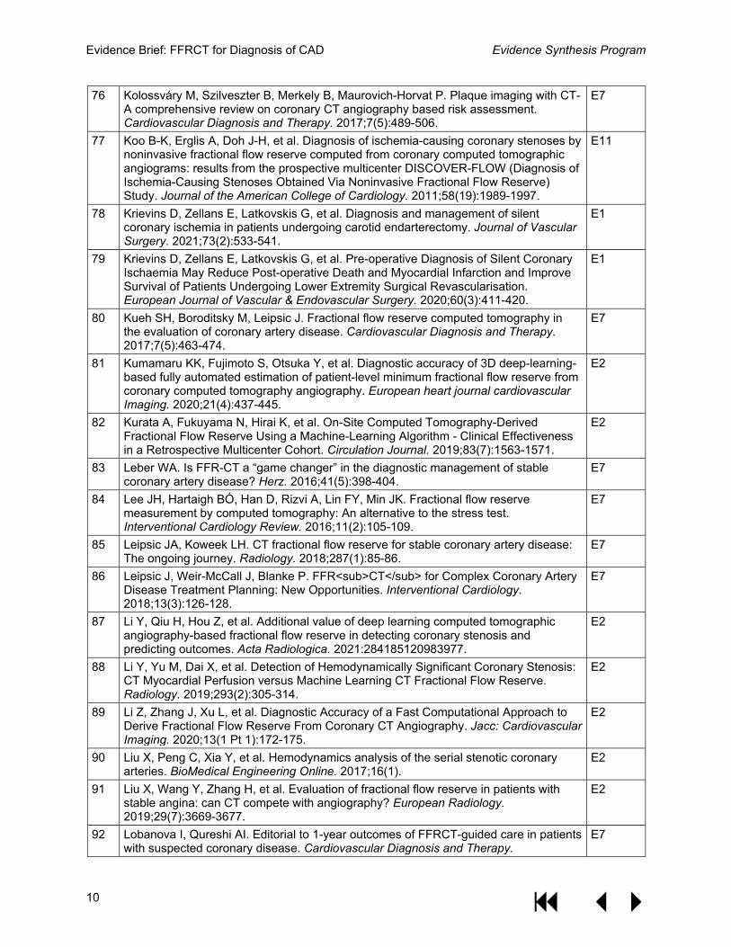
76 Kolossváry M, Szilveszter B, Merkely B, Maurovich-Horvat P. Plaque imaging with CT-A comprehensive review on coronary CT angiography based risk assessment. Cardiovascular Diagnosis and Therapy. 2017;7(5):489-506.
E7
77 Koo B-K, Erglis A, Doh J-H, et al. Diagnosis of ischemia-causing coronary stenoses by noninvasive fractional flow reserve computed from coronary computed tomographic angiograms: results from the prospective multicenter DISCOVER-FLOW (Diagnosis of Ischemia-Causing Stenoses Obtained Via Noninvasive Fractional Flow Reserve) Study. Journal of the American College of Cardiology. 2011;58(19):1989-1997.
E11
78 Krievins D, Zellans E, Latkovskis G, et al. Diagnosis and management of silent coronary ischemia in patients undergoing carotid endarterectomy. Journal of Vascular Surgery. 2021;73(2):533-541.
E1
79 Krievins D, Zellans E, Latkovskis G, et al. Pre-operative Diagnosis of Silent Coronary Ischaemia May Reduce Post-operative Death and Myocardial Infarction and Improve Survival of Patients Undergoing Lower Extremity Surgical Revascularisation. European Journal of Vascular & Endovascular Surgery. 2020;60(3):411-420.
E1
80 Kueh SH, Boroditsky M, Leipsic J. Fractional flow reserve computed tomography in the evaluation of coronary artery disease. Cardiovascular Diagnosis and Therapy. 2017;7(5):463-474.
E7
81 Kumamaru KK, Fujimoto S, Otsuka Y, et al. Diagnostic accuracy of 3D deep-learning-based fully automated estimation of patient-level minimum fractional flow reserve from coronary computed tomography angiography. European heart journal cardiovascular Imaging. 2020;21(4):437-445.
E2
82 Kurata A, Fukuyama N, Hirai K, et al. On-Site Computed Tomography-Derived Fractional Flow Reserve Using a Machine-Learning Algorithm - Clinical Effectiveness in a Retrospective Multicenter Cohort. Circulation Journal. 2019;83(7):1563-1571.
E2
83 Leber WA. Is FFR-CT a “game changer” in the diagnostic management of stable coronary artery disease? Herz. 2016;41(5):398-404.
E7
84 Lee JH, Hartaigh BÓ, Han D, Rizvi A, Lin FY, Min JK. Fractional flow reserve measurement by computed tomography: An alternative to the stress test. Interventional Cardiology Review. 2016;11(2):105-109.
E7
85 Leipsic JA, Koweek LH. CT fractional flow reserve for stable coronary artery disease: The ongoing journey. Radiology. 2018;287(1):85-86.
E7
86 Leipsic J, Weir-McCall J, Blanke P. FFR<sub>CT</sub> for Complex Coronary Artery Disease Treatment Planning: New Opportunities. Interventional Cardiology. 2018;13(3):126-128.
E7
87 Li Y, Qiu H, Hou Z, et al. Additional value of deep learning computed tomographic angiography-based fractional flow reserve in detecting coronary stenosis and predicting outcomes. Acta Radiologica. 2021:284185120983977.
E2
88 Li Y, Yu M, Dai X, et al. Detection of Hemodynamically Significant Coronary Stenosis: CT Myocardial Perfusion versus Machine Learning CT Fractional Flow Reserve. Radiology. 2019;293(2):305-314.
E2
89 Li Z, Zhang J, Xu L, et al. Diagnostic Accuracy of a Fast Computational Approach to Derive Fractional Flow Reserve From Coronary CT Angiography. Jacc: Cardiovascular Imaging. 2020;13(1 Pt 1):172-175.
E2
90 Liu X, Peng C, Xia Y, et al. Hemodynamics analysis of the serial stenotic coronary arteries. BioMedical Engineering Online. 2017;16(1).
E2
91 Liu X, Wang Y, Zhang H, et al. Evaluation of fractional flow reserve in patients with stable angina: can CT compete with angiography? European Radiology. 2019;29(7):3669-3677.
E2
92 Lobanova I, Qureshi AI. Editorial to 1-year outcomes of FFRCT-guided care in patients with suspected coronary disease. Cardiovascular Diagnosis and Therapy.
E7

Evidence Brief: FFRCT for Diagnosis of CAD Evidence Synthesis Program
11
2017;7:S115-S118. 93 Lossnitzer D, Chandra L, Rutsch M, et al. Additional Value of Machine-Learning
Computed Tomographic Angiography-Based Fractional Flow Reserve Compared to Standard Computed Tomographic Angiography. Journal of Clinical Medicine. 2020;9(3):03.
E2
94 Lu MT, Ferencik M, Roberts RS, et al. Noninvasive FFR derived from coronary CT angiography: management and outcomes in the PROMISE trial. JACC: Cardiovascular Imaging. 2017;10(11):1350-1358.
E4
95 Mahmoudi M, Nicholas Z, Nuttall J, et al. Fractional Flow Reserve Derived from Computed Tomography Coronary Angiography in the Assessment and Management of Stable Chest Pain: Rationale and Design of the FORECAST Trial. Cardiovascular Revascularization Medicine. 2020;21(7):890-896.
E7
96 Mangla A, Oliveros E, Williams KA, Sr., Kalra DK. Cardiac Imaging in the Diagnosis of Coronary Artery Disease. Current Problems in Cardiology. 2017;42(10):316-366.
E7
97 Mastrodicasa D, Albrecht MH, Schoepf UJ, et al. Artificial intelligence machine learning-based coronary CT fractional flow reserve (CT-FFR<sub>ML</sub>): Impact of iterative and filtered back projection reconstruction techniques. Journal of Cardiovascular Computed Tomography. 2018.
E3
98 Mathew RC, Gottbrecht M, Salerno M. Computed Tomography Fractional Flow Reserve to Guide Coronary Angiography and Intervention. Interventional Cardiology Clinics. 2018;7(3):345-354.
E7
99 Meier D, Skalidis I, De Bruyne B, et al. Ability of FFR-CT to detect the absence of hemodynamically significant lesions in patients with high-risk NSTE-ACS admitted in the emergency department with chest pain, study design and rationale. International Journal of Cardiology Heart & Vasculature. 2020;27:100496.
E7
100 Michail M, Ihdayhid AR, Comella A, et al. Feasibility and Validity of Computed Tomography-Derived Fractional Flow Reserve in Patients With Severe Aortic Stenosis: The CAST-FFR Study. Circulation: Cardiovascular Interventions. 2021;14(1):e009586.
E1
101 Min JK, Leipsic J, Pencina MJ, et al. Diagnostic accuracy of fractional flow reserve from anatomic CT angiography. JAMA. 2012;308(12):1237-1245.
E11
102 Min JK, Taylor CA, Achenbach S, et al. Noninvasive fractional flow reserve derived from coronary CT angiography clinical data and scientific principles. JACC: Cardiovascular Imaging. 2015;8(10):1209-1222.
E7
103 Miyajima K, Motoyama S, Sarai M, et al. On-site assessment of computed tomography-derived fractional flow reserve in comparison with myocardial perfusion imaging and invasive fractional flow reserve. Heart & Vessels. 2020;35(10):1331-1340.
E2
104 Miyoshi T, Osawa K, Ito H, et al. Non-invasive computed fractional flow reserve from computed tomography (CT) for diagnosing coronary artery disease - Japanese results from NXT trial (Analysis of Coronary Blood Flow Using CT Angiography: Next Steps). Circulation journal : official journal of the Japanese Circulation Society. 2015;79(2):406-412.
E11
105 Mordi IR, Badar AA, John Irving R, Weir-McCall JR, Houston JG, Lang CC. Efficacy of noninvasive cardiac imaging tests in diagnosis and management of stable coronary artery disease. Vascular Health and Risk Management. 2017;13:427-437.
E7
106 Nakanishi R, Budoff MJ. Noninvasive FFR derived from coronary CT angiography in the management of coronary artery disease: Technology and clinical update. Vascular Health and Risk Management. 2016;12:269-278.
E7
107 Nakazato R, Park HB, Gransar H, et al. Additive diagnostic value of atherosclerotic plaque characteristics to non-invasive FFR for identification of lesions causing
E2
Evidence Brief: FFRCT for Diagnosis of CAD Evidence Synthesis Program
12
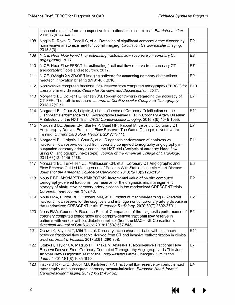
ischaemia: results from a prospective international multicentre trial. EuroIntervention. 2016;12(4):473-481.
108 Neglia D, Rovai D, Caselli C, et al. Detection of significant coronary artery disease by noninvasive anatomical and functional imaging. Circulation Cardiovascular imaging. 2015;8(3).
E2
109 NICE. HeartFlow FFRCT for estimating fractional flow reserve from coronary CT angiography. 2017.
E8
110 NICE. HeartFlow FFRCT for estimating fractional flow reserve from coronary CT angiography: Tools and resources. 2017.
E7
111 NICE. QAngio XA 3D/QFR imaging software for assessing coronary obstructions - medtech innovation briefing (MIB146). 2018.
E2
112 Noninvasive computed fractional flow reserve from computed tomography (FFRCT) for coronary artery disease. Centre for Reviews and Dissemination. 2017.
E10
113 Norgaard BL, Botker HE, Jensen JM. Recent controversy regarding the accuracy of CT-FFR. The truth is out there. Journal of Cardiovascular Computed Tomography. 2018;12(1):e1.
E7
114 Norgaard BL, Gaur S, Leipsic J, et al. Influence of Coronary Calcification on the Diagnostic Performance of CT Angiography Derived FFR in Coronary Artery Disease: A Substudy of the NXT Trial. JACC Cardiovascular imaging. 2015;8(9):1045-1055.
E11
115 Nørgaard BL, Jensen JM, Blanke P, Sand NP, Rabbat M, Leipsic J. Coronary CT Angiography Derived Fractional Flow Reserve: The Game Changer in Noninvasive Testing. Current Cardiology Reports. 2017;19(11).
E7
116 Norgaard BL, Leipsic J, Gaur S, et al. Diagnostic performance of noninvasive fractional flow reserve derived from coronary computed tomography angiography in suspected coronary artery disease: the NXT trial (Analysis of coronary blood flow using CT angiography: next steps). Journal of the American College of Cardiology. 2014;63(12):1145-1155.
E11
117 Norgaard BL, Terkelsen CJ, Mathiassen ON, et al. Coronary CT Angiographic and Flow Reserve-Guided Management of Patients With Stable Ischemic Heart Disease. Journal of the American College of Cardiology. 2018;72(18):2123-2134.
E3
118 Nous F BRLMYYMPBTAJKMKBGTNK. Incremental value of on-site computed tomography-derived fractional flow reserve for the diagnosis and management strategy of obstructive coronary artery disease in the randomized CRESCENT trials. European heart journal. 3782;40.
E2
119 Nous FMA, Budde RPJ, Lubbers MM, et al. Impact of machine-learning CT-derived fractional flow reserve for the diagnosis and management of coronary artery disease in the randomized CRESCENT trials. European Radiology. 2020;30(7):3692-3701.
E2
120 Nous FMA, Coenen A, Boersma E, et al. Comparison of the diagnostic performance of coronary computed tomography angiography-derived fractional flow reserve in patients with versus without diabetes mellitus (from the MACHINE Consortium). American Journal of Cardiology. 2019;123(4):537-543.
E2
121 Osawa K, Miyoshi T, Miki T, et al. Coronary lesion characteristics with mismatch between fractional flow reserve derived from CT and invasive catheterization in clinical practice. Heart & Vessels. 2017;32(4):390-398.
E11
122 Otake H, Taylor CA, Matsuo H, Tanaka N, Akasaka T. Noninvasive Fractional Flow Reserve Derived From Coronary Computed Tomography Angiography - Is This Just Another New Diagnostic Test or the Long-Awaited Game Changer? Circulation Journal. 2017;81(8):1085-1093.
E7
123 Packard RR, Li D, Budoff MJ, Karlsberg RP. Fractional flow reserve by computerized tomography and subsequent coronary revascularization. European Heart Journal Cardiovascular Imaging. 2017;18(2):145-152.
E4
Evidence Brief: FFRCT for Diagnosis of CAD Evidence Synthesis Program
13
124 Panchal HB, Veeranki SP, Bhatheja S, et al. Fractional flow reserve using computed tomography for assessing coronary artery disease: a meta-analysis. Journal of Cardiovascular Medicine. 2016;17(9):694-700.
E8
125 Park HB, Jang Y, Arsanjani R, et al. Diagnostic Accuracy of a Novel On-site Virtual Fractional Flow Reserve Parallel Computing System. Yonsei Medical Journal. 2020;61(2):137-144.
E2
126 Patel AR, Maffessanti F, Patel MB, et al. Hemodynamic impact of coronary stenosis using computed tomography: comparison between noninvasive fractional flow reserve and 3D fusion of coronary angiography with stress myocardial perfusion. The international journal of cardiovascular imaging. 2019;35(9):1733-1743.
E4
127 Podgorsak AR, Sommer KN, Reddy A, et al. Initial evaluation of a convolutional neural network used for noninvasive assessment of coronary artery disease severity from coronary computed tomography angiography data. Medical Physics. 2020;47(9):3996-4004.
E2
128 Pontone G, Andreini D, Guaricci AI, et al. Rationale and design of the PERFECTION (comparison between stress cardiac computed tomography PERfusion versus Fractional flow rEserve measured by Computed Tomography angiography In the evaluation of suspected cOroNary artery disease) prospective study. Journal of Cardiovascular Computed Tomography. 2016;10(4):330-334.
E7
129 Pontone G, Carita P, Verdecchia M, et al. Fractional flow reserve: Lessons from PLATFORM and future perspectives. Minerva Cardioangiologica. 2017;65(3):235-251.
E7
130 Pontone G, Muscogiuri G, Andreini D, et al. The New Frontier of Cardiac Computed Tomography Angiography: Fractional Flow Reserve and Stress Myocardial Perfusion. Current Treatment Options in Cardiovascular Medicine. 2016;18(12).
E7
131 Qiao HY, Li JH, Schoepf UJ, et al. Prognostic implication of CT-FFR based functional SYNTAX score in patients with de novo three-vessel disease. European heart journal cardiovascular Imaging. 2020;13:13.
E2
132 Qiao HY, Tang CX, Schoepf UJ, et al. Impact of machine learning-based coronary computed tomography angiography fractional flow reserve on treatment decisions and clinical outcomes in patients with suspected coronary artery disease. European Radiology. 2020;30(11):5841-5851.
E2
133 Rabbat M, Kauh B, Pontone G, Norgaard B, Lopez J, Mathew V. Fractional flow reserve derived from coronary computed tomography safely reduces invasive coronary angiography rates and cost in patients with stable coronary artery disease. Journal of the American College of Cardiology. 2017;69(11 Supplement):72.
E7
134 Raja J, Seitz MP, Yedlapati N, Khouzam RN. Can Computed Fractional Flow Reserve Coronary CT Angiography (FFRCT) Offer an Accurate Noninvasive Comparison to Invasive Coronary Angiography (ICA)? "The Noninvasive CATH." A Comprehensive Review. Current Problems in Cardiology. 2021;46(3):100642.
E7
135 Rajani R, Modi B, Ntalas I, Curzen N. Non-invasive fractional flow reserve using computed tomographic angiography: Where are we now and where are we going? Heart. 2017;103(15):1216-1222.
E7
136 Rajani R, Webb J, Marciniak A, Preston R. Comparative efficacy testing - fractional flow reserve by coronary computed tomography for the evaluation of patients with stable chest pain. International Journal of Cardiology. 2015;183:173-177.
E4
137 Renker M, Schoepf UJ, Becher T, et al. Computed tomography in patients with chronic stable angina: Fractional flow reserve measurement. Herz. 2017;42(1):51-57.
E9
138 Renker M, Schoepf UJ, Wang R, et al. Comparison of diagnostic value of a novel noninvasive coronary computed tomography angiography method versus standard coronary angiography for assessing fractional flow reserve. American Journal of Cardiology. 2014;114(9):1303-1308.
E2
Evidence Brief: FFRCT for Diagnosis of CAD Evidence Synthesis Program
14
139 Ronnow Sand NP, Nissen L, Winther S, et al. Prediction of Coronary Revascularization in Stable Angina: Comparison of FFR<sub>CT</sub> With CMR Stress Perfusion Imaging. Jacc: Cardiovascular Imaging. 2020;13(4):994-1004.
E4
140 Roobottom C. Radical changes to the investigation of stable chest pain following the 2016 NICE update. British Journal of Radiology. 2018;91(1087).
E10
141 Ropp A, White C. Current and Future Applications of Coronary CT Angiography with and Without FFR in the Emergency Room. Current Cardiovascular Imaging Reports. 2016;9(11).
E7
142 Rother J, Moshage M, Dey D, et al. Comparison of invasively measured FFR with FFR derived from coronary CT angiography for detection of lesion-specific ischemia: Results from a PC-based prototype algorithm. Journal of Cardiovascular Computed Tomography. 2018;12(2):101-107.
E2
143 Sand NPR, Veien KT, Nielsen SS, et al. Prospective comparison of FFR derived from coronary CT angiography with SPECT perfusion imaging in stable coronary artery disease: the ReASSESS Study. JACC: Cardiovascular Imaging. 2018;11(11):1640-1650.
E4
144 Schuijf JD, Ko BS, Di Carli MF, et al. Fractional flow reserve and myocardial perfusion by computed tomography: A guide to clinical application. European Heart Journal Cardiovascular Imaging. 2018;19(2):127-135.
E7
145 Sevag Packard RR, Karlsberg RP. Integrating FFRCT Into Routine Clinical Practice: A Solid PLATFORM or Slippery Slope?∗. Journal of the American College of Cardiology. 2016;68(5):446-449.
E7
146 Sigurdsson G. Improved Precision of Initial Chest Pain Evaluation With Fractional Flow Reserve Computed Tomography. Journal of the American Heart Association. 2017;6(8):22.
E7
147 Shah AB, Kirsch J, Bolen MA, et al. ACR Appropriateness Criteria((R)) Chronic Chest Pain-Noncardiac Etiology Unlikely-Low to Intermediate Probability of Coronary Artery Disease. Journal of the American College of Radiology. 2018;15(11S):S283-S290.
E7
148 Shi C, Zhang D, Cao K, et al. A study of noninvasive fractional flow reserve derived from a simplified method based on coronary computed tomography angiography in suspected coronary artery disease. Biomedical Engineering Online. 2017;16(1):43.
E2
149 Siontis GC, Mavridis D, Greenwood JP, et al. Outcomes of non-invasive diagnostic modalities for the detection of coronary artery disease: network meta-analysis of diagnostic randomised controlled trials. BMJ. 2018;360:k504.
E2
150 Skelly AC, Hashimoto R, Buckley DI, et al. Noninvasive Testing for Coronary Artery Disease. In: AHRQ Comparative Effectiveness Reviews. Rockville (MD): Agency for Healthcare Research and Quality (US); 2016.
E2
151 Takagi H, Ishikawa Y, Orii M, et al. Optimized interpretation of fractional flow reserve derived from computed tomography: Comparison of three interpretation methods. Journal of Cardiovascular Computed Tomography. 2018.
E3
152 Takahashi K KNTMCPCCCMROMKHRJADDTSP. TCT-326 Diagnostic Performance of Angiography-Based Quantitative Flow Ratio With Respect to Fractional Flow Reserve Derived From Computed Tomography Angiography: insight From the SYNTAX III Trial. Journal of the American College of Cardiology.74(13):B324-.
E4
153 Tan XW, Zheng Q, Shi L, et al. Combined diagnostic performance of coronary computed tomography angiography and computed tomography derived fractional flow reserve for the evaluation of myocardial ischemia: A meta-analysis. International Journal of Cardiology. 2017;236:100-106.
E3
154 Tan Y, Litt H. High-risk plaque features predict ischemia in acute chest pain-direct comparison to non-invasive FFR. Journal of cardiovascular computed tomography. 2017;Conference: 12th annual scientific meeting of the society of cardiovascular
E4
Evidence Brief: FFRCT for Diagnosis of CAD Evidence Synthesis Program
15
computed tomography. United states. 11(4 Supplement 1):S76-S77. 155 Tanaka K, Bezerra HG, Gaur S, et al. Comparison between non-invasive (coronary
computed tomography angiography derived) and invasive-fractional flow reserve in patients with serial stenoses within one coronary artery: A NXT Trial substudy. Annals of Biomedical Engineering. 2016;44(2):580-589.
E4
156 Tang CX, Liu CY, Lu MJ, et al. CT FFR for Ischemia-Specific CAD With a New Computational Fluid Dynamics Algorithm: A Chinese Multicenter Study. Jacc: Cardiovascular Imaging. 2020;13(4):980-990.
E2
157 Tesche C, De Cecco CN, Albrecht MH, et al. Coronary CT angiography-derived fractional flow reserve. Radiology. 2017;285(1):17-33.
E7
158 Tesche C, De Cecco CN, Baumann S, et al. Coronary CT Angiography-derived Fractional Flow Reserve: Machine Learning Algorithm versus Computational Fluid Dynamics Modeling. Radiology. 2018;288(1):64-72.
E3
159 Tesche C, Vliegenthart R, Duguay TM, et al. Coronary Computed Tomographic Angiography-Derived Fractional Flow Reserve for Therapeutic Decision Making. American Journal of Cardiology. 2017;120(12):2121-2127.
E2
160 Thompson AG, Raju R, Blanke P, et al. Diagnostic accuracy and discrimination of ischemia by fractional flow reserve CT using a clinical use rule: results from the Determination of Fractional Flow Reserve by Anatomic Computed Tomographic Angiography study. Journal of Cardiovascular Computed Tomography. 2015;9(2):120-128.
E2
161 van Assen M, De Cecco CN, Eid M, et al. Prognostic value of CT myocardial perfusion imaging and CT-derived fractional flow reserve for major adverse cardiac events in patients with coronary artery disease. Journal of cardiovascular computed tomography. 2019;13(3):26-33.
E2
162 Varga-Szemes A, Schoepf UJ, Maurovich-Horvat P, et al. Coronary plaque assessment of Vasodilative capacity by CT angiography effectively estimates fractional flow reserve. International Journal of Cardiology. 2021;30:30.
E2
163 Wang ZQ, Zhou YJ, Zhao YX, et al. Diagnostic accuracy of a deep learning approach to calculate FFR from coronary CT angiography. Journal of Geriatric Cardiology. 2019;16(1):42-48.
E2
164 Wardziak L, Kruk M, Pleban W, et al. Coronary CTA enhanced with CTA based FFR analysis provides higher diagnostic value than invasive coronary angiography in patients with intermediate coronary stenosis. Journal of Cardiovascular Computed Tomography. 2019;13(1):62-67.
E2
165 Wu W, Pan DR, Foin N, et al. Noninvasive fractional flow reserve derived from coronary computed tomography angiography for identification of ischemic lesions: A systematic review and meta-analysis. Scientific Reports. 2016;6.
E8
166 Xia G, Fan D, Yao X, Guan G, Wang J. Diagnostic efficacy of fractional flow reserve with coronary angiography in dual-source computed tomography scanner. Acta Cardiologica. 2018;73(1):76-83.
E11
167 Xie X, Zheng M, Wen D, Li Y, Xie S. A new CFD based non-invasive method for functional diagnosis of coronary stenosis. Biomedical Engineering Online. 2018;17(1):36.
E2
168 Xu PP, Li JH, Zhou F, et al. The influence of image quality on diagnostic performance of a machine learning-based fractional flow reserve derived from coronary CT angiography. European Radiology. 2020;30(5):2525-2534.
E2
169 Yang DH, Kang SJ, Koo HJ, et al. Incremental Value of Subtended Myocardial Mass for Identifying FFR-Verified Ischemia Using Quantitative CT Angiography: Comparison With Quantitative Coronary Angiography and CT-FFR. Jacc: Cardiovascular Imaging. 2018;12:12.
E2

Evidence Brief: FFRCT for Diagnosis of CAD Evidence Synthesis Program
16
170 Yang DH, Kim YH, Roh JH, et al. Diagnostic performance of on-site CT-derived fractional flow reserve versus CT perfusion. European heart journal cardiovascular Imaging. 2017;18(4):432-440.
E2
171 Yang J, Shan D, Dong M, et al. The effect of on-site CT-derived fractional flow reserve on the management of decision making for patients with stable chest pain (TARGET trial): objective, rationale, and design. Trials [Electronic Resource]. 2020;21(1):728.
E2
172 Yang L, Xu L, He J, et al. Diagnostic performance of a fast non-invasive fractional flow reserve derived from coronary CT angiography: an initial validation study. Clinical Radiology. 2019;74(12):973.e971-973.e976.
E2
173 Yoshikawa Y, Nakamoto M, Nakamura M, et al. On-site evaluation of CT-based fractional flow reserve using simple boundary conditions for computational fluid dynamics. The international journal of cardiovascular imaging. 2020;36(2):337-346.
E2
174 Zhou F, Wang YN, Schoepf UJ, et al. Diagnostic Performance of Machine Learning Based CT-FFR in Detecting Ischemia in Myocardial Bridging and Concomitant Proximal Atherosclerotic Disease. Canadian Journal of Cardiology. 2019;35(11):1523-1533.
E2
175 Zhuang B, Wang S, Zhao S, Lu M. Computed tomography angiography-derived fractional flow reserve (CT-FFR) for the detection of myocardial ischemia with invasive fractional flow reserve as reference: systematic review and meta-analysis. Eur Radiol. 2020;30(2):712-725.
E2
176 Zarins CK, Taylor CA, Min JK. Computed Fractional Flow Reserve (FFTCT) Derived from Coronary CT Angiography. Journal of Cardiovascular Translational Research. 2013;6(5):708-714.
E7

Evidence Brief: FFRCT for Diagnosis of CAD Evidence Synthesis Program
17
APPENDIX C. EVIDENCE TABLES DATA ABSTRACTION OF INCLUDED SYSTEMATIC REVIEWS Author, Year Search dates and
databases Population Included imaging
technologies Reference standard
HeartFlow Sensitivity (95% CI) Specificity (95% CI)
CCTA Sensitivity (95% CI) Specificity (95% CI)
# Included studies (HeartFlow)
Area Under Curve (HeartFlow)
Celeng, 20181
Timeframe: through September 7, 2017 Databases: PubMed, EMBASE, Web of Science
Study participants with suspected or known CAD
FFRCT, CTP, TAG (Transluminal attenuation gradient)
Sensitivity: 85% (81 to 90) Specificity: 73% (61 to 82)
Sensitivity: 87% (84 to 91) Specificity: 61% (54 to 68)
FFRCT: 18 HeartFlow: 6
0.87
Hamon, 20192
Timeframe: July 2018 Databases: Medline and Cochrane
Study participants with stable chest pain
FFRCT, CTA, CTP, TAG
Sensitivity: 84% (80 to 88) Specificity: 76% (73 to 79)
Sensitivity: 86% (85 to 88) Specificity: 64% (63 to 66)
FFRCT: 18 HeartFlow: 6
0.89
Pontone, 20203
Timeframe: through March 7, 2017 Databases: Medline and EMBASE
Study participants with suspected or known CAD
CCTA, stress ECHO, stress SPECT, PET, FFRCT, stress myocardial CT perfusion
Sensitivity: 85% (81 to 88) Specificity: 75% (72 to 78)
Sensitivity: 88% (85 to 90) Specificity: 64% (61 to 66)
HeartFlow: 7 0.89
Abbreviations: CAD=coronary artery disease; CCTA/CTA=coronary computed tomography angiography; CTP=coronary computed tomography myocardial perfusion; ECHO=Echocardiography; FFRCT=fractional flow reserve using computed tomography; PET=positron emission tomography; SPECT=single-photon emission computed tomography; TAG=transluminal attenuation gradient
DATA ABSTRACTION OF INCLUDED PRIMARY STUDIES Data Abstraction of Primary Studies Evaluating Diagnostic Accuracy of HeartFlow FFRCT
Author, Year N
Population Index Test Sensitivity (95% CI)* Specificity (95% CI)*
Trial Name Area Under Curve
Driessen, 20194 157
Patients with suspected stable CAD and who underwent CCTA, SPECT, PET, and FFR
Invasive FFR Sensitivity: 90 (84-95) Specificity: 86 (82-89)
PACIFIC FFRCT: 0.94 (0.92-0.96) CCTA: 0.83 (0.80-0.86)
Pontone, 20195 147
Symptomatic patients scheduled for clinically indicated ICA + invasive FFR
Invasive FFR Sensitivity: 88 (82-94) Specificity: 94 (91-96)
PERFECTION FFRCT: 0.93 (0.91-0.96) CCTA: 0.89 (0.86-0.93

Evidence Brief: FFRCT for Diagnosis of CAD Evidence Synthesis Program
18
Author, Year N
Population Index Test Sensitivity (95% CI)* Specificity (95% CI)*
Trial Name Area Under Curve
Pontone, 20196 85
Symptomatic patients scheduled for clinically indicated ICA + invasive FFR
ICA + Invasive FFR
Sensitivity: 86 (78–94) Specificity: 75 (68–82)
None FFRCT: 0.88 (0.83-0.92) CCTA: 0.83 (0.77-0.88)
Bom, 20217 132
Patients with suspected stable CAD and who underwent coronary CTA, SPECT, PET, and FFR and had vessels ≥30% angiographic stenosis on ICA
ICA + Invasive FFR
Sensitivity: 90 (83-96) Specificity: 68 (58-77)
PACIFIC FFRCT: 0.89 (0.83-0.93) CCTA: 0.79 (0.73-0.85)
Cami, 20208 1484
Patients referred for evaluation of myocardial ischemia
ICA + Invasive FFR
Distal: Sensitivity: 92, Specificity: 86 Terminal: Sensitivity: 92, Specificity: 50
None FFRCT, Distal: 0.91 (95% CI NR) FFRCT, Terminal: 0.83 (95% CI NR)
Ko, 20199 51
Symptomatic patients scheduled for ICA + invasive FFR
ICA + Invasive FFR
Sensitivity: 80.6 (62.5–92.5) Specificity: 85.0 (73.4–92.9)
None FFRCT: 0.90 (0.82-0.98) CCTA: 0.68 (0.56-0.80)
Tanigaki, 201910 152
Patients with stable CAD identified by CTA ICA + Invasive FFR
Sensitivity: 82 (76–88) Specificity: 70 (64–74)
ADVANCE FFRCT: 0.82 (0.76-0.87) CCTA: 0.70 (0.64-0.76)
*Per vessel Abbreviations: CAD=coronary artery disease; CCTA=coronary computed tomography angiography; FFR=fractional flow reserve; FFRCT=fractional flow reserve using computed tomography; ICA=invasive coronary angiography; PET=positron emission tomography; SPECT=single-photon emission computed tomography
Data Abstraction of Primary Studies Evaluating Clinical or Therapeutic Outcomes
Study Characteristics
Author, Year Study Design N
Location Follow-up
Population Patient Demographics
Cardiac Risk Factors Pre-test Probability of Disease
Comparator Adequacy of Images
ADVANCE Anastasius, 202011 Prospective cohort 4553
Intl Registry 1 year
Patients being investigated for clinically suspected CAD with documented atherosclerosis (>30%) on CCTA with FFRCT result
Age: 66.1 Male: 66.5% Race: NR
Diabetes: 22.1% Hypertension: 60.1% History of smoking: current (16.8%, previous:34.4%) Hyperlipidemia: 58.5%
Diamond-Forrester: 51.6%
None 96.8%
Fairbairn, 202012 (Gender diff.) Patel, 202013 Prospective cohort 4737
Intl Registry 90 days 1 year
Patients being investigated for clinically suspected CAD with documented atherosclerosis (>30%) on CCTA with FFRCT result
Age: 66.1 Male: 66.2% Race NR
Diabetes: 21.9% Hypertension: 59.8% History of smoking: current (16.8%, previous:34.1%) Hyperlipidemia: 58.1%
Diamond-Forrester: 51.6%
None 96.8%

Evidence Brief: FFRCT for Diagnosis of CAD Evidence Synthesis Program
19
Author, Year Study Design N
Location Follow-up
Population Patient Demographics
Cardiac Risk Factors Pre-test Probability of Disease
Comparator Adequacy of Images
Fairbairn, 201814 Nous, 202115 Prospective cohort 5083
Intl Registry 90 days 1 year
Patients being investigated for clinically suspected CAD with documented atherosclerosis (>30%) on CCTA
Age: 66 Male: 65.9% Race NR
Diabetes: 22.3% Hypertension: 59.9% History of smoking: current (16.6%, previous:34.1%) Hyperlipidemia: 58.2%
Diamond-Forrester: 51.3% for whole cohort, 51.6% for FFRCT pts
CCTA alone 96.8%
Pontone, 201916 (rejection rate) Prospective cohort 2778
Intl Registry NR
Patients being investigated for clinically suspected CAD with documented atherosclerosis (>30%) on CCTA using FFRCT 2.0 or later version
Age: 66 Male: 66% Race NR
Diabetes: 22% History of smoking: 61% Hyperlipidemia: 61%
NR None Rejection rate: 2.9% (95% CI 2.32 to 3.57).
Shiono, 201917 Prospective cohort 1829
Intl Registry 90 days
Japanese patients being investigated for clinically suspected CAD with documented atherosclerosis (>30%) on CCTA
Age: 69.4 Male: 65.4% Race: NR (Japanese centers)
Diabetes: 32.5% Hypertension: 60.2% History of smoking: current (17.5%, previous:33.5%) Hyperlipidemia: 60.2%
Diamond-Forrester: 55%
CCTA alone 96.3%
PLATFORM Colleran, 201718 Prospective cohort 116
Germany 1 year
Symptomatic adult patients with intermediate likelihood of obstructive CAD, without known CAD in Germany
Age: 59.9 Male: 57.7% 1.7% racial/ethnic minority
Diabetes: 13.0% Hypertension: 62.8% History of smoking: 50.9% Dyslipidemia: 21.5%
Diamond-Forrester: 50.1%
Originally planned testing ("usual care"): ICA
83.3%
Douglas, 201519 Douglas, 201620 Hlatky, 201521 Prospective cohort 584
11 European sites and Duke (US) 90 days 1 year
Symptomatic adult patients with intermediate likelihood of obstructive CAD, without known CAD
Age: 60.9 Male: 60.4% 1.5% racial/ethnic minority
Diabetes: 13.7% Hypertension: 54.3% History of smoking: 53.9% Dyslipidemia: 34.8%
Diamond-Forrester: 49%
Originally planned testing ("usual care"): non-invasive testing (any) or ICA
88%
OTHER Andreini, 201922 Prospective cohort 223
6 European sites NR
Patients with CAD diagnosed with ICA or CCTA and candidates for PCI or CABG
Age: 67.6 Male: 84.3% Race: NR
Diabetes: 37.7% Hypertension: 74.9% Current smoking: 22.6% Hyperlipidemia: 70%
NR CCTA alone or ICA alone
88%

Evidence Brief: FFRCT for Diagnosis of CAD Evidence Synthesis Program
20
Author, Year Study Design N
Location Follow-up
Population Patient Demographics
Cardiac Risk Factors Pre-test Probability of Disease
Comparator Adequacy of Images
Baggiano, 202023 Retrospective cohort 291
Italy NR
Symptomatic patients scheduled for ICA+invasive FFR
Age: 65 Male: 76% Race: NR
Diabetes: 19% Hypertension: 74% Current smoking: 32%
Diamond-Forrester: 65%
CCTA alone or CCTA + Stress CTP
89%
Curzen, 201624 Retrospective cohort 200
Intl NR
Patients with suspected stable CAD with at least one stenosis (30% - 90%) on CCTA undergoing nonemergent ICA
NR NR NR CCTA alone Only included those with FFRCT data
Fares, 201925 Retrospective cohort 207
US NR
Patients with suspected CAD referred for FFRCT
Age: 69.5 Male: 46.4% Race: 28.5 African American, 66.4% White
Diabetes: 21.5% Hypertension: 67.7% Smoking: current: 13.3%, past: 36.4%% Dyslipidemia: 66.7%
NR CCTA alone or C-FFRCT (algorithm for additional info)
79%
Ihdayhid, 201926 Case series 206
Intl 4.7 years (median)
Patients with suspected stable CAD with at least one stenosis (30% - 90%) on CCTA undergoing nonemergent ICA with FFRCT
Age: 64 Male: 64.1% Race: 68.4% White, 31.6% Asian
Diabetes: 22.8% Hypertension: 65.5% Smoking: 18.9% Hypercholesterolemia: 81.1%
Diamond-Forrester: 54.2%
None Excluded pts w/o FFRCT
Jang, 201627 Retrospective cohort 75
US NR
Patients with CCTA and referred for ICA.
Age: 60 Male: 75% Race NR
NR NR CCTA alone NR
Jensen, 201828 Prospective cohort 774
Denmark 90 days
Symptomatic patients referred to non-emergent ICA or CCTA on suspicion of stable CAD
Age: 59 Male: 52% Race: NR
Diabetes: 9% Hypertension: 37% History of smoking: 59% Hyperlipidaemia: 32%
Diamond-Forrester: 40%
CCTA alone (planned ICA [high risk] or planned CCTA [low risk])
98.6%
Norgaard, 202029 Case series 975
Denmark 2.2 years (median)
Patients with suspected chronic coronary syndrome with stenosis (30–70%) on CCTA.
Age: 61.9 Male: 59.1% Race NR
Diabetes: 12.0% Hypertension: 45.4% Current smoker: 23.0% Hyperlipidaemia: 37.7%
Diamond-Forrester: 44.8%
None 97.8%
Evidence Brief: FFRCT for Diagnosis of CAD Evidence Synthesis Program
21
Author, Year Study Design N
Location Follow-up
Population Patient Demographics
Cardiac Risk Factors Pre-test Probability of Disease
Comparator Adequacy of Images
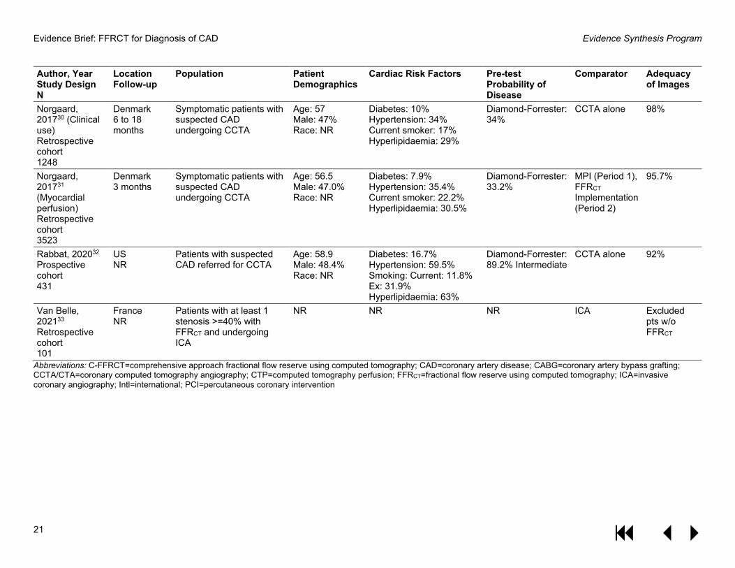
Norgaard, 201730 (Clinical use) Retrospective cohort 1248
Denmark 6 to 18 months
Symptomatic patients with suspected CAD undergoing CCTA
Age: 57 Male: 47% Race: NR
Diabetes: 10% Hypertension: 34% Current smoker: 17% Hyperlipidaemia: 29%
Diamond-Forrester: 34%
CCTA alone 98%
Norgaard, 201731 (Myocardial perfusion) Retrospective cohort 3523
Denmark 3 months
Symptomatic patients with suspected CAD undergoing CCTA
Age: 56.5 Male: 47.0% Race: NR
Diabetes: 7.9% Hypertension: 35.4% Current smoker: 22.2% Hyperlipidaemia: 30.5%
Diamond-Forrester: 33.2%
MPI (Period 1), FFRCT Implementation (Period 2)
95.7%
Rabbat, 202032 Prospective cohort 431
US NR
Patients with suspected CAD referred for CCTA
Age: 58.9 Male: 48.4% Race: NR
Diabetes: 16.7% Hypertension: 59.5% Smoking: Current: 11.8% Ex: 31.9% Hyperlipidaemia: 63%
Diamond-Forrester: 89.2% Intermediate
CCTA alone 92%
Van Belle, 202133 Retrospective cohort 101
France NR
Patients with at least 1 stenosis >=40% with FFRCT and undergoing ICA
NR NR NR ICA Excluded pts w/o FFRCT
Abbreviations: C-FFRCT=comprehensive approach fractional flow reserve using computed tomography; CAD=coronary artery disease; CABG=coronary artery bypass grafting; CCTA/CTA=coronary computed tomography angiography; CTP=computed tomography perfusion; FFRCT=fractional flow reserve using computed tomography; ICA=invasive coronary angiography; Intl=international; PCI=percutaneous coronary intervention

Evidence Brief: FFRCT for Diagnosis of CAD Evidence Synthesis Program
22
Outcomes
Author, Year Study Design N
ICA Use Change in Treatment Plan
MACE Other Clinical Outcomes Cost Quality of Life
ADVANCE Anastasius, 202011 Prospective cohort 4553
ICA w/o revascularization: 18.6% (suppl Table 2)
NR MACE events at 1 year 47 events (1%) No significant differences between age groups.
1 year: MI: FFRCT > 0.80: 11 events, FFRCT <=0.80: 31 events All-cause mortality: FFRCT > 0.80: 7 deaths, FFRCT <=0.80: 26 deaths Unplanned hospitalization: FFRCT > 0.80: 2 hospitalizations, FFRCT <=0.80: 4 hospitalizations Revascularizations: FFRCT > 0.80: 6%, FFRCT <= 0.80: 38%
NR
NR
Fairbairn, 202012 (Gender diff.) Patel, 202013 Prospective cohort 4737
ICA use at 90 days: Women: 54.5% vs Men: 56.5% (NSD) with FFRCT <=0.80. ICA without obstructive CAD at 90 days: Women: 32.1% vs Men 24.5% (p=0.0003)
Recommended treatment by FFRCT and actual clinical management at 1 year: Medical therapy: 92.9% received, 7.1% received revascularization, Revascularization: 68.9% received, 77% received revascularization to ICA
1 year: 55 MACE events (1.6%), 35 mortality events, 12 MI events, 8 ACS events
Revascularizations within 90 days: 1,026 (21.7%) by PCI, 150 (3.2%) by CABG
NR NR
Fairbairn, 201814 Nous, 202115 Prospective cohort 5083
90 day ICA use: Overall: 43.9% Over time: Cohort 1: 45.6%, Cohort 2: 41.9% Cohort 3: 44.3% (p=0.47)
Reclassification at 90 days between CCTA alone and CCTA plus FFRCT-based management plans in
90 days: No MACE events in patients with FFRCT >0.80. 19 (0.6%) MACE and 14 (0.3%) death/MI
Revascularizations within 90 days: 22.6% by PCI and 3.5% by CABG in Cohort 1; 19.8% by PCI and 3.2% by CABG in Cohort 2; 22.0% by
NR NR

Evidence Brief: FFRCT for Diagnosis of CAD Evidence Synthesis Program
23
Author, Year Study Design N
ICA Use Change in Treatment Plan
MACE Other Clinical Outcomes Cost Quality of Life
66.9% (95% CI 64.8–67.6) of patients.
occurred in subjects with FFRCT <0.80. 1 year: ~59 events overall. MACE over time in pts with an FFRCT result, 1.3%, 1.2% and 1.0% (p = 0.457) in cohort 1, 2, and 3, respectively.
PCI and 2.9% by CABG in Cohort 3
Pontone, 201916 (rejection rate) Prospective cohort 2778
NR
NR NR NR NR NR
Shiono, 201917 Prospective cohort 1829
ICA use at 90 days: After FFRCT: 50.4% had ICA (22.6% with negative FFRCT and 61.7% with positive FFRCT). ICA without obstructive CAD at 90 days: 20.5% with positive FFRCT and 46.1% with negative FFRCT (OR 3.29, 95% CI 2.19 to 4.95), p<0.0001)
Reclassification at 90 days between CCTA alone and CCTA plus FFRCT-based management plans in 55.8% of patients.
Pts with negative FFRCT (>0.8): No MACE events (n=509) at 90 days Pts with positive FFRCT (≤ 0.8): 5 (0.4%; n=1,249) MACE events at 90 days
Pts with negative FFRCT (>0.8): 3.9% underwent revascularization Pts with positive FFRCT (≤ 0.8): 67% underwent revascularization
NR NR
PLATFORM Colleran, 201718 Prospective cohort 116
ICA w/o obstructive CAD Planned ICA cohort: 90 days: 7.7% FFRCT vs 85.9% usual care.
NA
No events in either group
Revascularizations at 1 year: 12 by PCI usual care vs 10 FFRCT
Planned ICA cohort: Mean 1-year patient cost: €4217 FFRCT
Planned ICA cohort: QoL scores (FFRCT vs usual care): SAQ: +22.36 vs +18.68 (p=0.22), EQ-5D:

Evidence Brief: FFRCT for Diagnosis of CAD Evidence Synthesis Program
24
Author, Year Study Design N
ICA Use Change in Treatment Plan
MACE Other Clinical Outcomes Cost Quality of Life
Risk difference 78.2% (95% CI 67.1 to 89.4, p<0.001)
Stents per patient (mean): 2.1 usual care vs 1.6 FFRCT Bypass surgeries: 4 usual care vs 1 FFRCT Hospital days: 122 usual care vs 65 FFRCT
vs €6894 usual care (p<0.001).
+0.09 vs +0.03 (p=0.04), VAS: +5.09 vs -0.07 (p=0.51).
Douglas, 201519 Douglas, 201620 Hlatky, 201521 Prospective cohort 584
ICA w/o obstructive CAD Planned ICA cohort: 90 days: 12.4% FFRCT vs 73.3% usual care (p<0.0001). Risk difference: 60.8% (95% CI 53.0% to 68.7%) Planned non-invasive cohort: 90 days: 12.5% FFRCT vs 6.0% usual care (p=0.95). Risk difference: -6.5 (95% CI -14.4 to 1.4)
NA 90 days MACE: Planned ICA cohort: 2 FFRCT vs 0 usual care Planned non-invasive cohort: 0 events 1-year MACE: Planned ICA cohort: 2 FFRCT vs 2 usual care (0 in pts whose ICA was canceled based on FFRCT results) Planned non-invasive cohort: 0 FFRCT vs 1 usual care
90 days: 22,1% total (16.9% PCI, 5.1% CABG) 1 year: 23.1% total (17.8% PCI, 5.3% CABG)
Planned ICA cohort: 1-year per-patient mean costs: 32% lower in FFRCT vs usual care ($7,343 vs $10,734 p<0.0001) Planned non-invasive cohort: 1-year per-patient mean costs $2,679 vs $2,137; p=0.26
Planned ICA cohort: 1-year QOL scores (SAQ, EQ-5D, VAS) improved with both FFRCT and usual care (p<0.001). Improvements similar in FFRCT and usual care at both 90 days and 1 year. Planned non-invasive cohort: 1-year QOL scores (SAQ, EQ-5D, VAS) improved with both FFRCT and usual care (p<0.001). EQ-5D (mean change: FFRCT 0.12 vs usual care 0.07; p=0.02) 90-day QOL scores improved more in FFRCT than usual care: SAQ: 19.5 vs 11.4, p=0.003, EuroQOL: 0.08 vs 0.03, p=0.002, VAS: 4.1 vs 2.3, p=0.82.

Evidence Brief: FFRCT for Diagnosis of CAD Evidence Synthesis Program
25
Author, Year Study Design N
ICA Use Change in Treatment Plan
MACE Other Clinical Outcomes Cost Quality of Life
OTHER Andreini, 201922 Prospective cohort 223
NR Treatment decision change btwn PCI and CABG: Vs CCTA alone: 7% pts Vs ICA alone: 6.6% pts # pts with significant 3-vessel CAD: 92.3% CCTA alone to 78.8% FFRCT 86.1% ICA alone to 86.2% FFRCT
NR NA NR NR
Baggiano, 202023 Retrospective cohort 291
NR Reclassification of pts with FFRCT (vs CCTA alone): 28% Rate of agreement with final management decision: 63% CCTA alone, 71% FFRCT, 89% CCTA + stress CTP, 84% FFRCT + stress CTP
NR Rate of agreement on vessels to be revascularized: 57% CCTA alone, 63% FFRCT, 74% CCTA + stress CTP, 70% FFRCT + stress CTP
NR NR
Curzen, 201624 Retrospective cohort 200
NR Change in clinical management plan with FFRCT vs CCTA alone: 36%
NR 39.0% PCI and 4.5% CABG NR NR
Fares, 201925 Retrospective cohort 207
NR
Change in clinical recommendation: 24% with FFRCT vs CCTA alone
NR NR NR NR
Ihdayhid, 201926 Case series 206
NR NR MACE: Overall: 9.7% FFRCT ≤ 0.8: 15.6% vs FFRCT > 0.8: 3.1% (HR 5.5, 95% CI 1.6 to 19)
Composite outcome (death, MI, and any revascularization): Overall: 45.1%
NR NR

Evidence Brief: FFRCT for Diagnosis of CAD Evidence Synthesis Program
26
Author, Year Study Design N
ICA Use Change in Treatment Plan
MACE Other Clinical Outcomes Cost Quality of Life
FFRCT ≤ 0.8: 73.4% vs FFRCT > 0.8: 13.4% (HR 9.2, 95% CI 5.1 to 17).
Jang, 201627 NR Clinical management plan (ICA, OMT, PCI, CABG) changed in 55% of patients with FFRCT vs CCTA alone. 36 pts (48%) no longer planned for ICA with FFRCT vs CCTA alone.
No significant difference in 1 year cardiovascular events between patients with changed vs unchanged management after FFRCT (data NR)
37/75 (49.3%) referred for PCI based on FFRCT 2/75 (2.7%) referred for CABG based on FFRCT
NR NR
Jensen, 201828 Prospective cohort 774
NR ICA cancellation High risk: 75% with FFRCT vs 45%* with CCTA alone. Low-intermediate risk: 91%* with FFRCT vs 73%* with CCTA alone. *Est. from Fig. 2
14 (1.8%) experienced clinical adverse events (1 of which in patients where ICA was cancelled due to FFRCT results).
Revascularization: 54% (64/119) of patients in an unclear subgroup (PCI, 61%; CABG, 39%) 56/64 underwent revascularization after coronary CTA with optional FFRCT (59% (33/56) had FFRCT performed, 21% (12/56) had FFRCT plus FFR and/or iFR, and 20% (11/56) had CTA only)
NR NR
Norgaard, 202029 Case series 975
NR
NR NR Composite outcome (death, MI, hospitalization, revascularization): Unmatched: FFRCT ≤ 0.8: 2.9% vs FFRCT > 0.8: 1.2% Matched on CAC Score: CAC score 1-399: FFRCT ≤ 0.8: 8.3% vs FFRCT > 0.8: 3.9%
NR NR

Evidence Brief: FFRCT for Diagnosis of CAD Evidence Synthesis Program
27
Author, Year Study Design N
ICA Use Change in Treatment Plan
MACE Other Clinical Outcomes Cost Quality of Life
CAC score ≥ 400: FFRCT ≤ 0.8: 9.7% vs FFRCT > 0.8: 4.2%
Norgaard, 201730 (Clinical use) Retrospective cohort 1248
NR ICA use: 66% of patients with FFRCT had ICA deferred.
No patients having FFRCT, ICA, or MPI experienced a serious adverse cardiac event, including those in whom ICA was deferred.
Among pts referred to ICA (FFRCT ≤ 0.8): 45% (22 of 49) underwent coronary revascularization (PCI, n = 12; CABG, n = 10)
NR
NR
Norgaard, 201731 (Myocardial perfusion) Retrospective cohort 3523
ICA use: 12.9% period 1 vs 13.7% period 3. Adjusted risk difference: -4.2; 95% CI -6.9 to -1.6; p=0.002) ICA w/o obstructive CAD: 3.9% period 1 vs 2.3% period 2. Adjusted risk difference: -12.8%; 95% CI -22.2 to -3.4. p=0.008)
NA NR After clinical adoption of FFRCT: Rate of revascularization increased among pts who underwent ICA (14.1%; 95% CI, 3.3–24.9; P=0.01) Availability of information regarding lesion-specific ischemia for guiding therapeutic decisions increased (27.8%; 95% CI, 11.3–44.4; P<0.001)
NR NR
Rabbat, 202032 Prospective cohort 431
ICA use overall: FFRCT: 17% vs CCTA alone: 18% ICA use with ≥ 50%stenosis on CCTA: FFRCT: 45% vs CCTA alone: 80%
NR NR Revascularization: FFRCT: 10% vs 7% CCTA alone
NR NR
Van Belle, 202133
NR PCI strategy changed in 45% of patients
NR Revascularization: NR NR

Evidence Brief: FFRCT for Diagnosis of CAD Evidence Synthesis Program
28
Author, Year Study Design N
ICA Use Change in Treatment Plan
MACE Other Clinical Outcomes Cost Quality of Life
Retrospective cohort 101
FFRCT planner 78.2% vs ICA 71.9% (+6.3%; p = 0.01).
Abbreviations: ACS=acute coronary syndrome; CABG=coronary artery bypass graft; CAC=coronary artery calcium ; CAD=coronary artery disease; CCTA/CTA=coronary computed tomography angiography; CTP=computed tomography perfusion; EQ-5D=EuroQOL scale; FFR=fractional flow reserve; FFRCT=fractional flow reserve using computed tomography; ICA=invasive coronary angiography; iFR=instantaneous wave-free ratio; MACE=major adverse cardiovascular events; MI=myocardial infarction; OMT=optimal medical therapy; PCI=percutaneous coronary intervention; pts=patients; QoL=quality of life; SAQ=Seattle angina questionnaire; VAS=visual analog scale

Evidence Brief: FFRCT for Diagnosis of CAD Evidence Synthesis Program
29
QUALITY ASSESSMENT OF INCLUDED STUDIES Quality Assessment of Systematic Reviews using ROBIS-SR
Author, Year Study eligibility criteria Identification and selection of studies
Data collection and study appraisal
Synthesis and findings Overall risk of bias
Celeng, 20181 Low Pre-defined criteria, appropriate criteria for inclusion.
Low Multiple databases searched, no language or date restrictions, reference checking of included articles, dual independent study selection.
Low Sequential data abstraction, dual independent quality assessment with QUADAS. Supplemental file provides details on characteristics.
Low Analyses pre-defined, heterogeneity assessed. Did not discuss how quality of studies may have impacted results.
Low
Hamon, 20192 Low Defined criteria, appropriate criteria for inclusion.
Low Multiple databases searched, unclear language or date restrictions, reference checking of included articles, dual study selection (unclear if independent review).
Low Dual independent data abstraction, quality assessment with QUADAS (unclear if dual/independent). Table of study characteristics.
Low Analyses appropriate, heterogeneity assessed. Did not discuss how quality of studies may have impacted results.
Low
Pontone, 20203 Pre-defined criteria, appropriate criteria for inclusion.
Low Multiple databases searched, unclear language restrictions, 2 researchers reviewed studies, but unclear if dual independent review.
Low Sequential data abstraction and quality assessed (with QUADAS tool). Study characteristics available in supplemental table.
Low Analyses pre-defined, heterogeneity assessed. Did not discuss how quality of studies may have impacted results.
Low
Abbreviations: QUADAS=Quality assessment in diagnostic accuracy studies

Evidence Brief: FFRCT for Diagnosis of CAD Evidence Synthesis Program
30
Quality Assessment of Diagnostic Accuracy Studies Using QUADAS-2
Author, Year Could the selection of patients have introduced bias?
Could the conduct or interpretation of the index test have introduced bias?
Could the reference standard, its conduct, or its interpretation have introduced bias?
Could the patient flow have introduced bias?
Overall risk of bias
Driessen, 20194 Unclear Consecutively selected patients with stable new-onset chest pain and suspected CAD from PACIFIC trial. High proportion excluded for high a priori risk of CAD (n=28), declining to participate (n=44), and limited imaging availability (n=61). Unclear if these patients differed otherwise.
No Researcher extracting FFRCT values knew placement of pressure wire, but blinded to values.
No FFR gold standard functional assessment. Cardiologists blinded to CCTA, FFRCT results
No 83% of vessels evaluated by index test and reference standard
Unclear
Pontone, 20195 (stress computed)
Unclear Consecutive patients with suspected CAD referred for nonemergent, clinically indicated ICA, excluded pts with low to intermediate pre-test probability of CAD
No CCTA datasets sent to HeartFlow. The index test was conducted by a 3rd party, off-site, and blinded to the reference standard.
No FFR gold standard functional assessment. Cardiologists blinded to CCTA, FFRCT results
Unclear All patients underwent ICA, but invasive FFR measured in only 67%. 98% of patients had FFRCT. Test occurred within 60 days.
Unclear
Pontone, 20196 (dynamic stress)
Unclear All pts scheduled for ICA in certain timeframe. Excluded patients with low to intermediate pre-test likelihood of CAD
Unclear Appears that FFRCT analysis conducted before FFR, but not specifically mentioned.
No FFR gold standard functional assessment. Cardiologists blinded to CCTA, FFRCT, and CTP results
Unclear FFRCT successful in 95% of pts All pts had ICA with FFR. Tests occurred within 60 days.
Unclear
Bom, 20217 Yes Only included patients with at least 30% stenosis on ICA.
No Researcher extracting FFRCT values knew placement of pressure wire, but blinded to values.
No FFR gold standard functional assessment. FFR performed before FFRCT analysis.
No 82% of vessels had FFRCT analysis.
High
Cami, 20208 Unclear Unclear Unclear Unclear High

Evidence Brief: FFRCT for Diagnosis of CAD Evidence Synthesis Program
31
Author, Year Could the selection of patients have introduced bias?
Could the conduct or interpretation of the index test have introduced bias?
Could the reference standard, its conduct, or its interpretation have introduced bias?
Could the patient flow have introduced bias?
Overall risk of bias
Pts were consecutively enrolled. No description of inclusion/exclusion criteria.
Unclear if FFRCT value abstraction blinded to invasive FFR results
FR gold standard functional assessment. Unclear if invasive FFR blinded to CCTA or FFRCT results
Only 182/1910 vessels had invasive FFR, but it appears all of these had FFRCT. Reason for lack of invasive FFR data not given.
Ko, 20199 No Consecutive patients with no known CAD scheduled for ICA
Unclear Unclear if FFRCT value abstraction blinded to invasive FFR results
No FFR gold standard functional assessment. Cardiologists blinded to CCTA results.
Low 89% of patients included. 96% of patients had FFRCT analysis, all patients had invasive FFR. Unclear timing between measurements.
Unclear
Tanagaki, 201910 Yes Included patients with known CAD on CCTA
No FFRCT analyses conducted at core laboratory, blinded to the reference standard.
No FFR gold standard functional assessment. 3D coronary angiography assessment blinded.
Unclear 88% of patients included with both FFRCT and invasive FFR. No mention of timing between tests.
High
Abbreviations: CAD=coronary artery disease; CCTA=coronary computed tomography angiography; CTP=computed tomography perfusion; FFR=fractional flow reserve; FFRCT=fractional flow reserve using computed tomography; ICA=invasive coronary angiography; QUADAS=quality assessment in diagnostic accuracy studies
Quality Assessment of Cohort Studies Using ROBINS-I
Author, Year Selection bias (High, Low, Unclear)
Bias in classification of interventions (High, Low, Unclear)
Bias due to departures from intended interventions (High, Low, Unclear)
Bias due to measurement of outcomes? (High, Low, Unclear)
Bias due to confounding? (High, Low, Unclear)
Bias due to missing data? (High, Low, Unclear)
Bias in the selection of reported results (High, Low, Unclear)
Overall risk of bias
Andreini, 201922 Low Patients diagnosed with CAD by CCTA or ICA.
Low Separate heart teams analyzed distinct diagnostic
Unclear FFRCT available in 88% of patients.
Unclear Heart teams had knowledge of initial decision and
Unclear All patients included in both ICA and CCTA analysis.
Unclear 12% without FFRCT data excluded from analysis.
Low Prespecified outcomes reported
Unclear
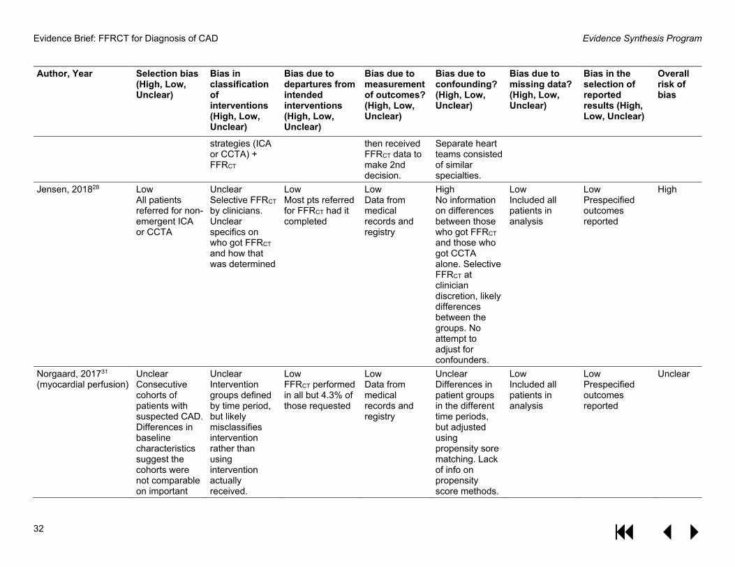
Evidence Brief: FFRCT for Diagnosis of CAD Evidence Synthesis Program
32
Author, Year Selection bias (High, Low, Unclear)
Bias in classification of interventions (High, Low, Unclear)
Bias due to departures from intended interventions (High, Low, Unclear)
Bias due to measurement of outcomes? (High, Low, Unclear)
Bias due to confounding? (High, Low, Unclear)
Bias due to missing data? (High, Low, Unclear)
Bias in the selection of reported results (High, Low, Unclear)
Overall risk of bias
strategies (ICA or CCTA) + FFRCT
then received FFRCT data to make 2nd decision.
Separate heart teams consisted of similar specialties.
Jensen, 201828 Low All patients referred for non-emergent ICA or CCTA
Unclear Selective FFRCT by clinicians. Unclear specifics on who got FFRCT and how that was determined
Low Most pts referred for FFRCT had it completed
Low Data from medical records and registry
High No information on differences between those who got FFRCT and those who got CCTA alone. Selective FFRCT at clinician discretion, likely differences between the groups. No attempt to adjust for confounders.
Low Included all patients in analysis
Low Prespecified outcomes reported
High
Norgaard, 201731 (myocardial perfusion)
Unclear Consecutive cohorts of patients with suspected CAD. Differences in baseline characteristics suggest the cohorts were not comparable on important
Unclear Intervention groups defined by time period, but likely misclassifies intervention rather than using intervention actually received.
Low FFRCT performed in all but 4.3% of those requested
Low Data from medical records and registry
Unclear Differences in patient groups in the different time periods, but adjusted using propensity sore matching. Lack of info on propensity score methods.
Low Included all patients in analysis
Low Prespecified outcomes reported
Unclear

Evidence Brief: FFRCT for Diagnosis of CAD Evidence Synthesis Program
33
Author, Year Selection bias (High, Low, Unclear)
Bias in classification of interventions (High, Low, Unclear)
Bias due to departures from intended interventions (High, Low, Unclear)
Bias due to measurement of outcomes? (High, Low, Unclear)
Bias due to confounding? (High, Low, Unclear)
Bias due to missing data? (High, Low, Unclear)
Bias in the selection of reported results (High, Low, Unclear)
Overall risk of bias
factors (type of angina, risk score, etc)
PLATFORM Douglas, 201519/201620; Hlatky, 201521; Colleran, 201718
Unclear Consecutive selection of participants into 2 cohorts, cohorts differed significantly by age, race, and other cardiac factors. Unclear if these differences were due to refusals to participate or other factors. Follow-up from study entry
Low Intervention groups clearly defined prior to measurement of outcomes.
Low All patients received planned usual care or CTA. 10-12% of requested FFRCT could not be completed
Low ICA determined by independent core laboratory. MACE data adjudicated by independent committee
Unclear Several baseline characteristics that differed between intervention groups were left out of propensity score model.
Low 95-100% follow-up. All participants included in analyses.
Low Prespecified outcomes reported
Unclear
Rabbat, 202032 High Consecutive patients referred to CCTA and FFRCT testing. Historical cohort with no info on selection. Baseline differences suggest
Low Intervention groups defined by test received
Low FFRCT available/possible for 92% of intervention group.
Unclear Clinical endpoints recorded in medical records. Intervention and control pts defined by change in hospital screening
High Important differences in baseline characteristics. No attempt to adjustment for confounding
Low Low level of missing data
Low Prespecified outcomes reported
High
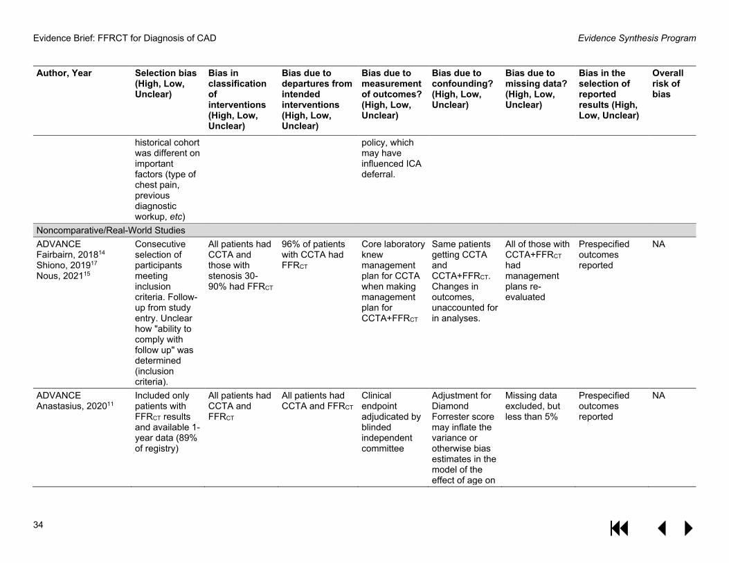
Evidence Brief: FFRCT for Diagnosis of CAD Evidence Synthesis Program
34
Author, Year Selection bias (High, Low, Unclear)
Bias in classification of interventions (High, Low, Unclear)
Bias due to departures from intended interventions (High, Low, Unclear)
Bias due to measurement of outcomes? (High, Low, Unclear)
Bias due to confounding? (High, Low, Unclear)
Bias due to missing data? (High, Low, Unclear)
Bias in the selection of reported results (High, Low, Unclear)
Overall risk of bias
historical cohort was different on important factors (type of chest pain, previous diagnostic workup, etc)
policy, which may have influenced ICA deferral.
Noncomparative/Real-World Studies ADVANCE Fairbairn, 201814 Shiono, 201917 Nous, 202115
Consecutive selection of participants meeting inclusion criteria. Follow-up from study entry. Unclear how "ability to comply with follow up" was determined (inclusion criteria).
All patients had CCTA and those with stenosis 30-90% had FFRCT
96% of patients with CCTA had FFRCT
Core laboratory knew management plan for CCTA when making management plan for CCTA+FFRCT
Same patients getting CCTA and CCTA+FFRCT. Changes in outcomes, unaccounted for in analyses.
All of those with CCTA+FFRCT had management plans re-evaluated
Prespecified outcomes reported
NA
ADVANCE Anastasius, 202011
Included only patients with FFRCT results and available 1-year data (89% of registry)
All patients had CCTA and FFRCT
All patients had CCTA and FFRCT
Clinical endpoint adjudicated by blinded independent committee
Adjustment for Diamond Forrester score may inflate the variance or otherwise bias estimates in the model of the effect of age on
Missing data excluded, but less than 5%
Prespecified outcomes reported
NA

Evidence Brief: FFRCT for Diagnosis of CAD Evidence Synthesis Program
35
Author, Year Selection bias (High, Low, Unclear)
Bias in classification of interventions (High, Low, Unclear)
Bias due to departures from intended interventions (High, Low, Unclear)
Bias due to measurement of outcomes? (High, Low, Unclear)
Bias due to confounding? (High, Low, Unclear)
Bias due to missing data? (High, Low, Unclear)
Bias in the selection of reported results (High, Low, Unclear)
Overall risk of bias
clinical outcomes in the cohort, as age is 1 of only 3 predictors used to calculate the score.
ADVANCE Fairbairn, 202012 Patel, 202013
Patients with FFRCT data from ADVANCE registry (excluded 6.8%). Unclear how "ability to comply with follow up" was determined (inclusion criteria).
All patients had CCTA and FFRCT
All patients had CCTA and FFRCT
Core laboratory knew management plan for CCTA when making management plan for CCTA+FFRCT. Clinical endpoint adjudicated by blinded independent committee
Adjusted for FFRCT only
90.% with data at 1 year, missing data excluded from MACE analysis
Prespecified outcomes reported
NA
ADVANCE Pontone, 201916 (rejection rate)
Only included 55% of ADVANCE registry in order to use only those with FFRCT 2.0 or greater
All pts had CCTA, classified based on rejection status
All patients had CCTA
Rejection data recorded by HeartFlow tech's blinded to clinical data
Looking for factors associated with rejection of CCTA, models adjusted for confounders
Missing data excluded, multivariable analysis only included 60% of cases, but not analysis of interest.
Prespecified outcomes reported
NA
Baggiano, 202023 Consecutive cohorts of patients with
All pts Received CCTA + FFRCT and either
FFRCT available in 96% of patients
CCTA, stress CTP, FFRCT analyses
Unclear if consecutive cohorts were
Overall evaluability of CCTA+FFRCT
Prespecified outcomes reported
NA

Evidence Brief: FFRCT for Diagnosis of CAD Evidence Synthesis Program
36
Author, Year Selection bias (High, Low, Unclear)
Bias in classification of interventions (High, Low, Unclear)
Bias due to departures from intended interventions (High, Low, Unclear)
Bias due to measurement of outcomes? (High, Low, Unclear)
Bias due to confounding? (High, Low, Unclear)
Bias due to missing data? (High, Low, Unclear)
Bias in the selection of reported results (High, Low, Unclear)
Overall risk of bias
suspected CAD referred for ICA. Unclear if there were any differences in the selection between dynamic and static stress CTP
dynamic or stress CTP (defined by original study cohort).
blinded to other test results.
similar receiving static and dynamic stress CTP (lumped together for analyses). Unclear if those unable to be analyzed were different than those analyzed
was 89%. Missing FFRCT were excluded.
Curzen, 201624 Patients from NXT trial with stable chest pain undergoing elective ICA
Intervention groups defined by test received
Appears all patients had FFRCT
Same cardiologists made plan with CCTA and FFRCT data, had knowledge of initial plan
Unclear time trends in outcomes.
Appears all patients had data for clinical management.
Prespecified outcomes reported
NA
Fares, 201925 Patients referred to FFRCT testing
Intervention groups defined by test received
21% of all FFRCTs were rejected
CCTA images interpreted after obtaining FFRCT results, readers blinded but also says CTAs were read "after receiving FFRCT result"
All patients received all diagnostic strategies, but results did not adjust for time trend in improvement of image adequacy
Appear to have outcome data for all FFRCT results, but unclear handling of those with missing FFRCTs.
Prespecified outcomes reported
NA
Norgaard, 201730 (clinical use)
All patients referred for CCTA during
Intervention groups defined by test received
FFRCT was available in 98% of those in whom it was requested.
Data from medical records and registry
Differences in symptoms and stenosis between
<10% missing outcome data for treatment plan, unclear
Prespecified outcomes reported
NA

Evidence Brief: FFRCT for Diagnosis of CAD Evidence Synthesis Program
37
Author, Year Selection bias (High, Low, Unclear)
Bias in classification of interventions (High, Low, Unclear)
Bias due to departures from intended interventions (High, Low, Unclear)
Bias due to measurement of outcomes? (High, Low, Unclear)
Bias due to confounding? (High, Low, Unclear)
Bias due to missing data? (High, Low, Unclear)
Bias in the selection of reported results (High, Low, Unclear)
Overall risk of bias
specific time period
different strategy groups, but only interested in outcomes in FFRCT group.
level and handling of missing data for MACE
Van Belle, 202133 Limited information on how patients were selected into study
Intervention groups defined by test received
Unclear if any CCTAs were rejected for FFRCT analysis
Same cardiologists made plan with CCTA and FFRCT data, had knowledge of initial plan
No information on patient demographics.
Unclear level and handling of missing data
Prespecified outcomes reported
NA
Abbreviations: CAD=coronary artery disease; CCTA=coronary computed tomography angiography; CTA=coronary computed tomography angiogram; CTP=computed tomography perfusion; FFRCT=fractional flow reserve using computed tomography; ICA=invasive coronary angiography; MACE=major adverse cardiovascular event; ROBINS-I=risk of bias in non-randomized studies of interventions
Quality Assessment of Case Series Using Murad et al
Author, Year
Do the patients represent the whole experience of the investigator or center? (Yes, No, Unclear)
Was the exposure adequately ascertained? (Yes, No, Unclear)
Was the outcome adequately ascertained? (Yes, No, Unclear)
Were other alternative causes that may explain the observation ruled out? (Yes, No, Unclear)
Was there a challenge/re-challenge phenomenon? (Yes, No, Unclear)
Was there a dose-response effect? (Yes, No, Unclear)
Was follow-up long enough for outcomes to occur? (Yes, No, Unclear)
Are the cases described with sufficient details? (Yes, No, Unclear
Overall bias (High, Low, Unclear)
Ihdayhid, 201926
No
Only 57% of initial
Yes
All included patients
Yes
Clinical events obtained from
Unclear
Other testing/ diagnosis
No No Yes Yes
All pts from NXT trial,
Fair

Evidence Brief: FFRCT for Diagnosis of CAD Evidence Synthesis Program
38
Author, Year
Do the patients represent the whole experience of the investigator or center? (Yes, No, Unclear)
Was the exposure adequately ascertained? (Yes, No, Unclear)
Was the outcome adequately ascertained? (Yes, No, Unclear)
Were other alternative causes that may explain the observation ruled out? (Yes, No, Unclear)
Was there a challenge/re-challenge phenomenon? (Yes, No, Unclear)
Was there a dose-response effect? (Yes, No, Unclear)
Was follow-up long enough for outcomes to occur? (Yes, No, Unclear)
Are the cases described with sufficient details? (Yes, No, Unclear
Overall bias (High, Low, Unclear)
population included: 13% rejected by FFRCT core laboratory, 10% incomplete dataset, 20% site declined involvement
underwent FFRCT, with data obtained from NXT trial
medical record review or interview (unclear if with pt or corresponding site/hospital). Events adjudicated by physicians blinded to FFRCT results.
strategies not reported; unclear if any pts sought them
which has publications describing methods
Norgaard, 202029
Yes
96% of those with FFRCT prescribed were included. Only included those with available FFRCT data and CAC score
Yes
All included patients underwent FFRCT, data obtained from national registry
Yes
Patient data obtained from national registry
Unclear
Adjusted for presence of stenosis >50%, but not other variables. Did not rule out potential time trend.
No No Yes
Median 2.2 years follow-up
Yes
Patients from national registry evaluated for CAD by CCTA with FFRCT
Good
Abbreviations: CAC=coronary artery calcium; CAD=coronary artery disease; CCTA=coronary computed tomography angiography; FFRCT=fractional flow reserve computed tomography

Evidence Brief: FFRCT for Diagnosis of CAD Evidence Synthesis Program
39
STRENGTH OF EVIDENCE OF INCLUDED STUDIES Outcome Studies Study
limitations Directness Consistency Precision Reporting
bias Summary of evidence
Diagnostic Accuracy (AUC)
3 SRs1-3 7 primary studies4-
10
Low (3 Low RoB SRs) to
Medium (7 unclear to high RoB studies)
Direct Consistent Precise Not detected
Several systematic reviews with low study limitations and narrow CIs reported consistently good diagnostic accuracy HeartFlow FFRCT, (AUC) range 0.87 to 0.89. Several primary studies published since the systematic reviews with medium study limitations generally supported these findings but had a broader range of estimates (AUC range 0.82 to 0.94). Moderate SOE
ICA Use
2 cohorts19,31 Medium (cohort with unclear RoB)
Indirect Inconsistent Imprecise Not detected
Compared to other noninvasive strategies HeartFlow FFRCT may increase ICA use compared to other noninvasive testing strategies. Two cohorts with medium study limitations, wide CIs, and limited comparisons to specific noninvasive testing strategies reported conflicting findings. Low SOE
1 cohort18,19 Medium (cohort with unclear RoB)
Direct Unknown Imprecise Not detected
Compared to direct referral to ICA HeartFlow FFRCT may reduce ICA use compared to planned ICA. A single cohort with medium study limitations and a wide CI directly compared HeartFlow FFRCT to planned ICA. Low SOE
1 cohort32 High (cohort with high RoB)
Direct Unknown Unknown Not detected
Compared to CCTA alone It is unclear whether HeartFlow FFRCT reduces ICA use compared to CCTA alone. A single cohort with high study limitations and unknown precision directly compared HeartFlow FFRCT to CCTA alone. Insufficient SOE
Change in Treatment Plan
3 cohorts22,28,30 and 6 case series14,17,23-
25,27,33
Medium (cohorts and case series
Direct Inconsistent Precise Not detected
HeartFlow FFRCT may change treatment plans in up to 70% of patients compared to CCTA alone (48% to 91% ICA cancellation). Several cohorts and case series comparing treatment

Evidence Brief: FFRCT for Diagnosis of CAD Evidence Synthesis Program
40
with unclear to high RoB)
plans between HeartFlow FFRCT and CCTA with medium study limitations and mostly narrow CIs reported a wide range of estimates of changes in treatment plans. Moderate SOE
MACE 3 cohorts19,20,28,30 and 4 case series11,13,14,17,26,27,29
Medium (cohorts and case series with unclear RoB)
Direct Consistent Unknown Not detected
MACE events may be very low (<1%) at 90 days to 1 year in patients receiving HeartFlow FFRCT and may increase in the longer-term (9.7% at ~5 years). Several cohorts and case series examining adverse clinical events with medium study limitations and unknown precision reported generally consistent findings. Low SOE
Abbreviations: AUC=area under curve; CCTA=coronary computed tomography angiography; FFRCT=fractional flow reserve using computed tomography; ICA=invasive coronary angiography; MACE=major adverse cardiovascular events; RoB=risk of bias; SOE=strength of evidence; SR=systematic review
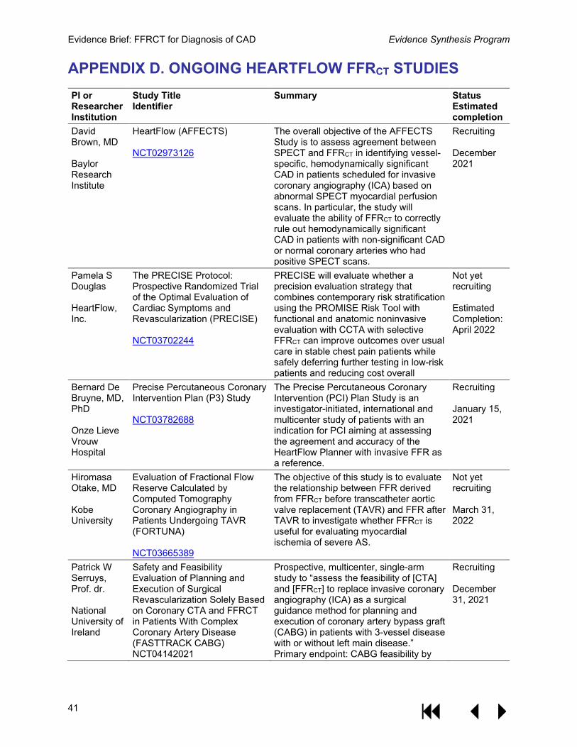
Evidence Brief: FFRCT for Diagnosis of CAD Evidence Synthesis Program
41
APPENDIX D. ONGOING HEARTFLOW FFRCT STUDIES PI or Researcher Institution
Study Title Identifier
Summary Status Estimated completion
David Brown, MD Baylor Research Institute
HeartFlow (AFFECTS) NCT02973126
The overall objective of the AFFECTS Study is to assess agreement between SPECT and FFRCT in identifying vessel-specific, hemodynamically significant CAD in patients scheduled for invasive coronary angiography (ICA) based on abnormal SPECT myocardial perfusion scans. In particular, the study will evaluate the ability of FFRCT to correctly rule out hemodynamically significant CAD in patients with non-significant CAD or normal coronary arteries who had positive SPECT scans.
Recruiting December 2021
Pamela S Douglas HeartFlow, Inc.
The PRECISE Protocol: Prospective Randomized Trial of the Optimal Evaluation of Cardiac Symptoms and Revascularization (PRECISE) NCT03702244
PRECISE will evaluate whether a precision evaluation strategy that combines contemporary risk stratification using the PROMISE Risk Tool with functional and anatomic noninvasive evaluation with CCTA with selective FFRCT can improve outcomes over usual care in stable chest pain patients while safely deferring further testing in low-risk patients and reducing cost overall
Not yet recruiting Estimated Completion: April 2022
Bernard De Bruyne, MD, PhD Onze Lieve Vrouw Hospital
Precise Percutaneous Coronary Intervention Plan (P3) Study NCT03782688
The Precise Percutaneous Coronary Intervention (PCI) Plan Study is an investigator-initiated, international and multicenter study of patients with an indication for PCI aiming at assessing the agreement and accuracy of the HeartFlow Planner with invasive FFR as a reference.
Recruiting January 15, 2021
Hiromasa Otake, MD Kobe University
Evaluation of Fractional Flow Reserve Calculated by Computed Tomography Coronary Angiography in Patients Undergoing TAVR (FORTUNA) NCT03665389
The objective of this study is to evaluate the relationship between FFR derived from FFRCT before transcatheter aortic valve replacement (TAVR) and FFR after TAVR to investigate whether FFRCT is useful for evaluating myocardial ischemia of severe AS.
Not yet recruiting March 31, 2022
Patrick W Serruys, Prof. dr. National University of Ireland
Safety and Feasibility Evaluation of Planning and Execution of Surgical Revascularization Solely Based on Coronary CTA and FFRCT in Patients With Complex Coronary Artery Disease (FASTTRACK CABG) NCT04142021
Prospective, multicenter, single-arm study to “assess the feasibility of [CTA] and [FFRCT] to replace invasive coronary angiography (ICA) as a surgical guidance method for planning and execution of coronary artery bypass graft (CABG) in patients with 3-vessel disease with or without left main disease.” Primary endpoint: CABG feasibility by
Recruiting December 31, 2021

Evidence Brief: FFRCT for Diagnosis of CAD Evidence Synthesis Program
42
CTA alone and occlusion rate for CTA-guided grafts at 1-month follow-up.
Nicolas van Mieghem Erasmus Medical Center
The Heartflow Coronary Disease Progression Evaluation Study (THRONE) NCT04052256
Prospective, single-center, single-arm study to “evaluate disease progression in intermediate lesions (invasive FFR 0.81-0.90 at baseline) using FFRCT at 2 years and determine whether CT characteristics may help to identify lesions that are more susceptible for FFR decline.”
Recruiting October 2023
Abbreviations: CABG= coronary artery bypass grafting; CAD=coronary artery disease; CCTA=coronary computed tomography angiography; FFR=fraction flow reserve; FFRCT=fractional flow reserve using computed tomography; ICA=invasive coronary angiography; PCI=percutaneous coronary intervention; SPECT=single-photon emission computed tomography; TAVR=transcatheter aortic valve replacement

Evidence Brief: FFRCT for Diagnosis of CAD Evidence Synthesis Program
43
APPENDIX E. DISPOSITION OF PEER REVIEWER COMMENTS Comment # Reviewer # Comment Author Response Are the objectives, scope, and methods for this review clearly described? 1 1 Yes None 2 2 Yes None 3 3 Yes None 4 4 Yes None 5 5 Yes None 6 6 Yes None Is there any indication of bias in our synthesis of the evidence? 7 1 No None 8 2 No None 9 3 No None 10 4 No None 11 5 No None 12 6 No None Are there any published or unpublished studies that we may have overlooked? 13 1 No None 14 2 No None 15 3 No None 16 4 No None 17 5 Yes - JAMA Netw Open. 2020;3(12):e2028312. We formally excluded this study as it uses data from
patients that have not received HeartFlow. As the implications and validity of this model are unclear, we have not discussed the study in the report.
18 6 No None Additional suggestions or comments can be provided below. If applicable, please indicate the page and line numbers from the draft report. 19 1 This is a very clear and excellently written review. I have
only two possible suggestions: 1) given the consistent evidence on higher specificity of CCTA-HF, and consistent findings from low quality studies, it seems one could state that it is likely that for patients undergoing CCTA, use of HF would modestly reduce the number referred for ICA. One could use the current VA data to calculate that impact. Given that only 20% are referred to ICA after CCTA, that number would likely be small.
We calculated the estimated number of ICAs prevented per 1000 ICAs in the previous ESP report. We have added a few sentences to the “Considerations for the Use of HeartFlow in VA” section on the findings from the calculation from the previous report.

Evidence Brief: FFRCT for Diagnosis of CAD Evidence Synthesis Program
44
20 1 2) I would more explicitly state the distinct types of research needed. Once could easily do a study of patients undergoing CCTA and randomize them to HF or not and measure change in ICA. More important given new recommendations would be to assess CT with HF as an alternative to other non-invasive strategies for moderate risk patients.
We have added a description of the specific types of research needed - controlled trials comparing CCTA alone to HeartFlow and HeartFlow to other noninvasive strategies to the executive summary and the future research section.
21 2 The clinical question addressed by the 2019 review was important and remains so. The earlier review was excellent and this update is a useful adjunct. I found no shortcomings in the methodology. It would probably be useful for general readers, to have a bit more description about how the technology functions and what is entailed.
We have added a bit more detail to the description of the HeartFlow technology. Readers are also referred to our earlier report for more detail.
22 2 In thinking about whether this technology could be adopted by VA it would be nice to have some information about relative cost.
We agree that information on relative cost could be useful, but we do not have ready access to this data and analysis of VA costs data was outside the scope of this report.
23 4 Page 21, line 4: The conclusion that "most patients receiving CCTA have normal or non-obstructive results and do not end up requiring ICA" is undoubtedly at least partially true, however a second viable explanation would be that the exam shows extensive coronary artery disease not amenable to ICA intervention. In my experience, this is not uncommon in the VA population, and is probably an equally valuable result by obviating the need for the patient to undergo an invasive procedure without significant therapeutic value. As I see it, one of the major roles of CCTA (with or without CT-FFR) should be to act as a gatekeeper to the cath lab, so that only those who will truly benefit will need an invasive (and relatively expensive) procedure.
We agree and have added that extensive coronary disease may also account for those not going on to ICA.
24 4 Page 21, line 16: correct term should be "CT technologist", not technician.
Corrected.
25 4 Page 25, line 44: correct title: Acting Director, VHA National Radiology Program
Corrected.

Evidence Brief: FFRCT for Diagnosis of CAD Evidence Synthesis Program
45
REFERENCES 1. Celeng C, Leiner T, Maurovich-Horvat P, et al. Anatomical and functional computed
tomography for diagnosing hemodynamically significant coronary artery disease: A meta-analysis. JACC: Cardiovascular Imaging. 2018:S1936-1878X(1918)30681-30688.
2. Hamon M, Geindreau D, Guittet L, Bauters C, Hamon M. Additional diagnostic value of new ct imaging techniques for the functional assessment of coronary artery disease: A meta-analysis. European Radiology. 2019;29(6):3044-3061.
3. Pontone G, Guaricci AI, Palmer SC, et al. Diagnostic performance of non-invasive imaging for stable coronary artery disease: A meta-analysis. Int J Cardiol. 2020;300:276-281.
4. Driessen RS, Danad I, Stuijfzand WJ, et al. Comparison of coronary computed tomography angiography, fractional flow reserve, and perfusion imaging for ischemia diagnosis. Journal of the American College of Cardiology. 2019;73(2):161-173.
5. Pontone G, Baggiano A, Andreini D, et al. Stress computed tomography perfusion versus fractional flow reserve ct derived in suspected coronary artery disease: The PERFECTION study. JACC: Cardiovascular Imaging. 2019;12(8 Pt 1):1487-1497.
6. Pontone G, Baggiano A, Andreini D, et al. Dynamic stress computed tomography perfusion with a whole-heart coverage scanner in addition to coronary computed tomography angiography and fractional flow reserve computed tomography derived. JACC: Cardiovascular Imaging. 2019;12(12):2460-2471.
7. Bom MJ, Driessen RS, Kurata A, et al. Diagnostic value of comprehensive on-site and off-site coronary ct angiography for identifying hemodynamically obstructive coronary artery disease. Journal of Cardiovascular Computed Tomography. 2021;15(1):37-45.
8. Cami E, Tagami T, Raff G, et al. Importance of measurement site on assessment of lesion-specific ischemia and diagnostic performance by coronary computed tomography angiography-derived fractional flow reserve. Journal of Cardiovascular Computed Tomography. 2020;29:29.
9. Ko BS, Linde JJ, Ihdayhid AR, et al. Non-invasive ct-derived fractional flow reserve and static rest and stress ct myocardial perfusion imaging for detection of haemodynamically significant coronary stenosis. The International Journal of Cardiovascular Imaging. 2019;35(11):2103-2112.
10. Tanigaki T, Emori H, Kawase Y, et al. Qfr versus FFR derived from computed tomography for functional assessment of coronary artery stenosis. JACC: Cardiovascular Interventions. 2019;12(20):2050-2059.
11. Anastasius M, Maggiore P, Huang A, et al. The clinical utility of FFR<sub>ct</sub> stratified by age. Journal of Cardiovascular Computed Tomography. 2020;23:23.
12. Fairbairn TA, Dobson R, Hurwitz-Koweek L, et al. Sex differences in coronary computed tomography angiography-derived fractional flow reserve: Lessons from advance. Jacc: Cardiovascular Imaging. 2020;13(12):2576-2587.
13. Patel MR, Norgaard BL, Fairbairn TA, et al. 1-year impact on medical practice and clinical outcomes of FFR<sub>ct</sub>: The advance registry. JACC: Cardiovascular Imaging. 2020;13(1 Pt 1):97-105.
14. Fairbairn TA, Nieman K, Akasaka T, et al. Real-world clinical utility and impact on clinical decision-making of coronary computed tomography angiography-derived

Evidence Brief: FFRCT for Diagnosis of CAD Evidence Synthesis Program
46
fractional flow reserve: Lessons from the advance registry. European Heart Journal. 2018;39(41):3701-3711.
15. Nous F, Budde RPJ, Fairbairn TA, et al. Temporal changes in FFR<sub>ct</sub>-guided management of coronary artery disease - lessons from the advance registry. Journal of Cardiovascular Computed Tomography. 2021;15(1):48-55.
16. Pontone G, Weir-McCall JR, Baggiano A, et al. Determinants of rejection rate for coronary ct angiography fractional flow reserve analysis. Radiology. 2019;292(3):597-605.
17. Shiono Y, Matsuo H, Kawasaki T, et al. Clinical impact of coronary computed tomography angiography-derived fractional flow reserve on japanese population in the advance registry. Circulation Journal. 2019;83(6):1293-1301.
18. Colleran R, Douglas PS, Hadamitzky M, et al. An FFRCT diagnostic strategy versus usual care in patients with suspected coronary artery disease planned for invasive coronary angiography at german sites: One-year results of a subgroup analysis of the platform (prospective longitudinal trial of FFRCT: Outcome and resource impacts) study. Open Heart. 2017;4(1).
19. Douglas PS, Pontone G, Hlatky MA, et al. Clinical outcomes of fractional flow reserve by computed tomographic angiography-guided diagnostic strategies vs Usual care in patients with suspected coronary artery disease: The prospective longitudinal trial of FFR(ct): Outcome and resource impacts study. European Heart Journal. 2015;36(47):3359-3367.
20. Douglas PS, De Bruyne B, Pontone G, et al. 1-year outcomes of FFRCT-guided care in patients with suspected coronary disease: The platform study. Journal of the American College of Cardiology. 2016;68(5):435-445.
21. Hlatky MA, De Bruyne B, Pontone G, et al. Quality-of-life and economic outcomes of assessing fractional flow reserve with computed tomography angiography: Platform. Journal of the American College of Cardiology. 2015;66(21):2315-2323.
22. Andreini D, Modolo R, Katagiri Y, et al. Impact of fractional flow reserve derived from coronary computed tomography angiography on heart team treatment decision-making in patients with multivessel coronary artery disease: Insights from the syntax iii revolution trial. Circulation: Cardiovascular Interventions. 2019;12(12):e007607.
23. Baggiano A, Fusini L, Del Torto A, et al. Sequential strategy including FFR<sub>ct</sub> plus stress-ctp impacts on management of patients with stable chest pain: The stress-ctp ripcord study. Journal of Clinical Medicine. 2020;9(7):08.
24. Curzen NP, Nolan J, Zaman AG, Norgaard BL, Rajani R. Does the routine availability of ct-derived FFR influence management of patients with stable chest pain compared to ct angiography alone?: The FFRCT ripcord study. JACC Cardiovascular Imaging. 2016;9(10):1188-1194.
25. Fares A, Alaiti MA, Alkhalil A, et al. Real world utilization of computed tomography derived fractional flow reserve: Single center experience in the united states. Cardiovascular Revascularization Medicine. 2019;20(12):1043-1047.
26. Ihdayhid AR, Norgaard BL, Gaur S, et al. Prognostic value and risk continuum of noninvasive fractional flow reserve derived from coronary ct angiography. Radiology. 2019;292(2):343-351.
27. Jang J, Krishnaswami A, Rogers C, Hung Y, Sengupta S. The impact of fractional flow reserve derived from coronary computed tomography angiography in a managed

Evidence Brief: FFRCT for Diagnosis of CAD Evidence Synthesis Program
47
healthcare system – kaiser permanente northern california [abstract #50]. Journal of Cardiovascular Computed Tomography. 2016;10(3):S1-S96.
28. Jensen JM, Bøtker HE, Mathiassen ON, et al. Computed tomography derived fractional flow reserve testing in stable patients with typical angina pectoris: Influence on downstream rate of invasive coronary angiography. European Heart Journal Cardiovascular Imaging. 2018;19(4):405-414.
29. Norgaard BL, Mortensen MB, Parner E, et al. Clinical outcomes following real-world computed tomography angiography-derived fractional flow reserve testing in chronic coronary syndrome patients with calcification. European heart journal cardiovascular Imaging. 2020;13:13.
30. Norgaard BL, Hjort J, Gaur S, et al. Clinical use of coronary cta-derived FFR for decision-making in stable cad. JACC: Cardiovascular Imaging. 2017;10(5):541-550.
31. Norgaard BL, Gormsen LC, Botker HE, et al. Myocardial perfusion imaging versus computed tomography angiography-derived fractional flow reserve testing in stable patients with intermediate-range coronary lesions: Influence on downstream diagnostic workflows and invasive angiography findings. Journal of the American Heart Association. 2017;6(8):22.
32. Rabbat M, Leipsic J, Bax J, et al. Fractional flow reserve derived from coronary computed tomography angiography safely defers invasive coronary angiography in patients with stable coronary artery disease. Journal of Clinical Medicine. 2020;9(2):24.
33. Van Belle E, Raposo L, Bravo Baptista S, et al. Impact of an interactive ct/FFR<sub>ct</sub> interventional planner on coronary artery disease management decision making. JACC: Cardiovascular Imaging. 2021;04:04.