evidence for vertical transmission of bacterial symbionts
TRANSCRIPT

APPLIED AND ENVIRONMENTAL MICROBIOLOGY, Oct. 2009, p. 6147–6156 Vol. 75, No. 190099-2240/09/$08.00�0 doi:10.1128/AEM.00023-09Copyright © 2009, American Society for Microbiology. All Rights Reserved.
Evidence for Vertical Transmission of Bacterial Symbionts from Adultto Embryo in the Caribbean Sponge Svenzea zeai�
On On Lee,1 Pui Yi Chui,1 Yue Him Wong,1 Joseph R. Pawlik,2 and Pei-Yuan Qian1*Department of Biology, Hong Kong University of Science and Technology, Clear Water Bay, Hong Kong,1 and Department of
Biology and Marine Biology and Center for Marine Science, University of North Carolina Wilmington,5600 Marvin K. Moss Lane, Wilmington, North Carolina 284092
Received 6 January 2009/Accepted 23 July 2009
The Caribbean reef sponge Svenzea zeai was previously found to contain substantial quantities of unicellularphotosynthetic and autotrophic microbes in its tissues, but the identities of these symbionts and their methodof transfer from adult to progeny are largely unknown. In this study, both a 16S rRNA gene-based finger-printing technique (denaturing gradient gel electrophoresis [DGGE]) and clone library analysis were appliedto compare the bacterial communities associated with adults and embryos of S. zeai to test the hypothesis ofvertical transfer across generations. In addition, the same techniques were applied to the bacterial communityfrom the seawater adjacent to adult sponges to test the hypothesis that water column bacteria could betransferred horizontally as sponge symbionts. Results of both DGGE and clone library analysis support thevertical transfer hypothesis in that the bacterial communities associated with sponge adults and embryos werehighly similar to each other but completely different from those in the surrounding seawater. Sequencing ofprominent DGGE bands and of clones from the libraries revealed that the bacterial communities associatedwith the sponge, whether adult or embryo, consisted of a large proportion of bacteria in the phyla Chloroflexiand Acidobacteria, while most of the sequences recovered from the community in the adjacent water columnbelonged to the class Alphaproteobacteria. Altogether, 21 monophyletic sequence clusters, comprising sequencesfrom both sponge adults and embryos but not from the seawater, were identified. More than half of thesponge-derived sequences fell into these clusters. Comparison of sequences recovered in this study with thosedeposited in GenBank revealed that more than 75% of S. zeai-derived sequences were closely related tosequences derived from other sponge species, but none of the sequences recovered from the seawater columnoverlapped with those from adults or embryos of S. zeai. In conclusion, there is strong evidence that a dominantproportion of sponge-specific bacteria present in the tissues of S. zeai are maintained through vertical transferduring embryogenesis rather than through acquisition from the environment (horizontal transfer).
Besides being the oldest metazoans, sponges are the sim-plest multicellular animals and possess a low degree of tissuedifferentiation and coordination (54). Sponges are sessile, fil-ter-feeding organisms that may harbor within their tissues aremarkable array of microorganisms, including bacteria (19,59, 64), archaea (41), zooxanthellae (22), diatoms (63), andfungi (35). In some cases, microbial consortia can make up to40 to 60% of the sponge tissue volume (21, 61) and exceed adensity of 109 microbial cells per ml of sponge tissue (62),which is several orders of magnitude higher than that found inseawater. Apart from being a source of food (43), bacterialsymbionts may participate in the acquisition and transfer ofnutrients inside sponges (67, 68), the recycling of insolubleprotein (69), the stabilization of the sponge skeleton (44), andthe processing of metabolic waste (4, 65). Many antimicrobialcompounds have been isolated from sponge bacterial symbi-onts (24, 47, 53), suggesting the involvement of symbiotic bac-teria in sponge chemical defenses. In some cases, bacterialsymbionts have been found to be the source of bioactive com-pounds that were isolated from sponges, which has opened up
new research directions in marine natural product chemistry,biotechnology, and pharmaceutical development (18, 23, 40).
Based on immunological evidence from the 1980s (66),sponge-bacterium symbioses are thought to have originated inthe Precambrian, when bacteria evolved to form a single cladeof sponge-specific bacteria that were distinct from isolatesfound in the surrounding seawater. Since then, many studieshave similarly documented a high level of consistency andspecificity in sponge-bacterium associations (20, 27, 59). Nev-ertheless, questions remain about the acquisition and mainte-nance of symbionts in host sponges. In general, the followingtwo hypotheses have been proposed: (i) a recently metamor-phosed sponge selectively retains specific groups of bacteriafrom the diverse pool of bacteria present in the water columnas it begins filter feeding (horizontal transfer) or (ii) specificbacterial strains are transmitted by the maternal sponge todeveloping embryos and are already present in the metamor-phosing sponge (vertical transfer) (58). The first hypothesisrequires some recognition of specific microbes by the sponge,perhaps through an innate immune system (36) or other meansto distinguish symbiont strains from food bacteria (70).
Vertical transfer of bacterial symbionts in sponges was firstproposed by Levi and Porte (29), who demonstrated the pres-ence of bacteria inside the larvae of the sponge Oscarella lobu-laris. Later, in 1976, Levi and Levi (30) studied the transmis-sion of bacteria in the sponge Chondrosia reniformis via sponge
* Corresponding author. Mailing address: Department of Biol-ogy, Hong Kong University of Science and Technology, Clear WaterBay, Hong Kong. Phone: (852) 2358-7331. Fax: (852) 2358-1559.E-mail: [email protected].
� Published ahead of print on 31 July 2009.
6147
at Univ of N
orth Carolina on A
pril 5, 2010 aem
.asm.org
Dow
nloaded from

oocytes. Since then, vertical transmission of bacterial symbi-onts via eggs or larvae has been documented for several spongespecies, including Tethya citrina (15), Geodia cydonium (50),Stelletta grubii (49), Hippospongia sp. (25), Spongia sp. (25),Halisarca dujardini (10), and Corticium candelabrum (8). How-ever, all of these studies employed transmission and scanningelectron microscopy and could only examine the presence ofbacteria in maternal sponges, oocytes, or larvae at the mor-phological level, with no determination of microbial identity.With advances in molecular techniques, Enticknap et al. (9)were the first to report the successful isolation of an alphapro-teobacterial symbiont, strain NW001, from both the adultsponge Mycale laxissima and its larvae. They also did a prelim-inary denaturing gradient gel electrophoresis (DGGE) analysisof the bacterial community in seawater and compared that withthe community in the sponge larval sample. However, such acomparison was not extended to the sponge adult, and no solidconclusion can be drawn for the horizontal transfer mechanismof sponge symbionts. More recently, Sharp et al. (52) usedfluorescence in situ hybridization (FISH) and clone librarytechniques to demonstrate the presence of proteobacteria, ac-tinobacteria, and a clade of sponge-associated bacteria in theembryos and mesohyl of the tropical sponge Corticium sp. Byclone library and DGGE analyses, Schmitt et al. (48a) identi-fied 28 vertical-transmission clusters in five different Caribbeansponge species and demonstrated that the complex sponge adultmicrobial community was collectively transmitted through repro-ductive stages. While these recent studies support the verticaltransfer hypothesis, they did not fully address the identities ofmicrobes in the water column surrounding the sponges, which iskey to determining whether horizontal transfer may also takeplace.
The Caribbean reef sponge Pseudaxinella zeai was reclassi-fied into a new genus, Svenzea (Demospongiae, Halichondria,Dictyonellidae), in 2002 because it has an unusual skeletonarrangement consisting mainly of short stout styles that arearranged in an isodictyal reticulation (2). It is a viviparoussponge that produces the largest embryos (�1 mm in diame-ter) and larvae (6 mm long) recorded for the phylum Porifera(45). Svenzea zeai has also been classified as a bacteriospongebecause it contains substantial amounts of unicellular photo-synthetic and autotrophic microbial symbionts in its tissues (2,45). Although bacteria were observed in the embryos and lar-vae of this sponge based on transmission electron microscopystudies (45), neither the direct linkage between the maternalsponge and the propagules nor the identity of the microbialsymbionts had been established.
In this study, our objective was to examine vertical versushorizontal transfer of bacterial symbionts in Svenzea zeai. Thiswas achieved by comparing the bacterial community profiles ofthe adults and embryos of the sponge by use of a combinationof molecular techniques, including DGGE and clone libraryanalysis. More than one technique was employed to compen-sate for deficiencies of each technique in revealing bacterialcommunity structure. Additionally, we used the same tech-niques to examine the bacterial community in the seawater thatsurrounded the sponge to determine whether horizontal trans-fer was evident.
MATERIALS AND METHODS
Sample collection and extraction of DNA. Tissue of the adult sponge Svenzeazeai was obtained at a depth of 12 m from San Salvador Island, Bahama Islands(24°03�N, 74°32�W), in June 2007. Three sponge individuals were carefullybrought to the water surface by scuba divers and flushed with autoclaved 0.22-�m-filtered seawater to remove loosely attached bacteria. The adult spongeswere dissected, and embryos in the adult tissues were carefully removed. Adultsponge tissue (0.5 ml) without any embryos was cut into small pieces and frozenin 0.8 ml of extraction buffer (100 mM Tris-HCl, 100 mM sodium EDTA, 100mM Na2HPO4, 1.5 M NaCl, 1% cetyltrimethylammonium bromide, pH 8). Theembryos removed from the adult tissue were first briefly washed with 70%ethanol and then thoroughly rinsed twice with autoclaved filtered seawater.Embryo mass (0.5 ml) removed from each of the three individual sponge adultswas homogenized and frozen in 0.8 ml of extraction buffer.
Bacterial communities in the surrounding seawater were collected in triplicateby filtering 1 liter of seawater onto 0.22-�m polycarbonate membranes (Osmon-ics). The membranes were then frozen in extraction buffer. The extraction andpurification of total bacterial DNA from the samples were performed followingthe sodium dodecyl sulfate-based method described by Liu et al. (31). PurifiedDNA was dissolved in 50 �l of double-distilled water (ddH2O) and kept at�20°C until use.
DNA fingerprinting analysis of bacterial communities. Bacterial communitystructure was revealed by DGGE (39). The 16S rRNA genes in the crude DNAextracts were amplified by PCR, using the universal primers 341F-GC (5�-CGCCCG CCG CGC CCC GCG CCC GTC CCG CCG CCC CCG CCC G CCTACG GGA GGC AGC AG) and 907R (5�-CCG TCA ATT CMT TTG AGTTT) (39). Each PCR mixture contained 2 �l of DNA template, 1.25 U of Taqpolymerase (Amersham Biosciences), a 0.25 mM concentration of each de-oxynucleoside triphosphate, 0.1 �M of each primer, and 1� PCR buffer in a totalvolume of 50 �l. PCR was performed in a thermal cycler (MJ Research) underthe following thermal conditions: initial denaturation at 95°C for 2 min; 10touchdown cycles of denaturation at 95°C for 1 min, annealing at 65°C (reducedto 55°C in increments of 1°C cycle�1) for 1 min, and extension at 72°C for 1 min;an additional 15 cycles with a constant annealing temperature of 55°C; and a finalextension at 72°C for 5 min. PCR products were mixed with loading buffer andloaded onto a 6% acrylamide gel with a denaturing gradient of 35 to 70% (100%denaturant � 7 M urea, 40% [vol/vol] formamide). Electrophoresis was per-formed using a D-Code system (Bio-Rad) with 1� TAE (20 mM Tris base, 10mM sodium acetate, and 0.5 mM EDTA) at a constant temperature of 60°C anda voltage of 125 V for 18 h. The gel was stained with 1� SYBR gold (MolecularProbes) for 15 min and photographed with an Alpha Imager 2200 gel documen-tation system (Alpha Innotech).
Sequencing analysis of DGGE bands. Major bands from the DGGE gel wereselected and excised from the gel for sequence analysis. Excised gel cubes werefirst washed with ddH2O and then immersed in 50 �l of ddH2O at 4°C overnight.Two microliters of DNA from each excised band was used as the template for thesame PCR-DGGE analysis to check for the band position and purity. PCRproducts were then purified, cloned into the pCR2.1-TOPO vector, and trans-formed into Escherichia coli competent cells by use of a Topo TA cloning kit(Invitrogen) according to the manufacturer’s manual. Transformants werescreened by blue-white selection on agar containing 5-bromo-4-chloro-3-indolyl-�-D-galactopyranoside (X-Gal)–isopropyl-�-D-thiogalactopyranoside (IPTG)and 100 �g ml�1 of ampicillin. White colonies were then transferred to freshplates and reincubated overnight. DNA was extracted from each positive cloneby picking a single white colony from the plate into 100 �l of ddH2O and lysingthe cells by heating at 99°C for 10 min. The lysates were used as DNA templatesfor subsequent PCR amplification, using the external vector primers M13F andM13R. Purified PCR products were then used as templates for cycle sequencingPCR, using either M13F or M13R primer and a DYEnamic ET dye terminatorkit (Amersham Biosciences). Cycle sequencing products were separated using aMegaBACE 500 genetic analyzer (Amersham Biosciences). The nucleotide se-quences obtained with the two primers were assembled using Sequencher 4.2(Gene Codes Corporation), and the assembled sequences were compared withsequences deposited in GenBank (http://www.ncbi.nlm.nih.gov/), using BLAST,to obtain their closest phylogenetic affiliations.
Clone library construction. Since the bacterial community structures for rep-licated samples were highly similar, as indicated by DGGE analysis, crude DNAextracts from the three replicates were pooled as templates for the constructionof clone libraries. The 16S rRNA genes in the crude DNA extracts were PCRamplified with the universal primers 8F (5�-AGA GTT TGA TCC TGG CTCAG) (3) and 1492R (5�-GGT TAC CTT GTT ACG ACT T) (28), using thefollowing PCR conditions: initial denaturation at 95°C for 5 min; 30 cycles of
6148 LEE ET AL. APPL. ENVIRON. MICROBIOL.
at Univ of N
orth Carolina on A
pril 5, 2010 aem
.asm.org
Dow
nloaded from

denaturation at 95°C for 1 min, annealing at 55°C for 1 min, and extension at72°C for 1 min; and a final extension at 72°C for 10 min. Successful PCRamplicons were purified using a PCR purification mini kit (Watson Biotechnol-ogies Inc., Shanghai, China). The quantities of purified PCR products weredetermined by GeneQuant (Amersham Biosciences). The same amounts ofpurified DNA from different samples were cloned into the pCR2.1-TOPO vectorand then transformed into E. coli competent cells by use of a Topo TA cloningkit (Invitrogen) according to the manufacturer’s manual. The same proceduresas those mentioned above were used for screening of positive clones, extractionof DNA, and subsequent PCR amplification. Aliquots (20 �l) of successful PCRamplicons were digested individually with two restriction enzymes (MspI andHaeIII) according to the manufacturer’s instructions (Invitrogen). Restrictionfragment length polymorphism (RFLP) patterns of each clone upon two differentrestriction enzyme digestions were obtained by electrophoresis on 3% agarosegels. Clones that showed the same RFLP patterns for the two enzymes wereclassified into the same operational taxonomic units (OTUs). One clone fromeach OTU was randomly selected and subjected to sequencing using vectorprimers M13F and M13R and internal primers 8F and 1492R. Sequencinganalysis was performed as mentioned above. Nearly the full lengths of bacterial16S rRNA gene sequences were obtained by assembling each fragment sequenceusing Sequencher 4.2 (Gene Codes Corporation). Chimera Check was used toexclude chimeras (34). The assembled sequences were then compared to theGenBank entries by using BLAST to obtain the closest phylogenetic affiliationfor each OTU. Phylogenetic analysis was performed with sequences retrieved foreach OTU in comparison with their closest affiliations, using ARB software (33).Phylogenetic trees were then constructed based on the neighbor-joining method(46), maximum parsimony (12), and the unweighted-pair group method usingaverage linkages (UPGMA) (56), using MEGA software (26).
Statistical analysis. Bacterial community structures, as revealed by DGGEband patterns, were compared among samples. Previous studies have suggestedthat major bands on DGGE gels represent the dominant bacterial speciespresent in the respective samples and that band intensity correlates with therelative abundance of the corresponding bacterial species within the sample (13,38). Therefore, each band in the DGGE gel was described by its position andrelative intensity in the profile, using GelCompar II software (Applied Maths).Band matching was performed with 1.00% position tolerance and 1.00% opti-mization. Similarity matrices were calculated based on the band position andintensity of each sample. Cluster analysis was performed based on the Pearsonsimilarity correlation, and dendrograms were constructed based on the Wardmethod, using GelCompar II software (Applied Maths).
For each clone library, the Chao estimator (7), the Shannon index (51), andthe coverage (37) were calculated. LIBSHUFF analysis was used to comparelibraries to determine if they were significantly different from each other (55). ALIBSHUFF comparison of three libraries yielded an experiment-wise critical Pvalue of 0.0085 according to the Bonferroni correction (http://libshuff.mib.uga
.edu). For each pairwise comparison, if the lower of the two P values calculatedby LIBSHUFF was less than or equal to the critical P value, then there was asignificant difference, with a confidence of 95%, in the composition of thecommunities sampled by each library.
Nucleotide sequence accession numbers. The 16S rRNA gene sequences ob-tained in this study were deposited in GenBank under the following accessionnumbers: FJ529257 to FJ529375.
RESULTS
DGGE analysis of bacterial communities. DGGE analysis ofthe bacterial communities associated with the adults and em-bryos of the sponge Svenzea zeai resulted in completely differ-ent band patterns in the DGGE gel than those associated withthe bacterial communities present in the water adjacent to theadult sponges (Fig. 1, right panel). The water column bacterialcommunities had an average of 17 bands that concentrated atthe upper area of the gel (where the denaturing concentrationof the gel was low). In contrast, the average numbers of bandsobserved for the adult- and embryo-associated bacterial com-munities were as high as 29 and 30, respectively, and the bandswere found mostly in the middle and lower parts of the gel(where the denaturant concentration was high). Cluster anal-ysis using a similarity matrix based on the band position andintensity confirmed the distinctiveness of the water columnbacterial community (Fig. 1, left panel). A distinct cluster wasformed with replicated samples from the water column, whichshared 0% similarity to the cluster formed from replicatedsamples from the sponge adults and embryos. In contrast, thesponge adult- and embryo-associated bacterial communitiesshared more than 50% similarity and formed a large clusterwhich consisted of two smaller clusters comprising the repli-cated samples from either the sponge adults or embryos.
Sequence analysis of excised DGGE bands. Altogether,12bands from the DGGE gel (2 from the water column samplesand 5 each from the sponge adults and embryos) were selected,excised, and sequenced. These bands were selected because oftheir prominence and uniqueness. Approximately 550 bp of
FIG. 1. DGGE band patterns (right) and dendrogram (left) showing the similarities of bacterial communities associated with the adults (A) andembryos (E) of the sponge Svenzea zeai and the planktonic bacterial community (W) in San Salvador, Bahama Islands. Labeled bands were excisedand sequenced. Details of the excised bands are given in Table 1.
VOL. 75, 2009 TRANSMISSION OF BACTERIAL SYMBIONTS IN SPONGES 6149
at Univ of N
orth Carolina on A
pril 5, 2010 aem
.asm.org
Dow
nloaded from

sequence was retrieved from each band and compared withnucleotide sequences deposited in GenBank. The closest phy-logenetic affiliation of each band sequence is listed in Table 1.All 12 sequences were affiliated with uncultured strains fromclones. The 2 sequences from the water samples (bands a andb) were closely related to the phylum Alphaproteobacteria,while the other 10 sequences, from the sponge adults (bands cto g) and embryos (bands h to l), belonged to the phyla Acidobac-teria and Chloroflexi. Bands excised from the same portions of thedenaturing gradient (i.e., bands c versus h, d versus i, e versus j, fversus k, and g versus l) shared the same closest matches. Theseresults indicate that strains belonging to the phylum Chloroflexicontributed large proportions of the bacterial communities asso-ciated with the adults and embryos of the sponge.
Clone library analysis. Altogether, 228 clones were re-trieved from the three clone libraries, 43 of which were fromthe seawater library, while 90 and 95 were from the spongeadult and embryo libraries, respectively. After RFLP screen-ing, 21, 53, and 44 OTUs were identified from the libraries ofseawater and sponge adults and embryos, respectively (Fig. 2).The Chao-1 estimator (7) and the Shannon index (H) wereused to estimate the species richness and to calculate the
diversity of the libraries, respectively. The higher the values,the richer and more diverse was the community. The resultsindicated that the sponge adults had the highest species rich-ness (Chao-1 � 125 25), followed by the sponge embryos(Chao-1 � 87 19), whereas the lowest species richness wasfound in the seawater (Chao-1 � 54 18; H � 2.71).
Nearly-full-length 16S rRNA gene sequences were obtainedfor each OTU and compared with sequences deposited inGenBank. Phylogenetic analysis of the 118 OTUs showed thatonly 3 of them (2.5%) were affiliated with sequences fromisolates, while the remaining 115 OTUs (97.5%) were closelyrelated to uncultured clones (Fig. 3). All clones from the sea-water library, except for one (W10), were affiliated with uncul-tured clones from different marine environments, includingseawater from Cocos Island, Monterey Bay, the Mediterra-nean Sea, and the Red Sea (Fig. 3). In contrast, among the 53and 44 OTUs retrieved from the sponge adult and embryolibraries, respectively, 42 and 33 of them (a total of 77%),respectively, were closely related to sequences retrieved fromother sponge species, including Plakortis sp., Corticium sp.,Theonella swinhoei, Agelas dilatata, and Aplysina aerophoba
TABLE 1. Closest phylogenetic affiliations of sequences retrieved from selected bands excised from DGGE gela
Band Source
Closest phylogenetic affiliation
Strain Source PhylumGenBankaccession
no.
Similarity(%)
a Water Uncultured Roseobacter sp. clone 2_C6 Coastal seawater from Ria de Vigo,Spain
Alphaproteobacteria EU600651 94
b Water Uncultured Rhodobacteriales bacteriumclone HF70_26K06
Seawater from Hawaii Ocean,North Pacific Subtropical Gyre
Alphaproteobacteria EU361404 94
c Adult Uncultured sponge symbiont PAUC37f Marine sponge Theonella swinhoeifrom the Western CarolineIslands in the Republic of Palau
Acidobacteria AF186413 97
d Adult Uncultured Chloroflexi bacterium clonePK067
Marine sponge Plakortis sp. fromLittle San Salvador Island,Bahamas
Chloroflexi EF076103 96
e Adult Uncultured Chloroflexi bacterium clonePK053
Marine sponge Plakortis sp. fromLittle San Salvador Island,Bahamas
Chloroflexi EF076110 95
f Adult Uncultured Chloroflexi bacterium clonePK017
Marine sponge Plakortis sp. fromLittle San Salvador Island,Bahamas
Chloroflexi EF076100 99
g Adult Uncultured Chloroflexi bacterium clonePK064
Marine sponge Plakortis sp. fromLittle San Salvador Island,Bahamas
Chloroflexi EF076111 96
h Embryo Uncultured sponge symbiont PAUC37f Marine sponge Theonella swinhoeifrom the Western CarolineIslands in the Republic of Palau
Acidobacteria AF186413 99
i Embryo Uncultured Chloroflexi bacterium clonePK067
Marine sponge Plakortis sp. fromLittle San Salvador Island,Bahamas
Chloroflexi EF076103 99
j Embryo Uncultured Chloroflexi bacterium clonePK053
Marine sponge Plakortis sp. fromLittle San Salvador Island,Bahamas
Chloroflexi EF076110 99
k Embryo Uncultured Chloroflexi bacterium clonePK017
Marine sponge Plakortis sp. fromLittle San Salvador Island,Bahamas
Chloroflexi EF076100 99
l Embryo Uncultured Chloroflexi bacterium clonePK064
Marine sponge Plakortis sp. fromLittle San Salvador Island,Bahamas
Chloroflexi EF076111 98
a The sequences of excised bands were compared to nucleotide sequences deposited in GenBank. The closest phylogenetic affiliation for the sequence from each bandis indicated by the strain name, phylum, accession number, and similarity. Refer to Fig. 1 for band positions.
6150 LEE ET AL. APPL. ENVIRON. MICROBIOL.
at Univ of N
orth Carolina on A
pril 5, 2010 aem
.asm.org
Dow
nloaded from

(Fig. 3). Only the remaining 23% were related to sequencesfrom other habitats, for instance, mangrove soil and seawater.
Of the 21 OTUs retrieved from the seawater library, 57%belonged to the phylum Alphaproteobacteria, 24% to Gammapro-teobacteria, 14% to Bacteroidetes, and 5% to Cyanobacteria (Fig.2). The sponge adult and embryo libraries comprised a widerrange of phyla than the seawater library did. Apart from theclones belonging to the phyla Alphaproteobacteria, Gammapro-teobacteria, and Bacteroidetes, clones closely related to six otherphyla were found (Deltaproteobacteria, Spirochaetes, Nitrospirae,Actinobacteria, Acidobacteria, and Chloroflexi) (Fig. 2). For boththe sponge adult and embryo libraries, the largest portion of theclones (�20% each) were affiliated with the phyla Chloroflexi andAcidobacteria, which were absent in the seawater library.
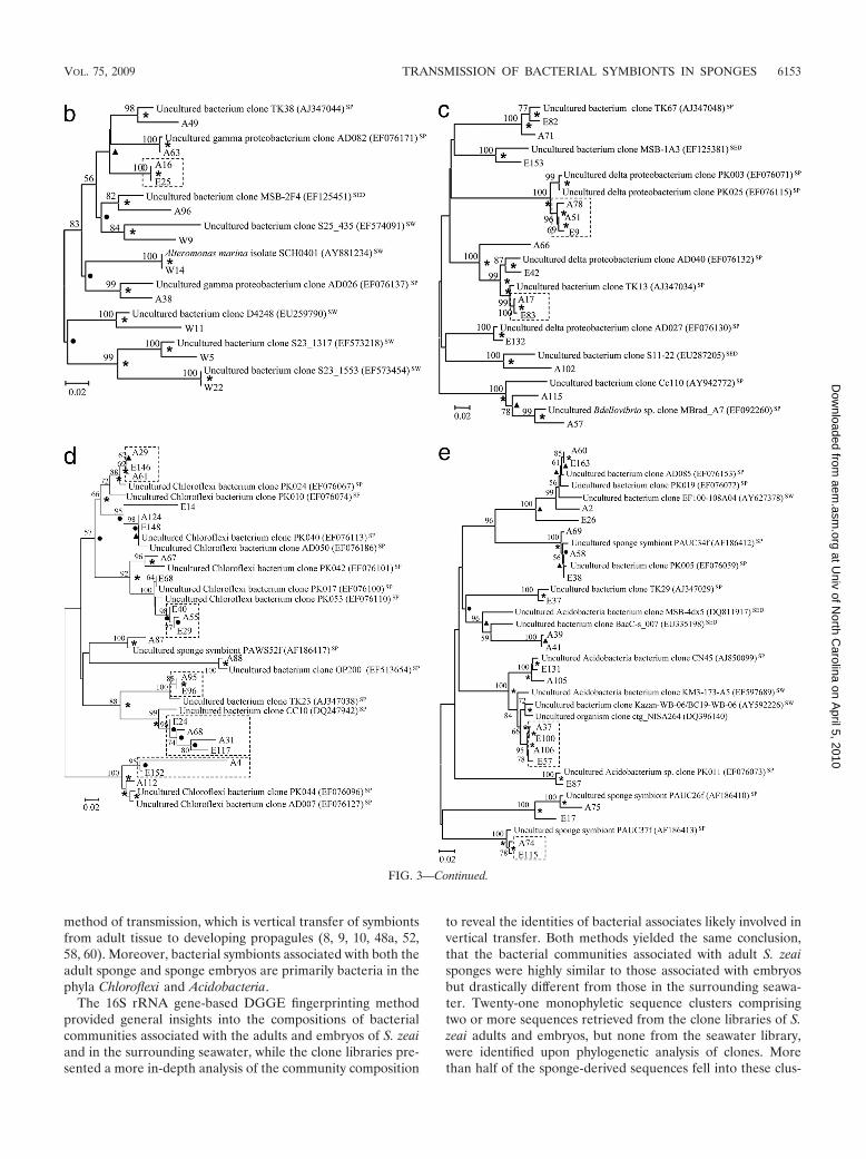
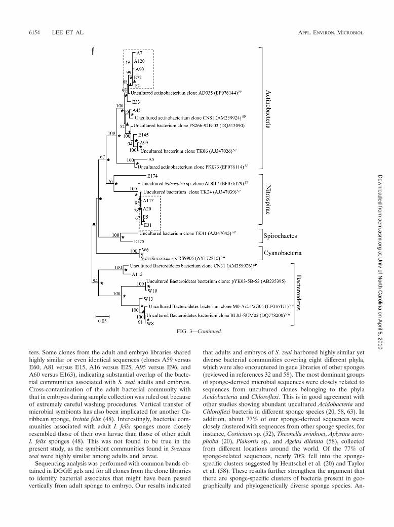
Figure 3 presents phylogenetic trees reflecting the geneticdistances among clones from different libraries with referenceto their closest relatives. For all phyla recovered in this study,except for Spirochaetes, Cyanobacteria, and Bacteroidetes, alarge proportion of clones from the sponge adults were clus-tered with those from the sponge embryos, showing extremelyhigh similarity in their 16S rRNA gene sequences. For in-stance, there was high similarity for clones A86 versus E140and A76 versus E70 in the Alphaproteobacteria (Fig. 3a), clonesA16 versus E25 in the Gammaproteobacteria (Fig. 3b), clonesA51 versus E9 in the Deltaproteobacteria (Fig. 3c), clones A29versus E146 and A124 versus E148 in the Chloroflexi (Fig. 3d),and clones A58 versus E38 and A74 versus E115 in theAcidobacteria (Fig. 3e). These highly similar clones were de-
fined as vertically transmitted phylotypes and formed 21 mono-phyletic sequence clusters (clusters of two or more sequencesretrieved from both the adult sponges and embryos but notfrom the seawater). More than half of the sequences derivedfrom the sponge adults and embryos fell into these clusters. Onthe other hand, clones from the seawater library normallyformed lineages distinct from those of the sponge libraries, asshown in the alphaproteobacterial (clones W2, W3, W12, W16,W19, W25, and W26) (Fig. 3a), gammaproteobacterial (clonesW5, W11, and W22) (Fig. 3b), and Bacteroidetes (clones W8,W10, and W13) (Fig. 3f) trees.
To determine the significance of the differences between theclone libraries based on available sequence data, LIBSHUFFanalysis was applied. Employing the Bonferroni correction, aLIBSHUFF comparison of three libraries yielded a critical Pvalue of 0.0085. LIBSHUFF analysis indicated that the spongeadult and embryo libraries were not significantly different fromeach other, as the lower of the two P values calculated fromLIBSHUFF analysis (P � 0.009) was larger than the critical Pvalue. In contrast, the seawater library was significantly differ-ent from the libraries of the sponge adults (lower P � 0.001)and the libraries of the sponge embryos (lower P � 0.001).
DISCUSSION
The Caribbean reef sponge Svenzea zeai was categorized asa bacteriosponge on the basis of transmission electron micros-copy observations because it contained substantial quantities
FIG. 2. 16S rRNA gene phylotype distribution and comparison of clone libraries from water samples and adults and embryos of the spongeSvenzea zeai. Species richness was estimated using the nonparametric Chao estimator (7). The Shannon index was calculated based on the numberof unique phylotypes detected by RFLP screening. Coverage of the library was expressed as a percentage and calculated from the equation C �1 � (n1/N), where n1 was the number of clones occurring only once in the library and N was the total number of clones examined (34).
VOL. 75, 2009 TRANSMISSION OF BACTERIAL SYMBIONTS IN SPONGES 6151
at Univ of N
orth Carolina on A
pril 5, 2010 aem
.asm.org
Dow
nloaded from

of unicellular photosynthetic and autotrophic bacteria (45).The method by which the very large embryos of Svenzea zeaiacquire their bacterial symbionts from the adult sponge re-mained unknown, as did the phylogenetic identities of thesymbionts. This study represents the only study on the associ-ated bacterial community in the sponge family Dictyonellidae
and is one of a few detailed studies combining different mo-lecular approaches to investigate vertical transmission as wellas horizontal transfer of sponge symbionts in the sponge byincorporating both the sponge embryo-associated and the in-digenous bacterioplankton communities for comparison. Theresults of our study corroborate the most commonly observed
FIG. 3. Phylogenetic trees showing genetic distances among clones retrieved from the libraries for seawater (prefixed with “W”), S. zeai adults(prefixed with “A”), and S. zeai embryos (prefixed with “E”) in reference to members of the Alphaproteobacteria (a), Gammaproteobacteria (b),Deltaproteobacteria (c), Chloroflexi (d), Acidobacteria (e), and Actinobacteria, Nitrospirae, Spirochaetes, Cyanobacteria, and Bacteroidetes (f). Thetrees were constructed based on the neighbor-joining method. Nodes which are observed in both maximum parsimony and UPGMA trees aremarked with asterisks, while those observed in either the maximum parsimony or UPGMA tree are marked with filled circles or triangles,respectively. Reference sequences originating from seawater, sediment, and sponges are indicated with the superscripts “SW,” “SED,” and “SP,”respectively. Nucleotide accession numbers of the reference sequences are given in parentheses. Dotted boxes show monophyletic clustersconsisting of sequences retrieved from both adult and embryo sponges but not from water. The scale bar represents percent substitutions pernucleotide position. Bootstrap values of �50% based on 1,000 resamplings are indicated by the numbers at the nodes.
6152 LEE ET AL. APPL. ENVIRON. MICROBIOL.
at Univ of N
orth Carolina on A
pril 5, 2010 aem
.asm.org
Dow
nloaded from
method of transmission, which is vertical transfer of symbiontsfrom adult tissue to developing propagules (8, 9, 10, 48a, 52,58, 60). Moreover, bacterial symbionts associated with both theadult sponge and sponge embryos are primarily bacteria in thephyla Chloroflexi and Acidobacteria.
The 16S rRNA gene-based DGGE fingerprinting methodprovided general insights into the compositions of bacterialcommunities associated with the adults and embryos of S. zeaiand in the surrounding seawater, while the clone libraries pre-sented a more in-depth analysis of the community composition
to reveal the identities of bacterial associates likely involved invertical transfer. Both methods yielded the same conclusion,that the bacterial communities associated with adult S. zeaisponges were highly similar to those associated with embryosbut drastically different from those in the surrounding seawa-ter. Twenty-one monophyletic sequence clusters comprisingtwo or more sequences retrieved from the clone libraries of S.zeai adults and embryos, but none from the seawater library,were identified upon phylogenetic analysis of clones. Morethan half of the sponge-derived sequences fell into these clus-
FIG. 3—Continued.
VOL. 75, 2009 TRANSMISSION OF BACTERIAL SYMBIONTS IN SPONGES 6153
at Univ of N
orth Carolina on A
pril 5, 2010 aem
.asm.org
Dow
nloaded from
ters. Some clones from the adult and embryo libraries sharedhighly similar or even identical sequences (clones A59 versusE60, A81 versus E15, A16 versus E25, A95 versus E96, andA60 versus E163), indicating substantial overlap of the bacte-rial communities associated with S. zeai adults and embryos.Cross-contamination of the adult bacterial community withthat in embryos during sample collection was ruled out becauseof extremely careful washing procedures. Vertical transfer ofmicrobial symbionts has also been implicated for another Ca-ribbean sponge, Ircinia felix (48). Interestingly, bacterial com-munities associated with adult I. felix sponges more closelyresembled those of their own larvae than those of other adultI. felix sponges (48). This was not found to be true in thepresent study, as the symbiont communities found in Svenzeazeai were highly similar among adults and larvae.
Sequencing analysis was performed with common bands ob-tained in DGGE gels and for all clones from the clone librariesto identify bacterial associates that might have been passedvertically from adult sponge to embryo. Our results indicated
that adults and embryos of S. zeai harbored highly similar yetdiverse bacterial communities covering eight different phyla,which were also encountered in gene libraries of other sponges(reviewed in references 32 and 58). The most dominant groupsof sponge-derived microbial sequences were closely related tosequences from uncultured clones belonging to the phylaAcidobacteria and Chloroflexi. This is in good agreement withother studies showing abundant uncultured Acidobacteria andChloroflexi bacteria in different sponge species (20, 58, 63). Inaddition, about 77% of our sponge-derived sequences wereclosely clustered with sequences from other sponge species, forinstance, Corticium sp. (52), Theonella swinhoei, Aplysina aero-phoba (20), Plakortis sp., and Agelas dilatata (58), collectedfrom different locations around the world. Of the 77% ofsponge-related sequences, nearly 70% fell into the sponge-specific clusters suggested by Hentschel et al. (20) and Tayloret al. (58). These results further strengthen the argument thatthere are sponge-specific clusters of bacteria present in geo-graphically and phylogenetically diverse sponge species. An-
FIG. 3—Continued.
6154 LEE ET AL. APPL. ENVIRON. MICROBIOL.
at Univ of N
orth Carolina on A
pril 5, 2010 aem
.asm.org
Dow
nloaded from

other discrepancy of the results obtained in the present studycompared with previous research was that the largest and best-known sponge-specific microbial clusters, “Candidatus Syn-echococcus spongiarum” (60) and “Poribacteria” (11), wereabsent from our sponge libraries. One possible reason may bedue to mismatches in the target region of the “Poribacteria”16S rRNA genes which make our universal primers not able toamplify them (11). Another possibility is that the abundance ofthese microbial groups may be too low to be recovered byclone library analysis. Sequencing of all minor bands in theDGGE gels was experimentally not feasible. In the future, ametagenomic approach may be a better way to reveal the mostcomplete picture of the sponge-associated microbial commu-nity. It would also be interesting to correlate the metabolicallyactive bacteria in the sponge and embryo tissues by using FISHor quantitative PCR so as to understand how they are trans-ferred between generations and their possible roles in hostsponges.
An important difference between this study and others inwhich the vertical transfer of microbial symbionts betweensponge adults and embryos has been implicated is the inclusionof a comparison with the indigenous bacterioplankton commu-nity. Analysis of the microbes in the water column adjacent toadult sponges allowed us to assess the possibility that horizon-tal transfer of symbionts was occurring. As indicated by theDGGE and clone library analyses, the bacterial communities inthe surrounding seawater were less diverse than and com-pletely different from the bacterial communities associatedwith S. zeai adults and embryos. Sequences belonging to thephyla Alpha- and Gammaproteobacteria dominated the bacte-rioplankton community, and most were closely related to se-quences recovered from the water column in geographicallydistinct locations, such as the Red Sea (14), the MediterraneanSea (1), the east coast of South Korea (5), Monterey Bay, andCocos Island. Distinct planktonic bacterial communities werealso recorded in many other studies (16, 17, 42). Hill (23)argued that considering the powerful filtration capacity ofsponges, with up to 2.4 � 1013 bacterial cells filtered per day,one might expect that even an extremely rare bacterium in thewater column could possibly be concentrated in a sponge tolevels detectable by PCR-based methods. As such, it would berisky to completely rule out the possibility that the bacterialsymbionts found in sponge samples can be present in seawatersamples, even though we did not detect them in the seawater inthis study. However, our observation that none of the bacteriadetected in the seawater samples resembled any of the bacteriarecovered from S. zeai adults or embryos strongly arguesagainst the horizontal transfer hypothesis. Therefore, environ-mental acquisition of bacterial associates is probably not animportant factor in maintaining the sponge-microbe associa-tion of S. zeai.
If vertical transmission of bacterial associates in sponges isthe rule, then the implication for the coevolution of spongesand sponge-associated microbes is considerable (21). However,the lack of a clearly resolved phylogeny of sponges (6) stillhampers our understanding of symbiont evolution in sponges.Although vertical transmission of Chloroflexi and Acidobacteriafrom adult to embryo was possible for S. zeai, the cellularmechanisms for the transfer remain to be explored. One tech-nique may be to design specific probes for in situ qualitative
and quantitative tracking of the presence of specific bacteriaduring different developmental stages of the sponge.
ACKNOWLEDGMENTS
We are grateful to the captain and crew of the R/V Seward Johnsonand to the Government of the Bahamas for allowing us to conductscientific research in their territorial waters.
This study was supported by grants from the China Ocean MineralResources Research and Development Association (COMRRDA06/07.SC02) and the KAUST Global Academic Partnership Program(KAUST005-CML07/08) to P.-Y. Qian and by a U.S. National ScienceFoundation Biological Oceanography Program grant to J. R. Pawlik(OCE-0550468).
REFERENCES
1. Alonso-Saez, L., V. Balague, E. L. Sa, O. Sanchez, J. M. Gonzalez, J.Pinhassi, R. Massana, J. Pernthaler, C. Pedros-Alio, and G. M. Gasol. 2007.Seasonality in bacterial diversity in north-west Mediterranean coastal waters:assessment through clone libraries, fingerprinting and FISH. FEMS Micro-biol. Ecol. 60:98–112.
2. Alvarez, B., R. W. M. van Soest, and K. Rutzler. 2002. Svenzea, a new genusof Dictyonellidae (Porifera: Demospongiae) from tropical reef environ-ments, with description of two new species. Contrib. Zool. 71:171–176.
3. Amann, R. I., W. Ludwig, and K. H. Schleifer. 1995. Phylogenetic identifi-cation and in situ detection of individual microbial cells without cultivation.Microbiol. Rev. 59:143–169.
4. Beer, S., and M. Ilan. 1998. In situ measurements of photosynthetic irradi-ance responses of two Red Sea sponges growing under dim light conditions.Mar. Biol. 131:613–617.
5. Bhattarai, H. D., Y. K. Lee, K. H. Cho, H. K. Lee, and H. W. Shin. 2006. Thestudy of antagonistic interactions among pelagic bacteria: a promising way tocoin environmental friendly antifouling compounds. Hydrobiologia 568:417–423.
6. Boury-Esnault, N. 2006. Systematics and evolution of Demospongiae. Can. J.Zool. 84:205–224.
7. Chao, A. 1984. Nonparametric estimation of the number of classes in apopulation. Scand. J. Stat. 11:265–270.
8. de Caralt, S., M. J. Uriz, and R. H. Wijffels. 2007. Vertical transmission andsuccessive location of symbiotic bacteria during embryo development andlarva formation in Corticium candelabrum (Porifera: Demospongiae). J. Mar.Biol. Assoc. U. K. 87:1693–1699.
9. Enticknap, J. J., M. Kelly, O. Peraud, and R. T. Hill. 2006. Characterizationof a culturable alphaproteobacterial symbiont common to many marinesponges and evidence for vertical transmission via sponge larvae. Appl.Environ. Microbiol. 72:3724–3732.
10. Ereskovsky, A. V., E. Gonobobleva, and A. Vishnyakov. 2005. Morphologicalevidence for vertical transmission of symbiotic bacteria in the viviparoussponge Halisarca dujardini Johnston (Porifera, Demospongiae, Halisarcida).Mar. Biol. 146:869–875.
11. Fieseler, L., M. Horn, M. Wagner, and U. Hentschel. 2004. Discovery of thenovel candidate phylum “Poribacteria” in marine sponges. Appl. Environ.Microbiol. 70:3724–3732.
12. Fitch, W. M. 1971. Toward defining the course of evolution: minimumchange for a specific tree topology. Syst. Zool. 20:406–416.
13. Fromin, N., J. Hamelin, S. Tarnawski, D. Roesti, K. Jourdain-Miserez, N.Forestier, S. Teyssier-Cuvelle, F. Gillet, M. Aragno, and P. Rossi. 2002.Statistical analysis of denaturing gel electrophoresis (DGE) fingerprintingpatterns. Environ. Microbiol. 4:634–643.
14. Fuller, N. J., D. Marie, F. Partensky, D. Vaulot, A. F. Post, and D. J. Scanlan.2003. Clade-specific 16S ribosomal DNA oligonucleotides reveal the pre-dominance of a single marine Synechococcus clade throughout a stratifiedwater column in the Red Sea. Appl. Environ. Microbiol. 69:2430–2443.
15. Gaino, E., B. Burlando, P. Puffa, and M. Sara. 1987. Ultrastructural study ofthe mature egg of Tethya citrina Sara and Melone (Porifera, Demospongiae).Gamete Res. 16:259–265.
16. Giovannoni, S. J., and M. S. Rappe. 2000. Evolution, diversity and molecularecology of marine prokaryotes, p. 47–84. In D. L. Kirchman (ed.), Microbialecology of the ocean. John Wiley & Sons, Inc., New York, NY.
17. Giovannoni, S. J., T. B. Britschgi, C. L. Moyer, and K. G. Field. 1990.Genetic diversity in Sargasso Sea bacterioplankton. Nature 345:60–63.
18. Haygood, M. G., E. W. Schmidt, S. K. Davidson, and D. J. Faulkner. 1999.Microbial symbionts of marine invertebrates: opportunities for microbialbiotechnology. J. Mol. Microbiol. Biotechnol. 1:33–43.
19. Hentschel, U., M. Schmid, M. Wanger, L. Fieseler, C. Gernert, and J.Hacker. 2001. Isolation and phylogenetic analysis of bacteria with antimi-crobial activities from the Mediterranean sponges Aplysina aerophoba andAplysina cavernicola. FEMS Microbiol. Ecol. 35:305–312.
20. Hentschel, U., J. Hopke, M. Horn, A. Friedrich, M. Wagner, J. Hacker, and
VOL. 75, 2009 TRANSMISSION OF BACTERIAL SYMBIONTS IN SPONGES 6155
at Univ of N
orth Carolina on A
pril 5, 2010 aem
.asm.org
Dow
nloaded from
B. Moore. 2002. Molecular evidence for a uniform microbial community insponges from different oceans. Appl. Environ. Microbiol. 68:4431–4440.
21. Hentschel, U., L. Fieseler, M. Wehrl, C. Gernert, M. Steinert, J. Hacker, andM. Horn. 2003. Microbial diversity of marine sponges. Prog. Mol. Subcell.Biol. 37:59–88.
22. Hill, M. S. 1996. Symbiotic zooxanthellae enhance boring and growth ratesof the tropical sponge Anthosigmella varians forma varians. Mar. Biol. 125:649–654.
23. Hill, R. T. 2004. Microbes from marine sponge: a treasure trove of biodi-versity of natural products discovery, p. 177–190. In A. T. Bull (ed.), Micro-bial diversity and bioprospecting. ASM Press, Washington, DC.
24. Jayatilake, G. S., M. P. Thornton, A. C. Leonard, J. E. Grimwade, and G. J.Baker. 1996. Metabolites from an Antarctic sponge associated bacteriumPseudomonas aeruginosa. J. Nat. Prod. 59:293–296.
25. Kaye, H. R. 1991. Sexual reproduction in four Caribbean commercialsponges. Oogenesis and transfer of bacterial symbionts. Invert. Reprod. Dev.19:13–24.
26. Kumar, S., K. Tamura, and M. Nei. 1994. MEGA: molecular evolutionarygenetics analysis software for microcomputers. Comput. Appl. Biosci. 10:189–191.
27. Lafi, F. F., M. J. Garson, and J. A. Fuerst. 2005. Culturable bacterialsymbionts isolated from two distinct sponge species (Pseudoceratina clavataand Rhabdastrella globostellata) from the Great Barrier Reef display similarphylogenetic diversity. Microb. Ecol. 50:213–220.
28. Lane, D. J. 1991. 16S/23S rRNA sequencing, p. 115–148. In E. Stackebrandtand M. Goodfellow (ed.), Nucleic acid techniques in bacterial systematics.John Wiley & Sons, Inc., New York, NY.
29. Levi, C., and A. Porte. 1962. Etude au microscope electronique de l’epongeOscarella lobularis Schmidt et de sa larve amphiblastula. Cah. Biol. Mar.3:307–315.
30. Levi, C., and P. Levi. 1976. Embryogenese de Chondrosia reniformis (Nardo),demosponge ovipare, et transmission des bacteries symbiotiques. Ann. Sci.Nat. Zool. Biol. Anim. 18:367–380.
31. Liu, W. T., T. L. Marsh, H. Cheng, and L. J. Forney. 1997. Characterizationof microbial diversity by determining terminal restriction fragment lengthpolymorphisms of genes encoding 16S rRNA. Appl. Environ. Microbiol.63:4516–4522.
32. Lopez, J., A. Ledger, C. Peterson, K. Sfanos, D. Harmody, S. Pomponi, andP. McCarthy. 2006. Molecular census and comparison of cultured and un-cultured microbial symbiont diversity from an ancient metazoan host, phy-lum Porifera, abstr. 6.15. Abstr. 5th Int. Symbiosis Soc. Congr., Vienna,Austria, 4 to 10 August 2006.
33. Ludwig, W., O. Strunk, R. Westram, et al. 2004. ARB: a software environ-ment for sequence data. Nucleic Acids Res. 32:1363–1371.
34. Maidak, B. L., J. R. Cole, T. G. Lilbum, C. T. Parker, P. R. Saxman, R. J.Farris, G. M. Garrity, G. J. Olsen, T. M. Schmidt, and J. M. Tiedje. 2001.The RDP-II (ribosomal database project). Nucleic Acids Res. 29:173–174.
35. Maldonado, M., N. Cortadellas, M. I. Trillas, and K. Rutzler. 2005. Endo-symbiotic yeast maternally transmitted in a marine sponge. Biol. Bull. 209:94–106.
36. Muller, W. E. G., and I. M. Muller. 2003. Origin of the metazoan immunesystem: identification of the molecules and their functions in sponges. Integr.Comp. Biol. 144:19–29.
37. Mullins, T. D., T. B. Britschgi, R. L. Krest, and S. J. Giovannoni. 1995.Genetic comparisons reveal the same unknown bacterial lineages in Atlanticand Pacific bacterioplankton communities. Limnol. Oceanogr. 40:148–158.
38. Murray, A. E., J. T. Hollibaugh, and C. Orrego. 1996. Phylogenetic compo-sitions of bacterioplankton from two California estuaries compared by de-naturing gradient gel electrophoresis of 16S rDNA fragments. Appl. Envi-ron. Microbiol. 62:2676–2680.
39. Muyzer, G., E. C. D. Waal, and A. G. Uitterlinden. 1993. Profiling of complexmicrobial populations by denaturing gradient gel electrophoresis analysis ofpolymerase chain reaction-amplified genes coding for 16S rRNA. Appl.Environ. Microbiol. 59:695–700.
40. Piel, J. 2006. Bacterial symbionts: prospects for the sustainable production ofinvertebrate-derived pharmaceuticals. Curr. Med. Chem. 13:39–50.
41. Preston, C. M., K. Y. Wu, T. F. Molinski, and E. F. DeLong. 1996. Apsychrophilic crenarchaeon inhabits a marine sponge: Cenarchaeum symbio-sum gen. nov., sp. nov. Proc. Natl. Acad. Sci. USA 93:6241–6246.
42. Rappe, M. S., K. Vergin, and S. J. Giovannoni. 2000. Phylogenetic compar-isons of a coastal bacterioplankton community with its counterparts in openocean and freshwater systems. FEMS Microbiol. Ecol. 33:219–232.
43. Reiswig, H. M. 1975. Bacteria as food for temperate-water marine sponges.Can. J. Zool. 53:493–502.
44. Rutzler, K. 1985. Associations between Caribbean sponges and photosyn-thetic organisms, p. 455–466. In K. Rutzler (ed.), New perspectives in spongebiology. Smithsonian Institution Press, Washington, DC.
45. Rutzler, K., R. W. M. van Soest, and B. Alvarez. 2003. Svenzea zeai, aCaribbean reef sponge with a giant larva, and Scopalina ruetzleri: a compar-ative fine structural approach to classification (Demospongiae, Halichondria,Dictyonellidae). Invert. Biol. 122:203–222.
46. Saitou, N., and M. Nei. 1987. The neighbor-joining method: a new methodfor reconstruction of phylogenetic trees. Mol. Biol. Evol. 4:406–425.
47. Schmidt, E. W., A. Y. Obraztsova, S. K. Davidson, D. J. Faulkner, and M. G.Haygood. 2000. Identification of the antifungal peptide-containing symbiontsof the marine sponge Theonella swinhoei as a novel -proteobacterium,“Candidatus Entotheonella palauensis.” Mar. Biol. 136:969–977.
48. Schmitt, S., J. B. Weisz, N. Lindquist, and U. Hentschel. 2007. Verticaltransmission of a phylogenetically complex microbial consortium in the vi-viparous sponge Ircinia felix. Appl. Environ. Microbiol. 73:2067–2078.
48a.Schmitt, S., H. Angermeier, R. Schiller, N. Lindquist, and U. Hentschel.2008. Molecular microbial diversity survey of sponge reproductive stages andmechanistic insights into vertical transmission of microbial symbionts. Appl.Environ. Microbiol. 74:7694–7708.
49. Sciscioli, M., L. S. Liaci, E. Lepore, M. Gherardi, and T. L. Simpson. 1991.Ultrastructural study of the mature egg of the marine sponge Stelletta grubii(Porifera, Demospongiae). Mol. Reprod. Dev. 28:346–350.
50. Sciscioli, M., E. Lepore, M. Gherardi, and L. S. Liaci. 1994. Transfer ofsymbiotic bacteria in the mature oocyte of Geodia cydonium (Porifera,Demospongiae): an ultrastructural study. Cah. Biol. Mar. 35:471–478.
51. Shannon, C. E. 1948. A mathematical theory of communication. Bell SystemTech. J. 27:379–423, 623–656.
52. Sharp, K. H., B. Eam, D. J. Faulkner, and M. G. Haygood. 2007. Verticaltransmission of diverse microbes in the tropical sponge Corticium sp. Appl.Environ. Microbiol. 73:622–629.
53. Shigemori, H., M. A. Bae, K. Yazava, T. Sasaki, and J. Kobayashi. 1992.Alteramida A, a new tetracyclic alkaloid from a bacterium Alteromonas sp.associated with the marine sponge Halichondria okadai. J. Org. Chem. 57:4317–4320.
54. Simpson, T. L. 1984. The cell biology of sponges. Springer-Verlag, NewYork, NY.
55. Singleton, D. R., M. A. Furlong, S. L. Rathbun, and W. B. Whitman. 2001.Quantitative comparisons of 16S rDNA gene sequence libraries from envi-ronmental samples. Appl. Environ. Microbiol. 67:4374–4376.
56. Sneath, P. H. A., and R. R. Sokal. 1973. Numerical taxonomy—the principlesand practice of numerical classification. W. H. Freeman, San Francisco, CA.
57. Reference deleted.58. Taylor, M. W., R. Radax, D. Steger, and M. Wagner. 2007. Sponge-associ-
ated microorganisms: evolution, ecology, and biotechnological potential. Mi-crobiol. Mol. Biol. Rev. 71:295–347.
59. Thacker, R. W., and S. Starnes. 2003. Host specificity of the symbioticcyanobacterium Oscillatoria spongeliae in marine sponge, Dysidea spp. Mar.Biol. 142:643–648.
60. Usher, K. M., S. Toze, J. Fromont, J. Kuo, and D. C. Sutton. 2004. A newspecies of cyanobacterial symbiont from the marine sponge, Chondrilla nu-cula. Symbiosis 36:183–192.
61. Vacelet, J., and C. Donadey. 1977. Electro microscope study of the associa-tion between sponges and bacteria. J. Exp. Mar. Biol. Ecol. 30:301–314.
62. Webster, N. S., and R. T. Hill. 2001. The culturable microbial community ofthe Great Barrier Reef sponge Rhopaloeides odorabile is dominated by analphaproteobacterium. Mar. Biol. 138:843–851.
63. Webster, N. S., K. J. Wilson, L. L. Blackall, and R. T. Hill. 2001. Phyloge-netic diversity of bacteria associated with the marine sponge Rhopaloeidesodorabile. Appl. Environ. Microbiol. 67:434–444.
64. Webster, N. S., A. P. Negri, M. M. Munro, and C. N. Battershill. 2004.Diverse microbial communities inhabit Antarctic sponges. Environ. Micro-biol. 6:288–300.
65. Wilkinson, C. R. 1978. Microbial associations in sponges. I. Ecology, physi-ology and microbial populations of coral reef sponges. Mar. Biol. 49:161–167.
66. Wilkinson, C. R. 1984. Immunological evidence for the Precambrian originof bacterial symbioses in marine sponges. Proc. R. Soc. Lond. B 220:509–517.
67. Wilkinson, C. R., and R. Garrone. 1980. Nutrition of marine sponges. In-volvement of symbiotic bacteria in the uptake of dissolved carbon, p. 157–161. In D. C. Smith and Y. Tiffon (ed.), Nutrition in the lower metazoan.Pergamon Press, Oxford, United Kingdom.
68. Wilkinson, C. R., and J. Vacelet. 1979. Transplantation of marine sponges todifferent conditions of light and current. J. Exp. Mar. Biol. Ecol. 37:91–104.
69. Wilkinson, C. R., R. Garrone, and D. Herbage. 1979. Sponge collagen deg-radation in vitro by sponge-specific bacteria. Colloq. Int. CNRS 291:361–364.
70. Wilkinson, C. R., R. Garrone, and J. Vacelet. 1984. Marine sponges discrim-inate between food bacteria and bacterial symbionts: electromicroscope ra-dioautography and in situ evidence. Proc. R. Soc. Lond. B 220:519–528.
6156 LEE ET AL. APPL. ENVIRON. MICROBIOL.
at Univ of N
orth Carolina on A
pril 5, 2010 aem
.asm.org
Dow
nloaded from