evidence from lesions: agnosia lesions (especially in the left hemisphere) of the inferior temporal...
TRANSCRIPT
Evidence from Lesions: Agnosia
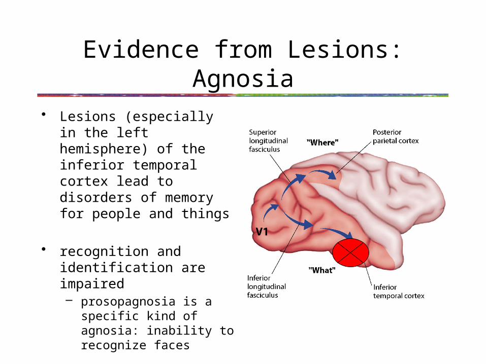
• Lesions (especially in the left hemisphere) of the inferior temporal cortex lead to disorders of memory for people and things
• recognition and identification are impaired– prosopagnosia is a specific
kind of agnosia: inability to recognize faces
• explicit (conscious) decisions about object features are disrupted
Ventral lesions can decouple awareness from action
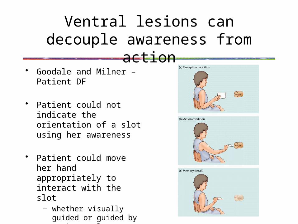
• Goodale and Milner – Patient DF
• Patient could not indicate the orientation of a slot using her awareness
• Patient could move her hand appropriately to interact with the slot
– whether visually guided or guided by an internal representation in memory
Ventral lesions can decouple awareness from action
• Single dissociation of action from conscious perception
• Dorsal pathway remained intact while ventral pathway was impaired
• Dorsal Pathway seems to guide motor actions, at least for ones that need spatial information
• Activity within the Dorsal Pathway seems not to be sufficient for consciousness
Lesions of “Retinostriate” Pathway
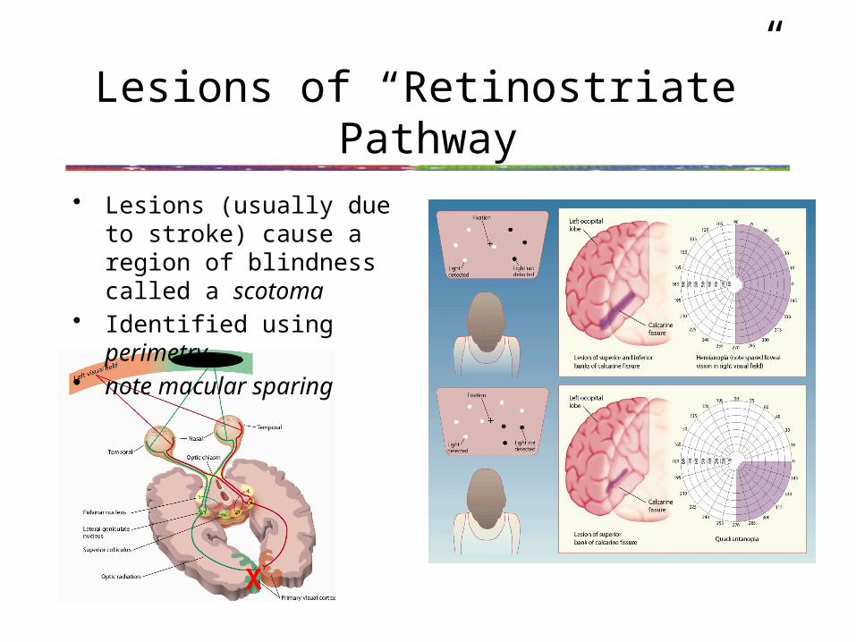
• Lesions (usually due to stroke) cause a region of blindness called a scotoma
• Identified using perimetry• note macular sparing
X
“Retinocollicular” Pathway independently mediates orienting
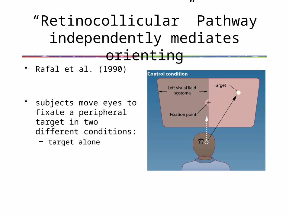
• Rafal et al. (1990)
• subjects move eyes to fixate a peripheral target in two different conditions:– target alone
Retinocollicular Pathway independently mediates orienting
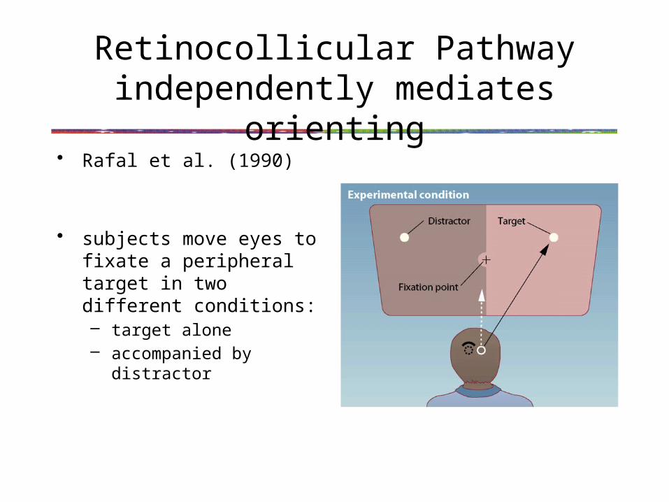
• Rafal et al. (1990)
• subjects move eyes to fixate a peripheral target in two different conditions:– target alone– accompanied by distractor

Retinocollicular Pathway independently mediates orienting
• Rafal et al. (1990) result
• Subjects were slower when presented with a distracting stimulus in the scotoma (359 ms vs. 500 ms)
• Residual vision in cortical blindness has been called “blindsight”
Retinocollicular Pathway independently mediates orienting
• Blindsight patients have since been shown to posses a surprising range of “residual” visual abilities– better than chance at detection and discrimination of some
visual features such as direction of motion
• These go beyond simple orienting - how can this be?

Retinocollicular Pathway independently mediates orienting
• Recall that the feed-forward sweep is not a single wave of information and that it doesn’t only go through V1
• In particular, MT seems to get very early and direct input
Retinocollicular Pathway independently mediates orienting
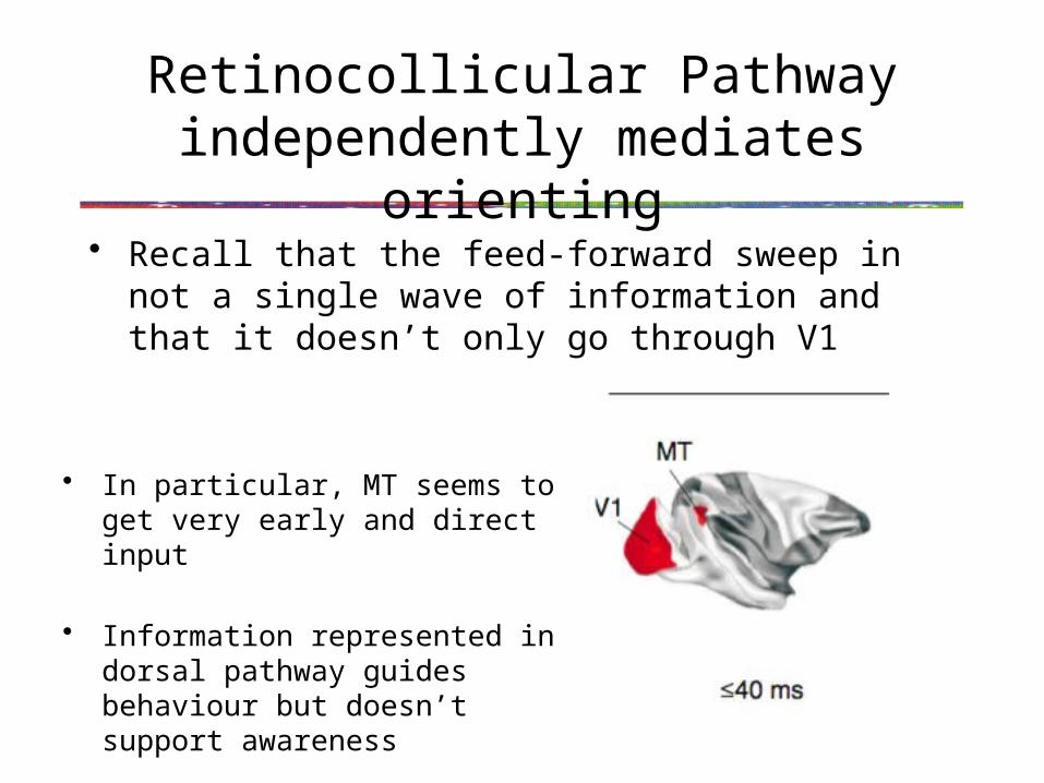
• Recall that the feed-forward sweep in not a single wave of information and that it doesn’t only go through V1
• In particular, MT seems to get very early and direct input
• Information represented in dorsal pathway guides behaviour but doesn’t support awareness