evidence - scottish intercollegiate guidelines network (sign) · sign 126 • diagnosis and...
TRANSCRIPT

SIGN 126 • Diagnosis and management of colorectal cancer
A national clinical guideline December 2011 • Revised August 2016
www.healthcareimprovementscotland.org
Edinburgh Office | Gyle Square |1 South Gyle Crescent | Edinburgh | EH12 9EB Telephone 0131 623 4300 Fax 0131 623 4299
Glasgow Office | Delta House | 50 West Nile Street | Glasgow | G1 2NPTelephone 0141 225 6999 Fax 0141 248 3776
The Healthcare Environment Inspectorate, the Scottish Health Council, the Scottish Health Technologies Group, the Scottish Intercollegiate Guidelines Network (SIGN) and the Scottish Medicines Consortium are key components of our organisation.
Evidence

KEY TO EVIDENCE STATEMENTS AND GRADES OF RECOMMENDATIONS
LEVELS OF EVIDENCE
1++ High quality meta-analyses, systematic reviews of RCTs, or RCTs with a very low risk of bias
1+ Well conducted meta-analyses, systematic reviews, or RCTs with a low risk of bias
1 - Meta-analyses, systematic reviews, or RCTs with a high risk of bias
2++
High quality systematic reviews of case control or cohort studies
High quality case control or cohort studies with a very low risk of confounding or bias and a high probability that the relationship is causal
2+ Well conducted case control or cohort studies with a low risk of confounding or bias and a moderate probability that the relationship is causal
2 - Case control or cohort studies with a high risk of confounding or bias and a significant risk that the relationship is not causal
3 Non-analytic studies, eg case reports, case series
4 Expert opinion
GRADES OF RECOMMENDATION
Note: The grade of recommendation relates to the strength of the evidence on which the recommendation is based. It does not reflect the clinical importance of the recommendation.
A
At least one meta-analysis, systematic review, or RCT rated as 1++, and directly applicable to the target population; or
A body of evidence consisting principally of studies rated as 1+, directly applicable to the target population, and demonstrating overall consistency of results
BA body of evidence including studies rated as 2++, directly applicable to the target population, and demonstrating overall consistency of results; or
Extrapolated evidence from studies rated as 1++ or 1+
CA body of evidence including studies rated as 2+, directly applicable to the target population and demonstrating overall consistency of results; or
Extrapolated evidence from studies rated as 2++
DEvidence level 3 or 4; or
Extrapolated evidence from studies rated as 2+
GOOD PRACTICE POINTS
Recommended best practice based on the clinical experience of the guideline development group
NHS Evidence has accredited the process used by Scottish Intercollegiate Guidelines Network to produce guidelines. Accreditation is valid for three years from 2009 and is applicable to guidance produced using the processes described in SIGN 50: a guideline developer’s handbook, 2008 edition (www.sign.ac.uk/guidelines/fulltext/50/index.html). More information on accreditation can be viewed at www.evidence.nhs.uk
Healthcare Improvement Scotland (HIS) is committed to equality and diversity and assesses all its publications for likely impact on the six equality groups defined by age, disability, gender, race, religion/belief and sexual orientation.
SIGN guidelines are produced using a standard methodology that has been equality impact assessed to ensure that these equality aims are addressed in every guideline. This methodology is set out in the current version of SIGN 50, our guideline manual, which can be found at www.sign.ac.uk/guidelines/fulltext/50/index.html. The EQIA assessment of the manual can be seen at www.sign.ac.uk/pdf/sign50eqia.pdf. The full report in paper form and/or alternative format is available on request from the NHS QIS Equality and Diversity Officer.
Every care is taken to ensure that this publication is correct in every detail at the time of publication. However, in the event of errors or omissions corrections will be published in the web version of this document, which is the definitive version at all times. This version can be found on our web site www.sign.ac.uk.
This document is produced from elemental chlorine-free material and is sourced from sustainable forests.

Scottish Intercollegiate Guidelines Network
Diagnosis and management of colorectal cancerA national clinical guideline
December 2011

Diagnosis and management of colorectal cancer
Scottish Intercollegiate Guidelines Network Gyle Square,1 South Gyle Crescent
Edinburgh EH12 9EB
www.sign.ac.uk
First published December 2011 Revised October 2015 Revised August 2016
ISBN 978 1 905813 82 7
Citation textScottish Intercollegiate Guidelines Network
(SIGN). Diagnosis and management of colorectal cancer. Edinburgh: SIGN; 2011. (SIGN publication no. 126). [December 2011].
Available from URL: http://www.sign.ac.uk
SIGN consents to the photocopying of this guideline for the purpose of implementation in NHSScotland

Contents
1 Introduction ............................................................................................................................................................1
1.1 The need for a guideline ............................................................................................................................................................1
1.2 Remit of the guideline ................................................................................................................................................................2
1.3 Definitions .......................................................................................................................................................................................2
1.4 Statement of intent ......................................................................................................................................................................2
2 Key recommendations ..........................................................................................................................................4
2.1 Primary care and referral ............................................................................................................................................................4
2.2 Diagnosis .........................................................................................................................................................................................4
2.3 Surgery .............................................................................................................................................................................................4
2.4 Pathology ........................................................................................................................................................................................4
2.5 Chemotherapy and radiotherapy ...........................................................................................................................................4
2.6 Follow up .........................................................................................................................................................................................5
3 Prevention and screening .....................................................................................................................................6
3.1 Diet and excess weight ...............................................................................................................................................................6
3.2 Alcohol..............................................................................................................................................................................................6
3.3 Smoking ...........................................................................................................................................................................................6
3.4 Physical activity .............................................................................................................................................................................7
3.5 Hormone therapy .........................................................................................................................................................................7
3.6 Chemoprevention using NSAIDs ............................................................................................................................................7
3.7 Screening .........................................................................................................................................................................................7
4 The impact of colorectal cancer on patients and their families ....................................................................11
4.1 Interventions to alleviate the impact of colorectal cancer ............................................................................................11
4.2 Information required to cope with and understand colorectal cancer ....................................................................11
4.3 Methods and sources of communication ............................................................................................................................12
4.4 Involving the patient in the decision making process ....................................................................................................12
4.5 The role of specialist nurses within the multidisciplinary team ..................................................................................12
5 Genetics ....................................................................................................................................................................13
6 Primary care and referral ......................................................................................................................................17
7 Diagnosis ..................................................................................................................................................................18
7.1 Endoscopy .......................................................................................................................................................................................18
7.2 Radiological diagnosis ................................................................................................................................................................18
7.3 Initial staging ..................................................................................................................................................................................18
7.4 Positron emission tomography ...............................................................................................................................................19
8 Surgery .....................................................................................................................................................................20
8.1 Preoperative staging ...................................................................................................................................................................20
8.2 Preoperative preparation ...........................................................................................................................................................20
8.3 Perioperative blood transfusion ..............................................................................................................................................20
8.4 Techniques in colorectal cancer surgery ..............................................................................................................................21
Diagnosis and management of colorectal cancer Contents
8.5 Local excision of colorectal cancers .......................................................................................................................................22
8.6 Laparoscopic surgery for colorectal cancer ........................................................................................................................23
8.7 Management of malignant colonic obstruction ...............................................................................................................23
8.8 Surgery for advanced disease ..................................................................................................................................................24
8.9 Specialisation and workload in colorectal cancer surgery ............................................................................................24
9 Pathology .................................................................................................................................................................25
9.1 Important pathological parameters in colorectal cancer ..............................................................................................25
9.2 Reporting in colorectal cancer .................................................................................................................................................25
10 Chemotherapy and radiotherapy........................................................................................................................26
10.1 Adjuvant chemotherapy ............................................................................................................................................................26
10.2 Management of patients with metastatic colorectal cancer ........................................................................................26
10.3 Management of patients with rectal cancer .......................................................................................................................28
11 Follow up of patients treated for colorectal cancer .........................................................................................30
12 Palliative care and the management of symptoms in advanced disease ....................................................31
12.1 Referral to palliative care ...........................................................................................................................................................31
12.2 Symptom management .............................................................................................................................................................31
13 Provision of information .......................................................................................................................................33
13.1 Checklist for provision of information ..................................................................................................................................33
13.2 Sources of further support ........................................................................................................................................................34
14 Implementing the guideline ................................................................................................................................37
14.1 Implementation strategy ...........................................................................................................................................................37
14.2 Resource implications of key recommendations .............................................................................................................37
14.3 Auditing current practice ..........................................................................................................................................................37
14.4 Advice to NHSScotland from the Scottish Medicines Consortium .............................................................................37
15 The evidence base ..................................................................................................................................................39
15.1 Systematic literature review .....................................................................................................................................................39
15.2 Recommendations for research ..............................................................................................................................................39
15.3 Review and updating ..................................................................................................................................................................39
16 Development of the guideline .............................................................................................................................40
16.1 Introduction....................................................................................................................................................................................40
16.2 The guideline development group ........................................................................................................................................40
16.3 Consultation and peer review ..................................................................................................................................................41
Abbreviations ......................................................................................................................................................................43
Annexes ................................................................................................................................................................................45
References ...........................................................................................................................................................................50
Diagnosis and management of colorectal cancerDiagnosis and management of colorectal cancer

| 1
Diagnosis and management of colorectal cancerDiagnosis and management of colorectal cancer 1 • Introduction
1 Introduction
1.1 THE NEED FOR A GUIDELINE
Scotland has one of the highest incidences of colorectal cancer (CRC) in the world (43.6 per 100,000 in men, 28.4 per 100,000 in women) and, as with many Western countries, the disease represents the second most common cause of cancer death.1 Encouragingly between 1998 and 2008 the incidence decreased by 4.1% in men and 1.6% in women and mortality decreased by 19.5% in men and 14.3% in women.1 The age standardised mortality rate for colorectal cancer has been decreasing for the past 25 years indicating an improvement in prognosis that may be related to improvements in disease management.1 The recent decline in incidence is encouraging but should not be seen as a cause for complacency. There is evidence that excess body weight, lack of exercise and alcohol intake are important risk factors2 and as body weight and alcohol intake is increasing amongst younger people in Scotland, the incidence of colorectal cancer may also increase.
The first SIGN guideline on colorectal cancer was published in 1996, and was prompted by evidence of poorer survival rates in Scotland relative to the United States of America and parts of Europe. Comparative data indicate that survival in Scotland continues to rank below average in Europe, although this may be accounted for in part by variations in data quality and evidence suggests that outcomes in Scotland are improving.3 The guideline was completely updated in 2003 and published as SIGN 67. New developments in the treatment of patients with colorectal cancer necessitate a revision of this guideline. The most radical changes have been in the area of non-surgical oncology but prevention, screening, family history, symptoms, investigations and surgical treatment have also been updated.
1.1.1 UPDATING THE EVIDENCE
This guideline updates SIGN 67 and reflects the most recent evidence available.
Where no new evidence was identified to support an update, text and recommendations are reproduced verbatim from SIGN 67. The original supporting evidence was not re-appraised by the current guideline development group.
1.1.2 SUMMARY OF UPDATES TO THE GUIDELINE, BY SECTION
2 Key recommendations New
3 Prevention and screening Updated
5 Genetics Completely revised
6 Primary care and referral Completely revised
7 Diagnosis Completely revised
8 Surgery Updated
10 Chemotherapy and radiotherapy Completely revised
11 Follow up Minor update
13 Provision of information Completely revised
14 Implementation New

2 |
Diagnosis and management of colorectal cancer
1.2 REMIT OF THE GUIDELINE
1.2.1 OVERALL OBJECTIVES
The main aims are:
y to encourage the adoption of measures in the general population and in high-risk groups to reduce the risk of developing colorectal cancer
y to promote early diagnosis in the general population and in high-risk groups y to guide more consistent referral y to improve all aspects of the management of patients with colorectal cancer in order to improve overall
and disease-free survival and improve health-related quality of life.
1.2.2 TARGET USERS OF THE GUIDELINE
It is recognised that the effective management of colorectal cancer requires a multidisciplinary approach. It follows that any unit treating patients with this disease must form an appropriate core multidisciplinary team (MDT) consisting of surgeon(s), oncologist(s), pathologist(s), radiologist(s) and nurse(s). In addition, this team should interact with a wider team including gastroenterologists, clinical geneticists, palliative care specialists and general practitioners (GPs). This guideline will be of particular interest to these professionals, as well as to patients and carers, managers and policy makers.
1.3 DEFINITIONS
Colorectal cancer, or bowel cancer, is defined as cancer arising from the epithelium (lining) of the colon or rectum.
1.4 STATEMENT OF INTENT
This guideline is not intended to be construed or to serve as a standard of care. Standards of care are determined on the basis of all clinical data available for an individual case and are subject to change as scientific knowledge and technology advance and patterns of care evolve. Adherence to guideline recommendations will not ensure a successful outcome in every case, nor should they be construed as including all proper methods of care or excluding other acceptable methods of care aimed at the same results. The ultimate judgement must be made by the appropriate healthcare professional(s) responsible for clinical decisions regarding a particular clinical procedure or treatment plan. This judgement should only be arrived at following discussion of the options with the patient, covering the diagnostic and treatment choices available. It is advised, however, that significant departures from the national guideline or any local guidelines derived from it should be fully documented in the patient’s case notes at the time the relevant decision is taken.
1.4.1 PRESCRIBING OF LICENSED MEDICINES OUTWITH THEIR MARKETING AUTHORISATION
Recommendations within this guideline are based on the best clinical evidence. Some recommendations may be for medicines prescribed outwith the marketing authorisation (product licence). This is known as ‘off label’ use. It is not unusual for medicines to be prescribed outwith their product licence and this can be necessary for a variety of reasons.
Generally the unlicensed use of medicines becomes necessary if the clinical need cannot be met by licensed medicines; such use should be supported by appropriate evidence and experience.
Medicines may be prescribed outwith their product licence in the following circumstances:
y for an indication not specified within the marketing authorisation y for administration via a different route y for administration of a different dose.

| 3
Diagnosis and management of colorectal cancer
“Prescribing medicines outside the recommendations of their marketing authorisation alters (and probably increases) the prescribers’ professional responsibility and potential liability. The prescriber should be able to justify and feel competent in using such medicines.”4
Any practitioner following a SIGN recommendation and prescribing a licensed medicine outwith the product licence needs to be aware that they are responsible for this decision, and in the event of adverse outcomes, may be required to justify the actions that they have taken.
Prior to prescribing, the licensing status of a medication should be checked in the current version of the British National Formulary (BNF).4 The summary of product characteristics (SPC) should also be consulted in the electronic medicines compendium (www.medicines.org.uk).
1.4.2 ADDITIONAL ADVICE TO NHSSCOTLAND FROM HEALTHCARE IMPROVEMENT SCOTLAND AND THE SCOTTISH MEDICINES CONSORTIUM
Healthcare Improvement Scotland processes multiple technology appraisals (MTAs) for NHSScotland that have been produced by the National Institute for Health and Clinical Excellence (NICE) in England and Wales.
The Scottish Medicines Consortium (SMC) provides advice to NHS Boards and their Area Drug and Therapeutics Committees about the status of all newly licensed medicines and any major new indications for established products.
SMC advice relevant to this guideline is summarised in section 14.4.
1 • Introduction

4 |
Diagnosis and management of colorectal cancer
2 Key recommendations
The following recommendations were highlighted by the guideline development group as the key clinical recommendations that should be prioritised for implementation. The grade of recommendation relates to the strength of the supporting evidence on which the recommendation is based. It does not reflect the clinical importance of the recommendation.
2.1 PRIMARY CARE AND REFERRAL
B Patients over the age of 40 who present with new onset, persistent or recurrent rectal bleeding should be referred for investigation.
C Review of the patient by a regional clinical genetics service is recommended for accurate risk assessment if family history of colorectal cancer is the principal indication for referral for investigation.
B All symptomatic patients should have a full blood count. In cases of anaemia the presence of iron deficiency should be determined.
2.2 DIAGNOSIS
D Colonoscopy is recommended as a very sensitive method of diagnosing colorectal cancer, enabling biopsy and polypectomy.
C CT colonography can be used as a sensitive and safe alternative to colonoscopy.
2.3 SURGERY
C Mesorectal excision is recommended for rectal cancers where the patient is fit for radical surgery. The mesorectal excision should be total for tumours of the middle and lower thirds of the rectum, and care should be taken to preserve the pelvic autonomic nerves wherever this is possible without compromising tumour clearance.
C When an abdominoperineal excision of the rectum is required for very low rectal cancers which cannot be adequately excised by a total mesorectal excision, then an extralevator approach to abdominoperineal excision of the rectum is recommended.
2.4 PATHOLOGY
B All reporting of colorectal cancer specimens should be done according to, or supplemented by, the Royal College of Pathologists’ minimum data set.
2.5 CHEMOTHERAPY AND RADIOTHERAPY
A All patients with Stage III colorectal cancer should be considered for adjuvant chemotherapy.
� The optimal treatment strategy for patients with metastatic colorectal cancer should be determined following discussion at a multidisciplinary team meeting and is dependent on the site and extent of metastatic disease and the performance status, organ function and comorbidity of the patient.
� Patients with rectal cancer who are potential surgical candidates need to be appropriately staged with MRI of the pelvis and discussed by a multidisciplinary team preoperatively. The risk of local recurrence based on MRI findings should be ascertained.

| 5
Diagnosis and management of colorectal cancer 2 • Key recommendations
2.6 FOLLOW UP
A Patients who have undergone curative resection for colorectal cancer should undergo formal follow up in order to facilitate the early detection of metastatic disease.

4
4
4
4
2++
6 |
Diagnosis and management of colorectal cancer
3 Prevention and screening
3.1 DIET AND EXCESS WEIGHT
Diet has long been regarded as the most important environmental influence in colorectal cancer and this is reflected in the volume of observational studies testing the dietary hypothesis. There are, however, two major problems in the interpretation of observational studies. Firstly, diet is related to other aspects of lifestyle that may influence risk, and secondly, people eat food rather than nutrients. In consequence, it is difficult to identify the specific components of diet that influence risk. The second expert report of the World Cancer Research Fund and the American Institute of Cancer Research has brought together the available literature through systematic review and, where possible, meta-analyses.2
3.1.1 WEIGHT
Both body fatness and abdominal fatness are categorised as convincing factors for developing colorectal cancer.2 In Caucasian populations the normal range of body mass index (BMI) is between 18.5 and 25 kg/ m².
D Maintaining a BMI close to the lower end of the normal range is advised for the general population to reduce the risk of developing colorectal cancer.
3.1.2 DIET
The consumption of foods containing dietary fibre, such as pulses and relatively unprocessed cereals, may help to decrease the risk of colorectal cancer.2 The consumption of fruit and non-starchy vegetables may also decrease the risk, although the evidence is limited and merely suggestive.2 The Scottish Government strategy also encourages people to eat 400 g (14 oz) of fruit and vegetables (in five portions) per day and increase fibre intake through consumption of breakfast cereal and wholemeal bread.5
The consumption of red meat and processed meat are convincing risk factors for colorectal cancer. Red meat should be restricted to less than 500 g (18 oz) per week. Processed meat should be avoided.2
D The general population should be advised to: y eat at least five portions (400 g or 14 oz) of non-starchy vegetables and fruits each day and to
eat relatively unprocessed cereal with every meal y keep consumption of red meat to less than 500 g (18 oz) per week and avoid processed meat.
3.2 ALCOHOL
Alcohol consumption is a convincing risk factor for colorectal cancer in men and a probable risk factor in women.2 It should be limited to no more than two drinks per day (30 g of ethanol or four units) for men and one drink per day (15 g of ethanol, two units) for women.
D The general population should be advised that if alcoholic drinks are consumed they should be limited to no more than two drinks (four units) per day for men and one drink (two units) per day for women.
3.3 SMOKING
Early studies of smoking and colorectal cancer showed no association. In later studies long term smokers have been found to be at elevated risk, with relative risks typically in the range of 1.5 to 3.0.6 The temporal pattern of the studies is consistent with an induction period of 30 to 40 years, and the emergence of an association for men before women is consistent with the pattern of smoking uptake occurring earlier in men in many countries. It has been estimated that one in five colorectal cancers in the USA might be attributed to tobacco use, and reducing the amount of smoking in the population may have effects on colorectal cancer as well as on other adverse outcomes of smoking.6
B The population of Scotland should be encouraged not to smoke, citing decreased colorectal cancer risk as one of the reasons.

4
1+
2++
2++
1++
| 7
Diagnosis and management of colorectal cancer 3 • Prevention and screening
3.4 PHYSICAL ACTIVITY
Physical activity is a factor that is convincingly associated with a decreased risk of colorectal cancer.2 UK guidelines on physical activity advise that adults should aim to have at least 150 minutes (2½ hours) of moderate exercise a week, either in bouts of 10 minutes or more or for 30 minutes on at least five days a week.7 Sedentary habits should be kept to a minimum.2,5,7
D Physical activity of at least moderate intensity (equivalent to brisk walking) for a minimum of 30 minutes five days a week is recommended for the whole population (unless contraindicated by a medical condition).
3.5 HORMONE THERAPY
Protective effects of both hormone replacement therapy (HRT) and oral contraceptives (OC) have been postulated. The majority of evidence, especially that from more rigorously designed studies, shows an inverse relationship between postmenopausal oestrogen replacement therapy and colorectal cancer.8,9 In all four meta-analyses, there was significant heterogeneity in the magnitude of the effect between studies.10-13 One randomised trial has shown a reduction in risk for colorectal cancer and hip fractures, but this risk reduction was outweighed by increased risk for coronary heart disease events, strokes, pulmonary embolism and invasive breast cancer.9 The relative risks appear to be lower for current than for past users. The protective effect reduces several years after stopping hormone use,11 and there appears to be no association with rectal cancer.13 Fewer data are available on OC use, although recent, rather than long term, intake appears to be related to some risk reduction.14 Despite consistent findings, there is concern that unidentified confounding factors or the ‘healthy user effect’ may have influenced the observed effect, and there is lack of information on the influence of hormone type, dose and duration of use.
B The use of hormone replacement therapy specifically to prevent colorectal cancer is not recommended.
3.6 CHEMOPREVENTION USING NSAIDs
The weight of evidence (covering more than 18,000 cases) for a protective effect of aspirin use against colorectal cancer, and the consistency of the effect in studies differing in design, location, population and motivating hypothesis means that chance alone cannot explain the inverse relation between aspirin use and colorectal cancer.15 Detection bias, bias due to indications for use of aspirin, other confounding factors, problems in the measurement of aspirin use and publication bias individually would not provide a reasonable explanation for the findings, although a possible cumulative effect of these issues cannot be completely excluded. The evidence relating to other types of non-steroidal anti-inflammatory drug (NSAID) is much less substantial.15
Detailed consideration of the total benefits in the prevention of colorectal cancer and other diseases in relation to toxicity will be required before use of aspirin in the prevention of colorectal cancer can be recommended.
3.7 SCREENING
Screening is the term used to describe the investigation of asymptomatic individuals in order to detect disease at an early stage when it is more amenable to treatment. In colorectal cancer screening may be applied to populations (limited only by age range) or to high risk groups. This section covers population screening and screening in two high risk groups: those who have had adenomas and those with inflammatory bowel disease. Patients who have had colorectal cancer are covered in section 11, and those with a family history are discussed in section 5.
3.7.1 POPULATION SCREENING
Population based trials of guaiac based faecal occult blood testing (FOBT) have consistently demonstrated significant reductions in colorectal cancer mortality and are summarised in a meta-analysis that indicates a reduction of 16% overall and 25% when adjusted for screening uptake.16 The majority of trials reported that the positive predictive value of the tests were low,16 which may have caused stress or anxiety for those receiving a false-positive result.

1+
2+
3
1++
1++
1++
4
4
41++
1++
8 |
Diagnosis and management of colorectal cancer
The guaiac test, however, is not specific for blood which creates a problem with sensitivity and specificity.17 One randomised controlled trial has compared the guaiac FOBT with faecal immunochemical testing (FIT) over one round, with the FIT set at an analytical sensitivity that gave a positivity rate approximately twice that of the guaiac test.17 This study demonstrated that participation and detection rates for advanced adenomas and cancer were significantly higher for FIT compared with guaiac FOBT but that FIT generated more than twice as many colonoscopies.17 Quantitative FIT, when set at a similar analytical cut-off for faecal haemoglobin as guaiac FOBT, performs similarly in terms of positivity, sensitivity, specificity and positive predictive value, but with the advantages of better participation rates and automated analytical processes that minimise error.212
It is important to acknowledge that no screening modality is 100% sensitive or specific, and that the guaiac FOBT has been shown to be associated with substantial interval cancer rates.19 Quantitative FIT, when set at a similar analytical cut-off for faecal haemoglobin as guaiac FOBT, has similar interval cancer rates.213 In addition, despite the advantages of FIT, its sensitivity is directly related to the cut-off used to trigger further investigation, and increasing the sensitivity through lowering the cut-off increases the positivity rate and can only be achieved at the expense of specificity.214
A multicentre randomised controlled trial (RCT) of single flexible sigmoidoscopy in a population aged between 55 and 64 who had expressed an interest in this type of screening demonstrated a significant reduction in both colorectal cancer mortality and incidence which was maintained for up to 12 years, although no effect on the incidence of right-sided cancer was seen.18 Owing to the selected population, this was an efficacy study and the performance of flexible sigmoidoscopy as a population screening tool will be dependent on uptake by an unselected population.
Although flexible sigmoidoscopy may have advantages over guaiac FOBT or FIT, how this test would perform in the Scottish population is not yet clear.
No evidence was identified to support colonoscopy or computed tomography colonography as a primary screening modality.
A Population screening for colorectal cancer should continue in the Scottish population using quantitative FIT set at a faecal haemoglobin concentration cut-off that is appropriate for investigative capacity, but no lower than the analytical sensitivity of the FOBT guaiac test.
� The versatility of quantitative FIT should be used to optimise the screening programme.
3.7.2 SCREENING AND SURVEILLANCE OF PATIENTS WITH INFLAMMATORY BOWEL DISEASE
It is generally accepted that patients with longstanding ulcerative colitis are at higher risk of developing colorectal cancer than the general population. The overall incidence of colorectal cancer in any patient with ulcerative colitis is 3.7%, with cumulative probabilities of 2% by 10 years, 8% by 20 years and 18% by 30 years.20 The risk of colorectal cancer in Crohn’s colitis is increased to a similar level to ulcerative colitis.21 There does not appear to be any significant risk of cancer associated with proctitis.21
Screening patients with inflammatory bowel disease detects cancer at an earlier stage, but at present there is no direct evidence that screening reduces mortality from colorectal cancer.21
The risk of colorectal cancer in patients with inflammatory bowel disease increases with the duration and extent of disease; other risk factors include severity of inflammation, primary sclerosing cholangitis (PSC), a family history of colorectal cancer (especially with a first degree relative <50 years of age), and possibly a young age at colitis diagnosis.21,22
Screening colonoscopy should be performed in all patients with ulcerative colitis or Crohn’s colitis after 10 years of disease; ideally, the procedure should be performed when the disease is in remission.22
Detection rates for dysplasia are higher for targeted as opposed to random biopsies.22 A meta-analysis found that chromoendoscopy using dye-spraying is significantly better than standard white-light endoscopy at detecting dysplasia, with a 44% higher yield.23 Methylene blue dye may, however, induce DNA damage, although the long term implications of these changes in patients with ulcerative colitis are not known.23 Indigo carmine dye may be safer as it does not induce similar DNA damage but is more expensive.23 If chromoendoscopy is not undertaken, two to four random biopsies should be taken from every 10 cm of the colon, in addition to biopsies of any suspicious areas.21,22 Narrow-band imaging does not appear to offer any advantages over standard white-light endoscopy in detection of dysplasia; the role of high-magnification endoscopy has not been adequately studied at present.22

4
4
1++
4
| 9
Diagnosis and management of colorectal cancer
It is difficult to define the optimal surveillance interval, but the following approach based on risk-stratification is advised:
Table 1: Optimal surveillance intervals for patients with inflammatory bowel disease22
Risk Description Surveillance
Low Extensive colitis with no active endoscopic or histological inflammation or left-sided colitis or Crohn’s colitis involving <50% of the colon
Repeat colonoscopy at five years
Medium Extensive colitis with mild active endoscopic or histological inflammation or post-inflammatory polyps or family history of colorectal cancer in a first degree relative <50 years age
Repeat colonoscopy at three years
High Extensive colitis with moderate/severe active endoscopic or histological inflammation or stricture in previous five years or dysplasia in previous five years when surgery has been declined or PSC/transplant for PSC or family history of colorectal cancer in a first degree relative <50 years age
Repeat colonoscopy at one year
Following colectomy, annual flexible sigmoidoscopic examination of the rectal stump/pouch should be performed if there is a past history of dysplasia or colorectal cancer or PSC or pouch mucosa demonstrating persistent atrophy and severe inflammation. If these risk factors are not present, flexible sigmoidoscopy can be performed at five-yearly intervals.22
Total colectomy is indicated in the following situations:21,22
y Detection of high-grade dysplasia or cancer; approximately 40% of patients with high-grade dysplasia either have colorectal cancer or develop it within a short time interval.
y Presence of a dysplasia-associated lesion/mass, unless the surrounding mucosa is clear of dysplasia. If a dysplastic polyp within an area of inflammation can be entirely resected endoscopically, it is not necessary to advise colectomy.
y Low-grade dysplasia confirmed by two expert gastrointestinal pathologists. Between 20% and 50% of patients with low-grade dysplasia will develop a more advanced lesion. If a patient declines colectomy, annual colonoscopic surveillance should be undertaken.
D All patients with ulcerative colitis or Crohn’s colitis of 10 years duration should undergo a screening colonoscopy.
D Chromoendoscopy with pan-colonic dye-spraying and targeted biopsy of abnormal areas is advised for detecting dysplasia. If chromoendoscopy is not used, 2-4 random biopsies should be taken from every 10 cm of the colon, in addition to biopsies of any suspicious areas.
D Surveillance colonoscopies should be performed yearly, 3-yearly or 5-yearly according to risk stratification.
D Colectomy should be performed for high-grade dysplasia, cancer, any dysplasia-associated lesion/mass that cannot be entirely resected endoscopically, and low-grade dysplasia confirmed by two expert gastrointestinal pathologists.
3 • Prevention and screening

1+
4
2+
4
4
4
4
4
4
1++
1+
10 |
Diagnosis and management of colorectal cancer
3.7.3 SURVEILLANCE OF PATIENTS AFTER REMOVAL OF ADENOMATOUS POLYPS
The majority of colorectal cancers arise from adenomatous polyps and it follows that these lesions should be removed.24 There is good evidence that this policy reduces the risk of developing colorectal cancer.25,26
Once an individual has been found to have one or more adenomas, follow-up colonoscopy to seek and remove further polyps is advised. The risk of recurrent adenomas appears to be increased by the number (≥3) and size of adenomatous polyps (>1 cm) at the first colonoscopy or if the index adenoma has a villous or high-grade dysplastic component.27 Surveillance intervals should be determined by risk stratification.22
Patients with one or two adenomas <1 cm in size without high-grade dysplasia or villous features are at low risk, and do not require routine colonoscopic surveillance. However, other factors may influence the decision (eg family history, quality and completeness of colonoscopy), and if further surveillance is indicated, follow-up colonoscopy should be performed at five years. If no adenomas are found, further surveillance is not required.22
The presence of either three or four small adenomas (<1 cm), or one adenoma >1 cm in size confers an intermediate risk, and surveillance colonoscopy should be undertaken at three years.22 Adenomas with villous features or high-grade dysplasia also fall into this category, provided that they have been completely resected (whole not piecemeal).28
Patients with ≥5 small adenomas, or ≥3 adenomas with at least one ≥1 cm in size are at high risk, and should undergo colonoscopy at one year.22
Surveillance colonoscopy should not routinely continue beyond the age of 75 years, as the lead time for an adenoma to progress to a cancer is similar to the life expectancy at that age.22
The performance of high-quality colonoscopy is vitally important. The accurate detection of adenomas is dependent upon a slow and careful examination of the colonic mucosa, including behind folds. Incomplete removal of polyps is associated with an increased risk of interval cancers. The site of large, sessile polyps, particularly if removed piecemeal, should be tattooed with India Ink and re-examined at three months.22
A meta-analysis of four randomised controlled trials (RCTs) has suggested that chromoendoscopy using pancolonic indigo-carmine dye spraying may improve the detection of small or flat lesions (both adenomatous and hyperplastic). Methodology was not consistent between the studies, however, the cost of dye and the increased procedure time might limit the usefulness of this approach in clinical practice.29
D Patients who have undergone colonoscopic polypectomy for adenomas should be offered follow-up colonoscopy based on risk stratification.
D Patients with one or two adenomas <1 cm in size without high-grade dysplasia are at low risk and only require follow-up colonoscopy at five years if other factors indicate the need for further surveillance. If no polyps are found, further surveillance is not required.
D The presence of either 3-4 small adenomas (<1 cm), or one adenoma >1 cm in size confers an intermediate risk, and surveillance colonoscopy should be undertaken at three years. If surveillance colonoscopy is subsequently normal on two consecutive occasions, it may cease.
D Patients with ≥5 small adenomas, or ≥3 adenomas with at least one polyp ≥1 cm in size are at high risk, and should undergo colonoscopy at one year.
� The accurate detection of adenomas is dependent upon the performance of high-quality colonoscopy.
3.7.4 PSYCHOLOGICAL CONSEQUENCES OF SCREENING
The psychological consequences of screening have not been systematically assessed. One RCT in Norway, in which healthy people were screened, identified no short term adverse psychological effect.30 Another RCT in the UK looked at the effect of information about cancer screening on those about to be screened and found no adverse effects.31
The long term effects of screening, such as reassurance in cases of false negative tests or increased distress in anxious individuals, have not been studied.

3
3
3
2++
2+
3
3
| 11
Diagnosis and management of colorectal cancer 4 • The impact of colorectal cancer on patients and their families
4 The impact of colorectal cancer on patients and their familiesColorectal cancer has a significant psychosocial impact on the individual and it is important to develop strategies to deal with this.32 In this section, the following issues are addressed: interventions to alleviate the impact of a diagnosis of colorectal cancer; information required by patients and their families to cope with and understand colorectal cancer; methods and sources of communication; involving the patient in the decision-making process and the role of specialist nurses within the multidisciplinary team.
4.1 INTERVENTIONS TO ALLEVIATE THE IMPACT OF COLORECTAL CANCER
Psychological distress is common in patients with all forms of cancer and usually remains undetected.33 Diagnosis is difficult because the symptoms of depression, anxiety, effects of treatment, and the cancer itself, overlap. Furthermore, differentiating depression from profound sadness and from demoralisation is not easy. Core features of depression include: persisting negative thoughts about self and the future, inability to take pleasure from day to day activities and a wish to self harm. Biological features such as insomnia are commonly due to the cancer itself and its treatment. Liaison psychiatrists are in a good position to advise on diagnosis and the use of medication in patients with psychological effects of physical illness.
There is some evidence that providing emotional and practical support may have a positive effect on patients’ well-being, although the types of help offered are very varied.34 As there are many national and local support services (eg Macmillan, clinical nurse specialists, liaison psychiatry, ‘drop-in’ centres, day centres, complementary therapy services etc) it is important that only those services which are relevant to the individual are offered.
Relatives of patients with cancer can have just as great, if not greater concerns than the patients themselves, and psychological morbidity can be detected in up to 50% of relatives.35-38
D Information about local support services should be made available to both the patient and their relatives.
� Clear follow-up arrangements to see specialists should be made as waiting and uncertainty add to distress. The reasons for these arrangements should be explained to the patient.
Systematic reviews of observational studies show that after potentially curative surgery, patients continue to experience problems in all areas of quality of life.39-41 There is also evidence that, although the prevalence of postoperative symptoms is greater after techniques which result in permanent stoma formation, sphincter-saving operations do not necessarily result in a good quality of life.39-41 There is no strong evidence that adjuvant chemotherapy adds to patient distress, however, patients do find the wait to see an oncologist particularly difficult.42
B Clinicians must be aware of the potential for physical, psychological, social and sexual problems after all colorectal cancer surgery, including sphincter-saving operations.
4.2 INFORMATION REQUIRED TO COPE WITH AND UNDERSTAND COLORECTAL CANCER
Many patients with cancer and their relatives feel poorly informed, and most patients prefer to have as much information about their illness as possible.37 Some patients do not want extensive information, and the reasons for this may be complex.43 In patients with colorectal cancer the greatest need for information appears to be at diagnosis; after discharge from surgery whilst waiting for an oncology appointment to discuss chemotherapy; and on completion of chemotherapy.42 A suggested checklist for the provision of information is available in section 13.1.
� Healthcare professionals should appreciate that information helps patients to understand how their disease may affect them and to anticipate problems and plan their lives.
� Patients should be offered the amount of information that is appropriate to their wishes in a way which is sensitive, understandable and accurate.

1+
1+
2++
3
12 |
Diagnosis and management of colorectal cancer
4.3 METHODS AND SOURCES OF COMMUNICATION
Complaints from patients with cancer about poor communication with healthcare professionals and lack of continuity of care are common. There is evidence that training programmes for nurses can improve listening and communication skills.44 Although the included trials were small and heterogeneous, one systematic review has suggested that providing a record of the consultation with a specialist can increase both the amount of information recalled and satisfaction with the information given.45 One randomised trial showed that patients preferred information based on their own medical records rather than general information about their type of cancer.46
B Listening and explaining skills can be improved by high-quality courses, and all healthcare professionals in cancer care should undergo such training.
B Healthcare professionals in cancer care should consider giving either written summaries or recordings of consultations to people who have expressed a preference for them.
4.4 INVOLVING THE PATIENT IN THE DECISION MAKING PROCESS
There is increasing evidence that cancer patients wish to be more involved in making decisions regarding their own care than clinicians may think.47 One systematic review of a large number of controlled studies was only able to conclude that interventions aimed at facilitating decision making are under-researched and that there was a need for more and better randomised trials.48 In a single descriptive study it was found that patients with colorectal cancer preferred a more passive role in decision making than patients with breast cancer, and this may be age- and sex-related.49
D Healthcare professionals should respect patients’ wishes to be involved when making plans about their own management.
D Patients should be given clear information about the potential risks and benefits of treatment, in order that they can make choices.
� Severe physical symptoms should be addressed before patients are asked to make complex treatment choices.
4.5 THE ROLE OF SPECIALIST NURSES WITHIN THE MULTIDISCIPLINARY TEAM
Patients with cancer often have complex needs that cannot be addressed by a single specialty or discipline. This has led to the development of multidisciplinary teams within Managed Clinical Networks to ensure a consistent and equitable approach to planning and managing care. It is now recognised that the clinical nurse specialist (CNS) should be an integral part of this network.50,51 A key component of the CNS role is to coordinate care between settings in addition to providing support, advice and information for patients and their carers throughout their illness.
� All patients, newly diagnosed or with a suspected diagnosis of colorectal cancer, should have access at diagnosis to a clinical nurse specialist for support, advice and information.
� All patients who may require stoma formation (permanent or temporary) should be referred and assessed by a stoma nurse specialist before admission to hospital.

2++
4
4
| 13
Diagnosis and management of colorectal cancer 5 • Genetics
5 Genetics
Individuals carrying mutations in genes responsible for hereditary non-polyposis colorectal cancer (HNPCC), familial adenomatous polyposis (FAP), Peutz Jegher, MUTYH-associated polyposis (MYH), and juvenile polyposis, have a significant lifetime risk of developing CRC which is reduced by regular colonoscopic screening.52,53
The British Society of Gastroenterology (BSG) and the Association of Coloproctology for Great Britain and Ireland (ACPGBI) have produced evidence based guidelines and a summary of their recommendations for CRC screening and surveillance in moderate- and high-risk family groups is provided in Tables 2 and 3 respectively.22
B Individuals at risk or known to be carrying a CRC syndrome gene mutation should be offered colonic screening according to the BSG/ACPGBI guidelines.
Table 2: Summary of BSG and ACPGBI recommendations for colorectal cancer screening and surveillance in moderate risk family groups22
Moderate risk family history categories
Life-time risk of CRC death (without surveillance)
Screening procedure
Age at initial screen (if older at presentation instigate forthwith)
Screening procedure and interval
Procedures/ yr/300,000
†Colorectal cancer in 3 FDR in first degree kinship*, none <50 yrs
~1 in 6-10 Colonoscopy 50 yrs 5 yearly colonoscopy to age 75 yrs
~18
†Colorectal cancer in 2 FDR in first degree kinship*, mean age <60 yrs
~1 in 6–10 Colonoscopy 50 yrs 5 yearly colonoscopy to age 75 yrs
~60
‡Colorectal cancer in 2 FDR ≥60 yrs
~1 in 12 Colonoscopy 55 yrs Once-only colonoscopy at age 55 yrs. If normal no follow-up
12
‡Colorectal cancer in 1 FDR <50 yrs
~1 in 12 Colonoscopy 55 yrs Once-only colonoscopy at age 55 yrs. If normal no follow-up
10
All other family history of colorectal cancer
>1 in 12 None N/A N/A None
Incident colorectal cancer case (age <50 yrs, or mismatch repair (MMR) prediction >10%), not fulfilling Lynch syndrome criteria
N/A Tumour MSI and/or IHC analysis§ If no tumour testing available consider genetics referral
N/A Standard post-op follow-up unless Lynch syndrome (LS) features on tumour analysis or a mutation identified, then LS surveillance applies.
20
* Affected relatives who are first-degree relatives (FDR) of each other AND at least one is a FDR of the consultand. No affected relative <50 years old (otherwise high-risk criteria would apply). Combinations of three affected relatives in a first degree kinship include: parent and aunt/uncle and/or grandparent; OR 2 siblings/1 parent; OR 2 siblings/1 offspring. Combinations of two affected relatives in a first degree kinship include a parent and grandparent, or >2 siblings, or >2 children, or child + sibling. Where both parents are affected, these count as being within the first degree kinship.
† Clinical Genetics referral recommended.‡ Centres may vary depending capacity and referral agreements. Ideally all such cases should be flagged systematically for future audit on a national scale.§ Refer to Clinical genetics if IHC loss or MSI-H.
Table reprinted from Cairns S, Scholefield JH, Steele RJ, Dunlop MG, Thomas HJ, Evans GD, et al. (2010). “Guidelines for colorectal cancer screening and surveillance in moderate and high risk groups (update from 2002).” Gut 59(5): 666-89 with permission from the BMJ Group.

14 |
Diagnosis and management of colorectal cancer
Table 3: Summary of BSG and ACPGBI recommendations for colorectal cancer screening and surveillance in high risk family groups22
Family history categories†
Life-time risk of CRC death (without surveillance)
Screening procedure
Age at initial screen
Screening interval and procedure
Procedures/yr/ 300,000
At-risk HNPCC (fulfils modified Amsterdam criteria*, or untested FDR of proven mutation carrier)
1 in 5 (male) 1 in 13 (female)
MMR gene testing of affected relative.Colonoscopy +/- oesophagog- astroduod- enoscopy (OGD)
Colonoscopy from age 25 yrs.OGD from age 50 yrs
18–24 months colonoscopy(2 yearly OGD from age 50 yrs)
50
MMR gene carrier 1 in 2.5 (male)1 in 6.5 (female)
Colonoscopy +/- OGD
At-risk FAP (member of FAP family with no mutation identified)
1 in 4 Adenomatous polyposis coli (APC) gene testing of affected relative.Colonoscopy or alternating colonoscopy/flexible sigmoidoscopy
Puberty Flexible approach important making allowance for variation in maturity
Annual colonoscopy or alternating colonoscopy/flexible sigmoidoscopy until aged 30 yrs, thereafter 3 to 5 yearly until 60 yrs.Procto-colectomy or colectomy if positive
2
Fulfils clinical FAP criteria, or proven APC mutation carrier opting for deferred surgery-prophylactic surgery normally strongly recommended
1 in 2 Colonoscopy or alternating Colonoscopy/flexible sigmoidoscopy.OGD with forward and side-viewing scope
Usually at diagnosis. Otherwise puberty.Flexible approach important making allowance for variation in maturity
Recommendation for procto-colectomy and pouch/colectomy before age 30 yrs.Cancer risk increases dramatically age >30 yrs. Twice yearly colonoscopy or alternating colonoscopy/flexible sigmoidoscopy
1
FAP post colectomy and ileorectal anastomosis
1 in 15 (rectal cancer)
Flexible rectoscopy Forward and side-viewing OGD
After surgery OGD from age 30 yrs
Annual flexible rectoscopy 3 - yearly forward and side-viewing OGD
3 (dependent on surgical practice)
FAP post procto-colectomy and pouch
Negligible DRE and pouch endoscopy. Forward and side-viewing OGD
After surgery OGD from age 30 yrs
Annual exams alternating flexible/rigid pouch endoscopy. 3 yearly forward and side-viewing OGD
3 (dependent on surgical practice)
MUTYH-associated polyposis
1 in 2–2.5 Genetic testing Colonoscopy +/- OGD
Colonoscopy from age 25 yrs. OGD from age 30 yrs
Mutation carriers should be counselled about the available limited evidenceOptions include prophylactic colectomy and ileorectal anastomosis; or biennial colonoscopy surveillance. 3 to 5 yearly gastro-duodenonoscopy
4

2+
4
4
2+
| 15
Diagnosis and management of colorectal cancer
Family history categories†
Life-time risk of CRC death (without surveillance)
Screening procedure
Age at initial screen
Screening interval and procedure
Procedures/yr/ 300,000
1 FDR with MSI-H colorectal cancer AND IHC shows loss of MSH2, MSH6 or PMS2 expression. MLH1 loss and MSI specifically excluded (MLH1 loss in elderly patient with right sided tumour is usually somatic epigenetic event)
1 in 5 (male) 1 in 13 (female (likely over-estimate)
Colonoscopy +/- OGD
Colonoscopy from age 25 yrs. OGD from age 50 yrs
2 yearly colonoscopy (with OGD aged >50 yrs)
<5 but variable, depending on extent of use of MSI and IHC tumour analysis
Peutz-Jeghers Syndrome
1 in 6 Genetic testing of affected relative. Colonoscopy +/- OGD
Colonoscopy from age 25 yrs. OGD from age 25 yrs. Small bowel MRI/enteroclysis
2 yearly colonoscopy.Consider colectomy and IRA for colonic cancer. Small bowel VCE or MRI/enteroclysis 2 to 4 yearly OGD 2 yearly
3
Juvenile polyposis 1 in 6 Genetic testing of affected rel. Colonoscopy +/- OGD
Colonoscopy from age 15 yrs. OGD from age 25 yrs
2 yearly colonoscopy and OGD. Extend interval aged >35 yrs
3
* The Amsterdam criteria for identifying HNPCC are: three or more relatives with colorectal cancer; one patient a first degree relative of another; two generations with cancer; and one cancer diagnosed below the age of 50 or other HNPCC-related cancers eg endometrial, ovarian, gastric, upper urethelial and biliary tree.
† Clinical Genetics referral and family assessment required, if not already in place or referral was not initiated by Clinical Genetics.
Reprinted from Cairns S, Scholefield JH, Steele RJ, Dunlop MG, Thomas HJ, Evans GD, et al. (2010). “Guidelines for colorectal cancer screening and surveillance in moderate and high risk groups (update from 2002).” Gut 59(5): 666-89 with permission from the BMJ Group.
Family history is an important indicator of CRC risk and should be used to inform decision making about colonoscopic screening in asymptomatic individuals.22,54 There is a lack of evidence for screening people at moderate-risk. BSG categorises high- to moderate-risk as:22
y people with three or more affected relatives in a first degree kinship with each other (none less than 50 years old)
y two affected relatives less than 60 years old in a first degree kinship with each other, or two affected relatives with a mean age less than 60 years old in a first degree kinship.
Five-yearly colonoscopy from age 50 to age 75 is recommended for high- to moderate-risk patients.
Those with only one affected relative less than 50 years old or only two affected first degree relatives aged 60 years or older are considered to be at low-moderate risk and should be offered a once-only colonoscopy at age 55.22
The risk of developing colorectal cancer under the age of 45 in the absence of a high-risk family history is low (see Figure 1).54
5 • Genetics

42+
2++
3
16 |
Diagnosis and management of colorectal cancer
Figure 1: Cumulative absolute risks of developing colorectal cancer over 10 yrs54
0%
1%
2%
3%
4%
5%
6%
7%
8%
9%
10%
20 25 30 35 40 45 50 55 60 65 70 75
Individual’s age
Cum
ulat
ive
risk
of d
evel
opin
g CR
C in
10
year
s
General population;
Case diagnosed at or above 50 yrs;
Case diagnosed at less than 50 yrs.
Reprinted from Butterworth, A. S., Higgins, J. P. T. and Pharoah, P. (2006). “Relative and absolute risk of colorectal cancer for individuals with a family history: a meta-analysis.” European Journal of Cancer 42: 216-27 with permission from Elsevier.
D Family history should be used to inform decision making about colonoscopic screening in asymptomatic individuals.
Individuals carrying mutations in genes responsible for HNPCC, FAP, Peutz-Jeghers syndrome, MYH and juvenile polyposis have a significant lifetime risk of developing CRC (70% risk by age 70, mean age of diagnosis of first cancer, mid-40s).22, 52, 53, 55 CRC tumours in HNPCC mutation carriers tend to show microsatellite instability with loss of MSH2, MLH1, PMS2 or MSH6 expression on immunohistochemistry. Analysis can play an important role in stratifying risk as, if a mutation were identified, testing can be offered and non-carriers reassured and discharged from screening. Some moderate risk families may have DNA mismatch repair mutations and analysis could therefore alter screening recommendations.
In addition emerging evidence suggests that families that fulfil Amsterdam Criteria, but have had analysis demonstrating that there is no microsatellite instability or loss of immunohistochemistry in tumour tissue, are at a level of colorectal cancer risk similar to moderate risk families. Colonoscopy screening could therefore be altered and recommendations similar to those for high to moderate-risk families discussed.56,57
B All individuals whose family history is suggestive of a CRC syndrome should be referred to a clinical genetics service for consideration of genetic testing to clarify the risk.
3
2++
2++
2++
2+
| 17
Diagnosis and management of colorectal cancer
6 Primary care and referral
In the management of colorectal cancer a crucial role for primary care is to recognise the patient who may have the disease, and to refer them promptly for investigation. Assessment of the risk of colorectal cancer can be made using the patient’s age and the presence or absence of presenting symptoms and signs.
Less than one per cent of colorectal cancers occur in patients under the age of 40 but the incidence increases significantly thereafter, reaching a peak in the eighth decade.1
In patients over 40 years of age new onset, persistent or recurrent rectal bleeding is recognised as an important symptom of colorectal cancer (positive predictive value 2.4%, 95% Confidence Interval (CI) 1.9 to 3.2).58,59 Combinations of symptoms, such as change in bowel habit or weight loss, can increase or reduce the possibility of a patient developing colorectal cancer. In a patient with rectal bleeding, however, no single symptom is likely to shift the probability of colorectal cancer to the extent of ‘ruling in’ or ‘ruling out’ the diagnosis with any degree of certainty.58
A family history should be obtained and might be relevant.58 Review of the patient by a regional clinical genetics service is recommended for accurate risk assessment if family history is the principal indication for referral for investigation (see section 5 and Annexes 2 and 3).
The presence of an abdominal or rectal mass or unexplained iron deficiency anaemia are important signs that increase the probability of a diagnosis of colorectal cancer and their presence should be determined (see Annexes 2 and 3).58,211 In one community based study, over one third of cancers were palpable on digital rectal examination.60 Inadequate examination (usually rectal) can delay diagnosis.61 Misinterpretation of false negative results has a similar impact61 and should not rule out referral if other symptoms are present.
B Patients over the age of 40 who present with new onset, persistent or recurrent rectal bleeding should be referred for investigation.
D For patients under the age of 40 with low-risk features and transient symptoms a watch and wait policy is recommended.
C Review of the patient by a regional clinical genetics service is recommended for accurate risk assessment if family history of colorectal cancer is the principal indication for referral for investigation.
B General practitioners should perform an abdominal and rectal examination on all patients with symptoms indicative of colorectal cancer. A positive finding should expedite referral, but a negative rectal examination should not rule out the need to refer.
B All symptomatic patients should have a full blood count. In cases of anaemia the presence of iron deficiency should be determined.
B All patients with unexplained iron deficiency anaemia should be referred for endoscopic investigation of upper and lower gastrointestinal tracts.
6 • Primary care and referral

34
2++
2++
2+
42+
2+
2++
2-
4
2+
2++
18 |
Diagnosis and management of colorectal cancer
7 Diagnosis
Three methods have been shown to be effective in the primary diagnosis of colorectal cancer: endoscopy, double contrast barium enema and computed tomography (CT) colonography. The success of each method depends on adequate bowel preparation.
� Where colorectal cancer is suspected clinically, the whole of the large bowel should be examined.
7.1 ENDOSCOPY
Colonoscopy is a very sensitive diagnostic test for colorectal cancer and has the major advantages of allowing both biopsy and polypectomy and does not involve exposure to ionising radiation.62 In a variable proportion of patients, however, the caecum is not reached,63,64 intravenous (IV) sedation may be required, the localisation of tumour may be inaccurate,65 and there is a small but significant risk of complications such as bleeding, bowel perforation and death.63, 66, 67
Colon capsule endoscopy is a new and relatively non-invasive modality for examining the colon. At present there is insufficient evidence to determine its role in the diagnosis of colorectal cancer.68
D Colonoscopy is recommended as a very sensitive method of diagnosing colorectal cancer, enabling biopsy and polypectomy.
7.2 RADIOLOGICAL DIAGNOSIS
When performed by trained radiologists CT colonography, has been shown to be the most accurate and best tolerated radiological imaging method of diagnosing colorectal cancer; it is gradually replacing the use of double contrast barium enema.69 It provides information from both within and outwith the large bowel and can be used where colonoscopy is deemed inappropriate.55,69-73
In frail and elderly patients, bowel preparation is often distressing and suboptimal. In these cases, minimal preparation CT colongraphy is an acceptable alternative to examine patients with colonic symptoms.74,75
Magnetic resonance colonography is a promising new imaging modality for the diagnosis of colorectal cancer that avoids radiation exposure.76,77 The technique is not yet standardised and its exact role remains to be determined.
C CT colonography can be used as a sensitive and safe alternative to colonoscopy.
D Minimal preparation CT is an alternative to CT colonography in frail elderly patients.
7.3 INITIAL STAGING
7.3.1 COLONIC CANCER
Contrast-enhanced CT of the chest, abdomen and pelvis is a sensitive method of determining the local staging of the primary tumour and the detection of distant metastasis. It has now replaced the use of chest X-ray and double contrast barium enema as the investigation of choice for the pre-treatment staging. Ultrasound and magnetic resonance imaging (MRI) of the liver is of value in characterising any indeterminate lesion found on CT. In addition, MRI of the liver can identify liver metastases accurately and is of value in evaluating these lesions if resection or radiofrequency ablation is being considered.78,79
7.3.2 RECTAL CANCER
Contrast-enhanced CT of the chest, abdomen and pelvis is used to detect local tumour spread and distant metastasis. For local staging, MRI of the rectum is superior to CT. It is an accurate and reproducible technique which is able to assess prognostic factors (tumour and nodal staging, circumferential resection margin and extramural venous involvement) that are of value in selecting patients for surgery or neoadjuvant therapy.76,80-83 Nodal staging by MRI is comparable to endoluminal ultrasound (US) with an average accuracy of 69%.84 This degree of uncertainty may pose a problem when selecting patients for preoperative radiotherapy.

2+
2++
2+
2++
2+
2++
42-
3
| 19
Diagnosis and management of colorectal cancer
Endoluminal US and MRI have complementary roles in the assessment of tumour depth. In patients with early tumours who may benefit from local excision, endoluminal US can be used to assess the degree of tumour penetration in relation to rectal wall layers.83-86
D All patients with colorectal cancer should be staged by contrast enhanced CT of the chest, abdomen and pelvis unless the use of intravenous iodinated contrast is contraindicated.
C MRI of the rectum is recommended for local staging of patients with rectal cancer.
C Endoluminal US can be used in a complementary role with MRI in staging patients with early rectal cancer.
7.4 POSITRON EMISSION TOMOGRAPHY
Positron emission tomography-computed tomography (PET/CT) is an imaging modality which superimposes the functional images obtained by PET with the anatomical details obtained from CT scanning. It can therefore depict the precise spatial distribution of abnormal metabolic activity in the body.
Fluoro-deoxy-glucose (FDG) is the most commonly used positron emission tomography radiotracer and has demonstrated utility in a variety of applications in oncology including the detection of colorectal cancer. At present, PET/CT scanning has no role in the primary diagnosis of colonic neoplasm, but colonic cancers may be detected incidentally on PET/CT performed for other indications.87 The use of PET/CT in monitoring and predicting response to therapy especially in locally advanced rectal cancer is still under investigation.88, 89
There is evidence to support the use of whole body FDG PET/CT in the following circumstances:
y In patients with apparently organ-restricted liver or lung metastases (either at primary presentation or during follow up) who are being considered for resection, a PET/CT scan should be considered prior to the administration of cytoreductive chemotherapy. The identification of occult metastatic disease prior to resection or chemotherapy may render resection inappropriate or may alter the patient’s management.90-93
y For evaluation of patients with raised tumour marker carcinoembryonic antigen (CEA) with negative or equivocal conventional imaging.93,94
y In assessment of possible pelvic recurrence and pre-sacral mass following treatment.93,95
C In patients with apparently organ-restricted liver or lung metastases (either at primary presentation or during follow up) who are being considered for resection, a PET/CT scan should be considered prior to the administration of cytoreductive chemotherapy. The identification of occult metastatic disease prior to resection or chemotherapy may render resection inappropriate or may alter patient’s management.
D FDG PET/CT should be used in the evaluation of patients with raised tumour marker CEA with negative or equivocal conventional imaging or assessment of possible pelvic recurrence and pre-sacral mass following treatment.
7 • Diagnosis

2+
41++
1+
1+
20 |
Diagnosis and management of colorectal cancer
8 Surgery
8.1 PREOPERATIVE STAGING
See section 7.3 for initial staging.
An important aspect of preoperative staging is complete visualisation of the large bowel. Synchronous cancers occur in 5% of patients, and these may not be readily detectable at surgery.96 When a cancer has been diagnosed, a complete colonoscopy, barium enema or CT colonography should be carried out before surgery wherever possible. When this is impossible owing to obstruction or other emergency presentation, it should be performed as soon as is feasible after resection.
C Complete colonic examination by colonoscopy, CT colonography or barium enema should be carried out, ideally preoperatively, in patients with colorectal cancer.
8.2 PREOPERATIVE PREPARATION
Patients undergoing surgery for colorectal cancer are at risk of both venous thromboembolism and wound infection. It is therefore recommended that prophylactic measures are taken as outlined in the appropriate SIGN guidelines.97,98 A Cochrane review incorporating 13 RCTs concluded that preoperative mechanical bowel preparation is of no benefit in elective colorectal surgery, but recommended that further research should be carried out on patients undergoing restoration of bowel continuity with stratification for colonic and rectal surgery.99 An RCT restricted to elective rectal cancer resection with restoration of continuity demonstrated an increase in total and infectious morbidity in those not receiving bowel preparation, indicating that restorative rectal cancer surgery may warrant preoperative bowel preparation.100
D Patients undergoing surgery for colorectal cancer should have: y venous thomboembolism prophylaxis y antibiotic prophylaxis consisting of a single dose of antibiotics providing both aerobic and
anaerobic cover given within 30 minutes of induction of anaesthesia.
B Preoperative mechanical bowel preparation is recommended for patients undergoing restorative rectal resection.
8.3 PERIOPERATIVE BLOOD TRANSFUSION
Concern has been raised over the potential for increased risk of cancer recurrence following perioperative blood transfusion.101 A meta-analysis of three randomised and two cohort studies where control groups received either leucodepleted or autologous blood transfusion found no significant difference in cancer recurrence. Due to the small number of patients taking part in the trials, the meta-analysis was insufficiently powered to detect a difference of less than 20% in risk. The inability of these studies to exclude a small effect is of less significance since leucodepletion of blood for transfusion has become universal in the UK.102
B If a patient undergoing colorectal cancer surgery is deemed to require a blood transfusion, this should not be withheld on account of a possible association with increased risk of cancer recurrence.

| 21
Diagnosis and management of colorectal cancer
8.4 TECHNIQUES IN COLORECTAL CANCER SURGERY
8.4.1 RECTAL CANCER
There is evidence from large cohort studies using historical controls that the use of total mesorectal excision (TME) reduces the risk of local recurrence after rectal cancer surgery, and improves survival.103-105 This appears to be due to good circumferential clearance of tumour. In preoperative imaging, circumferential resection margin or CRM is defined as the distance between the invasion front of the tumour or any mesorectal nodal/tumour deposit and the adjacent mesorectal fascia. A circumferential dissection that characterises TME eliminates the inherent risk of involved lymph nodes as long as the margin is greater than 1 mm. This circumferential margin is also an independent risk factor for the development of distant metastases and mortality and can be accurately predicted by the use of MRI.80,106 It is unlikely that tumours of the upper rectum will benefit from total excision of the mesorectum, as long as the principles of careful dissection in the plane immediately outside the mesorectum are applied.107 The low anastomosis necessitated by TME results in poorer functional results than a higher anastomosis, and should be avoided unless doing so would compromise adequate mesorectal excision.108 It is also important to preserve the autonomic nerves in the pelvis to minimise bladder and sexual dysfunction.109
Large cohort studies demonstrate that patients undergoing abdominoperineal excision of the rectum rather than anterior resection are more likely to develop local recurrence after the surgery and that this is related to a higher likelihood of circumferential margin involvement.110 The newly developed technique of extralevator abdominoperineal excision which produces a cylindrical resection specimen has been shown to be associated with a lower rate of circumferential margin involvement than the conventional approach and is therefore likely to be associated with lower local recurrence rates.111,112
C Mesorectal excision is recommended for rectal cancers where the patient is fit for radical surgery. The mesorectal excision should be total for tumours of the middle and lower thirds of the rectum, and care should be taken to preserve the pelvic autonomic nerves wherever this is possible without compromising tumour clearance.
C When an abdominoperineal excision of the rectum is required for very low rectal cancers which cannot be adequately excised by a total mesorectal excision, then an extralevator approach to abdominoperineal excision of the rectum is recommended.
� Extralevator abdominoperineal excision of the rectum leads to a large perineal defect that is challenging to close, and the involvement of plastic surgeons should be considered.
8.4.2 COLON CANCER
A cohort study of the use of complete mesocolic excision and flush ligation of the colonic vessels in the treatment of patients with colon cancer has demonstrated reduced risk of local recurrence and improved survival when compared with historical controls.113 In addition, a comparison of this technique used in a German hospital with a conventional technique used in Leeds demonstrated a significantly larger harvest of lymph nodes and more mesorectal tissue.114 A retrospective observational study has also indicated that complete mesocolic excision may be associated with an overall survival advantage particularly in Stage III cancers.115
C It is recommended that colon cancer is treated with radical surgery involving complete mesocolic excision and flush ligation of the colonic vessels.
2+
2++
4
2+
2+
2-
8 • Surgery
1++
2++
2++
1+
2+
1+
1+
4
2+
4
22 |
Diagnosis and management of colorectal cancer
8.4.3 ANASTOMOSES
Anastomotic leakage is an important and potentially fatal complication of colorectal cancer surgery, and measures to minimise it should be taken. There is no high quality evidence to support any specific technique, but a meta-analysis indicated that the only difference between hand-sewn and stapled anastomoses is a slightly increased risk of anastomotic stricture with stapling.116
Risk factors for anastomotic dehiscence are well documented and include male sex, increasing age and obesity, but in anterior resection leakage is increased with a low (<5 cm from anorectal junction) anastomosis.117
The results from three meta-analyses and a systematic review indicate that after a low anterior resection, a defunctioning stoma reduces the risk of clinical anastomotic leakage and return to the operating theatre.118-121 No clear conclusion can be drawn regarding the effect on post-operative mortality and there is insufficient high quality evidence to recommend either a defunctioning colostomy or a defunctioning ileostomy.
Another disadvantage of the low anastomosis is poor function, and there is good evidence from randomised trials to support the use of a colopouch in this situation.122-124
C With a low rectal anastomosis, consider giving a defunctioning stoma.
C With a low rectal anastomosis after TME, consider a colopouch.
� Not all patients will benefit from a low rectal anastomosis, and if the patient is deemed to be at unacceptable risk of anastomotic breakdown or poor function, then a permanent colostomy should be employed.
B After a low rectal anastomosis (ie after a TME) a defunctioning stoma should be constructed unless there are compelling reasons not to do so.
� The defunctioning stoma is only likely to be effective if the patient has had full preoperative bowel preparation.
8.5 LOCAL EXCISION OF COLORECTAL CANCERS
Certain rectal cancers are technically amenable to local excision, and there is evidence from two randomised trials that this is associated with less morbidity than radical surgery.125,126 There is also evidence from non-randomised trials that local excision is associated with higher rates of local recurrence than radical surgery, presumably owing to residual tumour in lymph nodes.127
Adjuvant radiotherapy and chemotherapy may reduce local recurrence rates, but a reliable and widely accepted regimen has not yet been developed.127 T1 tumours (those with the smallest local spread, see Annex 4) are often deemed suitable for local excision, but it must be stressed that extensive involvement of the submucosa is associated with a 17% rate of lymph node involvement.128 Minimal involvement of the submucosa (T1 sm1 tumours) appears to be associated with minimal risk of lymph node involvement. Colonic (and some rectal) cancers may be excised by polypectomy at colonoscopy (polyp cancers), and cohort studies indicate that such lesions do not require further surgery unless there is histopathological evidence of tumour at the margin (incomplete excision), lymphovascular invasion or if the invasive tumour is poorly differentiated.129,130
Currently, it is not possible to identify a subgroup of patients with rectal cancer in whom regional lymph node involvement can be comprehensively excluded thus allowing unreserved recommendation for local excision, although those with T1 sm1 tumours may be suitable.
C The relative risks of operative morbidity and recurrence must be carefully weighed and explained fully to the patient so that an informed decision can be made regarding local excision and rectal cancer.
C Further surgery for pedunculated polyp cancers that have been removed endoscopically is indicated if:
y there is histological evidence of tumour at, or within 1 mm of, the resection margin y there is lymphovascular invasion y the invasive tumour is poorly differentiated.
1+
1++
2+
1++
2+
2+
| 23
Diagnosis and management of colorectal cancer
8.6 LAPAROSCOPIC SURGERY FOR COLORECTAL CANCER
Two systematic reviews covering studies up to 2008 indicate no significant differences in terms of outcome when comparing survival after laparoscopic and open colorectal resection. There is no difference in operative mortality or in recurrence at site of primary. Three RCTs of rectal cancer dissection performed laparoscopically reported less blood loss and higher lymph node counts compared with open total mesorectal excision.131,132
One RCT showed a higher conversion rate for T4 tumours (see Annex 4). There is no difference in three- and five-year disease-specific survival.131,132
A Laparoscopic and open surgery can be offered for resection of colorectal cancer.
8.7 MANAGEMENT OF MALIGNANT COLONIC OBSTRUCTION
When a mechanical large bowel obstruction is suspected, a water-soluble contrast enema can confirm this and avoid operative intervention for pseudo-obstruction.133 Abdominal CT may also be used in the context, although there is no robust evidence base for this.
C Mechanical large bowel obstruction should be distinguished from pseudo-obstruction before surgery.
There is evidence that, in suitable patients, and with sufficient surgical expertise, removal of the tumour at the first operation is feasible.134 If primary resection is carried out and immediate anastomosis is feasible, given a suitable patient and appropriate surgical expertise, there is RCT evidence that segmental resection is preferable to subtotal colectomy in terms of functional outcome.135
C Patients with malignant obstruction of the large bowel should be considered for immediate resection.
A If immediate reconstruction after resection is deemed feasible, segmental resection is preferred for left-sided lesions.
A 2007 systematic review suggested that placement of self expanding metallic stents is both a safe and effective technique for relieving left-sided malignant colonic obstruction.136 Since then two RCTs have closed prematurely because of colonic perforations caused by stenting.137,138 One of these trials was in patients with Stage IV disease and it was hypothesised that stenting may be unsafe in patients undergoing chemotherapy. The other trial was closed because two colonic perforations directly related to stent placement occurred among 30 randomised patients. There is, however, an ongoing randomised trial of colonic stenting as a bridge to surgery versus emergency surgery for the management of malignant colonic obstruction.139
B Where facilities and expertise are available, colonic stenting can be considered for the palliation of patients with obstructing colon cancer, ie in those who are not fit for immediate resection or in those with advanced disease. The risk of colonic perforation should be taken into account.
� Stenting as a bridge to surgery in patients fit for immediate resection should only be performed as part of a randomised controlled trial.
8 • Surgery

42+
4
2++
24 |
Diagnosis and management of colorectal cancer
8.8 SURGERY FOR ADVANCED DISEASE
There is evidence from cohorts with historical controls that survival can be improved by hepatic resection for technically suitable metastatic disease,140 and the same may be true of lung resection.141 In situ ablation for liver metastases which are not suitable for resection is also feasible, but the benefit is less clear.141 This approach can also be used for lung disease.142
In patients with locally advanced primary or recurrent disease surgical removal offers the only chance of cure, but quality of life may be adversely affected by inappropriate attempts at resection.143 For disease that is clearly inoperable, interventions such as stenting or laser ablation may provide useful palliation.144
D Patients with liver and lung metastases should be considered for resection or, in the case of liver disease, in situ ablation.
D In patients with advanced local or recurrent disease, careful consideration should be given to surgical excision or palliative intraluminal procedures.
8.9 SPECIALISATION AND WORKLOAD IN COLORECTAL CANCER SURGERY
There is evidence from cohorts and historical controls that morbidity and survival are affected by surgeon and hospital workload but the evidence is insufficient to recommend a specific yearly volume.145 Evidence from North America, where specific colorectal accreditation is available, indicates better outcomes from specialists,146 and evidence from Europe convincingly demonstrates better outcomes after specialist training in rectal cancer surgery.104
B Surgery for colorectal cancer should only be carried out by appropriately trained surgeons whose work is audited. Low rectal cancer surgery should only be performed by those trained to carry out TME.

2++
2+
1+
2+
| 25
Diagnosis and management of colorectal cancer
9 Pathology
Pathological examination of the resection specimen is of the utmost importance in determining prognosis and the need for adjuvant therapy.
There is good evidence that staging identifies those patients who might benefit from adjuvant chemotherapy, and circumferential resection margin reporting helps to select patients with rectal cancer who might benefit from postoperative radiotherapy (see section 10).
9.1 IMPORTANT PATHOLOGICAL PARAMETERS IN COLORECTAL CANCER
Resection specimens for colorectal cancer need to be carefully prepared and dissected to obtain accurate assessment of the important prognostic parameters. Cohort studies have shown that tumour stage (Dukes or tumour, node, metastasis (TNM) system, see Annex 4) is an important prognostic parameter. The presence of clear vascular invasion outwith the bowel wall (assessed on routine haematoxylin and eosin stained preparations) is a further adverse parameter, correlating particularly with the development of hepatic metastases.147,148 Ulceration of the peritoneum, defined by the presence of tumour cells directly on the surface, is another important microscopic indicator of a poor outcome.107,147 Rectal cancer presents problems of its own in that much of the rectum lies embedded in the soft tissues of the pelvis. It is recognised that local recurrence of rectal cancer can be accurately predicted by pathological assessment of circumferential margin involvement in these tumours.107
B Pathological reporting of colorectal cancer resection specimens should include information on: y tumour differentiation y staging (Dukes and TNM systems) y margins (peritoneal and CRM) y extramural vascular invasion.
� Specimens should ideally be received fresh in the laboratory and opened from either end up to, but not through, the tumour. The peritoneal and/or circumferential margins are marked with ink and the bowel pinned and fixed for at least 48 hours. The region of the tumour is examined by slicing in serial thin (5 mm) transverse sections to allow for optimum assessment of depth of invasion and margins. As a routine four blocks of tumour are taken for microscopy. The fat is carefully dissected to retrieve all lymph nodes.107
� For rectal cancers comments should be made on the quality of the surgical mesorectal resection specimen and on whether or not the anterior quadrant is involved for tumours lying below the peritoneal reflection.
9.2 REPORTING IN COLORECTAL CANCER
Studies have shown that template proformas significantly increase the rate of inclusion of data items in reports of colorectal cancer resection specimens.149,150
B All reporting of colorectal cancer specimens should be done according to, or supplemented by, the Royal College of Pathologists’ minimum data set.
9 • Pathology

3
1++
1+
4
4
26 |
Diagnosis and management of colorectal cancer
10 Chemotherapy and radiotherapy
10.1 ADJUVANT CHEMOTHERAPY
10.1.1 STAGE II COLORECTAL CANCER (T1-4 N0 M0)
A wide variety of prognostic factors influence outcome in patients with Stage II colorectal cancer. Whether or not these factors are independently more informative than careful consideration of clinical variables (age, tumour sub-site, comorbidity) and pathological variables (T stage, N stage, surgical margin, tumour grade, number of nodes retrieved, number of nodes involved, extramural vascular invasion) is unclear.151,152
The evidence identified was of insufficient quality to determine the use of any novel prognostic or predictive marker to aid decision making in this group of patients. The decision whether or not to use adjuvant chemotherapy for a patient with Stage II colorectal cancer should be based upon a consideration of the balance between competing risks: the increased risk of treatment-related morbidity and mortality associated with increasing age and comorbidity and the increased risk of relapse associated with the presence of T4 disease, high grade tumour or extramural vascular invasion.
10.1.2 STAGE III COLORECTAL CANCER (T1-4 N1,2 M0)
Evidence from randomised trials and systematic reviews shows that, at least for patients under the age of 75, adjuvant chemotherapy improves the survival of patients with Stage III colorectal cancer.153-161 In younger patients, regimens combining oxaliplatin with thymidylate synthase (TS) inhibition (capecitabine, 5-Fluorouracil/folinic acid) may be more effective than TS inhibitors alone.162 Combined treatment is more toxic than TS inhibition alone, which should be considered when assessing any relative advantage that it confers.
A All patients with Stage III colorectal cancer should be considered for adjuvant chemotherapy.
� Decisions concerning adjuvant therapy for patients over the age of 75 with Stage III colorectal cancer should be based on a balance between the risks and the potential benefits of treatment. Biological age may be more relevant than chronological age in making these decisions.
10.2 MANAGEMENT OF PATIENTS WITH METASTATIC COLORECTAL CANCER
� The optimal treatment strategy for patients with metastatic colorectal cancer should be determined following discussion at a multidisciplinary team meeting and is dependent on the site and extent of metastatic disease and the performance status, organ function and comorbidity of the patient.
10.2.1 RESECTABLE LIVER METASTASES
For patients with liver-only metastases complete surgical resection appears to offer the best chance of long term survival (>30% in some series).163 Some studies have demonstrated that cure is possible in this population. The criterion for determining patient suitability for resection is the likelihood of achieving complete resection of all evident disease with negative surgical margins while maintaining adequate liver reserve.163
In patients with resectable liver metastases there are data suggesting that perioperative chemotherapy with the Fluorouracil (5-FU)/leucovorin/oxaliplatin regimen improves progression-free survival by 7-8% at three years compared to resection alone.163
� All patients with liver-only metastases should be discussed at an MDT meeting which includes a liver surgeon in order to determine resectability.
D Surgical resection should be considered for all patients with resectable liver metastases.
D Patients with resectable liver metastases should be considered for perioperative chemotherapy with a combination of oxaliplatin and 5-FU/leucovorin for a total period of six months.

4
4
1++
1+
1++
4
4
341+
| 27
Diagnosis and management of colorectal cancer
10.2.2 UNRESECTABLE LIVER METASTASES
The majority of patients diagnosed with metastatic colorectal cancer have unresectable disease.163 In selected cases, patients with significant response to downstaging chemotherapy can be converted from unresectable to resectable status.163 An important factor to consider is that irinotecan- and oxaliplatin-based chemotherapeutic regimens may cause liver steatohepatitis and sinusoidal liver injury respectively and it is generally recommended that surgical re-evaluation is undergone approximately two to three months after initiation of chemotherapy.163
D Patients with unresectable liver metastases should be considered for downstaging chemotherapy using a combination of oxaliplatin (or irinotecan) and 5-FU/leucovorin.
Selective internal radiation therapy may prolong time to progression of liver metastases in patients who have previously been treated with chemotherapy. Further trials are needed for patients who have not received chemotherapy for metastatic colorectal cancer’.164
10.2.3 FIRST LINE CHEMOTHERAPY
Evidence from several systematic reviews has confirmed that chemotherapy improves survival in patients with metastatic colorectal cancer.165-167 Various active drugs are available that can be used either in combination (5-FU/leucovorin, capecitabine, irinotecan, oxaliplatin, bevacizumab, cetuximab) or as single agents (5-FU/leucovorin, capecitabine, irinotecan, tegafur-uracil, cetuximab, panitumumab). The backbone of first line chemotherapy for patients with metastatic colorectal cancer consists of a fluoropyrimidine (capecitabine or 5-FU/leucovorin or tegafur-uracil).
Combination chemotherapy with 5-FU/leucovorin/oxaliplatin or 5-FU/leucovorin/irinotecan provides higher response rates and longer progression-free survival than 5-FU alone in patients with good performance status and organ function.168-170 These combinations have similar activity but differ in toxicity profile.167 The combination of capecitabine and oxaliplatin is an alternative to the combination of 5-FU/lecovorin/oxaliplatin with similar activity and safety although evidence from a meta-analysis suggests that the latter may have a slightly higher response rate.171
Two randomised controlled trials have found that combination chemotherapy was not superior to sequential chemotherapy in terms of overall survival and therefore sequential treatment starting with 5-FU monotherapy remains a valid option in selected patients.172
A combined analysis of data from seven phase III clinical trials in advanced colorectal cancer provided support for a correlation between an increase in median survival and administration of all of the three cytotoxic agents (ie 5-FU/LV, oxaliplatin, irinotecan).163
A All patients with metastatic colorectal cancer should be considered for chemotherapy.
A Combination treatment with 5-FU/leucovorin/oxaliplatin or capecitabine and oxaliplatin or 5-FU/leucovorin/irinotecan are the preferred options in patients with good performance status and organ function.
� The choice of first line chemotherapy for patients with metastatic colorectal cancer will depend on patient fitness, comorbidity, and overall aim of treatment.
There is little good quality evidence for the use of raltitrexed in patients with metastatic colorectal cancer although there are data in the adjuvant setting and a number of phase II trials of metastatic patients which show an association between raltitrexed and an increased incidence of toxicity and treatment-related deaths.173-175 Nevertheless, raltitrexed may be useful in the management of patients with severe coronary artery disease as it does not, in contrast to other regimens, induce coronary vasospasm.176
D Consider raltitrexed for patients with metastatic colorectal cancer who are intolerant to 5-FU and leucovorin, or for whom these drugs are not suitable.
10 • Chemotherapy and radiotherapy

1+
1++
2++
1++
1+
1++
2+
28 |
Diagnosis and management of colorectal cancer
10.2.4 SECOND LINE CHEMOTHERAPY
Decisions regarding second line therapy for patients with metastatic colorectal cancer depend on patient symptoms, overall fitness and previous chemotherapy exposure. In patients with metastatic colorectal cancer previously treated with 5-FU, irinotecan has been found to improve overall survival by approximately two months compared to best supportive care.170, 177 Tumour-related symptoms and pain-free survival were significantly better, although there was a higher incidence of neutropaenia, nausea, vomiting and diarrhoea. Irinotecan has also been shown to improve overall survival by approximately two months compared to 5-FU. Irinotecan is sometimes used as part of second line combination therapy, but this is currently an unlicensed use. The addition of oxaliplatin to 5-FU improves response rate compared to 5-FU alone in patients previously treated with 5-FU/leucovorin/irinotecan.177
A Second line chemotherapy should be considered for patients with metastatic colorectal cancer with good performance status and adequate organ function.
A Irinotecan should be used as second line therapy following first line oxaliplatin (or vice versa).
� The choice of second line chemotherapy for patients with metastatic colorectal cancer will depend on patient fitness, comorbidity and previous chemotherapy exposure.
10.2.5 BIOLOGICAL THERAPY
The vascular endothelial growth factor (VEGF) antibody, bevacizumab, increases response rate, progression-free survival and overall survival in first line treatment in combination with 5-FU/leucovorin/irinotecan and in combination with 5-FU alone. It also improves progression-free survival in combination with 5-FU/leucovorin/oxaliplatin in second line treatment.178,179 Treatment with bevacizumab is associated with increased toxicity including risk of hypertension, proteinuria, bleeding, thromboembolic complications or rarely, gastrointestinal perforation.179
Although the use of bevacizumab is associated with improved outcomes in patients with metastatic colorectal cancer it is currently not recommended by the Scottish Medicines Consortium (see section 14.4).
The epidermal growth factor receptor (EGFR) antibodies cetuximab and panitumumab improve survival in chemo-refractory patients with metastatic colorectal cancer.180-182 Activity appears to be confined to patients with K-ras wild-type and a sizeable body of recent literature has now demonstrated that tumours that have a mutation in codon 12 or codon 13 of the KRAS gene are insensitive to EGFR inhibitors.181
Although the use of cetuximab or panitumumab is associated with improved outcomes it is currently not recommended by the SMC in patients with chemo-refractory metastatic colorectal cancer (see section 14.4).
When used in the first line treatment of patients with metastatic colorectal cancer cetuximab is associated with increased response rates when added to combination chemotherapy. The CRYSTAL study found that progression-free and overall survival are prolonged in patients with K-ras wild-type tumours who received cetuximab in addition to 5-FU/leucovorin/irinotecan,183 whereas the COIN trial found that cetuximab increased response rate with no evidence of benefit in progression-free or overall survival when added to oxaliplatin and capecitabine or 5-FU/leucovorin/oxaliplatin.184 SMC criteria on the use of cetuximab are detailed in section 14.4.
B Cetuximab should be considered in combination with 5-FU/leucovorin/oxaliplatin or 5-FU/ leucovorin/irinotecan chemotherapy as first-line treatment for patients with RAS wild-type metastatic colorectal cancer. The use of cetuximab in combination with oxaliplatin and capecitabine cannot currently be recommended.
10.3 MANAGEMENT OF PATIENTS WITH RECTAL CANCER
Following optimal surgery, the most important risk factors for local recurrence and survival in rectal cancer are T stage, nodal status and circumferential resection margin. Magnetic resonance imaging has been demonstrated to accurately predict the CRM, and therefore the clinical outcome (see section 8.4.1).80,82
As ‘thin slice’ MRI was not routinely used during the planning and conduct of almost all of the studies analysed in the meta-analyses and reviews, it is difficult to apply the data to current UK practice in which highly selective MRI criteria are used to classify patients into categories with low or high risk of local recurrence.

1++
1+
1+
1+
1+
1++
1+
2+
| 29
Diagnosis and management of colorectal cancer 10 • Chemotherapy and radiotherapy
Preoperative pelvic radiotherapy (biological effective dose (BED) 30 Gy or higher) can reduce local recurrence rates in rectal cancer compared to surgery alone (hazard ratio (HR) 0.71, 95% CI 0.64 to 0.78, number needed to treat NNT 22). The improvement in overall mortality for preoperative radiotherapy is of borderline significance (HR 0.9, 95% CI 0.87 to 1.00).185
Short course preoperative radiotherapy (SCPRT) (25 Gy in 5 fractions) has been shown to reduce the relative risk of local recurrence by 61% (HR 0.39, 95% CI 0.27 to 0.58; p<0.0001) compared to selective postoperative chemoradiation.186
Mature 12 year data from the Dutch SCPRT randomised controlled TME trial have shown that the 10 year cumulative incidence of local recurrence was five per cent in the group assigned to SCPRT (25 Gy in five fractions) and surgery and 11% in the surgery alone group (p<0.0001). SCPRT does not compensate for an involved CRM and when these patients were excluded from the analysis, SCPRT significantly improved the 10 year survival in patients with a negative CRM and TNM stage III disease (p=0.032).187
Preoperative concurrent chemoradiation results in lower local recurrence rates (relative risk 0.46, 95% CI 0.26 to 0.82) and reduced acute and late toxicities compared to postoperative concurrent chemoradiation.188
The use of preoperative chemoradiotherapy is preferable to postoperative chemoradiotherapy in the management of rectal cancer.189
The addition of chemotherapy to preoperative radiotherapy improves the response rate, pathological complete response rate (pCR) and local control rates compared to radiotherapy alone (local recurrence odds ratio (OR) 0.53, 95% CI 0.39-0.72, p<0.001 complete pathological response OR 2.52- 5.27, p<0.001). It does not improve the anterior resection rate (OR 1.11, 95% CI 0.92-1.31, p=0.29). The improvement in local control in patients receiving concurrent chemoradiation does not translate into long term outcome with no difference in disease-free survival or overall survival compared to radiotherapy alone (HR 0.99, 95% CI 0.84 to 1.17; p=0.89; and HR 1.02, 95% CI 0.89 to 1.17, p=0.79 respectively).190
The long term morbidity in terms of bowel dysfunction is higher in those patients who have received preoperative radiotherapy compared to surgery alone.191 In a review of short course preoperative adjuvant radiotherapy 5-13% of patients showed benefit without additional harm, 0-2% had benefit with additional harm, 74-87% had neither benefit nor additional harm, and 6-11% had no benefit but additional harm.192
A subset analysis of a large RCT showed that the role of adjuvant fluorouracil-based chemotherapy following preoperative chemoradiotherapy remains unclear.193 Some evidence suggests that those patients responding to chemoradiation (ypT0-2) benefit, but non-responders (ypT3-ypT4) do not.193 Overall, however, the study showed no significant impact of adjuvant chemotherapy on progression-free survival or overall survival in patients with resectable T3-4 disease receiving preoperative chemoradiation. On the basis of insufficient evidence on the benefit of adjuvant chemotherapy after preoperative chemoradiation, no recommendation can be made.
� Patients with rectal cancer who are potential surgical candidates need to be appropriately staged with MRI of the pelvis and discussed by a multidisciplinary team preoperatively. The risk of local recurrence based on MRI findings should be ascertained.
A Patients considered to have a moderate risk of local recurrence with total mesorectal excision surgery alone, and in whom the CRM is not threatened or breached on MRI, could be considered for preoperative radiotherapy (25 Gy in five fractions over one week) and immediate TME surgery.
� The long term detrimental impact of preoperative radiotherapy on bowel function needs to be balanced against the degree of benefit conferred. The associated morbidity of preoperative radiotherapy should be discussed with the patient. Careful preoperative staging with MRI should identify the patients for whom radiotherapy can be safely omitted.
A Patients who require downstaging of the tumour because of encroachment on the mesorectal fascia should receive combination chemotherapy and radiotherapy, (BED >30 Gy) followed by surgery at an interval to allow cytoreduction.
� Patients requiring chemoradiotherapy prior to surgery to downstage the tumour should be considered for entry into randomised clinical trials.

1++
1+
4
30 |
Diagnosis and management of colorectal cancer
11 Follow up of patients treated for colorectal cancer
Patients who have undergone apparently curative resection for colorectal cancer are followed up for four reasons:
1. to detect metastatic disease in the hope that early detection and treatment will result in improved survival;
2. to survey the remaining colon and rectum to detect intraluminal recurrence and/or other cancer or adenomatous polyps;
3. for the psychological support of the patient;
4. for audit purposes.
In this guideline, only the first two reasons are addressed.
Individual randomised trials show no advantage of follow up as measured by survival.194-198 Meta-analyses indicate that follow up can offer survival benefit by means of earlier detection of metastatic disease. In particular, interval CT scanning and serial carcinoembryonic antigen levels appear to be useful in this respect.199-201
There is no evidence that FOBT is of any value in follow up of patients after curative resective surgery.
Colonoscopic follow up has not been demonstrated to improve survival.22 As the incidence of colorectal cancer is increased after the first occurrence, and adenomatous polyps occur with increased frequency, colonoscopic follow up is advised five years after surgery and at subsequent five-year intervals.22 The decision to cease surveillance will be determined by the presence of comorbidity and should be made in consultation with the patient.
A Patients who have undergone curative resection for colorectal cancer should undergo formal follow up in order to facilitate the early detection of metastatic disease.
� Interval CT scanning and CEA estimation may be of value in the follow up of patients who have undergone curative resection for colorectal cancer but further studies are required to define an optimum approach.
� Colonoscopic follow up is advised five-yearly after curative resection for colorectal cancer.
� Where the clinician suspects intraluminal recurrence, colonoscopy is indicated.
� Clinicians should be aware of the need to have symptoms and signs of metastatic recurrence promptly investigated.

3
1+
4
| 31
Diagnosis and management of colorectal cancer
12 Palliative care and the management of symptoms in advanced disease
As in advanced cancer of any site it is important to help the patients to understand where they are in their illness with regard to stage of advancement and what may or may not be realistically achieved.
12.1 REFERRAL TO PALLIATIVE CARE
There are many reports suggesting unmet needs, both physical and emotional, in patients with advanced colorectal cancer leading to the view that patients may benefit from access to palliative care services before the ‘terminal’ phase.202
� Patients with advanced colorectal cancer whose physical or emotional symptoms are difficult to control should be referred to a specialist in palliative care without delay.
12.2 SYMPTOM MANAGEMENT
Patients with advanced disease frequently have multiple symptoms. Pain, fatigue and emotional distress are those most commonly reported, and the number and severity of symptoms increases as the cancer advances.203
12.2.1 PAIN
Pain is still common in severely ill patients with cancer and its severity underestimated.204 In a national audit, 58% of cancer patients in the acute hospital setting recorded their pain as either moderate or severe.205
Abdominal pain is common, and may be caused by the tumour itself or bowel obstruction. It may also be due to liver metastases or coeliac plexus involvement.
Involvement of the coeliac plexus, lumbosacral root, spinal cord or cauda equina can cause pain in a nerve root distribution which is difficult to describe and may be burning, numbing, tingling, shooting, or like toothache. Treatment of the pain requires a multidisciplinary approach, and although the pain may respond to opioids, additional drugs such as gabapentin, amitriptyline or ketamine may be used. Perineal pain and tenesmus may respond to opioids and to agents such as gabapentin.
For a more detailed discussion of pain assessment and management see the SIGN guideline on control of pain in patients with cancer.153
12.2.2 MALIGNANT BOWEL OBSTRUCTION
Patients who develop small or large bowel obstruction, and in whom surgery is inappropriate, can be managed in most cases without intravenous fluids or a nasogastric tube. Pain (visceral and colic), nausea and vomiting, can often be controlled for weeks using analgesics, anti-emetics and antisecretory drugs parenterally - most often given by syringe driver. Patients may then be able to eat and drink. Parenteral hydration is sometimes indicated to control nausea, whereas regular mouth care is the treatment of choice for dry mouth.206 A Cochrane review concluded that there was weak evidence that corticosteroids (dexamethasone 6-16 mg IV) may help the resolution of inoperable obstruction in some patients with few side effects.207
D Medical measures such as analgesics, antiemetics and antisecretory drugs should be used alone or in combination to relieve the symptoms of bowel obstruction.
12 • Palliative care and the management of symptoms in advanced disease

4
32 |
Diagnosis and management of colorectal cancer
12.2.3 FATIGUE
Fatigue has been identified as a common problem for patients.208 In the absence of any correctable cause corticosteroids may be of some benefit.209
12.2.4 NUTRITION AND WEIGHT LOSS
Patients and families understandably focus on what patients are able to eat. Although there is no evidence that nutritional supplements, parenteral or enteral feeding are of benefit in preventing cancer cachexia when the disease is advanced, evidence is emerging that it may be of value at an earlier stage.210 Referral to a specialist state registered dietitian or advice from a nutrition support team should be sought where appropriate.
� As anorexia and weight loss are so distressing for the patient and their family, the issue of nutrition must be addressed.

| 33
Diagnosis and management of colorectal cancer
13 Provision of informationThis section reflects the issues likely to be of most concern to patients and their carers. These points are provided for use by health professionals when discussing colorectal cancer with patients and carers and in guiding the production of locally produced information materials.
13.1 CHECKLIST FOR PROVISION OF INFORMATION
This section gives examples of the information patients/carers may find helpful at the key stages of the patient journey. The checklist was designed by members of the guideline development group based on their experience and their understanding of the evidence base. The checklist is neither exhaustive nor exclusive.
Initial presentation and referral
y Explain to patients that the symptoms they report may be caused by colorectal cancer or other conditions.
y Advise patients of the need for referral to a specialist.
y Explain to patients that colorectal cancer is diagnosed by physical examination and one or more diagnostic tests including:
- colonoscopy
- CT colonography
- biopsy.
Diagnosis
y Ensure patients understand what colorectal cancer is, including genetic risk.
y Explain to patients that further tests may be done to ‘stage’ the cancer. This helps to establish the stage to which the cancer has grown and perhaps spread.
y Discuss treatment options with patients and offer written and verbal information outlining a clear pathway of how they will be treated and cared for. The amount of information given should be appropriate to their wishes and level of understanding, and be delivered in a way which is sensitive, understandable to them, and accurate.
y Allow sufficient time to discuss the following issues and ensure patients are involved in discussions:
- aims of treatments
- prognosis (without surgery and after surgery)
- depression and anxiety.
y Ensure patients are aware of where they can go for further information and support (see section13.2).
y Ensure patients are aware of how they can access a clinical nurse specialist for support, advice and information.
y Explain about stoma to patients who require stoma formation and advise that they will be referred to and assessed by a stoma nurse specialist before admission to hospital.
Treatment
y Inform patients of treatment plans and advise them of the timeframe for treatment.
y Discuss adjuvant chemotherapy/radiotherapy, its procedures and adverse side effects.
y Discuss side effects of other treatments with patients and how they can be managed.
y Advise patients of where they can receive information about financial issues.
y Ensure patients understand the importance of attending ongoing follow-up appointments after discharge and inform them of how they are likely to be followed up, ie by whom, where and when.
13 • Provision of information
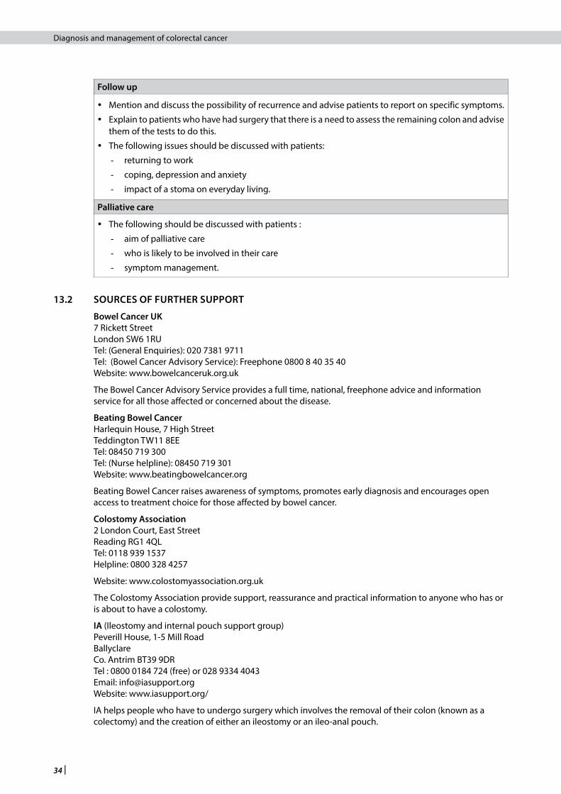
34 |
Diagnosis and management of colorectal cancer
Follow up
y Mention and discuss the possibility of recurrence and advise patients to report on specific symptoms.
y Explain to patients who have had surgery that there is a need to assess the remaining colon and advise them of the tests to do this.
y The following issues should be discussed with patients:
- returning to work
- coping, depression and anxiety
- impact of a stoma on everyday living.
Palliative care
y The following should be discussed with patients :
- aim of palliative care
- who is likely to be involved in their care
- symptom management.
13.2 SOURCES OF FURTHER SUPPORT
Bowel Cancer UK 7 Rickett Street London SW6 1RU Tel: (General Enquiries): 020 7381 9711 Tel: (Bowel Cancer Advisory Service): Freephone 0800 8 40 35 40 Website: www.bowelcanceruk.org.uk
The Bowel Cancer Advisory Service provides a full time, national, freephone advice and information service for all those affected or concerned about the disease.
Beating Bowel Cancer Harlequin House, 7 High Street Teddington TW11 8EE Tel: 08450 719 300 Tel: (Nurse helpline): 08450 719 301 Website: www.beatingbowelcancer.org
Beating Bowel Cancer raises awareness of symptoms, promotes early diagnosis and encourages open access to treatment choice for those affected by bowel cancer.
Colostomy Association 2 London Court, East Street Reading RG1 4QL Tel: 0118 939 1537 Helpline: 0800 328 4257
Website: www.colostomyassociation.org.uk
The Colostomy Association provide support, reassurance and practical information to anyone who has or is about to have a colostomy.
IA (Ileostomy and internal pouch support group) Peverill House, 1-5 Mill Road Ballyclare Co. Antrim BT39 9DR Tel : 0800 0184 724 (free) or 028 9334 4043 Email: [email protected] Website: www.iasupport.org/
IA helps people who have to undergo surgery which involves the removal of their colon (known as a colectomy) and the creation of either an ileostomy or an ileo-anal pouch.

| 35
Diagnosis and management of colorectal cancer
Cancer Research UK/CancerHelp UK Tel: 0800 800 4040 Email: [email protected] www.cancerhelp.org.uk
CancerHelp UK is a free information service about cancer and cancer care for people with cancer and their families. It is provided by Cancer Research UK. The site includes a comprehensive range of information including cancer prevention, diagnosis, treatment and follow up.
Macmillan Cancer Support (Scotland) 132 Rose Street Edinburgh EH2 3JD Tel: 0131 260 3270 Email: [email protected] Website: www.macmillan.org.uk
The Scottish office of the UK charity, which supports people with cancer (and their families) with specialist information, treatment and care.
Maggie’s Centres Scotland www.maggiescentres.org Email: [email protected]
Maggie’s provides practical, emotional and social support to people with cancer, their family and friends. Built alongside NHS cancer hospitals and staffed with professional experts, Maggie’s Centres are warm and welcoming, full of light and open space, with a big kitchen table at their heart.
Maggie’s Dundee Tom McDonald Avenue, Ninewells Hospital Dundee DD2 1NH Tel: 01382 632999 Email: [email protected]
Maggie’s Edinburgh The Stables, Western General Hospital Crewe Road South Edinburgh EH4 2XU Tel: 0131 537 3131 Email: [email protected]
Maggie’s Fife Victoria Hospital, Hayfield Road Kirkcaldy KY2 5AH Tel: 01592 647997 Email: [email protected]
Maggie’s Glasgow The Gatehouse, Western Infirmary 10 Dumbarton Road Glasgow G11 6PA Tel: 0141 330 3311
Gartnavel General Hospital 1053 Great Western Road Glasgow G12 0YN Tel: 0141 357 2269 Email: [email protected]
Maggie’s Highlands Raigmore Hospital, Old Perth Road Inverness IV2 3UJ Tel: 01463 706306 Email: [email protected]
13 • Provision of information

36 |
Diagnosis and management of colorectal cancer
Maggie’s Lanarkshire Flat 78, Residential accommodation Wishaw General Hospital 50 Netherton Road Wishaw ML2 ODP Tel: 01698 358392 Email: [email protected]
Marie Curie Cancer Care (Scotland) 14 Links Place Edinburgh EH6 7EB Tel: 0800 716 146 www.mariecurie.org.uk
Marie Curie Cancer Care, a care charity, provides practical nursing care at home and specialist care across its ten Marie Curie centres.
Tak Tent Support (Scotland) Flat 5, 30 Shelley Court Gartnavel Complex Glasgow G12 0YN Tel: 0141 211 0122 Email: [email protected] www.taktent.org
Tak Tent offers information, support, education and care for people with cancer, their families and friends and professionals. They have support groups throughout Scotland.

| 37
Diagnosis and management of colorectal cancer
14 Implementing the guideline
This section provides advice on the resource implications associated with implementing the key clinical recommendations, and advice on audit as a tool to aid implementation.
14.1 IMPLEMENTATION STRATEGY
Implementation of national clinical guidelines is the responsibility of each NHS Board and is an essential part of clinical governance. Mechanisms should be in place to review care provided against the guideline recommendations. The reasons for any differences should be assessed and addressed where appropriate. Local arrangements should then be made to implement the national guideline in individual hospitals, units and practices.
Implementation of this guideline will be encouraged and supported by SIGN.
14.2 RESOURCE IMPLICATIONS OF KEY RECOMMENDATIONS
No recommendations were identified as having significant budgetary impact.
14.3 AUDITING CURRENT PRACTICE
A first step in implementing a clinical practice guideline is to gain an understanding of current clinical practice. Audit tools designed around guideline recommendations can assist in this process. Audit tools should be comprehensive but not time consuming to use. Successful implementation and audit of guideline recommendations requires good communication between staff and multidisciplinary team working.
The guideline development group has identified the following as key points to audit to assist with the implementation of this guideline:
y number and frequency of surveillance colonoscopies performed on patients with inflammatory bowel disease at low, medium and high risk
y number of patients who receive follow-up colonoscopy after colonoscopic polypectomy y number and frequency of surveillance colonoscopies performed in patients with ≥5 small adenomas, or
≥3 adenomas with at least one polyp ≥1 cm in size y number of referrals to the Clinical Genetics Service of individuals whose family history is suggestive of
a CRC syndrome y the inclusion of tumour differentiation, staging (Dukes’ and TNM systems), margins (peritoneal and CRM)
and extramural vascular invasion in pathological reports of colorectal cancer resection specimens y outcomes of treatment, including treatment-related morbidity and mortality, disease-free survival and
overall survival.
14.4 ADVICE TO NHSSCOTLAND FROM THE SCOTTISH MEDICINES CONSORTIUM
The Scottish Medicines Consortium concluded in 2005 that cetuximab is not recommended for use within NHSScotland in combination with irinotecan for the treatment of patients with epidermal growth factor receptor (EGFR)-expressing metastatic colorectal cancer after failure of irinotecan-including cytotoxic therapy.
Following a further submission in 2010 the SMC recommended that cetuximab is accepted for restricted use within NHSScotland for the treatment of patients with epidermal growth factor receptor (EGFR)-expressing, K-ras wild-type metastatic colorectal cancer in combination with chemotherapy. Cetuximab is restricted to use in patients who have not previously received chemotherapy for their metastatic disease, with liver metastases only that are considered non-resectable but in whom potentially curative liver metastasis resection would be undertaken if the lesions became resectable after treatment with chemotherapy and cetuximab.
In January 2015 SMC accepted cetuximab for restricted use as treatment for patients with EGFR-expressing, RAS wild-type metastatic colorectal cancer:
14 • Implementing the guideline

38 |
Diagnosis and management of colorectal cancer
SMC has restricted cetuximab for use in patients with RAS wild-type metastatic colorectal cancer, in combination with irinotecan or oxaliplatin-based chemotherapy, in patients who have not previously received chemotherapy for their metastatic disease (first-line treatment).
Panitumumab (Vectibix) is not recommended as monotherapy for the treatment of patients with EGFR-expressing metastatic colorectal carcinoma with non-mutated (wild-type) K-ras after failure of fluoropyrimidine-, oxaliplatin-, and irinotecan- containing chemotherapy regimens.
Bevacizumab (Avastin) is not recommended for use within NHSScotland in combination with fluoropyrimidine-based chemotherapy for treatment of patients with metastatic carcinoma of the colon or rectum due to insufficient evidence of cost effectiveness.
Further information is available from the SMC website www.scottishmedicines.org.uk

| 39
Diagnosis and management of colorectal cancer
15 The evidence base
15.1 SYSTEMATIC LITERATURE REVIEW
The evidence base for SIGN 67: Management of colorectal cancer was synthesised in accordance with SIGN methodology. A systematic review of the literature was carried out using an explicit search strategy devised by the SIGN Information Officer in collaboration with members of the guideline development group. The search for systematic reviews and meta-analyses covered the Cochrane Library, MEDLINE, and EMBASE databases, from January 2002 to March 2011. The search for randomised controlled trials, cohort studies, case control studies, and cross-sectional surveys covered the Cochrane Library, MEDLINE, EMBASE, and CINAHL databases, from January 2002 to March 2011. The evidence base was updated during the course of development of the guideline, and the search was supplemented by reviewing references identified from papers from the searches and from personal databases of the guideline development group members.
15.1.1 LITERATURE SEARCH FOR PATIENT ISSUES
At the start of the guideline development process, a SIGN Information Officer conducted a literature search for qualitative and quantitative studies that addressed patient issues of relevance to the management of colorectal cancer. Databases searched include Medline, Embase, CINAHL and PsycINFO, and the results were summarised and presented to the guideline development group. A copy of the Medline version of the patient search strategy is available on the SIGN website.
15.2 RECOMMENDATIONS FOR RESEARCH
The guideline development group was not able to identify sufficient evidence to answer all of the key questions asked in this guideline (see Annex 1). The following areas for further research have been identified:
y screening using faecal immunological testing and flexible sigmoidoscopy within the Scottish population y well designed prospective population based studies using robust and reproducible assays to determine
the use of any novel prognostic or predictive marker to aid decision making on patient selection for adjuvant therapy
y RCTs to assess the optimal duration and timing of second line chemotherapy.
15.3 REVIEW AND UPDATING
This guideline was issued in 2011 and will be considered for review in three years. The review history, and any updates to the guideline in the interim period, will be noted in the review report, which is available in the supporting material section for this guideline on the SIGN website: www.sign.ac.uk
Comments on new evidence that would update this guideline are welcome and should be sent to the SIGN Executive, Gyle Square, 1 South Gyle Crescent, Edinburgh, EH12 9EB (email: [email protected]).
15 • The evidence base

40 |
Diagnosis and management of colorectal cancer
16 Development of the guideline
16.1 INTRODUCTION
SIGN is a collaborative network of clinicians, other healthcare professionals and patient organisations and is part of Healthcare Improvement Scotland. SIGN guidelines are developed by multidisciplinary groups of practising clinicians using a standard methodology based on a systematic review of the evidence. Further details about SIGN and the guideline development methodology are contained in SIGN 50: A Guideline Developer’s Handbook, available at www.sign.ac.uk
16.2 THE GUIDELINE DEVELOPMENT GROUP
Professor Robert Steele Professor of Surgery, Ninewells Hospital, Dundee (Chair)
Dr George Barlow MBE General Practitioner (retired), Glasgow
Miss Nicola Bradshaw Macmillan Cancer Genetic Counsellor, Royal Hospital for Sick Children, Glasgow
Dr Ewan Brown Consultant Medical Oncologist, Western General Hospital, Edinburgh
Ms Juliet Brown Information Officer, SIGN
Mr Alan Campbell Pharmacist, The Beatson West of Scotland Cancer Centre, Glasgow
Professor Frank Carey Consultant Pathologist, Ninewells Hospital, Dundee
Miss Jen Layden Programme Manager, SIGN
Mr Tim McAdam Consultant Colorectal/General Surgeon, Aberdeen Royal Infirmary
Mr Ian McEwan Patient representative, Midlothian
Dr Catriona McLean Consultant Clinical Oncologist, Western General Hospital, Edinburgh
Professor Alistair Munro Consultant Oncologist, Ninewells Hospital, Dundee
Mr Terence O’Kelly Consultant Colorectal Surgeon, Aberdeen Royal Infirmary
Dr Perminder Phull Consultant Gastroenterologist, Aberdeen Royal Infirmary
Dr Fat Wui Poon Consultant Radiologist, Glasgow Royal Infirmary
Dr Mary Porteous Consultant Clinical Geneticist, Western General Hospital, Edinburgh
Dr Leslie Samuel Macmillan Consultant Oncologist, Aberdeen Royal Infirmary
Ms Ailsa Stein Programme Manager, SIGN
The membership of the guideline development group was confirmed following consultation with the member organisations of SIGN. All members of the guideline development group made declarations of interest and further details of these are available on request from the SIGN Executive.
Guideline development and literature review expertise, support and facilitation were provided by the SIGN Executive. All members of the SIGN Executive make yearly declarations of interest and further details of these are available on request.
Mr Euan Bremner Guideline Coordinator
Mrs Lesley Forsyth Events Coordinator
Mrs Karen Graham Patient Involvement Officer
Mr Stuart Neville Publications Designer

| 41
Diagnosis and management of colorectal cancer
16.2.1 ACKNOWLEDGEMENTS
SIGN would like to acknowledge the guideline development group responsible for the development of SIGN 67: Management of colorectal cancer, on which this guideline is based.
SIGN is grateful to the following former members of the guideline development group who have contributed to the development of the guideline.
Mrs Inge MacKenzie Patient representative, Sandbank, Argyll
Ms Jackie Rodger Colorectal Clinical Nurse Specialist, Ninewells Hospital, Dundee
16.3 CONSULTATION AND PEER REVIEW
16.3.1 PUBLIC CONSULTATION
The draft guideline was available on the SIGN website for a month to allow all interested parties to comment. All contributors made declarations of interest and further details of these are available on request from the SIGN Executive.
16.3.2 SPECIALIST REVIEW
This guideline was also reviewed in draft form by the following independent expert referees, who were asked to comment primarily on the comprehensiveness and accuracy of interpretation of the evidence base supporting the recommendations in the guideline. The guideline group addresses every comment made by an external reviewer, and must justify any disagreement with the reviewers’ comments. All expert referees made declarations of interest and further details of these are available on request from the SIGN Executive.
SIGN is very grateful to all of these experts for their contribution to the guideline.
Dr Paul Baughan General Practitioner and Chair of Scottish Primary Care Cancer Group, Clackmannanshire
Mrs Alyson Branch Senior Cancer Care Pharmacist, Beatson West of Scotland Cancer Centre, Glasgow
Ms Carly Brownlie Patient Involvement Representative, SIGN Council
Mr John Camilleri-Brennan Consultant General and Colorectal Surgeon, Stirling Royal Infirmary
Dr Patrick Cadigan Registrar, Association of Cancer Physicians and Royal College of Physicians, London
Professor James Garden Regius Professor of Clinical Surgery, Edinburgh Royal Infirmary
Dr Derek Gillen Clinical Lead for Endoscopy and Clinical Lead for Bowel Cancer Screening, NHS Greater Glasgow and Clyde
Ms Rachel Haigh Colorectal Cancer Clinical Nurse Specialist, Western General Hospital, Edinburgh
Dr Chris Johnstone General Practitioner, Barony Practice, Paisley
Mr James Mander Consultant Colorectal Surgeon, Western General Hospital, Edinburgh
Dr Alison McCallum Director of Public Health and Health Policy, NHS Lothian
Dr Alec McDonald Consultant Clinical Oncologist, Beatson West of Scotland Cancer Centre, Glasgow
Mr Graham McKay Consultant Colorectal Surgeon, Glasgow Royal Infirmary
Mr Mark Potter Consultant Colorectal Surgeon, Western General Hospital, Edinburgh
Mr James Powell Consultant Hepatobiliary/Transplant Surgeon, Queen Margaret Hospital, Dunfermline
Professor Greg Rubin Professor of General Practice and Primary Care, Durham University
Dr Matt Rutter Consultant Gastroenterologist, University Hospital of North Tees, Stockton-on-Tees
Professor David Sebag-Montefiore Consultant Clinical Oncologist, St James Hospital, Leeds
16 • Development of the guideline

42 |
Diagnosis and management of colorectal cancer
Dr Alasdair Taylor Consultant Radiologist, University Hospitals of Morecambe Bay NHS Foundation Trust
Mr Graeme Wilson Consultant Colorectal Surgeon, Western General Hospital, Edinburgh
Mr Satheesh Yalamarthi Consultant Colorectal Surgeon, Queen Margaret Hospital, Dunfermline
Dr Steven Yule Consultant Radiologist, Aberdeen Royal Infirmary
16.3.3 SIGN EDITORIAL GROUP
As a final quality control check, the guideline is reviewed by an editorial group comprising the relevant specialty representatives on SIGN Council to ensure that the specialist reviewers’ comments have been addressed adequately and that any risk of bias in the guideline development process as a whole has been minimised.
All members of the SIGN Editorial group make yearly declarations of interest and further details of these are available on request from the SIGN Executive.
The Editorial group for this guideline was as follows.
Dr Keith Brown Chair of SIGN; Co-Editor
Ms Beatrice Cant Programme Manager, SIGN
Dr Bernard Croal Royal College of Pathologists
Mr Andrew de Beaux Royal College of Surgeons of Edinburgh
Dr Roberta James Acting Programme Director, SIGN; Co-Editor
Dr Elizabeth Junor Royal College of Radiologists
Dr Sara Twaddle Director of SIGN; Co-Editor
Dr Christine Walker Royal College of Radiologists

| 43
Diagnosis and management of colorectal cancer
Abbreviations5-FU fluorouracil
ACPGBI Association of Coloproctology for Great Britain and Ireland
APC adenomatous polyposis coli
BED biological effective dose
BMI body mass index
BNF British National Formulary
BSG British Society of Gastroenterology
CEA carcinoembryonic antigen
CI confidence interval
CNS clinical nurse specialist
CRC colorectal cancer
CRM circumferential resection margin
CT computed tomography
DNA deoxyribonucleic acid
DRE digital rectal examination
EGFR epidermal growth factor receptor
FAP familial adenomatous polyposis
FDG fluoro-deoxy-glucose
FDR first degree relative
FIT faecal immunological testing
FOBT faecal occult blood testing
GP general practitioner
GY gray
HNPCC hereditary non-polyposis colorectal cancer
HR hazard ratio
HRT hormone replacement therapy
IDA iron deficiency anaemia
IHC immunohistochemistry
IV intravenous
K-ras Kirsten rat sarcoma proto-oncogene
LS Lynch syndrome
MDT multidisciplinary team
MMR mismatch repair
MRI magnetic resonance imaging
MSI microsatellite instability
MTA multiple technology appraisal
MYH MUTYH-associated polyposis
NICE National Institute for Health and Clinical Excellence
Abbreviations

44 |
Diagnosis and management of colorectal cancer
NNT number needed to treat
NSAID non-steroidal anti-inflammatory drug
OC oral contraceptive
OR odds ratio
OGD oesophagogastroduodenoscopy
pCR pathological complete response rate
PET positron emission tomography
PLR positive likelihood ratio
PSC primary sclerosing cholangitis
RCT randomised controlled trial
SCPRT short course preoperative radiotherapy
SIGN Scottish Intercollegiate Guidelines Network
SMC Scottish Medicines Consortium
SPC summary of product characteristics
TME total mesorectal excision
TNM tumour, node, metastasis staging system
TS thymidylate synthase
US ultrasound
USA United States of America
VEGF vascular endothelial growth factor
VCE video capsule endoscopy
WHO World Health Organisation

| 45
Diagnosis and management of colorectal cancer
Annex 1Key questions addressed in this update
THE KEY QUESTIONS USED TO DEVELOP THE GUIDELINE
The update of this guideline is based on a series of structured key questions that define the target population, the intervention, diagnostic test, or exposure under investigation, the comparison(s) used and the outcomes used to measure efficacy, effectiveness, or risk. These questions form the basis of the systematic literature search.
Key questionSee guideline section
1. What new evidence exists in the prevention of colorectal cancer through specific dietary measures and weight monitoring?
3.1
2. What screening modalities are appropriate (for the general population, inflammatory bowel disease, adenomatous polyps and family history of CRC), and when and how frequently should these be performed?
Consider: colonoscopy, biopsy, faecal occult blood testing (FOBT), FIT test (faecal immunological testing)
3.7
3. What is the risk of developing colorectal cancer in individuals with a family history of CRC?
Consider: MSI, familial risk, mismatch repair, MYH, FAP, APC HNPCC
5
4. How should patients with CRC be selected for genetic analysis?
Consider: germline mutations in mismatch repair genes (APC, MYH), tumour indicators – MSI and immunohistochemistry for MLH, MSH2 and MSH6
5
5. What signs/symptom combinations are a predictive value of CRC and do signs/symptoms inform the investigations?
Consider: abdominal symptoms eg pain, distention, iron deficiency anaemia, weight loss, bleeding (gastrointestinal, rectal), irritable bowel syndrome, coeliac disease
6
6. Is there evidence for undertaking specific investigations for CRC within the primary care setting at the time of referral?
Consider: FOBT, full blood count, family history, digital rectal examination, abdominal examination
6
7. What is the accuracy of endorectal ultrasound and high resolution MRI of the rectum in the T and N staging of rectal cancer?
7
8. What is the role of CT colonoscopy and barium testing in the diagnosis of CRC?
Consider: specificity and sensitivity
7
9. What investigation(s) for elective and emergency patients should be considered for preoperative staging in colon and rectal cancer?
Consider: MRI, CT, Chest X-ray
7, 8.1
10. What is the evidence for specific surgical techniques in the treatment of patients with colon and rectal cancer, and the effectiveness of these techniques on patient outcomes?
Consider: radical surgery, laparoscopic surgery and abdomino-perineal excision of rectum, colonic resectionConsider outcomes: lymph node numbers, pathology scoring in macroscopic specimens
8
11. What is the role of stenting in malignant obstruction?
Consider: palliative, bridge to surgery, tumour dissemination
8.7
Annexes

46 |
Diagnosis and management of colorectal cancer
12. What is the role of protective stomas in anterior resection?
Consider: preoperative radiotherapy, defunctioning and ileostomy
8.4.3
13. Which patients are suitable for adjuvant chemotherapy in colon cancer?
Key search terms: adjuvant, colon cancer, colon neoplasm, node positive, node negative, Dukes staging
10.1
14. Which chemotherapy regimen is optimal in the treatment of patients with colon cancer and rectal cancer?
Consider: a) Metastatic
b) Adjuvant
c) Dose, route, schedule, duration of treatment
10.110.210.3
15. What is the optimum treatment regimen for patients with advanced (metastatic) colon cancer?
Key search terms: intensive regimen, palliative regimen, curative, non-curative, liver metastases, colon, metastatic metastectomy, KRAS and BRAF mutations.
10.2
16. Which patient group should be considered for adjuvant treatment in rectal cancer and when in treatment should patients be considered?
Consider: radiotherapy, chemotherapy, chemoradiation, circumferential resection margin (CRM), neo-adjuvant, adjuvant, intraoperative, pre- and postoperative, contact therapy
10.3
17. What treatment is optimal for patients with operable rectal cancer and locally advanced rectal cancer?
Consider: downstaging, Dukes stage, chemotherapy, radiotherapy, cyberknife, gamma knife, stereotactic, radionucleide, radio-embolisation (palliative)
10.3

| 47
Diagnosis and management of colorectal cancer
Annex 2Scottish referral guidelines for suspected cancer: lower gastrointestinal cancer211
LOWER GASTROINTESTINAL CANCER
Lower gastrointestinal symptoms are common in the community. Rectal bleeding for instance is estimated to affect 140,000 individuals per 1 million population each year. There are large differences in the predictive value of rectal bleeding for cancer according to its association with other symptoms and signs and the age of the patient. Different management strategies should be adopted according to cancer risk so that those patients with transient low-risk symptoms caused by benign disease avoid unnecessary investigation. The following protocol is recommended for managing patients with rectal bleeding and features associated with a possible diagnosis of colorectal cancer:
KEY POINTS:
i "Watch and wait" is appropriate for patients less than 40 years of age with low risk features and particularly those with very transient symptoms. The duration of "watch and wait" can be flexible and tailored to individual patients but a period of six weeks is recommended. A clear mechanism for follow-up is needed but this will not necessarily require return to the clinic. Review by telephone or e-mail might be appropriate. If the presenting problem resolves then no further action is required. If it does not, or if there is continuing concem then the patient should be referred for investigation/treatment as per care plan.
i i Family history: This should be obtained and might be relevant but review by a Regional Clinical Genetics Service is recommended for accurate risk assessment if this is the principal indication for referral for investigation.
i i i Investigations: No examinations or investigations other than abdominal and rectal examination and full blood count are recommended. Faecal occult blood testing (FOBt) is not indicated and should not influence decision making in symptomatic patients.
Annexes
Low risk features
• Transient symptoms (<6 weeks)
• Patient <40 years
• Structured proforma
• Decision support
Watch and wait (6 weeks)
• Assessment and Review
• Setting determined locally
• Patient agreement required
• Appropriate information and counselling
Review
No further symptoms
• Discharge
Symptoms
• Persist or recur
• Deteriorate
• Will not agree to “watch and wait”
High risk features
• Persistent rectal bleeding without anal symptoms
• Persistent change in bowel habit (>6 weeks)
• Significant family history
• Right sided abdominal mass
• Palpable rectal mass
• Unexplained iron deficiency anaemia
• Patients with persistent diarrhoea
• Patients in whom there is clinical doubt
Refer
• E- communication
• Structured proforma
• Decision support
Visualisation of the large bowel
Refer
• E- communication
• Structured proforma
• Decision supportReview

48 |
Diagnosis and management of colorectal cancer
Annex 3Risk based guideline for investigating patients with symptoms and/or signs suggestive of large bowel pathology (including colorectal cancer).This guideline is relevant to patients who are suspected of having significant colorectal pathology and who have been referred from primary care to secondary care in accordance with the current referral guidance.
≥ 40 years Rectal bleeding and change in bowel habit with more frequent and/or loose stools
y Colonoscopy
y Patients should proceed directly to test where possible.
≥ 40 years Rectal bleeding and change in bowel habit with less frequent firm stools (ie constipation)
y Visualisation of the large bowel in its entirety is recommended. Colonoscopy or flexible sigmoidoscopy and barium enema or CT colonography are suitable. Choice of modality will depend upon clinical decision and local capacity.
y Patients should wherever possible proceed directly to test.
≥ 40 years Rectal bleeding in isolation y Consider on individual merits.
y Most patients should proceed directly to test.
y Colonoscopy should be considered in all patients over 50 years of age.
y Under 50 years, flexible sigmoidoscopy may be appropriate in the first instance.
> 40 years A symptom/symptoms outwith above categories
y Consider on individual merits.
y In many cases outpatient assessment will be appropriate in the first instance.
< 40 years Rectal bleeding +/- peri-anal symptoms (discomfort/pain, soreness, itching and prolapse)
Note: This refers to patients with persistent symptoms ie those lasting more than 6 weeks.
y Clinical assessment in appropriate setting with procto-sigmoidoscopy as minimum requirement for examination.
< 40 years Any other combination of symptoms y Assess each referral on individual merit.
y Outpatient review recommended in the first instance.
Any age Unexplained iron deficiency anaemia (IDA)*
(*IDA confirmed and cause remains unexplained in accordance with: Guidelines for management of iron deficiency anaemia, BSG Guidelines in Gastroenterology 2005)
y Simultaneous colonoscopy and upper gastrointestinal endoscopy.
Any age Suspected abdominal or rectal mass y Assessment in outpatient clinic.

| 49
Diagnosis and management of colorectal cancer
Annex 4TNM (tumour, node, metastasis) staging
T Primary Tumour
TX Primary tumour cannot be assessed
T0 No evidence of primary tumour
Tis Carcinoma in situ1
T1 Tumour invades submucosa.
T2 Tumour invades muscularis propria
T3 Tumour invades through the muscularis propria into the subserosa, or into the non- peritonealised pericolic or perirectal tissues
T4 Tumour directly invades other organs or structures, and/or perforates visceral peritoneum2,3
N Regional Lymph Nodes
NX Regional lymph nodes cannot be assessed (eg previously removed)
N0 No regional lymph node metastasis
N1 Metastasis in 1 to 3 regional lymph nodes
N2 Metastasis in 4 or more regional lymph nodes
M Distant Metastates
MX Distant metastasis cannot be assessed
M0 No distant metastasis
M1 Distant metastasis present
1 Tis includes cancer cells confined within the glandular basement membrane (intraepithelial) or lamina propria (intramucosal) with no extension through muscularis mucosae into the submucosa.
2 Direct invasion in T4 includes invasion of other segments of the colorectum by way of the serosa, eg invasion of the sigmoid colon by a carcinoma of the caecum.
3 Tumour that is adherent to other organs or structures, macroscopically, is classified T4. However, if no tumour is present in the adhesion, microscopically, the classification should be pT3.
Dukes StagingA Limited to the submucosa B1 Tumour invades into but not through the muscularis propria, no lymph node involvementB2 Tumour invades through the muscularis propria, no lymph node involvementB3 Tumour directly invades other organs or structures (added)C1 Regional lymph nodes involvedC2 Metastases present in nodes at mesenteric artery ligature (apical nodes)
Annexes

50 |
Diagnosis and management of colorectal cancer
References
1. Cancer statistics: colorectal cancer. [cited 22 February 2011]. Available from url: http://www.isdscotland.org/isd/1425.html#Colorectal cancer
2. World Cancer Research Fund (WCRF) / American Institute for Cancer Research. Food, Nutrition, Physical Activity, and the Prevention of Cancer: a Global Perspective. Washington DC: AICR; 2007.
3. Gatta G, Faivre J, Capocaccia R, Ponz de Leon M. Survival of colorectal cancer patients in Europe during the period 1978-1989. Eur J Cancer 1998;34:2176-83.
4. Guidance on prescribing. In: The British National Formulary No. 59. London: British Medical Association and Royal Pharmaceutical Society of Great Britain; 2010.
5. Healthy Eating, Active Living: An action plan to improve diet, increase physical activity and tackle obesity (2008-2011). Edinburgh: Scottish Government; 2008. Available from url: http://www.scotland.gov.uk/Resource/Doc/228860/0061963.pdf
6. Giovannucci E. An updated review of the epidemiological evidence that cigarette smoking increases risk of colorectal cancer. Cancer Epidemiol Biomarkers Prev 2001;10(7):725-31.
7. Department of Health Physical Activity, Health Improvement and Protection. Start Active, Stay Active: A report on physical activity from the four home coutnries’ Chief Medical Officers. London: Department of Health; 2011.
8. Crandall CJ. Estrogen replacement therapy and colon cancer: A clinical review. J Womens Health Gend Based Med 1999;8(9):1155-66.
9. Rossouw J, Anderson G, Prentice R, LaCroix A, Kooperberg C, Stefanick M, et al. Risks and benefits of estrogen plus progestin in healthy postmenopausal women: principal results From the Women’s Health Initiative randomized controlled trial. JAMA 2002;288(3):321-33.
10. Beral V, Banks E, Reeves G, Appleby P. Use of HRT and the subsequent risk of cancer. J Epidemiol Biostat 1999;4(3):191-210.
11. Grodstein F, Newcomb PA, Stampfer MJ. Postmenopausal hormone therapy and the risk of colorectal cancer: a review and meta-analysis. Am J Med 1999;106(5):574-82.
12. Hebert-Croteau N. A meta-analysis of hormone replacement therapy and colon cancer in women. Cancer Epidemiol Biomarkers Prev 1998; 7: 653-9.
13. Nanda K, Bastian L, Hasselblad V, Simel D. Hormone replacement therapy and the risk of colorectal cancer: a meta-analysis. Obstet Gynecol 1999;93(5):880-8.
14. Franceschi S, La Vecchia C. Colorectal cancer and hormone replacement therapy: an unexpected finding. Eur Journal Cancer Prev 1998;7(6):427-38.
15. IARC Working Group on the Evaluation of Cancer Preventive Agents. Non-steroidal anti-inflammatory drugs. Lyon: International Agency for Research on Cancer; 1997.
16. Hewitson P, Glasziou PP, Irwig L, Towler B, Watson E. Screening for colorectal cancer using the faecal occult blood test, Hemoccult. Cochrane Database of Systematic Reviews, 2007 Issue 1.
17. van Rossum LG, van Rijn, AF Laheij RJ, van Oijen, MG, Fockens P, van Krieken HH, et al. Random Comparison of Guaiac and Immunochemical Fecal Occult Blood Tests for Colorectal Cancer in a Screening Population. Gastroenterology 2008;135:82-90.
18. Atkin WS, Edwards R, Kralj-Hans I, Wooldrage K, Hart AR, Northover JM, Parkin DM, et al. Once-only flexible sigmoidoscopy screening in prevention of colorectal cancer: a multicentre randomised controlled trial. Lancet 2010 375 1624-33.
19. Steele R, McClements PL, Libby G, Black R, Morton C, Birrell J, Mowat NA, et al. Results from the first three rounds of the Scottish demonstration pilot of FOBT screening for colorectal cancer. Gut 2009;58(4):530-5.
20. Eaden J, Abrams K, Mayberry J. The risk of colorectal cancer in ulcerative colitis: a meta-analysis. Gut 2001;48(4):526-35.
21. Collins PD, Mpofu C, Watson AJ, Rhodes JM. Strategies for detecting colon cancer and/or dysplasia in patients with inflammatory bowel disease. Cochrane Database of Systematic Reviews 2006, Issue 2.
22. Cairns S, Scholefield JH, Steele RJ, Dunlop MG, Thomas HJ, Evans GD, Eaden JA, Rutter MD, Atkin WP, Saunders BP, Lucassen A, Jenkins P, Fairclough PD, Woodhouse CR; British Society of Gastroenterology; Association of Coloproctology for Great Britain and Ireland. Guidelines for colorectal cancer screening and surveillance in moderate and high risk groups (update from 2002). Gut 2010;59(5):666-89.
23. Subramanian V, Mannath J, Ragunath K, Hawkey CJ. Meta-analysis: The diagnostic yield of chromoendoscopy for detecting dysplasia in patients with colonic inflammatory bowel disease. Aliment Pharmacol Ther 2011;33(3):304-3011.
24. Bond J. Colorectal cancer update. Prevention, screening, treatment, and surveillance for high-risk groups. . Med Clin North Am 2000;84(5):1163-82.
25. Mandel J, Church T, Bond J, Ederer F, Geisser M, Mongin S, et al. The effect of fecal occult-blood screening on the incidence of colorectal cancer. N Engl J Med 2000;343(22):1603-7.
26. Winawer SJ, Fletcher RH, Miller L, Godlee F, Stolar MH, Mulrow CD, et al. Colorectal cancer screening: Clinical guidelines and rationale. Gastroenterology 1997;112(2):594-642.
27. Saini SD, Kim HM, Schoenfeld P. Incidence of advanced adenomas at surveillance colonoscopy in patients with a personal history of colon adenomas: a meta-analysis and systematic review. Gastroint Endosc 2006;64(4):614-26.
28. Winawer S, Zauber AG, Fletcher RH, Stillman JS, O’Brien MJ, Levin B, et al. Guidelines for colonoscopy surveillance after polypectomy: a consensus update by the US Multi-Society Task Force on Colorectal Cancer and the American Cancer Society. CA Cancer J Clin 2006;56(3):143-59.
29. Brown SR, Baraza W, Hurlstone P. Chromoscopy versus conventional endoscopy for the detection of polyps in the colon and rectum. Cochrane Database of Systematic Reviews 2007, Issue 4.
30. Thiis-Evensen E, Wilhelmsen I, Hoff GS, Blomhoff S, Sauar J. The psychologic effect of attending a screening program for colorectal polyps. Scand J Gastroenterol 1999;34:103-9.
31. Wardle J, Taylor T, Sutton S, Atkin W. Does publicity about cancer screening raise fear of cancer? Randomised trial of the psychological effect of information about cancer screening. BMJ 1999;319(7216):1037-8.
32. Derogatis L, Morrow GR, Fetting J, Penman D, Piasetsky S, Schmale AM, et al. The prevalence of psychiatric disorders among cancer patients. JAMA 1983;249(6):751-7.

| 51
Diagnosis and management of colorectal cancer
33. Fallowfield L, Ratcliffe D, Jenkins V, Saul J. Psychiatric morbidity and its recognition by doctors in patients with cancer. Br J Cancer 2001;84(8):1011-5.
34. Slevin M, Nichols SE, Downer SM, Wilson P, Lister TA, Arnott S,et al. Emotional support for cancer patients: what do patients really want? Br J Cancer 1996;74(8):1275-9.
35. Baider L, Koch U, Esacson R, De-Nour AK. Prospective study of cancer patients and their spouses: the weakness of marital strength. Psychooncology 1998;7(1):49-56.
36. Harrison J, Haddad P, Maguire P. The impact of cancer on key relatives: a comparison of relative and patient concerns. Eur J Cancer 1995;31A(11):1736-40.
37. Nijboer C, Triemstra M, Tempelaar R, Sanderman R, van den Bos GA. Determinants of caregiving experiences and mental health of partners of cancer patients. Cancer 1999;86(4):577-88.
38. Northouse LL, Schafer JA, Tipton J, Metivier L. The concerns of patients and spouses after the diagnosis of colon cancer: a qualitative analysis. J Wound Ostomy Continence Nurs 1999;26(1):8-17.
39. Bekkers M, van Knippenberg FC, van Dulmen AM, van den Borne HW, van Berge Henegouwen GP. Survival and psychosocial adjustment to stoma surgery and nonstoma bowel resection: a 4-year follow-up. J Psychosom Res 1997;42(3):235-44.
40. Camilleri-Brennan J, Steele RJ. Quality of life after treatment for rectal cancer. Br J Surg 1998;85(8):1036-43.
41. Sprangers M, Taal BG, Aaronson NK, te Velde A. Quality of life in colorectal cancer. Stoma vs. nonstoma patients. Dis Colon Rectum 1995;38(4):361-9.
42. Knowles G, Tierney A, Jodrell D, Cull A. The perceived information needs of patients receiving adjuvant chemotherapy for surgically resected colorectal cancer. Eur J Oncol Nurs 1999;31:208-22.
43. Jenkins V, Fallowfield L, Saul J. Information needs of patients with cancer: results from a large study in UK cancer centres. Br J Cancer 2001;84(1):48-51.
44. Fallowfield L, Saul J, Gilligan B. Teaching senior nurses how to teach communication skills in oncology. Cancer Nurs 2001;24(3):185-91.
45. Scott JT, Entwistle VA, Sowden AJ, Watt I. Recordings or summaries of consultations for people with cancer. Cochrane Database of Systematic Reviews 2002, Issue 4 2002(4).
46. Jones R, Pearson J, McGregor S, Cawsey AJ, Barrett A, Craig N, et al. Randomised trial of personalised computer based information for cancer patients. BMJ 1999;319(7219):1241-7.
47. Bruera E, Pituskin E, Calder K, Neumann CM, Hanson J. The addition of an audiocassette recording of a consultation to written recommendations for patients with advanced cancer: A randomized, controlled trial. Cancer 1999;86(11):2420-5.
48. Bekker H, Thornton JG, Airey CM, Connelly JB, Hewison J, Robinson MB, et al. Informed decision making: An annotated bibliography and systematic review. Health Technology Assessment (Rockville, Md) 1999;3(1):iii-150.
49. Beaver K, Bogg J, Luker KA. Decision-making role preferences and information needs: a comparison of colorectal and breast cancer. Health Expect 1999;2(4):266-76.
50. Clinical Standards Board for Scotland. Clinical standards: colorectal cancer. Edinburgh: Clinical Standards Board for Scotland; 2001.
51. Scottish Cancer Co-ordinating and Advisory Committee. Commissioning Cancer Services Sub-Committee. Interim report to the Chief Medical Officer, Scottish Office Department of Health. Edinburgh: The Scottish Office; 1995.
52. Lindor NM, Petersen GM, Hadley DW, Kinney AY, Miesfeldt S, Lu KH, et al. Recommendations for the care of individuals with an inherited predisposition to Lynch syndrome: A systematic review. JAMA 2006;296(12):1507-17.
53. Vasen HF, Moslein, G, Alonso, A, Aretz, S, Bernstein, I, Bertario, L, et al. Guidelines for the clinical management of familial adenomatous polyposis (FAP). Gut 2008;57(5): 704-13.
54. Butterworth AS, Higgins, JPT and Pharoah, P. Relative and absolute risk of colorectal cancer for individuals with a family history: a meta-analysis. Eur J Cancer 2006;42:216-27.
55. Mulhall BP, Veerappan GR, Jackson JL. Meta-analysis: Computed tomographic colonography. Ann Intern Med 2005;142(8):635-50.
56. Aaltonen L, Johns L, Järvinen H, Mecklin JP, Houlston R. Explaining the familial colorectal cancer risk associated with mismatch repair (MMR)-deficient and MMR-stable tumors. Clin Cancer Res 2007;13(1):356-61.
57. Lindor N, Rabe K, Petersen GM, Haile R, Casey G, Baron J, et al. Lower cancer incidence in Amsterdam-I criteria families without mismatch repair deficiency: familial colorectal cancer type X. JAMA 2005;293(16):1979-85.
58. Bekkink OM, McCowan C, Falk GA, Teljeur C, Van De Laar FA, Fahey T. Diagnostic accuracy systematic review of rectal bleeding in combination with other symptoms, signs and tests in relation to colorectal cancer. Br J Cancer 2010;102(1):48-58.
59. Hamilton W. The CAPER studies: Five case-control studies aimed at identifying and quantifying the risk of cancer in symptomatic primary care patients. Br J Cancer 2009;101(SUPPL. 2):S80-S6.
60. Ellis BG, Thompson MR. Factors identifying higher risk rectal bleeding in general practice. Br J Gen Pract 2005;55(521):949-55.
61. Mitchell E, Macdonald S, Campbell NC, Weller D, Macleod U. Influences on pre-hospital delay in the diagnosis of colorectal cancer: A systematic review. Br J Cancer 2008;98(1):60-70.
62. Rex D, Rahmani EY, Haseman JH, Lemmel GT, Kaster S, Buckley JS. Relative sensitivity of colonoscopy and barium enema for detection of colorectal cancer in clinical practice. Gastroenterology 1997;112(1):17-23.
63. Bowles C, Leicester R, Romaya C, Swarbrick E, Williams CB, Epstein O. A prospective study of colonoscopy practice in the UK today: are we adequately prepared for national colorectal cancer screening tomorrow? Gut 2004;53(2):277-83.
64. Thoeni R, Laufer I. Polyps and cancer. Textbook of gastrointestinal radiology 1994; 1160.
65. Bartram C. The colon and rectum. In: Curati WL CD, Lipton MJ, Allison DJ, editor. Imaging in oncology. London: Greenwich Medical Media; 1998. p.99-110.
66. Atkin W. Implementing screening for colorectal cancer. BMJ 1999;319(7219):1212-3.
67. Waye J, Kahn O, Auerbach ME. Complications of colonoscopy and flexible sigmoidoscopy. Gastrointest Endosc Clin N Am 1996;6(2):343-77.
68. Rokkas T, Papaxoinis K, Triantafyllou K, Ladas SD. A meta-analysis evaluating the accuracy of colon capsule endoscopy in detecting colon polyps. Gastrointest Endosc 2010;71(4):792-8.
69. Rosman AS, Korsten MA. Meta-analysis comparing CT colonography, air contrast barium enema, and colonoscopy. Am J Med 2007;120(3):203-10.
70. Chaparro M, Gisbert JP, Del Campo L, Cantero J, Mate J. Accuracy of computed tomographic colonography for the detection of polyps and colorectal tumors: A systematic review and meta-analysis. Digestion 2009;80(1):1-17.
References
52 |
Diagnosis and management of colorectal cancer
71. Pickhardt PJ, Kim DH, Meiners RJ, Wyatt KS, Hanson ME, Barlow DS, et al. Colorectal and extracolonic cancers detected at screening CT colonography in 10,286 asymptomatic adults. Radiology 2010;255(1):83-8.
72. Regge D, Laudi C, Galatola G, Della Monica P, Bonelli L, Angelelli G, et al. Diagnostic accuracy of computed tomographic colonography for the detection of advanced neoplasia in individuals at increased risk of colorectal cancer. JAMA 2009;301(23):2453-61.
73. Rockey DC, Paulson E, Niedzwiecki D, Davis W, Bosworth HB, Sanders L, et al. Analysis of air contrast barium enema, computed tomographic colonography, and colonoscopy: prospective comparison. Lancet 2005;365(9456):305-11.
74. Koo BC, Ng CS, J UK-I, Prevost AT, Freeman AH. Minimal preparation CT for the diagnosis of suspected colorectal cancer in the frail and elderly patient. Clin Radiol 2006;61(2):127-39.
75. Ng C, Wei W, Doyle TC, Courtney HM, Dixon AK, Freeman AH. Minimal-preparation abdomino-pelvic CT in frail and elderly patients: prognostic value of colonic and extracolonic findings. Clin Radiol 2008;63(4):424-32.
76. Purkayastha S, Tekkis PP, Athanasiou T, Tilney HS, Darzi AW, Heriot AG. Diagnostic precision of magnetic resonance imaging for preoperative prediction of the circumferential margin involvement in patients with rectal cancer. Colorectal Dis 2007;9(5):402-11.
77. Zijta FM, Bipat S, Stoker J. Magnetic resonance (MR) colonography in the detection of colorectal lesions: A systematic review of prospective studies. Eur Radiol 2010;20(5):1031-46.
78. Mainenti P, Mancini M, Mainolfi C, Camera L, Maurea S, Manchia A,et al. Detection of colo-rectal liver metastases: prospective comparison of contrast enhanced US, multidetector CT, PET/CT, and 1.5 Tesla MR with extracellular and reticulo-endothelial cell specific contrast agents. Abdom Imaging 2010;35(5):511-21.
79. Muhi A, Ichikawa T, Motosugi U, Sou H, Nakajima H, Sano K, et al. Diagnosis of colorectal hepatic metastases: Contrast-enhanced ultrasonography versus contrast-enhanced computed tomography versus superparamagnetic iron oxide-enhanced magnetic resonance imaging with diffusion-weighted imaging. J Magn Reson Imaging 2010;32(5):1132-40
80. Mercury Study Group. Diagnostic accuracy of preoperative magnetic resonance imaging in predicting curative resection of rectal cancer: prospective observational study. BMJ 2006;333(7572):14.
81. Kim JH, Beets GL, Kim MJ, Kessels AGH, Beets-Tan RGH. High-resolution MR imaging for nodal staging in rectal cancer: Are there any criteria in addition to the size? Eur J Radiol 2004;52(1):78-83.
82. Lahaye MJ, Engelen SM, Nelemans PJ, Beets GL, van de Velde CJ, van Engelshoven JM, et al. Imaging for predicting the risk factors--the circumferential resection margin and nodal disease--of local recurrence in rectal cancer: a meta-analysis. Semin Ultrasound CT MR 2005;26(4):259-68.
83. Bipat S, Glas AS, Slors FJ, Zwinderman AH, Bossuyt PM, Stoker J. Rectal cancer: local staging and assessment of lymph node involvement with endoluminal US, CT, and MR imaging--a meta-analysis. Radiology 2004;232(3):773-83.
84. Skandarajah AR, Tjandra JJ. Preoperative loco-regional imaging in rectal cancer. ANZ J Surg 2006;76(6):497-504.
85. Puli SR, Bechtold ML, Reddy JB, Choudhary A, Antillon MR. Can endoscopic ultrasound predict early rectal cancers that can be resected endoscopically? A meta-analysis and systematic review. Dig Dis Sci 2009;55(5):1221-9.
86. Puli SR, Bechtold ML, Reddy JB, Choudhary A, Antillon MR, Brugge WR. How good is endoscopic ultrasound in differentiating various T stages of rectal cancer? Meta-analysis and systematic review. Ann Surg Oncol 2009;16(2):254-65.
87. Gutman F, Alberini JL, Wartski M, Vilain D, Le Stanc E, Sarandi F, et al. Incidental colonic focal lesions detected by FDG PET/CT. AJR Am J Roentgenol 2005;185(2):495-500.
88. Capirci C, Rampin L, Erba PA, Galeotti F, Crepaldi G, Banti E, et al. Sequential FDG-PET/CT reliably predicts response of locally advanced rectal cancer to neo-adjuvant chemo-radiation therapy. Eur J Nucl Med Mol Imaging 2007;34(10):1583-93.
89. De Geus-Oei L, Vriens D, Van Laarhoven HWM, Van Der Graaf WTA, Oyen WJG. Monitoring and predicting response to therapy with 18F-FDG PET in colorectal cancer: A systematic review. J Nucl Med 2009;50(Suppl 1):43S-54S.
90. Bipat S, van Leeuwen MS, Comans EF, Pijl ME, Bossuyt PM, Zwinderman AH, Stoker J. Colorectal liver metastases: CT, MR imaging, and PET for diagnosis--meta-analysis. Radiology 2005;237(1):123-31.
91. Huebner RH, Park KC, Shepherd JE, Schwimmer J, Czernin J, Phelps ME, et al. A meta-analysis of the literature for whole-body FDG PET detection of recurrent colorectal cancer. J Nucl Med 2000;41(7):1177-89.
92. Watson AJM, Lolohea S, Robertson GM, Frizelle FA. The role of positron emission tomography in the management of recurrent colorectal cancer: a review. Dis Colon Rectum 2007;50(1):102-14.
93. Positron Emission Tomography Advisory Group. Statement of Best Practice and Current Agreed PET Clinical Protocols. Edinburgh: The Scottish Government Health and Socail Care Directorates, Health and Healthcare Improvement Directorate and Healthcare Planning; 2011.
94. Metser U, You J, McSweeney S, Freeman M, Hendler A. Assessment of tumor recurrence in patients with colorectal cancer and elevated carcinoembryonic antigen level: FDG PET/CT versus contrast-enhanced 64-MDCT of the chest and abdomen. AJR Am J Roentgenol 2010;194(3):766-71.
95. Moore H, Akhurst T, Larson SM, Minsky BD, Mazumdar M, Guillem JG. A case-controlled study of 18-fluorodeoxyglucose positron emission tomography in the detection of pelvic recurrence in previously irradiated rectal cancer patients. J Am Coll Surg 2003;197(1):22-8.
96. Williams N, Jass JR, Hardcastle JD. Clinicopathological assessment and staging of colorectal cancer. Br J Surg 1988;75(7):649-52.
97. Scottish Intercollegiate Guidelines Network. Antibiotic prophylaxis in surgery. Edinburgh: SIGN; 2008 (SIGN Guideline no. 104).
98. Scottish Intercollegiate Guidelines Network. Prevention and management of venous thromboembolism. Edinburgh: SIGN; 2010 (SIGN Guideline no. 122).
99. Guenaga KK, Matos Delcio, Wille-Jørgensen Peer. Mechanical bowel preparation for elective colorectal surgery. Cochrane Database of Systematic Reviews, John Wiley & Sons, Ltd Chichester 2009(1).
100. Bretagnol F, Panis Y, Rullier E, Rouanet P, Berdah S, Dousset B, et al; French Research Group of Rectal Cancer Surgery, (GRECCAR). Rectal cancer surgery with or without bowel preparation: the French GRECCAR III multicenter single-blinded randomized trial. Ann Surg Oncol 2010;252(5):863-8.
101. Foster RJ, Costanza MC, Foster JC, Wanner MC, Foster CB. Adverse relationship between blood transfusions and survival after colectomy for colon cancer. Cancer 1985;55(6):1195-201
.
| 53
Diagnosis and management of colorectal cancer
102. McAlister FA, Clark HD, Wells PS, Laupacis A. Perioperative allogeneic blood transfusion does not cause adverse sequelae in patients with cancer: A meta-analysis of unconfounded studies. Br J Surg 1998;85(2):171-8.
103. Heald R, Moran BJ, Ryall RD, Sexton R, MacFarlane JK. Rectal cancer: the Basingstoke experience of total mesorectal excision, 1978-1997. Arch Surg 1998;133(8):894-9.
104. Martling A, Holm T, Rutqvist LE, Moran BJ, Heald RJ, Cedemark B. Effect of a surgical training programme on outcome of rectal cancer in the County of Stockholm. Stockholm Colorectal Cancer Study Group, Basingstoke Bowel Cancer Research Project. Lancet 2000;356(9224):93-6.
105. Wiig JN, Carlsen E, Soreide O. Mesorectal excision for rectal cancer: a view from Europe. Semin Surg Oncol 1998;15(2):78-86.
106. Wibe A, Rendedal PR, Svensson E et al. Prognostic significance of the circumferential resection margin following total mesorectal excision for rectal cancer. Br J Surg 2002;89:327-34.
107. Adam I, Mohamdee MO, Martin IG, Scott N, Finan PJ, Johnston D, et al. Role of circumferential margin involvement in the local recurrence of rectal cancer. Lancet 1994;344(8924):707-11.
108. Lewis W, Martin IG, Williamson ME, Stephenson BM, Holdsworth PJ, Finan PJ, et al. Why do some patients experience poor functional results after anterior resection of the rectum for carcinoma? Dis Colon Rectum 1995;38(3):259-63.
109. Havenga K, Maas CP, DeRuiter MC, Welvaart K, Trimbos JB. Avoiding long-term disturbance to bladder and sexual function in pelvic surgery, particularly with rectal cancer. Semin Surg Oncol 2000;18(3):235-43.
110. Marr R, Birbeck K, Garvican J, Macklin CP, Tiffin NJ, Parsons WJ, et al. The modern abdominoperineal excision: the next challenge after total mesorectal excision. Ann Surg 2005;242(1):74-82.
111. West NP, Anderin C, Smith KJE, Holm T, Quirke P, European Extralevator Abdominoperineal Excision Study Group. Multicentre experience with extralevator abdominoperineal excision for low rectal cancer. Br J Surg 2008;97(4):588-99.
112. West NP, Finan PJ, Anderin C, Lindholm J, Holm T, Quirke P. Evidence of the oncologic superiority of cylindrical abdominoperineal excision for low rectal cancer. Br J Surg 2010;97(4):588-99.
113. Hohenberger W, Weber K, Matzel K, Papadopoulos T, Merkel S. Standardized surgery for colonic cancer: complete mesocolic excision and central ligation--technical notes and outcome. Colorectal Dis 2009;11(4):354-64; discussion 64-5.
114. West NP, Hohenberger W, Weber K, Perrakis A, Finan PJ, Quirke P. Complete mesocolic excision with central vascular ligation produces an oncologically superior specimen compared with standard surgery for carcinoma of the colon. J Clin Oncol 2010;28(2):272-8.
115. West NP, Morris EJA, Rotimi O, Cairns A, Finan PJ, Quirke P. Pathology grading of colon cancer surgical resection and its association with survival: a retrospective observational study. Lancet Oncol 2008;9(9):857-65.
116. MacRae H, McLeod RS. Handsewn vs. stapled anastomoses in colon and rectal surgery: a meta-analysis. Dis Colon Rectum 1998;41(2):180-9.
117. Rullier E, Laurent C, Garrelon JL, Michel P, Saric J, Parneix M. Risk factors for anastomotic leakage after resection of rectal cancer. Br J Surg 1998;85(3):355-8.
118. Huser N, Michalski CW, Erkan M, Schuster T, Rosenberg R, Kleeff J, et al. Systematic review and meta-analysis of the role of defunctioning stoma in low rectal cancer surgery. Ann Surg 2008;248(1):52-60.
119. Montedori A, Cirocchi R, Farinella E, Sciannameo F, Abraha I. Covering ileo- or colostomy in anterior resection for rectal carcinoma. Cochrane Database of Systematic Reviews 2010, Issue 5.
120. Pata G, D’Hoore A, Fieuws S, Penninckx F. Mortality risk analysis following routine vs selective defunctioning stoma formation after total mesorectal excision for rectal cancer. Colorectal Dis 2009;11(8):797-805.
121. Tan WS, Tang CL, Shi L, Eu KW. Meta-analysis of defunctioning stomas in low anterior resection for rectal cancer. Br J Surg 2009;96(5):462-72.
122. Ho Y, Tan M, Leong AF, Seow-Choen F. Ambulatory manometry in patients with colonic J-pouch and straight coloanal anastomoses: randomized, controlled trial. . Dis Colon Rectum 2000 43(6):793-9.
123. Huber F, Herter B, Siewert JR. Colonic pouch vs. side-to-end anastomosis in low anterior resection. Dis Colon Rectum 1999;42(7):896-902.
124. Williams N, Seow-Choen F. Physiological and functional outcome following ultra-low anterior resection with colon pouch-anal anastomosis. Br J Surg 1998;85(8):1029-35.
125. Winde G, Nottberg H, Keller R, Schmid KW, Bünte H. Surgical cure for early rectal carcinomas (T1). Transanal endoscopic microsurgery vs. anterior resection. Dis Colon Rectum 1996;39(9):969-76.
126. Lezoche G, Baldarelli M, Guerrieri M, Paganini AM, De Sanctis A, Bartolacci S, Lezoche E. A prospective randomized study with a 5-year minimum follow-up evaluation of transanal endoscopic microsurgery versus laparoscopic total mesorectal excision after neoadjuvant therapy. Surg Endosc 2008;22(2):352-8.
127. Breen E, Bleday R. Preservation of the anus in the therapy of distal rectal cancers. Surg Clin North Am 1997;77(1):71-83.
128. Kikuchi R, Takano M, Takagi K, Fujimoto N, Nozaki R, Fujiyoshi T, et al. Management of early invasive colorectal cancer. Risk of recurrence and clinical guidelines [see comments]. Dis Colon Rectum 1995;38(12):1286-95.
129. Wolff WL, Shinya H, Cwern M, Hsu M. Cancerous colonic polyps. “Hands on” or “hands off?”. Am Surg 1990;56(3):148-52.
130. Chapman M, Scholefield JH, Hardcastle JD. Management and outcome of patients with malignant colonic polyps identified fromteh Nottingham Colorectal Screening Study. Colorect Dis 2000;2:8-12.
131. Breukink S, Pierie J, Wiggers T. Laparoscopic versus open total mesorectal excision for rectal cancer. Cochrane Database of Systematic Reviews 2006, Issue 4.
132. Kuhry E, Schwenk W, Gaupset R, Romild U, Bonjer HJ. Long-term results of laparoscopic colorectal cancer resection. Cochrane Database of Systematic Reviews 2008, Issue 2.
133. Koruth N, Koruth A, Matheson NA. The place of contrast enema in the management of large bowel obstruction. J R Coll Surg Edinb 1985;30:258-60.
134. Deans GT, Krukowski ZH, Irwin ST. Malignant obstruction of the left colon. Br J Surg 1994;81(9):1270-6.
135. Single-stage treatment for malignant left-sided colonic obstruction: a prospective randomized clinical trial comparing subtotal colectomy with segmental resection following intraoperative irrigation. The SCOTIA Study Group. Subtotal Colectomy versus On-table irrigation and anastomosis. Br J Surg 1995;82:1622-7.
References
54 |
Diagnosis and management of colorectal cancer
136. Watt A, Faragher IG, Griffin TT, Rieger NA, Maddern GJ. Self-expanding metallic stents for relieving malignant colorectal obstruction: A systematic review. Ann Surg 2007;246(1):24-30.
137. Pirlet I, Slim K, Kwiatkowski F, Michot F, Millat BL. Emergency preoperative stenting versus surgery for acute left-sided malignant colonic obstruction: a multicenter randomized controlled trial. Surg Endosc 2010.
138. van Hooft JE, Fockens P, Marinelli AW, Timmer R, van Berkel AM, Bossuyt PM, et al. Early closure of a multicenter randomized clinical trial of endoscopic stenting versus surgery for stage IV left-sided colorectal cancer. Endoscopy 2008;40(3):184-91.
139. van Hooft JE, Bemelman WA, Breumelhof R, Siersema PD, Kruyt PM, van der Linde K, et al. Colonic stenting as bridge to surgery versus emergency surgery for management of acute left-sided malignant colonic obstruction: a multicenter randomized trial (Stent-in 2 study). BMC Surg 2007;7(12).
140. Scheele J, Stang R, Altendorf-Hofman A, Paul M. Resection of colorectal liver metastases. World J Surg 1995;19:59-71.
141. Garden O, Rees M, Poston GJ, Mirza D, Saunders M, Ledermann J, et al. Guidelines for resection of colorectal cancer liver metastases. Gut 2006;55(Suppl 3):iii1-8.
142. Yamakado K, Hase S, Matsuoka T, Tanigawa N, Nakatsuka A, Takaki H,et al. Radiofrequency ablation for the treatment of unresectable lung metastases in patients with colorectal cancer: a multicenter study in Japan. J Vasc Interv Radiol 2007;18(3):393-8.
143. Sagar PM, Pemberton JH. Surgical management of locally recurrent rectal cancer. Br J Surg 1996;83(3):293-304.
144. Dohmoto M, Hunerbein M, Schlag PM. Palliative endoscopic therapy of rectal carcinoma. Eur J Cancer 1996;32A(1):25-9.
145. Hillner B, Smith TJ, Desch CE. Hospital and physician volume or specialisation and outcomes incancer treatment: importance in quality of cancer care. J Clin Oncol 2000;18:2327-40.
146. Meagher AP. Colorectal cancer: is the surgeon a prognostic factor? A systematic review [see comments]. Med J Aust 1999;171(6):308-10.
147. Shepherd N, Baxter KJ, Love SB. The prognostic importance of peritoneal involvement in colonic cancer: a prospective evaluation. Gastroenterology 1997;112(4):1096-102.
148. Talbot I, Ritchie S, Leighton MH, Hughes AO, Bussey HJ, Morson BC. The clinical significance of invasion of veins by rectal cancer. Br J Surg 1980;67(6):439-42.
149. Branston L, Greening S, Newcombe RG, Daoud R, Abraham JM, Wood F, Dallimore NS, et al. The implementation of guidelines and computerised forms improves the completeness of cancer pathology reporting. The CROPS project: a randomised controlled trial in pathology. Eur J Cancer 2002;38(6):764-72.
150. Cross S, Feeley KM, Angel CA. The effect of four interventions on the informational content of histopathology reports of resected colorectal carcinomas. J Clin Pathol 1998;51(6):481-2.
151. Morris EJ, Maughan NJ, Forman D, Quirke P. Who to treat with adjuvant therapy in Dukes B/stage II colorectal cancer? The need for high quality pathology. Gut 2007;56(10):1419-25.
152. Petersen VC, Baxter KJ, Love SB, Shepherd NA. Identification of objective pathological prognostic determinants and models of prognosis in Dukes’ B colon cancer. Gut 2002;51(1):65-9.
153. Scottish Intercollegiate Guidelines Network. Control of pain in patients with cancer. Edinburgh: SIGN; 2008.(SIGN Guideline no.106)
154. Anonymous. Efficacy of adjuvant fluorouracil and folinic acid in B2 colon cancer. International Multicentre Pooled Analysis of B2 Colon Cancer Trials (IMPACT B2) Investigators [see comments]. J Clin Oncol 1999;17(5):1356-63.
155. Dube S, Heyen F, Jenicek M. Adjuvant chemotherapy in colorectal carcinoma: results of a meta-analysis. Dis Colon Rectum 1997;40(1):35-41.
156. Figueredo A, Fine S, Maroun J, Walker-Dilks C, Wong S. Adjuvant therapy for stage III colon cancer after complete resection. Provincial Gastrointestinal Disease Site Group. . Cancer Prev Control 1997;1(4):304-19.
157. Gill S, Loprinzi CL, Sargent DJ, Thomé SD, Alberts SR, Haller DG, et al. Pooled analysis of fluorouracil-based adjuvant therapy for stage II and III colon cancer: who benefits and by how much? J Clin Oncol 2004;22(10):1797-806.
158. Glimelius B, Dahl O, Cedermark B, Jakobsen A, Bentzen SM, Starkhammar H, et al. Nordic, Group GTAT. Adjuvant chemotherapy in colorectal cancer: a joint analysis of randomised trials by the Nordic Gastrointestinal Tumour Adjuvant Therapy Group. Acta Oncol 2005;44(8):904-12.
159. O’Connell M, Mailliard JA, Kahn MJ, Macdonald JS, Haller DG, Mayer RJ, et al. Controlled trial of fluorouracil and low-dose leucovorin given for 6 months as postoperative adjuvant therapy for colon cancer. J Clin Oncol 1997;15:246-50.
160. Quasar Collaborative Group, Gray R, Barnwell J, McConkey C, Hills RK, Williams NS, Kerr DJ. Adjuvant chemotherapy versus observation in patients with colorectal cancer: a randomised study. Lancet 2007;370(9604):2020-9.
161. Sakamoto J, Ohashi Y, Hamada C, Buyse M, Burzykowski T, Piedbois P; Meta-Analysis Group of the Japanese Society for Cancer of the Colon and Rectum; Meta-Analysis Group in Cancer. Efficacy of oral adjuvant therapy after resection of colorectal cancer: 5-year results from three randomized trials. J Clin Oncol 2004;22(3):484-92.
162. André T, Boni C, Navarro M, Tabernero J, Hickish T, Topham C, et al. Improved overall survival with oxaliplatin, fluorouracil, and leucovorin as adjuvant treatment in stage II or III colon cancer in the MOSAIC trial. J Clin Oncol 2009;27(19):3109-16.
163. National Comprehensive Cancer Network. Colon Cancer. National Comprehensive Cancer Network; 2011.
164. National Institute for Health and Clinical Excellence. Selective internal radiation therapy for non-resectable colorectal metastases in the liver. London: NICE; 2011.
165. Jonker DJ, Maroun JA, Kocha W. Survival benefit of chemotherapy in metastatic colorectal cancer: a meta-analysis of randomized controlled trials. Br J Cancer 2000;82(11):1789-94.
166. Simmonds P, Colorectal Cancer Collaborative Group. Palliative chemotherapy for advanced colorectal cancer: systematic review and meta-analysis. BMJ 2000;321(7260):531-5.
167. Golfinopoulos V, Salanti G, Pavlidis, N and Ioannidis, JP. Survival and disease-progression benefits with treatment regimens for advanced colorectal cancer: a meta-analysis. Lancet Oncol 2007;8:898-911.
168. Douillard J, Cunningham D, Roth AD, Navarro M, James RD, Karasek P, et al. Irinotecan combined with fluorouracil compared with fluorouracil alone as first-line treatment for metastatic colorectal cancer: a multicentre randomised trial. Lancet 2000;355(9209):1041-7.
169. Saltz L, Cox JV, Blanke C, Rosen LS, Fehrenbacher L, Moore MJ, et al. Irinotecan plus fluorouracil and leucovorin for metastatic colorectal cancer. Irinotecan Study Group. . N Engl J Med 2000;343(13):905-14.

| 55
Diagnosis and management of colorectal cancer
170. Hind D, Tappenden, P, Tumur, I, Eggington, S, Sutcliffe, P and Ryan, A. The use of irinotecan, oxaliplatin and raltitrexed for the treatment of advanced colorectal cancer: systematic review and economic evaluation 2008.
171. Arkenau H, Arnold D, Cassidy J, Diaz-Rubio E, Douillard JY, Hochster H, et al. Efficacy of oxaliplatin plus capecitabine or infusional fluorouracil/leucovorin in patients with metastatic colorectal cancer: a pooled analysis of randomized trials. J Clin Oncol 2008;26(36):5910-7.
172. Van Cutsem E, Nordlinger, B, Cervantes A and On behalf of the ESMO Guidelines Working Group. Advanced colorectal cancer: ESMO Clinical Practice Guidelines for treatment. Ann Oncol 2010;21(suppl 5):v93-v7.
173. Judson I, Maughan T, Beale P, Primrose J, Hoskin P, Hanwell J, et al. Effects of impaired renal function on the pharmacokinetics of raltitrexed (Tomudex ZD1694). Br J Cancer 1998;78(9):1188-93.
174. Köhne C, Thuss-Patience P, Friedrich M, Daniel PT, Kretzschmar A, Benter T, et al. Raltitrexed (Tomudex): an alternative drug for patients with colorectal cancer and 5-fluorouracil associated cardiotoxicity. Br J Cancer 1998;77(6):973-7.
175. Feliu J, Castañón C, Salud A, Mel JR, Escudero P, Pelegrín A, et al. Phase II randomised trial of raltitrexed-oxaliplatin vs raltitrexed-irinotecan as first-line treatment in advanced colorectal cancer. Br J Cancer 2005;93(11):1230-5.
176. Maughan T, James RD, Kerr DJ, Ledermann JA, McArdle C, Seymour MT, et al; British MRC Colorectal Cancer Working Party. Comparison of survival, palliation, and quality of life with three chemotherapy regimens in metastatic colorectal cancer: a multicentre randomised trial. Lancet 2002;359(9317):1555-63.
177. Roque IFM, Sola, I, Martin-Richard, M, Lopez, J J and Cosp, XB. Second-line chemotherapy in advanced and metastatic CRC. Cochrane Database of Systematic Reviews 2009, Issue 2.
178. Tappenden P, Chilcott J, Brennan A, Pilgrim H. Systematic review of economic evidence for the detection, diagnosis, treatment, and follow-up of colorectal cancer in the United Kingdom. Int J Technol Assess Health Care 2009;25(4):470-8.
179. Welch S, Spithoff, K. Rumble, RB and Maroun, J. Bevacizumab combined with chemotherapy for patients with advanced colorectal cancer: A systematic review. Ann Oncol 2010;1152(21):1152-62.
180. Amado R, Wolf M, Peeters M, Van Cutsem E, Siena S, Freeman DJ, et al. Wild-type KRAS is required for panitumumab efficacy in patients with metastatic colorectal cancer. J Clin Oncol 2008;26(10):1626-34.
181. Ibrahim E, Zekri JM, Bin Sadiq BM. Cetuximab-based therapy for metastatic colorectal cancer: a meta-analysis of the effect of K-ras mutations. Int J Colorectal Dis 2010;25(6):713-21.
182. Van Cutsem E, Peeters M, Siena S, Humblet Y, Hendlisz A, Neyns B, et al. Open-label phase III trial of panitumumab plus best supportive care compared with best supportive care alone in patients with chemotherapy- refractory metastatic colorectal cancer. Clin Colorectal Cancer 2007;6(suppl2):S60-S5.
183. Van Cutsem E, Kohne CH, Lang I, Folprecht G, Nowacki MP, Cascinu S, et al. Cetuximab plus irinotecan, fluorouracil, and leucovorin as first-line treatment for metastatic colorectal cancer: Updated analysis of overall survival according to tumor KRAS and BRAF mutation status. J Clin Oncol 2011;29(15):2011-9.
184. Maughan TS, Adams RA, Smith CG, Meade AM, Seymour MT,
Wilson RH, et al. Addition of cetuximab to oxaliplatin-based first-line combination chemotherapy for treatment of advanced colorectal cancer: results of the randomised phase 3 MRC COIN trial. Lancet 2011;377(9783):2103-14.
185. Wong RK, Tandan V, De Silva S, Figueredo A. Pre-operative radiotherapy and curative surgery for the management of localized rectal carcinoma. Cochrane Database of Systematic Reviews 2007;2.
186. Sebag-Montefiore DSR, Steele R, Monson J, Grieve R, Khanna S, et al. Preoperative radiotherapy versus selective postoperative chemoradiotherapy in patients with rectal cancer (MRC CR07 and NCIC-CTG C016): a multicentre, randomised trial. Lancet 2009;373(9666):811-20.
187. van Gijn W, Marijnen CAM, Nagtegaal ID, Kranenbarg EM-K, Putter H, Wiggers T, et al. Preoperative radiotherapy combined with total mesorectal excision for resectable rectal cancer: 12-year follow-up of the multicentre, randomised controlled TME trial. Lancet Oncol 2011;12(6):575-82.
188. Sauer R, Becker H, Hohenberger W, Rödel C, Wittekind C, Fietkau R, et al; German Rectal Cancer Study Group. Preoperative versus postoperative chemoradiotherapy for rectal cancer. N Engl J Med 2004;351(17):1731-40.
189. Wong RKS, Berry S, Spithoff K, Simunovic M, Chan K, Agboola O, et al. Preoperative or postoperative therapy for stage II or III rectal cancer: An updated practice guideline. Clin Oncol 2010;22(4):265-71.
190. Ceelen WP, Van Nieuwenhove Y, Fierens K. Preoperative chemoradiation versus radiation alone for stage II and III resectable rectal cancer. Cochrane Database of Systematic Reviews 2009, Issue 1.
191. Birgisson H, Pahlman L, Gunnarsson U, Glimelius B. Late adverse effects of radiation therapy for rectal cancer - A systematic overview. Acta Oncol 2007;46(4):504-16.
192. Bakx R, Emous M, Legemate DA, Zoetmulder FA, van Tienhoven G, Bemelman WA, et al. Harm and benefits of short-term pre-operative radiotherapy in patients with resectable rectal carcinomas. Eur J Surg Oncol 2006;32(5):520-6.
193. Collette L, Bosset JF, Den Dulk M, Nguyen F, Mineur L, Maingon P, et al. Patients with curative resection of cT3-4 rectal cancer after preoperative radiotherapy or radiochemotherapy: Does anybody benefit from adjuvant fluorouracil-based chemotherapy? A trial of the European Organisation for Research and Treatment of Cancer Radiation Oncology Group. J Clin Oncol 2007;25(28):4379-86.
194. Audisio R, Setti-Carraro P, Segala M, Capko D, Andreoni B, Tiberio G. Follow-up in colorectal cancer patients: a cost-benefit analysis. Ann Surg Oncol 1996;3(4):349-57.
195. Kjeldsen B, Kronborg O, Fenger C, Jørgensen OD. A prospective randomized study of follow-up after radical surgery for colorectal cancer. Br J Surg 1997;84(5):666-9.
196. Pietra N, Sarli L, Costi R, Ouchemi C, Grattarola M, Peracchia A. Role of follow-up in management of local recurrences of colorectal cancer: a prospective, randomized study. Dis Colon Rectum 1998;41(9):1127-33.
197. Schoemaker D, Black R, Giles L, Toouli J. Yearly colonoscopy, liver CT, and chest radiography do not influence 5-year survival of colorectal cancer patients. Gastroenterology 1998;114(1):7-14.
198. Secco G, Fardelli R, Rovida S, Gianquinto D, Baldi E, Bonfante P, et al. Is intensive follow-up really able to improve prognosis of patients with local recurrence after curative surgery for rectal cancer? Ann Surg Oncol 2000;7(1):32-7.
References

Diagnosis and management of colorectal cancer
199. Jeffery G, Hickey BE, Hider P. Follow-up strategies for patients treated for non-metastatic colorectal cancer. Cochrane Database of Systematic Reviews 2002, Issue 1.
200. Renehan A, Egger M, Saunders MP, O’Dwyer ST. Impact on survival of intensive follow up after curative resection for colorectal cancer: systematic review and meta-analysis of randomised trials. BMJ 2002;324(7341):813.
201. Rosen M, Chan L, Beart RW Jr, Vukasin P, Anthone G. Follow-up of colorectal cancer: a meta-analysis. Dis Colon Rectum 1998;41(9):1116-26.
202. McCarthy E, Phillips RS, Zhong Z, Drews RE, Lynn J. Dying with cancer: patients’ function, symptoms, and care preferences as death approaches. J Am Geriatr Soc 2000;48(5 Suppl):S110-21.
203. Twycross R, Wilcock, A. Symptom management in advanced cancer. 3rd. Abingdon: Radcliffe Medical Press; 2001.
204. Portenoy R, Thaler HT, Kornblith AB, Lepore JM, Friedlander-Klar H, Coyle N, et al. Symptom prevalence, characteristics and distress in a cancer population. Qual Life Res 1994;3(3):183-9.
205. Scottish Executive Health Department. Scottish national audit of the prevalence of pain in patients with active cancers recognised to cause pain. Edinburgh; 2003.
206. Ripamonti C, Twycross R, Baines M, Bozzetti F, Capri S, De Conno F,et al. Clinical-practice recommendations for the management of bowel obstruction in patients with end-stage cancer. Support Care Cancer 2001;9(4):223-33.
207. Feuer D, Broadley KE. Corticosteroids for the resolution of malignant bowel obstruction in advanced gynaecological and gastrointestinal cancer (Cochrane Review). Cochrane Database of Systematic Reviews 2002, Issue 1.
208. Stone P, Richardson A, Ream E, Smith AG, Kerr DJ, Kearney N. Cancer Fatigue Forum. Cancer-related fatigue: inevitable, unimportant and untreatable? Results of a multi-centre patient survey. Ann Oncol 2000 11(8):971-5.
209. Bruera E, Ernst S, Hagen N, Spachynski K, Belzile M, Hanson J, et al. Effectiveness of megestrol acetate in patients with advanced cancer: a randomized, double-blind, crossover study. Cancer Prev Control 1998;2(2):74-8.
210. Fearon K, Barber MD, Moses AG. The cancer cachexia syndrome. Surg Oncol Clin N Am 2001;10(1):109-26.
211 Scottish Executive Health Department, Scottish referral guidelines for suspected cancer, NHS HDL (2007) 09.
212 Steele RJ, McDonald PJ, Digby J, Brownlee L, Strachan JA, Libby G, et al. Clinical outcomes using a faecal immunochemical test for haemoglobin as a first-line test in a national programme constrained by colonoscopy capacity. United European Gastroenterol J 2013;1(3):198-205.

ISBN 978 1 905813 82 7www.sign.ac.uk
www.healthcareimprovementscotland.org
Edinburgh Office | Gyle Square |1 South Gyle Crescent | Edinburgh | EH12 9EB Telephone 0131 623 4300 Fax 0131 623 4299
Glasgow Office | Delta House | 50 West Nile Street | Glasgow | G1 2NPTelephone 0141 225 6999 Fax 0141 248 3776
The Healthcare Environment Inspectorate, the Scottish Health Council, the Scottish Health Technologies Group, the Scottish Intercollegiate Guidelines Network (SIGN) and the Scottish Medicines Consortium are key components of our organisation.