evidence that the dna mismatch repair system removes 1 ... · evidence that the dna mismatch repair...
TRANSCRIPT

Southern Illinois University CarbondaleOpenSIUC
Articles Biochemistry & Molecular Biology
10-2-2015
Evidence that the DNA mismatch repair systemremoves 1-nucleotide Okazaki fragment flaps.Lyudmila Y Kadyrova
Basanta K Dahal
Farid A Kadyrov
Follow this and additional works at: http://opensiuc.lib.siu.edu/bmb_articles
This Article is brought to you for free and open access by the Biochemistry & Molecular Biology at OpenSIUC. It has been accepted for inclusion inArticles by an authorized administrator of OpenSIUC. For more information, please contact [email protected].
Recommended CitationKadyrova, Lyudmila Y, Dahal, Basanta K and Kadyrov, Farid A. "Evidence that the DNA mismatch repair system removes 1-nucleotideOkazaki fragment flaps.." The Journal of Biological Chemistry 290, No. 40 (Oct 2015): 24051-24065. doi:10.1074/jbc.M115.660357.

The MMR system and Okazaki fragment maturation
Evidence that the DNA Mismatch Repair System Removes 1-nt Okazaki Fragment Flaps*
Lyudmila Y. Kadyrova, Basanta K. Dahal, and Farid A. Kadyrov1
1From the Department of Biochemistry and Molecular Biology, Southern Illinois University School of
Medicine, Carbondale, IL 62901, USA.
*Running title: The MMR system and Okazaki fragment maturation
To whom correspondence should be addressed: Farid Kadyrov, Department of Biochemistry and
Molecular Biology, Southern Illinois University School of Medicine, Carbondale, IL 62901, USA, Tel.:
(618) 453-6405; e-mail: [email protected]
Keywords: Genomic instability, cancer, DNA mismatch repair, mutL homolog 1 (MLH1), DNA
endonuclease, DNA replication, Okazaki fragment maturation.
Background: The DNA mismatch repair (MMR) system protects humans from cancer. Results: Combining an MMR system defect (msh2Δ) with rad27Δ causes a strong synergistic increase in the rate of 1-bp insertions and a reconstituted MMR system removes 1-nt flaps. Conclusion: The MMR system removes 1-nt Okazaki fragment flaps. Significance: A new function of the MMR system was identified.
ABSTRACT
The MMR system plays a major role in promoting genome stability and suppressing carcinogenesis. In this work, we investigated whether the MMR system is involved in Okazaki fragment maturation. We found that in the yeast Saccharomyces cerevisiae the MMR system and the flap endonuclease Rad27 act in overlapping pathways that protect the nuclear genome from 1-bp insertions. In addition, we determined that purified yeast and human MutSα proteins recognize 1-nt DNA and RNA flaps. In reconstituted human systems, MutSα , PCNA, and RFC activate MutLα endonuclease to remove the flaps. ATPase and endonuclease mutants of MutLα are defective in the flap removal. These results suggest that the MMR
system contributes to the removal of 1-nt Okazaki fragment flaps.
INTRODUCTION
Genome stability is essential for maintaining life and preventing numerous genetic disorders. The MMR system promotes genome stability by correcting replicative DNA polymerase errors, removing mismatches formed during homologous recombination, impeding homeologous recombination, and participating in DNA damage response (1-5). Genetic or epigenetic inactivation of the MMR system strongly predisposes humans to several types of cancers (6). MMR has been extensively studied in E. coli and eukaryotes (7,8).
MutLα (MLH1-PMS2 heterodimer in humans and MLH1-PMS1 heterodimer in yeast), MutSα (MSH2-MSH6 heterodimer), MutSβ (MSH2-MSH3 heterodimer), EXO1, PCNA, and RFC are the key eukaryotic MMR factors (9-23). Eukaryotic MMR occurs both on the leading and lagging strands, but mismatches on the lagging strands are corrected more efficiently than those on the leading strands (24). The first step in eukaryotic MMR is recognition of the mismatch by the MutS homolog MutSα or MutSβ (11,12,15,19). After mismatch recognition, MutSα or MutSβ and loaded PCNA activate MutLα to incise the discontinuous daughter strand in the
1

The MMR system and Okazaki fragment maturation
vicinity of the mismatch (25-28). The endonuclease activity of MutLα depends on the integrity of its ATPase sites and the DQHA(X)2E(X)4E motif (25,26). A strand break generated by MutLα 5′ to the mismatch serves as the entry site for MutSα-activated Exonuclease 1 to degrade a mismatch-containing segment of the daughter strand in a 5′→3′ excision reaction (21,25). The resulting gap is repaired by DNA polymerase δ holoenzyme (29). The loss of Exonuclease 1 causes only a modest MMR defect in yeast and mice (18,22). Consistent with these observations, a reconstituted system lacking Exonuclease 1 is proficient in MMR (30). The reconstituted system bypasses the requirement for Exonuclease 1 in the mismatch removal by relying on the strand-displacement activity of DNA polymerase δ holoenzyme.
In addition to mismatches, several other aberrant structures with significant mutagenic potential are formed during DNA replication. Among them are Okazaki fragment flaps (31,32). Okazaki fragment maturation is a process that removes the flaps and joins the trimmed ends together producing continuous strands (33,34). Genetic evidence indicates that defective removal of Okazaki fragments flaps causes genome instability (31,32). In eukaryotes, Rad27/FEN1 endonuclease, Dna2 helicase/nuclease, and the 3′→5′ exonuclease activity of DNA polymerase δ remove Okazaki fragment flaps (32-36). PCNA interacts with Rad27, and this interaction strongly stimulates the flap endonuclease activity of Rad27 (37). An important question is whether there are additional proteins that contribute to the removal of Okazaki fragment flaps.
The MMR system corrects DNA polymerase errors on newly replicated DNA (38-41). It has been unknown whether the MMR system plays a direct role in DNA replication. In this report, we describe genetic and biochemical experiments that indicate that the MMR system removes 1-nt Okazaki fragment flaps.
EXPERIMENTAL PROCEDURES Yeast strains and genetic methods
S. cerevisiae wild-type haploid strains used in this study were FKY688 (MATα ade5-1 lys2::InsE-A14 trp1-289 his7-2 leu2-3,112 ura3-52 V29617::URA3) (42), E134 (MATα ade5-1 lys2::InsE-A14 trp1-289 his7-2 leu2-3,112 ura3-52) (43), E35 (MATα ade5-1 lys2::InsE-A8 trp1-
289 his7-2 leu2-3,112 ura3-52) (43), BY4742 (MATα his3Δ1 leu2Δ0 lys2Δ0 ura3Δ0), and 1B-D770 (MATa ade5-1 lys2::Tn5-13 trp1-289 his7-2 leu2-3,112 ura3-4) (44). The wild-type diploid strain FKY1037 was prepared by crossing the E134 and 1B-D770 strains. Gene replacements were generated by transforming yeast haploid or diploid cells with disruption cassettes in the presence of lithium acetate/PEG4,000/DMSO. The PMS1 gene located in its natural chromosomal location was mutated to the pms1-E707K allele using the “dellitto perfetto” technique (45). Spontaneous mutation rates were measured and mutation spectra were determined as previously described (42). Oligonucleotides
Oligonucleotides used in this work were synthesized by IDT (Coralville, IA). The sequences of the indicated oligonucleotides are shown in Table 1. Proteins
Human MutSα, MutLα, MutLα-D699N, MutLα-E705K, MutLα-EA, PCNA, RFC, RPA, CAF-1, histone H3-H4 complex, and FEN1 were isolated in nearly homogenous forms as previously described (23,25,30,46). Yeast MutSα containing the FLAG tag at the N-terminus of its Msh6 subunit was expressed in and purified from insect Sf9 cells. The protein that was used in the DNA-binding reactions was more than 95% pure. Gel mobility shift assays
Gel mobility shift assays that used the oligonucleotide-based substrates (Fig. 2) were carried out as described below. The oligonucleotide-based substrates were produced using oligonucleotides 1-8 (Table 1). Each of the substrates contained oligonucleotide 1 which was labeled with 32P at the 5′ end with T4 polynucleotide kinase. In addition, the homoduplex, 1-nt insertion, dynamic 1-nt DNA flap, static 1-nt 3′ DNA flap, static 1-nt 5′ DNA flap, and nicked substrates contained oligonucleotides 2, 3, 4 and 5, 6 and 7, 4 and 8, and 4 and 7, respectively. To make the DNA substrates, the indicated oligonucleotides were mixed and annealed. The annealing was carried out in a buffer containing 20 mM HEPES-NaOH, pH 7.4, and 100 mM KCl at 40°C for 4 h, followed by incubation of the mixtures at 20°C for 30 min. After annealing, the resulting duplex DNAs were separated on native 6% polyacrylamide gels and then purified from the gels. The gel-purified DNAs were used as
2
The MMR system and Okazaki fragment maturation
substrates in the DNA-binding reactions. The DNA-binding reactions were carried out in 20-µl mixtures each containing 20 mM HEPES-NaOH, pH 7.4, 5 mM MgCl2, 140 mM KCl, 0.2 mg/ml BSA, 2 mM DTT, 20 nM of a competitor 40-bp DNA, 2 nM of the indicated 32P-labeled DNA substrate, and purified yeast or human MutSα. Yeast MutSα concentration in the mixtures varied in the range of 5-1600 nM (the actual concentrations used were 5 nM, 10 nM, 20 nM, 40 nM, 100 nM, 200 nM, 400 nM, 550 nM, 800 nM, 1200 nM, and 1600 nM). Human MutSα concentration in the mixtures was in the range of 5-800 nM (the actual concentrations used were 5 nM, 10 nM, 20 nM, 40 nM, 100 nM, 200 nM, 400 nM, 550 nM, and 800 nM). The competitor 40-bp DNA was prepared by annealing two complementary phosphorylated 40-mer oligonucleotides 9 and 10. Reaction mixtures containing yeast MutSα were incubated for 10 min at 30°C, and reaction mixtures containing human MutSα were incubated for 5 min at 37°C. The reaction products were immediately subjected to electrophoresis on 6% polyacrylamide gels in the 0.5 x Tris-Borate-EDTA running buffer at 4°C. The gels were dried and 32P-labeled DNAs were visualized with a Typhoon phosphorimager (GE HealthCare). Each experiment was repeated at least twice. After quantification of the images with ImageQuant software (GE HealthCare), the apparent Kds were determined using GraphPad Prism 6 software. The data were fit into the equation of nonlinear regression curve with variable Hill slope (Y=Bmax* Xh/(Kd+Xh)). In this equation, Y is the concentration of MutSα-DNA complexes, Bmax is the maximum concentration of MutSα-DNA complexes, X is the concentration of MutSα, Kd is the apparent dissociation constant, and h is the Hill coefficient.
Gel mobility shift assays that used 2-kb circular DNA substrates (Fig. 3) were performed as detailed below. The substrates were prepared using the pSYAH1A plasmid DNA containing a 36-nt gap (47). The gap was generated according to a described protocol (47). The no-flap, G-T, 1-nt DNA flap, and 1-nt RNA flap substrates were prepared by annealing the gapped pSYAH1A DNA with oligonucleotide 11, 12, 13, and 14, respectively. The G-T and no-flap substrates each contain two ligatable nicks that are 36-nt apart. Cleavage with restriction endonucleases HindIII and HpyCH4III was utilized to determine what
fraction of each of the substrates contains the annealed oligonucleotide. These restriction endonucleases do not cleave DNA within a gap due to the destruction of their sites by the gap. Based on this approach, we determined that ~95% of each of the circular substrates contained the annealed oligonucleotide.
To determine apparent Kds for binding of human MutSα to the circular DNAs, the reactions were carried out in 20-µl mixtures each containing 20-mM HEPES-NaOH, pH 7.4, 120 mM KCl, 5 mM MgCl2, 0.2 mM ATP, 0.2 mg/ml BSA, 2 mM DTT, 1.9 nM (50 ng) of the indicated circular 2-kb DNA, 50 nM of the competitor 40-bp DNA, and human MutSα (5 nM, 10 nM, 20 nM, 40 nM, 100 nM, 200 nM, 400 nM, 550 nM, or 800 nM). After a 5-min incubation at 37°C, each reaction mixture was mixed with 3-µl loading buffer (1xTAE, 40% glycerol, and 0.02% bromophenol blue), and the reaction products were immediately subjected to electrophoresis on 1.2% agarose gels in 1xTAE at 4°C, followed by ethidium bromide staining of the gels. The separated DNAs were transferred onto nylon membranes and hybridized with 32P-labeled oligonucleotide 15. The labeled DNAs were visualized with a Typhoon phosphorimager. The data were quantified and analyzed as described above. DNA incision reactions
Circular DNAs were used as substrates in the incision reactions (Fig. 4-8). Each of the substrates was prepared by annealing of an appropriate 5′-phosphorylated or 5′-32P-labeled oligonucleotide to the gapped pSYAH1A DNA in a mixture containing the oligonucleotide and gapped DNA in a 1:1 molar ratio. The diagnostic cleavage with HindIII and HpyCH4III outlined above showed that 92%-96% of each of the substrates contained the annealed oligonucleotide. The 5′-32P-label was introduced into the oligonucleotides by T4 polynucleotide kinase. The incision reactions were performed in 25-40 µl mixtures each containing 20-mM HEPES-NaOH, pH 7.4, 120 mM KCl, 5 mM MgCl2, 3 mM ATP, 0.2 mg/ml BSA, 2 mM DTT, 1.5 nM (60 fmol) of the indicated DNA substrate, and the indicated human proteins. When MutSα, MutLα, PCNA, RFC, RPA, CAF-1, MutLα-E705K, MutLα-D699N, and MutLα-EA were present in the reaction mixtures, their concentrations were 40 nM, 16 nM, 24 nM, 4 nM, 40 nM, 24 nM, 16 nM, 16 nM, and 16 nM, respectively. Some DNA incision reactions (Fig. 6-8) occurred in the presence of histone H3-H4
3

The MMR system and Okazaki fragment maturation
heterodimer (22 nM, 44 nM, or 88 nM). The DNA incision reactions were incubated at 37°C for 10-30 min as indicated. Unless noted otherwise, the reactions were stopped and analyzed as described below. At the specified times, 8-µl or 11-µl aliquots of the reactions were mixed with 20-µl of a gel-loading buffer containing 90% formamide and 20 mM EDTA. DNA products of the stopped reactions were separated on 15% polyacrylamide gels containing 6 M urea. The gels were dried and the 32P-labeled DNA species were visualized by phosphorimaging. The data were quantified using ImageQuant software (GE HealthCare).
RESULTS The MMR system and Rad27 flap endonuclease have overlapping functions involved in the maintenance of genome stability
We began this work to investigate whether the MMR system contributes to the removal of Okazaki fragment flaps. The Rad27/FEN1 endonuclease is the key enzyme that removes short flaps during Okazaki fragment maturation (33,34). Accordingly, S. cerevisiae strains lacking Rad27 are genetically unstable (31,32,48,49). Previous research has demonstrated that both the MMR system and Rad27 are necessary for the suppression of mutations in the +1 frameshift reporter his7-2 (32,38-40,44). +1 frameshifts that occur in a 51-bp his7-2 sequence containing an A7 run revert the phenotype of the cells to His+ (42,44). To study whether there is a functional overlap between the MMR system and the Rad27 flap endonuclease, we determined the his7-2 mutation rates in the haploid and diploid yeast strains shown in Tables 2-3. The his7-2 mutation rate in the haploid double mutant msh2Δ rad27Δ (6,700x10-8) was 33 times higher than the sum of the his7-2 mutation rates in the haploid single mutants msh2Δ and rad27Δ (i.e. combining msh2Δ with rad27Δ resulted in a 33-fold synergistic increase in the his7-2 mutation rate) (Table 2). Likewise, the his7-2 mutation rate for the diploid double mutant msh2Δ/msh2Δ rad27Δ/rad27Δ (13,000x10-8) was increased 36 times relative to the sum of the his7-2 mutation rates for the diploid single mutants msh2Δ/msh2Δ RAD27/RAD27 and MSH2/MSH2 rad27Δ/rad27Δ (Table 3). These findings indicate that there is a functional overlap between the MMR system and Rad27 in haploid and diploid yeast S. cerevisiae.
lys2::InsE-A8 is a yeast +1 frameshift reporter (43). +1 frameshifts that are formed within a 71-bp
lys2::InsE-A8 region including an A8 run produce Lys+ cells (43). To ascertain that the above findings (Tables 2-3) were not reporter-specific, we measured the lys2::InsE-A8 mutation rates in the msh2Δ, rad27Δ, and msh2Δ rad27Δ mutants (Table 4). Analysis of the data demonstrated that the lys2::InsE-A8 mutation rate in the msh2Δ rad27Δ double mutant (21,000x10-8) was 24 times higher than the sum of the lys2::InsE-A8 mutation rates in the msh2Δ and rad27Δ single mutants. Thus, the use of the lys2::InsE-A8 mutation assay provided additional evidence that a genetic stabilization function of the MMR system overlaps with a genetic stabilization function of the Rad27 flap endonuclease. Collectively, these genetic experiments suggest that an MMR system-dependent mechanism and a different mechanism dependent on the Rad27 flap endonuclease repair the same or related types of pre-mutagenic intermediates which, if left unrepaired, give rise to +1 frameshifts.
Next, we used DNA sequencing to identify +1 frameshifts that reverted his7-2 in the msh2Δ, rad27Δ, and msh2Δ rad27Δ mutants (Table 2). The results revealed that all of the his7-2 reversions in the msh2Δ and msh2Δ rad27Δ spectra and a majority of the reversions in the rad27Δ spectrum were 1-bp insertions, each of which extended the A7 run into an A8 run (Table 2). In addition, we found that combining msh2Δ with rad27Δ led to a 40-fold synergistic increase in the rate of 1-bp insertions (Table 2). This finding implies that one or several related types of pre-mutagenic intermediates producing 1-bp insertions are repaired by both an MMR system-dependent mechanism and a Rad27-dependent mechanism.
The MMR system contains two mismatch recognition complexes, MutSα and MutSβ. As shown in Table 5, the his7-2 mutation rate in the msh3Δ msh6Δ mutant was indistinguishable from that in the msh2Δ mutant, but 23 times higher than the sum of those in the msh3Δ and msh6Δ mutants. This result indicates that the partially overlapping activities of MutSα and MutSβ (19,50-52) are engaged in the suppression of +1 frameshifts in his7-2. To study whether an MMR system-dependent function overlapping with a Rad27 function involves MutSα and/or MutSβ, we determined the his7-2 mutation rates for the msh2Δ, rad27Δ, msh2Δ rad27Δ, msh3Δ msh6Δ rad27Δ, msh3Δ rad27Δ, and msh6Δ rad27Δ
4

The MMR system and Okazaki fragment maturation
mutants (Table 5). We found that the his7-2 mutation rate for the msh3Δ msh6Δ rad27Δ mutant did not differ from the his7-2 mutation rate for the msh2Δ rad27Δ mutant, but was ~12 or ~70 times higher than the rate for the msh6Δ rad27Δ or msh3Δ rad27Δ mutants, respectively. These data indicate that both MutSα and MutSβ participate in an MMR system-dependent function that overlaps with a Rad27 function. We also found that the his7-2 mutation rate in msh6Δ rad27Δ exceeded that in msh3Δ rad27Δ by six fold (Table 5). This result is consistent with the view that compared to MutSβ, MutSα plays a more important role in an MMR system-dependent function that overlaps with a Rad27 function.
MutLα endonuclease is a key component of the eukaryotic MMR system (9,13,14,25,26,38,39). The endonuclease activity of yMutLα depends on the integrity of the Pms1 DQHA(X)2E(X)4E motif, which is part of the putative active site of the endonuclease (25,26,53,54). The E707K substitution, which replaces the first glutamate residue in the DQHA(X)2E(X)4E motif of yMutLα, inactivates the yeast MMR system (26). We found that combining rad27Δ with mlh1Δ, pms1Δ, or pms1-E707K resulted in a 20-26 times synergistic increase in the his7-2 mutation rate (Table 5). Nevertheless, the his7-2 mutation rate in the pms1-E707K rad27Δ, pms1Δ rad27Δ, or mlh1Δ rad27Δ strain was half that in the msh2Δ rad27Δ strain (Table 5). Taken together, these data suggest that an MMR system-dependent function overlapping with a Rad27 function often involves the endonuclease activity of MutLα.
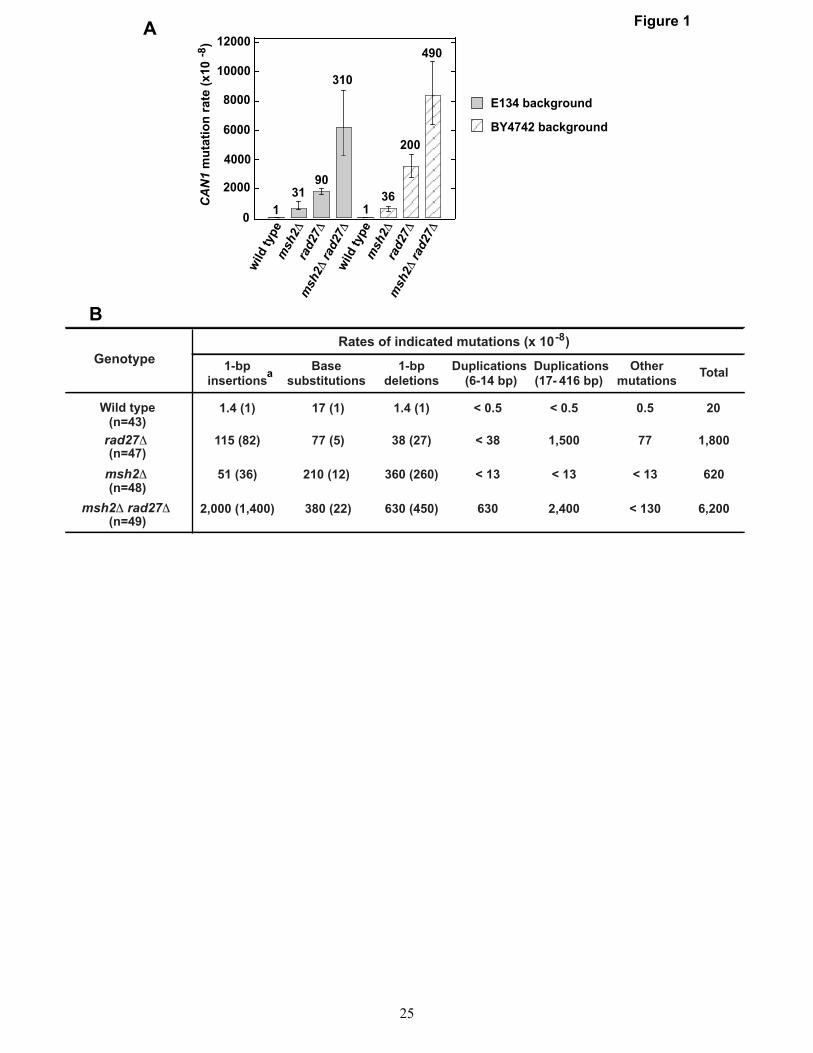
The results described above were obtained using the his7-2 and lys2::InsE-A8 reversion assays that only allow scoring of +1 frameshifts. Unlike the his7-2 and lys2::InsE-A8 reversion assays, the CAN1 forward mutation assay allows scoring of many different types of genetic alterations including 1-bp insertions, base substitutions, and 1-bp deletions. The CAN1 forward mutation assay takes advantage of the fact that mutational inactivation of CAN1 gene encoding arginine permease makes the yeast cell resistant to canavanine, a structural analogue of arginine. In this assay, Canr cells are selected on a synthetic media that lacks arginine and contains canavanine. To determine can1 mutation spectrum in an msh2Δ rad27Δ strain, we performed a series of experiments summarized in Fig. 1. We started this series of experiments by measuring the CAN1
mutation rates in two sets of msh2Δ, rad27Δ, and msh2Δ rad27Δ strains (Fig. 1A). One set of the strains was prepared on the wild-type strain E134 background and the other on the wild-type strain BY4742 background. We chose to measure CAN1 mutation rates in two sets of yeast strains to exclude the possibility that the data are strain-specific. The results demonstrated that the relative CAN1 mutation rate in either msh2Δ rad27Δ mutant was ~2-times higher than the sum of the relative CAN1 mutation rates in the isogenic single mutants (i.e., the relative CAN1 mutation rates in the isogenic msh2Δ and rad27Δ mutants are in a weak synergistic relationship) (Fig. 1A). Similar results were obtained in two earlier studies (48,49). We next determined the can1 mutation spectra in the wild-type, msh2Δ, rad27Δ, and msh2Δ rad27Δ mutants (Fig. 1B). The rates of base substitutions and 1-nt deletions in the msh2Δ rad27Δ mutant did not differ significantly from those in the msh2Δ mutant. On the other hand, the rate of 1-nt insertions in the msh2Δ rad27Δ mutant was 12 times higher than sum of those in the msh2Δ and rad27Δ mutants. This information supports the view that one or several related types of pre-mutagenic intermediates causing 1-nt insertions are removed by both an MMR-dependent mechanism and a Rad27-dependent mechanism.
Duplications are formed at a high rate in rad27Δ mutants (31,32). These duplications have been suggested to be the products of unprocessed Okazaki fragment flaps (31,49). Strikingly, 6-14-bp duplications were produced at a rate of 630x10-8 in CAN1 in the msh2Δ rad27Δ strain, but were absent in the can1 spectra of the rad27Δ and msh2Δ mutants (Fig. 1B). These data suggest that one or several related types of pre-mutagenic intermediates triggering 6-14-bp duplications are removed by both an MMR-dependent mechanism and a Rad27-dependent mechanism.
The Dna2 helicase/nuclease is an essential enzyme that participates in the removal of flaps during Okazaki fragment maturation (33-36). Yeast strains carrying a dna2 allele, dna2-1, are temperature-sensitive (55,56) and show a weak defect in the maintenance of dinucleotide repeats (56). We established that the his7-2 mutation rate in the dna2-1 strain was increased 10-fold relative to that in the wild-type strain (Table 6). Sequencing of ten independent HIS7 revertants produced in the dna2-1 background showed that
5

The MMR system and Okazaki fragment maturation
nine mutants contained an identical mutation, which was an A insertion in the his7-2 A7 run, and one mutant had a deletion of two As in the same run. We then studied the effect of combining dna2-1 with msh2Δ on the his7-2 mutation rate (Table 6). We found that the his7-2 mutation rate in the dna2-1 msh2Δ double mutant was 2-times higher than the sum of those in the single mutants. This observation is consistent with the idea that one or several related types of pre-mutagenic intermediates causing +1 frameshifts are repaired by both an MMR system-dependent mechanism and a Dna2-dependent mechanism.
Recognition of 1-nt DNA flaps by MutSα
We considered two models to explain the observation that combining msh2Δ with rad27Δ leads to the strong synergistic increases in the rates of spontaneous 1-bp insertions (Table 2 and Fig. 1B). In the first model, DNA polymerase α errors are corrected not only by MMR (57), but also by a Rad27-dependent mechanism, and DNA polymerase α errors that escape both MMR and the Rad27-dependent mechanism produce mutations including 1-bp insertions. However this model is not supported by the observation that the deletion of RAD27 in the msh2Δ strain does not significantly increase the rate of base substitutions (Fig. 1B), which are the most common products of DNA polymerase α errors (57,58). Thus, it is unlikely that a considerable fraction of 1-bp insertions formed in msh2Δ rad27Δ mutants originate from DNA polymerase α errors. The second model is based on the knowledge that the key function of the 5′ flap endonuclease Rad27 is the removal of short Okazaki fragment flaps (33,34). In this model, 1-nt Okazaki fragment flaps are removed by both a Rad27-dependent mechanism and an MMR system-dependent mechanism, and the unprocessed flaps are converted by misalignment and ligation into 1-bp insertions. Thus, this model suggests that the majority of 1-bp insertions produced in msh2Δ rad27Δ mutants are formed from 1-nt Okazaki fragment flaps. Since Okazaki fragment flaps do not cause base substitutions, the second model is consistent with our genetic data (Table 2 and Fig. 1B).
The second model postulates that the MMR system removes 1-nt Okazaki fragment flaps. To determine whether there is evidence for this, we carried out the biochemical experiments described
below. We first examined whether yeast MutSα recognizes 1-nt DNA flaps present on the 32P-labeled oligonucleotide-based substrates (Fig. 2). The data revealed that yeast MutSα bound the substrate containing the dynamic 1-nt flap with an apparent Kd of 38 ± 2 nM (Fig. 2A-B). The control experiments indicated that yeast MutSα bound the 1-nt insertion-containing DNA, nicked DNA, and homoduplex DNA with apparent Kds of 25 ± 1 nM, 180 ± 10 nM, and 200 ± 8 nM, respectively (Fig. 2A-B). Therefore, these experiments demonstrate that yeast MutSα recognizes the dynamic 1-nt flap nearly as efficiently as the 1-nt insertion. We then investigated whether yeast MutSα recognizes static 1-nt 3′ and 5′ flaps. The experiments showed that yeast MutSα bound the static 1-nt 3′ and 5′ flaps with apparent Kds of 60 ± 2 nM and 55 ± 3 nM, respectively. Thus, yeast MutSα recognizes the static 1-nt 3′ and 5′ flaps with the same affinity. Surprisingly, yeast MutSα detected the static 1-nt 3′ and 5′ flaps somewhat less efficiently than the dynamic 1-nt flap (Fig. 2A-B). Since a dAMP residue forms the flap in the dynamic substrate and a dCMP residue produces the flaps in the static substrates, it is possible that yeast MutSα recognizes a flapped dCMP residue less efficiently than a flapped dAMP residue.
We also studied whether human MutSα recognizes the dynamic 1-nt flap (Fig. 2C). Our experiments indicated that human MutSα bound the dynamic 1-nt flap with an apparent Kd of 30 ± 1 nM. An apparent Kd for binding of human MutSα to the 1-nt insertion is 30 ± 6 nM. These Kd values are 7-12 times lower than those for binding of human MutSα to the nicked and homoduplex DNAs (Fig. 2C). Thus, human MutSα efficiently recognizes the dynamic 1-nt flap. Collectively, these findings support the view that the ability to recognize 1-nt DNA flaps is conserved in eukaryotic MutSα proteins.
We also analyzed whether human MutSα recognizes a dynamic 1-nt flap present on a circular 2-kb DNA (Fig. 3). Each of the substrates contained a 1-nt DNA flap, a 1-nt RNA flap, no flap, or a G-T mispair (Fig. 3A). The results revealed that MutSα bound the 1-nt DNA and RNA flap-containing DNAs with Kd values of 119 ± 3 nM and 115 ± 10 nM, respectively (Fig. 3B). These Kd values are half that of 254 ± 35 nM for the binding of MutSα to the control no-flap DNA. Thus, MutSα detects that the circular DNA carries
6

The MMR system and Okazaki fragment maturation
a 1-nt flap, which may be a deoxyribonucleotide or ribonucleotide residue.
MutLα endonuclease-dependent removal of 1-nt flaps
Having shown that MutSα recognizes the 1-nt DNA and RNA flaps on the circular DNA, we carried out and analyzed the reconstituted reactions to determine whether these flaps activate human MutLα endonuclease to incise the discontinuous strand in the presence of human MutSα, PCNA, RFC, and RPA (Fig. 4). The circular DNAs were used as substrates in these reactions because loaded PCNA, required for the activation of MutLα endonuclease (25-27), slides off of linear DNA. The reactions were performed under conditions that were very similar to those used for the identification of the MutSα-, PCNA-, RFC-, mismatch-, and ATP-dependent endonuclease activity of human MutLα (25). Analysis of the reactions (Fig. 4A-B) led to the following observations. First, 34 ± 5% of the discontinuous strand of the 1-nt DNA flap-containing substrate was incised by MutLα, whereas the endonuclease cleaved only 10 ± 2% of the discontinuous strand of the control flap-free substrate. Second, MutLα incised 30 ± 1% of the discontinuous strand of the 1-nt RNA flap. Third, an endonuclease-deficient MutLα variant, MutLα-E705K (25), did not incise the discontinuous strands of the tested substrates. Together, these observations indicate that 1-nt flaps activate MutLα endonuclease to incise the discontinuous strand in the presence of MutSα, PCNA, RFC, and RPA.
To determine whether incision of the discontinuous strand by MutLα results in the removal of flaps, we performed experiments summarized in Fig. 5. As shown in lane 2 of Fig. 5A, the incubation of MutLα, MutSα, PCNA, RFC, and RPA with the 1-nt DNA flap-containing circular substrate led to incision of the 32P-labeled 37-nt fragment at several sites. The most abundant product of the incision reaction had an apparent length of 5 nt, indicating that the incision occurred at a site that is four nucleotides 3′ to the flap. The incision products were not formed when MutSα, MutLα, RFC, or PCNA was omitted from the reaction mixture, but the omission of RPA did not have a significant effect on the incision (Fig. 5A, lanes 3, 4, 6 and 7, and Fig.5B). These results indicate that MutSα, MutLα, RFC, and PCNA are
required for the incision, but RPA is not. The time-course experiments demonstrated that the incision reaction produced the 5-nt fragment in a time-dependent manner (Fig. 5C). The efficiency of the incision of the site located 4 nt downstream from a 1-nt flap was three times higher than that of the same site on the control flap-free substrate (Fig. 5A, lanes 2 and 10, and Fig.5B-C). Thus, the flap dependence of the MutLα incision was three-fold. Changing the incubation temperature from 37°C to 25°C decreased the flap dependence of the MutLα incision from three- to two-fold (data not shown). MutLα, MutSα, PCNA, and RFC were also required for the incision of the 1-nt RNA flap-containing substrate (Fig. 5A, lanes 18-20,22, and 23, and Fig.5B). Consistent with previous study (59), the 5-nt incision product containing the 5′ ribonucleotide residue migrated in the gel slightly slower than the 5-nt incision product lacking a ribonucleotide residue (Fig. 5A, lanes 2 and 18).
We also studied whether the endonuclease activity of MutLα is necessary for the incision of the discontinuous strand at a 1-nt flap (Fig. 5A-B). The replacement of the wild-type MutLα with the endonuclease-deficient MutLα-E705K led to the disappearance of the incision products indicating that the endonuclease activity of MutLα is responsible for the incisions (Fig. 5A, lanes 5 and 21, and Fig. 5B). Further analysis revealed that the presence of a 1-nt flap did not activate MutLα endonuclease to incise the discontinuous strand immediately upstream from the flap (data not shown). Taken together, these experiments demonstrate that MutSα, RFC, and PCNA activate MutLα endonuclease to incise the discontinuous strand 4 nt downstream from a 1-nt DNA or RNA flap. Since the incision is so close to the flaps, it triggers their dissociation from the substrates.
Newly replicated DNA is rapidly assembled into nucleosomes by a mechanism that depends on the histone H3-H4 chaperone CAF-1 (60-62). The first step in CAF-1-dependent nucleosome assembly is the deposition of histone H3-H4 tetramers. CAF-1-dependent nucleosome assembly probably impacts many processes that take place on the nascent DNA. Consistent with this idea, CAF-1-dependent nucleosome assembly modulates MMR (46,63). Because the MMR system-dependent flap removal (Fig. 5) is likely to occur during CAF-1-dependent nucleosome assembly, we studied whether histone H3-H4 deposition by CAF-1 affects the flap-removing
7

The MMR system and Okazaki fragment maturation
activity of the MMR system. We determined that CAF-1-dependent histone H3-H4 deposition stimulated the flap-removing activity of the MMR system by two-fold (Fig. 6A, lanes 11 and 12, and Fig. 6B) and increased the flap dependence of the incision from three- to six-fold (Fig. 6C). The efficiency of the flap removal was not changed when MutSα and MutLα were added to the reaction mixtures that were incubated with CAF-1, the histone H3-H4 complex, PCNA, RFC, and RPA for 15 min suggesting that the MMR system efficiently removes 1-nt DNA flaps in the presence of pre-loaded H3-H4 tetramers (data not shown). The omission of CAF-1 significantly decreased both the efficiency and flap dependence of the incision (Fig. 6C). Control experiments revealed that the flap removal occurring in the presence of CAF-1-dependent histone H3-H4 deposition required both MutSα and MutLα (Fig. 6A-B). An endonuclease-deficient MutLα variant, MutLα-D699N (25), as well as a MutLα ATPase mutant, MutLα-EA (64), could not substitute for the wild-type MutLα in the incision reaction. Thus, these experiments demonstrate that the CAF-1-dependent histone H3-H4 deposition promotes the removal of 1-nt DNA flaps by the activated MutLα endonuclease.
We then studied how CAF-1 and the histone H3-H4 complex affect the incision of the discontinuous strand at sites that are distant from the 1-nt flap (Fig. 7). Strikingly, the presence of CAF-1 and the histone H3-H4 complex suppressed the MutLα endonuclease-dependent incision of the discontinuous strand at the remote sites (Fig. 7A, lanes 9 and 14-16, and Fig. 7B). A similar suppression of the MutLα endonuclease-dependent incision of the discontinuous strand was observed in the 6 protein-system containing the histone H3-H4 complex, but not CAF-1 (Fig. 7A, lanes 9-12, and Fig. 7B). These findings imply that both CAF-1-dependent histone H3-H4 deposition onto the DNA and non-specific binding of the histone H3-H4 complex to the DNA protect the remote sites from the incision by the activated MutLα endonuclease.
Next, we performed experiments to study whether the reconstituted MMR system is able to remove flaps in the presence of FEN1 (Fig. 8). The data showed that increasing FEN1 concentration decreased the yield of the product of MutLα endonuclease-dependent flap removal and increased the yield of the product of FEN1-
dependent flap removal (Fig. 8A-C). In addition, the data indicated that one or several proteins present in the eight-protein system suppressed the flap endonuclease activity of FEN1 (Fig. 8A, lanes 3-10, and Fig. 8C). These experiments provide evidence that the MMR system removes flaps in the presence of FEN1 and suggest that the flap endonuclease activities of FEN1 and the MMR system compete with each other.
DISCUSSION
High-fidelity DNA replication is required for the maintenance of genome integrity and the suppression of human diseases (65). The MMR system improves the fidelity of DNA replication by correcting the errors of DNA polymerization (3,4,7). We have used genetic analysis and reconstituted systems to study whether the MMR system contributes to the removal of Okazaki fragment flaps. The major findings described in this report are (1) combining rad27Δ with msh2Δ produces strong synergistic increases in the rates of 1-bp insertions in his7-2 and CAN1 (Table 2 and Fig. 1B); (2) combining rad27Δ with mlh1Δ, pms1Δ, or pms1-E707K causes a 20-26 times synergistic increase in the rate of +1 frameshifts in his7-2 (Table 5); (3) purified yeast and human MutSα proteins recognize 1-nt flaps (Fig. 2 and 3); (4) MutLα endonuclease activated by MutSα, RFC, and PCNA removes 1-nt flaps (Fig. 5); (5) the flap-removing activity of the reconstituted MMR system is stimulated by CAF-1-dependent histone H3-H4 deposition (Fig. 6); and (6) the reconstituted MMR system removes 1-nt flaps in the presence of FEN1 (Fig. 8).
These findings indicate that the eukaryotic MMR system removes a subset of 1-nt Okazaki fragment flaps and support a model illustrated in Fig. 9. This model suggests that MutSα, MutLα, PCNA, and RFC provide the minimal set of activities required for the removal of 1-nt Okazaki fragment flaps by the MMR system. According to this model, the mechanism of the removal of a 1-nt Okazaki fragment flap by the MMR system can be divided into three key steps: recognition of the flap by MutSα, activation of MutLα endonuclease by MutSα, PCNA, and RFC, and the removal of the flap by the activated MutLα endonuclease. Our genetic results also suggest that there is an Msh2-dependent, MutLα-independent mechanism of removal of 1-nt Okazaki fragment flaps (Table 5). In addition, our genetic results are compatible with
8

The MMR system and Okazaki fragment maturation
another model. In this model, misalignment and ligation converts some 1-nt Okazaki fragment flaps into 1-nt loops, which are then removed by the strand-specific MMR (8,11,19,28,66). However, it has not yet been demonstrated that a replicative DNA ligase is able to convert 1-nt flaps into 1-nt loops in the presence of Rad27/FEN1 and/or the MMR system.
The absolute his7-2 mutation rate in the rad27Δ/rad27Δ msh2Δ/msh2Δ diploid (Table 3) is half that of the previously described strong mutator diploid pol3-01/pol3-01 msh2Δ/msh2Δ (40). (The pol3-01 mutation inactivates the proofreading activity of DNA polymerase δ.) This observation reveals that the MMR system is nearly as important for the removal of +1 frameshift intermediates in rad27Δ strains as for the repair of +1 frameshift intermediates in pol3-01 strains. Genetic interactions between the MMR system and Rad27 have been investigated in the past (31,48,49), but none of the previous studies utilized a +1 frameshift assay or determined can1 mutation spectrum in a strain that lacks an MMR gene and RAD27. Nevertheless, Johnson et al. (48) reported that the relative CAN1 mutation rate in the msh2Δ mutant is in a weak synergistic relationship with that in the rad27Δ mutant. Thus, the results of the measurements of the relative CAN1 mutation rates in the msh2Δ, rad27Δ, and msh2Δ rad27Δ mutants obtained in this work (Fig. 1A) and the study of Johnson et al. (48) are consistent with each other.
MutSα was initially identified as an MMR factor that detects single base-base mismatches (11,12). Subsequent work established that MutSα recognizes 1-12 nt insertion/deletion loops (15,19) and damaged base pairs (67). We have described in this report that MutSα recognizes 1-nt DNA/RNA flaps (Fig. 2-3). This finding extends the range of potentially mutagenic DNA structures recognized by MutSα. Our genetic experiments support the idea that MutSβ plays a role in the MMR system-dependent removal of 1-nt Okazaki fragment flaps (Table 5). Thus, it is possible that MutSβ, like MutSα, recognizes 1-nt DNA/RNA flaps and activates MutLα endonuclease to remove them. This would be in line with previous work that identified that MutSβ specifically binds a variety of DNA recombination structures including the non-complementary 5′ DNA flaps and 3′ tails (68).
In the crystal structure of MutSα-G-T DNA complex, the E434 residue of the conserved mismatch recognition F-X-E motif forms a hydrogen bond with the mispaired T, the conserved F432 residue stacks onto the T, DNA is sharply bent at the mismatch, and there are several non-specific protein-DNA interactions (69). These features are also present in the structures of the prokaryotic MutS-mismatch-containing DNA complexes (70,71). It has been proposed that during mismatch recognition, MutS stacks the conserved F on an unpaired nucleotide residue and bends DNA (70). The intrinsic bendability of duplex DNA at a mismatch is thought to strongly contribute to the recognition of the mismatch by MutS. A recent work has shown that the same mechanism of mismatch recognition is employed by MutSα (69). We speculate that the MSH6 F-X-E motif is responsible for the recognition of flaps by MutSα. If this is the case, the conserved E434 is a strong candidate to interact with a flapped deoxy- or ribonucleotide residue via a hydrogen bond. It has been described that duplex DNA bends at nicks (72). Therefore, DNA bending at a nick that accompanies the flap may facilitate the flap recognition by MutSα. Since the MMR system is conserved from bacteria to humans (69-71), it is possible that the MMR system also contributes to the removal of Okazaki fragment flaps in bacteria.
Eukaryotic DNA transactions occur in the nucleosomal environment. The fact that the size of naked nascent DNA strands at a eukaryotic replication fork is only ~450 bp (73) is consistent with the view that the newly replicated DNA is rapidly assembled into nucleosomes by the histone chaperone CAF-1 (60). Our analysis demonstrates that the CAF-1-dependent histone H3-H4 deposition increases the efficiency and specificity of the flap removal by MutLα and protects the discontinuous strand from MutLα incision at the remote sites (Fig. 6 and 7). The mechanism behind these effects is not known. We speculate that the loaded histones H3-H4 tetramers trap the MutLα-containing incision complex at the flap-containing site where it was assembled, and as a result the MutLα is not able to incise the discontinuous strand at the remote sites and instead removes the flap.
Previous research demonstrated that during eukaryotic Okazaki fragment maturation, the strand displacement activity of DNA polymerase δ (30,33,74) produces flaps that are removed by the
9
The MMR system and Okazaki fragment maturation
Rad27/FEN1 endonuclease (33,34), the 3′-5′ exonuclease activity of DNA polymerase δ (32), and the nuclease/helicase Dna2 (36). In this report,
we have described evidence that the eukaryotic MMR system contributes to the removal of Okazaki fragment flaps.
ACKNOWLEDGEMENTS
We are grateful to Paul Modrich for his advice and insightful discussions, Farid F. Kadyrov for critical reading of the manuscript, Francesca Storici and Kirill Lobachev for help with the “dellitto perfetto” technique. We thank Tim Formosa for supplying a dna2-1 strain and Paul Modrich and Mike Resnick for providing plasmids used in this work.
CONFLICT OF INTEREST The authors declare that they have no conflicts of interest with the contents of this article.
AUTHOR CONTRIBUTIONS
FK and LK designed experiments. LK, BD, and FK performed experiments and analyzed data. FK and LK wrote the paper.
REFERENCES
1. Modrich, P. (1995) Mismatch repair, genetic stability and tumour avoidance. Philos. Trans. R. Soc. Lond. B Biol. Sci. 347, 89-95
2. Harfe, B. D., and Jinks-Robertson, S. (2000) DNA Mismatch Repair and Genetic Instability. Annu. Rev. Genet. 34, 359-399
3. Kunkel, T. A., and Erie, D. A. (2005) DNA Mismatch Repair. Annu. Rev. Biochem. 74, 681-710
4. Li, G. M. (2008) Mechanisms and functions of DNA mismatch repair. Cell Res. 18, 85-98
5. Jiricny, J. (2013) Postreplicative mismatch repair. Cold Spring Harb. Perspect. Biol. 5, a012633
6. Lynch, H. T., Snyder, C. L., Shaw, T. G., Heinen, C. D., and Hitchins, M. P. (2015) Milestones of Lynch syndrome: 1895-2015. Nat. Rev. Cancer 15, 181-194
7. Iyer, R. R., Pluciennik, A., Burdett, V., and Modrich, P. L. (2006) DNA mismatch repair: functions and mechanisms. Chem. Rev. 106, 302-323
8. Modrich, P. (2006) Mechanisms in eukaryotic mismatch repair. J. Biol. Chem. 281, 30305-30309
9. Strand, M., Prolla, T. A., Liskay, R. M., and Petes, T. D. (1993) Destabilization of tracts of simple repetitive DNA in yeast by mutations affecting DNA mismatch repair. Nature 365, 274-276
10. de Wind, N., Dekker, M., Berns, A., Radman, M., and te Riele, H. (1995) Inactivation of the mouse Msh2 gene results in mismatch repair deficiency, methylation tolerance, hyperrecombination, and predisposition to cancer. Cell 82, 321-330
11. Drummond, J. T., Li, G.-M., Longley, M. J., and Modrich, P. (1995) Isolation of an hMSH2•p160 heterodimer that restores mismatch repair to tumor cells. Science 268, 1909-1912
12. Palombo, F., Gallinari, P., Iaccarino, I., Lettieri, T., Hughes, M., D'Arrigo, A., Truong, O., Hsuan, J. J., and Jiricny, J. (1995) GTBP, a 160-kilodalton protein essential for mismatch-binding activity in human cells. Science 268, 1912-1914
10

The MMR system and Okazaki fragment maturation
13. Li, G.-M., and Modrich, P. (1995) Restoration of mismatch repair to nuclear extracts of H6 colorectal tumor cells by a heterodimer of human MutL homologs. Proc. Natl. Acad. Sci. U. S. A. 92, 1950-1954
14. Edelmann, W., Cohen, P. E., Kane, M., Lau, K., Morrow, B., Bennett, S., Umar, A., Kunkel, T., Cattoretti, G., Chaganti, R., Pollard, J. W., Kolodner, R. D., and Kucherlapati, R. (1996) Meiotic pachytene arrest in MLH1-deficient mice. Cell 85, 1125-1134
15. Palombo, F., Iaccarino, I., Nakajima, E., Ikejima, M., Shimada, T., and Jiricny, J. (1996) hMutSβ, a heterodimer of hMSH2 and hMSH3, binds to insertion/deletion loops in DNA. Curr. Biol. 6, 1181-1184
16. Johnson, R. E., Kovvali, G. K., Guzder, S. N., Amin, N. S., Holm, C., Habraken, Y., Sung, P., Prakash, L., and Prakash, S. (1996) Evidence for involvement of yeast proliferating cell nuclear antigen in DNA mismatch repair. J. Biol. Chem. 271, 27987-27990
17. Umar, A., Buermeyer, A. B., Simon, J. A., Thomas, D. C., Clark, A. B., Liskay, R. M., and Kunkel, T. A. (1996) Requirement for PCNA in DNA mismatch repair at a step preceding DNA resynthesis. Cell 87, 65-73
18. Tishkoff, D. X., Boerger, A. L., Bertrand, P., Filosi, N., Gaida, G. M., Kane, M. F., and Kolodner, R. D. (1997) Identification and characterization of Saccharomyces cerevisiae EXO1, a gene encoding an exonuclease that interacts with MSH2. Proc. Natl. Acad. Sci. U. S. A. 94, 7487-7492
19. Genschel, J., Littman, S. J., Drummond, J. T., and Modrich, P. (1998) Isolation of hMutSβ from human cells and comparison of the mismatch repair specificities of hMutSβ and hMutSα. J. Biol. Chem. 273, 19895-19901
20. de Wind, N., Dekker, M., Claij, N., Jansen, L., van Klink, Y., Radman, M., Riggins, G., van der Valk, M., van't Wout, K., and te Riele, H. (1999) HNPCC-like cancer predisposition in mice through simultaneous loss of Msh3 and Msh6 mismatch-repair protein functions. Nat. Genet. 23, 359-362
21. Genschel, J., Bazemore, L. R., and Modrich, P. (2002) Human exonuclease I is required for 5' and 3' mismatch repair. J. Biol. Chem. 277, 13302-13311
22. Wei, K., Clark, A. B., Wong, E., Kane, M. F., Mazur, D. J., Parris, T., Kolas, N. K., Russell, R., Hou, H., Jr., Kneitz, B., Yang, G., Kunkel, T. A., Kolodner, R. D., Cohen, P. E., and Edelmann, W. (2003) Inactivation of Exonuclease 1 in mice results in DNA mismatch repair defects, increased cancer susceptibility, and male and female sterility. Genes Dev. 17, 603-614
23. Dzantiev, L., Constantin, N., Genschel, J., Iyer, R. R., Burgers, P. M., and Modrich, P. (2004) A defined human system that supports bidirectional mismatch-provoked excision. Mol. Cell 15, 31-41
24. Pavlov, Y. I., Mian, I. M., and Kunkel, T. A. (2003) Evidence for preferential mismatch repair of lagging strand DNA replication errors in yeast. Curr. Biol. 13, 744-748
25. Kadyrov, F. A., Dzantiev, L., Constantin, N., and Modrich, P. (2006) Endonucleolytic function of MutLalpha in human mismatch repair. Cell 126, 297-308
26. Kadyrov, F. A., Holmes, S. F., Arana, M. E., Lukianova, O. A., O'Donnell, M., Kunkel, T. A., and Modrich, P. (2007) Saccharomyces cerevisiae MutLalpha is a mismatch repair endonuclease. J. Biol. Chem. 282, 37181-37190
11
The MMR system and Okazaki fragment maturation
27. Pluciennik, A., Dzantiev, L., Iyer, R. R., Constantin, N., Kadyrov, F. A., and Modrich, P. (2010) PCNA function in the activation and strand direction of MutLalpha endonuclease in mismatch repair. Proc. Natl. Acad. Sci. US A 107, 16066-16071
28. Iyer, R. R., Pluciennik, A., Genschel, J., Tsai, M. S., Beese, L. S., and Modrich, P. (2010) MutLalpha and proliferating cell nuclear antigen share binding sites on MutSbeta. J. Biol. Chem. 285, 11730-11739
29. Constantin, N., Dzantiev, L., Kadyrov, F. A., and Modrich, P. (2005) Human mismatch repair: Reconstitution of a nick-directed bidirectional reaction. J. Biol. Chem. 280, 39752-39761
30. Kadyrov, F. A., Genschel, J., Fang, Y., Penland, E., Edelmann, W., and Modrich, P. (2009) A possible mechanism for exonuclease 1-independent eukaryotic mismatch repair. Proc. Natl. Acad. Sci. USA 106, 8495-8500
31. Tishkoff, D. X., Filosi, N., Gaida, G. M., and Kolodner, R. D. (1997) A novel mutation avoidance mechanism dependent on S. cerevisiae RAD27 is distinct from DNA mismatch repair. Cell 88, 253-263
32. Jin, Y. H., Obert, R., Burgers, P. M., Kunkel, T. A., Resnick, M. A., and Gordenin, D. A. (2001) The 3'-->5' exonuclease of DNA polymerase delta can substitute for the 5' flap endonuclease Rad27/Fen1 in processing Okazaki fragments and preventing genome instability. Proc. Natl. Acad. Sci. U S A 98, 5122-5127
33. Burgers, P. M. (2009) Polymerase dynamics at the eukaryotic DNA replication fork. J. Biol. Chem. 284, 4041-4045
34. Balakrishnan, L., and Bambara, R. A. (2011) Eukaryotic lagging strand DNA replication employs a multi-pathway mechanism that protects genome integrity. J. Biol. Chem. 286, 6865-6870
35. Budd, M. E., and Campbell, J. L. (1995) A yeast gene required for DNA replication encodes a protein with homology to DNA helicases. Proc. Natl. Acad. Sci. U .S .A. 92, 7642-7646
36. Ayyagari, R., Gomes, X. V., Gordenin, D. A., and Burgers, P. M. (2003) Okazaki fragment maturation in yeast. I. Distribution of functions between FEN1 AND DNA2. J. Biol. Chem. 278, 1618-1625
37. Li, X., Li, J., Harrington, J., Lieber, M. R., and Burgers, P. M. (1995) Lagging strand DNA synthesis at the eukaryotic replication fork involves binding and stimulation of FEN-1 by proliferating cell nuclear antigen. J. Biol. Chem. 270, 22109-22112
38. Morrison, A., Johnson, A. L., Johnston, L. H., and Sugino, A. (1993) Pathway correcting DNA replication errors in Saccharomyces cerevisiae. EMBO J. 12, 1467-1473
39. Morrison, A., and Sugino, A. (1994) The 3'-->5' exonucleases of both DNA polymerases delta and epsilon participate in correcting errors of DNA replication in Saccharomyces cerevisiae. Mol. Gen. Genet. 242, 289-296
40. Tran, H. T., Gordenin, D. A., and Resnick, M. A. (1999) The 3'-->5' exonucleases of DNA polymerases delta and epsilon and the 5'-->3' exonuclease Exo1 have major roles in postreplication mutation avoidance in Saccharomyces cerevisiae. Mol. Cell. Biol. 19, 2000-2007
41. Greene, C. N., and Jinks-Robertson, S. (2001) Spontaneous frameshift mutations in Saccharomyces cerevisiae: accumulation during DNA replication and removal by proofreading and mismatch repair activities. Genetics 159, 65-75
12
The MMR system and Okazaki fragment maturation
42. Kadyrova, L. Y., Mertz, T. M., Zhang, Y., Northam, M. R., Sheng, Z., Lobachev, K. S., Shcherbakova, P. V., and Kadyrov, F. A. (2013) A reversible histone H3 acetylation cooperates with mismatch repair and replicative polymerases in maintaining genome stability. PLoS Genet. 9, e1003899
43. Tran, H. T., Keen, J. D., Kricker, M., Resnick, M. A., and Gordenin, D. A. (1997) Hypermutability of homonucleotide runs in mismatch repair and DNA polymerase proofreading yeast mutants. Mol. Cell. Biol. 17, 2859-2865
44. Shcherbakova, P. V., and Kunkel, T. A. (1999) Mutator phenotypes conferred by MLH1 overexpression and by heterozygosity for mlh1 mutations. Mol. Cell. Biol. 19, 3177-3183
45. Storici, F., Lewis, L. K., and Resnick, M. A. (2001) In vivo site-directed mutagenesis using oligonucleotides. Nat. Biotechnol. 19, 773-776
46. Kadyrova, L. Y., Rodriges Blanko, E., and Kadyrov, F. A. (2011) CAF-I-dependent control of degradation of the discontinuous strands during mismatch repair. Proc. Natl. Acad. Sci. U S A 108, 2753-2758
47. York, S. J., and Modrich, P. (2006) Mismatch repair-dependent iterative excision at irreparable O6-methylguanine lesions in human nuclear extracts. J. Biol. Chem. 281, 22674-22683
48. Johnson, R. E., Kovvali, G. K., Prakash, L., and Prakash, S. (1995) Requirement of the yeast RTH1 5' to 3' exonuclease for the stability of simple repetitive DNA. Science 269, 238-240
49. Xie, Y., Liu, Y., Argueso, J. L., Henricksen, L. A., Kao, H. I., Bambara, R. A., and Alani, E. (2001) Identification of rad27 mutations that confer differential defects in mutation avoidance, repeat tract instability, and flap cleavage. Mol. Cell. Biol. 21, 4889-4899
50. Johnson, R. E., Kovvali, G. K., Prakash, L., and Prakash, S. (1996) Requirement of the yeast MSH3 and MSH6 genes for MSH2-dependent genomic stability. J. Biol. Chem. 271, 7285-7288
51. Marsischky, G. T., Filosi, N., Kane, M. F., and Kolodner, R. (1996) Redundancy of Saccharomyces cerevisiae MSH3 and MSH6 in MSH2-dependent mismatch repair. Genes Dev. 10, 407-420
52. Harfe, B. D., and Jinks-Robertson, S. (1999) Removal of frameshift intermediates by mismatch repair proteins in Saccharomyces cerevisiae. Mol. Cell. Biol. 19, 4766-4773
53. Pillon, M. C., Lorenowicz, J. J., Uckelmann, M., Klocko, A. D., Mitchell, R. R., Chung, Y. S., Modrich, P., Walker, G. C., Simmons, L. A., Friedhoff, P., and Guarne, A. (2010) Structure of the endonuclease domain of MutL: unlicensed to cut. Mol. Cell 39, 145-151
54. Gueneau, E., Dherin, C., Legrand, P., Tellier-Lebegue, C., Gilquin, B., Bonnesoeur, P., Londino, F., Quemener, C., Le Du, M. H., Marquez, J. A., Moutiez, M., Gondry, M., Boiteux, S., and Charbonnier, J. B. (2013) Structure of the MutLalpha C-terminal domain reveals how Mlh1 contributes to Pms1 endonuclease site. Nat. Struct. Mol. Biol. 20, 461-468
55. Formosa, T., and Nittis, T. (1999) Dna2 mutants reveal interactions with Dna polymerase alpha and Ctf4, a Pol alpha accessory factor, and show that full Dna2 helicase activity is not essential for growth. Genetics 151, 1459-1470
56. Budd, M. E., and Campbell, J. L. (2000) The pattern of sensitivity of yeast dna2 mutants to DNA damaging agents suggests a role in DSB and postreplication repair pathways. Mutat. Res. 459, 173-186
13

The MMR system and Okazaki fragment maturation
57. Nick McElhinny, S. A., Kissling, G. E., and Kunkel, T. A. (2010) Differential correction of lagging-strand replication errors made by DNA polymerases {alpha} and {delta}. Proc. Natl. Acad. Sci. U S A 107, 21070-21075
58. Kunkel, T. A. (1985) The mutational specificity of DNA polymerases-alpha and -gamma during in vitro DNA synthesis. J. Biol. Chem. 260, 12866-12874
59. Nick McElhinny, S. A., Watts, B. E., Kumar, D., Watt, D. L., Lundstrom, E. B., Burgers, P. M., Johansson, E., Chabes, A., and Kunkel, T. A. (2010) Abundant ribonucleotide incorporation into DNA by yeast replicative polymerases. Proc. Natl. Acad. Sci. U S A 107, 4949-4954
60. Smith, S., and Stillman, B. (1989) Purification and characterization of CAF-I, a human cell factor required for chromatin assembly during DNA replication in vitro. Cell 58, 15-25
61. Smith, S., and Stillman, B. (1991) Stepwise assembly of chromatin during DNA replication in vitro. EMBO J 10, 971-980
62. Kadyrova, L. Y., Rodriges Blanko, E., and Kadyrov, F. A. (2013) Human CAF-1-dependent nucleosome assembly in a defined system. Cell Cycle 12, 3286-3297
63. Schopf, B., Bregenhorn, S., Quivy, J. P., Kadyrov, F. A., Almouzni, G., and Jiricny, J. (2012) Interplay between mismatch repair and chromatin assembly. Proc .Natl. Acad. Sci. U S A 109, 1895-1900
64. Raschle, M., Dufner, P., Marra, G., and Jiricny, J. (2002) Mutations within the hMLH1 and hPMS2 subunits of the human MutLalpha mismatch repair factor affect its ATPase activity, but not its ability to interact with hMutSalpha. J. Biol. Chem. 277, 21810-21820
65. Kunkel, T. A. (2004) DNA replication fidelity. J. Biol. Chem. 279, 16895-16898
66. Pena-Diaz, J., and Jiricny, J. (2012) Mammalian mismatch repair: error-free or error-prone? Trends Biochem. Sci. 37, 206-214
67. Duckett, D. R., Drummond, J. T., Murchie, A. I. H., Reardon, J. T., Sancar, A., Lilley, D. M. J., and Modrich, P. (1996) Human MutSα recognizes damaged DNA base pairs containing O6-methylguanine, O4-methylthymine or the cisplatin-d(GpG) adduct. Proc. Natl. Acad. Sci. U. S. A. 93, 6443-6447
68. Surtees, J. A., and Alani, E. (2006) Mismatch repair factor MSH2-MSH3 binds and alters the conformation of branched DNA structures predicted to form during genetic recombination. J. Mol. Biol. 360, 523-536
69. Warren, J. J., Pohlhaus, T. J., Changela, A., Iyer, R. R., Modrich, P. L., and Beese, L. S. (2007) Structure of the human MutSalpha DNA lesion recognition complex. Mol. Cell 26, 579-592
70. Lamers, M. H., Perrakis, A., Enzlin, J. H., Winterwerp, H. H., de Wind, N., and Sixma, T. K. (2000) The crystal structure of DNA mismatch repair protein MutS binding to a G-T mismatch. Nature 407, 711-717
71. Obmolova, G., Ban, C., Hsieh, P., and Yang, W. (2000) Crystal structures of mismatch repair protein MutS and its complex with a substrate DNA. Nature 407, 703-710
72. Kuhn, H., Protozanova, E., and Demidov, V. V. (2002) Monitoring of single nicks in duplex DNA by gel electrophoretic mobility-shift assay. Electrophoresis 23, 2384-2387
14

The MMR system and Okazaki fragment maturation
73. Lucchini, R., Wellinger, R. E., and Sogo, J. M. (2001) Nucleosome positioning at the replication fork. EMBO J. 20, 7294-7302
74. Garg, P., Stith, C. M., Sabouri, N., Johansson, E., and Burgers, P. M. (2004) Idling by DNA polymerase delta maintains a ligatable nick during lagging-strand DNA replication. Genes Dev. 18, 2764-2773
FOOTNOTES Research reported in this publication was supported by the National Institute of General Medical Sciences of the National Institutes of Health under Award Number R01GM095758. The content is solely the responsibility of the authors and does not necessarily represent the official views of the National Institutes of Health.
15

The MMR system and Okazaki fragment maturation
FIGURE LEGENDS Figure 1. CAN1 mutation rates and can1 mutation spectra in the wild-type, msh2Δ , rad27Δ , and msh2Δ rad27Δ strains. (A) CAN1 mutation rates. Each of the mutants was made in the two different wild-type backgrounds: E134 and BY4742. The numbers above the bars are the relative mutation rates. (B) can1 mutation spectra in the wild-type strain E134 and its mutant derivatives. The relative mutation rates are in parentheses. a, all 1-bp insertions were formed in mononucleotide runs that were ≥2N.
Figure 2. Human and yeast MutSα proteins recognize 1-nt DNA flaps. The gel mobility shift assays with the oligonucleotide-based DNA substrates and calculations of the apparent Kds were performed as described in “EXPERIMENTAL PROCEDURES”. All six substrates had the same bottom strand. The DNA sequences of the homoduplex and nicked DNA substrates were identical to each other and to the his7-2 sequence, in which the majority of +1 frameshifts are formed. Compared to the top strand of the homoduplex or nicked substrate, the top strands of the flapped and 1-nt insertion substrates each contained an extra nucleotide residue, which was necessary to produce the 1-nt flap or 1-nt insertion. (A) Representative images showing binding of yeast MutSα to the different DNA substrates. Each DNA-binding reaction was carried out in the mixture containing the indicated concentration of yeast MutSα and the indicated DNA substrate (2 nM). (B) and (C) Apparent Kds for binding of yeast MutSα (B) and human MutSα (C) to the indicated DNA substrates. The apparent Kds were calculated using the data that were obtained by quantification of images including those shown in (A). The numbers above the bars are the apparent Kds.
Figure 3. Human MutSα recognizes 1-nt DNA and RNA flaps on 2-kb circular DNA molecules. (A) Diagrams of the 2-kb circular DNAs used in the DNA-binding reactions. Each diagram also shows the relative position of the hybridization probe (a bar with an asterisk). The hybridization probe is complementary to the continuous strand. (B) Apparent Kds for binding of human MutSα to the indicated circular substrates. The numbers above the bars are the apparent Kds. The gel mobility shift assays and calculations of the apparent Kds were carried out as detailed in “EXPERIMENTAL PROCEDURES”.
Figure 4. 1-nt DNA and RNA flaps activate MutLα endonuclease to incise the discontinuous strands in the presence of MutSα , PCNA, RFC, and RPA. Each DNA incision reaction was carried out in the mixture containing the indicated human proteins and DNA substrate (1.5 nM). When MutSα, MutLα, MutLα-E705K, PCNA, RFC, and RPA were present in the reaction mixtures, their concentrations were 40 nM, 16 nM, 16 nM, 24 nM, 4 nM, and 40 nM, respectively. After a 10-min incubation, the reactions were stopped by the addition of NaOH and EDTA to the final concentrations of 40 mM and 5 mM, respectively. The reaction products were separated on alkaline 1.2% agarose gels, transferred onto nylon membranes, hybridized with 32P-labeled oligonucleotide 16, and visualized by phosphorimaging. (A) Representative images showing incision of the discontinuous strands in the presence of MutLα, MutSα, PCNA, RFC, and RPA. The diagrams outline the circular DNA substrates. Each diagram also shows the relative position of the hybridization probe (a bar with an asterisk). The hybridization probe is complementary to the discontinuous strand. (B) Summary of incision of the discontinuous strands of the indicated DNA substrates at sites that are 4-nt 3′ to the flap or control nick. The data were obtained by quantification of images including those shown in (A) and are presented as averages ± 1 SD, n≥3.
Figure 5. MutLα endonuclease incises the discontinuous strand four nucleotides downstream from a 1-nt DNA or RNA flap. The 37-nt fragments of the 1-nt DNA and RNA flap-containing substrates and the 36-nt fragments of the control flap-free and G-T substrates were labeled at their 5′ ends with 32P. Each DNA incision reaction was performed in the mixture containing the indicated human proteins and 32P-labeled DNA substrate (1.5 nM). When MutSα, MutLα, MutLα-E705K, PCNA, RFC, and RPA were present in the reaction mixtures, their concentrations were 40 nM, 16 nM, 16 nM, 24 nM, 4 nM, and 40 nM, respectively. The DNA incision reactions were stopped and analyzed as described in “EXPERIMENTAL PROCEDURES”. (A) Representative image showing MutLα endonuclease-
16

The MMR system and Okazaki fragment maturation
dependent incision of the discontinuous strand 4 nt downstream from the 1-nt flap. The incision reactions were incubated for 10 min. The diagrams outline the circular DNA substrates. (B) Summary of incision of the discontinuous strands of the indicated substrates at sites that are 4-nt 3′ to the flap or control nick. The DNA incision reactions were incubated for 10 min. (C) Time course of incision of the discontinuous strands of the indicated substrates at sites that are 4-nt 3′ to the flap or control nick. The incision reactions were carried out in the mixtures containing MutSα (40 nM), MutLα (16 nM), PCNA (24 nM), RFC (4 nM), RPA (40 nM), and the indicated DNA substrate (1.5 nM). The data in (B) and (C) are averages ± 1 SD ((B): n≥4 and (C): n≥3) and were obtained by quantification of images including the one shown in (A).
Figure 6. CAF-1-dependent histone H3-H4 deposition stimulates the removal of 1-nt flaps by the activated MutLα endonuclease. The 37-nt fragment of the 1-nt DNA flap-containing substrate and the 36-nt fragment of the control flap-free substrate were labeled at their 5′ ends with 32P. Each DNA incision reaction was performed in the mixture containing the indicated human proteins and 32P-labeled DNA substrate (1.5 nM). When MutSα, MutLα, MutLα-D699N, MutLα-EA, PCNA, RFC, RPA, CAF-1, and the histone H3-H4 heterodimer were present in the reaction mixtures, their concentrations were 40 nM, 16 nM, 16 nM, 16 nM, 24 nM, 4 nM, 40 nM, 24 nM, and 88 nM, respectively. The reactions were incubated for 30 min and then stopped and analyzed as described in “EXPERIMENTAL PROCEDURES”. (A) Representative image showing the effects of the indicated protein combinations on incision of the discontinuous strands of the indicated substrates at sites that are 4-nt 3′ from the flap or control nick. The diagrams outline the circular DNA substrates. (B) Graphical representation the effects of the indicated protein combinations on incision of the discontinuous strands of the indicated substrates at sites that are 4-nt 3′ from the flap or control nick. The data were obtained by quantification of images including the one shown in (A) and are averages ± 1 SD (n≥4). (C) Dependence of the incision on the presence of the 1-nt DNA flap. The flap dependence values were calculated from the data shown in (B). The presence of a statistically significant difference between the flap dependences of the two indicated reactions was identified by unpaired t-test.
Figure 7. CAF-1-dependent histone H3-H4 deposition protects the remote sites from incision by MutLα endonuclease. Each DNA incision reaction was performed in the mixture containing the indicated human proteins and DNA substrate (1.5 nM). When MutSα, MutLα, PCNA, RFC, RPA, and CAF-1 were present in the reaction mixtures, their concentrations were 40 nM, 16 nM, 24 nM, 4 nM, 40 nM, and 24 nM, respectively. After a 30-min incubation, the incision reactions were stopped and analyzed as described in Fig. 4. (A) Image showing the effects of the different protein combinations on incision of the discontinuous strands of the 1-nt flap-containing and flap-free DNA substrates. The diagrams outline the DNA substrates. Each diagram also shows the relative position of the hybridization probe (a bar with an asterisk), which is complementary to the discontinuous strand. (B) and (C) Incision of the discontinuous strands of the 1-nt flap-containing and flap-free DNA substrates as a function of concentration of histone H3-H4 heterodimers. The data were obtained by quantification of images including the one shown in (A) and presented as averages ± 1 SD, n=2.
Figure 8. Flap removal in a reconstituted human system containing FEN1 and MutLα endonucleases. The DNA incision reactions were carried out in the mixtures containing the indicated human proteins and 32P-labeled circular DNA substrate (1.5 nM). When MutSα, MutLα, PCNA, RFC, RPA, CAF-1, and the histone H3-H4 heterodimer were present in the reaction mixtures, their concentrations were 40 nM, 16 nM, 24 nM, 4 nM, 40 nM, 24 nM, and 88 nM, respectively. After incubation for 10 min, the DNA incision reactions were stopped and analyzed as described in “EXPERIMENTAL PROCEDURES”. (A) Representative image showing the effects of the different protein combinations on the removal of the 1-nt DNA flaps. The arrows indicate the positions of the 1-nt and 5-nt cleavage products generated by FEN1 and MutLα, respectively. The diagram outlines the circular DNA substrate. (B) Graphical representation of the effects of the different FEN1 concentrations on the yield of the product of MutLα endonuclease-dependent flap removal in the eight-protein system. The eight-protein system contained MutLα (16 nM), MutSα (40 nM), PCNA (24 nM), RFC (4 nM), RPA
17

The MMR system and Okazaki fragment maturation
(40 nM), CAF-1 (24 nM), histone H3-H4 heterodimer (88 nM), and FEN1 (0.3 nM, 0.6 nM, 1.2 nM, or 2.4 nM). (C) Graphical representation of the effects of the different FEN1 concentrations on the yield of the product of FEN1-dependent flap removal in the one-protein and eight-protein systems. The one-protein system contained FEN1 (0.3 nM, 0.6 nM, 1.2 nM, or 2.4 nM). The data in (B) and (C) were obtained by quantification of images including the one shown in panel (A), and are averages ± 1 SD (n≥4).
Figure 9. Role for the MMR system in DNA replication. The model suggests that the MMR system supports DNA replication by removing 1-nt Okazaki fragment flaps. The process of the removal of a 1-nt Okazaki fragment flap by the MMR system is initiated by the recognition of the flap by MutSα. In the next step, MutSα acts in conjunction with PCNA and RFC to activate MutLα endonuclease. The activated MutLα endonuclease then removes the flap.
18

The MMR system and Okazaki fragment maturation
TABLES
Table 1. The sequences of oligonucleotides described in this report Oligonucleotide Oligonucleotide sequence
1 5′-AACCGTCATTTTCTAGGTTTTTTTCTTTTCTGAATTCAGAA-3′ 2 5′-TTCTGAATTCAGAAAAGAAAAAAACCTAGAAAATGACGGTT-3′ 3 5′-TTCTGAATTCAGAAAAGAAAAAAAACCTAGAAAATGACGGTT-3′ 4 5′-TTCTGAATTCAGAAAAGAAAA-3′ 5 5′-AAAACCTAGAAAATGACGGTT-3′ 6 5′-TTCTGAATTCAGAAAAGAAAAC-3′ 7 5′-AAACCTAGAAAATGACGGTT-3′ 8 5′-CAAACCTAGAAAATGACGGTT-3′ 9 5′-CGCCGAATTGCTAGCAAGCTTTCGAGTCTAGAAATTCGGC-3′ 10 5′-GCCGAATTTCTAGACTCGAAAGCTTGCTAGCAATTCGGCG-3′ 11 5'-GCTACCGTCCTCGAAGCTTCCGCATCGGAGTCGACG-3' 12 5'-GCTACCGTCCTCGAGGCTTCCGCATCGGAGTCGACG-3' 13 5'-CGCTACCGTCCTCGAAGCTTCCGCATCGGAGTCGACG-3' 14 5'-rCGCTACCGTCCTCGAAGCTTCCGCATCGGAGTCGACG-3' 15 5′-GACAGTTACCAATGCTTAATCAGTG-3′ 16 5′-GCAGCGAGGCAGTGAGCGAGGAAGC-3′
Oligonucleotides 1-14 were gel-purified by IDT (Coralville, IA).
19

The MMR system and Okazaki fragment maturation
Table 2. Impact of deletion of MSH2 and RAD27 on rates of his7-2 mutations his7-2 mutation rate Rates of indicated his7-2 mutations (x 10-8) Relevant genotype
Absolute mutation rate (x 10-8)
Relative rate
1-bp insertions in the A7 run
(A7→A8)
Complex mutations a
Other +1 frameshifts
Wild typeb (n=42)
0.6 (0.5 – 1.2)
1 0.5 [1] 0.03 0.06
msh2Δ (n=41)
120 (88 – 150)
200 120 [240]
< 3 < 3
rad27Δ (n=39)
82 (66 – 110)
140 48 [96]
27 6
msh2Δ rad27Δ (n=41)
6,700 (5,900 – 9,400)
11,000 6,700 [13,400] < 160 < 160
The mutant strains are isogenic to E134 (wild type) and were obtained by dissection of tetrads of MSH2/msh2Δ RAD27/rad27Δ diploids. 95% confidence intervals are in parentheses and the relative rates of 1-bp insertions are in brackets. a , each of the complex mutations consisted of an insertion and four or more other genetic alterations, all located within an ~20-bp DNA segment. b, the wild-type data are from a previous report (42).
20

The MMR system and Okazaki fragment maturation
Table 3. Effect of deletion of MSH2 and RAD27 on his7-2 mutation rate in the diploid S. cerevisiae his7-2 mutation rate Genotype
Absolute rate (x10-8) 95% CI
Relative rate
wild type 0.9 0.7 – 1.3 1
RAD27/rad27Δ MSH2/msh2Δ 1.1 0.9 – 1.5 1
RAD27/RAD27 msh2Δ /msh2Δ 160 140 – 210 180
rad27Δ /rad27Δ MSH2/MSH2 200 150 – 230 220
RAD27/rad27Δ msh2Δ /msh2Δ 150 30 – 190 160
rad27Δ /rad27Δ MSH2/msh2Δ 610 320 – 860 680
rad27Δ /rad27Δ msh2Δ /msh2Δ 13,000 11,000 – 16,000 14,000
The mutant diploid strains are derivatives of FKY1037 (wild type) and were prepared using the lithium acetate/PEG/DMSO transformation method.
21

The MMR system and Okazaki fragment maturation
Table 4. Effect of combining msh2Δ and rad27Δ on lys2::InsE-A8 mutation rate lys2::InsE-A8 mutation rate Genotype
Absolute rate (x10-8) Relative rate wild type 1.6
(1.4 – 4.2) 1
msh2Δ 750 (640 – 970)
460
rad27Δ 110 (76 – 160)
68
msh2Δ rad27Δ 21,000 (17,000 – 27,000)
13,000
The strains are isogenic to E35 (wild type) and were prepared using the lithium acetate/PEG/DMSO transformation method. 95% confidence intervals are in parentheses.
22

The MMR system and Okazaki fragment maturation
Table 5. Effects of the different mutant combinations on his7-2 mutation rate his7-2 mutation rate Genotype
Absolute rate (x10-8) Relative rate wild type 0.7
(0.5 – 0.9) 1
msh3Δ 2.3 (1.6 – 3.1)
3
msh6Δ 3.0 (2.2 – 3.8)
4
msh3Δ msh6Δ 110 a (93 – 140)
160
msh2Δ 140 a (100 – 260)
200
rad27Δ 46 (41 – 55)
66
msh2Δ rad27Δ 6,800 b, c (4,500 – 9,900)
9,700
msh3Δ msh6Δ rad27Δ 6,100 d, e (4,500 – 8,800)
8,800
msh3Δ rad27Δ 91 (71 – 94)
130
msh6Δ rad27Δ 520 (450 – 660)
740
mlh1Δ 100 (88 – 130)
150
mlh1Δ rad27Δ 3,500 b, d (2,500 – 5,100)
5,000
pms1Δ 100 (72 – 120)
140
pms1Δ rad27Δ 3,800 (2,500 – 4,300)
5,500
pms1-E707K 150 (110 – 230)
210
pms1-E707K rad27Δ 3,900 c, e (2,600 – 5,100)
5,500
The mutant haploid strains are isogenic to FKY688 (wild type) and were constructed using the lithium acetate/PEG/DMSO transformation method. 95% confidence intervals are in parentheses. a,b,c,d, and e, the indicated mutation rates were analyzed by Mann-Whitney U two-tailed test. The two mutation rates marked a do not statistically differ from each other (ap=0.15). The difference between two mutation rates labeled with the same letter (b,c,d or e) is statistically significant (bp=0.008, cp=0.012, dp=0.01, and ep=0.021).
23

The MMR system and Okazaki fragment maturation
Table 6. Effects of dna2-1 msh2Δ on his7-2 mutation rate his7-2 mutation rate a Genotype
Absolute rate (x10-8) Relative rate wild type 0.8
(0.5 – 1.7) 1
dna2-1 8 (4 – 10)
10
msh2Δ 120 (100 – 140)
160
dna2-1 msh2Δ 280 (260 – 400)
340
The strains are isogenic to E134 (wild type) and were prepared using the lithium acetate/PEG/DMSO transformation method. a, the mutation rates were measured at 25°C. 95% confidence intervals are in parentheses.
24
Rates of indicated mutations (x 10 )Genotype
Wild type(n=43)
1-bpinsertions
Basesubstitutions
1-bpdeletions
Duplications(6-14 bp)
Duplications(17- 416 bp)
Othermutations Total
1.4 (1) 17 (1) 1.4 (1) < 0.5 < 0.5 0.5 20
115 (82) 77 (5) 38 (27) < 38 77 1,800rad27Δ (n=47)
-8
B
A
msh2Δ rad27Δ (n=49)
2,000 (1,400) 380 (22) 630 (450) 2,400630
1,500
< 130 6,200
msh2Δ (n=48)
62051 (36) 210 (12) < 13< 13 < 13360 (260)
CA
N1
mut
atio
n ra
te (x
10
)-80 1
363190
310
200
490
1
2000
4000
BY4742 background
E134 background
6000
8000
10000
12000
rad2
7Δm
sh2Δ
rad2
7Δ
msh
2Δ
wild
type
rad2
7Δm
sh2Δ
rad2
7Δ
msh
2Δ
wild
type
Figure 1
a
25

nicked duplex
yeast MutSα
human MutSα
dynamic 1-nt DNA flap
free DNA
bound DNA
bound DNA
bound DNA
yMutSα (nM)
homoduplex0 5
0 5 0 510 20 40 10
020
040
055
080
012
0016
00
0 5 10 20 40 100
200
400
550
800
1200
1600
yMutSα (nM) 0 5 10 20 40 100
200
400
550
800
1200
1600
static 1-nt 3' DNA flap
10 20 40 100
200
400
550
800
1200
1600
static 1-nt 5' DNA flap
10 20 40 100
200
400
550
800
1200
1600
0 5 10 20 40 100
200
400
550
800
1200
1600
1-nt insertion
free DNA
free DNA
yMutSα (nM)
K
(nM
)d
3825
60 55
180200
nick
ed D
NA
hom
odup
lex
DNA
1-nt
inse
rtion
dyna
mic
1-n
t DNA
flap
stat
ic 1
-nt 3
' DNA
flap
stat
ic 1
-nt 5
' DNA
flap
A B
C
0
50
100
150
200
250
3030
220
360
nick
ed D
NA
hom
odup
lex
DNA
1-nt
inse
rtion
dyna
mic
1-n
t DNA
flap
K
(nM
)d
0
100
200
300
400
500
Figure 2
26

0
50
100
150
200
250
300K
(n
M)
dDNA w
ithou
t flap
G-T D
NA
DNA with
1-nt
DNA fla
p
B
DNA with
1-nt
RNA fla
p
DNA
a flap
A
G-T DNA
G
Twithout
** *
Figure 3
DNA
DNA flapwith 1-nt
*
DNA
RNA flapwith 1-nt
254
119
38
115
human MutSα
27

MutSα
PCNA, RFC,RPAMutLα - E705K
2.0 kb -
1.0 kb -
B
DNA
Incision (%)Protein mixture
A
MutLα- +
- + +-
-+-- + +
- +- + +-
-+-- + +
- +- + +-
-+-- + +
1.5 kb -
- +- + +-
-+-- + +
*
G-T
10 ± 22 ± 2
48 ± 2 1 ± 1
1-nt DNA
34 ± 5
flap
2 ± 1 30 ± 1 0 ± 1
Incisionproducts
Figure 4
MutLα-E705K, MutSα, PCNA, RFC, RPA MutLα, MutSα, PCNA, RFC, RPA
G-T DNA
DNA with
flap1-nt RNA
DNA
a flapwithout
* *DNA with
flap1-nt DNA
without a flap
DNADNA with
1-nt RNA flap
DNA with
*
28

3 nt -5 nt -6 nt7 nt -
14 nt -
25 nt -
+ ++- -+MutSα
PCNARFCRPA
++ + ++- -+ ++ + ++- -+ ++ + ++- -+ +++ +- -+ ++ + +- -+ ++ + +- -+ ++ + +- -+ ++
+-- - - -
- - - - - - +- - - - - - - +- - - - - - +- - - - - - -++ +- -+ ++ ++ +- -+ ++ ++ +- -+ ++ ++ +- -+ +++ ++- -+ ++ + ++- -+ ++ + ++- -+ ++ + ++- -+ +++ ++- -+ ++ + ++- -+ ++ + ++- -+ ++ + ++- -+ ++
B
complete (MutLα, MutSα, PCNA, RFC, & RPA)
- MutSα
- MutLα- MutLα, + MutLα-E705K
- PCNA- RFC- RPA
4.5 ± 0.8 1.5 ± 0.3
Incision (%)
5.4 ± 0.5 2.8 ± 0.6
< 0.1 < 0.1 < 0.1 < 0.1< 0.1 < 0.1 < 0.1 < 0.1< 0.1 < 0.1 < 0.1 < 0.1< 0.1 < 0.1 < 0.1 < 0.1< 0.1 < 0.1 < 0.1 < 0.1
1.3 ± 0.2 4.6 ± 0.7 2.6 ± 0.63.8 ± 0.2
Protein mixture
A
-MutLα
MutLα-E705K
Inci
sion
(%)
0
5
10
15
Time (min)0 5 10 15 20 25 30 35
C
DNA with 1-nt DNA flap
36 nt37 nt -
32P
DNA with
flap
32P
1-nt RNA G-T DNA
32P32P
DNA
a flapwithout
DNA with
flap1-nt DNA
1 3 5 72 4 6 8
9 11 13 1510 12 14 16
17 19 21 2318 20 22 24
25 27 29 3126 28 30 32
M2 M2 M2 M2M1
Figure 5
DNA without flap
DNA G-T1-nt DNA
flapwithout a flap
DNADNA with
1-nt RNA flap
DNA with
29

MutSα
PCNA, RFC, RPACAF-1
+ ++- + +++ +- -+ +
--
-
+ +- +
+
+++- - ++
+
-
MutLα
MutLα-D699NH3-H4
MutLα-EA+- - - - - -
+ +++ ++
+
++
+
-- +- - - - - --
+ +
+ ++- + +++ +- -+ +
--
-
+ +- +
+
+++- - ++
+
-+- - - - - -
+ +++ ++
+
++
+
-- +- - - - - --
+ +
- - - -
3 nt -
M1M2 M21 2 3 4 5 6 7 8 9 10 11 12 13 14 15 16 17 18
5 nt -6 nt7 nt -
14 nt -
25 nt -
36 nt37 nt -
++ +
+-
---
0
2
4
6
8
CAF-1H3-H4
Flap
dep
ende
nce
of th
e in
cisi
on (f
old)
p < 0.05
A
C
MutSα, MutLαPCNA, RFC, RPA + + + +
+ + + +PCNA, RFC, RPACAF-1
MutSα + + + + + + +MutLα + + + + +
-- - -
+ + + + + + +++
++ + + +- +-
-H3-H4 +
++ + +- +
-MutLα-D699N -- --- -+-MutLα-EA -- --- -
0
5
10
15
20
25
30
Inci
sion
(%)
DNA with1-nt DNA flap
B
Figure 6
--
--
32P
DNA with
flap1-nt DNA
32P
DNA
a flapwithout
DNA without a flap
30

MutSα,CAF-1
Histone H3-H4 (nM)
- +-- -
++-+
+-+
+-+
+++
+++
+++
*
MutLα
2.0 kb -
1.5 kb -
1.0 kb -
*DNA
a flapwithout
DNA with
flap1-nt DNA
21 3 4 5 6 7 8 109 11 12 13 14 15 16Lane
Incisionproducts
- +-- -
++-+
+-+
+-+
+++
+++
+++
A
CB
0
10
20
30
40
50In
cisi
on (%
)
0
10
20
30
40
50
0 20 40 60 80 100 0 20 40 60 80 100
- CAF-1
- CAF-1
DNA without a flapDNA with 1-nt DNA flap
+ CAF-1
+ CAF-1
Inci
sion
(%)
Histone H3-H4 (nM) Histone H3-H4 (nM)
Figure 7
00 22 44 88 22 44 88 00 22 44 88 22 44 88
PCNA,RFC,RPA
31

RFC, PCNACAF-1, H3-H4, RPA
MutLα, MutSα
FEN1 (nM)
- +-- + +
++ +++
+
++++ +- - - -
- - - -- - - -
+
A
C
B
3 nt -5 nt -
7 nt -
25 nt -
37 nt -
M 1 2 3 4 5 6 7 8 9 10
FEN1 (nM)
FEN1 (nM)
eight-protein systemone-protein system
flap
rem
oval
(fm
ol)
Prod
uct o
f Mut
Lα-d
epen
dent
flap
rem
oval
(fm
ol)
Prod
uct o
f FEN
1-de
pend
ent
Figure 832P
DNA with
flap1-nt DNA
0
1
2
3
4
5
6
0 0.5 1 1.5
eight-protein system
2 2.5 3
0
10
20
30
40
50
60
0
00
0.5
0.3
0.6
1.2
2.4
0.3
0.6
1.2
2.4
1 1.5 2 2.5 3
32
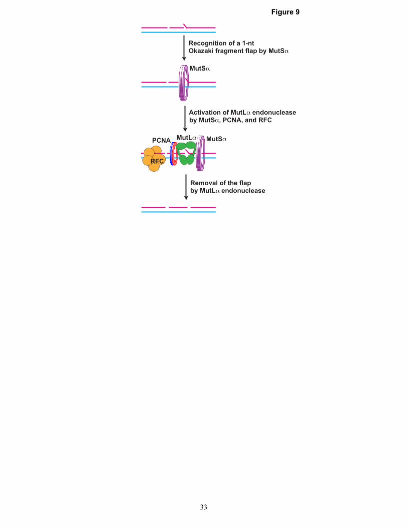
Figure 9
MutSα
PCNA
Activation of MutLα endonuclease
Removal of the flapby MutLα endonuclease
by MutSα, PCNA, and RFC
Recognition of a 1-ntOkazaki fragment flap by MutSα
MutSα
RFC
MutLα
33