evolution of radiology: page 1 of 22 for non-radiologists...©ken l schreibman, phd/md 7/1/14...
TRANSCRIPT

©Ken L Schreibman, PhD/MD 7/1/14 www.schreibman.info
page 1 of 22 Evolution of Radiology: For Non-Radiologists
TOPICS
Radiographs CT X-rays (EMS) Nuclear Med Ultrasound MRI
Physics Coils Magnets Safety
WOW
© 2014 Ken L Schreibman, PhD/MD
www.schreibman.info
Evolution of Radiology An Introduction for Non-Radiologists
Slide 1/132 Jump to next slide
Jump to last slide viewed
S
Ken L Schreibman, PhD/MD Professor of Radiology
UW – Madison Fellow, ACR Musculoskeletal Section 10 Staff Radiologists 5 Fellows
Chief Bone CT www.radiology.wisc.edu
TOPICS
Radiographs CT X-rays (EMS) Nuclear Med Ultrasound MRI
Physics Coils Magnets Safety
WOW
© 2014 Ken L Schreibman, PhD/MD
www.schreibman.info
Evolution of Radiology An Introduction for Non-Radiologists
Slide 2/132 Jump to next slide
Jump to last slide viewed
S
Nov 8th, 1895: The Birth of Radiology 11/8/95 Wilhelm Conrad Röntgen produces “X-rays” 12/28/95 Röntgen presents: “On a New Kind of Rays” 2/11/96 Jones publishes: “The Discovery of a Bullet
Lost in the Wrist by Means of the Roentgen Rays” First Radiograph Röntgen’s wife
1901:Röntgen wins 1st Nobel prize in physics
www.wikipedia.com
TOPICS
Radiographs CT X-rays (EMS) Nuclear Med Ultrasound MRI
Physics Coils Magnets Safety
WOW
© 2014 Ken L Schreibman, PhD/MD
www.schreibman.info
Evolution of Radiology An Introduction for Non-Radiologists
Slide 3/132 Jump to next slide
Jump to last slide viewed
S
How We Make Radiographs Light rays bounce off my hand and into my camera. We call the image: “Light-Ray” “Photograph” Image of the light
photons that bounce off my hand and into my image capture device.
How We Make Photographs
TOPICS Radiographs CT X-rays (EMS) Nuclear Med Ultrasound MRI
Physics Coils Magnets Safety
WOW
© 2014 Ken L Schreibman, PhD/MD
www.schreibman.info
Evolution of Radiology An Introduction for Non-Radiologists
Slide 4/132 Jump to next slide
Jump to last slide viewed
S
How We Make Shadows
Flashlight
TOPICS
Radiographs CT X-rays (EMS) Nuclear Med Ultrasound MRI
Physics Coils Magnets Safety
WOW
© 2014 Ken L Schreibman, PhD/MD
www.schreibman.info
Evolution of Radiology An Introduction for Non-Radiologists
Slide 5/132 Jump to next slide
Jump to last slide viewed
S
“Shadow-graph” Everywhere hand blocks the light is dark… Everywhere hand doesn’t block the light is illuminated. Now, if we hang photographic film on wall we get…
TOPICS
Radiographs CT X-rays (EMS) Nuclear Med Ultrasound MRI
Physics Coils Magnets Safety
WOW
© 2014 Ken L Schreibman, PhD/MD
www.schreibman.info
Evolution of Radiology An Introduction for Non-Radiologists
Slide 6/132 Jump to next slide
Jump to last slide viewed
S
“Negative-graph” Everywhere hand blocks light the film is not exposed and stays white...
Everywhere hand doesn’t block the light the film gets exposed and turns dark.

©Ken L Schreibman, PhD/MD 7/1/14 www.schreibman.info
page 2 of 22 Evolution of Radiology: For Non-Radiologists
TOPICS
Radiographs CT X-rays (EMS) Nuclear Med Ultrasound MRI
Physics Coils Magnets Safety
WOW
© 2014 Ken L Schreibman, PhD/MD
www.schreibman.info
Evolution of Radiology An Introduction for Non-Radiologists
Slide 7/132 Jump to next slide
Jump to last slide viewed
S
How We Make Radiographs
Terminology:
“X-rays”: Rays that pass
thru the patient.
The image is called a
“radiograph”
X-ray Tube
X-ray Detector
TOPICS
Radiographs CT X-rays (EMS) Nuclear Med Ultrasound MRI
Physics Coils Magnets Safety
WOW
© 2014 Ken L Schreibman, PhD/MD
www.schreibman.info
Evolution of Radiology An Introduction for Non-Radiologists
Slide 8/132 Jump to next slide
Jump to last slide viewed
S
20th Century: Images = Film
Film
X-ray Tube
X-ray Detector
Processed
in the dark
Developer
Allowed
to dry
Radiologist
Lost/Damaged
Primary
Specialist
Outside
File room
TOPICS
Radiographs CT X-rays (EMS) Nuclear Med Ultrasound MRI
Physics Coils Magnets Safety
WOW
© 2014 Ken L Schreibman, PhD/MD
www.schreibman.info
Evolution of Radiology An Introduction for Non-Radiologists
Slide 9/132 Jump to next slide
Jump to last slide viewed
S
How many of you, right now, have some sort of image capture device? Camera? Smart phone? perhaps even a laptop/tablet that can take
pictures?
How many of you have an image capture device that uses film?
21st Century: Images = Film
http://static.photo.net
TOPICS
Radiographs CT X-rays (EMS) Nuclear Med Ultrasound MRI
Physics Coils Magnets Safety
WOW
© 2014 Ken L Schreibman, PhD/MD
www.schreibman.info
Evolution of Radiology An Introduction for Non-Radiologists
Slide 10/132 Jump to next slide
Jump to last slide viewed
S
21st Century: Digital Imaging
X-ray Tube
X-ray Detector
PACS Picture Archive & Communication System Computer Server
Radiologist
Primary
Specialist
Outside
Lost/Damaged
File room Burn
CD for
Patients
Requires investment
in new digital X-ray
detectors
TOPICS
Radiographs CT X-rays (EMS) Nuclear Med Ultrasound MRI
Physics Coils Magnets Safety
WOW
© 2014 Ken L Schreibman, PhD/MD
www.schreibman.info
Evolution of Radiology An Introduction for Non-Radiologists
Slide 11/132 Jump to next slide
Jump to last slide viewed
S
Old Terms Still Used “Film”: What I tend to call radiographs “Wet read”: Look at the film STAT Refers to when we would look at films right
out of the developing solution, before they had time to dry.
“Dial a telephone” “Ring tones”
www.telephonearchive.com
TOPICS
Radiographs CT X-rays (EMS) Nuclear Med Ultrasound MRI
Physics Coils Magnets Safety
WOW
© 2014 Ken L Schreibman, PhD/MD
www.schreibman.info
Evolution of Radiology An Introduction for Non-Radiologists
Slide 12/132 Jump to next slide
Jump to last slide viewed
S
Radiographs are Limited Radiographs can detect only four densities of tissue:
Air (black)
Soft Tissues (dark gray)
Bones (light gray)
Metal (white)
All soft tissues look the same on radiographs: Muscles/Tendons Vessels/Nerves Organs/Blood

©Ken L Schreibman, PhD/MD 7/1/14 www.schreibman.info
page 3 of 22 Evolution of Radiology: For Non-Radiologists
TOPICS
Radiographs CT X-rays (EMS) Nuclear Med Ultrasound MRI
Physics Coils Magnets Safety
WOW
© 2014 Ken L Schreibman, PhD/MD
www.schreibman.info
Evolution of Radiology An Introduction for Non-Radiologists
Slide 13/132 Jump to next slide
Jump to last slide viewed
S
X-rays as Diagnostic Tool Can see:
Bones Fractures
Joint width, surfaces Arthritis Osteophytes Erosions
Radiographs: 2D projection of 3D patient Radiographs flatten everything Can’t tell what’s in front, what’s behind
With radiographs: NEED MULTIPLE VIEWS! “One view = No views”
Can’t see: Inside skull Can’t see the brain
Inside joints Can’t see tears Ligaments, Tendons Menisci, Cartilage
TOPICS
Radiographs CT X-rays (EMS) Nuclear Med Ultrasound MRI
Physics Coils Magnets Safety
WOW
© 2014 Ken L Schreibman, PhD/MD
www.schreibman.info
Evolution of Radiology An Introduction for Non-Radiologists
Slide 14/132 Jump to next slide
Jump to last slide viewed
S
“One view = No views”
TOPICS
Radiographs CT X-rays (EMS) Nuclear Med Ultrasound MRI
Physics Coils Magnets Safety
WOW
© 2014 Ken L Schreibman, PhD/MD
www.schreibman.info
Evolution of Radiology An Introduction for Non-Radiologists
Slide 15/132 Jump to next slide
Jump to last slide viewed
S
Need Multiple Views Small finger Not a subtle
fracture Fragment
overlap each other so perfectly on PA view, are undetectable
PA Obl Lat
TOPICS
Radiographs CT X-rays (EMS) Nuclear Med Ultrasound MRI
Physics Coils Magnets Safety
WOW
© 2014 Ken L Schreibman, PhD/MD
www.schreibman.info
Evolution of Radiology An Introduction for Non-Radiologists
Slide 16/132 Jump to next slide
Jump to last slide viewed
S
For Joints: Need 3 Views!
PA
PIP
Obl
PIP
Lat
PIP
TOPICS
Radiographs CT X-rays (EMS) Nuclear Med Ultrasound MRI
Physics Coils Magnets Safety
WOW
© 2014 Ken L Schreibman, PhD/MD
www.schreibman.info
Evolution of Radiology An Introduction for Non-Radiologists
Slide 17/132 Jump to next slide
Jump to last slide viewed
S
Tomography: Small Step Forward To overcome flat 2D nature of radiographs… Structures in the
Focal Plane are in focus. Structures out of
focal plane are blurred out.
At best, we got blurry pictures.
Long exposures = high radiation.
Film
1935 Grossmann Tomograph
Focal Plane
Can’t use this to see the brain
British Journal of Radiology 8:733-751,1935
TOPICS
Radiographs CT X-rays (EMS) Nuclear Med Ultrasound MRI
Physics Coils Magnets Safety
WOW
© 2014 Ken L Schreibman, PhD/MD
www.schreibman.info
Evolution of Radiology An Introduction for Non-Radiologists
Slide 18/132 Jump to next slide
Jump to last slide viewed
S
CT: Giant Leap Forward CT: Computed Tomography (Tomo [Gr]: part, slice)
CAT: Computed Axial Tomography 1917 Johann Radon, Austrian mathematician, proved image of a 3D object could be reconstructed from an infinite number of 2D projection images of the object.
Had to await the advent of main-frame computers in the 1970’s.
www.wikipedia.com

©Ken L Schreibman, PhD/MD 7/1/14 www.schreibman.info
page 4 of 22 Evolution of Radiology: For Non-Radiologists
TOPICS
Radiographs CT X-rays (EMS) Nuclear Med Ultrasound MRI
Physics Coils Magnets Safety
WOW
© 2014 Ken L Schreibman, PhD/MD
www.schreibman.info
Evolution of Radiology An Introduction for Non-Radiologists
Slide 19/132 Jump to next slide
Jump to last slide viewed
S
Hounsfield & EMI Brain Scanner 1972: Godfrey Hounsfield, a British electrical engineer at EMI Laboratories, developed EMI Brain Scanner. Finally, could see through the skull into the brain! Awarded Nobel Prize for Medicine 1979; Knighted 1981. “Hounsfield Units” is the scale we use to measure CT density.
EMI: “Electric and Musical Industries”
www.sciencemuseum.org.uk
TOPICS
Radiographs CT X-rays (EMS) Nuclear Med Ultrasound MRI
Physics Coils Magnets Safety
WOW
© 2014 Ken L Schreibman, PhD/MD
www.schreibman.info
Evolution of Radiology An Introduction for Non-Radiologists
Slide 20/132 Jump to next slide
Jump to last slide viewed
S
CT = Rotating X-rays
X-ray Tube
X-ray Tube
X-ray Tube
Inside CT Scanner
TOPICS
Radiographs CT X-rays (EMS) Nuclear Med Ultrasound MRI
Physics Coils Magnets Safety
WOW
© 2014 Ken L Schreibman, PhD/MD
www.schreibman.info
Evolution of Radiology An Introduction for Non-Radiologists
Slide 21/132 Jump to next slide
Jump to last slide viewed
S
Computed AXIAL Tomography Axial Plane: Top to Bottom
O,G 19yoM
Base of skull
Arch of C1
Body of C2
TOPICS
Radiographs CT X-rays (EMS) Nuclear Med Ultrasound MRI
Physics Coils Magnets Safety
WOW
© 2014 Ken L Schreibman, PhD/MD
www.schreibman.info
Evolution of Radiology An Introduction for Non-Radiologists
Slide 22/132 Jump to next slide
Jump to last slide viewed
S
Computed VOLUME Tomography Thin, continuous slices= Solid volume of data Can reformat data: Any 2-D plane Coronal, Sagittal, Oblique
Even in 3-D!
O,G 19yoM
TOPICS
Radiographs CT X-rays (EMS) Nuclear Med Ultrasound MRI
Physics Coils Magnets Safety
WOW
© 2014 Ken L Schreibman, PhD/MD
www.schreibman.info
Evolution of Radiology An Introduction for Non-Radiologists
Slide 23/132 Jump to next slide
Jump to last slide viewed
S
Multi-Planar Reformat
Coronal Plane Front to Back Like AP view
Sagittal Plane Left to Right Like a Lateral view
Dens Fx (Odontoid)
Fx easy to see on CT
Fx hard to see on
radiographs
O,G 19yoM
TOPICS
Radiographs CT X-rays (EMS) Nuclear Med Ultrasound MRI
Physics Coils Magnets Safety
WOW
© 2014 Ken L Schreibman, PhD/MD
www.schreibman.info
Evolution of Radiology An Introduction for Non-Radiologists
Slide 24/132 Jump to next slide
Jump to last slide viewed
S
Why CT is So Great Can see fractures otherwise missed Cervical spine, pelvis
Can see the brain! Strokes, bleeds, tumors
Can see organs (lungs, liver, bowel) Tumors, trauma, acute/chronic diseases
And now with ultra-fast, multi-slice… Can scan the heart in a single beat! Can see coronary arteries, pulmonary emboli
Hospitals have CT scanners in the ER

©Ken L Schreibman, PhD/MD 7/1/14 www.schreibman.info
page 5 of 22 Evolution of Radiology: For Non-Radiologists
TOPICS
Radiographs CT X-rays (EMS) Nuclear Med Ultrasound MRI
Physics Coils Magnets Safety
WOW
© 2014 Ken L Schreibman, PhD/MD
www.schreibman.info
Evolution of Radiology An Introduction for Non-Radiologists
Slide 25/132 Jump to next slide
Jump to last slide viewed
S
CT Usage Increasing in ERs
pediatrics.aappublications.org
0.9% 1.5%
6.0% 7.4%
4.3%
9.2%
11.2%
15.4%
13.1%
16.4% 15.4%
0%
5%
10%
15%
20%
1998 1999 2000 2001 2002 2003 2004 2005 2006 2007 2008
Computed Tomography Use Among Children Presenting to Emergency Departments With Abdominal Pain
Pediatrics Nov 2012 Vol 130, # 5, p 1–7
Published online October 8, 2012
TOPICS
Radiographs CT X-rays (EMS) Nuclear Med Ultrasound MRI
Physics Coils Magnets Safety
WOW
© 2014 Ken L Schreibman, PhD/MD
www.schreibman.info
Evolution of Radiology An Introduction for Non-Radiologists
Slide 26/132 Jump to next slide
Jump to last slide viewed
S
Problems with CT Usually requires IV contrast 1% patients are allergic to CT contrast Can affect renal function
Costs more than radiographs Knee radiographs (4 views): $154 Knee CT (no contrast): $1,200
Can’t see structures inside joints Knee: Menisci, Ligaments, Cartilage Shoulder: Rotator Cuff, Labrum Spine: Disks, Spinal Cord
TOPICS
Radiographs CT X-rays (EMS) Nuclear Med Ultrasound MRI
Physics Coils Magnets Safety
WOW
© 2014 Ken L Schreibman, PhD/MD
www.schreibman.info
Evolution of Radiology An Introduction for Non-Radiologists
Slide 27/132 Jump to next slide
Jump to last slide viewed
S
Biggest Problem with CT High radiation dose
We are exposed to low levels of radiation every day, “Background Radiation” Earth: naturally occurring radionuclides Uranium-238, potassium-40
Atmosphere: Radon-222 (from U-238) 2nd leading cause of lung cancer after smoking
Space: cosmic rays Airline crews are more exposed to cosmic rays,
doubling their background exposure.
Ave background dose ≈ 2.4mSv/year www.wikipedia.com
TOPICS
Radiographs CT X-rays (EMS) Nuclear Med Ultrasound MRI
Physics Coils Magnets Safety
WOW
© 2014 Ken L Schreibman, PhD/MD
www.schreibman.info
Evolution of Radiology An Introduction for Non-Radiologists
Slide 28/132 Jump to next slide
Jump to last slide viewed
S
Radiation from Diagnostic Imaging Ave background dose ≈ 2.4mSv/year
Chest Radiograph ≈ 0.06mSv ≈1 week of background radiation
Chest CT ≈ 7.0mSv ≈3 YEARS of background radiation
How much radiation is too much?
Health Physics Society
“Risks of medical imaging at effective doses below 50 mSv for single procedures or 100 mSv for multiple procedures over short time periods are too low to be detectable and may be nonexistent.”
www.aapm.org 12/13/11
TOPICS
Radiographs CT X-rays (EMS) Nuclear Med Ultrasound MRI
Physics Coils Magnets Safety
WOW
© 2014 Ken L Schreibman, PhD/MD
www.schreibman.info
Evolution of Radiology An Introduction for Non-Radiologists
Slide 29/132 Jump to next slide
Jump to last slide viewed
S
What are X-rays? X-rays are not naturally occurring Produced by X-ray tubes Used for diagnostic imaging Radiographs Tomography CT Fluoroscopy (radiographs in real-time)
Used for radiation therapy Treating tumors Orders of magnitude higher radiation dose than in
diagnostic imaging
X-rays part of Electromagnetic Spectrum
TOPICS
Radiographs CT X-rays (EMS) Nuclear Med Ultrasound MRI
Physics Coils Magnets Safety
WOW
© 2014 Ken L Schreibman, PhD/MD
www.schreibman.info
Evolution of Radiology An Introduction for Non-Radiologists
Slide 30/132 Jump to next slide
Jump to last slide viewed
S
Electromagnetic Spectrum

©Ken L Schreibman, PhD/MD 7/1/14 www.schreibman.info
page 6 of 22 Evolution of Radiology: For Non-Radiologists
TOPICS
Radiographs CT X-rays (EMS) Nuclear Med Ultrasound MRI
Physics Coils Magnets Safety
WOW
© 2014 Ken L Schreibman, PhD/MD
www.schreibman.info
Evolution of Radiology An Introduction for Non-Radiologists
Slide 31/132 Jump to next slide
Jump to last slide viewed
S
Electromagnetic Spectrum
Iguaçu (Iguazú) Falls Brazil-Argentina Border
TOPICS
Radiographs CT X-rays (EMS) Nuclear Med Ultrasound MRI
Physics Coils Magnets Safety
WOW
© 2014 Ken L Schreibman, PhD/MD
www.schreibman.info
Evolution of Radiology An Introduction for Non-Radiologists
Slide 32/132 Jump to next slide
Jump to last slide viewed
S
Electromagnetic Spectrum
This is the part of the EMS we can see. But there’s much more…
V i s i b l e L i g h t
R O Y G B I V
TOPICS
Radiographs CT X-rays (EMS) Nuclear Med Ultrasound MRI
Physics Coils Magnets Safety
WOW
© 2014 Ken L Schreibman, PhD/MD
www.schreibman.info
Evolution of Radiology An Introduction for Non-Radiologists
Slide 33/132 Jump to next slide
Jump to last slide viewed
S
Electromagnetic Spectrum
Infrared (below red) IR: Heat Vision Nocturnal animals
Ultraviolet (above violet) UV: Sun Tans/Burns Bees see in UV
Infra- Red
Ultra- Violet Visible Light
www.naturfotograf.com
Oenothera biennis
Potentilla intermedia
UV Light White Light
UV Light White Light
TOPICS
Radiographs CT X-rays (EMS) Nuclear Med Ultrasound MRI
Physics Coils Magnets Safety
WOW
© 2014 Ken L Schreibman, PhD/MD
www.schreibman.info
Evolution of Radiology An Introduction for Non-Radiologists
Slide 34/132 Jump to next slide
Jump to last slide viewed
S
Electromagnetic Spectrum
Visible Light Infra-Red Ultra-Violet Light Infra-Red Ultra-Violet Radio Waves
X-rays (man-made)
-rays (atomic decay)
Cosmic (space)
Ionizing Radiation Detach Electrons Atoms ionized “Free Radicals”
Damage DNA
Non-Ionizing Radiation No Free Radicals Doesn’t Damage DNA May cause Heat Damage
The Way Energy Moves Throughout the Universe
TOPICS
Radiographs CT X-rays (EMS) Nuclear Med Ultrasound MRI
Physics Coils Magnets Safety
WOW
© 2014 Ken L Schreibman, PhD/MD
www.schreibman.info
Evolution of Radiology An Introduction for Non-Radiologists
Slide 35/132 Jump to next slide
Jump to last slide viewed
S
Electromagnetic Spectrum
Light Infra-Red Ultra-Violet Radio Waves
X-rays (man-made)
-rays (atomic decay)
Cosmic (space)
To Perceive Energy Detector sensitive to energy frequency Human retina sensitive to visible light
Energy able to reach the detector
“X-ray Vision Specs” even if they could detect X-rays they still wouldn’t work…
There are no naturally occurring X-rays out there to detect!
TOPICS
Radiographs CT X-rays (EMS) Nuclear Med Ultrasound MRI
Physics Coils Magnets Safety
WOW
© 2014 Ken L Schreibman, PhD/MD
www.schreibman.info
Evolution of Radiology An Introduction for Non-Radiologists
Slide 36/132 Jump to next slide
Jump to last slide viewed
S
X-ray Vision
“SUPERMAN IS ABLE TO FOCUS ON OBJECTS FAR BEYOND THE RANGE OF NORMAL HUMAN SIGHT.
HIS EYES CAN PERCEIVE VIRTUALLY THE ENTIRE
ELECTROMAGNETIC SPECTRUM,
ENABLING HIM TO SEE THROUGH MOST SOLID
OBJECTS.”
©Ken L Schreibman, PhD/MD 7/1/14 www.schreibman.info
page 7 of 22 Evolution of Radiology: For Non-Radiologists
TOPICS
Radiographs CT X-rays (EMS) Nuclear Med Ultrasound MRI
Physics Coils Magnets Safety
WOW
© 2014 Ken L Schreibman, PhD/MD
www.schreibman.info
Evolution of Radiology An Introduction for Non-Radiologists
Slide 37/132 Jump to next slide
Jump to last slide viewed
S
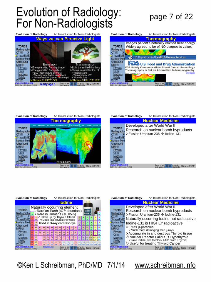
Ways we can Image Patients Ways we can Perceive Light
Transmission Light transmitted thru lamp X-rays transmitted thru pt Radiographs Tomography, CT Fluoroscopy
Shows STRUCTURE
Emission Energy emitted from light saber Energy emitted from patient EKG (Heart’s natural electricity) Thermography (Body’s natural heat) Nuclear Medicine (injected radioisotope)
Shows FUNCTION
Marty age 5
TOPICS
Radiographs CT X-rays (EMS) Nuclear Med Ultrasound MRI
Physics Coils Magnets Safety
WOW
© 2014 Ken L Schreibman, PhD/MD
www.schreibman.info
Evolution of Radiology An Introduction for Non-Radiologists
Slide 38/132 Jump to next slide
Jump to last slide viewed
S
Thermography Images patient’s naturally emitted heat energy. Widely agreed to be of NO diagnostic value.
www.meditherm.com
www.fda.gov
TOPICS
Radiographs CT X-rays (EMS) Nuclear Med Ultrasound MRI
Physics Coils Magnets Safety
WOW
© 2014 Ken L Schreibman, PhD/MD
www.schreibman.info
Evolution of Radiology An Introduction for Non-Radiologists
Slide 39/132 Jump to next slide
Jump to last slide viewed
S
Thermography
www.meditherm.com
TOPICS
Radiographs CT X-rays (EMS) Nuclear Med Ultrasound MRI
Physics Coils Magnets Safety
WOW
© 2014 Ken L Schreibman, PhD/MD
www.schreibman.info
Evolution of Radiology An Introduction for Non-Radiologists
Slide 40/132 Jump to next slide
Jump to last slide viewed
S
Nuclear Medicine Developed after World War II Research on nuclear bomb byproducts Fission Uranium-235 Iodine-131
TOPICS
Radiographs CT X-rays (EMS) Nuclear Med Ultrasound MRI
Physics Coils Magnets Safety
WOW
© 2014 Ken L Schreibman, PhD/MD
www.schreibman.info
Evolution of Radiology An Introduction for Non-Radiologists
Slide 41/132 Jump to next slide
Jump to last slide viewed
S
Iodine
wikipedia.org
Naturally occurring element Rare on Earth (47th abundant) Rare in Humans (<0.05%) Taken up by Thyroid Gland Made into Thyroid Hormone
Used in X-ray contrast dye
TOPICS
Radiographs CT X-rays (EMS) Nuclear Med Ultrasound MRI
Physics Coils Magnets Safety
WOW
© 2014 Ken L Schreibman, PhD/MD
www.schreibman.info
Evolution of Radiology An Introduction for Non-Radiologists
Slide 42/132 Jump to next slide
Jump to last slide viewed
S
Nuclear Medicine Developed after World War II Research on nuclear bomb byproducts Fission Uranium-235 Iodine-131
Naturally occurring Iodine not radioactive Iodine-131 is HIGHLY radioactive Emits β-particles Much more damaging than -rays
Accumulate in and destroys Thyroid tissue Nuclear Reactor Fallout Hypothyroid Take Iodine pills to block I-131 from Thyroid
Useful for treating Thyroid Cancer

©Ken L Schreibman, PhD/MD 7/1/14 www.schreibman.info
page 8 of 22 Evolution of Radiology: For Non-Radiologists
TOPICS
Radiographs CT X-rays (EMS) Nuclear Med Ultrasound MRI
Physics Coils Magnets Safety
WOW
© 2014 Ken L Schreibman, PhD/MD
www.schreibman.info
Evolution of Radiology An Introduction for Non-Radiologists
Slide 43/132 Jump to next slide
Jump to last slide viewed
S
Nuclear Medicine
wikipedia.org
Developed more agents to accumulate in specific tissues, emit low-energy -rays. “Radiopharmaceuticals” Many use Technetium Not naturally occurring 1936: First element to be
artificially produced
TOPICS
Radiographs CT X-rays (EMS) Nuclear Med Ultrasound MRI
Physics Coils Magnets Safety
WOW
© 2014 Ken L Schreibman, PhD/MD
www.schreibman.info
Evolution of Radiology An Introduction for Non-Radiologists
Slide 44/132 Jump to next slide
Jump to last slide viewed
S
Nuclear Medicine Technetium-99m: Ideal Imaging Agent Short half-life (6 hours) After 24 hours 94% gone
Emits -rays -rays pass out of
the patient without accumulating
Good energy for gamma-camera detection Dual-head cameras:
Image -rays emitted front & back
Marty age 14
TOPICS
Radiographs CT X-rays (EMS) Nuclear Med Ultrasound MRI
Physics Coils Magnets Safety
WOW
© 2014 Ken L Schreibman, PhD/MD
www.schreibman.info
Evolution of Radiology An Introduction for Non-Radiologists
Slide 45/132 Jump to next slide
Jump to last slide viewed
S
Nuclear Medicine: Bone Scan Was used a lot before CT & MR Shows bone pathology earlier than radiographs
Nowadays, seldom used for focal lesions We use MR for: Focal bone pain not seen on radiographs Infections (osteomyelitis) Imaging primary bone tumors
We still use Nuc Med Bone Scans for: Looking for bone metastases in entire body Breast Cancer Prostate Cancer
TOPICS
Radiographs CT X-rays (EMS) Nuclear Med Ultrasound MRI
Physics Coils Magnets Safety
WOW
© 2014 Ken L Schreibman, PhD/MD
www.schreibman.info
Evolution of Radiology An Introduction for Non-Radiologists
Slide 46/132 Jump to next slide
Jump to last slide viewed
S
Nuclear Medicine: PET/CT Most recent innovation in Nuc Med PET: Positron Emission Tomography Uses agents with very short half-lives Flourine-18 (100 min) Oxygen-15 (2 minutes) Made onsite with cyclotron
Agents taken up by tumors, metastases Well shows abnormal FUNCTION
Combined with CT (Computed Tomography) Well shows underlying ANATOMY
Used for staging cancer patients
TOPICS
Radiographs CT X-rays (EMS) Nuclear Med Ultrasound MRI
Physics Coils Magnets Safety
WOW
© 2014 Ken L Schreibman, PhD/MD
www.schreibman.info
Evolution of Radiology An Introduction for Non-Radiologists
Slide 47/132 Jump to next slide
Jump to last slide viewed
S
Ways we can Perceive Light Transmission Emission
Marty age 5
Reflection Light reflected off
Superman’s chest Ultrasound waves
reflected off patient’s organs
TOPICS
Radiographs CT X-rays (EMS) Nuclear Med Ultrasound MRI
Physics Coils Magnets Safety
WOW
© 2014 Ken L Schreibman, PhD/MD
www.schreibman.info
Evolution of Radiology An Introduction for Non-Radiologists
Slide 48/132 Jump to next slide
Jump to last slide viewed
S
Ultrasound Developed after World War II Based upon SONAR “SOund Navigation And Ranging” Sound wave sent out If sound hits an object get reflected back Measure time for the
reflected echo to return Multiplying the time by
speed of sound (2) = distance from the object
Works best in water Water transmits sound well

©Ken L Schreibman, PhD/MD 7/1/14 www.schreibman.info
page 9 of 22 Evolution of Radiology: For Non-Radiologists
TOPICS
Radiographs CT X-rays (EMS) Nuclear Med Ultrasound MRI
Physics Coils Magnets Safety
WOW
© 2014 Ken L Schreibman, PhD/MD
www.schreibman.info
Evolution of Radiology An Introduction for Non-Radiologists
Slide 49/132 Jump to next slide
Jump to last slide viewed
S
Sonography Uses radio waves (Not X-rays, -rays)
Light Infra-Red Ultra-Violet Radio Waves
X-rays (man-made)
-rays (atomic decay)
Cosmic (space)
Ionizing Radiation Non-Ionizing Radiation
Radiographs CT Fluoroscopy
Nuclear Med
Ultrasound (3-17MHz)
AM>530KHz
FM>87MHz
TOPICS
Radiographs CT X-rays (EMS) Nuclear Med Ultrasound MRI
Physics Coils Magnets Safety
WOW
© 2014 Ken L Schreibman, PhD/MD
www.schreibman.info
Evolution of Radiology An Introduction for Non-Radiologists
Slide 50/132 Jump to next slide
Jump to last slide viewed
S
Sonography Useful for… Tissues that contain/surrounded by water Abdominal organs Gall bladder (Gall Stones) Kidneys (Kidney Stones)
Blood vessels Blood clots (DVT: Deep Venous Thrombosis)
Imaging without Ionizing Radiation Pelvic organs Uterus, Ovaries Testes
Babies (Newborn) Babies… before birth
TOPICS
Radiographs CT X-rays (EMS) Nuclear Med Ultrasound MRI
Physics Coils Magnets Safety
WOW
© 2014 Ken L Schreibman, PhD/MD
www.schreibman.info
Evolution of Radiology An Introduction for Non-Radiologists
Slide 51/132 Jump to next slide
Jump to last slide viewed
S
Obstetric Ultrasound
Marty age -5 months
TOPICS
Radiographs CT X-rays (EMS) Nuclear Med Ultrasound MRI
Physics Coils Magnets Safety
WOW
© 2014 Ken L Schreibman, PhD/MD
www.schreibman.info
Evolution of Radiology An Introduction for Non-Radiologists
Slide 52/132 Jump to next slide
Jump to last slide viewed
S
Next… MRI
www.dreamstime.com
TOPICS
Radiographs CT X-rays (EMS) Nuclear Med Ultrasound MRI
Physics Coils Magnets Safety
WOW
© 2014 Ken L Schreibman, PhD/MD
www.schreibman.info
Evolution of Radiology An Introduction for Non-Radiologists
Slide 53/132 Jump to next slide
Jump to last slide viewed
S
MRI doesn’t rely on X-rays to see projected shadows of patients Unlike radiographs, CT, fluoroscopy
MRI sees tissues based upon sub-atomic characteristics Proton nucleus of Hydrogen
“NMR” “Nuclear Magnetic Resonance” “No More Radiologists”
MRI “Magnetic Resonance Imaging”
+
-
MRI: Giant Leap Sideways
TOPICS Radiographs CT X-rays (EMS) Nuclear Med Ultrasound MRI
Physics Coils Magnets Safety
WOW
© 2014 Ken L Schreibman, PhD/MD
www.schreibman.info
Evolution of Radiology An Introduction for Non-Radiologists
Slide 54/132 Jump to next slide
Jump to last slide viewed
S
MRI Scanner: 2 Components
THE COIL THE MAGNET
N
S

©Ken L Schreibman, PhD/MD 7/1/14 www.schreibman.info
page 10 of 22 Evolution of Radiology: For Non-Radiologists
TOPICS
Radiographs CT X-rays (EMS) Nuclear Med Ultrasound MRI
Physics Coils Magnets Safety
WOW
© 2014 Ken L Schreibman, PhD/MD
www.schreibman.info
Evolution of Radiology An Introduction for Non-Radiologists
Slide 55/132 Jump to next slide
Jump to last slide viewed
S
THE MAGNET
MRI Scanner: 2 Components
THE COIL : Jobs
Radio Frequency Transmitter Radio
Frequency Receiver
TOPICS
Radiographs CT X-rays (EMS) Nuclear Med Ultrasound MRI
Physics Coils Magnets Safety
WOW
© 2014 Ken L Schreibman, PhD/MD
www.schreibman.info
Evolution of Radiology An Introduction for Non-Radiologists
Slide 56/132 Jump to next slide
Jump to last slide viewed
S
How MR Scanner Works Magnet Aligns spins of protons in hydrogen nuclei Align in direction of magnetic field, B0
Coil 1)Sends RF pulse to flip spinning protons After RF pulse is off, protons realign to B0 As protons realign, resonate RF energy
2)Measures strength of resonant RF echo At a specific time, TE, “Echo Time”
Steps 1&2 repeated many times / image slice At a specific “Repetition Time”, TR
TOPICS
Radiographs CT X-rays (EMS) Nuclear Med Ultrasound MRI
Physics Coils Magnets Safety
WOW
© 2014 Ken L Schreibman, PhD/MD
www.schreibman.info
Evolution of Radiology An Introduction for Non-Radiologists
Slide 57/132 Jump to next slide
Jump to last slide viewed
S
Key to MRI Specific tissue types
have specific resonant echoes (T1, T2) depending upon specified TR & TE
Fluid (Hydrogen protons in H2O) Cysts Joint effusions Edema (in soft tissues, in bone marrow)
Fat (Hydrogen protons in fat) Sub-cutaneous fat Fatty yellow bone marrow
Dense Stuff (with few Hydrogen protons) Cortical bone Ligaments, tendons Menisci
TOPICS Radiographs CT X-rays (EMS) Nuclear Med Ultrasound MRI
Physics Coils Magnets Safety
WOW
© 2014 Ken L Schreibman, PhD/MD
www.schreibman.info
Evolution of Radiology An Introduction for Non-Radiologists
Slide 58/132 Jump to next slide
Jump to last slide viewed
S
T1 Recovery Curve (TR~500ms)
T1-weighted image (TR: short, TE: short)
Fat: High Fluid: Low Dense Stuff: Low
Signal
Low
High
Time to Echo TE (ms)
Fat
Fluid
Dense Stuff
10ms
TOPICS
Radiographs CT X-rays (EMS) Nuclear Med Ultrasound MRI
Physics Coils Magnets Safety
WOW
© 2014 Ken L Schreibman, PhD/MD
www.schreibman.info
Evolution of Radiology An Introduction for Non-Radiologists
Slide 59/132 Jump to next slide
Jump to last slide viewed
S
Fat
Dense Stuff
T2 Decay Curve (TR~2,000ms)
T2-weighted image (TR: long, TE: long)
Fluid: Intermed. Fat: Intermediate Dense Stuff: Low
Signal
Low
High
Time to Echo TE (ms) 80ms
To increase separation of fluid from fat, can apply “Fat-Suppression” (“Fat-Saturation”)
Fluid
TOPICS
Radiographs CT X-rays (EMS) Nuclear Med Ultrasound MRI
Physics Coils Magnets Safety
WOW
© 2014 Ken L Schreibman, PhD/MD
www.schreibman.info
Evolution of Radiology An Introduction for Non-Radiologists
Slide 60/132 Jump to next slide
Jump to last slide viewed
S
Fat
Fluid Dense Stuff
T2 Decay Curve (TR~2,000ms)
T2-weighted image (TR: long, TE: long)
Fluid: Intermed. Fat: Low (suppressed)
Dense Stuff: Low Signal
Low
High
Time to Echo TE (ms) 80ms
To increase separation of fluid from fat, can apply “Fat-Suppression” (“Fat-Saturation”)
It is always preferable to suppress the fat on T2 to increase fluid conspicuity.
Inversion Recovery (IR, STIR) is equivalent
Fat-Sat

©Ken L Schreibman, PhD/MD 7/1/14 www.schreibman.info
page 11 of 22 Evolution of Radiology: For Non-Radiologists
TOPICS
Radiographs CT X-rays (EMS) Nuclear Med Ultrasound MRI
Physics Coils Magnets Safety
WOW
© 2014 Ken L Schreibman, PhD/MD
www.schreibman.info
Evolution of Radiology An Introduction for Non-Radiologists
Slide 61/132 Jump to next slide
Jump to last slide viewed
S
Fat
Fluid Dense Stuff
T2 Decay Curve (TR~2,000ms)
T2-weighted image (TR: long, TE: long)
Fluid: High (relatively)
Fat: Low (suppressed)
Dense Stuff: Low
Signal
Low
High
Time to Echo TE (ms) 80ms
Fat-Sat
Compress signal scale
TOPICS
Radiographs CT X-rays (EMS) Nuclear Med Ultrasound MRI
Physics Coils Magnets Safety
WOW
© 2014 Ken L Schreibman, PhD/MD
www.schreibman.info
Evolution of Radiology An Introduction for Non-Radiologists
Slide 62/132 Jump to next slide
Jump to last slide viewed
S
How We Make MR Images Magnetic field divides body into slices
Each slice is subdivided into “voxels” voxel: 3D pixel voxel size = 2D pixel size X slice thickness
Coil measures signal in each voxel
Computer maps this onto 2D slices
High signal: White (“Bright”)
Intermediate signal: Gray (“Iso-intense”)
Low signal: Black (“Dark”)
TOPICS
Radiographs CT X-rays (EMS) Nuclear Med Ultrasound MRI
Physics Coils Magnets Safety
WOW
© 2014 Ken L Schreibman, PhD/MD
www.schreibman.info
Evolution of Radiology An Introduction for Non-Radiologists
Slide 63/132 Jump to next slide
Jump to last slide viewed
S
Comparing Sequences T1 T2 T2fs
TR=400 TE=10ms
TR=2500 TE=80ms
TR=2500 TE=80ms
Fat Pad
Bone Marrow
Sub Q
Fat Pad
Bone Marrow
Sub Q
Fat Pad
Bone Marrow
Sub Q
Fluid: Low
Cyst Fluid: High
Cyst Fluid: High
Cyst
Fat: High
Fat: Inter
Fat: Low
Knee, same mid-sagittal slice
TOPICS
Radiographs CT X-rays (EMS) Nuclear Med Ultrasound MRI
Physics Coils Magnets Safety
WOW
© 2014 Ken L Schreibman, PhD/MD
www.schreibman.info
Evolution of Radiology An Introduction for Non-Radiologists
Slide 64/132 Jump to next slide
Jump to last slide viewed
S
Fat
Dense Stuff
T2 Decay Curve (TR~2,000ms)
PD-weighted image (TR: long, TE: short)
Fluid: Intermed.
Fat: Intermediate Dense Stuff: Low
Signal
Low
High
Time to Echo TE (ms) 20ms
Fluid
80ms
Poor separation fat from fluid
Great separation of the Dense Stuff from fluid & fat
“Proton Density”
TOPICS
Radiographs CT X-rays (EMS) Nuclear Med Ultrasound MRI
Physics Coils Magnets Safety
WOW
© 2014 Ken L Schreibman, PhD/MD
www.schreibman.info
Evolution of Radiology An Introduction for Non-Radiologists
Slide 65/132 Jump to next slide
Jump to last slide viewed
S
Comparing Sequences T1 PD T2fs
TR=400 TE=10ms
TR=2500 TE=20ms
TR=2500 TE=80ms
Knee, same mid-sagittal slice
Fluid: Low
Cyst
Fluid: Inter
Cyst
Fluid: High
Cyst
Fat: High
Fat: Inter
Fat: Low
Dense Stuff: Low
Menisci
Dense Stuff: Low
Menisci
Dense Stuff: Low
Menisci
TOPICS
Radiographs CT X-rays (EMS) Nuclear Med Ultrasound MRI
Physics Coils Magnets Safety
WOW
© 2014 Ken L Schreibman, PhD/MD
www.schreibman.info
Evolution of Radiology An Introduction for Non-Radiologists
Slide 66/132 Jump to next slide
Jump to last slide viewed
S
MRI: Need Multiple Sequences T1 shows Fat best Most normal anatomy surrounded by fat In essence, T1 shows anatomy best
T2 shows Fluid best Most pathology contains fluid (edema) In essence, T2 shows pathology best Fat-suppression makes fluid more conspicuous
PD shows Dense Stuff best Good for meniscal and tendon tears Used mostly for MRI of joint pain

©Ken L Schreibman, PhD/MD 7/1/14 www.schreibman.info
page 12 of 22 Evolution of Radiology: For Non-Radiologists
TOPICS
Radiographs CT X-rays (EMS) Nuclear Med Ultrasound MRI
Physics Coils Magnets Safety
WOW
© 2014 Ken L Schreibman, PhD/MD
www.schreibman.info
Evolution of Radiology An Introduction for Non-Radiologists
Slide 67/132 Jump to next slide
Jump to last slide viewed
S
Seeing in 4-Dimensions Radiographs are flat projections Only give information in 2-D Need 2+ projections to fully see patient
CT is a stack of slices Images the patient in 3-D
MR is a stack of slices… and more Not only shows tissues in 3-D It shows the composition of the tissues T1: How Fatty, T2: How Wet
MR shows more than just 3-D
TOPICS
Radiographs CT X-rays (EMS) Nuclear Med Ultrasound MRI
Physics Coils Magnets Safety
WOW
© 2014 Ken L Schreibman, PhD/MD
www.schreibman.info
Evolution of Radiology An Introduction for Non-Radiologists
Slide 68/132 Jump to next slide
Jump to last slide viewed
S
Seeing in 5-Dimensions!
What’s left vs right? What’s front vs behind? What’s up vs down? What’s the temperature? What’s the time of day?
TOPICS
Radiographs CT X-rays (EMS) Nuclear Med Ultrasound MRI
Physics Coils Magnets Safety
WOW
© 2014 Ken L Schreibman, PhD/MD
www.schreibman.info
Evolution of Radiology An Introduction for Non-Radiologists
Slide 69/132 Jump to next slide
Jump to last slide viewed
S
Seeing in 5-Dimensions!
TOPICS Radiographs CT X-rays (EMS) Nuclear Med Ultrasound MRI
Physics Coils Magnets Safety
WOW
© 2014 Ken L Schreibman, PhD/MD
www.schreibman.info
Evolution of Radiology An Introduction for Non-Radiologists
Slide 70/132 Jump to next slide
Jump to last slide viewed
S
Limitations of MRI Limited Field of View (FOV)
Image resolution related to voxel size Smaller FOV = smaller voxels Smaller voxels = higher resolution To maximize resolution, try to limit FOV
Can only image inside the coil Requires an assortment of
different coils for different body parts
TOPICS
Radiographs CT X-rays (EMS) Nuclear Med Ultrasound MRI
Physics Coils Magnets Safety
WOW
© 2014 Ken L Schreibman, PhD/MD
www.schreibman.info
Evolution of Radiology An Introduction for Non-Radiologists
Slide 71/132 Jump to next slide
Jump to last slide viewed
S
Knee Coil
TOPICS Radiographs CT X-rays (EMS) Nuclear Med Ultrasound MRI
Physics Coils Magnets Safety
WOW
© 2014 Ken L Schreibman, PhD/MD
www.schreibman.info
Evolution of Radiology An Introduction for Non-Radiologists
Slide 72/132 Jump to next slide
Jump to last slide viewed
S
Knee Coil for the Ankle

©Ken L Schreibman, PhD/MD 7/1/14 www.schreibman.info
page 13 of 22 Evolution of Radiology: For Non-Radiologists
TOPICS
Radiographs CT X-rays (EMS) Nuclear Med Ultrasound MRI
Physics Coils Magnets Safety
WOW
© 2014 Ken L Schreibman, PhD/MD
www.schreibman.info
Evolution of Radiology An Introduction for Non-Radiologists
Slide 73/132 Jump to next slide
Jump to last slide viewed
S
Foot Coil
TOPICS Radiographs CT X-rays (EMS) Nuclear Med Ultrasound MRI
Physics Coils Magnets Safety
WOW
© 2014 Ken L Schreibman, PhD/MD
www.schreibman.info
Evolution of Radiology An Introduction for Non-Radiologists
Slide 74/132 Jump to next slide
Jump to last slide viewed
S
Elbow Coil
TOPICS
Radiographs CT X-rays (EMS) Nuclear Med Ultrasound MRI
Physics Coils Magnets Safety
WOW
© 2014 Ken L Schreibman, PhD/MD
www.schreibman.info
Evolution of Radiology An Introduction for Non-Radiologists
Slide 75/132 Jump to next slide
Jump to last slide viewed
S
Wrist Coil
TOPICS Radiographs CT X-rays (EMS) Nuclear Med Ultrasound MRI
Physics Coils Magnets Safety
WOW
© 2014 Ken L Schreibman, PhD/MD
www.schreibman.info
Evolution of Radiology An Introduction for Non-Radiologists
Slide 76/132 Jump to next slide
Jump to last slide viewed
S
2 Part Torso Coil
TOPICS
Radiographs CT X-rays (EMS) Nuclear Med Ultrasound MRI
Physics Coils Magnets Safety
WOW
© 2014 Ken L Schreibman, PhD/MD
www.schreibman.info
Evolution of Radiology An Introduction for Non-Radiologists
Slide 77/132 Jump to next slide
Jump to last slide viewed
S
Many Coils are Needed
TOPICS Radiographs CT X-rays (EMS) Nuclear Med Ultrasound MRI
Physics Coils Magnets Safety
WOW
© 2014 Ken L Schreibman, PhD/MD
www.schreibman.info
Evolution of Radiology An Introduction for Non-Radiologists
Slide 78/132 Jump to next slide
Jump to last slide viewed
S
MRI Scans are Expensive Coils are expensive: >$25,000 EACH! Scanners are expensive: >$2,000,000 Specialty trained technologists are expensive
MR scans take 30-60 minutes Run several sequences in several planes Can scan only a limited number of patients per day Have to charge a lot per scan
Knee Radiographs (4 views): $154 Knee CT (no contrast): $1,200 Knee MR (no contrast): $2,400 Don’t order MSK MR before getting Radiographs!

©Ken L Schreibman, PhD/MD 7/1/14 www.schreibman.info
page 14 of 22 Evolution of Radiology: For Non-Radiologists
TOPICS
Radiographs CT X-rays (EMS) Nuclear Med Ultrasound MRI
Physics Coils Magnets Safety
WOW
© 2014 Ken L Schreibman, PhD/MD
www.schreibman.info
Evolution of Radiology An Introduction for Non-Radiologists
Slide 79/132 Jump to next slide
Jump to last slide viewed
S
MR Scans are Long MR scans take 30-60 min Patient’s need to lie still… like a statue… for the entire time.
If the patient is ill the day of the scan and can’t stop coughing or sneezing, should reschedule.
Patients who can’t lie flat, severe heart failure (CHF), can’t get MRI.
Marty age 6
TOPICS
Radiographs CT X-rays (EMS) Nuclear Med Ultrasound MRI
Physics Coils Magnets Safety
WOW
© 2014 Ken L Schreibman, PhD/MD
www.schreibman.info
Evolution of Radiology An Introduction for Non-Radiologists
Slide 80/132 Jump to next slide
Jump to last slide viewed
S
Scanners A CT scanner… is a doughnut
TOPICS
Radiographs CT X-rays (EMS) Nuclear Med Ultrasound MRI
Physics Coils Magnets Safety
WOW
© 2014 Ken L Schreibman, PhD/MD
www.schreibman.info
Evolution of Radiology An Introduction for Non-Radiologists
Slide 81/132 Jump to next slide
Jump to last slide viewed
S
Scanners A CT scanner… is a doughnut
An MR scanner… is a cannoli
TOPICS
Radiographs CT X-rays (EMS) Nuclear Med Ultrasound MRI
Physics Coils Magnets Safety
WOW
© 2014 Ken L Schreibman, PhD/MD
www.schreibman.info
Evolution of Radiology An Introduction for Non-Radiologists
Slide 82/132 Jump to next slide
Jump to last slide viewed
S
MR Scanner is a Tube
They don’t build tubes to torture patients. Tubular design is needed to achieve the
high magnetic fields inherent to MRI. This is a 1.5 T magnet
TOPICS
Radiographs CT X-rays (EMS) Nuclear Med Ultrasound MRI
Physics Coils Magnets Safety
WOW
© 2014 Ken L Schreibman, PhD/MD
www.schreibman.info
Evolution of Radiology An Introduction for Non-Radiologists
Slide 83/132 Jump to next slide
Jump to last slide viewed
S
Tesla: Measure Magnetic Field Strength Earth's magnetic field: 30 µT (3×10⁻⁵ T)
Typical refrigerator magnet: 3 mT (3×10⁻³ T)
High Field MRI scanner: 1.5 – 3 T 1,000 times the strength refrigerator magnet 100,000 times the Earth’s magnetic field
TOPICS
Radiographs CT X-rays (EMS) Nuclear Med Ultrasound MRI
Physics Coils Magnets Safety
WOW
© 2014 Ken L Schreibman, PhD/MD
www.schreibman.info
Evolution of Radiology An Introduction for Non-Radiologists
Slide 84/132 Jump to next slide
Jump to last slide viewed
S
“Open” MRI = Low Field Favored by commercial stand-alone MRI sites “Our MRI scanner is open on all four sides; that’s a major advantage for large people who find a tunnel too confining, for children who might become frightened inside a tunnel, and for anyone with a touch of claustrophobia.”
open-mri-inc.com

©Ken L Schreibman, PhD/MD 7/1/14 www.schreibman.info
page 15 of 22 Evolution of Radiology: For Non-Radiologists
TOPICS
Radiographs CT X-rays (EMS) Nuclear Med Ultrasound MRI
Physics Coils Magnets Safety
WOW
© 2014 Ken L Schreibman, PhD/MD
www.schreibman.info
Evolution of Radiology An Introduction for Non-Radiologists
Slide 85/132 Jump to next slide
Jump to last slide viewed
S
“Open” MRI = Low Field Favored by commercial stand-alone MRI sites
Typical open MR: 0.1-0.3T
1/10th strength of a high field scanner…
1/10th image resolution of a high field scanner. Costs 1/10th the price to buy low field scanner… They charge the same price as a high field scan.
Diagnostic value of low field MR is inferior to that of high field MR.
TOPICS
Radiographs CT X-rays (EMS) Nuclear Med Ultrasound MRI
Physics Coils Magnets Safety
WOW
© 2014 Ken L Schreibman, PhD/MD
www.schreibman.info
Evolution of Radiology An Introduction for Non-Radiologists
Slide 86/132 Jump to next slide
Jump to last slide viewed
S
UW Experience with Open MR 0.7 T “Mid Field”
This is highest field open scanner made
Our accuracy: Knee In 1.5 T MR: ≈ 95% In this scanner: 75% Same UW radiologists Same UW protocols
Diagnostic value of low field MR is inferior to that of high field MR.
TOPICS
Radiographs CT X-rays (EMS) Nuclear Med Ultrasound MRI
Physics Coils Magnets Safety
WOW
© 2014 Ken L Schreibman, PhD/MD
www.schreibman.info
Evolution of Radiology An Introduction for Non-Radiologists
Slide 87/132 Jump to next slide
Jump to last slide viewed
S
UW Experience with Open MR Our surgeons refused to schedule patients in our open scanner. Ran it only 2 days/week
Primarily: Obese patients As bad as this scanner
was, it did a particularly poor job with…
obese patients. Got rid of it for a 3 T !
TOPICS
Radiographs CT X-rays (EMS) Nuclear Med Ultrasound MRI
Physics Coils Magnets Safety
WOW
© 2014 Ken L Schreibman, PhD/MD
www.schreibman.info
Evolution of Radiology An Introduction for Non-Radiologists
Slide 88/132 Jump to next slide
Jump to last slide viewed
S
My Recommendations For yourself or your patients: Don’t use open low field scanners Always want to use at least a 1.5 T scanner Go to a 3 T if available!
What about obese patients? Patients who don’t fit in the standard 1.5 T? We now have an alternative to low field
open scanners for the “Wisconsin-sized” patient…
TOPICS
Radiographs CT X-rays (EMS) Nuclear Med Ultrasound MRI
Physics Coils Magnets Safety
WOW
© 2014 Ken L Schreibman, PhD/MD
www.schreibman.info
Evolution of Radiology An Introduction for Non-Radiologists
Slide 89/132 Jump to next slide
Jump to last slide viewed
S
New Wide Bore 1.5T It’s still a tube…
But it’s a much wider tube
Same size opening as a CT scanner Table can hold up to 500 lbs!
Marty age 14
TOPICS
Radiographs CT X-rays (EMS) Nuclear Med Ultrasound MRI
Physics Coils Magnets Safety
WOW
© 2014 Ken L Schreibman, PhD/MD
www.schreibman.info
Evolution of Radiology An Introduction for Non-Radiologists
Slide 90/132 Jump to next slide
Jump to last slide viewed
S
Wide Bore 1.5T, also Short Bore Wide bore + short bore
= less “closed in” feeling
Marty age 14

©Ken L Schreibman, PhD/MD 7/1/14 www.schreibman.info
page 16 of 22 Evolution of Radiology: For Non-Radiologists
TOPICS
Radiographs CT X-rays (EMS) Nuclear Med Ultrasound MRI
Physics Coils Magnets Safety
WOW
© 2014 Ken L Schreibman, PhD/MD
www.schreibman.info
Evolution of Radiology An Introduction for Non-Radiologists
Slide 91/132 Jump to next slide
Jump to last slide viewed
S
MR scanner is a tube
Claustrophobia Don’t make patients claustrophobic
Things I’ve seen clinicians write: I told my patient how traumatic an MR scan is I told my patient it’s like laying inside a COFFIN I told my patient it’s like laying in a SEWER PIPE
TOPICS
Radiographs CT X-rays (EMS) Nuclear Med Ultrasound MRI
Physics Coils Magnets Safety
WOW
© 2014 Ken L Schreibman, PhD/MD
www.schreibman.info
Evolution of Radiology An Introduction for Non-Radiologists
Slide 92/132 Jump to next slide
Jump to last slide viewed
S
MR scanner is just a tube Nothing happens inside the tube Nothing moves Nothing crushes Open at both ends Plenty of air No radiation No X-rays No flashing lights
If it didn’t make any noise you wouldn’t even know anything was happening.
TOPICS
Radiographs CT X-rays (EMS) Nuclear Med Ultrasound MRI
Physics Coils Magnets Safety
WOW
© 2014 Ken L Schreibman, PhD/MD
www.schreibman.info
Evolution of Radiology An Introduction for Non-Radiologists
Slide 93/132 Jump to next slide
Jump to last slide viewed
S
MR scanners make lots of noise We protect the patient’s ears Ear plugs Headphones Can play radio station or CD or patient’s iPod
Our goal is to make patient relaxed We get our best pictures of people sleeping
TOPICS
Radiographs CT X-rays (EMS) Nuclear Med Ultrasound MRI
Physics Coils Magnets Safety
WOW
© 2014 Ken L Schreibman, PhD/MD
www.schreibman.info
Evolution of Radiology An Introduction for Non-Radiologists
Slide 94/132 Jump to next slide
Jump to last slide viewed
S
Can take something mild as an outpatient Valium (Diazepam) Ativan (Lorazepam) Cocktail? (not all 3) Patient should not drive! NOT Haldol (Haloperidol)
If patient is really problematic We can provide conscience sedation at hospital Not at outpatient facility
If patient is really really problematic General anesthesia can be arranged (It rarely comes to that)
If your patient is still anxious
TOPICS
Radiographs CT X-rays (EMS) Nuclear Med Ultrasound MRI
Physics Coils Magnets Safety
WOW
© 2014 Ken L Schreibman, PhD/MD
www.schreibman.info
Evolution of Radiology An Introduction for Non-Radiologists
Slide 95/132 Jump to next slide
Jump to last slide viewed
S
The Big Problem with MRI
It’s a Big Magnet
It’s a Big Magnet It’s Always On
TOPICS
Radiographs CT X-rays (EMS) Nuclear Med Ultrasound MRI
Physics Coils Magnets Safety
WOW
© 2014 Ken L Schreibman, PhD/MD
www.schreibman.info
Evolution of Radiology An Introduction for Non-Radiologists
Slide 96/132 Jump to next slide
Jump to last slide viewed
S
Why is it Always On? Isn’t it an electromagnet? Can’t we just flick a switch and turn it off? It’s not that simple… Yes, it’s an electromagnet. Yes, it works by passing current through wire To achieve 1.5T, need to pass A LOT of current through wire Requires low resistance wire… …super-conducting wire Super-conducting materials operate at
CRYOGENIC TEMPERATURES! Can’t turn off magnet with venting cryogens.

©Ken L Schreibman, PhD/MD 7/1/14 www.schreibman.info
page 17 of 22 Evolution of Radiology: For Non-Radiologists
TOPICS
Radiographs CT X-rays (EMS) Nuclear Med Ultrasound MRI
Physics Coils Magnets Safety
WOW
© 2014 Ken L Schreibman, PhD/MD
www.schreibman.info
Evolution of Radiology An Introduction for Non-Radiologists
Slide 97/132 Jump to next slide
Jump to last slide viewed
S
Occasionally Replenish Cryogens
TOPICS Radiographs CT X-rays (EMS) Nuclear Med Ultrasound MRI
Physics Coils Magnets Safety
WOW
© 2014 Ken L Schreibman, PhD/MD
www.schreibman.info
Evolution of Radiology An Introduction for Non-Radiologists
Slide 98/132 Jump to next slide
Jump to last slide viewed
S
MRI Safety Everyone in the entire medical center needs to respect MRI safety
Can’t bring into the scanner room anything that is: Ferromagnetic Electronic that is not certified MRI compatible.
TOPICS
Radiographs CT X-rays (EMS) Nuclear Med Ultrasound MRI
Physics Coils Magnets Safety
WOW
© 2014 Ken L Schreibman, PhD/MD
www.schreibman.info
Evolution of Radiology An Introduction for Non-Radiologists
Slide 99/132 Jump to next slide
Jump to last slide viewed
S
Safety Videos
www.patiencys.com/mri-safety
TOPICS
Radiographs CT X-rays (EMS) Nuclear Med Ultrasound MRI
Physics Coils Magnets Safety
WOW
© 2014 Ken L Schreibman, PhD/MD
www.schreibman.info
Evolution of Radiology An Introduction for Non-Radiologists
Slide 100/132 Jump to next slide
Jump to last slide viewed
S
Things Stuck in Magnets: Floor Buffer
www.MRImetalDetector.com
TOPICS
Radiographs CT X-rays (EMS) Nuclear Med Ultrasound MRI
Physics Coils Magnets Safety
WOW
© 2014 Ken L Schreibman, PhD/MD
www.schreibman.info
Evolution of Radiology An Introduction for Non-Radiologists
Slide 101/132 Jump to next slide
Jump to last slide viewed
S
www.simplyphysics.com
Things Stuck in Magnets: Gas Tank
TOPICS Radiographs CT X-rays (EMS) Nuclear Med Ultrasound MRI
Physics Coils Magnets Safety
WOW
© 2014 Ken L Schreibman, PhD/MD
www.schreibman.info
Evolution of Radiology An Introduction for Non-Radiologists
Slide 102/132 Jump to next slide
Jump to last slide viewed
S
Things Stuck in Magnets: ICU Bed
www.MRImetalDetector.com

©Ken L Schreibman, PhD/MD 7/1/14 www.schreibman.info
page 18 of 22 Evolution of Radiology: For Non-Radiologists
TOPICS
Radiographs CT X-rays (EMS) Nuclear Med Ultrasound MRI
Physics Coils Magnets Safety
WOW
© 2014 Ken L Schreibman, PhD/MD
www.schreibman.info
Evolution of Radiology An Introduction for Non-Radiologists
Slide 103/132 Jump to next slide
Jump to last slide viewed
S
Things Stuck in Magnets: Chair
www.simplyphysics.com
TOPICS
Radiographs CT X-rays (EMS) Nuclear Med Ultrasound MRI
Physics Coils Magnets Safety
WOW
© 2014 Ken L Schreibman, PhD/MD
www.schreibman.info
Evolution of Radiology An Introduction for Non-Radiologists
Slide 104/132 Jump to next slide
Jump to last slide viewed
S
Things Stuck in Magnets: Drug Cart
TOPICS
Radiographs CT X-rays (EMS) Nuclear Med Ultrasound MRI
Physics Coils Magnets Safety
WOW
© 2014 Ken L Schreibman, PhD/MD
www.schreibman.info
Evolution of Radiology An Introduction for Non-Radiologists
Slide 105/132 Jump to next slide
Jump to last slide viewed
S
Warning Signs
TOPICS Radiographs CT X-rays (EMS) Nuclear Med Ultrasound MRI
Physics Coils Magnets Safety
WOW
© 2014 Ken L Schreibman, PhD/MD
www.schreibman.info
Evolution of Radiology An Introduction for Non-Radiologists
Slide 106/132 Jump to next slide
Jump to last slide viewed
S
Warning Signs
TOPICS
Radiographs CT X-rays (EMS) Nuclear Med Ultrasound MRI
Physics Coils Magnets Safety
WOW
© 2014 Ken L Schreibman, PhD/MD
www.schreibman.info
Evolution of Radiology An Introduction for Non-Radiologists
Slide 107/132 Jump to next slide
Jump to last slide viewed
S
Metal Objects May Become Airborne
TOPICS Radiographs CT X-rays (EMS) Nuclear Med Ultrasound MRI
Physics Coils Magnets Safety
WOW
© 2014 Ken L Schreibman, PhD/MD
www.schreibman.info
Evolution of Radiology An Introduction for Non-Radiologists
Slide 108/132 Jump to next slide
Jump to last slide viewed
S
MRI Safety in China

©Ken L Schreibman, PhD/MD 7/1/14 www.schreibman.info
page 19 of 22 Evolution of Radiology: For Non-Radiologists
TOPICS
Radiographs CT X-rays (EMS) Nuclear Med Ultrasound MRI
Physics Coils Magnets Safety
WOW
© 2014 Ken L Schreibman, PhD/MD
www.schreibman.info
Evolution of Radiology An Introduction for Non-Radiologists
Slide 109/132 Jump to next slide
Jump to last slide viewed
S
MRI Safety in China
TOPICS Radiographs CT X-rays (EMS) Nuclear Med Ultrasound MRI
Physics Coils Magnets Safety
WOW
© 2014 Ken L Schreibman, PhD/MD
www.schreibman.info
Evolution of Radiology An Introduction for Non-Radiologists
Slide 110/132 Jump to next slide
Jump to last slide viewed
S
Limit Access to MR Suite
TOPICS
Radiographs CT X-rays (EMS) Nuclear Med Ultrasound MRI
Physics Coils Magnets Safety
WOW
© 2014 Ken L Schreibman, PhD/MD
www.schreibman.info
Evolution of Radiology An Introduction for Non-Radiologists
Slide 111/132 Jump to next slide
Jump to last slide viewed
S
A True Tragedy
Freak MRI Accident Kills Westchester Boy
New York Daily News Online Tuesday, July 31, 2001
Magnet send canister flying into him
6-year-old boy undergoing an MRI exam at a Westchester hospital died after the machine’s powerful 10-ton magnet turned
an oxygen canister into a missile that smashed his skull, officials said yesterday. Michael Colombini
mrimetaldetector.com
TOPICS
Radiographs CT X-rays (EMS) Nuclear Med Ultrasound MRI
Physics Coils Magnets Safety
WOW
© 2014 Ken L Schreibman, PhD/MD
www.schreibman.info
Evolution of Radiology An Introduction for Non-Radiologists
Slide 112/132 Jump to next slide
Jump to last slide viewed
S
MRI Safety Everyone in the entire medical center needs to respect MRI safety
Can’t bring into the scanner room anything that is: Ferromagnetic Electronic that is not certified MRI compatible.
TOPICS
Radiographs CT X-rays (EMS) Nuclear Med Ultrasound MRI
Physics Coils Magnets Safety
WOW
© 2014 Ken L Schreibman, PhD/MD
www.schreibman.info
Evolution of Radiology An Introduction for Non-Radiologists
Slide 113/132 Jump to next slide
Jump to last slide viewed
S
No Implanted Electronics No pacemakers Magnet won’t suck
pacer out of chest But magnet may… Drain the battery
Make pacer fire erratically
Scramble electronics
May even reprogram pacer
www.dotmed.com
TOPICS
Radiographs CT X-rays (EMS) Nuclear Med Ultrasound MRI
Physics Coils Magnets Safety
WOW
© 2014 Ken L Schreibman, PhD/MD
www.schreibman.info
Evolution of Radiology An Introduction for Non-Radiologists
Slide 114/132 Jump to next slide
Jump to last slide viewed
S
No Implanted Electronics No pacemakers
No cochlea implants
www.advancedbionics.com

©Ken L Schreibman, PhD/MD 7/1/14 www.schreibman.info
page 20 of 22 Evolution of Radiology: For Non-Radiologists
TOPICS
Radiographs CT X-rays (EMS) Nuclear Med Ultrasound MRI
Physics Coils Magnets Safety
WOW
© 2014 Ken L Schreibman, PhD/MD
www.schreibman.info
Evolution of Radiology An Introduction for Non-Radiologists
Slide 115/132 Jump to next slide
Jump to last slide viewed
S
No Implanted Electronics No pacemakers
No cochlea implants
No neuro-stimulators
memory-alpha.org
TOPICS
Radiographs CT X-rays (EMS) Nuclear Med Ultrasound MRI
Physics Coils Magnets Safety
WOW
© 2014 Ken L Schreibman, PhD/MD
www.schreibman.info
Evolution of Radiology An Introduction for Non-Radiologists
Slide 116/132 Jump to next slide
Jump to last slide viewed
S
Metal Inside Patients Safety Issues
Metal that can’t move is not a safety issue Fillings in the teeth Orthopedic hardware
Need to worry about metal that CAN move Metal in/around eyes Welding equipment Grinding equipment Fire guns w/o protection People who’ve been shot
Old aneurysm clips
Imaging Issues
from patient blog
TOPICS
Radiographs CT X-rays (EMS) Nuclear Med Ultrasound MRI
Physics Coils Magnets Safety
WOW
© 2014 Ken L Schreibman, PhD/MD
www.schreibman.info
Evolution of Radiology An Introduction for Non-Radiologists
Slide 117/132 Jump to next slide
Jump to last slide viewed
S
New UW Screening Sheet
TOPICS Radiographs CT X-rays (EMS) Nuclear Med Ultrasound MRI
Physics Coils Magnets Safety
WOW
© 2014 Ken L Schreibman, PhD/MD
www.schreibman.info
Evolution of Radiology An Introduction for Non-Radiologists
Slide 118/132 Jump to next slide
Jump to last slide viewed
S
An actual case… We’re screening the patient to see if he’s MR compatible. We ask the patient if he has any metal in his body. He replies, “… yeah… I think I was shot in the head once.” Is this patient MR compatible? Maybe yes, maybe no. We get a skull radiograph… What do you say now? “One view = no views”
TOPICS
Radiographs CT X-rays (EMS) Nuclear Med Ultrasound MRI
Physics Coils Magnets Safety
WOW
© 2014 Ken L Schreibman, PhD/MD
www.schreibman.info
Evolution of Radiology An Introduction for Non-Radiologists
Slide 119/132 Jump to next slide
Jump to last slide viewed
S
Need to have Multiple Views
What’s the answer?
Waters View
Bullet projecting next to the orbit
AP View
Bullet nowhere near the eye
TOPICS
Radiographs CT X-rays (EMS) Nuclear Med Ultrasound MRI
Physics Coils Magnets Safety
WOW
© 2014 Ken L Schreibman, PhD/MD
www.schreibman.info
Evolution of Radiology An Introduction for Non-Radiologists
Slide 120/132 Jump to next slide
Jump to last slide viewed
S
Need to have Multiple Views Here’s the answer on the lateral view!
On the Waters view the bullet just happened to project over the eye.
Lateral View Bullet embedded in back of calvarium!
Waters View
AP View
This patient IS MR Compatible

©Ken L Schreibman, PhD/MD 7/1/14 www.schreibman.info
page 21 of 22 Evolution of Radiology: For Non-Radiologists
TOPICS
Radiographs CT X-rays (EMS) Nuclear Med Ultrasound MRI
Physics Coils Magnets Safety
WOW
© 2014 Ken L Schreibman, PhD/MD
www.schreibman.info
Evolution of Radiology An Introduction for Non-Radiologists
Slide 121/132 Jump to next slide
Jump to last slide viewed
S
This Patient is NOT MR Compatible Don’t want this knife blade to move from its current position.
History?
“Stabbing Chest Pain”
TOPICS
Radiographs CT X-rays (EMS) Nuclear Med Ultrasound MRI
Physics Coils Magnets Safety
WOW
© 2014 Ken L Schreibman, PhD/MD
www.schreibman.info
Evolution of Radiology An Introduction for Non-Radiologists
Slide 122/132 Jump to next slide
Jump to last slide viewed
S
Thought Experiment: Magnetic Attraction
N S
Glass of Water
N S
Glass of Jell-O
Magnetic attraction > Weight paperclip
Magnetic attraction < Weight paperclip
+ glass + Jell-O
Like bullet in eye
Like bullet in brain
Iron Cup
N S
Like ICU bed
TOPICS
Radiographs CT X-rays (EMS) Nuclear Med Ultrasound MRI
Physics Coils Magnets Safety
WOW
© 2014 Ken L Schreibman, PhD/MD
www.schreibman.info
Evolution of Radiology An Introduction for Non-Radiologists
Slide 123/132 Jump to next slide
Jump to last slide viewed
S
Metal Inside Patients Safety Issues
No implanted electronics No metal that can move
OK: Orthopedic hardware OK: Modern aneurysm
clips OK: Modern heart valves OK: Vascular stents OK: IVC filters
Imaging Issues Metal can affect the
magnetic field “Susceptibility artifact”
May limit diagnostic value of the scan…
But often the scans come out just fine. As long as the patient is
MR safe, we’re willing to try. If we can’t get useful
images, cancel all charges
TOPICS
Radiographs CT X-rays (EMS) Nuclear Med Ultrasound MRI
Physics Coils Magnets Safety
WOW
© 2014 Ken L Schreibman, PhD/MD
www.schreibman.info
Evolution of Radiology An Introduction for Non-Radiologists
Slide 124/132 Jump to next slide
Jump to last slide viewed
S
Patient with lots of metal
Is it unsafe to put this patient in the magnet? Of course not!
Patient has unexplained knee pain.
Metal Example: Femoral Rod T1
Fracture!
Rod causes slight artifact
Even in retrospect this fracture cannot
be seen on the radiograph.
TOPICS
Radiographs CT X-rays (EMS) Nuclear Med Ultrasound MRI
Physics Coils Magnets Safety
WOW
© 2014 Ken L Schreibman, PhD/MD
www.schreibman.info
Evolution of Radiology An Introduction for Non-Radiologists
Slide 125/132 Jump to next slide
Jump to last slide viewed
S
T2fs Coronal
artifact
artifact
Metal Example: Interference Screws
ACL graft intact
T2fs Sagittal TOPICS
Radiographs CT X-rays (EMS) Nuclear Med Ultrasound MRI
Physics Coils Magnets Safety
WOW
© 2014 Ken L Schreibman, PhD/MD
www.schreibman.info
Evolution of Radiology An Introduction for Non-Radiologists
Slide 126/132 Jump to next slide
Jump to last slide viewed
S
PD Sagittal T2fs Sagittal
ACL graft intact
PD Sagittal Medial
Tear Posterior Horn Medial Medicus
Metal Example: Interference Screws

©Ken L Schreibman, PhD/MD 7/1/14 www.schreibman.info
page 22 of 22 Evolution of Radiology: For Non-Radiologists
TOPICS
Radiographs CT X-rays (EMS) Nuclear Med Ultrasound MRI
Physics Coils Magnets Safety
WOW
© 2014 Ken L Schreibman, PhD/MD
www.schreibman.info
Evolution of Radiology An Introduction for Non-Radiologists
Slide 127/132 Jump to next slide
Jump to last slide viewed
S
What to Order When (WOW): MSK Should always start with radiographs Least expensive study May show the answer Needed for planning other studies CT In ER for fracture detection (spine) For surgical planning of known fractures To assess degree of surgical fusion MRI Joints: Tears, internal derangement Spine: Disk bulges, cord compression Bones: Occult fractures, infection, tumors,…
TOPICS
Radiographs CT X-rays (EMS) Nuclear Med Ultrasound MRI
Physics Coils Magnets Safety
WOW
© 2014 Ken L Schreibman, PhD/MD
www.schreibman.info
Evolution of Radiology An Introduction for Non-Radiologists
Slide 128/132 Jump to next slide
Jump to last slide viewed
S
Putting it all together: Case example 41 yo F Neck pain Numbness/tingling Radiating down both arms Down to the fingers Spares the thumbs
Radiculopathy C7, C8
What to order first? Radiographs… of what? Cervical spine
Dermatome Map
warwickphysio.com
TOPICS
Radiographs CT X-rays (EMS) Nuclear Med Ultrasound MRI
Physics Coils Magnets Safety
WOW
© 2014 Ken L Schreibman, PhD/MD
www.schreibman.info
Evolution of Radiology An Introduction for Non-Radiologists
Slide 129/132 Jump to next slide
Jump to last slide viewed
S
AP View
Putting it all together: Case example
Z,K 41yoF
C1
C2
C3
C4
C5
C6
C7
T1
Lateral View
DDD: C5-C6 C6-C7
Bulging disks? Cord compression? Nerve impingement? Can’t tell w/radiograph Need MR
T2 Sagittal STIR
C1
C2
C3
C4
C5
C6
C7
T1
C1
C2
C3
C4
C5
C6
C7
T1
Bulges: C5-C6 C6-C7
Spare: C6 nerve
Impinge: C7 nerve C8 nerve
TOPICS
Radiographs CT X-rays (EMS) Nuclear Med Ultrasound MRI
Physics Coils Magnets Safety
WOW
© 2014 Ken L Schreibman, PhD/MD
www.schreibman.info
Evolution of Radiology An Introduction for Non-Radiologists
Slide 130/132 Jump to next slide
Jump to last slide viewed
S
Putting it all together: Case example
Z,K 41yoF
Surgery Remove bulging
C5-C6 & C6-C7 disks
Fuse vertebral bodies of C5 to C6 & C6 to C7
Did well for nearly two years…
C1
C2
C3
C4
C5
C6
C7
C1
C2
C3
C4
C5
C6
C7
T1
Post-Operative
TOPICS
Radiographs CT X-rays (EMS) Nuclear Med Ultrasound MRI
Physics Coils Magnets Safety
WOW
© 2014 Ken L Schreibman, PhD/MD
www.schreibman.info
Evolution of Radiology An Introduction for Non-Radiologists
Slide 131/132 Jump to next slide
Jump to last slide viewed
S
Two years later… Increasing
arm pain, L>R, dysfunction, weakness
Putting it all together: Case example
Z,K 41yoF
C1
C2
C3
C4
C5
C6
C7
T1
T2 Sagittal C1
C2
C3
C4
C5
C6
C7
T1
New bulge C7-T1 Junctional
Fusions? Can’t tell
by MR
CT Sagittal C1
C2
C3
C4
C5
C6
C7
T1
CT Coronal
C1
C2
C3
C4
C5
C6
C7
T1
Fused
Unfused
TOPICS
Radiographs CT X-rays (EMS) Nuclear Med Ultrasound MRI
Physics Coils Magnets Safety
WOW
© 2014 Ken L Schreibman, PhD/MD
www.schreibman.info
Evolution of Radiology An Introduction for Non-Radiologists
Slide 132/132 Jump to next slide
Jump to last slide viewed
S
WOW: Practical Considerations
Charge Time Radiation
Radiographs $154 < 1 sec ~0.06 mSv (1 week background)
CT $1,200 ~ 5 min ~7.0 mSv (3 years background)
MR $2,400 30 - 60 min NONE
US (abdomen)
(extremity) $1,200
$700 ~30 min NONE
Bone Scan
SPECT
$1,500
$2,500
~30 min
4hr post inject ~3.5 mSv (1.5 years background)
PET/CT $7,400 30 - 60 min
1hr post inject
PET: 7 mSv
CT (whole body) : 18mSv Total: 10 years background
www.wikipedia.com J Nucl Med 2005:46;608-13