evolution of the and cognition in
TRANSCRIPT

. 4
SIXTY-SECOND
JAMES ARTHUR LECTURE ONTHE EVOLUTION OF THE HUMAN BRAIN
1992
EVOLUTION OF THE BRAIN ANDCOGNITION IN HOMINIDS
DEAN FALK
AMERICAN MUSEUM OF NATURAL HISTORYNEW YORK : 1992


SIXTY-SECOND
JAMES ARTHUR LECTURE ON
THE EVOLUTION OF THE HUMAN BRAIN


SIXTY-SECOND
JAMES ARTHUR LECTURE ONTHE EVOLUTION OF THE HUMAN BRAIN
1992
EVOLUTION OF THE BRAIN ANDCOGNITION IN HOMINIDS
DEAN FALK
AMERICAN MUSEUM OF NATURAL HISTORYNEW YORK : 1992


JAMES ARTHUR LECTURES ONTHE EVOLUTION OF THE HUMAN BRAIN
Frederick Tilney, The Brain in Relation to Behavior: March 1 5. 1932
C. Judson Herrick, Brains as Instruments of Biological Values; April 6, 1933
D. M. S. Watson, The Story of Fossil Brains front Fish to Man; April 24, 1934
C. U . Ariens Kappers, Structural Principles in the Nervous System; The Development
of the Forebrain in Animals and Prehistoric Human Races; April 25, 1935
Samuel T. Orton, The Language Area of the Human Brain and Some of Its Dis-
orders; May 15, 1936
R. W. Gerard, Dynamic Neural Patterns; April 15, 1937
Franz Weidenreich, The Phylogenetic Development of the Hominid Brain and Its
Connection with the Transformation of the Skull; May 5, 1938
G. Kingsley Noble, The Neural Basis of Social Behavior of Vertebrates; May 1 1,
1939
John F. Fulton, A Functional Approach to the Evolution ofthe Primate Brain; May2, 1940
Frank A. Beach, Central Nervous Mechanisms Involved in the Reproductive Behavior
of Vertebrates; May 8, 1 94
1
George PinkJey, A History of the Human Brain; May 14, 1942
James W. Papez, Ancient Landmarks of the Human Brain and Their Origin; May27. 1943
James Howard McGregor, The Brain of Primates; May 1 1, 1944
K. S. Lashley, Neural Correlates of Intellect; April 30, 1945
Warren S. McCuIloch, Finality and Form in Nervous Activity; May 2, 1946
S. R. Detwiler, Structure-Function Correlations in the Developing Nervous System
as Studied by Experimental Methods; May 8, 1947
Tilly Edinger, The Evolution of the Brain; May 20, 1948
Donald O. Hebb, Evolution of Thought and Emotion; April 20, 1 949
Ward Campbell Halstead, Brain and Intelligence; April 26, 1950
Harry F. Harlow, The Brain and Learned Behavior; May 10, 1951
Clinton N. Woolsey, Sensory and Motor Systems of the Cerebral Cortex; May 7,
1952
Alfred S. Romer, Brain Evolution in the Light of Vertebrate History; May 21, 1953
Horace W. Magoun, Regulatory Functions of the Brain Stem; May 5, 1954
*Fred A. Metller, Culture and the Structural Evolution of the Neural System; April
21, 1955
*Pinckney J. Harman, Paleoneurologic, Neoneurologic. and Ontogenetic Aspects of
Brain Phylogeny; April 26, 1956

**Davenport Hooker, Evidence of Prenatal Function of the Central Nervous System
in Man; April 25, 1957
*David P. C. Lloyd, The Discrete and the Diffuse in Nervous Action; May 8, 1958
**Charles R. Noback, The Heritage of the Human Brain; May 6, 1959
**Emst Scharrer, Brain Function and the Evolution ofCerebral Vascularization; May26, 1960
Paul I. Yakovlev, Brain, Body and Behavior. Stereodynamic Organization of the
Brain and of the Motility-Experience in Man Envisaged as a Biological
Action System; May 16, 1961
H. K. Hartline, Principles ofNeural Interaction in the Retina; May 29, 1962
Harry Grundfest, Specialization and Evolution ofBioelectric Activity; May 28, 1963
**Roger W. Sperry, Problems Outstanding in the Evolution of Brain Function; June
3, 1964
*Jose M. R. Delgado, Evolution of Physical Control of the Brain; May 6, 1965
Seymour S. Kety, Adaptive Functions and the Biochemistry of the Brain; May 19,
1966
Dominick P. Purpura, Ontogenesis ofNeuronal Organizations in the MammalianBrain; May 25, 1967
*Kenneth D. Roeder, Three Views of the Nervous System; April 2, 1968
tPhillip V. Tobias, Some Aspects of the Fossil Evidence on the Evolution of the
Hominid Brain; April 2, 1 969
*Karl H. Pribram, What Makes Man Human; April 23, 1970
Walle J. H. Nauta, A New View ofthe Evolution ofthe Cerebral Cortex ofMammals;May 5, 1971
David H. Hubel, Organization of the Monkey Visual Cortex; May 11, 1972
Janos Szentagothai, The World ofNerve Nets; ianuary 16, 1973
*Ralph L. Holloway, The Role ofHuman Social Behavior in the Evolution of the
Brain; May 1, 1973
*Elliot S. Valenstein, Persistent Problems in the Physical Control of the Brain; May16, 1974
Marcel Kinsboume, Development and Evolution of the Neural Basis ofLanguage;
April 10, 1975
*John Z. Young, What Squids and Octopuses Tell Us About Brains and Memories;
May 13, 1976
*Berta Scharrer, An Evolutionary Interpretation of the Phenomenon of Neurosecre-
tion; April 12, 1977
Lester R. Aronson, Forebrain Function in Vertebrate Evolution; April 18, 1978
*Leonard Radinsky, The Fossil Record ofPrimate Brain Evolution; March 26, 1979
Norman Geschwind, Anatomical Asymmetry ofthe Brain in Humans and Animals:
An Evolutionary Perspective: April 7, 1980
Irving T. Diamond, Evolution of the Primate Neocortex: March 23, 1981
*Robert D. Martin, Human Brain Evolution in an Ecological Context: April 27,
1982
Eric Kandel, Molecular Explorations into Learning and Memory: April 27, 1983
Alexander Marshack, Hierarchical Evolution of the Human Capacity: The Paleo-
lithic Evidence: May 1, 1984
Yves Coppens, Environment. Hominid Evolution, and the Evolution of the Brain;
April 16, 1985
Roger A. Gorski, Sexual Differentiation of the Brain: from Birds to Rats to Man:April 22, 1986
Nicholas K. Humphrey, The Uses of Consciousness: April 7, 1987
Stephen J. Gould, Chomsky Under the Spandrels ofSan Marco: April 5, 1988
*Harry J. Jerison, Brain Size and the Evolution ofMind: October 10, 1989
Paul H. Harvey, Comparing Brains: March 20, 1990
Jeffrey T. Laitman, Evolution of the Vocal Tract and the Origins of Speech: May7, 1991
*Dean Falk, The Evolution of the Human Brain and Cognition in Hominids: April
14, 1992
Published versions of these lectures can be obtained from Publications, Dept. of
Anthropology, The American Museum of Natural History, Central Park
West at 79th St.. New York, N.Y. 10024.
"Out of print.
tPublished version: The Brain in Hominid Evolution, New York: Columbia Uni-
versity Press, 1971.

JAMES ARTHUR1842-1930
Bom in Ireland and brought up in Glasgow, Scotland, James Arthur
came to New York in 1871. Trained in mechanics and gear-cutting,
he pursued a career in the manufacture and repair of machinery,
during the course of which he founded a number of successful busi-
nesses and received patents on a variety of mechanical devices. His
mechanical interests evolved early into a lifelong passion for ho-
rology, the science of measuring time, and he both made some
remarkable clocks and assembled an important collection ofold and
rare timepieces.
Early in this century James Arthur became associated with the
American Museum of Natural History, and began to expand his
interest in time to evolutionary time, and his interest in mechanisms
to that most precise and delicate mechanism ofthem all, the humanbrain. The ultimate expression of his fascination with evolution and
the brain was James Arthur's bequest to the American Museumpermitting the establishment of the James Arthur Lectures on the
Evolution of the Human Brain. The first James Arthur Lecture was
delivered on March 15, 1932, two years after Mr. Arthur's death,
and the series has since continued annually, without interruption.

-^^f*«>^ O^Wfejb

EVOLUTION OF THE BRAIN ANDCOGNITION IN HOMINIDS
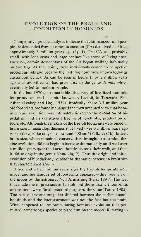
Comparative genetic analyses indicate that chimpanzees and peo-
ple are descended from a common ancestor (CA) that lived in Africa
approximately 5 million years ago (fig. 1). The CA was probably
small, with long arms and large canines like those of living apes.
Early on, certain descendants of the CA began walking habitually
on two legs. At that point, these individuals ceased to be apelike
protohominids and became the first true hominids, known today as
australopithecines. As can be seen in figure 1, by 2 million years
ago, australopithecines had given rise to the genus Homo, which
eventually led to modem people.
In the late 1970s, a remarkable discovery of fossilized hominid
footprints occurred at a site known as Laetoli, in Tanzania, East
Africa (Leakey and Hay, 1979). Ironically, these 3.5 million year
old footprints profoundly changed the then accepted view that hom-
inid brain evolution was intimately linked to the evolution of bi-
pedalism and its consequent freeing of forelimbs, production of
tools, etc. Although the makers ofthe Laetoli footprints were bipedal,
brain size in australopithecines that lived over 3 million years ago
was in the apelike range, i.e., around 400 cm^ (Falk, 1987b). Indeed
brain size, which remained conservative throughout australopithe-
cine evolution, did not begin to increase dramatically until well over
a million years after the Laetoli hominids took their walk, and then
it did so only in the genus Homo (fig. 2). Thus the origin and initial
evolution o^hip^dsMsva preceded XhQ dramatic increase in brain size
that characterized Homo.Three and a half million years after the Laetoli footprints were
made, another famous set of footprints appeared— this time left on
the moon by the astronaut Neil Armstrong (Falk, 1991). The feet
that made the impressions at Laetoli and those that left footprints
on the moon were, for all practical purposes, the same (Tuttle, 1985).
The part of the anatomy that differed between the earlier Laetoli
hominids and the later astronaut was not the feet but the brain.
What happened to the brain during hominid evolution that per-
mitted Armstrong's species to place him on the moon? Referring to
1
GORILLAS CHIMPANZEES
^-=i-^
HUMANSAUSTRALOPITHECINES
/Ih.mo hi, hill, ./— / .6 Homo erectus
5 0—/ Common Ancestor
/
/
^^^^'^i^f/^
Fig. 1. Molecular evidence suggests that people and chimpanzees are descended
from a common ancestor that lived approximately 5 million years ago. The fossil
record for australopithecines and for Homo is indicated by solid lines. (Reprinted
from Falk, 1 99 1
)
figure 2, this question may be divided into three parts: (1) What
modifications occurred in the direct ancestors of Homo that facili-
tated or permitted the initial take-off" in brain size? (2) Once brain
size began to increase, how was the continued increase in brain size
sustained, i.e., what behavior(s) were "targeted" by natural selection?
(3) How did the external and internal organization of the human
brain change as it enlarged and what were the cognitive correlates
of these neurological changes?
Paleoanthropologists use two methods to address these questions.
The "direct method" relies on examination of fossil hominids for
information about the external morphology of the convolutions and
blood vessels of the brain (as reflected on casts of the interior of the
braincase, or endocranial casts), and for estimates ofbrain size (which
is approximated by cranial capacity). These features may then be
interpreted in light of information determined from other parts of

1800
1600 -

I. WHAT FACILITATED THE INITIAL TAKE-OFFIN BRAIN SIZE?
Discovery of the Laetoli footprints has caused paleoanthropolo-
gists to abandon the belief that bipedahsm was intimately linked to
an increase in brain size in early hominids. The feet went first. But
why? As is frequently the case in evolutionary studies, a clue to this
mystery may be found by examining the environment of the par-
ticular group of australopithecines that gave rise to Homo. These
early hominids (known as gracile australopithecines) are believed to
have spent their days roaming across open savanna grasslands in
search of widely dispersed food, water, and trees. A number of
workers have speculated that australopithecines were probably sub-
ject to intense solar radiation as they went about their business in
the tropical savanna habitat (Wheeler, 1988). This is significant for
hominid brain evolution because brains are exquisitely heat sensi-
tive. In fact, according to one noted vascular physiologist:
A rise ofonly four or five degrees C above normal begins to disturb brain functions.
For example, high fevers in children are sometimes accompanied by convulsions;
these are manifestations of the abnormal functioning of the nerve cells of the
overheated brain. Indeed, it may be that the temperature of the brain is the single
most important factor limiting the survival of man and other animals in hot en-
vironments (Baker, 1979: 1 36, emphasis added).
For various reasons, I have hypothesized that selection for specific
vascular features that controlled brain temperature released a phys-
iological constraint on brain size, which then became a focus of
natural selection in Homo (Falk, 1 990).
The Radiator Theory
If a species evolves a new form of locomotion or habitual body
posture, its circulatory system will be modified because of changed
gravitational (or hydrostatic) forces associated with the new modeof life. Hydrostatic pressures may be envisioned by imagining a long
tube half-filled with water. If the tube is horizontal, the water (and
associated gravitational stress) is distributed along its entire length.
However, if the tube is tilted to the vertical, all of the water is in its
bottom half and that part of the vessel takes the stress from the

weight of the fluid. Natural selection favors vascular systems that
are designed to cope with hydrostatic stresses. For example, arboreal
snakes have evolved special mechanisms for pumping blood through
vertically oriented vessels, as opposed to the situation in their hor-
izontally inclined relatives (Lillywhile, 1987a, 1987b).
For physiological reasons, the circulatory systems of early hom-
inids had to become modified during the evolution of bipedalism.
Indeed, evidence of those modifications appears in modern humans.
Because people sleep horizontally but move about in the world ver-
tically, they have complex vascular systems that are designed to cope
with hydrostatic pressures in a variety of postures by shifting the
flow of blood with changing body position. Thus, if a person is lying
down, blood flows out of the skull through the internal jugular veins.
When the individual stands up, however, most of the exiting cranial
blood shifts away from the jugulars and into a network of veins that
surrounds the spinal cord. (Monkeys and apes have vascular systems
that are tailored somewhat differently to their own forms of loco-
motion.)
Fortunately for paleoanthropology, certain details of the cranial
vascular system appear in skulls of fossil hominids. These features
include grooves that represent venous sinuses and meningeal vessels,
as well as emissary foramina through which emissary veins penetrate
the skull. Taken together, these features show that the cranial vas-
cular systems of different groups of early hominids were adapted in
one of two different ways as each group refined bipedalism in its
own particular environment. On the one hand, the hominids from
Ethiopia that are associated with the famous "Lucy" skeleton have
an unusual venous sinus in the occipital region (the "occipital/mar-
ginal" sinus) that they share with later living australopithecines whowere probably their descendants (Falk and Conroy, 1983). These
australopithecines (known as robust australopithecines) ultimately
became extinct without giving rise to more modem hominids. Onthe other hand, data regarding the emissary veins suggest that a
different cranial vascular system developed in the gracile australo-
pithecines who lived in thermally stressful habitats and eventually
gave rise to Homo.Innumerable tiny veins penetrate the outside of the human skull

and communicate with the diploic veins that are found within the
bones of the skull itself. The diploic veins, in turn, penetrate the
inside surface of the skull bones, where they communicate with
meningeal veins that course on the surface of the dura mater that
covers the brain. Thus, the human skull is covered on its outside
and inside surfaces with a complex web of communicating veins
that happen to lack valves. Significantly, several named emissary
veins that are part of this network penetrate through specific foram-
ina of the skull. Consequently, the presence of emissary veins can
be surmised from fossil crania. Comparative data from skulls of
living apes and humans (Boyd, 1930) and direct data determined
from the hominid fossil record (Falk, 1986) reveal that frequencies
of the mastoid and parietal emissary foramina increased dramati-
cally in the lineage leading from gracile australopithecines to extant
Homo, but not in the other early hominids (fig. 3). Moreover, the
frequencies of these emissary veins seem to have increased in step
with increasing cranial capacity (Falk, 1990). If emissary veins are
viewed as a "window" into the wider network of cranial veins, these
data suggest that cranial vasculature became increasingly complex
as evolution progressed and brain size increased in Homo. Whatfunction could this network of veins have served?
The answer to this question is provided by physiological research
that compared the direction of blood flow in emissary veins of hu-
man volunteers who were subjected to severe cold stress at one time,
and heat stress at another (Cabanac and Brinnel, 1985). Cabanac
and Brinnel found that under conditions of hyperthermia, cranial
blood that had been cooled by the actions of sweating and evapo-
ration at the scalp was brought from the outside surface of the skull
into the brain case. (In cold subjects, on the other hand, blood flow
was slow and flowed out from the cranium.) The researchers con-
cluded that the entire network of cranial veins which the emissary
veins represent functions to selectively cool the brain under con-
ditions of severe heat stress. Thus, like the engine ofa car, the humanbrain has a radiator that prevents overheating.
To summarize the radiator theory, evolution of a prototype net-
work of cranial veins in gracile australopithecines released a thermal
constraint that had previously kept brain size in check. This facil-

^fifnf^y '//'/^'f/^
Fig. 3. The mastoid and parietal emissary foramina and the vertebral plexus of
veins that receives cranial blood when people are upright. The emissary veins are
part of a wider network of cranial veins that communicate between the outside and
inside of the skull. This network helps cool the brain under conditions of hyperther-
mia, and also drains blood to the vertebral plexus. (Reprinted from Falk, 1992)
itated the take-off in brain size in Homo. Bipedalism in gracile
australopithecines preceded the increase in brain size in Homo for
two reasons. First, because of the constraints of gravity, bipedaUsm
necessitated a rearrangement in cranial blood vessels. (Since this was
also true for robust australopithecines, it was a necessary but not
sufficient condition for the subsequent evolution in brain size.) Sec-
ond, bipedalism allowed gracile australopithecines to minimize the

amount of body surface exposed to the sun, thereby reducing their
heat loads and making it easier to adapt to thermally stressful sa-
vanna habitats (Wheeler, 1988). Thus, the vasculature of gracile
australopithecines became modified in response to gravitational andthermal pressures that were associated with refinement ofbipedalism
on the savanna. One result was the beginning of a cranial radiator
network of veins that could help cool the brain under conditions of
intense exercise. More important, once in place, this system was
itself modifiable and could therefore keep up with the increasing
thermolytic needs of an enlarging brain.
The radiator theory is mechanistic. The dramatic increase in brain
size in Homo is viewed as having simply been facilitated by the
release of thermal constraints that previously kept brain size in
check. Thus, the radiator network ofveins is seen as a prime releaser,
not a prime mover of brain evolution. One must turn to other
theories for speculation about the behaviors that were selected for
once the brain had acquired an adjustable radiator and could get
bigger.
II. WHAT BEHAVIORS WERE TARGETED BYNATURAL SELECTION?
Brain size doubled during the past 2 million years in the genus
Homo, increasing from an average of approximately 700 cm^ to a
modem mean of about 1400 cm^ (fig. 2). Although body size (or
stature) also enlarged during this time, it did not keep up with the
increase in brain size. Consequently, the brain of humans is three
times as large as is mathematically expected for an ape of equivalent
body size (Passingham, 1975; Falk, 1980b). What factors were re-
sponsible for the sustained increase in brain size? Since at least
Darwin's time, scientists have attempted to answer this question by
identifying specific behaviors that were the primary target of natural
selection. Some workers have gone so far as to suggest that a single
behavior was responsible for human brain evolution. Classic "prime
mover" candidates include warfare, language, tool production, and
hunting. These are discussed elsewhere (Falk, 1980b). Two newcandidates may now be added to this list— throwing (Calvin, 1982)
and social (or Machiavellian) intelligence (Byrne and Whiten, 1987).
8

According lo Calvin's (1982) throwing hypothesis, one of the ear-
liest lateralizations that occurred in the hominid brain was for rapid
motor sequencing of the right hand and arm (represented in the left
frontal lobe). Calvin believes that the cause of this lateralization was
natural selection for right-handed throwing of stones at prey. Hefurther suggests that neural machinery for throwing may also have
been used for the oral-facial musculature (which is represented near
the hand in the brain) and that, if so, this sequencing machinery
provided the scaffolding for the subsequent development of another
left hemisphere product, human language. Although the throwing
hypothesis is consistent with some research concerning neurophys-
iology ofthe motor cortex (Ojemann, 1 983), the assertion that skilled
xhTov,\ng preceded the evolution of language remains interesting but
speculative.
Because nonhuman primates are extremely complex in their social
relationships, primatologists have recently focused attention on so-
cial intelligence as a possible prime mover of brain evolution. For
example. Byrne and Whiten (1987) hypothesize that selection oc-
curred for "Machiavellian intelligence" in which individuals re-
lentlessly selected for still more cleverness (including deceptive be-
havior) in their companions. Other workers extend this concept by
suggesting that humans evolved as a result of a process of "runaway
social competition" between competing groups (Alexander, 1989).
This latter form of "intelligence" shades into another prime mover
candidate, namely warfare. A problem with social intelligence as a
prime mover ofhominid brain evolution is the fact that many species
of nonhuman primates are extremely clever in their social interac-
tions, yet without benefit of the extremely encephalized brain that
characterizes humans.
.As discussed elsewhere (Falk. 1980b), although entertaining, the
search for prime movers of human brain evolution is highly spec-
ulative and does not lend itself well to hypothesis testing. Further-
more, whether or not it is reasonable to attribute all of hominid
brain evolution to selection for only one behavior is debatable. As
described below, the human brain underwent a remarkable reor-
ganization as it enlarged. One would suppose that if selection of one
behavior was primarily responsible for brain evolution in Homo,

that behavior would be tied functionally to the neurological reor-
ganization that occurred. It should also clearly distinguish all people
from other primates. These issues will be returned to later.
III. NEUROLOGICAL REORGANIZATION ANDITS COGNITIVE CORRELATES
Under some circumstances, details of the cerebral cortex, blood
vessels, venous sinuses, and sutures are reproduced on casts of the
interior of the braincase (endocranial casts or endocasts). Endocasts
may occur naturally when fine sediment consolidates inside a skull
or can be prepared artificially with latex. One limitation of endocasts
is that the relatively largest-brained species within various groups
of mammals, including primates, fail to reproduce clear details of
the pattern of convolutions on endocasts (Radinsky, 1972). Thus,
endocasts from skulls of Homo sapiens reveal little information
about the surface of the brain, whereas small-brained australopith-
ecines are associated with a fossil record of nicely detailed natural
endocasts.
A second limitation of endocasts is that they fail to reproduce all
areas of the cerebral cortex equally well. For example, early hominid
endocasts usually do not reveal many details from the occipital
region of the brain, while the frontal lobe is reproduced in muchbetter detail. Interpretation of the external morphology of austra-
lopithecine occipital cortices has been subject to a good deal of
controversy, partly because of this limitation. Ralph Holloway be-
lieves that the outside surface of australopithecine occipital lobes
appears humanlike in certain respects, whereas my comparison of
chimpanzee, gorilla, and human brains led me to conclude that
australopithecine sulcal patterns are apelike in the occipital and all
other regions of the brain (Falk, 1980a; Radinsky, 1979). During
the past decade, a good deal has been published on this controversy,
and details are available elsewhere (Falk, 1992).
One is on firmer ground with the frontal lobe, not only because
it happens to show up better on endocasts, but because it is the one
area of the brain in which sulcal patterns clearly distinguish humanbrains from ape brains (Connolly, 1950). In the frontal lobes ofgreat
10

Fig. 4. Left frontal lobes (lateral view above, rotated to basal view below): (A)
chimpanzee, (B) orangutan, (C) gorilla, and (D) human. Orbitofrontal sulci are dark-
ened in the ape brains. Sulci darkened in the human brain include the inferior frontal
and those limiting the pars triangularis (arrow) associated with Broca's speech area.
(Reprinted from Falk, 1983)
apes (fig. 4A-C), an orbitofrontal sulcus (darkened in the illustration)
incises the lateral border and continues to course back toward the
temporal lobes on the underneath surface. This sulcus is not visible
on human brains, however, because it has been displaced deep within
the Sylvian fissure by the expanded convolutions of the frontal lobe.
Instead, human frontal lobes are usually characterized by two small
sulci that delimit a triangular patch of gray matter (arrow in fig. 4D)
that forms part of Broca's speech area in the left hemisphere. (As
discussed below, although this pattern is generally similar on the
right and left sides, the functions of the two hemispheres differ—
i.e., the human brain is lateralized.) In keeping with its expanded
size and reorganized sulcal pattern, the human brain also has frontal
lobes that appear squared in front (from a dorsal view) as compared
to ape brains (Falk, 1983).
The earliest indication of a humanlike frontal lobe in the known
hominid fossil record occurs in KNM ER 1470, a Homo habilis
11

Fig. 5. Endocast of left frontal lobe ofKNM ER 1470, a Homo habilis specimen
that is a little less than 2 million years old. Dots are reconstructed portion of frontal
lobe, hatching represents damaged area, fi, inferior frontal sulcus; arrows indicate
sulci that may delimit an area that is homologous with the pars triangularis ofhumanbrains. (Reprinted from Falk. 1983)
specimen from Kenya that is almost 2 million years old (fig. 5).
Although brain size in this specimen was only about 750 cm^ (Hol-
loway, 1978), its endocast reproduces the sulcal pattern in the left
frontal lobe that is associated with Broca's speech area in living
people. This suggests that Homo habilis may have been capable of
rudimentary speech (Tobias, 1981; Falk, 1983). Corroborative ev-
idence that the frontal lobe was already lateralized in early Homohas been provided by an archaeological analysis of stone flakes,
which indicates that knappers may already have been right-handed
by 2 million years ago (Toth, 1985). (Recall that the speech organs
and the right hand are represented by adjacent areas in the left frontal
lobe.)
As noted above, the australopithecine endocasts appear apelike
in the sulcal patterns of their frontal lobes. They also lack the squared
shape of the frontal lobe that can be seen in the earliest represen-
tatives ofHomo, including ER 1470. Furthermore, these differences
between australopithecines and Homo are not merely due to scaling
(allometric) factors whereby enlarged brains have more sulci than
smaller brains. (For discussion of allometry, see Jerison, 1991.) In
12
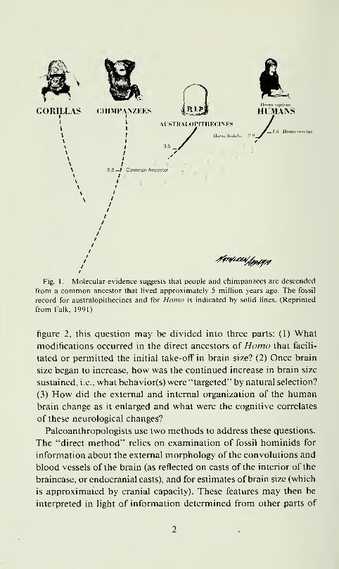
Fig. 6. Right hemispheres of a juvenile chimpanzee brain (top), the Taung en-
docast (left), and a human newborn (right). In all major respects, the Taung endocast
appears apelike, not humanlike. (Reprinted from Falk et al., 1989)
order to control for allometry, sulci from the frontal and temporal
lobes of the right hemisphere of the Taung australopithecine en-
docast were precisely measured and compared to corresponding sulci
in the brains of a juvenile chimpanzee and a human newborn whose
cranial capacity was close to the size of Taung's (Falk et al., 1989).
(See fig. 6.) Once size differences had been taken into account, the
ratio of the summed frontal lobe sulci of Taung relative to that for
the human baby brain was markedly impoverished—even compared
to the chimpanzee brain! Since this was not the case for the temporal
lobe, it appears Xhatfrontal lobe expansion was particularly dramatic
during the subsequent evolution ofHomo .
Other evidence points to the frontal lobes (or the behaviors that
depend upon them) as a particular focus for natural selection during
the evolution of Homo. Using a new gyrification index (GI) that
measures the degree of convolutedness in any given location of the
brain, Armstrong and her colleagues compared the extent of folding
in the various lobes of monkeys, apes, and humans (Armstrong et
13

al., 1991). They found that whereas the GIs of apes and humanscompletely overlap in the occipital region, the human brain is muchmore convoluted in the frontal lobes than are ape brains. The find-
ings for the occipital region suggest that changes in sulcal pattern
occurred only when brain size increased and that the occipital cortex
in the Taung endocast was therefore probably apelike. Having stud-
ied the entire surface of the brain, the authors concluded (on p. 347):
"that the proximal cause or mechanisms by which the hominid brain
increased in size are more likely to have been either a response to
changes in the frontal lobes or to events that influenced all regions
of the brain."
Besides language (which will be discussed below), what behaviors
were elaborated with the continued expansion of the frontal lobes
during the evolution of //cmo.^ Although any answer to this question
will necessarily be speculative, it is informative to consider frontal
lobe functions that occur in monkeys (and presumably apes) and
note their elaborations in humans. Because medical research has
focused extensively on macaques, the circuitry in the prefrontal
cortex and its regulation of behavior are fairly well understood for
these monkeys. A major function of macaque prefrontal cortex is
to assess memories (symbolic representations related to visuospatial
information, emotions, etc.), keep them "on line," and use them to
guide motor behavior in the absence of external stimuli (Goldman-
Rakic, 1987). According to Goldman-Rakic (p. 406), "many inte-
grated higher-order functions including language, concept formation,
and planning for the future may be built on this functional element."
In keeping with this, it is well known (from the old days of lobot-
omies) that damage to the human prefrontal cortex frequently causes
disturbances of attention, an inability to use past experience to grasp
the essence of a situation, and a loss of the ability to plan ahead. A"flattening" of personality and inappropriate social behaviors may
also result from prefrontal damage.
The archaeological record provides glimpses that suggest frontal
lobe abilities may have increased along with increasing brain size
in the genus Homo (at least up to the relatively recent time of Ne-
anderthals). Preparation of even simple stone tools (associated with
early Homo) takes some ability to keep a task in mind and execute
14

a plan. Apparently, abilities pertaining to the manufacture of stone
tools continued to evolve so that, by 300,000 years ago. Homoercctus was demonstrating relatively sophisticated notions of per-
spective, control of spatial quantity, and an understanding of com-
position (Wynn, 1 989). Although the human propensity for worrying
about the future is difficult to gauge from the archaeological record,
most anthropologists would probably agree that the first deliberate
burials (attributed to Neanderthals) indicate an upper boundary for
the appearance of an acute consciousness of time.
Brain Lateralization
Turning our attention to brain lateralization, its evolution maybe investigated by comparing shapes of lobes (petalia patterns) in
the brains ofnonhuman primates and people. Analyses ofCAT scans
and radiographs reveal that right-handed people tend to have right
frontal lobes that project farther and are wider than the left frontal
lobes (i.e., a right frontal petalia), whereas their left occipital lobes
are usually wider and protrude more (LeMay, 1976; Galaburda et
al., 1978). (The reverse condition of left frontal and right occipital
petalias is more likely to be found in left-handers.) Petalias give both
the brain and the overlying skull a characteristic lopsided appearance
that can be detected on endocasts. These shape asymmetries occur
in monkeys and apes, but not to the extent that they do in humans
(LeMay et al., 1982). The human pattern of left-occipital-right-
frontal petalias appears early in Homo and, in keeping with the
comparative findings, may have been foreshadowed in australo-
pithecines (Holloway and de Lacoste-Lareymondie, 1982).
Not too long ago, it was thought that humans were unique among
primates in manifesting asymmetries in lengths and configurations
of certain cortical features, e.g., having a longer left Sylvian fissure
and larger left planum temporale. Largely because ofrecent advances
in medical imaging, the alternative idea that cortical asymmetries
of humans are end products of a long evolutionary history is nowsupported by comparative evidence regarding sulcal lengths and
areas of cortex delimited by sulci. Thus, rhesus monkeys exhibit
numerous asymmetries in their frontal lobes including right frontal
15

petalias (Falk et al., 1990). These asymmetries, which may involve
short-term memory for visual information, correlate with functional
lateralization for certain vocal and visual processing in macaques.
Analysis of a large sample of macaque endocasts revealed that both
gross and detailed morphology of the brain are heritable (Cheverud
et al., 1990), a finding that is consistent with a hypothetical genetic
component for cortical lateralization.
A greater number of cortical asymmetries have been discovered
in humans (Falk et al., 1991) than in monkeys or apes, not just in
frontal lobes, but throughout the brain. Taken together, the evidence
from endocasts of fossil hominids and the comparative data re-
garding the external morphology of the cerebral cortex suggest that
brain lateralization became more elaborate as brain size increased
in Homo . What were the cognitive correlates of an increasingly
lateralized brain during the evolution ofHomo?One way to approach
this question, is first to observe the functional aspects of brain la-
teralization in people, and then to compare the cognitive abilities
of humans with those of their closest nonhuman cousin, the chim-
panzee.
The literature on functional brain lateralization in humans is mul-
tifaceted and sometimes difficult to interpret because differences
between hemispheres are often subtle but statistically significant
(Falk, 1987a, 1992). Upon close inspection, however, contradictory
data obtained by separate studies may frequently be attributed to
slightly different methodologies or to differences in the populations
that were tested. For example, the musically sophisticated and left-
handers do not test like the general population on certain measures
of brain lateralization. Nevertheless, as a result of various kinds of
tests (e.g., dichotic listening, tachistoscopic viewing, etc.) performed
on enormous numbers of normal and clinical populations, certain
generalizations may be made about the specializations of right and
left hemispheres (fig. 7).
As illustrated in figure 7, the left hemisphere is associated with
language functions, skilled movements (e.g., like those engaged in
by over 90% of right hands), and analytical, time-sequencing pro-
cesses. This hemisphere is also involved in processing positive emo-
tions. The right hemisphere, on the other hand, engages in holistic.
16

f.ORPl'S CALLOSl'M
Fig. 7. General specializations of left and right hemispheres. Dorsal view of the
brain, which is shown connected by the corpus callosum. (Reprinted from Falk, 1992)
global pursuits. Mental imaging, visuospatial skills, and musical
abilities are also associated with the right hemisphere. The right
hemisphere processes negative emotions and has a noticeably greater
role than the left hemisphere in generally expressing emotions, as
well as reading them in other people. In fact, although the left hemi-
sphere processes language, it is the right hemisphere that provides
tone of voice, an important and sometimes decisive aspect of verbal
communication. The right hemisphere also seems to have an edge
over the left in recognizing faces, in the ability to understand met-
aphor, and in certain aspects of humor. It is important to note,
however, that the right and left hemispheres are, ofcourse, connected
in normal individuals. (Figure 7 is therefore an oversimplification,
i.e., most tasks will have some input from both sides of the brain.)
There is an incredible amount of variation and complexity in the
17
patterns of asymmetries for various skills expressed by different
individuals. Furthermore, males and females seem to be organized
somewhat differently with respect to both functional and anatomical
brain lateralization. Lateralized behaviors in humans have been hy-
pothesized to be the result of asymmetrical distributions of neuro-
chemicals, as has been shown for certain rodents and birds. Inter-
estingly, it has also been suggested that different patterns of brain
lateralization in humans may be the result of developmental events
(such as prenatal exposure to testosterone) that differentially influ-
ence males and females (Geschwind and Galaburda, 1987). (For
further discussion see Falk, 1992.)
One way to assess which behaviors "drove" hominid brain evo-
lution is to compare the frontal lobe and asymmetrically mediated
behaviors in chimpanzees and humans (including where the two
categories overlap). What do the human and chimpanzee have in
common? Beginning with the frontal lobe, both species can bring
visuospatial memories on line and carry out related tasks. In fact,
chimpanzees are every bit as good as humans at remembering the
location of cached food and retrieving it at some later time. Unlike
most animals, chimpanzees and humans both recognize themselves
in mirrors, a feat that my colleague Gordon Gallup attributes to a
frontal-lobe sense of "me" (Maser and Gallup, 1990). Furthermore,
both species have fairly good motor skills, e.g., they can work puzzles
that require manipulation of parts, and each expresses emotions
through gestures that are controlled by frontal lobe motor cortex.
But the similarities stop here. According to Savage-Rumbaugh
(Falk, 1992), chimpanzees cannot keep very many tasks in mind at
once, the way that humans can. In terms of frontal lobe planning,
chimpanzees do not have a humanlike awareness of death or a
religion (Maser and Gallup, 1990). With respect to regulating be-
havior through accessing symbolic representations, chimpanzees are
not very good at controlling emotional outbursts— a frontal lobe
mediated activity in which almost all human children eventually
become proficient. Although they can manipulate objects, chim-
panzees as a population are not lateralized for handedness, unlike
humans. Finally, the most dramatic (lateralized) frontal lobe differ-
18

encc between chimpanzees and humans is, of course, thai chimpan-
zees lack the human capacity for speech.
There are other important cognitive attributes of humans (com-
pared to chimpanzees) that depend on an overall lateralized brain
(fig. 7). As noted elsewhere:
No chimpan/cc ever wrote a book, composed a symphony, painted a realistic
portrait, performed a Singing in the /?a;>7-quality tap dance, calculated the day of
the week upon which a certain date will fall next year, developed a math proof,
took apart and reassembled a watch, programmed a computer, designed a bridge,
or theorized about the origin of the universe (Falk, 1992: 58).
In short, these human arts and sciences spring from an enlarged
lateralized brain that has numerous circuits subserving specialized
functions. These circuits are sometimes referred to as modules (Fo-
dor, 1983).
Can these modules be understood in terms of the specific internal
reorganization that took place in the brain during the course of brain
evolution in Homo? Since one cannot turn to endocasts for the
answer to this question, investigation of brain growth during humandevelopment may be informative. Although humans are bom with
their full complement of neurons, profound changes occur in the
nervous system postnatally (Gibson, 1991; Konner, 1991). As the
human brain enlarges after birth, there is a proliferation of synapse
formation, dendritic branching, and division of glial support cells
that form the myelin sheaths around axons (thus giving the white
matter its characteristic appearance). Myelination, which increases
the speed and specificity of nerve transmission, occurs in specific
sequences that appear to correlate with the development of motor,
social, and intellectual skills (Gibson, 1991). For example, the ap-
pearance of social smiling and fear ofstrangers in infants is correlated
with myelination of specific neurological structures during the first
year of life, whereas cortical association areas subserving "higher"
intellectual functions continue to myelinate up to the age of 30 years
(Konner, 1991).
Comparative studies provide another important source of infor-
mation regarding hominid brain evolution. Since larger mammalian
brains have larger, more widely spaced neurons than do smaller
19

brains, this trend and other related scaling (allometric) factors prob-
ably occurred during brain enlargement in Homo. Thus, although
the absolute number of neurons would have increased with brain
size, their density would have decreased. There also should have
been an increase in the overall fraction of the cortex devoted to
axonal interconnections (i.e., cortical white matter), along with an
increase in dendritic branching. Significantly (see below), the fraction
of cells with which any one cell communicates directly ("percent
connectedness") would have decreased as brain size increased in
//omo(Ringo, 1991).
Based on these comparative data, Ringo (1991) has developed an
eloquent model that accounts for a general trend toward cortical
specialization within larger-brained mammals. As brain size (and
therefore neuron number) increases in the various species, each neu-
ron must be connected to a decreased fraction of the total number
of neurons. Otherwise large brains would be "swamped" by geo-
metrically increasing numbers of connections relative to their ab-
solute number of neurons. (To use Ringo's example, a 100% con-
nected two-neuron system would have two connections whereas a
100% connected four-neuron system would have 12 connections,
etc.) Because absolute connectivity nevertheless increases with en-
larged brain size, the "interconnection problem" is resolved by de-
creased neuronal density, i.e., more volume is available per neuron
in larger brains. Since long axons take more space than short axons
in such a system, they are minimized, and specializations result. In
Ringo's words:
One interesting possibility is that this increasing load from interconnectedness is
avoided by utilizing specialization so that only within major groupings of neurons
need there be full interconnection, while between major groupings only 'results'
need to be passed. This then suggests the argument that big brains need hemispheric
specialization, because of interconnections getting out of hand, and further suggests
that large hemispheres will be more specialized than small ones. This simply means
that cortical areas will be more specialized. This tendency appears to follow across
the best investigated species. . . . (Ringo, 1991: 5)
Comparative data from prosimians (Galago) and monkeys {Macaco)
suggest that, in keeping with Ringo's hypothesis, new areas were
added to the cerebral cortex as brain size increased early in anthro-
20

poid evolution (Preuss and Goldman-Rakic, 1991). Interestingly,
the addition of new areas occurred specifically in prefrontal cortex.
LANGUAGE: THE PRIME MOVER?
What behaviors were targeted by natural selection during the ex-
traordinary' neurological evolution of Homo? Converging evidence
suggests that, during the past 2 million years, changes were especially
dramatic in the prefrontal cortex and that, at the same time, overall
brain lateralization increased. The prime mover candidate that clear-
ly spans both of these themes is language, which is also the one
candidate that clearly separates all people from apes. Ringo's (1991)
hypothesis, on the other hand, suggests that various concomitant
specializations (modules) that are associated with lateralized brains
may have been the focus of natural selection. Ringo, however, notes
that his theory is general and that it does not preclude specific ad-
aptations for individual species. Thus, any one of numerous spe-
cializations that occurred in the enlarging hemispheres could have
been selected for during the course ofhominid evolution. If selection
were strong enough, the end result would have been to fix the target
cortical specialization (and its related behaviors) in all members of
the population, while retaining a variety of other specializations that
are distributed across the cerebral cortex and manifested in varying
degrees in different individuals. Since all people talk (but not ev-
eryone is musical or mechanically inclined), the frontal lobe's role
in accessing symbolic representations, holding them on line, and
using them to guide motor output in the absence of external stimuli
may have been elaborated during hominid evolution by selection
for language-related behaviors. Indeed, the notion of language as a
focus for natural selection together with the mechanism outlined by
Ringo forms a compelling model that is consistent with much of the
evidence outlined in this lecture.
ACKNOWLEDGMENTS
Preparations ofthis manuscript was supported by NSF grant BNS-
9008179. I am grateful to Patrick Ganon, Ken Kramer, Jim Neely,
21

and Ken Weiss for helpful discussions, and to George Gumermanfor calling my attention to Neil Armstrong's moonprints,
REFERENCES
Alexander, R. D.
1989. Evolution of the human psyche. In P. Mellars, and C. Stringer (eds.), Thehuman revolution, pp. 455-513. Princeton: Princeton Univ. Press.
Armstrong, E., K. Zilles, M. Curtis, and A. Schleicher
1991. Cortical folding, the lunate sulcus and the evolution of the human brain.
J. Human Evol. 20: 341-348.
Baker, M. A.
1979. A brain-cooling system in mammals. Sci. Am. 240: 130-139.
Boyd, G. I.
1930. The emissary foramina in the cranium of man and the anthropoids. J.
Anat. 65: 108-121.
Byrne, R. W., and A. Whiten
1987. Machiavellian intelligence. Oxford: Oxford Univ. Press.
Cabanac, M., and H. Brinnel
1985. Blood flow in the emissary veins of the human head during hyp>erthermia.
European J. Appl. Physiol. 54: 172-176.
Calvin, W. H.
1982. Did throwing stones shape hominid brain evolution? Ethol. Sociobiol. 3:
115-124.
Cheverud, J. M., D. Falk, M. Vannier, L. Konigsberg, R. C. Helmkamp, and C.
Hildebolt
1 990. Heritability of brain size and surface features in rhesus macaques {Macaca
mulatta). J. Hered. 81: 51-57.
Connolly, C. J.
1950. External morphology of the primate brain. Springfield, 111: C. C. Thomas.
Falk, D.
1980a. A reanalysis of the South African natural endocasts. Am. J. Phys. Anthrop.
53: 525-539.
1980b. Hominid brain evolution: the approach from paleoneurology. Yearb. Phys.
Anthrop. 23: 93-107.
1983. Cerebral cortices of East African early hominids. Science 221: 1072-1074.
1986. Evolution of cranial blood drainage in hominids: enlarged occipital/mar-
ginal sinuses and emissary foramina. Am. J. Phys. Anthrop. 70: 31 1-324.
1 987a. Brain lateralization in primates and its evolution in hominids. Yearb. Phys.
Anthrop. 30: 107-125.
1987b. Hominid paleoneurology. Ann. Rev. Anthrop. 16: 13-30.
1 990. Brain evolution in Homo: the "radiator" theory. Behav. Brain Sci. 13:333-
381.
1991. 3.5 Million years of hominid brain evolution. Semin. Neurosci. 3: 409-
416.
1992. Braindance: new discoveries about human origins and brain evolution.
New York: Henry Holt.
22

Falk, D., and G. Conroy
1 983. The cranial venous sinus system in Australopithecus afarensis. Nature 306:
779-781.
Falk. D.. C. Hildeboit. J. Cheverud, L. A. P. Kohn. G. Figiel, and M. Vannier
1991. Human cortical asymmetries determined with 3D MR technology. J. Neu-
rosci. Methods 39: 185-191.
Falk, D., C. Hildeboit. J. Cheverud. M. Vannier, R. C. Helmkamp. and L. Konigsberg
1 990. Cortical asymmetries in frontal lobes of rhesus monkeys (Macaco mulatta).
Bram Res. 512: 40-45.
Falk, D.. C. Hildeboit, and M. W. Vannier
1989. Reassessment of the Taung early hominid from a neurological perspective.
J. Human Evol. 18: 485--t92.
Fodor, J. A.
1983. The modularity of mind. Cambridge, MA: MIT Press.
Galaburda. .\. M.. M. LeMay, T. L. Kemper, and N. Geschwind
1978. Right-left asymmetries m the brain. Science 199: 852-856.
Geschwind, N., and A. M. Galaburda
1987. Cerebral lateralization: biological mechanisms, associations, and pathol-
ogy. Cambridge. MA: MIT Press.
Gibson, K. R.
1991. Myelination and behavioral development: a comparative perspective on
questions of neoteny, altriciality and intelligence. In K. R. Gibson and
A. C. Petersen (eds.), Brain maturation and cognitive development, pp.
29-63. New York: Aldine de Gruyter.
Goldman-Rakic, P. S.
1987. Circuitry of primate prefrontal cortex and regulation of behavior by rep-
resentational memory. In V. B. Mountcastle, F. Plum, and S. R. Geiger
(eds.). Handbook of physiology, the nervous system V, pp. 373^17.Bethesda: .American Physiological Society.
Holloway. R. L.
1978. Problems of brain endocast interpretation and African hominid evolution.
In C. Jolly (ed.). Early hominids in Africa, pp. 379^01. New York: St.
Martins Press.
Holloway. R. L.. and M. C. de LaCoste-Lareymondie
1982. Brain endocast asymmetry in pongids and hominids: some preliminary
findings on the paleontology ofcerebral dominance. Am. J. Phys. Anthrop.
58: 108-110.
Jerison, H. J.
1991. Brain size and the evolution of mind. The James Arthur Lecture. NewYork: American Museum of Natural History
.
Konner, M.1991. Universals of behavioral development in relation to brain myelination. In
K. R. Gibson and A. C. Petersen (eds.). Brain maturation and cognitive
development, pp. 181-223. New York: Aldine de Gruyter.
Leakey, M. D., and R. L. Hay1979. Pliocene footprints in the Laetoli beds at Laetoli, northern Tanzania. Na-
ture 278: 317-323.
23

LeMay, M.1976. Morphological cerebral asymmetries ofmodem man, fossil man, and non-
human primates. Ann. New York Acad. Sci. 280: 349-360.
LeMay, M., M. S. Billig, and N. Geschwind
1982. Asymmetries of the brains and skulls of nonhuman primates. In E. Arm-strong and D. Falk (eds.). Primate brain evolution: methods and concepts,
pp. 263-278. New York: Plenum Press.
Lillywhite, H. B.
1987a. Circulatory adaptations of snakes to gravity. Am. Zool. 27: 81-95.
1987b. Snakes and pressure. Nat. Hist., Nov., pp. 59-67.
Maser, J. D., and G. Gallup
1990. Theism as a by-product of natural selection. J. Religion 70: 515-532.
Ojemann, G. A.
1983. Brain organization for language from the perspective of electrical stimu-
lation mapping. Behav. Brain Sci. 6: 189-230.
Passingham, R. E.
1975. Changes in the size and organization of the brain in man and his ancestors.
Brain Behav. Evol. 11: 73-90.
Preuss, T. M., and P. S. Goldman-Rakic
1991. Myelo- and cytoarchitecture of the granular frontal cortex and surrounding
regions in the strepsirhine primate Galago and the anthropoid primate
Macaca. J. Comp. Neurol. 310: 429-474.
Radinsky, L. B.
1972. Endocasts and studies of primate brain evolution. In R. Tuttle (ed.). The
functional and evolutionary biology of primates, pp. 175-184. Chicago:
Aldine.
1979. The fossil record of primate brain evolution. The James Arthur Lecture.
New York: American Museum of Natural History.
Ringo, J. L.
1991. Neuronal interconnection as a function of brain size. Brain, Behav. Evol.
38: 1-6.
Tobias, P. V.
1981. The emergence ofman in Africa and beyond. Philos. Trans. R. Soc. London
B, 292: 43-56.
Toth, N.
1985. Archeological evidence for preferential right-handedness in the lower and
middle Pleistocene, and its possible implications. J. Hum. Evol. 14: 607-
614.
Tuttle, R. H.
1985. Ape footprints and Laetoli impressions: a response to the SUNY claims.
In P. V. Tobias (ed.), Hominid evolution: past, present and future, pp.
129-133. New York: Alan R. Liss.
Wheeler, P.
1988. Stand tall and stay cool. New Scientist 12 (May): 62-65.
Wynn, T.
1989. The evolution of spatial competence. Champaign-Urbana: Univ. IlHnois
Press.
24


James Arthur lecture on the
evolution ol the human brainAM. MUS. NAT. HIST. LIBRARYReceived on: (^8-26-92