ewma 2014 - ep425 comparative study of human full thickness skin graft survival with different...
TRANSCRIPT

COMPARATIVE STUDY OF HUMAN FULL THICKNESS SKIN GRAFT SURVIVAL WITH DIFFERENT APPLICATION
METHODS OF HUMAN ADIPOSE-DERIVED STEM CELLS IN NUDE MOUSE
Chung Ho Yun1, Ryu Jeong Yeop1, Oh Eun Jung1, Kim Tae Jung1, Choi Jin Hyun2 1Depatment of Plastic and Reconstructive Surgery, School of Medicine, Kyungpook National University, Daegu, Korea 2Department of Advanced Organic Materials Science and Engineering, Kyungpook National University, Daegu, Korea
Introduction
Materials and Methods
The main benefit of using Adipose-Derived stem cells (ADSCs) is that they can be easily harvested from patients in a simple, minimally invasive liposuction procedure and then be readily available for autologous cell therapy. ADSCs secrete multiple angiogenic growth factors at levels that are bioactive. Our purpose of this study was to compare the effect of ADSCs by different application methods on skin graft survival rate and timing, models of skin grafting on skin defect was designed.
Control Experiment(ASCs)
Applications Normal Saline Intrafascia Topically apply
Animals 40 BALB/nude mouse (5.5wks, 20-25g)
Number 12 12 12
Anesthesia Ketamine HCl 80mg/kg & Xylazine Hydrochloride 10mg/kg, Intramuscular
Skin graft Human full thickness skin
Sample Collection Gross photography, Histology, Biomolecular study (days 7,14 months1,2,3,6)
Full thickness skin defect
Application of ASCs
Covered with human FTSG

Figure 1. (Left) The take number of FTSG on nude mouse. A: Intrafascial injection of adipose-derived stem cells. B: Topical application of adipose-derived stem cells. C: Control group.. (Right) On the 7th, 14th postoperative day and 1st, 2nd, 3rd, 6th postoperative month, we checked photograph and the survival rate of skin graft and take timing was measured by grossly.
Results
Figure 2. (Left)A comparison of the angiogenesis and collagen fiber on the 7th and 14th post-operative day (Masson's trichrome stain, ×100). The abundant angiogenesis and the thick and regularly aligned collagen fiber was observed in Group A. The red arrows are dedicate neo-vascularized vessels. (Right)
POD #7days
POD #14days
POD #1month
POD #2months
POD #3months
POD #6months
Intrafascial injection group
Topically application group
Control group
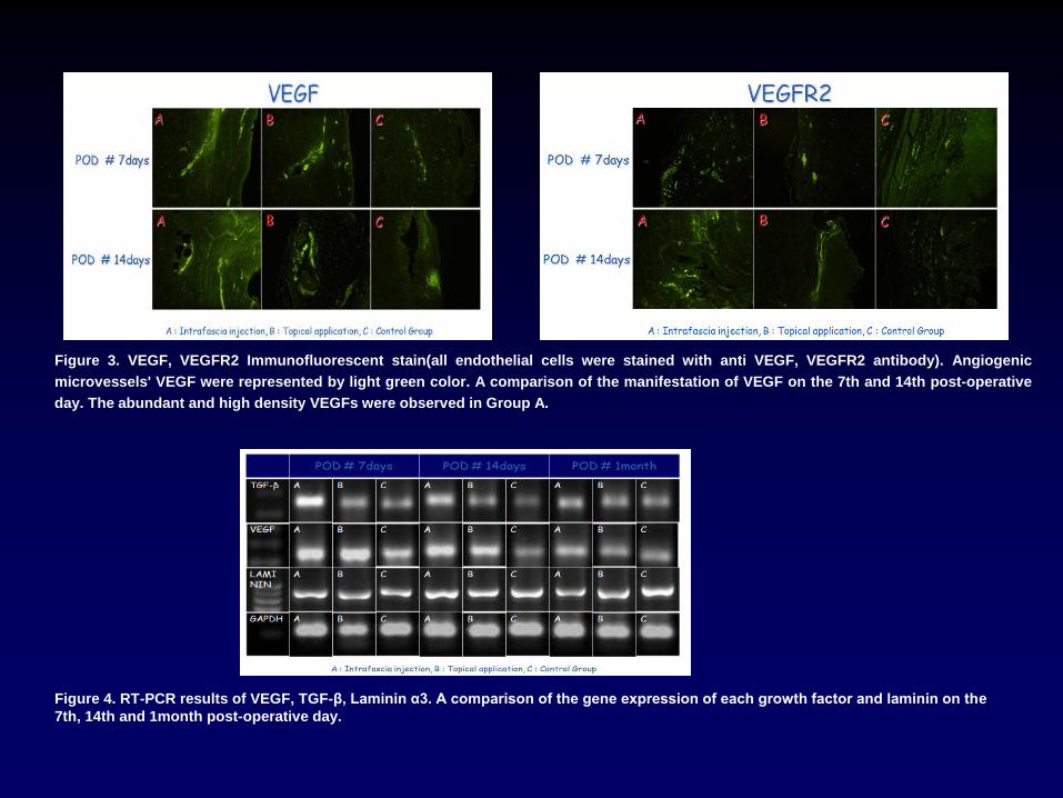
Figure 3. VEGF, VEGFR2 Immunofluorescent stain(all endothelial cells were stained with anti VEGF, VEGFR2 antibody). Angiogenic microvessels' VEGF were represented by light green color. A comparison of the manifestation of VEGF on the 7th and 14th post-operative day. The abundant and high density VEGFs were observed in Group A.
Figure 4. RT-PCR results of VEGF, TGF-β, Laminin α3. A comparison of the gene expression of each growth factor and laminin on the 7th, 14th and 1month post-operative day.

The rate of success in human full-thickness skin graft was statistically significantly high (p<0.05) in the group A (100%) which is
Intrafascial injection group compared to all the other groups and together with the group B (75%) which is topical application
group the two groups both showed the higher rate of success in skin graft than the control group (50%). In the result of
experiment to know the time taken for skin graft after grafting in the individual groups which were successful in skin graft, the
time taken in intrafascial injection group A (7 days) and the topical application group B (7.2 days) showed the result of shorter
time than that in the control group (7.7 days) so it showed the result that the time for engraftment becomes shorter in the group A
and B than that in the control group. Compared to the group C which is histological control group, more proliferated blood
vessels were shown in the group A and B. The formation of collagenous fiber showed more concentrated and regular
arrangement in the group A in 14 days after creation of cut, and in the control group the irregular and loose appearance of the
formation of arrangement of collagenous fiber compared to in other groups was observed. In immunocytochemical opinion the
manifestation of the growth factor VEGF showed more and more concentrated form in the group A. Resultantly, in the human full-
thickness skin graft model in nude mouse, the group A appeared to be the best in the engraftment of skin graft and also in the
group B appeared to be effective in engraftment.
The data demonstrated that administration of ADSCs into recipient beds by intrafascial injection for skin grafting can improve the
skin graft survival and improve the angiogenesis more than topical application of ADSCs
Summary & Conclusions
Figure 5. Western blot results of TGF-β. A comparison of the protein analysis with electrophoresis of TGF-β on the 7th, 14th and 1month post-operative day.