examination of b cell and virus interactions through...
TRANSCRIPT
PhD thesis
Examination of B cell and virus interactions through transformation by a pathogen (EBV) and transduction by
a lentiviral vector (HIV-1)
Krisztián Kvell MD
Department of Immunology and Biotechnology Faculty of Medicine, University of Pécs, Hungary
Program leader: Prof. Péter Németh MD PhD
Project leaders: Péter Balogh MD PhD1, Prof. Rudolf H. Zubler MD PhD2 and Prof. Péter Németh MD PhD1
1: Department of Immunology and Biotechnology Faculty of Medicine, University of Pécs, Hungary
2: Research and Development Unit, Division of Hematology
Department of Internal Medicine, Geneva University Hospitals, Switzerland
P é c s
2007
LIST OF CONTENTS
List of abbreviations…………………….………………………………………………3 General introduction………………………..…………………………..……….……...4 Aims……………………………………………………………………………………...4 Section I. – Establishment and fine-tuning of the EBV-associated post-transplantation
lymphoproliferative disease model system
1.1 Summary……..………….…….……………………………………………….5 1.2 Introduction.........................................................................................................6 1.3 Materials and methods……...……….…………………………………………7 1.4 Results…………..………….…………………….……………………………10 1.5 Discussion…..……..…………………………………………………………..13
Section II. – Establishment of a human primary B cell monoculture system for efficient lentiviral transduction and gene function assay
2.1 Summary……..………….…….………………………………………..……..18
2.2 Introduction........................................................................................................20 2.3 Materials and methods…..….……….………………………………………...22 2.4 Results and discussion…………………….…………………………………..24
Future perspectives..........……….…….………………………………………..……..32
Acknowledgements…………………………………………………………………….34 References……………………………………………………………………………...35 List of publications…………………………………………………………………….39 Appendix – Thesis-related personal publications..…………………………………......41
2
LIST OF ABBREVIATIONS
7AAD: 7 amino-actinomycin D
cDNA: complementary DNA
CMV: cytomegalovirus
cPPT: central polypurine tract
EBNA: Epstein-Barr nuclear antigen
EBV: Epstein-Barr virus
EF1α: elongation factor 1α
FACS: fluorescence-activated cell sorter
FCS: fetal calf serum
FLIP: FLICE inhibitory molecule
GFP: green fluorescent protein
HIV: human immune-deficiency virus
IRES: internal ribosome entry site
LMP-1: latent membrane protein 1
LPD: lymphoproliferative disease
LTR: long terminal repeat
MFI: mean fluorescence intensity
MIB-1: monoclonal antibody for Ki-67
M-MLV: Moloney murine leukemia virus
MOI: multiplicity of infection
PALS: periarteriolar lymphoid sheath
PBL: peripheral blood lymphocyte
PBS: phosphate-buffered saline
PCR: polymerase chain reaction
SCID: severe combined immune deficiency
SEM: standard error of mean
SIN: self inactivating
scFv: single-chain variable fragment
TLR: Toll-like receptor
VSV-G: vesicular stomatitis virus G-protein
WPRE/WHV: Woodchuck-hepatitis virus post-transcriptional responsive element
3

GENERAL INTRODUCTION In a simplistic way innate immunity in vertebrates fights cellular intruders (bacteria, fungi, parasites), while acquired immunity is more specialized for subcellular pathogens (viruses). This is in part supported by the complex relationship between viruses and lymphocytes. There are numerous known receptor-ligand pairs that allow for specific and strong interaction between viruses and lymphocytes. Depending on the context i.e. the actual status of continuous co-evolution such physical connections may help efficient viral infection of lymphocytes or specific immune recognition of viruses. Both lymphocytes and viruses display extensive genetic variability. The rapid and mutation-prone replication of viruses has been such an evolutionary demand that perhaps only gene-rearrangement processes restricted to antigen-receptor bearing lymphocytes could bring back balance for long-lived and genetically otherwise stable vertebrates in terms of co-evolution. In most cases viruses infect non-lymphoid cells, but elicit immune responses dominated by lymphoid cells. However, in the cases investigated here it is a part of the lymphoid cell population that becomes either transformed by wild-type or transduced by artificial viruses. Having said these it is especially exciting to investigate certain virus-triggered lymphoid diseases and to utilise domesticated viral vectors for research purposes in lymphocytes.
AIMS
1. In vivo modelling of the post-transplantation lymphoproliferative disease (PTLD) triggered by endogenous EBV-reactivation in human primary B-lymphocytes in a human-mouse (hu-SCID) chimera system. 2. Standardisation of the chimeric PTLD model system by performing an in vivo superinfection and confirming the dominance of the introduced EBV strain. 3. Establishment of human primary B cell monoculture system allowing for efficient lentiviral transduction, investigation of gene transfer efficiency by marker gene expression (GFP). 4. Transduction of representative intracellular (vFLIP) and secreted (IL4) transgenes in parallel with GFP, investigation of their expression or activity in functional assays.
4
SECTION I.
Establishment and fine-tuning of the EBV-associated post-transplantation lymphoproliferative disease model system 1.1 Summary
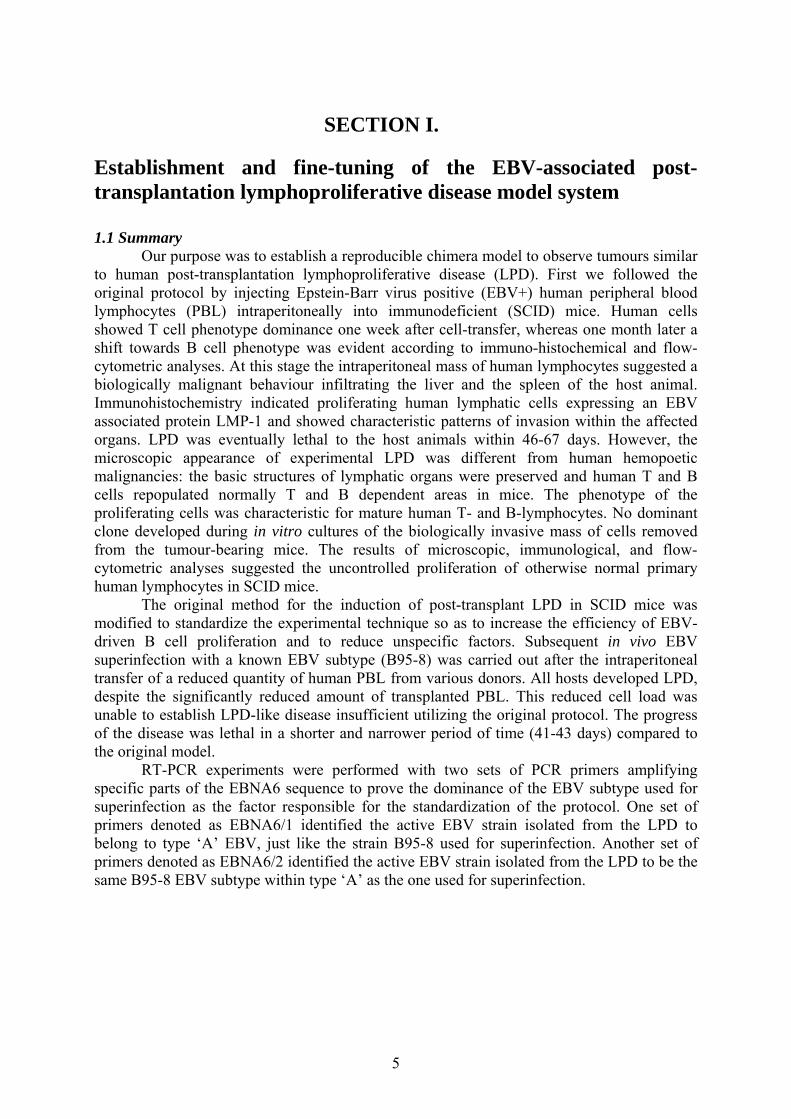
Our purpose was to establish a reproducible chimera model to observe tumours similar to human post-transplantation lymphoproliferative disease (LPD). First we followed the original protocol by injecting Epstein-Barr virus positive (EBV+) human peripheral blood lymphocytes (PBL) intraperitoneally into immunodeficient (SCID) mice. Human cells showed T cell phenotype dominance one week after cell-transfer, whereas one month later a shift towards B cell phenotype was evident according to immuno-histochemical and flow-cytometric analyses. At this stage the intraperitoneal mass of human lymphocytes suggested a biologically malignant behaviour infiltrating the liver and the spleen of the host animal. Immunohistochemistry indicated proliferating human lymphatic cells expressing an EBV associated protein LMP-1 and showed characteristic patterns of invasion within the affected organs. LPD was eventually lethal to the host animals within 46-67 days. However, the microscopic appearance of experimental LPD was different from human hemopoetic malignancies: the basic structures of lymphatic organs were preserved and human T and B cells repopulated normally T and B dependent areas in mice. The phenotype of the proliferating cells was characteristic for mature human T- and B-lymphocytes. No dominant clone developed during in vitro cultures of the biologically invasive mass of cells removed from the tumour-bearing mice. The results of microscopic, immunological, and flow-cytometric analyses suggested the uncontrolled proliferation of otherwise normal primary human lymphocytes in SCID mice.
The original method for the induction of post-transplant LPD in SCID mice was modified to standardize the experimental technique so as to increase the efficiency of EBV-driven B cell proliferation and to reduce unspecific factors. Subsequent in vivo EBV superinfection with a known EBV subtype (B95-8) was carried out after the intraperitoneal transfer of a reduced quantity of human PBL from various donors. All hosts developed LPD, despite the significantly reduced amount of transplanted PBL. This reduced cell load was unable to establish LPD-like disease insufficient utilizing the original protocol. The progress of the disease was lethal in a shorter and narrower period of time (41-43 days) compared to the original model. RT-PCR experiments were performed with two sets of PCR primers amplifying specific parts of the EBNA6 sequence to prove the dominance of the EBV subtype used for superinfection as the factor responsible for the standardization of the protocol. One set of primers denoted as EBNA6/1 identified the active EBV strain isolated from the LPD to belong to type ‘A’ EBV, just like the strain B95-8 used for superinfection. Another set of primers denoted as EBNA6/2 identified the active EBV strain isolated from the LPD to be the same B95-8 EBV subtype within type ‘A’ as the one used for superinfection.
5
1.2 Introduction
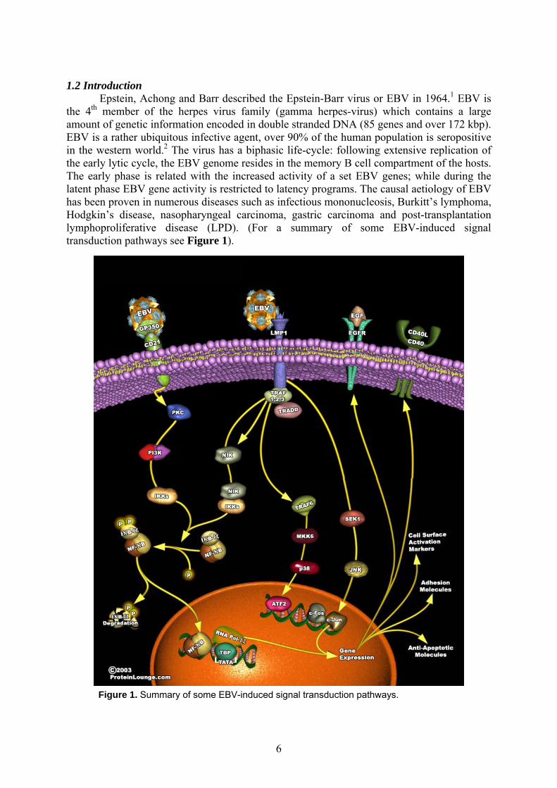
Epstein, Achong and Barr described the Epstein-Barr virus or EBV in 1964.1 EBV is the 4th member of the herpes virus family (gamma herpes-virus) which contains a large amount of genetic information encoded in double stranded DNA (85 genes and over 172 kbp). EBV is a rather ubiquitous infective agent, over 90% of the human population is seropositive in the western world.2 The virus has a biphasic life-cycle: following extensive replication of the early lytic cycle, the EBV genome resides in the memory B cell compartment of the hosts. The early phase is related with the increased activity of a set EBV genes; while during the latent phase EBV gene activity is restricted to latency programs. The causal aetiology of EBV has been proven in numerous diseases such as infectious mononucleosis, Burkitt’s lymphoma, Hodgkin’s disease, nasopharyngeal carcinoma, gastric carcinoma and post-transplantation lymphoproliferative disease (LPD). (For a summary of some EBV-induced signal transduction pathways see Figure 1).
Figure 1. Summary of some EBV-induced signal transduction pathways.
6

LPD may develop in EBV seropositive immunodeficient patients. These immune-
deficiencies by origin can be ‘natural’ (e.g. in AIDS, Duncan’s disease), or ‘artificial’ (as a result of chronic immunosuppressive treatment e.g. in post-transplantation medication). Among transplanted patients the occurrence of LPD is approximately 1%.3 Since chronic immunosuppression is becoming the part of everyday medicine the occurrence of LPD is likely to grow in the future. EBV subtypes differ in their ability to create LPD: ‘type A’ EBV is more potent than ‘type B’ EBV.2 ‘Type A’ LPD in younger patients has symptoms similar to infectious mononucleosis, infiltrating organs including the transplanted graft as well as the lymph nodes, bone marrow, liver, gastrointestinal tract, lungs, heart, nasal pharynx, thyroid glands and the central nervous system. ‘Type B’ LPD develops in senior patients in the form of solid tumors within the central nervous system and in the liver or elsewhere in the abdomen.4, 5 Ceasing immunosuppression usually heals LPD, but may simultaneously trigger graft rejection or the progression of other primary diseases. Different animal models are available for experimental LPD, but the induction of xenogeneic lymphoid chimeras in genetically immunodeficient mice shows most resemblance with human LPD.
The C.B.17 scid/scid mice were first described in 1983. These mice have a spontaneous point mutation resulting a special defect of V(D)J recombinase system.2 As functional lymphocytes do not develop, these animals lack specific immune reactions and can easily host xenogeneic tissues. There are several methods to create chimeras by repopulating SCID mice with human lymphoid cells.6 One involves the transfer human PBL into mature SCID mice generally via an intraperitoneal injection thus creating what is called ‘hu-(PBL)-SCID’. Although this method provides only a transient chimera condition for about six months, it is convenient and usually stable for a period long enough to examine some human diseases in vivo.7, 8, 9, 10 We refer to the above protocol as the original protocol. This method is still relevant, however it possesses some disadvantages: usually the amount of human PBL necessary for successful chimerism is rather high, around 5x10⁷ cells per mouse per inoculation. In addition, different human blood donors most likely harbour EBV strains of various transforming capacities. These factors result irregular and highly unpredictable chimera-development and survival of host mice.11, 12, 13
The viral EBNA6 DNA sequence is highly diverse among different subtypes of EBV. Some features of the EBNA6 sequence are still subjects of debate. The very same fragments appear to be necessary for successful viral activity and to encode viral proteins that efficiently elicit immune responses. It is intriguing to have tandem repeats in a DNA sequence that encodes such an important yet immunogenic protein sequence. A primer pair referred to as EBNA6/2 partially overlaps one such tandem sequence. RT-PCR with these primers differentiated between subtypes of type ‘A’ EBV and identify dominant active EBV strains in tissue samples of the chimeras.14, 15
1.3 Materials and methods 1.3.1 Animals
An ethic committee has approved all experiments involving live animals and all necessary guidelines concerning the treatment of animals were observed. C.B.17 scid/scid homozygous mice were kept inbred in the animal facility at the Faculty of Medicine, University of Pécs, under specific pathogen free conditions. During experiments the animals were kept in a laminar cabinet in the Department of Immunology and Biotechnology. Ciprofloxacin (Ciprobay, Bayer) containing water was administered per os as antibiotic prophylaxis.
7

1.3.2 Cell lines The B95-8 cell line harboring type ‘A’ EBV and the AG876 cell line containing type ‘B’ EBV, both secreting complete infectious Epstein-Barr virions were cultured in DMEM containing 20% FCS for fourteen days in the Regional Laboratory of Virology, Baranya County Institute of State Public Health Service, Pécs, Hungary. 1.3.3 Transplantation of human PBL
Following the approval of the appropriate ethic committee and along with the informed consent of six different human blood donors, buffy-coats were prepared from their blood donations as sources of human PBL. According to attached qualifications mostly EBV+ buffy-coats were used except for donor No.3, who was used as negative control. A suspension enriched in lymphocytes was obtained by density-gradient centrifugation (20°C, 20 minutes, 2000/min.) over Ficoll solution (ρ=1.077 g/cm3). Cells were counted in Burker-chamber after staining with the tripane-blue cell-exclusion dye. The viability of PBL was higher than 90% in all cases. Intraperitoneal cell transfer was achieved with 1-6x10⁷ cells per mouse per inoculation, depending on the protocol used (3-6x10⁷ cells per inoculation following the original protocol and 1-2x10⁷ cells per inoculation following the modified protocol). Altogether sixteen hu-PBL-SCID xenogeneic chimeras were created
1.3.4 In vivo superinfection
Three of the sixteen chimeras created altogether by the original and modified protocols were subjected to the in vivo superinfection protocol described below (Tables 1-2). The supernatant of the B95-8 cell line was centrifuged (20°C, 5 minutes, 1000/min) then filtered through 0.22 µm Whatman GF/C filter in order to eliminate cells and cellular debris. Ten days after intraperitoneal PBL transfer 1.5 ml purified supernatant was injected intraperitoneally into the host animals. The other three chimeras were not subjected to in vivo superinfection. An extra SCID mouse was control treated, EBV positive B95-8 supernatant was given without former PBL transfer. 1.3.5 Antibodies
The following anti-human monoclonal antibodies were used: anti- CD3, CD4, CD5, CD8, CD10, CD19, CD20, CD23, CD45, κ light chain, λ light chain, (all of them from DAKO, Denmark). Anti-mouse CD45 (IBL-5/25, developed in our laboratory), a-LMP 1and MIB-1 (both from DAKO, Denmark) antibodies were also used. 1.3.6 Immunohistochemistry
Histological examinations were performed on the intraperitoneal lymphoid mass and organs infiltrated by LPD (liver, spleen) from mice bearing clinical signs of malignant disease. Cryostat sections of frozen organs were fixed in acetone for 5 minutes then left to air-dry for 2 minutes. Further reactions were performed in a humid chamber. After rehydration with PBS the inhibition of endogenous peroxidase activity was performed by using phenyl-hydrasine-hydrochloride (1mg/ml) for 20 minutes. Non-specific binding sites were saturated by 5% BSA. Each antibody was incubated for 45 minutes followed by HRPO-conjugated anti-mouse IgG. The colour reaction was developed in Na-acetate buffer containing H2O2 and 3-amino-9-ethyl carbasol (AEC). After 15 minutes colour development was stopped by PBS solution. Hematoxilin counter staining was used. Classical hematoxilin-eosin staining was also made from all the samples for microscopic classification of the pathologic events. Normal human lymphoid tissues and monoclonal antibodies with irrelevant specificity were used as positive and negative controls, respectively.
8
1.3.7 Flow cytometric analysis
FACS phenotype analysis was performed on cells forming the intraperitoneal mass within LPD bearing mice and on cells cultured later on in vitro. Vortex-homogenization of the intraperitoneal mass of cells yielded a cell-suspension in PBS solution. Cells were incubated with FITC (fluorescein-isothyocianat) or PE (phycoerythrin) labelled antibodies for 30 minutes on ice. Measurements were validated by carefully selected positive and negative controls. FACSCalibur type (Becton Dickinson, USA) flow-cytometer, and the CellQuest software (Becton Dickinson, USA) were used for analysis. 1.3.8 In vitro cultures
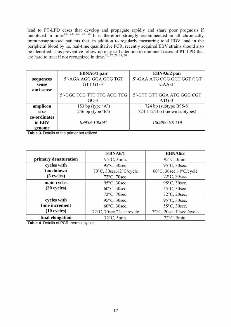
Following sterile biopsy from the infiltrated murine organs i.e. liver, spleen and from the peritoneal lymphoid mass, cells were mechanically dispersed and in vitro cultures were maintained in DMEM medium containing 10% FCS in a thermostat (Forma Scientific, USA) (37°C, 5% CO2 and saturated humidity) for a period of 6 weeks before cultures exhausted. 1.3.9 Nucleic acid isolation DNA of the B95-8 and AG876 cell lines was isolated by the GenElute Mammalian Genomic DNA Kit (Sigma). DNA sample isolated from the Akata cell line was a kind gift of János Minarovits M.D. Ph.D. (National Centre of Epidemiology, Budapest, Hungary). RNA was isolated from the peritoneal lymphoid mass using the GenElute Total RNA Kit (Sigma). 1.3.10 Primers The EBNA6/1 and EBNA6/2 primer pairs were synthesised by IDT (Integrated DNA Technologies, Inc.) and Sigma-Genosys Ltd, respectively. These primers cannot differentiate between DNA and cDNA as no introns exist within the amplified sequences. Therefore we performed DNase treatment. The used two pairs of primers both bind and amplify sequences of the EBNA6 gene. The EBNA6/1 pair helps to differentiate type ‘A’ and type ‘B’ EBV, while the EBNA6/2 pair surrounds a 39 bp tandem repeat sequence allowing the identification of various type ‘A’ EBV subtypes.16, 17, 18, 19 Details of the primers used are summarized in Table 3. 1.3.11 DNase treatment, reverse transcription and polymerase chain reaction, visualization DNase treatment of the RNA sample was carried out to eliminate DNA contamination, preventing false positive PCR results. Prior to reverse transcription and polymerase chain reaction the DNase enzyme was heat-inactivated to prevent decreased PCR sensitivity. Control reactions using RNA samples after DNase treatment but prior to cDNA synthesis were included in each set of PCR. For the above described procedures the DNase I. Kit (Sigma) was used. Reverse transcription was performed starting from 1 µg of RNA sample and using random hexamer primers and the M-MLV enzyme according to the vendor’s instructions. All reagents used for reverse transcription were part of the SuperScript First Strand Synthesis system for RT-PCR (Gibco). Following reverse transcription 1µl of cDNA was further used as sample for polymerase chain reaction. All PCR reagents came with the AdvanTaq kit (Clontech) and we performed reactions as recommended in the kit manual. EBNA6/1 and EBNA6/2 PCR products required 45 in a PCR Sprint device (Hybaid). For high specificity at maintained sensitivity we performed ‘touchdown’ for the first 5 annealing and `time-increment` for the last 10 elongation stages. Details are summarized in Table 4.
9
The PCR products were run in 2% agarose gels containing ethydium-bromide. Both analogue and digital pictures were taken and analysed with the Scion Image software. 1.4 Results 1.4.1 Stages of LPD development following the original protocol
Using the original protocol we created 10 hu-PBL-SCID xenogeneic chimeras with PBL from three different human blood donors (Table 1). LPD developed from the PBL of two donors. In the case of the third donor no LPD occurred since this control person was EBV seronegative. One week after cell transfer at the site of inoculation T-lymphocytes still represented the majority of human cells as FACS analysis showed the dominance of CD3+ cells compared to CD19+ cells (Figure 3A) isolated from the peritoneal mass, similar to human peripheral blood lymphocytes under normal conditions.
The first sign of the developing LPD was the occurrence of a loosely structured tissue-like mass of cells in the peritoneal cavity, in our case it was located adjacent to the portal region of the liver. The cells prepared from the peritoneal ‘tumour-like’ mass displayed human CD45+ according to flow cytometry (Figures 3B-3C). At this stage of the disease B cells began to proliferate and formed a dominant subpopulation. Among B cells some were CD23+ activated B cells. The T cell subpopulation was in minority by this time (Figures 3D-3G). Immunohistochemistry revealed further details. The proliferative capacity of cells that formed the intraperitoneal mass could be demonstrated by their MIB-1 staining; such cells were scattered in a diffuse pattern indicating multifocal increased growth-rate (Figure 4A). The majority of tumour forming cells was κ light chain positive, that were polyclonal (Figures 4C-4D). The presence of EBV was confirmed by the expression of LMP-1 (latent membrane protein type 1) in a fraction of human B cells (Figure 4B).
Dissemination of the human lymphoid cells occurred during the next stage of LPD. In addition to the mass of proliferating lymphoid cells located at the hilus of the liver and the spleen macroscopic haemorrhage-infarcts appeared on the surface of the liver. Tissue samples of the liver and the spleen were analyzed by histology. Proliferating lymphocytes infiltrating the tissue were easily distinguished from the hepatocellular background following hematoxilin-eosin staining by microscopy examination (Figure 4E). Immunohistochemistry revealed characteristic localization of human lymphocyte subsets in the spleen. The CD3+ T cells preferred the periarteriolar lymphatic sheaths (PALS) in the spleen, while the CD20+ B cells clustered into follicles (Figures 4F-4H) similar to physiological distribution. LPD was lethal to mice in 46-67 days, the moribund state was accompanied by palpable abdominal tumour, weakness, hypokinesis and ruffled fur (Table 1 and Figure 2). 1.4.2 In vitro cultures of tumour cells
Cells removed from the host animals could be cultured in vitro for approx. 6 weeks. At small magnification colonies formed large spherical groups due to increased growth rate and cells sticking together following division. Based on flow-cytometry the CD19+ tumour cells were CD5- and CD10- indicating that they were B cells, but not B1 or centroblast cells (Figures 3H-3I). No dominant clone developed, though light chain profile still favoured the κ positive population compared to the λ positive population (Figure 3J). 1.4.3 Inducing LPD by the modified protocol
Using the modified protocol we created altogether 6 chimeras with PBL from three different human blood donors (Table 2). In each pair of mice one was treated with subsequent in vivo superinfection: an i.p. injection of 1.5 ml B95-8 supernatant. A SCID mouse injected with 1.5 ml of B95-8 supernatant without prior cell transfer served as negative control. Note
10

No. of mouse
No. of donor
PBL injection
PBL per mouse
LPD emergence
survival (days)
1. 1. Yes 4x10⁷ * * 2. 1. Yes 5x10⁷ Yes 55 3. 2. Yes 4x10⁷ * * 4. 2. Yes 5x10⁷ Yes 46 5. 2. Yes 6x10⁷ Yes 67 6. 3. Yes 4x10⁷ * * 7. 3. Yes 5x10⁷ * * 8. 3. Yes 3x10⁷ No >122 9. 3. Yes 5x10⁷ No >122 10. 3. Yes 6x10⁷ No >122
Table 1. Summary of original chimera creation protocol. Asterisks (*) show animals sacrificed for FACS measures. Donor No. 3 was EBV seronegative.
No. of mouse
No. of donor
PBL injection
EBV injection
LPD emergence
survival (days)
11. 4. Yes No No >74 12. 4. Yes Yes Yes 41 13. 5. Yes No No >74 14. 5. Yes Yes Yes 42 15. 6. Yes No No >74 16. 6. Yes Yes Yes 43 17. --- No Yes No >74
Table 2. Summary of modified chimera creation protocol. All mice subjected to PBL injection received 1-2x10⁷ cells per mouse per inoculation. Survival was followed for 74 days.
0
10
20
30
40
50
60
days
average ofsurvival
standarddeviationof survival
original protocol modified protocol
Figure 2. Differences between the protocols in the average of survival and the standard deviation of survival
11

Figure 3. Characterization of the hu-SCID chimera system by flow-cytometry.
9 mouse CD45 human CD45
CD1Figure 3A. Site of inoculation prior to LPD development.
Figure 3B. Species origin of the periportal mass of cells.
Figure 3C. Species origin of the periportal mass of cells.
9 D4
CD13 CD3
Figure 3D. Phenotype of the periportal mass of cells.
Figure 3E. Phenotype of the peritoneal mass of cells.
Figure 3F. Phenotype of the peritoneal mass of cells.
9D8
CD3
Figure 3G. Phenotype of periportal mass of cells.
a-κ
Figure 3J. Clonality of in vitro cultured cells.
CD1
CD5 CD10
Figure 3H. Phenotype of in vitro cultured cells.
Figure 3I. Phenotype of in vitro cultured cells.
D19
human CD45
uman CD45
CD3
Figure 3K. Phenotype of ex vivo peritoneal LPD.
Figure 3L. Phenoex vivo peritoneal
12
C
9
CD1CD3
CD
CD3CD2
3 CC
a-λ
htype of LPD.

that neither the transfer of a smaller amount of PBL alone from EBV seropositive donors nor the in vivo administration of B95-8 supernatant on its own caused the emergence of LPD. In chimeras that received subsequent in vivo superinfection LPD developed and proved to be lethal in 41-43 days (Table 2 and Figure 2). At the autopsy of affected mice an intraperitoneal mass of cells in the ascitic fluid and enlarged, unusually pale liver and spleen were observed. Flow-cytometric analysis of the intraperitoneal mass of cells showed mainly human CD45+ phenotype and the dominance of CD19+ B cells compared to CD3+ T cells (Figures 3K-3L). The double negative population contained murine cells (figures not shown). In vitro cultures of cells removed from the peritoneal cavity of moribund LPD hosts showed high capacity for proliferation for over a month before cell division halted. At small magnification colonies formed large spherical groups of cells adhering to each other following division. Six chimeras were created following our modified protocol (Table 2). Three mice received PBL transfer alone and three others were also subjected to in vivo superinfection ten days after PBL inoculation. When the PBL transfer was not followed by in vivo EBV superinfection all animals survived beyond 74 days (time of follow-up), due to the insufficient amount of human cells for LPD induction. However, all three mice died in 41-43 days if PBL inoculation was followed by in vivo EBV superinfection. In our earlier experiments following the original protocol of chimera creation, survival figures varied between 46-67 days with an average of 56 days (Tables 1-2 and Figure 2). 1.4.4 PCR differentiation of EBV types and subtypes First we tested our EBNA6/1 PCR system for its ability to distinguish the two major types of EBV. Figure 5 demonstrates that by using the EBNA6/1 primer pair our PCR system clearly differentiates type ‘A’ and type ‘B’ EBV due to the size difference of amplicons characteristic for the two major types of EBV. Figure 6 illustrates that if RT-PCR is performed with the DNase treated RNA sample extracted from the peritoneal lymphoid mass it produces only one amplicon, the one that is characteristic of type ‘A’ EBV. The EBNA6/2 primer pair was also tested in our PCR system before application. As shown on Figure 7 the EBNA6/2 system can differentiate between different subtypes within type ‘A’ EBV. Figure 8 shows that if RT-PCR is performed with the DNase treated RNA sample isolated from the peritoneal lymphoid mass only one amplicon is produced, that is characteristic of subtype B95-8 of type ‘A’ EBV based on the length of the amplicon. In all PCR experiments the lane for genomic DNA contamination verifies the efficiency of DNase treatment and the purity of the resulting RNA sample. Software assisted image analysis confirms the absence of contaminating PCR products as seen attached below the PCR images. (Tables 3-4 summarize the specifications of the primers and PCR conditions used.) 1.5 Discussion There is always uncertainty in creating a hu-PBL-SCID xenogeneic chimera model. The survival of the graft depends on many factors that are not all clearly understood or can not be interfered properly.20 For example: graft versus host reaction can drastically influence the biological response of the recipient. The likelihood of graft acceptance can be raised with the increase of input cell quantity and careful protocols that spare cell functions. In the original protocol for creating hu-PBL-SCID models the minimally required amount of cells is approx. 5x10⁷ cells per inoculation, as smaller quantities usually fail to successfully repopulate the host.7 In vitro activation of human cells prior to transfer may also be helpful. PBL donors can be divided into at least two groups based on the incidence of LPD. The ability of donors to create LPD in SCID mice is constant with time and it is characteristic to donors. Approx. 40% of donors belong to the high incidence group and 60% to the low-
13

Figure 4. Characterization of the hu-SCID chimera system by histology.
100 µm
Figure 4A. Intraperitoneal mass of cells, MIB-1 immunohistology, 40x
Figure 4B. Intraperitoneal mass of cells, a-LMP-1 immunohistology, 40x
Figure 4C. Intraperitoneal mass of cells, a-κ light chain immunohistology, 40x
Figure 4D. Intraperitoneal mass of cells, a-λ light chain immunohistology, 40x
Figure 4F. Infiltration of the spleen, a-CD20 immunohistology, 100x
200 µm
40 µm
Figure 4E. Infiltration of the liver, hematoxilin-eosin staining, 40x
Figure 4G. Infiltration of the spleen,a-CD3 immunohistology, 20x
Figure 4H. Infiltration of the spleen,a-CD20 immunohistology, 20x
14
marker - CTRL type ‘A’ type ‘B’ marker -CTRL + CTRL gen. CTRL LPD
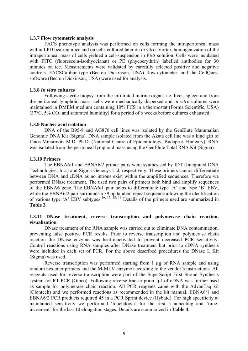
Figure 5. PCR differentiating between type ‘A’ and type ‘B’ EBV using the EBNA6/1 primer pair. Above: PCR picture. Below: image analysis. The lanes are: (left to right) DNA size marker, negative control reaction, sample from B95-8 EBV (type ‘A’), sample from AG876 EBV (type ‘B’).
Figure 6. RT-PCR identifying the type of EBV in the sample from a hu-(PBL)-SCID chimera with LPD using the EBNA6/1 primer pair. Above: RT-PCR picture. Below: image analysis. The lanes are: (left to right) DNA size marker, negative control reaction, sample from B95-8 EBV (type ‘A’) used as positive control, genomic control reaction, LPD sample from a chimera.
marker - CTRL B95-8 Akata marker - CTRL + CTRL gen. CTRL LPD
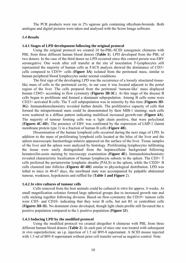
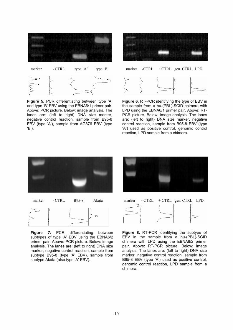
Figure 8. RT-PCR identifying the subtype of EBV in the sample from a hu-(PBL)-SCID chimera with LPD using the EBNA6/2 primer pair. Above: RT-PCR picture. Below: image analysis. The lanes are: (left to right) DNA size marker, negative control reaction, sample from B95-8 EBV (type ‘A’) used as positive control, genomic control reaction, LPD sample from a chimera.
Figure 7. PCR differentiating between subtypes of type ‘A’ EBV using the EBNA6/2 primer pair. Above: PCR picture. Below: image analysis. The lanes are: (left to right) DNA size marker, negative control reaction, sample from subtype B95-8 (type ‘A’ EBV), sample from subtype Akata (also type ‘A’ EBV).
15
intermediate incidence group. The incidence of LPD in the two groups shows no direct relation with the concentration of latently infected B cells in PBL. The frequency of latently infected cells among B-lymphocytes in the peripheral blood is individual and varies considerably. In a population of healthy carriers this is between 5-500x10-7 with a mean of 50x10-7 and this value is characteristic to each donor. The low concentration of latently infected cells is due to the fact that it concerns only a fraction of memory B cells and can also explain the relatively high number of cells required for successful LPD formation in the chimeras. Immunosuppression during human allograft transplantation protocols causes an average 40-50 fold increase of latently infected B cells in the peripheral blood of patients. Their increased numbers may be due to the prolonged survival or increased production of precursors that are under direct cytotoxic control.21 The prevalence of CD8+ cells within the graft is also variable, hence inadequate cytotoxic immune-surveillance may also play a part in lymphoproliferation. Besides the mentioned uncertainties of creating chimera models, the exact time-span necessary for LPD emergence can not be precisely forecasted. In case of an EBV+ PBL donor the chances of developing LPD within 60-110 days is about 80-100%.
The original method for inducing post-transplantation LPD in SCID mice was modified during our experiments to standardize and increase the efficiency of B cell tumour occurrence with the reduction of unspecific factors. We transferred a smaller quantity of human PBL compared to the original protocol and then administered EBV from the B95-8 cell line as subsequent in vivo superinfection. These alterations apparently circumvented some points of variation. The well-known human LPD developed in the animals, but a fraction of the formerly used amount of cells of EBV seropositive donors was sufficient to develop the disease in a slightly shorter and in a narrower time-span regardless of further donor characteristics. Following the modified protocol the intraperitoneal transfer of 1-2x10⁷ lymphocytes was sufficient to create LPD compared to 5x10⁷ with the original protocol, average survival decreased from 56 to 42 days and more importantly standard deviation of survival fell from 10.5 days to 1 day. Based on serology data, the donors of human lymphocytes were all EBV positive . However, the type or subtype of pre-existing EBV in donors was not identified prior to PBL transfer. There are many subtypes of EBV present in human population. By the EBNA6/2 system alone, 13 cell lines harbouring different type ‘A’ subtypes of EBV were tested and described in literature.16, 17 It is most unlikely that originally all three buffy-coat donors shared the same subtype of EBV during our experiments. Yet, based on RT-PCR, in all three chimeras the only active EBV strain was the B95-8 subtype used for superinfection. Also survival figures of the chimeras following the modified protocol were rather uniform. The dominance of subtype B95-8 compared to viral strains pre-existent in B-lymphocytes is supported by repeated RT-PCR experiments. Apparently the viral strain used for superinfection had an advantage of mutliplication compared to the EBV strains acquired previously by the B-lymphocytes. We believe that much of this advantage comes from the that the hu-(PBL)-SCID chimeras provide an initially constitutive, sustained immune-compromised environment and in vivo superinfection occurred under such circumstances. It seems that if EBV superinfection takes place parallel to immuno-suppression, the EBV subtype used for superinfection has an advantage of replication compared to EBV subtypes acquired earlier, present in a quiescent form within the B-lymphocytes. In turn this leads to a faster lymphoproliferation and a consequently more malignant biological behaviour ultimately resulting PBL donor independent survival figures of chimeras created following our modified protocol. The situation described above closely resembles the immunosuppressed state found in many post-transplant patients due to their immunosuppressive medication. EBV superinfection occurring in post-transplant patients receiving immunosuppressive therapy can
16
lead to PT-LPD cases that develop and propagate rapidly and share poor prognosis if unnoticed in time.18, 22, 23, 24, 25 It is therefore strongly recommended in all chronically immunosuppressed patients that, in addition to regularly measuring total EBV load in the peripheral blood by i.e. real-time quantitative PCR, recently acquired EBV strains should also be identified. This preventive follow-up may call attention to imminent cases of PT-LPD that are hard to treat if not recognized in time.26, 27, 28, 29, 30 EBNA6/1 pair EBNA6/2 pair
sequences sense
anti-sense
5’-AGA AGG GGA GCG TGT GTT GT-3’
5’-GGC TCG TTT TTG ACG TCG
GC-3’
5’-GAA ATG CGG GCT GGT CGT GAA-3’
5’-CTT GTT GGA ATG GGG CGT
ATG-3’ amplicon
size 153 bp (type ‘A’) 246 bp (type ‘B’)
724 bp (subtype B95-8) 724-1124 bp (known subtypes)
co-ordinates in EBV genome
99939-100091
100395-101119
Table 3. Details of the primer set utilized.
EBNA6/1 EBNA6/2 primary denaturation 95°C, 3min. 95°C, 3min.
cycles with `touchdown`
(5 cycles)
95°C, 30sec. 70°C, 30sec.↓2°C/cycle
72°C, 70sec.
95°C, 30sec. 60°C, 30sec.↓1°C/cycle
72°C, 20sec. main cycles (30 cycles)
95°C, 30sec. 60°C, 30sec. 72°C, 70sec.
95°C, 30sec. 55°C, 30sec. 72°C, 20sec.
cycles with time increment
(10 cycles)
95°C, 30sec. 60°C, 30sec.
72°C, 70sec.↑2sec./cycle
95°C, 30sec. 55°C, 30sec.
72°C, 20sec.↑1sec./cycle final elongation 72°C, 5min. 72°C, 5min.
Table 4. Details of PCR thermal cycles.
17

SECTION II.
Establishment of a human primary B cell monoculture system for efficient lentiviral transduction and gene function assay
2.1 Summary Recently, using HIV-1-derived lentivectors, we have obtained efficient transduction of primary human B lymphocytes co-cultured with murine EL-4 B5 thymoma cells. However, efficient lentiviral transduction of human primary B cells has so far only been feasible in such co-culture assays, not in mono-cultures of B cells. We have now found that transduction of B cells in a mono-culture system using CpG DNA was comparable to that in the EL-4 B5 co-culture system. Following the stimulation of human primary B cells by CpG motifs (see Figure 9 as the summary of TLR-mediated signal transduction pathways), anti-Ig and the human recombinant cytokines IL2 and IL10 a monocistronic vector with a CMV promoter gave 32 ± 4.7% GFP+ human primary B cells cells, which value for transduction efficiency is considered as robust, given the natural variety of human blood-donors. The EF1α promoter has also been tested, but performed inferior as seen earlier in our experiments and in accordance with previous reports. Also we have selected and transduced transgenes other than GFP as ‘proof-of-principle’ for the method. We have utilized both a secreted and an intracellular transgene to prove functionality of the expressed proteins following lentiviral transduction. In the same B/CpG system as above a bicistronic vector under the control of the CMV promoter, encoding either IL-4 or vFLIP as the first cistron and than GFP as the second cistron, gave 14.2 ± 2.1% GFP+ cells. The EF1α promoter showed even more impaired capabilities in these bicistronic than monocistronic vectors. We measured transgenic IL-4 secretion to be 1.3±0.2ng for 105 B cells in 24 hours using carefully selected experimental conditions and controls. In secondary culture systems transgenic IL-4 proved to be fully functional on B cells (promoting IgE switch in the EL4 B5 co-culture system and increased proliferation in the CD40L system, data not shown). Following transduction with a bicistronic vector encoding the viral FLIP molecule, vFLIP was detectable by both flow-cytometry and Western blot in GFP+, but not in GFP- B cells. When functional assays were employed to measure vFLIP-mediated rescue from FasL-triggered apoptosis, GFP+ (thus vFLIP+) cells were enriched among viable cells from 4% to 16% and 22% by FasL in a dose dependent manner (0 ng/ml, 100 ng/ml and 200 ng/ml, respectively). (Initial transduction efficiencies were kept low by utilizing low viral MOI to keep headroom for the anticipated enrichment of protected cells). In another assay following sort-enrichment of GFP+ (thus vFLIP+) cells 57% of the sorted GFP+ B cells were rescued from FasL-induced cell death as proved by their increased thymidine-incorporation
18
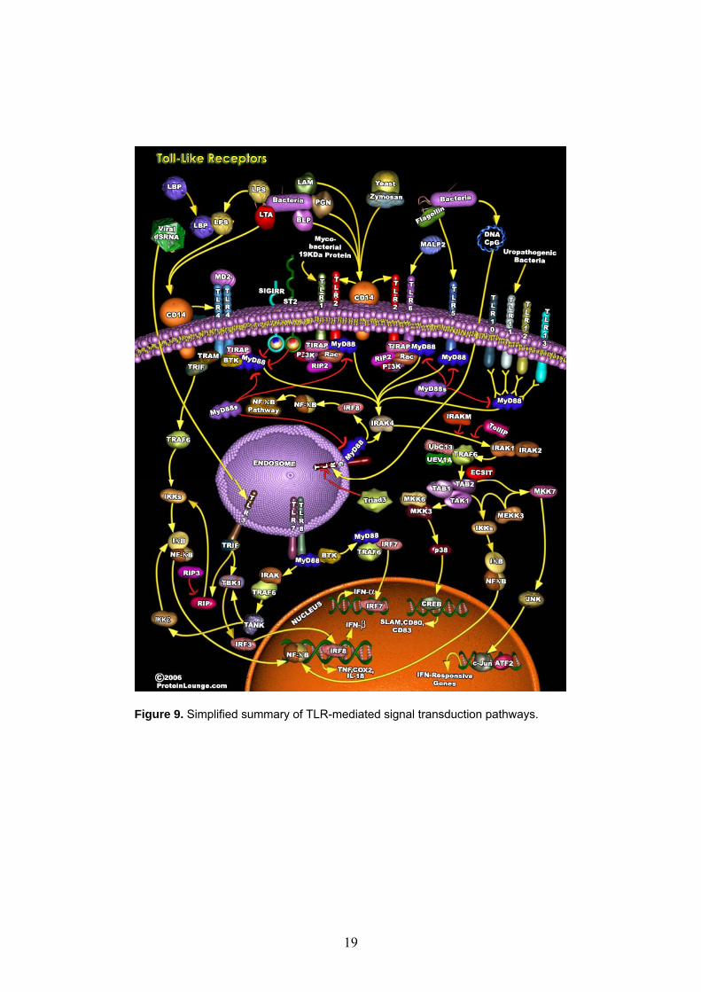
Figure 9. Simplified summary of TLR-mediated signal transduction pathways.
19
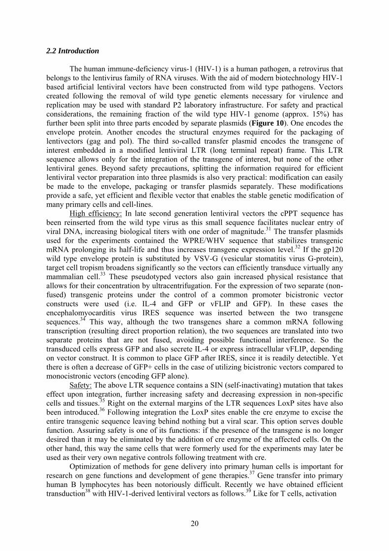
2.2 Introduction The human immune-deficiency virus-1 (HIV-1) is a human pathogen, a retrovirus that belongs to the lentivirus family of RNA viruses. With the aid of modern biotechnology HIV-1 based artificial lentiviral vectors have been constructed from wild type pathogens. Vectors created following the removal of wild type genetic elements necessary for virulence and replication may be used with standard P2 laboratory infrastructure. For safety and practical considerations, the remaining fraction of the wild type HIV-1 genome (approx. 15%) has further been split into three parts encoded by separate plasmids (Figure 10). One encodes the envelope protein. Another encodes the structural enzymes required for the packaging of lentivectors (gag and pol). The third so-called transfer plasmid encodes the transgene of interest embedded in a modified lentiviral LTR (long terminal repeat) frame. This LTR sequence allows only for the integration of the transgene of interest, but none of the other lentiviral genes. Beyond safety precautions, splitting the information required for efficient lentiviral vector preparation into three plasmids is also very practical: modification can easily be made to the envelope, packaging or transfer plasmids separately. These modifications provide a safe, yet efficient and flexible vector that enables the stable genetic modification of many primary cells and cell-lines. High efficiency: In late second generation lentiviral vectors the cPPT sequence has been reinserted from the wild type virus as this small sequence facilitates nuclear entry of viral DNA, increasing biological titers with one order of magnitude.31 The transfer plasmids used for the experiments contained the WPRE/WHV sequence that stabilizes transgenic mRNA prolonging its half-life and thus increases transgene expression level.32 If the gp120 wild type envelope protein is substituted by VSV-G (vesicular stomatitis virus G-protein), target cell tropism broadens significantly so the vectors can efficiently transduce virtually any mammalian cell.33 These pseudotyped vectors also gain increased physical resistance that allows for their concentration by ultracentrifugation. For the expression of two separate (non-fused) transgenic proteins under the control of a common promoter bicistronic vector constructs were used (i.e. IL-4 and GFP or vFLIP and GFP). In these cases the encephalomyocarditis virus IRES sequence was inserted between the two transgene sequences.34 This way, although the two transgenes share a common mRNA following transcription (resulting direct proportion relation), the two sequences are translated into two separate proteins that are not fused, avoiding possible functional interference. So the transduced cells express GFP and also secrete IL-4 or express intracellular vFLIP, depending on vector construct. It is common to place GFP after IRES, since it is readily detectible. Yet there is often a decrease of GFP+ cells in the case of utilizing bicistronic vectors compared to monocistronic vectors (encoding GFP alone). Safety: The above LTR sequence contains a SIN (self-inactivating) mutation that takes effect upon integration, further increasing safety and decreasing expression in non-specific cells and tissues.35 Right on the external margins of the LTR sequences LoxP sites have also been introduced.36 Following integration the LoxP sites enable the cre enzyme to excise the entire transgenic sequence leaving behind nothing but a viral scar. This option serves double function. Assuring safety is one of its functions: if the presence of the transgene is no longer desired than it may be eliminated by the addition of cre enzyme of the affected cells. On the other hand, this way the same cells that were formerly used for the experiments may later be used as their very own negative controls following treatment with cre. Optimization of methods for gene delivery into primary human cells is important for research on gene functions and development of gene therapies.37 Gene transfer into primary human B lymphocytes has been notoriously difficult. Recently we have obtained efficient transduction38 with HIV-1-derived lentiviral vectors as follows.39 Like for T cells, activation
20
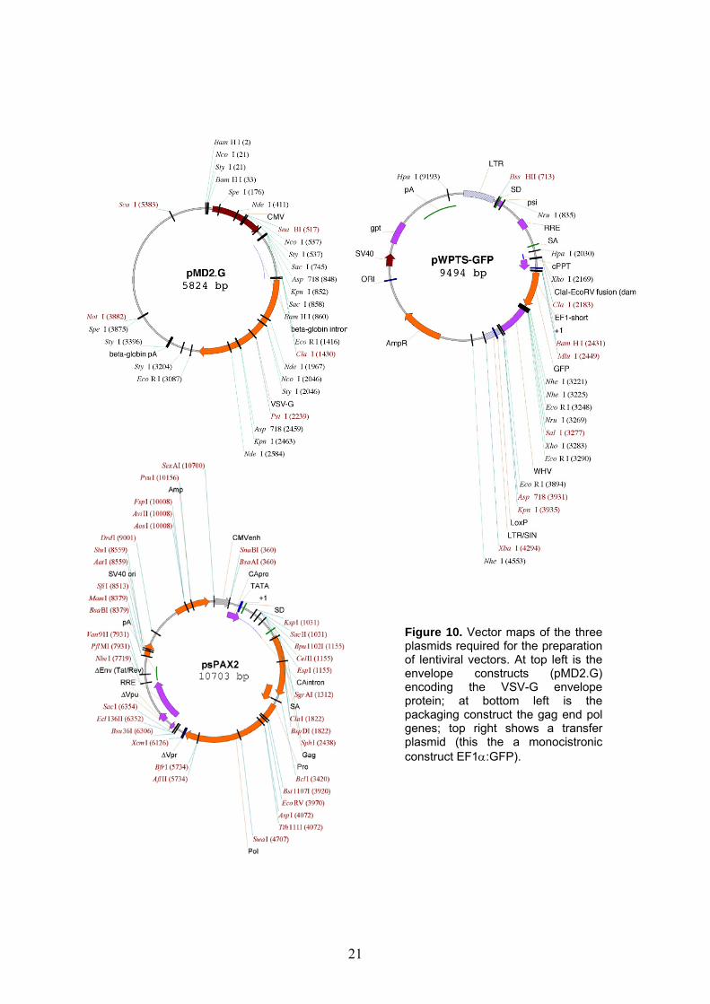
Figure 10. Vector maps of the three plasmids required for the preparation of lentiviral vectors. At top left is the envelope constructs (pMD2.G) encoding the VSV-G envelope protein; at bottom left is the packaging construct the gag end pol genes; top right shows a transfer plasmid (this the a monocistronic construct EF1α:GFP).
21

of the B cells was necessary.40, 41 In B cells co-cultured with irradiated murine EL-4 B5 thymoma cells and human cytokines, HIV-1 based lentiviral vectors with the human cytomegalovirus (CMV) or elongation factor-1α (EF-1α) internal promoter provided efficient transduction efficiencies.38 On the other hand, T-B co-culture is cumbersome, and a disadvantage for various research applications is that the functions of the thymoma cells are not yet molecularly characterized. Isolated B cells activated by cross-linking of CD40 in the presence of various cytokines were very poorly transducible with HIV vectors.38, 42
Therefore our aim was to find a mono-culture system for the efficient transduction with HIV vectors of isolated B cells activated by defined stimuli. The LPS receptor, Toll-like receptor-4 (TLR4), is absent on/in human B cells.43, 44 But TLR9, the receptor for immune-stimulatory CpG DNA, is expressed by memory human B cells, and up-regulated on naive B cells by anti-immunoglobulin (Ig) antibody, which mimics an antigen signal.43, 44 We have found that monocistronic and bicistronic HIV-1-derived vectors ensure an efficient means of gene transfer in the CpG DNA-dependent B cell monoculture system that operates with defined stimuli of activation. 2.3 Materials and methods 2.3.1 HIV-1-derived vectors A second-generation packaging system and self-inactivating HIV-1 vectors with the LoxP site in the 3’ LTR were utilized.45, 46 The monocistronic vector with the EF-1α internal promoter (called vector EF1α-GFP in this study) was plasmid pWPTS-GFP. This plasmid carries, in 5’ to 3’ direction, the cPPT element, the intronless (‘short’) EF-1α promoter, the expression cassette, and the WPRE.47, 48 Vector CMV-GFP was made by replacing the promoter with the human CMV promoter between the ClaI and the BamHI restriction sites.49 In the bicistronic vectors, which were derived from the above vectors, the first cistron was the transgene of interest, between the BamHI and the SalI restriction sites, followed by the encephalomyocarditis virus IRES and GFP encoded downstream of the IRES.46 In the vectors EF1α-IL-4-GFP and CMV-IL-4-GFP the transgene was the complete human IL-4 cDNA sequence (a kind gift from Dr. J.-F. Gauchat, formerly at the Institut Pierre Fabre, St. Julien, France). In vector CMV-vFLIP-GFP the transgene was the FLAG-tagged viral FLIP of human molluscum contagiosum virus (open reading frame 159L), kindly provided by Dr. Margot Thome of the Department of Biochemistry, University of Lausanne.50 Virus particles pseudotyped with vesicular stomatitis virus G glycoprotein were produced, concentrated 100-fold by ultracentrifugation, and titrated on HeLa cells by measurement of GFP+ cells by FACS to establish HeLa transducing units per milliliter (usually 0.5-2x10⁸), as described.51 Viral stocks were suspended in B cell culture medium and stored at -70°C. 2.3.2 Cell cultures and transduction Approval for research on human cells was obtained from the Ethics Committee of the Geneva University Hospitals. Written consent was provided for all cell samples. B cells were isolated to more than 98% purity from buffy-coats of virus-screened blood donations obtained from the Geneva Transfusion Center using Ficoll–Hypaque centrifugation and anti-CD19 magnetic beads (Dynal). All B cell cultures were grown in RPMI 1640 medium supplemented with 10% fetal calf serum, 2-mercaptoethanol, Hepes buffer, and antibiotics as described. 2.3.3 CpG DNA system B cells (25,000 cells/200 µl culture; flat-bottom 96-well plates) were stimulated with CpG DNA (2.5 µg/ml), anti-Ig antibody (2 µg/ml), IL-2 (50 ng/ml), and IL-10 (10 ng/ml). CpG oligonucleotides of the published sequences C274, 2006, and C661 were obtained as complete
22

phosphorothioates synthesized to our order by Microsynth (Balgach, Switzerland). Anti-Ig was goat F(abV)2 anti-human IgA + IgG + IgM (H+L) (Jackson ImmunoResearch). The cytokines were from PeproTech. FACS sorting of CD27+/CD27- cells was done as described. 52, 53
2.3.4 CD40L system B cells (50,000 cells/200 µl) were stimulated with a soluble oligomeric CD40L (300 ng/ml), IL-2 (50 ng/ml), and IL-10 (10 ng/ml) as described.52 When CD40L was added to the CpG DNA system or the EL-4 B5 system, its concentration was 300 ng/ml. 2.3.5 EL-4 B5 system B cells (3,000 cells/200 µl) were stimulated with 50,000 irradiated EL-4 B5 murine thymoma cells and a mixture of human cytokines acting on the EL-4 B5 cells (IL-1β) or the B cells (TNF-α, IL-2, and IL-10), as described.51 To transduce B cell cultures at different time points, the big cell clusters, which formed in all culture systems, were dispersed by gentle pipetting (five times aspiration of the culture using 200µl tips). Virus particles were then added to the cultures in a volume of 2 to 4 µl, at the indicated MOI according to cell counts by microscope (B cells or B plus EL-4 B5 cells), without further manipulations or cell wash. We found that centrifugation of virus particles in new culture plates (spinoculation) with cell transfer did not give better results. 2.3.6 Sorting and FACS analysis of transduced cells GFP+ cell populations were sorted on a FACStar sorter, and analyses of GFP+ cells were performed using CellQuest software (Becton–Dickinson), at the cytofluorometry laboratory of the Geneva Medical Center. Cells were pretreated with polyclonal mouse Ig before staining with phycoerythrin (PE)-coupled anti-CD19 and viable (7-amino-actinomycin-D low) cells were gated for analysis of GFP expression.51 For analysis of cytoplasmic IL-4, cells were cultured with brefeldin A (Sigma) 2.5 µg/m for 3 hours. For analysis of cytoplasmic IL-4 or viral FLIP, cells were treated with Cytofix/Cytoperm and Perm/Wash solutions, followed by AB+ serum, and then stained, respectively, with PE-coupled anti-IL-4 mAb (Pharmingen) or anti-FLAG-M2 mAb (Sigma), which we coupled to biotin, followed by PE-coupled streptavidin (Jackson ImmunoResearch). The isotype control was mouse IgG1. Apoptosis was assessed using the Annexin V–PE Apoptosis Detection Kit I (Becton–Dickinson). 2.3.7 IL-4 secretion rate On day 2 or 3 after addition of viral particles, B cells cells were harvested and washed twice in culture medium in Eppendorf tubes, and viable, trypan blue-excluding cells were counted. The cells were re-cultured as before, and after 24 h soluble IL-4 was measured in culture supernatants by Quantikine Human IL-4 Immunoassay (R&D Systems). 2.3.8 Real-time quantitative PCR The LightCycler system, High Pure PCR Template Preparation Kit, and LightCycler FastStart DNA MasterPLUS SYBR Green I kit (all from Roche) were utilized. PCR was performed in separate assays for genomic DNA, using intron-binding primers for β2-microglobulin (5’-GGCACTGCTGAGATACTGAT-3’, reverse 5’-GCTAGGACAGCAGGACTTA-3’; 215-bp product) and for vector DNA, using GFP primers (5’-GGCAAGCTGACCCTGAAGTT-3’, 5’-GCATGGCGGACTTGAAGAAGT-3’; 149-bp product); respective plasmid standards were utilized.
23

2.3.9 FasL assays The protective effect of vFLIP against apoptosis was assayed both by flow-cytometric and cell-proliferation assays (thymidine incorporation). Flow cytometry: B cells in the CpG system were transduced on day 2 by low quantities of lentiviruses (CMV:GFP or CMV:vFLIP-GFP) at MOI of 1. Some cultures were not transduced as control cultures. Then 3 days following transduction the cells were given different concentrations of FasL (0, 100 or 200 ng/ml) for 48 hours. Then on day 7 of the culture viable B cells in the gate of living cells (7AAD negative) were assayed for GFP expression by flow-cytometry. Cell-proliferation: Following transduction of B cells as above, GFP+ cells were sorted on day 5, that is 3 days after exposure to vector and re-cultured (20,000 trypan blue-excluding cells/200 µl, triplicate cultures) in the CpG DNA system (without anti-Ig) in the presence of soluble oligomeric FasL (200 ng/ml) prepared as described.54 Thymidine incorporation was measured after 48 hours including the pulsing during the last 12 hours with 1 µCi (0.037 MBq) [methyl-3H] thymidine (Amersham Pharmacia Biotech).52
2.3.10 Western blot for viral FLIP This was done with the ECL Western blotting analysis system (Amersham) using 0.45µm nitrocellulose membranes (Bio-Rad) and Biomax chemiluminescence films (Kodak). The antibodies were IgG1 mouse anti-FLAG-M2 mAb (Sigma) or polyclonal goat IgG anti-actin (Santa Cruz Biotechnologies) in conjunction with horseradish peroxidase-coupled anti-mouse or anti-goat antibodies, respectively (Santa Cruz Biotechnologies). 2.4 Results and discussion 2.4.1 B cell transduction in the CpG DNA system We found that peripheral blood B cells stimulated by CpG DNA, anti-Ig antibody, and the cytokines IL-2 and IL-10, according to Bernasconi et al., were efficiently transduced by HIV vectors. For monocistronic (GFP) and bicistronic (IL-4–GFP or vFLIP-GFP) vectors, the mean fluorescence intensity (MFI) of GFP+ B cells was always higher (2.5-fold or more) with the CMV than with the EF-1α internal promoter (Figure 11), confirming our previous findings with monocistronic vectors.51, 55 As expected, the percentages of GFP+ cells were lower with the bicistronic compared to the monocistronic vectors (3- or 2.3-fold by the mean, respectively, for the EF-1α or CMV promoter; for the CMV promoter, mean data are shown on Figure 12). The CG-rich oligonucleotides C274 (21 bases) and 2006 (24 bases), which triggered optimal B cell proliferation in the study by Marshall et al., both allowed for efficient transduction, whereas oligonucleotide C661, with all CG inverted to GC, gave 20-fold lower proliferation and transduction.[16] Vectors had to be added to the B cell cultures on day 2 or later. At an of MOI 20 HeLa cell-transducing units per B cell the numbers of GFP+ cells were 1.2-fold higher by the mean than at an MOI of 10, but at an MOI of 30 or higher the numbers progressively decreased. 2.4.2 Comparison of different B cell culture systems Viable cell expansion in the CpG DNA system set up with total peripheral blood B cells was fivefold by the mean in 10 days. Individual B cells proliferate very heterogeneously in this system.[10] At least fivefold higher B cell expansion was obtained in the system using EL-4 B5 murine thymoma cells as helper T cells.51, 56 But the percentages of transduced B cells among the viable cells analyzed on day 6 or 7 were very similar in both systems (Figure 12,
24
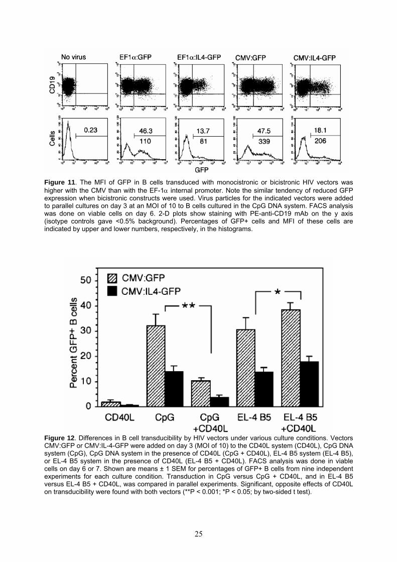
Figure 11. The MFI of GFP in B cells transduced with monocistronic or bicistronic HIV vectors was higher with the CMV than with the EF-1α internal promoter. Note the similar tendency of reduced GFP expression when bicistronic constructs were used. Virus particles for the indicated vectors were added to parallel cultures on day 3 at an MOI of 10 to B cells cultured in the CpG DNA system. FACS analysis was done on viable cells on day 6. 2-D plots show staining with PE-anti-CD19 mAb on the y axis (isotype controls gave <0.5% background). Percentages of GFP+ cells and MFI of these cells are indicated by upper and lower numbers, respectively, in the histograms.
Figure 12. Differences in B cell transducibility by HIV vectors under various culture conditions. Vectors CMV:GFP or CMV:IL-4-GFP were added on day 3 (MOI of 10) to the CD40L system (CD40L), CpG DNA system (CpG), CpG DNA system in the presence of CD40L (CpG + CD40L), EL-4 B5 system (EL-4 B5), or EL-4 B5 system in the presence of CD40L (EL-4 B5 + CD40L). FACS analysis was done in viable cells on day 6 or 7. Shown are means ± 1 SEM for percentages of GFP+ B cells from nine independent experiments for each culture condition. Transduction in CpG versus CpG + CD40L, and in EL-4 B5 versus EL-4 B5 + CD40L, was compared in parallel experiments. Significant, opposite effects of CD40L on transducibility were found with both vectors (**P < 0.001; *P < 0.05; by two-sided t test).
25

CpG and EL-4 B5; there were about 30 and 14% GFP+ cells, respectively, for vectors CMV:GFP and CMV:IL-4-GFP). Since activated B cells proliferated and outnumbered non-proliferating cells, this means that proliferating B cells were similarly transduced in both systems. Various cell types, e.g., T cells, after some activation become transducible with lentivectors even if they do not divide.41, 42 So far this could not be studied with B cells, because - unlike for T cells - B cell populations could not be maintained in an activated, yet non-dividing state. In agreement with our previous data, however, transducibility was very low in B cells proliferating in response to soluble CD40 ligand, IL-2, and IL-10 (CD40L, Figure 12).51 Moreover, when we added CD40L to the CpG DNA system (CpG + CD40L), thymidine incorporation was enhanced 2-fold compared to the standard CpG DNA system, but transducibility became 3.2-fold lower by the mean (P<0.001 by two-sided t test; nine independent, paired experiments). By contrast, adding CD40L to the EL-4 B5 system (EL-4 B5 + CD40L) gave not only slightly increased thymidine incorporation compared to the standard EL-4 B5 system, but also 1.3-fold higher B cell transducibility (P<0.05; also nine independent, paired experiments). In those experiments in which the B response in the EL-4 B5 system was relatively weak, addition of CD40L could double the percentage of GFP+ B cells. Anti-Ig did not enhance transducibility in the CD40L system, but B cells stimulated with CpG DNA only were transducible (about 15% GFP+ cells with monocistronic vector). Thus, it appeared that, in particular, B cells stimulated via CD40 in the absence of other T cell signals (an artificial condition) somehow resisted transduction by HIV vectors. The vectors did not cause elevated surface annexinV expression in CD40L-stimulated than in CpG DNA stimulated B cells. Serafini et al. found that in B cells activated via CD40 the transduction was blocked at an early stage.57 It is possible that innate cellular defenses, such as mechanisms involving TRIM5 or APOBEC3 antiretroviral factors, are involved; they might be triggered under certain abnormal cell activation conditions in vivo (possibly mimicked by sole CD40L activation).58, 59
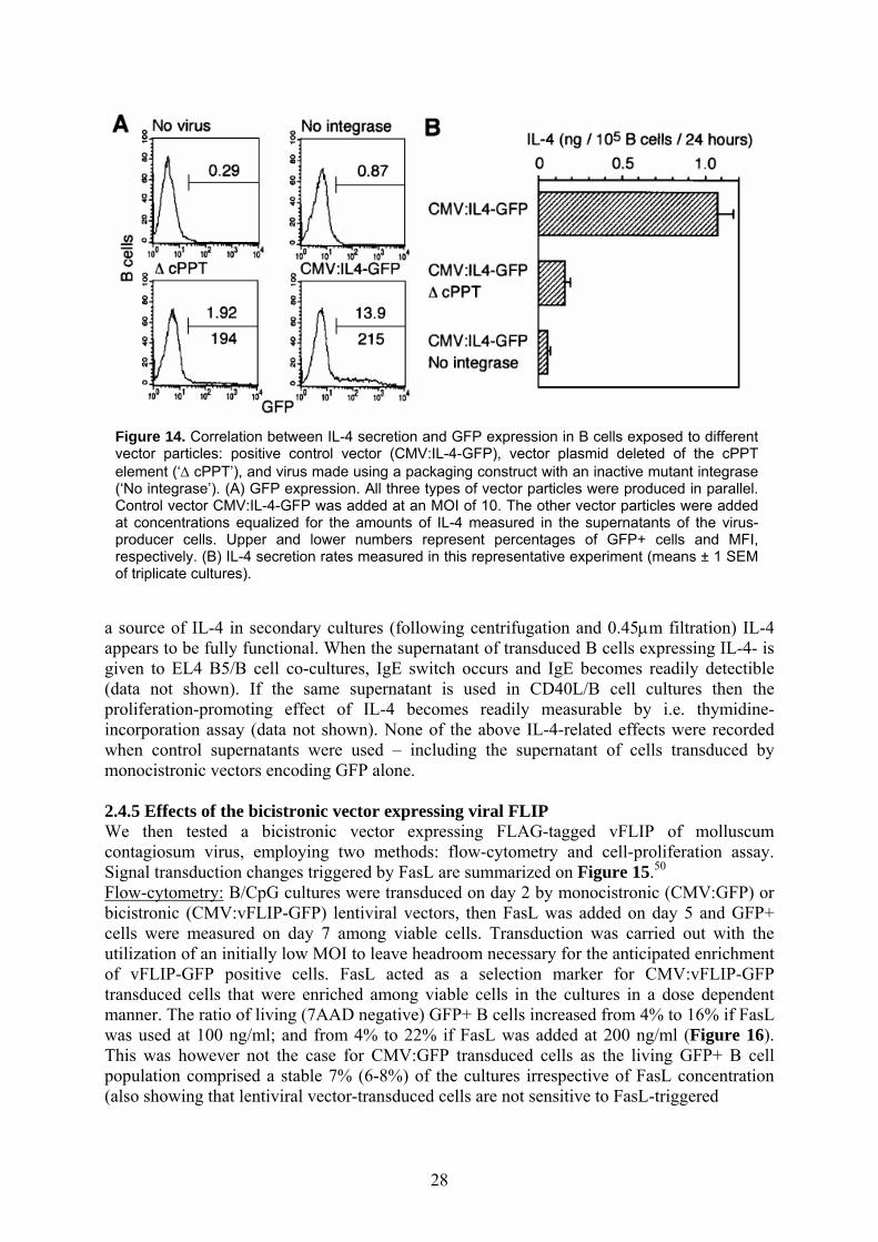
2.4.3 IL-4 secretion by transduced B cells Changes in signal transduction promoted by IL-4 in B cells are summarized on Figure 13. B cells transduced with IL-4 encoding vectors, after cell wash, released IL-4 that proved to functionally intact during secondary cultures. We studied whether pseudo-transduction played a role, i.e., any viral integration-independent effects, including passive transfer of transgene proteins by the virus particles (cells become exposed to numerous defective particles).51 Therefore, we tested viruses produced using packaging constructs with a loss-of-function mutant integrase or a vector plasmid (CMV:IL-4-GFP) lacking the central polypurine tract (cPPT) involved in nuclear transportation. We added control virus at an MOI of 10 and the others, produced in parallel, at concentrations equalized for IL-4 concentrations in the virus-producer 293T culture supernatant. The control vector gave 13.9% GFP+ B cells (Figure 14A) and an IL-4 release of 1.1 ng IL-4/105 B cells/24 hours (Figure 14B). The virus lacking integrase activity gave only 0.87% GFP+ cells, but no bright GFP+ cells (low MFI), i.e., this was pseudo-transduction that weakly occurs with GFP - however this phenomenon is intense if membrane-bound transgenic proteins are expressed due to viral budding.51 The IL-4 release was also very low; indicating that about 5% of the IL-4 release obtained with the positive control may have been due to pseudo-transduction. The virus lacking the cPPT gave 1.92% GFP+ cells, including about 1% bright cells. The IL-4 release (6.7-fold lower than control) was close to what one would expect for this number of bright GFP+ cells plus pseudo-transduction as estimated above. Thus, pseudo-transduction played only a negligible role. GFP expression and IL-4 secretion were stable in B cells between days 3 and 8 after exposure to vector. On day 8 after exposure to vectors with the respective optimal promoters, at an
26
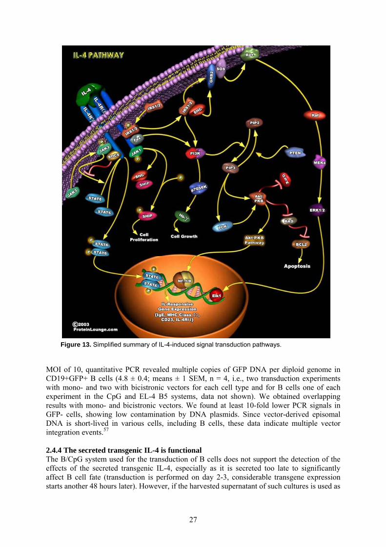
Figure 13. Simplified summary of IL-4-induced signal transduction pathways.
MOI of 10, quantitative PCR revealed multiple copies of GFP DNA per diploid genome in CD19+GFP+ B cells (4.8 ± 0.4; means ± 1 SEM, n = 4, i.e., two transduction experiments with mono- and two with bicistronic vectors for each cell type and for B cells one of each experiment in the CpG and EL-4 B5 systems, data not shown). We obtained overlapping results with mono- and bicistronic vectors. We found at least 10-fold lower PCR signals in GFP- cells, showing low contamination by DNA plasmids. Since vector-derived episomal DNA is short-lived in various cells, including B cells, these data indicate multiple vector integration events.57 2.4.4 The secreted transgenic IL-4 is functional The B/CpG system used for the transduction of B cells does not support the detection of the effects of the secreted transgenic IL-4, especially as it is secreted too late to significantly affect B cell fate (transduction is performed on day 2-3, considerable transgene expression starts another 48 hours later). However, if the harvested supernatant of such cultures is used as
27
aag(piwm 2WcSFbcuotmwTp(
Figure 14. Correlation between IL-4 secretion and GFP expression in B cells exposed to different vector particles: positive control vector (CMV:IL-4-GFP), vector plasmid deleted of the cPPT element (‘∆ cPPT’), and virus made using a packaging construct with an inactive mutant integrase (‘No integrase’). (A) GFP expression. All three types of vector particles were produced in parallel. Control vector CMV:IL-4-GFP was added at an MOI of 10. The other vector particles were added at concentrations equalized for the amounts of IL-4 measured in the supernatants of the virus-producer cells. Upper and lower numbers represent percentages of GFP+ cells and MFI, respectively. (B) IL-4 secretion rates measured in this representative experiment (means ± 1 SEM of triplicate cultures).
source of IL-4 in secondary cultures (following centrifugation and 0.45µm filtration) IL-4 ppears to be fully functional. When the supernatant of transduced B cells expressing IL-4- is iven to EL4 B5/B cell co-cultures, IgE switch occurs and IgE becomes readily detectible data not shown). If the same supernatant is used in CD40L/B cell cultures then the roliferation-promoting effect of IL-4 becomes readily measurable by i.e. thymidine-ncorporation assay (data not shown). None of the above IL-4-related effects were recorded hen control supernatants were used – including the supernatant of cells transduced by onocistronic vectors encoding GFP alone.
.4.5 Effects of the bicistronic vector expressing viral FLIP e then tested a bicistronic vector expressing FLAG-tagged vFLIP of molluscum
ontagiosum virus, employing two methods: flow-cytometry and cell-proliferation assay. ignal transduction changes triggered by FasL are summarized on Figure 15.50
low-cytometry: B/CpG cultures were transduced on day 2 by monocistronic (CMV:GFP) or icistronic (CMV:vFLIP-GFP) lentiviral vectors, then FasL was added on day 5 and GFP+ ells were measured on day 7 among viable cells. Transduction was carried out with the tilization of an initially low MOI to leave headroom necessary for the anticipated enrichment f vFLIP-GFP positive cells. FasL acted as a selection marker for CMV:vFLIP-GFP ransduced cells that were enriched among viable cells in the cultures in a dose dependent anner. The ratio of living (7AAD negative) GFP+ B cells increased from 4% to 16% if FasL as used at 100 ng/ml; and from 4% to 22% if FasL was added at 200 ng/ml (Figure 16). his was however not the case for CMV:GFP transduced cells as the living GFP+ B cell opulation comprised a stable 7% (6-8%) of the cultures irrespective of FasL concentration also showing that lentiviral vector-transduced cells are not sensitive to FasL-triggered
28

Figure 15. Summary of FasL-triggered signal transduction pathways.
apoptosis more than untransduced B cells). Not only did the percentage of the CMV:vFLIP-GFP cells increase but also their mean fluorescence intensity (data not indicated). Cell-proliferation assay: We added the same bicistronic vector (CMV:vFLIP-GFP) or control vector (CMV:GFP) to B cells in the CpG DNA system on day 2 and sorted GFP+ cells on day 5 (Figure 17A). We re-cultured the cells in the presence or absence of soluble FasL. In three experiments, we measured thymidine incorporation after 48 hours to measure cell survival (Figure 17B). Only 14 ± 4% of B cells transduced with control vector, but 63 ± 12% of B cells transduced with vFLIP vector, resisted killing by FasL. Thus, among those cells that could be killed by FasL, 57% (range 45 to 71%) were protected by vFLIP. In B cells transduced in the EL-4 B5 system, intracellular vFLIP was detectable with anti-FLAG mAb by FACS and its expression correlated with GFP expression in individual cells (Figure 17C). Viral FLIP was also detectable with this antibody by Western blot in sorted GFP+, but not in GFP-, B cells (Figure 17D). 2.4.6 Proof of principle for functional lentiviral transduction in the B/CpG system In the B/CpG system utilizing late second generation lentiviral vectors we have introduced a representative secreted transgene (hu-rIL-4) and an intracellular transgene (vFLIP). Both representative genes yielded high amounts of easily detectable transgenic proteins. IL-4 was
29
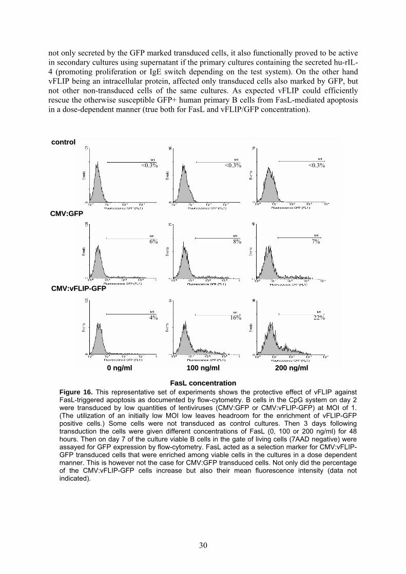
not only secreted by the GFP marked transduced cells, it also functionally proved to be active in secondary cultures using supernatant if the primary cultures containing the secreted hu-rIL-4 (promoting proliferation or IgE switch depending on the test system). On the other hand vFLIP being an intracellular protein, affected only transduced cells also marked by GFP, but not other non-transduced cells of the same cultures. As expected vFLIP could efficiently rescue the otherwise susceptible GFP+ human primary B cells from FasL-mediated apoptosis in a dose-dependent manner (true both for FasL and vFLIP/GFP concentration).
22% 4% 16%
7% 6% 8%
<0.3% <0.3% <0.3%
220000 nngg//mmll 110000 nngg//mmll00 nngg//mmll
CCMMVV::vvFFLLIIPP--GGFFPP
CCMMVV::GGFFPP
ccoonnttrrooll
Figure 16. This representative set of experiments shows the protective effect of vFLIP against FasL-triggered apoptosis as documented by flow-cytometry. B cells in the CpG system on day 2 were transduced by low quantities of lentiviruses (CMV:GFP or CMV:vFLIP-GFP) at MOI of 1. (The utilization of an initially low MOI low leaves headroom for the enrichment of vFLIP-GFP positive cells.) Some cells were not transduced as control cultures. Then 3 days following transduction the cells were given different concentrations of FasL (0, 100 or 200 ng/ml) for 48 hours. Then on day 7 of the culture viable B cells in the gate of living cells (7AAD negative) were assayed for GFP expression by flow-cytometry. FasL acted as a selection marker for CMV:vFLIP-GFP transduced cells that were enriched among viable cells in the cultures in a dose dependent manner. This is however not the case for CMV:GFP transduced cells. Not only did the percentage of the CMV:vFLIP-GFP cells increase but also their mean fluorescence intensity (data not indicated).
FFaassLL ccoonncceennttrraattiioonn
30
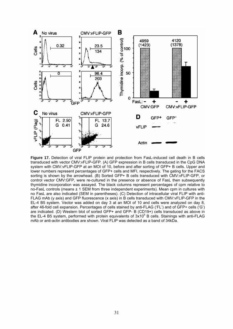
Figure 17. Detection of viral FLIP protein and protection from FasL-induced cell death in B cells
transduced with vector CMV:vFLIP-GFP. (A) GFP expression in B cells transduced in the CpG DNA system with CMV:vFLIP-GFP at an MOI of 10, before and after sorting of GFP+ B cells. Upper andlower numbers represent percentages of GFP+ cells and MFI, respectively. The gating for the FACS sorting is shown by the arrowhead. (B) Sorted GFP+ B cells transduced with CMV:vFLIP-GFP, or control vector CMV:GFP, were re-cultured in the presence or absence of FasL then subsequently thymidine incorporation was assayed. The black columns represent percentages of cpm relative to no-FasL controls (means ± 1 SEM from three independent experiments). Mean cpm in cultures with no FasL are also indicated (SEM in parentheses). (C) Detection of intracellular viral FLIP with anti-FLAG mAb (y axis) and GFP fluorescence (x axis) in B cells transduced with CMV:vFLIP-GFP in the EL-4 B5 system. Vector was added on day 3 at an MOI of 10 and cells were analyzed on day 8, after 46-fold cell expansion. Percentages of cells stained by anti-FLAG (‘FL’) and of GFP+ cells (‘G’) are indicated. (D) Western blot of sorted GFP+ and GFP- B (CD19+) cells transduced as above in the EL-4 B5 system, performed with protein equivalents of 3x10⁵ B cells. Stainings with anti-FLAG mAb or anti-actin antibodies are shown. Viral FLIP was detected as a band of 34kDa.
31

Future perspectives The first section of this PhD thesis demonstrated that we have successfully established a human-SCID chimera system for the investigation of EBV-induced post-transplantation lymphoproliferative disorder in vivo triggered by endogenous EBV strains of healthy blood-donors. The resulting chimera system may serve as a model system for EBV reactivation in transplanted patients. With the introduction of an active, dominant EBV by performing in vivo superinfection we have significantly modified the protocol. This latter method is also a model system for post transplantation lymphoproliferative disorders that occur upon infection by EBV in an immunologically compromised status. Both systems should be useful for testing therapeutic strategies and novel agents in an in vivo system that resembles the human PT-LPD syndrome. This stage of drug-trials may be placed between in vitro and human trials as an intermediate therapeutic checkpoint.60 Moreover, the combination of EBNA1 qPCR with the EBNA6 genomic PCR and RT-PCR assays employed in this work should also be able to monitor the peripheral blood EBV load of patients and identify all intrinsic and the dominantly active EBV strains, respectively.61, 16, 17, 18, 19 Another use of lymphotropic viruses in studying lymphocyte functions is reported in the second section of this thesis in the combination of the CpG-DNA motif based human B cell monoculture and HIV1-based lentiviral vectors as a to date unique platform for the ex vivo assessment of gene function testing and possible gene therapy applications. The expressed transgenes proved to be functionally active as seen in functional assays fulfilling the ‘proof-of-principle’ both for secreted (IL-4) and intracellular (vFLIP) transgenic proteins. Here this second section has joining points with the first section. EBV has now been studied for decades, yet even recently novel characteristics of this virus have been discovered i.e. in the fields of RNA interference and regulatory T cells. 62, 63, 64, 65 EBV has a vast genome encoding numerous genes that interact and reprogram human primary B cells. Therefore, it is certain that specific EBV-genes or their combinations are promising candidates for investigation in the proposed B/CpG system employing lentiviral vectors. (Although EBV-derived vectors are also already available for research, their utilization is technically demanding and such vectors are unlikely to be inert carriers in such experiments).66, 67, 68
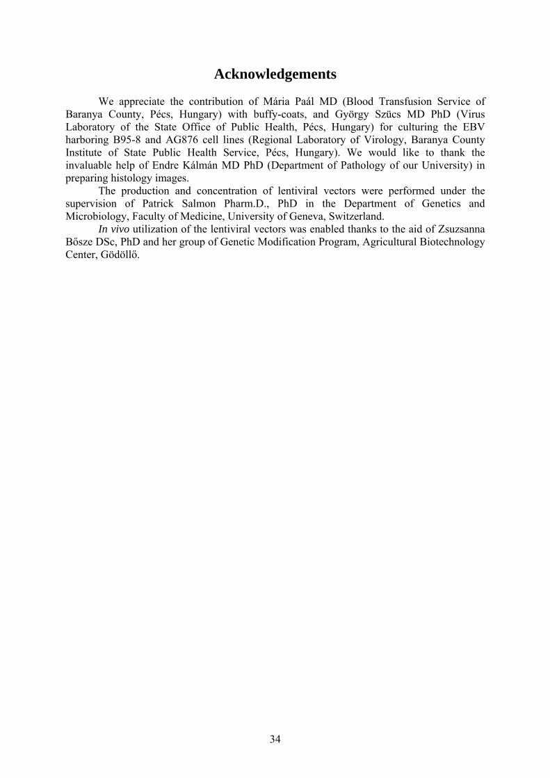
Due to the special characteristics of lentiviral vectors – the ability to transduce differentiated and even resting primary cells (including neurons), stable integration of multiple copies into the target cell genome, low toxicity, safety and the relative ease of production technique – these vectors have lately been used for the establishment of stable transgenic animals.69, 70, 71, 72, 73, 74 Our group – in collaboration with the Agricultural Biotechnology Center in Gödöllő – also began to create such animals for research purposes. Although the in vivo utilization of lentiviral vectors requires special safety precautions as well as concentrated, high quality vector preparations, the collaboration is already fruitful as the first transgenic animals have been born: GFP+ Balb/C mice (Figures 18A-B). Combining lentiviral methodology with scFv targeting is a very promising field of targeting vector transduction in vivo. In contrast to whole antibodies, an artificially engineered scFv is composed of a single protein chain where the originally separately expressed variable domains of the light and the heavy chains are connected by a linker amino-acid sequence (Figures 19A-B). This allows for simple molecular biology manipulation while preserving idiotype specificity. Lentiviral vectors pseudotyped for scFv envelope proteins can selectively bind and transduce cells both in vitro and in vivo.75, 76, 77 Specificity is provided by the scFv component while efficiency and long-term genetic manipulation are guaranteed by lentiviral vectors. In collaboration with other research groups we plan to develop and utilize such vectors for research purposes both in vitro and in vivo.
32
A B
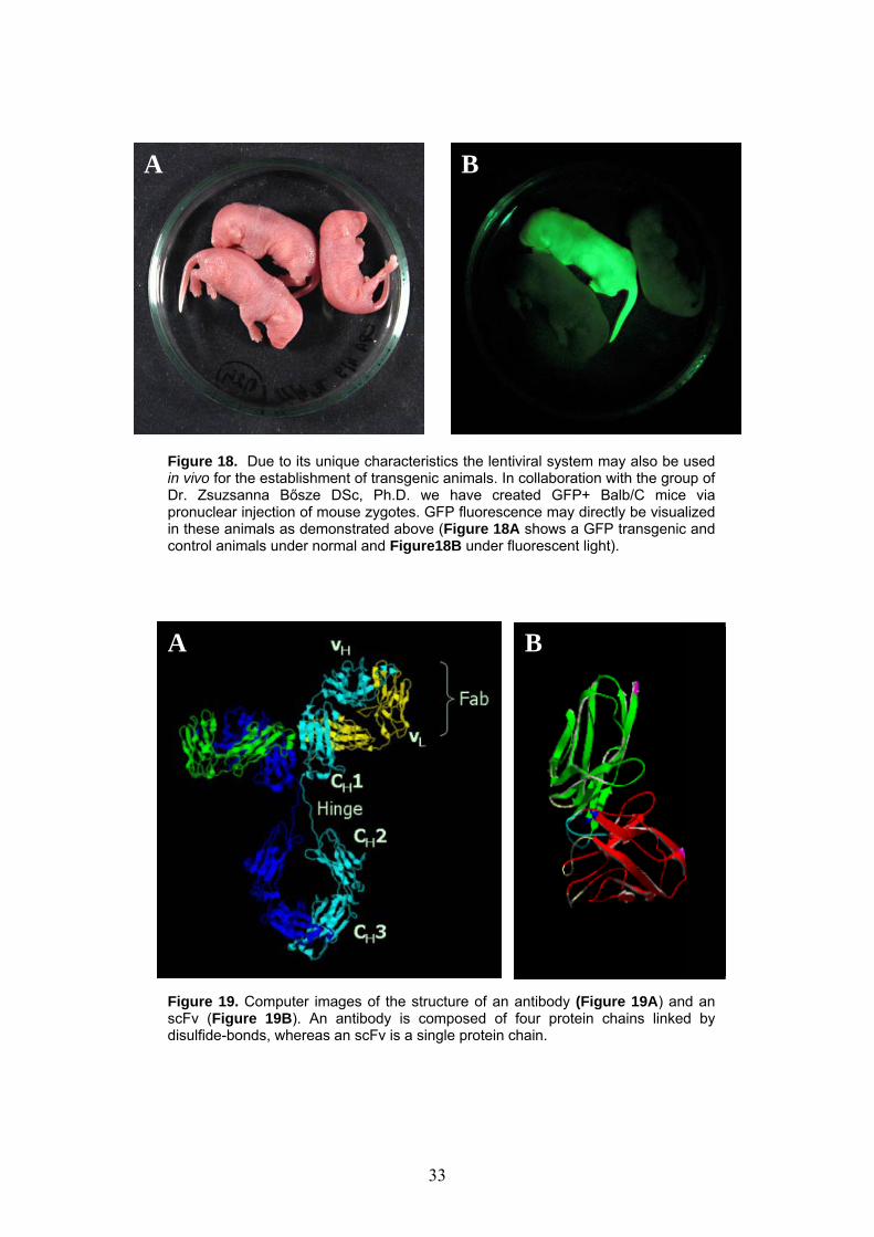
Figure 18. Due to its unique characteristics the lentiviral system may also be used in vivo for the establishment of transgenic animals. In collaboration with the group of Dr. Zsuzsanna Bősze DSc, Ph.D. we have created GFP+ Balb/C mice via pronuclear injection of mouse zygotes. GFP fluorescence may directly be visualized in these animals as demonstrated above (Figure 18A shows a GFP transgenic and control animals under normal and Figure18B under fluorescent light).
A B
Figure 19. Computer images of the structure of an antibody (Figure 19A) and an scFv (Figure 19B). An antibody is composed of four protein chains linked by disulfide-bonds, whereas an scFv is a single protein chain.
33
Acknowledgements We appreciate the contribution of Mária Paál MD (Blood Transfusion Service of Baranya County, Pécs, Hungary) with buffy-coats, and György Szücs MD PhD (Virus Laboratory of the State Office of Public Health, Pécs, Hungary) for culturing the EBV harboring B95-8 and AG876 cell lines (Regional Laboratory of Virology, Baranya County Institute of State Public Health Service, Pécs, Hungary). We would like to thank the invaluable help of Endre Kálmán MD PhD (Department of Pathology of our University) in preparing histology images. The production and concentration of lentiviral vectors were performed under the supervision of Patrick Salmon Pharm.D., PhD in the Department of Genetics and Microbiology, Faculty of Medicine, University of Geneva, Switzerland. In vivo utilization of the lentiviral vectors was enabled thanks to the aid of Zsuzsanna Bősze DSc, PhD and her group of Genetic Modification Program, Agricultural Biotechnology Center, Gödöllő.
34
References [1] Joklik WK, Willett HP: Epstein-Barr virus. Microbiology pp.954 [2] Fuzzati-Armento M-T, Duchosal MA: hu-PBL-SCID mice: an in vivo model of Epstein-
Barr virus-dependent lymphoproliferative disease. Histol. Histopathol. 13:155-168, 1998 [3] Boyle TJ, Tamburini M, Berend KR et al: Human B cell lymphoma in severe combined
immunodeficient mice after active infection with Epstein-Barr virus. Surgery 112-2:378-386, 1992
[4] Hanto DW, Sakamoto K, Purtilo DT et al: The Epstien-Barr virus in the pathogenesis of posttransplant lymphoproliferative disorders - clinical, pathologic and virologic correlation. Surgery 90:204-213, 1981
[5] Capello D, Rossi D, Gaidano G. Post-transplant lymphoproliferative disorders: molecular basis of disease histogenesis and pathogenesis. Hematol. Oncol. (2):61-7. Review, 2005
[6] Nakamine H, Okano M, Taguchi Y et al: Hematopathologic features of Epstein-Barr virus-induced human B-lymphoproliferation in mice with severe combined immune deficiency. Lab. Invest. 65-4:389-399, 1991
[7] Hesselton RM, Koup RA, Cromwell MA et al: Human peripheral blood xenografts in the SCID mouse: characterisation of immunologic reconstitution. J. Infect. Dis. 168: 630-640, 1993
[8] Martino G, Anastasi J, Feng J et al: The fate of human peripheral blood lymphocytes after transplantation into SCID mice. Eur. J. Immunol. 23:1023-1028, 1993
[9] Mosier DE: Humanising the mouse – introduction. Sem. in Immunol. 8-4:185, 1996 [10] Murphy WJ, Taub DD, Longo DL: The hu-PBL-SCID mouse as a means to examine
human immune function in vivo. Sem. in Immunol. 8-4:233-242, 1996 [11] Custer RP, Bosma GC, Bosma MJ. Severe combined immunodeficiency (SCID) in the
mouse – pathology, reconstitution, neoplasms. Am. J. of Pathol. 120: 464-77,1985 [12] Mosier DE. Viral pathogenesis in hu-PBL-SCID mice. Sem. in Immunol. 8: 255-62,
1996 [13] Johannessen I, Crawford DH. In vivo models for Epstein-Barr virus (EBV)-associated B
cell lymphoproliferative disease (BLPD).Rev Med Virol. (4):263-77. Review, 1999 [14] Parker GA, Touitou R, Allday MJ. Epstein-Barr virus EBNA3C can disrupt multiple cell
cycle checkpoints and induce nuclear division divorced from cytokinesis. Oncogene 19: 700-9, 2000
[15] Rajnavolgyi E, Nagy N, Thuresson B et al. A repetitive sequence of Epstein-Barr virus nuclear antigen 6 comprises overlapping T cell epitopes which induce HLA-DR-restricted CD4+ T lymphocytes. Int. Immunol. 12: 281-93, 2000
[16] Falk K, Gratama JW, Rowe M et al. The role of repetitive DNA sequences in the size variation of Epstein-Barr virus (EBV) nuclear antigens, and the identification of different EBV isolates using RFLP and PCR analysis. J. Gen. Virol. 76: 779-90, 1995
[17] Falk KI, Zou J-Z, Lucht E et al. Direct identification by PCR of EBV types and variants in clinical samples. J. Med. Virol. 51: 355-63, 1997
[18] Gratama JW, Oosterveer MAP, Weimar W et al. Detection of multiple 'Ebnotypes' in individual Epstein-Barr virus carriers following lymphocyte transformation by virus derived from peripheral blood and oropharynx. J. Gen. Virol. 75: 85-94, 1994
[19] Menin C, Ometto L, Veronesi A et al. Dominance of a single Epstein-Barr virus strain in SCID-mouse tumors induced by injection of peripheral blood mononuclear cells from healthy human donors. Virus Res. 36: 215-31, 1995
[20] Gleick J: Chaos - the birth of a new science pp.305-335, 1987 [21] Thorley-Lawson DA, Babcock GJ: A model for persistent infection with Epstein-Barr
virus: the stealth virus of human B cells. Life Science 65-14:1433-1453, 1999
35
[22] Cox KL, Lawrence-Miyasaki LS, Garcia-Kennedy R et al. An increased incidence of Epstein-Barr virus infection and lymphoproliferative disorder in young children on FK506 after liver transplantation. Transplantation 59: 524-9, 1995
[23] Hopwood P, Crawford DH. The role of EBV in post-transplant malignancies: a review. J. Clin. Pathol. 53: 248-54, 2000
[24] Knowles DM, Cesarman E, Chadburn A et al. Correlative morphologic and molecular genetic analysis demonstrates three distinct categories of posttransplantation lymphoproliferative disorders. Blood 85: 552-65, 1995
[25] Kvell K, Balogh P, Németh P. Clinical aspects of the EBV associated post-transplantation lymphoproliferative disease – Mini-review. Med. W. (Hungarian) 143: 713-9. 2002
[26] Gustaffson A, Levitsky V, Zou J-Z et al. Epstein-Barr virus (EBV) load in bone marrow transplant recipients at risk to develop posttransplant lymphoproliferative disease: prophylactic infusion of EBV-specific cytotoxic T cells. Blood 95: 807-14, 2000
[27] Jabs WJ, Hennig H, Kittel M et al. Normalised quantification by real-time PCR of Epstein-Barr virus load in patients at risk for posttransplant lymphoproliferative disorders. J. Clin. Microbiol. 39: 564-9, 2001
[28] Lucas KG, Burton RL, Zimmerman SE et al. Semiquantitative Epstein-Barr virus (EBV) polymerase chain reaction for the determination of patients at risk for EBV-induced lymphoproliferative disease after stem cell transplantation. Blood 91: 3654-61, 1998
[29] Niesters HGM, van Esser J, Fries E et al. Development of a real-time quantitative assay for detection of Epstein-Barr virus. J. Clin. Microbiol. 38: 712-5, 2000
[30] Rogers BB, Sommerauer J, Quan A et al. Epstein-Barr virus polymerase chain reaction and serology in pediatric post-transplant lymphoproliferative disorder: three year experience. Ped. Devel. Pathol. 1: 480-6, 1998
[31] Park F, Kay MA. Modified HIV-1 based lentiviral vectors have an effect on viral transduction efficiency and gene expression in vitro and in vivo. Mol Ther. (3):164-73., 2001
[32] Schambach A, Galla M, Maetzig T et al. Improving transcriptional termination of self-inactivating gamma-retroviral and lentiviral vectors. Mol Ther. (6):1167-73, 2007
[33] Cronin J, Zhang XY, Reiser J. Altering the tropism of lentiviral vectors through pseudotyping. Curr Gene Ther. (4):387-98. Review, 2005 Erratum in: Curr Gene Ther. (5):531, 2005
[34] Ogueta SB, Yao F, Marasco WA. Design and in vitro characterization of a single regulatory module for efficient control of gene expression in both plasmid DNA and a self-inactivating lentiviral vector. Mol Med. (8):569-79., 2001
[35] Logan AC, Haas DL, Kafri T et al. Integrated self-inactivating lentiviral vectors produce full-length genomic transcripts competent for encapsidation and integration. J. Virol. (16):8421-36, 2004
[36] Chang LJ, Zaiss AK. Self-inactivating lentiviral vectors and a sensitive Cre-loxP reporter system. Methods Mol Med. 76:367-82, 2003
[37] Somia N, Verma I M Gene therapy: trials and tribulations. Nat. Rev. Genet. 1: 91 – 99, 2000
[38] Basu U, Banerjee S. An engineered EBV vector expressing human factor VIII and von Willebrand factor in cultured B cells. J. Gene Med. 6: 760 – 768, 2004
[39] Ballantyne J, Henry DL, Müller JR et al. Efficient recombination of a switch substrate retrovector in CD40-activated B lymphocytes: implications for the control of CH gene switch recombination. J. Immunol. 161: 1336 – 1337, 1998
[40] Zufferey R, Nagy D, Mandel RJ et al. Multiply attenuated lentiviral vector achieves efficient gene delivery in vivo. Nat. Biotechnol. 15: 871 – 87, 1997
36

[41] Unutmaz D, KewalRamani VN, Marmon S et al. Cytokine signals are sufficient for HIV-1-infection of resting human T lymphocytes. J. Exp. Med. 189: 1735 – 1746, 1999
[42] Ducrey-Rundquist O, Guyader M, Trono D. Modalities of interleukin-7-induced human immunodeficiency virus permissiveness in quiescent T lymphocytes. J. Virol. 76: 9103 – 9111, 2002
[43] Moreau T, Bardin F, Imbert J et al. Restriction of a transgene expression to the B-lymphoid progeny of human lentivirally transduced CD34+ cells. Mol. Ther. 10: 45 – 56, 2004
[44] Janssens W, Chuah MK, Naldini L et al. Efficiency of onco-retroviral and lentiviral gene transfer into primary mouse and human B-lymphocytes is pseudotype dependent. Hum. Gene Ther. 14: 263 – 276, 2003
[45] Zufferey R, Dull T, Mandel RJ et al. Self-inactivating lentivirus vector for safe and efficient in vivo gene delivery. J. Virol. 72: 9873 – 9880, 1998
[46] Salmon P, Oberholzer J, Occhiodoro T et al. Reversible immortalization of human primary cells by lentivector-mediated transfer of specific genes. Mol. Ther. 2: 404 – 414, 2000
[47] Zennou V, Petit C, Guetard D et al. HIV-1 genome nuclear import is mediated by a central DNA flap. Cell 101: 173 – 185, 2000
[48] Zufferey R, Donello JE, Trono D et al. Woodchuck hepatitis virus post-transcriptional regulatory element enhances expression of transgenes delivered by retroviral vectors. J. Virol. 73: 2886 – 2892, 1999
[49] Salmon P, Kindler V, Ducrey O et al. High-level transgene expression in human hematopoietic progenitors and differentiated blood lineages after transduction with improved lentiviral vectors. Blood 96: 3392 – 3398, 2000
[50] Thome M, Schneider P, Hofmann K et al. Viral FLICE-inhibitory proteins (FLIPs) prevent apoptosis induced by death receptors. Nature 386: 517 – 521, 1997
[51] Bovia F, Salmon P, Matthes T et al. Efficient transduction of primary human B lymphocytes and nondividing myeloma B cells with HIV-1-derived lentiviral vectors. Blood 101: 1727 – 1733, 2003
[52] Werner-Favre C, Bovia F, Schneider P et al. IgG subclass-switch capacity is low in switched and in IgM-only, but high in IgD+IgM+, post-germinal center (CD27+) human B cells. Eur. J. Immunol. 31: 243 – 249, 2001
[53] Marshall JD, Fearon K, Abbate C et al. Identification of a novel CpG DNA class and motif that optimally stimulate B cell and plasmocytoid dendritic cell functions. J. Leucocyte Biol. 73: 781 – 792, 2003
[54] Holler N, Tardivel A, Kovacsovics-Bankowksi M et al. Two adjacent trimeric Fas ligands are required for Fas signaling and formation of a death-inducing signaling complex. Mol. Cell. Biol. 23: 1428 – 1440, 2003
[55] Bernasconi NL, Onai N, Lanzavecchia A. A role for Toll-like receptors in acquired immunity: up-regulation of TLR9 by BCR triggering in naive B cells and constitutive expression in memory B cells. Blood 101: 4500 – 4504, 2003
[56] Grimaitre M, Werner-Favre C, Kindler V et al. Human naive B cells cultured with EL-4 T cells mimic a germinal center-related B cell stage: concordant changes in Bcl-2 protein and messenger RNA levels. Eur. J. Immunol. 27: 199 – 205, 1997
[57] Serafini M, Naldini L, Introna M. Molecular evidence of inefficient transduction of proliferating human B lymphocytes by VSV-pseudotyped HIV-1-derived lentivectors. Virology 325: 413 – 424, 2004
[58] Bieniasz PD. Intrinsic immunity: a front-line defense against viral attack. Nat. Immunol. 5: 1109 – 1115, 2004
37
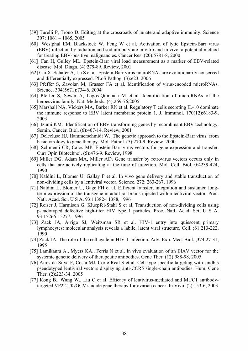
[59] Turelli P, Trono D. Editing at the crossroads of innate and adaptive immunity. Science 307: 1061 – 1065, 2005
[60] Westphal EM, Blackstock W, Feng W et al. Activation of lytic Epstein-Barr virus (EBV) infection by radiation and sodium butyrate in vitro and in vivo: a potential method for treating EBV-positive malignancies. Cancer Res. (20):5781-8, 2000
[61] Fan H, Gulley ML. Epstein-Barr viral load measurement as a marker of EBV-related disease. Mol. Diagn. (4):279-89. Review, 2001
[62] Cai X, Schafer A, Lu S et al. Epstein-Barr virus microRNAs are evolutionarily conserved and differentially expressed. PLoS Pathog. (3):e23, 2006
[63] Pfeffer S, Zavolan M, Grasser FA et al. Identification of virus-encoded microRNAs. Science. 304(5671):734-6, 2004
[64] Pfeffer S, Sewer A, Lagos-Quintana M et al. Identification of microRNAs of the herpesvirus family. Nat. Methods. (4):269-76,2005
[65] Marshall NA, Vickers MA, Barker RN et al. Regulatory T cells secreting IL-10 dominate the immune response to EBV latent membrane protein 1. J. Immunol. 170(12):6183-9, 2003
[66] Izumi KM. Identification of EBV transforming genes by recombinant EBV technology. Semin. Cancer. Biol. (6):407-14. Review, 2001
[67] Delecluse HJ, Hammerschmidt W. The genetic approach to the Epstein-Barr virus: from basic virology to gene therapy. Mol. Pathol. (5):270-9. Review, 2000
[68] Sclimenti CR, Calos MP. Epstein-Barr virus vectors for gene expression and transfer. Curr Opin Biotechnol. (5):476-9. Review, 1998
[69] Miller DG, Adam MA, Miller AD. Gene transfer by retrovirus vectors occurs only in cells that are actively replicating at the time of infection. Mol. Cell. Biol. 0:4239-424, 1990
[70] Naldini L, Blomer U, Gallay P et al. In vivo gene delivery and stable transduction of non-dividing cells by a lentiviral vector. Science. 272: 263-267, 1996
[71] Naldini L, Blomer U, Gage FH et al. Efficient transfer, integration and sustained long-term expression of the transgene in adult rat brains injected with a lentiviral vector. Proc. Natl. Acad. Sci. U S A. 93:11382-11388, 1996
[72] Reiser J, Harmison G, Kluepfel-Stahl S et al. Transduction of non-dividing cells using pseudotyped defective high-titer HIV type 1 particles. Proc. Natl. Acad. Sci. U S A. 93:15266-15277, 1996
[73] Zack JA, Arrigo SJ, Weitsman SR et al. HIV-1 entry into quiescent primary lymphocytes: molecular analysis reveals a labile, latent viral structure. Cell. ;61:213-222, 1990
[74] Zack JA. The role of the cell cycle in HIV-1 infection. Adv. Exp. Med. Biol. ;374:27-31, 1995
[75] Lamikanra A., Myers KA., Ferris N et al. In vivo evaluation of an EIAV vector for the systemic genetic delivery of therapeutic antibodies. Gene Ther. (12):988-98, 2005
[76] Aires da Silva F, Costa MJ, Corte-Real S et al. Cell type-specific targeting with sindbis pseudotyped lentiviral vectors displaying anti-CCR5 single-chain antibodies. Hum. Gene Ther. (2):223-34. 2005
[77] Kong B., Wang W., Liu C et al. Efficacy of lentivirus-mediated and MUC1 antibody-targeted VP22-TK/GCV suicide gene therapy for ovarian cancer. In Vivo. (2):153-6, 2003
38

List of publications Total cumulative impact factors: 27.35 Publications related to the thesis (cumulative impact factors: 23.64) Kvell K, Nguyen TH, Salmon P, Glauser F, Werner-Favre C, Barnet M, Schneider P, Trono D, Zubler RH. Transduction of CpG DNA-stimulated primary human B cells with bicistronic lentivectors. Mol Ther. 2005 Nov;12(5):892-9. impact factor: 6.13 Bovia F, Salmon P, Matthes T, Kvell K, Nguyen TH, Werner-Favre C, Barnet M, Nagy M, Leuba F, Arrighi JF, Piguet V, Trono D, Zubler RH. Efficient transduction of primary human B lymphocytes and nondividing myeloma B cells with HIV-1-derived lentiviral vectors. Blood. 2003 Mar 1;101(5):1727-33. impact factor: 10.12 Kvell K, Balogh P, Nemeth P. [Clinical aspects of Epstein-Barr-virus-associated lymphoproliferative disease] Orv Hetil. 2002 Apr 7;143(14):713-9. Review. Hungarian. Kvell K, Balogh P, Nemeth P. Fine-tuning the EBV+ hu-PBL-SCID xenogeneic chimera model using in vivo superinfection. Pathol Oncol Res. 2000;6(4):280-6. Cooper EL, Kvell K, Engelmann P, Nemeth P. Still waiting for the toll? Immunol Lett. 2006 Apr 15;104(1-2):18-28. impact factor: 1.71 Kvell K, Cooper EL, Engelmann P, Nemeth P. Blurring borders: innate immunity with adaptive features. Clin Develop Immunol 2007 (under review) Kvell K, Czompoly T, Pikkarainen T, Balogh P. Species-specific restriction of cell surface expression of mouse MARCO glycoprotein in murine cell lines. Biochem Biophys Res Commun. 2006 Mar 24;341(4):1193-202. impact factor: 2.84 Bartis D, Boldizsar F, Kvell K, Szabo M, Palinkas L, Nemeth P, Monostori E, Berki T. Intermolecular relations between the glucocorticoid receptor, ZAP-70 kinase, and Hsp-90. Biochem Biophys Res Commun. 2007 Mar 2;354(1):253-8. impact factor: 2.84 Other publications (cumulative impact factors: 3.71) Pal J, Nyarady Z, Marczinovits I, Par A, Ali YS, Berencsi G, Kvell K, Nemeth P. Comprehensive regression analysis of hepatitis B virus X antigen level and anti-HBx antibody titer in the sera of patients with HBV infection. Pathol Oncol Res. 2006;12(1):34-40. Bagamery K, Landau R, Kvell K, Graham J. Different platelet activation levels in non-pregnant, normotensive pregnant, pregnancy-induced hypertensive and pre-eclamptic women. A pilot study of flow cytometric analysis. Eur J Obstet Gynecol Reprod Biol. 2005 Jul 1;121(1):117-8. impact factor: 1.00
39

Bagamery K, Kvell K, Landau R, Graham J. Flow cytometric analysis of CD41-labeled platelets isolated by the rapid, one-step OptiPrep method from human blood. Cytometry A. 2005 May;65(1):84-7. impact factor: 2.10 Bagamery K, Kvell K, Barnet M, Landau R, Graham J. Are platelets activated after a rapid, one-step density gradient centrifugation? Evidence from flow cytometric analysis. Clin Lab Haematol. 2005 Feb;27(1):75-7. impact factor: 0.61 Nagy G, Szekeres G, Kvell K, Berki T, Nemeth P. Development and characterisation of a monoclonal antibody family against aquaporin 1 (AQP1) and aquaporin 4 (AQP4). Pathol Oncol Res. 2002;8(2):115-24.
40
METHODS
© 2000 W. B. Saunders & Company Ltd on behalf of the Arányi Lajos Foundation 1219-4956/00/040280+07 $ 12.00/0
PATHOLOGY ONCOLOGY RESEARCH Vol 6, No 4, 2000
10.1053.paor.2000.0300 available online at http://www.idealibrary.com on
Fine-tuning the EBV+ hu-PBL-SCID Xenogeneic Chimera ModelUsing In Vivo Superinfection
Krisztián KVELL, Péter BALOGH, Péter NÉMETH
University of Pécs, Faculty of Medicine, Department of Immunology and Biotechnology, Pécs, Hungary
Received: Nov 11, 2000; accepted: Dec 13, 2000Correspondence: Péter NÉMETH, MD, PhD; Department ofImmunology and Biotechnology, University of Pécs, Faculty ofMedicine, Szigeti u. 12., Pécs, H-7643, Hungary; E-mail: [email protected]
Our purpose was to establish a reproducible xeno-geneic chimera model to observe tumors similar tothe well-known human posttransplant lymphopro-liferative disease (LPD). First we followed the origi-nal protocol injecting Epstein-Barr virus positive(EBV+) human peripheral blood lymphocytes (PBL)intraperitoneally into immunodeficient (SCID)mice. Human cells showed T cell phenotype inmajority one week after the transfer, whereas onemonth later a shift towards B cell phenotype wasevident according to immunohistochemical andflow cytometric analysis. At this stage the intraperi-toneal mass of cells suggested a biologically malig-nant behaviour infiltrating the liver and the spleenof the host animal. Immunohistochemistry indicatedproliferating human lymphatic cells expressing EBVassociated proteins and characteristic patterns ofinvasion within the affected organs. Eventually LPDwas lethal to the host animals in 46-67 days. Howev-er, the microscopic appearance of experimental LPDwas different from the human haemopoietic malig-nancies: the basic structures of lymphatic organswere preserved and the human T and B cells repop-ulated the normally T and B dependent areas inmice. The phenotypes of the proliferating cells werehuman and characteristic for the mature T- and B-lymphocytes. No dominant clone developed during
in vitro culturing of the biologically invasive massof cells removed from the tumor-bearing mice. Theresults of microscopical, immunological, and flowcytometrical analysis suggested a mature but uncon-trolled proliferation of human lymphocytes in SCIDmice. The original method for the induction of post-transplant LPD in SCID mice was modified in ourfurther experiments to standardise the experimentaltechnique increasing the efficiency of B cell prolif-eration and the reducing the number of unspecificfactors. Subsequent in vivo EBV superinfection wascarried out after the intraperitoneal transfer of areduced quantity of human PBL from differentdonors. The same disease developed in our modi-fied chimera model as by the use of original protocolexcept for some valuable differences. All hostsdeveloped LPD regardless the significantly reducedamount of transplanted PBL and it was lethal in ashorter period of time (41-43 days) compared to theoriginal model. The decreased quantity of trans-planted human lymphatic cells was formerly insuf-ficient using the original protocol. Therefore thismodified and standardised protocol can lead to amore predictable and reproducible model allowingus to examine fine details of posttransplant lympho-proliferative disease. (Pathology Oncology ResearchVol 6, No 4, 280–286, 2000)
Keywords: SCID mice, lymphoproliferative disease, EBV superinfection, B cell proliferation
Introduction
Epstein, Achong and Barr described first the “Epstein-Barr virus” (EBV) in 1964.9 EBV is the 4th member of theherpes virus family (gamma herpesviridae) contains dou-ble stranded DNA and it is a polyclonal stimulator of
B-lymphocytes. EBV is a rather ubiquitous infectiveagent, 70–90% of the human population is seropositive.5
The causal aetiology of EBV is evident and proved insome disease, in some others the correlation is revealed bymeans of statistics only. Relations to many known humandiseases such as infectious mononucleosis, Burkitt’s lym-phoma, Hodgkin’s disease, nasopharyngeal carcinoma,gastric carcinoma and posttransplant lymphoproliferativedisease (LPD) were drawn.32
The LPD may develop in EBV seropositive and immun-odeficient humans. These immunodeficiencies by origincan be “natural” (e.g. in AIDS, Duncan’s disease), or
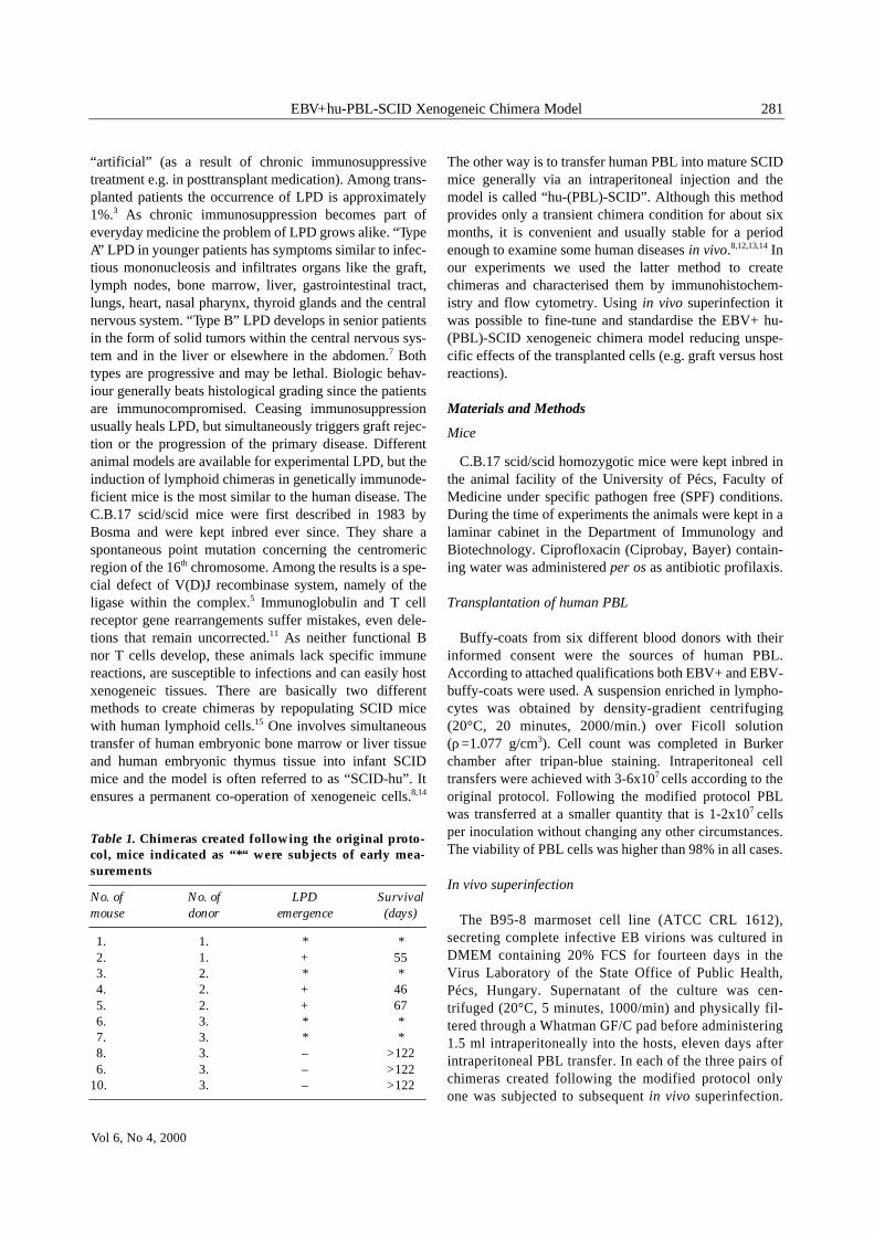
“artificial” (as a result of chronic immunosuppressivetreatment e.g. in posttransplant medication). Among trans-planted patients the occurrence of LPD is approximately1%.3 As chronic immunosuppression becomes part ofeveryday medicine the problem of LPD grows alike. “TypeA” LPD in younger patients has symptoms similar to infec-tious mononucleosis and infiltrates organs like the graft,lymph nodes, bone marrow, liver, gastrointestinal tract,lungs, heart, nasal pharynx, thyroid glands and the centralnervous system. “Type B” LPD develops in senior patientsin the form of solid tumors within the central nervous sys-tem and in the liver or elsewhere in the abdomen.7 Bothtypes are progressive and may be lethal. Biologic behav-iour generally beats histological grading since the patientsare immunocompromised. Ceasing immunosuppressionusually heals LPD, but simultaneously triggers graft rejec-tion or the progression of the primary disease. Differentanimal models are available for experimental LPD, but theinduction of lymphoid chimeras in genetically immunode-ficient mice is the most similar to the human disease. TheC.B.17 scid/scid mice were first described in 1983 byBosma and were kept inbred ever since. They share aspontaneous point mutation concerning the centromericregion of the 16th chromosome. Among the results is a spe-cial defect of V(D)J recombinase system, namely of theligase within the complex.5 Immunoglobulin and T cellreceptor gene rearrangements suffer mistakes, even dele-tions that remain uncorrected.11 As neither functional Bnor T cells develop, these animals lack specific immunereactions, are susceptible to infections and can easily hostxenogeneic tissues. There are basically two differentmethods to create chimeras by repopulating SCID micewith human lymphoid cells.15 One involves simultaneoustransfer of human embryonic bone marrow or liver tissueand human embryonic thymus tissue into infant SCIDmice and the model is often referred to as “SCID-hu”. Itensures a permanent co-operation of xenogeneic cells.8,14
The other way is to transfer human PBL into mature SCIDmice generally via an intraperitoneal injection and themodel is called “hu-(PBL)-SCID”. Although this methodprovides only a transient chimera condition for about sixmonths, it is convenient and usually stable for a periodenough to examine some human diseases in vivo.8,12,13,14 Inour experiments we used the latter method to createchimeras and characterised them by immunohistochem-istry and flow cytometry. Using in vivo superinfection itwas possible to fine-tune and standardise the EBV+ hu-(PBL)-SCID xenogeneic chimera model reducing unspe-cific effects of the transplanted cells (e.g. graft versus hostreactions).
Materials and Methods
Mice
C.B.17 scid/scid homozygotic mice were kept inbred inthe animal facility of the University of Pécs, Faculty ofMedicine under specific pathogen free (SPF) conditions.During the time of experiments the animals were kept in alaminar cabinet in the Department of Immunology andBiotechnology. Ciprofloxacin (Ciprobay, Bayer) contain-ing water was administered per os as antibiotic profilaxis.
Transplantation of human PBL
Buffy-coats from six different blood donors with theirinformed consent were the sources of human PBL.According to attached qualifications both EBV+ and EBV-buffy-coats were used. A suspension enriched in lympho-cytes was obtained by density-gradient centrifuging(20°C, 20 minutes, 2000/min.) over Ficoll solution(ρ=1.077 g/cm3). Cell count was completed in Burkerchamber after tripan-blue staining. Intraperitoneal celltransfers were achieved with 3-6x10⁷ cells according to theoriginal protocol. Following the modified protocol PBLwas transferred at a smaller quantity that is 1-2x10⁷ cellsper inoculation without changing any other circumstances.The viability of PBL cells was higher than 98% in all cases.
In vivo superinfection
The B95-8 marmoset cell line (ATCC CRL 1612),secreting complete infective EB virions was cultured inDMEM containing 20% FCS for fourteen days in theVirus Laboratory of the State Office of Public Health,Pécs, Hungary. Supernatant of the culture was cen-trifuged (20°C, 5 minutes, 1000/min) and physically fil-tered through a Whatman GF/C pad before administering1.5 ml intraperitoneally into the hosts, eleven days afterintraperitoneal PBL transfer. In each of the three pairs ofchimeras created following the modified protocol onlyone was subjected to subsequent in vivo superinfection.
281EBV+hu-PBL-SCID Xenogeneic Chimera Model
Vol 6, No 4, 2000
Table 1. Chimeras created following the original proto-col, mice indicated as “*“ were subjects of early mea-surements
No. of No. of LPD Survivalmouse donor emergence (days)
1. 1. * *2. 1. + 553. 2. * *4. 2. + 465. 2. + 676. 3. * *7. 3. * *8. 3. – >1226. 3. – >122
10. 3. – >122

An extra SCID mouse was given intraperitoneally 1.5 mlof EBV+ B95-8 supernatant without former PBL transferas a negative control.
Antibodies
The following anti-human monoclonal antibodies wereused: anti- CD3, CD4, CD5, CD8, CD10, CD19, CD20,CD23, CD45, κ light chain, λ light chain, (all of them fromDAKO, Denmark). Anti-mouse CD45 (IBL-5/25, devel-oped in our laboratory), a-LMP 1 and MIB-1 (both fromDAKO, Denmark) antibodies were also used.
Immunohistochemistry
Histologic examinations were performed on the intra-peritoneal lymphoid mass and organs infiltrated by LPD(liver, spleen) from mice bearing clinical signs of malig-nant disease. Cryostat slides of frozen organs were fixed inacetone for 5 minutes then left to air-dry for 2 minutes.Further reactions were performed in a humidity chamber.After rehydration with PBS the inhibition of endogenousperoxidase activity was performed by using phenil-hydrasin-hydrochloride (1 mg/ml) for 20 minutes. Satura-tion of the unspecific binding sites with 5% FCS albuminwas completed. Antibodies were incubated for 45 minuteseach on the slides including the streptavidin-peroxidaseconjugated antibody. Signals were developed in a citratebuffer containing H2O2 and 3-amino-9-ethyl carbasol(AEC). After 15 minutes development was halted by PBSsolution. Hematoxylin nuclear counter staining was used.First classical hematoxylin-eosin stain made from all thesamples for microscopic classification of the pathologicevents. Normal human lymphoid tissues and monoclonalantibodies with irrelevant specificity were used as positiveand negative controls, respectively.
Flow cytometric analysis
FACS phenotype analysis was performed on cellsforming the intraperitoneal mass within LPD bearingmice and on cells cultured in vitro later on. Centrifuga-tion (4°C, 5 minutes, 1000/min) following the homo-genising of the intraperitoneal mass resulted in a suspen-sion of cells in PBS solution. Cells were incubated withFITC (fluorescein-isothyocianat) or PE (phycoerythrin)labelled antibodies for 30 minutes over ice. Measure-ments were validated by carefully selected positive andnegative controls. FACSCalibur type (Becton Dickinson,USA) flow cytometer, and the CellQuest software (Bec-ton Dickinson, USA) were used for analysis.
In vitro culturing
Following sterile biopsy from the affected organs cellswere mechanically dispersed to perform in vitro culturingover continuous control in DMEM medium containing10% FCS in a tissue culture thermostat (Forma Scientific,USA) (37°C, 5% CO2, saturated humidity).
Results
Engraftment of human cells
Using the original protocol we created altogether 10hu-PBL-SCID xenogeneic chimeras with PBL fromthree different human blood donors (Table 1). PBL fromtwo donors could develop LPD in the mice. In case of the
282 KVELL et al
PATHOLOGY ONCOLOGY RESEARCH
Figure 1. Site of inoculationprior to LPD, FACS analysis
Figure 3. Origin of periportalmass of cells, FACS analysis
Figure 4. Phenotype of peripor-tal mass of cells, FACS analysis
Figure 5. Phenotype of peri-toenal mass of cells, FACSanalysis
Figure 7. Phenotype of peri-portal mass of cells, FACSanalysis
Figure 6. Phenotype of peri-toenal mass of cells, FACSanalysis
Figure 2. Origin of periportalmass of cells, FACS analysis
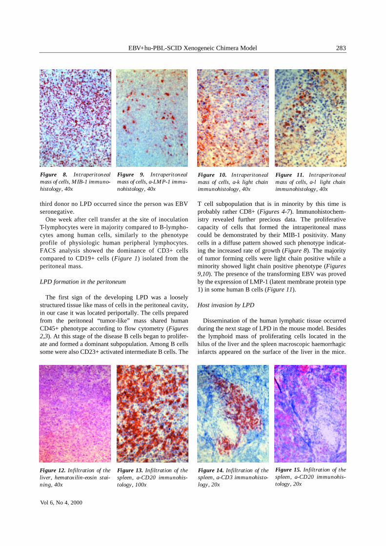
third donor no LPD occurred since the person was EBVseronegative.
One week after cell transfer at the site of inoculation T-lymphocytes were in majority compared to B-lympho-cytes among human cells, similarly to the phenotypeprofile of physiologic human peripheral lymphocytes.FACS analysis showed the dominance of CD3+ cellscompared to CD19+ cells (Figure 1) isolated from theperitoneal mass.
LPD formation in the peritoneum
The first sign of the developing LPD was a looselystructured tissue like mass of cells in the peritoneal cavity,in our case it was located periportally. The cells preparedfrom the peritoneal “tumor-like” mass shared humanCD45+ phenotype according to flow cytometry (Figures2,3). At this stage of the disease B cells began to prolifer-ate and formed a dominant subpopulation. Among B cellssome were also CD23+ activated intermediate B cells. The
T cell subpopulation that is in minority by this time isprobably rather CD8+ (Figures 4-7). Immunohistochem-istry revealed further precious data. The proliferativecapacity of cells that formed the intraperitoneal masscould be demonstrated by their MIB-1 positivity. Manycells in a diffuse pattern showed such phenotype indicat-ing the increased rate of growth (Figure 8). The majorityof tumor forming cells were light chain positive while aminority showed light chain positive phenotype (Figures9,10). The presence of the transforming EBV was provedby the expression of LMP-1 (latent membrane protein type1) in some human B cells (Figure 11).
Host invasion by LPD
Dissemination of the human lymphatic tissue occurredduring the next stage of LPD in the mouse model. Besidesthe lymphoid mass of proliferating cells located in thehilus of the liver and the spleen macroscopic haemorrhagicinfarcts appeared on the surface of the liver in the mice.
283EBV+hu-PBL-SCID Xenogeneic Chimera Model
Vol 6, No 4, 2000
Figure 8. Intraperitonealmass of cells, MIB-1 immuno-histology, 40x
Figure 9. Intraperitonealmass of cells, a-LMP-1 immu-nohistology, 40x
Figure 10. Intraperitonealmass of cells, a-k light chainimmunohistology, 40x
Figure 11. Intraperitonealmass of cells, a-λ light chainimmunohistology, 40x
Figure 12. Infiltration of theliver, hematoxilin-eosin stai-ning, 40x
Figure 13. Infiltration of thespleen, a-CD20 immunohis-tology, 100x
Figure 14. Infiltration of thespleen, a-CD3 immunohisto-logy, 20x
Figure 15. Infiltration of thespleen, a-CD20 immunohis-tology, 20x

Tissue samples of the liver and the spleen were analysedhistologically. Proliferating lymphocytes infiltrating thetissue regardless of hepatic lobuli were easy to distinguishfrom the hepatocellular background after hematoxilin-eosin staining during microscopic examination (Figure12). Immunohistochemistry revealed characteristic locali-sation of human lymphocyte subsets. The CD3+ T cellspreferred periarteriolar lymphatic sheaths (PALS) in thespleen, while the CD20+ B cells favoured lymphatic folli-cles (Figures 13-15) similarly to the normal anatomiclocation. LPD was lethal to mice in 46-67 days, agony wasaccompanied by palpable abdominal tumor, weakness,hypokinesis and ruffled hair.
In vitro culturing of tumor cells
Transformed cells removed from the host animals couldbe cultured in vitro for approx. 6 weeks. At small magni-fication colonies formed large spherical groups with manyfission. Based on the findings of flow cytometry besides aCD19+ phenotype tumor cells also shared and CD5- andCD10- phenotype meaning that they were B cells but didnot belong to the B1 or centroblast subgroups (Figures16,17). No dominant clone developed, light chain profilestill favoured the positive population to the positive popu-lation at a ratio of approx. 3:1 (Figure 18).
Reproducing LPD by using the modified protocol
Using the modified protocol we created altogether 6chimeras with PBL from three different human blooddonors. Activation by the xenogeneic environmentincreased the susceptibility of human lymphocytes toEBV. In each pair of mice only one was treated with sub-sequent in vivo superinfection. An extra SCID mouse wasintraperitoneally injected 1.5 ml of B95-8 supernatant asnegative control (Table 2). Note that neither the transfer ofa smaller amount of PBL from EBV seropositive donorsnor the in vivo administration of B95-8 supernatant byitself caused the emergence of LPD. Only the constellationof the two mentioned circumstances could create the dis-ease. In chimeras that received subsequent in vivo super-infection LPD developed and proved to be lethal in 41-43
days. Palpable abdominal tumor, weakness, hypokinesisand ruffled hair accompanied their agony. At autopsy ofaffected mice an intraperitoneal loosely structured mass ofcells in some ascitic fluid and enlarged, unusually paleliver and spleen were observed. Flow cytometric analysisof the intraperitoneal mass of cells showed mainly humanCD45+ phenotype and the dominance of CD19+ B cellscompared to CD3+ T cells (Figures 19-20). The doublenegative population contained murine cells (figures notshown). In vitro cultures of cells removed from the peri-toneal cavity of moribund LPD hosts showed high capac-ity for proliferation for over a month before fission halted.At small magnification colonies formed large sphericalgroups of cells adhered to each other after mitosis.
Discussion
There is always uncertainty in creating a hu-PBL-SCIDxenogeneic chimera model. Reproducibility is a major prob-lem concerning experiments involving biological systems.According to some literature data, since biological systemsare very complex and sensitive to starting conditions andnon-linear formulas rule them, at the level of present mea-surement accuracy results can not be forecasted even if start-ing parameters are clear. The adherence of the graft dependson many factors that are not clearly understood or cannot beinterfered properly6 (E.g. the graft versus host reaction caninfluence the biologic response of the recipient).
The suggested minimal amount of cells required for suc-cessful transfer, the protocol of preparing a cell suspensionand the treatment of SCID mice prior to transplantationalmost differs in each laboratory. The likelihood of graftacceptance can be raised with an increase of input cellquantity and careful protocols that spare cell functions. Ifthe original protocol is followed for creating hu-PBL-SCIDmodels the minimally required amount of cells is approx.5x107 cells per inoculation, smaller quantities usually failto successfully repopulate the host.8 Yet we must admitthat increased cell input quantity raises not only thechances of host repopulation but also points of variationthat lead to unspecific side effects of grafting and unpre-dictable survival of chimeras.
Since in SCID mice myelopoietic cells develop ade-quately, intact innate immunity including granulocytes,monocytes, macrophages and NK cells decrease the prob-
284 KVELL et al
PATHOLOGY ONCOLOGY RESEARCH
Figure 16. Phenotype of invitro culture, FACS analysis
Figure 17. Phenotype of invitro culture, FACS analysis
Figure 18. Clonality of invitro culture, FACS analysis

ability of graft adherence and influences LPD as well.Against murine NK cells an antibody known as anti-asia-lo GM1 or a sublethal irradiation of 1-4 Gy can be useful.10
Unfortunately SCID mice are sensitive to irradiation and Tcell lymphomas may emerge.
According to some experiments not only murine NKcells, but simultaneously transferred human NK cells canalso decrease the activity of autologous B cells.4 In vitroactivation of human cells prior to transfer may also behelpful. This can help lymphocyte homing processes too.14
PBL donors can be divided into at least two groups basedon the incidence of LPD. The ability of donors to createLPD is constant with time and it is characteristic to donors.Approx. 40% of donors belong to the high incidence groupand 60% to the low-intermediate incidence group. It is aninteresting observation that the number of clones is greaterin LPD developing from PBL of high incidence donors.16
The incidence of LPD in the two groups shows no directrelation with the concentration of latently infected B cellsin PBL. The frequency of latently infected cells among B-lymphocytes in the peripheral blood is individual andvaries considerably. In a population of healthy carriers it isbetween 5-500x10-7 with a mean of 50x10-7 and is alsocharacteristic to donors. This low number of latentlyinfected cells is due to the fact that it concerns only a frac-tion of memory B cells and can also explain the minimumamount of cells required for successful repopulation.Immunosuppression during human allograft transplanta-tion protocols causes an average 40-50 fold increase oflatently infected B cells in the peripheral blood of patients
as it may perturb mechanisms that regulate the absolutenumbers. Memory cells are unlikely to be under directimmunosurveillance therefore their increased numbersmust be due to the prolonged survival or increased pro-duction of precursors that are under direct cytotoxic con-trol.17 Donors possess different Th1/Th2 cytokine profilesthat influence both graft adherence, B cell proliferationand the development of graft versus host reactions.1 Theprevalence of CD8+ cells within the graft is various.Therefore inadequate cytotoxic immunosurveillance mayalso play a part in lymphoproliferation.
EBV subtypes differ in their ability to create LPD, “typeA” EBV is more potent than “type B” EBV.5 The B95-8marmoset cell line secretes “type A” EBV. Genetic differ-ences i.e. deletions within the EBNA-2 gene can consider-ably decrease transforming capacity since almost all viralpromoters contain EBNA-2 dependent elements. The pro-tein product of the EBNA-2 gene is considered to be afunctional homologue of Notch IC and results in differen-tiation arrest and continued proliferation. Obviously it isalso a main target of cytotoxic activity.17
Besides the mentioned uncertainties of creating chimeramodels, the exact timespan necessary for LPD emergencecan not be forecasted either. In case of an EBV+ PBLdonor the chances of developing LPD in the system with-
in 60-110 days is about 80-100%. The matter is complexand that means variation is results.
The original method for inducing posttransplant LPD inSCID mice was modified during our experiments to stan-dardise and increase the efficiency of B cell tumor occur-rence with the reduction of unspecific factors. We trans-ferred a smaller quantity of human PBL compared to theoriginal protocol and administered EBV from the B95-8cell line as subsequent in vivo superinfection. These alter-ations apparently circumvented some points of variation.The well-documented human LPD developed in the ani-mals except for some valuable differences. A fraction ofthe formerly used cell number of EBV seropositive donorswas sufficient to develop the disease in a slightly shorter
285EBV+hu-PBL-SCID Xenogeneic Chimera Model
Vol 6, No 4, 2000
Table 2. Chimeras created following the modified pro-tocol, mouse No. 7 was negative control
No. of No. of In vivo LPD Survivalmouse donor infection emergence (days)
1. 1. – – >742. 1. + + 413. 2. – – >744. 2. + + 425. 3. – – >746. 3. + + 437. – + – >74
Figure 19. Phenotype of peri-toneal LPD, FACS analysis
Figure 20. Phenotype of peri-toneal LPD, FACS analysis
averageofsurvival
60
50
40
30
20
10
0originalprotocol
modifiedprotocol
standarddeviationofsurvival
day
s
Figure 21. Differences between the protocols in the average ofsurvival and the standard deviation of survival

286 KVELL et al
PATHOLOGY ONCOLOGY RESEARCH
and in a more narrow time-span regardless of other donorcharacteristics. That is following the modified protocol theintraperitoneal transfer of 1-2x10⁷ lymphocytes was suffi-cient to create LPD, average survival decreased from 56 to42 days and more importantly standard deviation of sur-vival fell from 10.5 days to 1 day for all three donors. (Fig-ure 21). Standard deviation describes the reproducibilityof individual measurements and the number of observa-tions does not influence its value. It is therefore possible tostate that subsequent in vivo superinfection can be a usefulalteration to fine-tune the EBV+ hu-PBL-SCID model andour modified in vivo model of human LPD can be helpfulin detailing the steps of lymphoproliferation and in testingnew tactics of treating the disease.
Acknowledgements
We appreciate the contribution of Mária Paál M.D. (Blood Trans-fusion Service of Baranya County, Pécs, Hungary) with buffy-coats, and György Szûcs M.D. Ph.D. (Virus Laboratory of the StateOffice of Public Health, Pécs, Hungary) with B95-8 marmoset cellline cultures. We would like to thank the invaluable help of EndreKálmán M.D., PhD (Department of Pathology of our University) inpreparing histologic pictures and György Lustyik PhD (Departmentof Biophysics of our University) in flow cytometric analysis.
References
1.²Amadori A, Veronesi A, Coppola V, et al: The hu-PBL-SCIDmouse in human lymphocyte function and lymphomagenesisstudies: achievements and caveats. Seminars in Immunology 8-4:249-254, 1996.
2.²Belágyi J: Medical biometry – an introduction. pp.21, 1999.3.²Boyle TJ, Tamburini M, Berend KR, et al: Human B cell lym-
phoma in severe combined immunodeficient mice after activeinfection with Epstein-Barr virus. Surgery 112-2:378-386, 1992.
4.²Carlsson R, Martensson C, Kalliomaki S, et al: Human periph-eral blood lymphocytes transplanted into SCID mice reconsti-tute an in vivo culture system exhibiting several parametersfound in a normal humoral immune response and are a source
of immunocytes for the production of human monoclonal anti-bodies. The Journal Of immunology 148:1065-1071, 1992.
5.²Fuzzati-Armento M-T, Duchosal MA: hu-PBL-SCID mice: an invivo model of Epstein-Barr virus-dependent lymphoprolifera-tive disease. Histology and Histopathology 13:155-168, 1998.
6.²Gleick J: Chaos – the birth of a new science pp.305-335, 1987.7.²Hanto DW, Sakamoto K, Purtilo DT, et al: The Epstien-Barr
virus in the pathogenesis of posttransplant lymphoproliferativedisorders – clinical, pathologic and virologic correlation.Surgery 90:204-213, 1981.
8.²Hesselton RM, Koup RA, Cromwell MA, et al: Human periph-eral blood xenografts in the SCID mouse: characterisation ofimmunologic reconstitution. The Journal of Infectious Diseases168:630-640, 1993.
9.²Joklik WK, Willett HP: Epstein-Barr virus. Microbiologypp.954.
10.²Lacerda JF, Ladanyi M, Jagiello C, et al: Administration ofrabbit anti-asialo GM1 antiserum facilitates the development ofhuman Epstein-Barr virus-induced lymphoproliferations inxenografted C.B.-17 scid/scid mice. Transplantation 61-3:492-497, 1996.
11.²Malynn BA, Blackwell TK, Fulop GM, et al: The SCID defectaffects the final step of the immunoglobulin VDJ recombinasemechanism. Cell 54:453-460, 1988.
12.²Martino G, Anastasi J, Feng J, et al: The fate of human periph-eral blood lymphocytes after transplantation into SCID mice.European Journal of Immunology 23:1023-1028, 1993.
13.²Mosier DE: Humanising the mouse – introduction. Seminars inImmunology 8-4:185, 1996.
14.²Murphy WJ, Taub DD, Longo DL: The hu-PBL-SCID mouse asa means to examine human immune function in vivo. Semi-nars in Immunology 8-4:233-242, 1996.
15.²Nakamine H, Okano M, Taguchi Y, et al: Hematopathologic fea-tures of Epstein-Barr virus-induced human B-lymphoprolifera-tion in mice with severe combined immune deficiency. Labora-tory Investigation 65-4:389-399, 1991.
16.²Picchio G, Kobayashi R, Kirven M, et al: Heterogeneity amongEpstein-Barr virus-seropositive donors in the generation ofimmunoblastic B cell lymphomas in SCID mice receivinghuman peripheral blood leukocyte grafts. Cancer Research52:2468-2477, 1992.
17.²Thorley-Lawson DA, Babcock GJ: A model for persistent infec-tion with Epstein-Barr virus: the stealth virus of human B cells.Life Science 65-14:1433-1453, 1999.

ARTICLE doi:10.1016/j.ymthe.2005.05.010
Transduction of CpG DNA-Stimulated Primary Human BCells with Bicistronic Lentivectors
Krisztian Kvell,1 Tuan H. Nguyen,2 Patrick Salmon,2 Frederic Glauser,1
Christiane Werner-Favre,1 Marc Barnet,1 Pascal Schneider,3
Didier Trono,2 and Rudolf H. Zubler1,*
1Division of Hematology, Department of Internal Medicine, and 2Department of Genetics and Microbiology, University Hospitals,
1211 Geneva-14, Switzerland3Department of Biochemistry, University of Lausanne, 1066 Epalinges, Switzerland
*To whom correspondence and reprint requests should be addressed. Fax: 41 22 372 72 88. E-mail: [email protected].
Available online 11 July 2005
892
Recently, using HIV-1-derived lentivectors, we obtained efficient transduction of primary human Blymphocytes cocultured with murine EL-4 B5 thymoma cells, but not of isolated B cells activated byCD40 ligation. Coculture with a cell line is problematic for gene therapy applications or study ofgene functions. We have now found that transduction of B cells in a system using CpG DNA wascomparable to that in the EL-4 B5 system. A monocistronic vector with a CMV promoter gave 32 F4.7% green fluorescent protein (GFP)+ cells. A bicistronic vector, encoding IL-4 and GFP in the firstand second cistrons, respectively, gave 14.2 F 2.1% GFP+ cells and IL-4 secretion of 1.3 F 0.2 ng/105
B cells/24 h. This was similar to results obtained in CD34+ cells using the elongation factor-1Apromoter. Activated memory and naive B cells were transducible. After transduction with abicistronic vector encoding a viral FLIP molecule, vFLIP was detectable by FACS or Western blot inGFP+, but not in GFP�, B cells, and 57% of sorted GFP+ B cells were protected against Fas ligand-induced cell death. This system should be useful for gene function research in primary B cells anddevelopment of gene therapies.
Key Words: HIV-1-derived lentivectors, bicistronic vectors, human primary B lymphocytes,CpG DNA, viral FLIP
INTRODUCTION
Optimization of methods for gene delivery into primaryhuman cells is important for research on gene functionsand development of gene therapies [1]. Gene transfer intoprimary human B lymphocytes has been notoriouslydifficult. Successful applications of nonviral methods tofunctional assays of transgenes have not been reportedfor these cells. Epstein–Barr virus vectors so far have beentested only in B cell lines [2]. Murine oncoretroviralvectors gave low transduction efficiency (of up to about4%) in primary human B cells; they could be utilized tostudy effects detectable by very sensitive methods, suchas immunoglobulin (Ig) class-switch recombinationdetectable by PCR [3]. HIV-1-derived lentivectors gaveefficient transduction of various immature and maturehuman hematopoietic cells [4–6]. Transgene expressionrestricted to B lymphocytes was obtained by graftinglentivector-transduced CD34+ progenitors into NOD/SCID mice [7]. T lymphocytes could be transduced afteractivation, at least from the G0 to the G1 stage of the cellcycle [5,6]. But primary human B cells activated into
proliferation by crosslinking of CD40 in the presence ofvarious cytokines were very poorly transducible [8,9].Recently with such vectors we obtained efficient trans-duction of primary B cells cocultured with irradiatedmurine EL-4 B5 thymoma cells; monocistronic vectorswith the human cytomegalovirus (CMV) or elongationfactor-1a (EF-1a) internal promoter gave 27 F 12% greenfluorescent protein (GFP)+ cells [9].
For gene therapy applications, however, a cell linepotentially adds risks, such as generation of recombinantviruses. For studies of gene functions a handicap is thatthe functions of the thymoma cells have not yet beenmolecularly characterized. The first aim of this study wasto find a culture system for transduction with HIV vectorsof isolated B cells activated by defined stimuli. The LPSreceptor, Toll-like receptor-4 (TLR4), is lacking in humanB cells. But TLR9, the endosomal receptor for bacterialDNA (short single-stranded DNA containing nonmethy-lated CpG motifs; CpG DNA), is constitutively expressedin memory human B cells and rapidly upregulated byanti-Ig antibody—which mimics an antigen signal—in
MOLECULAR THERAPY Vol. 12, No. 5, November 2005
Copyright C The American Society of Gene Therapy
1525-0016/$30.00
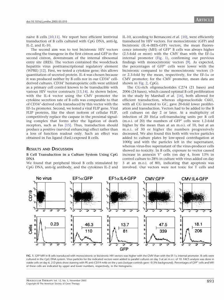
ARTICLEdoi:10.1016/j.ymthe.2005.05.010
naRve B cells [10,11]. We report here efficient lentiviraltransduction of B cells cultured with CpG DNA, anti-Ig,IL-2, and IL-10.
The second aim was to test bicistronic HIV vectorsencoding the transgene in the first cistron and GFP in thesecond cistron, downstream of the internal ribosomalentry site (IRES). The vectors contained the woodchuckhepatitis virus posttranscriptional regulatory element(WPRE) [12]. First, we tested a cytokine gene; this allowsquantitation of secreted protein. IL-4 was chosen becauseit was produced neither by B cells nor in our CD34+ cell-derived cultures. CD34+ hematopoietic cells were utilizedas a primary cell control known to be transducible withvarious HIV vector constructs [13,14]. As shown below,with the IL-4 vector using the CMV promoter thecytokine secretion rate of B cells was comparable to thatof CD34+-derived cells transduced by this vector with theEF-1a promoter. Second, we tested a viral FLIP gene. ViralFLIP proteins, like the short isoform of cellular FLIP,competitively replace the caspase in the proximal signal-ing complex that forms after the ligation of deathreceptors, such as Fas [15]. Thus, transduction shouldproduce a positive (survival enhancing) effect rather thana loss of function readout only. Such an effect wasobtained in Fas ligand (FasL)-exposed B cells.
RESULTS AND DISCUSSION
B Cell Transduction in a Culture System Using CpGDNAWe found that peripheral blood B cells stimulated byCpG DNA, anti-Ig antibody, and the cytokines IL-2 and
FIG. 1. GFP MFI in B cells transduced with monocistronic or bicistronic HIV vector
cultured in the CpG DNA system. Virus particles for the indicated vectors were ad
viable cells on day 6. 2-D plots show staining with PE-anti-CD19 mAb on the y axi
of these cells are indicated by upper and lower numbers, respectively, in the his
MOLECULAR THERAPY Vol. 12, No. 5, November 2005
Copyright C The American Society of Gene Therapy
IL-10, according to Bernasconi et al. [10], were efficientlytransduced by HIV vectors. For monocistronic (GFP) andbicistronic (IL-4–IRES–GFP) vectors, the mean fluores-cence intensity (MFI) of GFP+ B cells was always higher(2.5-fold or more) with the CMV than with the EF-1ainternal promoter (Fig. 1), confirming our previousfindings with monocistronic vectors [9]. As expected,the percentages of GFP+ cells were lower with thebicistronic compared to the monocistronic vectors (3-or 2.3-fold by the mean, respectively, for the EF-1a orCMV promoter; for the CMV promoter, mean data areshown in Fig. 2, CpG).
The CG-rich oligonucleotides C274 (21 bases) and2006 (24 bases), which caused optimal B cell proliferationin the study by Marshall et al. [16], both allowed forefficient transduction, whereas oligonucleotide C661,with all CG inverted to GC, gave 20-fold lower prolifer-ation and transduction. Vectors had to be added to the Bcell cultures on day 2 or later. At a multiplicity ofinfection of 20 HeLa cell-transducing units per B cell(m.o.i. of 20) the numbers of GFP+ cells were 1.2-foldhigher by the mean than at an m.o.i. of 10, but at anm.o.i. of 30 or higher the numbers progressivelydecreased. We also found this both with vector particlesadded to culture plates by low-speed centrifugation at1000g and with the particles left in the supernatant,whereas virus-free supernatant of the virus-producer cellsshowed no toxicity. In B cells, exposure to vector causedincrease in annexin V+ cells (on day 4, from 13% incontrol culture to 28% in culture with virus added on day3 at an m.o.i. of 80), indicating that apoptosis wasinvolved. Our vectors were not toxic for T cells and
s was higher with the CMV than with the EF-1a internal promoter. B cells were
ded to parallel cultures on day 3 at an m.o.i. of 10. FACS analysis was done in
s (isotype controls gave b0.5% background). Percentages of GFP+ cells and MFI
tograms.
893
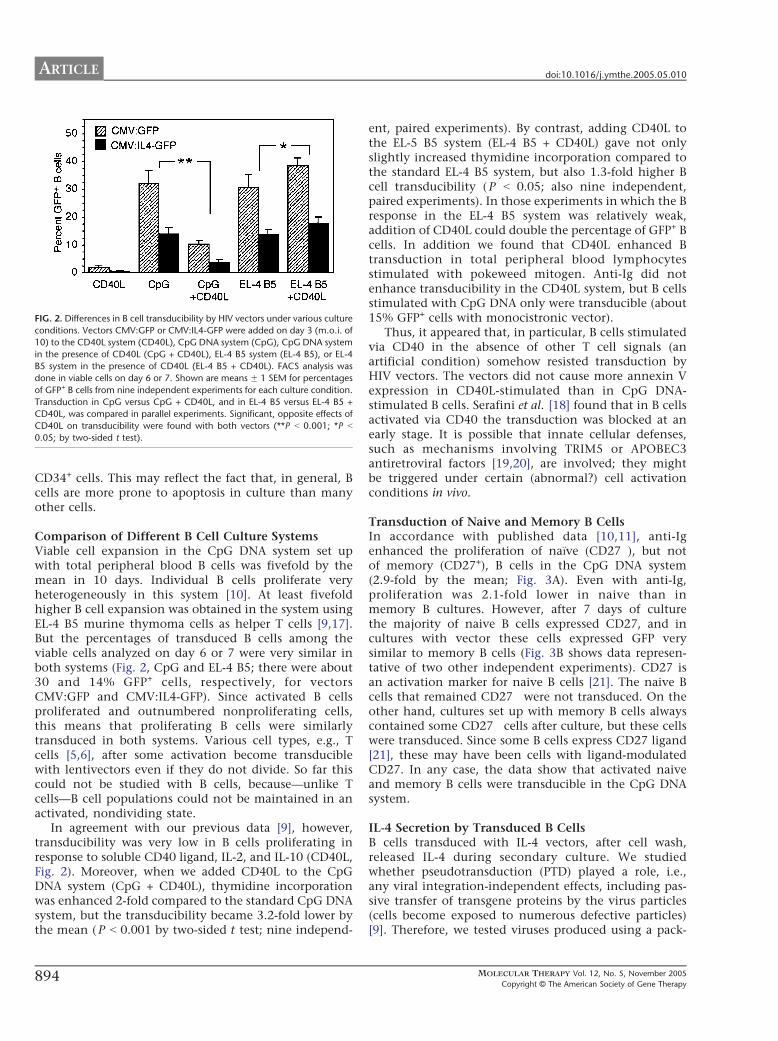
FIG. 2. Differences in B cell transducibility by HIV vectors under various culture
conditions. Vectors CMV:GFP or CMV:IL4-GFP were added on day 3 (m.o.i. o
10) to the CD40L system (CD40L), CpG DNA system (CpG), CpG DNA system
in the presence of CD40L (CpG + CD40L), EL-4 B5 system (EL-4 B5), or EL-4
B5 system in the presence of CD40L (EL-4 B5 + CD40L). FACS analysis wa
done in viable cells on day 6 or 7. Shown are means F 1 SEM for percentage
of GFP+ B cells from nine independent experiments for each culture condition
Transduction in CpG versus CpG + CD40L, and in EL-4 B5 versus EL-4 B5 +
CD40L, was compared in parallel experiments. Significant, opposite effects o
CD40L on transducibility were found with both vectors (**P b 0.001; *P b
0.05; by two-sided t test).
ARTICLE doi:10.1016/j.ymthe.2005.05.010
894
f
s
s
.
f
CD34+ cells. This may reflect the fact that, in general, Bcells are more prone to apoptosis in culture than manyother cells.
Comparison of Different B Cell Culture SystemsViable cell expansion in the CpG DNA system set upwith total peripheral blood B cells was fivefold by themean in 10 days. Individual B cells proliferate veryheterogeneously in this system [10]. At least fivefoldhigher B cell expansion was obtained in the system usingEL-4 B5 murine thymoma cells as helper T cells [9,17].But the percentages of transduced B cells among theviable cells analyzed on day 6 or 7 were very similar inboth systems (Fig. 2, CpG and EL-4 B5; there were about30 and 14% GFP+ cells, respectively, for vectorsCMV:GFP and CMV:IL4-GFP). Since activated B cellsproliferated and outnumbered nonproliferating cells,this means that proliferating B cells were similarlytransduced in both systems. Various cell types, e.g., Tcells [5,6], after some activation become transduciblewith lentivectors even if they do not divide. So far thiscould not be studied with B cells, because—unlike Tcells—B cell populations could not be maintained in anactivated, nondividing state.
In agreement with our previous data [9], however,transducibility was very low in B cells proliferating inresponse to soluble CD40 ligand, IL-2, and IL-10 (CD40L,Fig. 2). Moreover, when we added CD40L to the CpGDNA system (CpG + CD40L), thymidine incorporationwas enhanced 2-fold compared to the standard CpG DNAsystem, but the transducibility became 3.2-fold lower bythe mean ( P b 0.001 by two-sided t test; nine independ-
ent, paired experiments). By contrast, adding CD40L tothe EL-5 B5 system (EL-4 B5 + CD40L) gave not onlyslightly increased thymidine incorporation compared tothe standard EL-4 B5 system, but also 1.3-fold higher Bcell transducibility ( P b 0.05; also nine independent,paired experiments). In those experiments in which the Bresponse in the EL-4 B5 system was relatively weak,addition of CD40L could double the percentage of GFP+ Bcells. In addition we found that CD40L enhanced Btransduction in total peripheral blood lymphocytesstimulated with pokeweed mitogen. Anti-Ig did notenhance transducibility in the CD40L system, but B cellsstimulated with CpG DNA only were transducible (about15% GFP+ cells with monocistronic vector).
Thus, it appeared that, in particular, B cells stimulatedvia CD40 in the absence of other T cell signals (anartificial condition) somehow resisted transduction byHIV vectors. The vectors did not cause more annexin Vexpression in CD40L-stimulated than in CpG DNA-stimulated B cells. Serafini et al. [18] found that in B cellsactivated via CD40 the transduction was blocked at anearly stage. It is possible that innate cellular defenses,such as mechanisms involving TRIM5 or APOBEC3antiretroviral factors [19,20], are involved; they mightbe triggered under certain (abnormal?) cell activationconditions in vivo.
Transduction of Naive and Memory B CellsIn accordance with published data [10,11], anti-Igenhanced the proliferation of naRve (CD27�), but notof memory (CD27+), B cells in the CpG DNA system(2.9-fold by the mean; Fig. 3A). Even with anti-Ig,proliferation was 2.1-fold lower in naive than inmemory B cultures. However, after 7 days of culturethe majority of naive B cells expressed CD27, and incultures with vector these cells expressed GFP verysimilar to memory B cells (Fig. 3B shows data represen-tative of two other independent experiments). CD27 isan activation marker for naive B cells [21]. The naive Bcells that remained CD27� were not transduced. On theother hand, cultures set up with memory B cells alwayscontained some CD27� cells after culture, but these cellswere transduced. Since some B cells express CD27 ligand[21], these may have been cells with ligand-modulatedCD27. In any case, the data show that activated naiveand memory B cells were transducible in the CpG DNAsystem.
IL-4 Secretion by Transduced B CellsB cells transduced with IL-4 vectors, after cell wash,released IL-4 during secondary culture. We studiedwhether pseudotransduction (PTD) played a role, i.e.,any viral integration-independent effects, including pas-sive transfer of transgene proteins by the virus particles(cells become exposed to numerous defective particles)[9]. Therefore, we tested viruses produced using a pack-
MOLECULAR THERAPY Vol. 12, No. 5, November 2005
Copyright C The American Society of Gene Therapy

FIG. 3. Naive and memory B cells proliferated and were transducible in the CpG DNA system. (A) Anti-Ig antibody (aIg) enhanced thymidine incorporation by
naive (CD27�, white columns), but not by memory (CD27+, black columns), B cells in the presence of CpG DNA, with or without IL-2 and IL-10 (means F 1 SEM
of counts per minute (cpm) from three independent experiments). (B) The 2-D plots show CD27 expression and FL1 fluorescence in sorted memory and naive B
cells before (Day 0) and after culture in the CpG DNA system (Day 7), with no virus or with vector CMV:GFP added on day 3 an m.o.i. of 10. Percentages of
CD27+ cells (27+) and of GFP+ cells among the CD27+ cells (27+GF+) are indicated (experiment representative for three independent experiments). Only those
naive B cells that became CD27+ were transduced.
ARTICLEdoi:10.1016/j.ymthe.2005.05.010
aging construct with a loss-of-function mutant integraseor a vector plasmid (CMV:IL4-GFP) lacking the centralpolypurine tract (cPPT) involved in nuclear translocation[22]. We added control virus at an m.o.i. of 10 and theothers, produced in parallel, at concentrations equalizedfor IL-4 concentrations in the 293T culture supernatant.Control vector gave 13.9% GFP+ B cells (Fig. 4A) and anIL-4 release of 1.1 ng IL-4/105 B cells/24 h (Fig. 4B). Thevirus lacking integrase activity gave 0.87% GFP+ cells, butno bright GFP+ cells, i.e., this was weak PTD that occurswith GFP [9]. The IL-4 release was low, indicating thatabout 5% of the IL-4 release obtained with the positivecontrol may have been due to PTD. The virus lacking thecPPT gave 1.92% GFP+ cells, including about 1% brightcells. The IL-4 release (6.7-fold lower than control) wasclose to what one would expect for this number of brightGFP+ cells plus PTD as estimated above. Thus, PTD playeda negligible role.
Comparison of IL-4 Secretion by B Cells and CD34+
CellsWe used CD34+ cells as a reference for work withlentivectors. After transduction with the IL-4 vector withthe EF-1a promoter, which is optimal in these cells [13],the GFP expression in CD34+ cell-derived cultures (Fig. 5A;the upper histograms) was similar to that obtained in B
MOLECULAR THERAPY Vol. 12, No. 5, November 2005
Copyright C The American Society of Gene Therapy
cells with vector CMV:IL4-GFP. In CD34+-derived cells,intracytoplasmic IL-4 was detectable by FACS and corre-lated with GFP expression among individual transducedcells (lower histograms in Fig. 5A). Although B cells donot produce IL-4 [23], a strong nonspecific staining bythe anti-IL-4 antibody in transduced and nontransducedB cells made such detection impossible. IL-4 secretion wasalso similar in B cells and CD34+-derived cells (Fig. 5B;about 1.2 ng IL-4/105 cells/24 h). In B cells, IL-4 secretionwith the EF-1a promoter was 3.4-fold lower than with theCMV promoter. In FACS-sorted GFP+ B cells, the IL-4secretion increased in parallel with their enrichment (notshown). Thus, transducibility with bicistronic vector wassimilar for B and CD34+ cells. That many B cells becomesecretory cells (plasma cells) was apparently not relevantfor the cytokine secretion rate. However, a considerablylower cytokine secretion rate was reported for primary Bleukemia cells transduced with a bicistronic HIV vectorlacking the WPRE [24]. This element enhances mRNAstability [12].
GFP expression and IL-4 secretion were stable in B cellsbetween days 3 and 8 after exposure to vector and inCD34+-derived cells when followed up for 21 days. Onday 8 after exposure to vectors with the respectiveoptimal promoters, at an m.o.i. of 10, quantitative PCRrevealed multiple copies of GFP DNA per diploid genome
895

FIG. 4. Correlation between IL-4 secretion and GFP expression in B cells exposed to different vector particles: positive control vector (CMV:IL4-GFP), vecto
plasmid deleted of the cPPT element (D cPPT), and virus made using a packaging construct with an inactive mutant integrase (No integrase). (A) GFP expression
All three types of vector particles were produced in parallel. Control vector CMV:IL4-GFP was added at an m.o.i. of 10. The other vector particles were added a
concentrations equalized for the amounts of IL-4 measured in the supernatants of the virus-producer cells. Upper and lower numbers represent percentages o
GFP+ cells and MFI, respectively. (B) IL-4 secretion rates measured in this representative experiment (means F 1 SEM of triplicate cultures).
ARTICLE doi:10.1016/j.ymthe.2005.05.010
in CD34+-derived GFP+ cells (4.5 F 0.3), as expected [25],and also in CD19+GFP+ B cells (4.8 F 0.4; means F 1 SEM,n = 4, i.e., two transduction experiments with mono- andtwo with bicistronic vectors for each cell type and for Bcells one of each experiment in the CpG and B5 systems).
FIG. 5. GFP expression and IL-4 secretion by B cells transduced with vector CMV:IL4-GFP were similar to those found in cell cultures derived from cord blood
CD34+ cells transduced with vector EF1a:IL4-GFP. (A) The upper plots show GFP expression in cell culture derived from 97% pure cord blood CD34+ cells from a
representative experiment. EF1a:IL4-GFP was added on day 1 at an m.o.i. of 20; cells were analyzed on day 5. Upper and lower numbers represent percentage
of GFP+ cells and MFI, respectively. The lower plots show a staining with PE–anti-IL-4 mAb on the y axis and GFP fluorescence on the x axis on day 5. Percentage
of cells stained by anti-IL-4 (,,IL
,,),and of GFP+ cells (
,,G
,,) are indicated. (B) IL-4 secretion rates measured in B cells and CD34+ cell-derived cultures transduced
with the indicated vectors at an m.o.i. of 10 (means F 1 SEM from six independent experiments).
MOLECULAR THERAPY Vol. 12, No. 5, November 2005896Copyright C The American Society of Gene Therap
r
.
t
f
We obtained overlapping results with mono- and bicis-tronic vectors. We found at least 10-fold lower PCRsignals in GFP� cells, showing low contamination byDNA plasmids. Since vector-derived episomal DNA isshort-lived in various cells, including B cells [18], these
s
s
y

ARTICLEdoi:10.1016/j.ymthe.2005.05.010
data indicate multiple vector integration in both celltypes.
Effects of Bicistronic Vector Expressing Viral FLIPWe then tested a bicistronic vector expressing FLAG-tagged vFLIP of molluscum contagiosum virus [15]. Weadded this vector (CMV:vFLIP-GFP) or control vector(CMV:GFP) to B cells in the CpG DNA system on day 2and sorted GFP+ cells on day 5 (Fig. 6A). We reculturedthe cells in the presence or absence of soluble FasL. In twoexperiments, transduction of vFLIP caused a 1.8- to 2.4-fold reduction in the number of FasL-induced annexin V+
apoptotic cells after 20 h (without and with FasL,respectively, there were 32 F 3.9 and 79 F 5.6% annexinV+ cells for control vector versus 35 F 4.4 and 57 F 1.5 for
FIG. 6. Detection of viral FLIP protein and of protection from FasL-induced cell d
cells transduced in the CpG DNA system with CMV:vFLIP-GFP at an m.o.i. of 10
percentages of GFP+ cells and MFI, respectively. The gating for the FACS sorting i
transduced with CMV:vFLIP-GFP, or control vector CMV:GFP, after reculture in t
counts per minute (cpm) relative to no-FasL controls (means F 1 SEM from three
(SEM in parentheses). (C) Detection of intracellular viral FLIP with anti-FLAG mAb (
in the EL-4 B5 system. Vector was added on day 3 at an m.o.i. of 10 and cells were
anti-FLAG (,,FL
,,) and of GFP+ cells (
,,G
,,) are indicated. (D) Western blot of sorte
performed with protein equivalents of 3 � 105 B cells. Stainings with anti-FLAG m
kDa.
MOLECULAR THERAPY Vol. 12, No. 5, November 2005
Copyright C The American Society of Gene Therapy
vFLIP vector). In three other experiments, we measuredthymidine incorporation after 48 h to detect cell survival(Fig. 6B). Only 14 F 4% of B cells transduced with controlvector, but 63 F 12% of B cells transduced with vFLIPvector, resisted killing by FasL. Thus, among those cellsthat could be killed by FasL, 57% (range 45 to 71%) wereprotected by vFLIP.
In B cells transduced in the EL-4 B5 system, intra-cellular vFLIP was detectable with anti-FLAG mAb byFACS and its expression correlated with GFP expressionin individual cells (Fig. 6C). Viral FLIP was also detectablewith this antibody by Western blot in sorted GFP+, butnot in GFP�, B cells (Fig. 6D).
In conclusion, with an HIV-1-derived bicistronicvector we obtained proof of principle for the testing of
eath in B cells transduced with vector CMV:vFLIP-GFP. (A) GFP expression in B
, before and after sorting of GFP+ B cells. Upper and lower numbers represent
s shown by the arrowhead. (B) Thymidine incorporation by sorted GFP+ B cells
he presence or absence of FasL. The black columns represent percentages of
independent experiments). Mean cpm in cultures with no FasL are indicated
y axis) and GFP fluorescence (x axis) in B cells transduced with CMV:vFLIP-GFP
analyzed on day 8, after 46-fold cell expansion. Percentages of cells stained by
d GFP+ and GFP� B (CD19+) cells transduced as above in the EL-4 B5 system,
Ab or anti-actin antibodies are shown. Viral FLIP was detected as a band of 34
897
ARTICLE doi:10.1016/j.ymthe.2005.05.010
a gene function in primary human B cells. The CpG DNAculture system should be useful for such studies.
MATERIALS AND METHODS
HIV-1-Derived Vectors
A second-generation packaging system and self-inactivating HIV-1
vectors [26] with the pLoxP site in the 3VLTR [27] were utilized. The
monocistronic vector with the EF-1a internal promoter (called vector
EF1a:GFP in this study) was plasmid pWPT-GFP for which the map and
sequence are available at http://www.tronolab.unige.ch. This plasmid
carries, in the 5V to 3V direction, the cPPT element [22], the intronless
(bshortQ) EF-1a promoter, the expression cassette, and the WPRE [12].
Vector CMV:GFP was made by replacing the promoter with the human
CMV promoter [13] between the ClaI and the BamHI restriction sites. In
the bicistronic vectors, which were derived from the above vectors, the
first cistron was the transgene of interest, between the BamHI and the
SalI restriction sites, followed by the encephalomyocarditis virus IRES
[27] and GFP encoded downstream of the IRES. In the vectors EF1a:IL4-
GFP and CMV:IL4-GFP the transgene was the complete human IL-4
cDNA sequence (a kind gift from Dr. J.-F. Gauchat, formerly at the
Institut Pierre Fabre, St. Julien, France). In vector CMV:vFLIP-GFP the
transgene was the FLAG-tagged viral FLIP of human molluscum
contagiosum virus (open reading frame 159L), kindly provided by Dr.
Margot Thome of the Department of Biochemistry, University of
Lausanne [15]. In some experiments we used a vector plasmid as above,
except that it lacked the cPPT element, or a packaging construct [9]
with a loss-of-function mutant integrase (mutation D64V) [28].
Virus particles pseudotyped with vesicular stomatitis virus G glyco-
protein were produced, concentrated 100-fold by ultracentrifugation, and
titrated on HeLa cells by measurement of GFP+ cells by FACS to establish
HeLa transducing units per milliliter (usually 0.5 to 2 � 108), as described
[9]. Viral stocks were suspended in B cell culture medium and stored at
�708C.
Cell Cultures and Transduction
Approval for research on human cells was obtained from the Ethics
Committee of the Geneva University Hospitals. Written consent was
provided for all cell samples. B cells were isolated to more than 99% purity
from buffy coats of virus-screened blood donations obtained from the
Geneva Transfusion Center using Ficoll–Hypaque centrifugation and anti-
CD19 magnetic beads (Dynal) [29]. All B cell cultures were grown in RPMI
1640 medium supplemented with 10% fetal calf serum, 2-mercaptoetha-
nol, Hepes buffer, and antibiotics as described [29].
CpG DNA system. B cells (25,000 cells/200 Al culture; flat-bottom 96-
well plates) were stimulated with CpG DNA (2.5 Ag/ml), anti-Ig
antibody (2 Ag/ml), IL-2 (50 ng/ml), and IL-10 (10 ng/ml). CpG
oligonucleotides of the published sequences C274, 2006, and C661
[16] were obtained as complete phosphorothioates synthesized to our
order by Microsynth (Balgach, Switzerland). Anti-Ig was goat F(abV)2
anti-human IgA + IgG + IgM (H+L) (Jackson ImmunoResearch). The
cytokines were from PeproTech. FACS sorting of CD27+/CD27� cells
was done as described [29].
CD40L system. B cells (50,000 cells/200 Al) were stimulated with a soluble
oligomeric CD40L (300 ng/ml), IL-2 (50 ng/ml), and IL-10 (10 ng/ml) as
described [29]. When CD40L was added to the CpG DNA system or the EL-
4 B5 system, its concentration was also 300 ng/ml.
EL-4 B5 system. B cells (3000 cells/200 Al) were stimulated with 50,000
irradiated EL-4 B5 murine thymoma cells and a mixture of human
cytokines acting on the EL-4 B5 cells (IL-1h) or the B cells (TNF-a, IL-2,
and IL-10), as described [9].
To transduce B cell cultures at different times, the big cell clusters,
which formed in all culture systems, were dispersed by gentle pipetting
(five times aspiration of the culture using 200-Al tips). Virus particles were
then added to the cultures in a volume of 2 to 4 Al, at the indicated m.o.i.
according to cell counts by microscope (B cells or B plus EL-4 B5 cells),
898
without further manipulations or cell wash. We found that centrifugation
of virus particles in new culture plates (spinoculation) with cell transfer
did not give better results.
CD34+ cell culture and transduction. Umbilical cord blood CD34+ cells
were isolated to N95% purity with anti-CD34 magnetic beads (Dynal),
cultured, and transduced as described [13]. Briefly, the cells (105/200 Al)
were first cultured and transduced on day 1 in the presence of
thrombopoietin (TPO). After 48 h, the cells were recultured in Iscove’s
modified Dulbecco’s medium supplemented with 10% fetal calf serum,
antibiotics, TPO, Flt3-ligand, and stem cell factor; the cells were
recultured every 6 days.
Sorting and FACS Analysis of Transduced Cells
Various GFP+ cell populations were sorted on a FACStar sorter, and
analyses of GFP+ cells were performed on a FACScan analyzer using
CellQuest software (Becton–Dickinson), at the cytofluorometry laboratory
of the Geneva Medical Center. Cells were pretreated with polyclonal
mouse Ig before staining with phycoerythrin (PE)-coupled anti-CD19 [9],
anti-CD27 [29], or anti-CD34 [13] mAb, and viable (7-amino-actino-
mycin-D low) cells were gated for analysis of GFP expression [9]. For
analysis of cytoplasmic IL-4, cells were cultured with brefeldin A (Sigma),
2.5 Ag/ml, for 3 h. For analysis of cytoplasmic IL-4 or viral FLIP, cells were
treated with Cytofix/Cytoperm and Perm/Wash solutions, followed by
AB+ serum, and then stained, respectively, with PE-coupled anti-IL-4 mAb
(Pharmingen) or anti-FLAG-M2 mAb (Sigma), which we coupled to biotin,
followed by PE-coupled streptavin (Jackson ImmunoResearch). The
isotype control was mouse IgG1. Apoptosis was assessed using the
Annexin V–PE Apoptosis Detection Kit I (Becton–Dickinson).
Other Analyses of Transduced Cells
IL-4 secretion rate. On day 2 or 3 after addition of viral particles, B cells or
CD34+ cells were harvested and washed twice in culture medium in
Eppendorf tubes, and viable, trypan blue-excluding cells were counted.
The cells were recultured as before, and after 24 h soluble IL-4 was
measured in culture supernatants by Quantikine Human IL-4 Immuno-
assay (R&D Systems).
Real-time quantitative PCR. The LightCycler system, High Pure PCR
Template Preparation Kit, and LightCycler FastStart DNA MasterPLUS SYBR
Green I kit (all from Roche) were utilized. Cells were transduced with
vectors treated with DNase I before ultracentrifugation [7]. PCR was
performed in separate assays for genomic DNA, using intron-binding
primers for h2-microglobulin (5V-GGCACTGCTGAGATACTGAT-3V,
reverse 5V-GCTAGGACAGCAGGACTTA-3V; 215-bp product) and for vector
DNA, using GFP primers (5V-GGCAAGCTGACCCTGAAGTT-3V, 5V-
GCATGGCGGACTTGAAGAAGT-3V; 149-bp product); respective plasmid
standards were utilized.
FasL assay. GFP+ cells were sorted 3 days after exposure to vector and
recultured (20,000 trypan blue-excluding cells/200 Al, triplicate cultures)
in the CpG DNA system (without anti-Ig) in the presence of soluble
oligomeric FasL (200 ng/ml) prepared as described [30]. Thymidine
incorporation was measured after 48 h including the pulsing during the
last 12 h [29] with 1 ACi (0.037 MBq) [methyl-3H]thymidine (Amersham
Pharmacia Biotech).
Western blot for viral FLIP. This was done with the ECL Western blotting
analysis system (Amersham) using 0.45-Am nitrocellulose membranes
(Bio-Rad) and Biomax chemiluminescence films (Kodak). The antibodies
were IgG1 mouse anti-FLAG-M2 mAb (Sigma) or polyclonal goat IgG anti-
actin (Santa Cruz Biotechnologies) in conjunction with horseradish
peroxidase-coupled anti-mouse or anti-goat antibodies, respectively
(Santa Cruz Biotechnologies).
ACKNOWLEDGMENTS
These studies were supported by grants from the Swiss National Science
Foundation (to D.T. and R.Z.), the European Community, and the Institut
Clayton pour la Recherche (to D.T.). K. Kvell is a member of the M.D.–Ph.D.
Program of the University of Pecs, Hungary.
MOLECULAR THERAPY Vol. 12, No. 5, November 2005
Copyright C The American Society of Gene Therapy

ARTICLEdoi:10.1016/j.ymthe.2005.05.010
RECEIVED FOR PUBLICATION JANUARY 25, 2005; REVISED APRIL 13, 2005;
ACCEPTED MAY 2, 2005.
REFERENCES1. Somia, N., and Verma, I. M. (2000). Gene therapy: trials and tribulations. Nat. Rev.
Genet. 1: 91 – 99.
2. Basu, U., and Banerjee, S. (2004). An engineered EBV vector expressing human factor
VIII and von Willebrand factor in cultured B cells. J. Gene Med. 6: 760 – 768.
3. Ballantyne, J., et al. (1998). Efficient recombination of a switch substrate retrovector in
CD40-activated B lymphocytes: implications for the control of CH gene switch
recombination. J. Immunol. 161: 1336 – 1337.
4. Zufferey, R., et al. (1997). Multiply attenuated lentiviral vector achieves efficient gene
delivery in vivo. Nat. Biotechnol. 15: 871 – 875.
5. Unutmaz, D., et al. (1999). Cytokine signals are sufficient for HIV-1-infection of resting
human T lymphocytes. J. Exp. Med. 189: 1735 – 1746.
6. Ducrey-Rundquist, O., Guyader, M., and Trono, D. (2002). Modalities of interleukin-7-
induced human immunodeficiency virus permissiveness in quiescent T lymphocytes.
J. Virol. 76: 9103 – 9111.
7. Moreau, T., Bardin, F., Imbert, J., Chabannon, C., and Tonnelle, C. (2004). Restriction
of a transgene expression to the B-lymphoid progeny of human lentivirally transduced
CD34+ cells. Mol. Ther. 10: 45 – 56.
8. Janssens, W., et al. (2003). Efficiency of onco-retroviral and lentiviral gene transfer into
primary mouse and human B-lymphocytes is pseudotype dependent. Hum. Gene Ther.
14: 263 – 276.
9. Bovia, F., et al. (2003). Efficient transduction of primary human B lymphocytes and
nondividing myeloma B cells with HIV-1-derived lentiviral vectors. Blood 101:
1727 – 1733.
10. Bernasconi, N. L., Onai, N., and Lanzavecchia, A. (2003). A role for Toll-like receptors in
acquired immunity: up-regulation of TLR9 by BCR triggering in naRve B cells and
constitutive expression in memory B cells. Blood 101: 4500 – 4504.
11. Bourke, E., et al. (2003). The toll-like receptor repertoire of human B lymphocytes:
inducible and selective expression of TLR9 and TLR10 in normal and transformed cells.
Blood 102: 956 – 963.
12. Zufferey, R., Donello, J. E., Trono, D., and Hope, T. J. (1999). Woodchuck hepatitis virus
posttranscriptional regulatory element enhances expression of transgenes delivered by
retroviral vectors. J. Virol. 73: 2886 – 2892.
13. Salmon, P., et al. (2000). High-level transgene expression in human hematopoietic
progenitors and differentiated blood lineages after transduction with improved
lentiviral vectors. Blood 96: 3392 – 3398.
14. Schomber, T., Kalberer, C. P., Wodnar-Filipowicz, A., and Skoda, R. C. (2004). Gene
silencing by lentivirus-mediated delivery of siRNA in human CD34+ cells. Blood 103:
4511 – 4513.
MOLECULAR THERAPY Vol. 12, No. 5, November 2005
Copyright C The American Society of Gene Therapy
15. Thome, M., et al. (1997). Viral FLICE-inhibitory proteins (FLIPs) prevent apoptosis
induced by death receptors. Nature 386: 517 – 521.
16. Marshall, J. D., et al. (2003). Identification of a novel CpG DNA class and motif that
optimally stimulate B cell and plasmocytoid dendritic cell functions. J. Leucocyte Biol.
73: 781 – 792.
17. Grimaıtre, M., Werner-Favre, C., Kindler, V., and Zubler, R. H. (1997). Human naive B
cells cultured with EL-4 T cells mimic a germinal center-related B cell stage: concordant
changes in Bcl-2 protein and messenger RNA levels. Eur. J. Immunol. 27: 199 – 205.
18. Serafini, M., Naldini, L., and Introna, M. (2004). Molecular evidence of inefficient
transduction of proliferating human B lymphocytes by VSV-pseudotyped HIV-1-derived
lentivectors. Virology 325: 413 – 424.
19. Bieniasz, P. D. (2004). Intrinsic immunity: a front-line defense against viral attack. Nat.
Immunol. 5: 1109 – 1115.
20. Turelli, P., and Trono, D. (2005). Editing at the crossroads of innate and adaptive
immunity. Science 307: 1061 – 1065.
21. Lens, S. M. A., Tesselaar, K., van Oers, M. H. J., and van Lier, R. A. W. (1998).
Control of lymphocyte function through CD27–CD70 interactions. Semin. Immunol.
10: 491 – 499.
22. Zennou, V., Petit, C., Guetard, D., Nehrbass, U., Mantagnier, L., and Charneau, P.
(2000). HIV-1 genome nuclear import is mediated by a central DNA flap. Cell 101:
173 – 185.
23. Matthes, T., et al. (1993). Cytokine mRNA expression during an in vitro response of
human B lymphocytes: kinetics of B cell tumor necrosis factor a, interleukin (IL) 6, IL-10
and transforming growth factor h1 mRNAs. J. Exp. Med. 178: 512 – 528.
24. Stripecke, R., et al. (2000). Lentiviral vectors for efficient delivery of CD80 and
granulocyte–macrophage-colony-stimulating factor in human acute lymphoblastic
leukemia and acute myeloid leukemia cells to induce antileukemic immune responses.
Blood 96: 1317 – 1326.
25. Woods, N. B., et al. (2003). Lentiviral vector transduction of NOD/SCID repopulating
cells results in multiple vector integrations per transduced cell: risk of insertional
mutagenesis. Blood 101: 1284 – 1289.
26. Zufferey, R., et al. (1998). Self-inactivating lentivirus vector for safe and efficient in vivo
gene delivery. J. Virol. 72: 9873 – 9880.
27. Salmon, P., et al. (2000). Reversible immortalization of human primary cells by
lentivector-mediated transfer of specific genes. Mol. Ther. 2: 404 – 414.
28. Leavitt, A. D., Robles, G., Alesandro, N., and Varmus, H. E. (1996). Human
immunodeficiency virus type 1 integrase mutants retain in vitro integrase activity yet
fail to integrate viral DNA efficiently during infection. J. Virol. 70: 721 – 728.
29. Werner-Favre, C., et al. (2001). IgG subclass-switch capacity is low in switched and in
IgM-only, but high in IgD+IgM+, post-germinal center (CD27+) human B cells. Eur. J.
Immunol. 31: 243 – 249.
30. Holler, N., et al. (2003). Two adjacent trimeric Fas ligands are required for Fas signaling
and formation of a death-inducing signaling complex. Mol. Cell. Biol. 23: 1428 – 1440.
899