examination of thin films with stm and afmreu-mse.mme.wsu.edu/past/1999/gallaherben1999.pdf ·...
TRANSCRIPT
Examination of Thin Films With STM and AFM
Presented by:Ben Gallaher
Acknowledgments:Matt Kontz
Dr. K.W. HippsDan Barlow
Naoko MiyashitaMeredith Pritchard
Louis ScudieroIntroduction:The goal of the experiments performed was to examine alkanethiols and phthalocyanines. Self assembled monolayers of alkanethiols are of interest because of their application in the lubricating and wetting of surfaces, contolling corrosion, and designing biological sensor surfaces. Phthalocyanines are of interest because of their durability and potential applications to molecular circuitry. They are already widely used as dyes and coatings on rewriteable CD’s.In order to study a surface with molecular resolution Scanning Probe Microscopy (SPM) must be used. A Digital Instruments in air, stand alone Scanning Tunneling Microscope (STM) was used. The controller was a Digital Instruments Nanoscope IIIa. To study Langmuir-Blodgett films of phthalocyanines, an Atomic Force
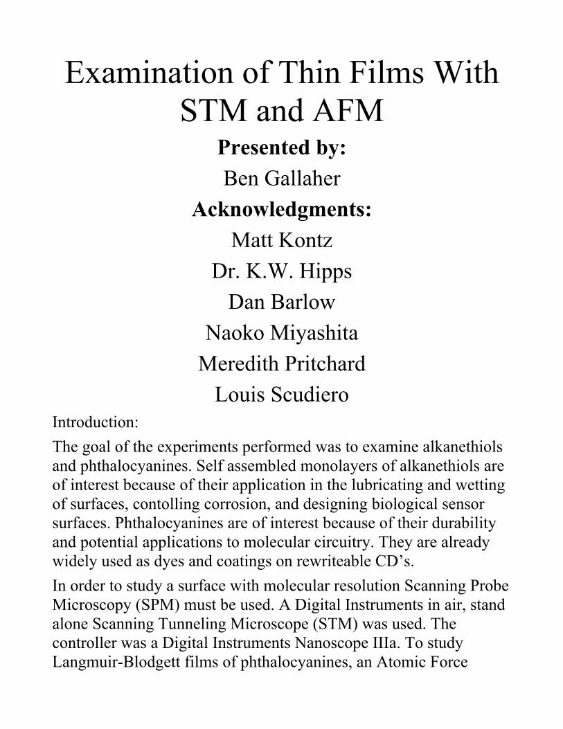
Microscope (AFM)--a Digital Instuments AFM with the same controller--was used. Because an STM requires an electrical current to function, conductive substrates are necessary. Cleaved graphite and atomically smooth gold on mica were used to accomplish this.Self Assembled Monolayers (SAMs) are formed by soaking a substrate in a solvent which has low levels of the compound of interest. Initially, reagent grade benzene served as the solvent. Imaged taken showed that the benzene contained too many impurities. Because of complications with impurities in the benzene solvent, SAMs are not discussed here.
Basic Schematic of an STM (above)
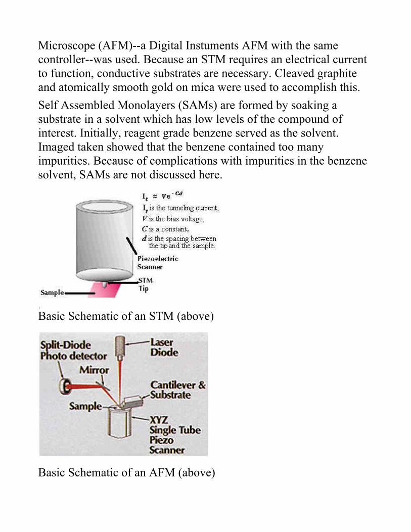
Basic Schematic of an AFM (above)
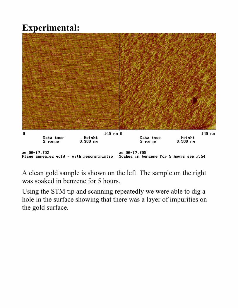
Experimental:
A clean gold sample is shown on the left. The sample on the right was soaked in benzene for 5 hours.Using the STM tip and scanning repeatedly we were able to dig a hole in the surface showing that there was a layer of impurities on the gold surface.

This image was taken at a 1:4 scan ration to save time scanning.In order to reduce impurities, spectroscopic grade benzene was used as the solvent.

The sample was also soaked in the spectroscopic benzene for 15 hours in new glassware and exposed to vacuum for 3 hours. While the gold is not perfect, it is much cleaner.
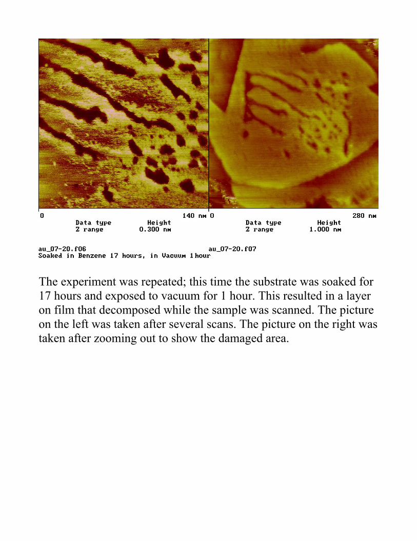
The experiment was repeated; this time the substrate was soaked for 17 hours and exposed to vacuum for 1 hour. This resulted in a layer on film that decomposed while the sample was scanned. The picture on the left was taken after several scans. The picture on the right was taken after zooming out to show the damaged area.
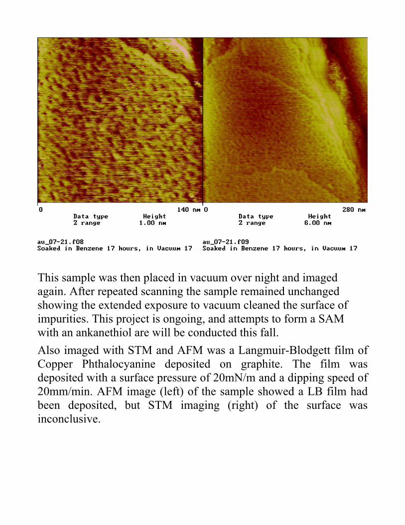
This sample was then placed in vacuum over night and imaged again. After repeated scanning the sample remained unchanged showing the extended exposure to vacuum cleaned the surface of impurities. This project is ongoing, and attempts to form a SAM with an ankanethiol are will be conducted this fall.Also imaged with STM and AFM was a Langmuir-Blodgett film of Copper Phthalocyanine deposited on graphite. The film was deposited with a surface pressure of 20mN/m and a dipping speed of 20mm/min. AFM image (left) of the sample showed a LB film had been deposited, but STM imaging (right) of the surface was inconclusive.
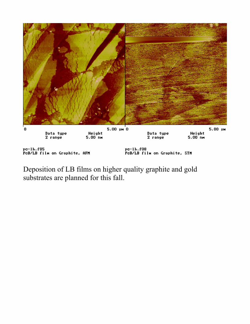
Deposition of LB films on higher quality graphite and gold substrates are planned for this fall.
References:Sarid, Dror. "Exploring Scanning Probe Microscopy with Mathematica." John Wiley and Sons, Inc. New York. 1997.Strausser, Y.E. and M.G. Heaton "Application Notes: Scanning Probe Microscopy: Technology Overview." Digital Instruments. May 1994. http://www.di.com/appnotes/AmLab/AL-SPMMain.html (29 July 1998)Stroscio, Joseph A., William J. Kaiser. "Methods of Experimental Physics Volume 27: Scanning Tunneling Microscopy." Academic Press, Inc. New York. 1993.Wiesendanger, Roland. "Scanning Probe Microscopy and Spectroscopy: Methods and Applications." Cambridge University Press. Cambridge, Great Britain. 1994.