excited states of bromopyrimidines probed by vuv
TRANSCRIPT
International Journal of
Molecular Sciences
Article
Excited States of Bromopyrimidines Probed by VUVPhotoabsorption Spectroscopy and Theoretical Calculations
Mónica Mendes 1,* , Fábris Kossoski 2,* , Ana I. Lozano 1 , João Pereira-da-Silva 1, Rodrigo Rodrigues 1 ,João Ameixa 1 , Nykola C. Jones 3 , Søren V. Hoffmann 3 and Filipe Ferreira da Silva 1
�����������������
Citation: Mendes, M.; Kossoski, F.;
Lozano, A.I.; Pereira-da-Silva, J.;
Rodrigues, R.; Ameixa, J.; Jones, N.C.;
Hoffmann, S.V.; Ferreira da Silva, F.
Excited States of Bromopyrimidines
Probed by VUV Photoabsorption
Spectroscopy and Theoretical
Calculations. Int. J. Mol. Sci. 2021, 22,
6460. https://doi.org/10.3390/
ijms22126460
Academic Editor: Paschalis
Alexandridis
Received: 21 May 2021
Accepted: 12 June 2021
Published: 16 June 2021
Publisher’s Note: MDPI stays neutral
with regard to jurisdictional claims in
published maps and institutional affil-
iations.
Copyright: © 2021 by the authors.
Licensee MDPI, Basel, Switzerland.
This article is an open access article
distributed under the terms and
conditions of the Creative Commons
Attribution (CC BY) license (https://
creativecommons.org/licenses/by/
4.0/).
1 CEFITEC, Departamento de Física, NOVA School of Science and Technology, FCT NOVA,Universidade NOVA de Lisboa, 2829-516 Caparica, Portugal; [email protected] (A.I.L.);[email protected] (J.P.-d.-S.); [email protected] (R.R.); [email protected] (J.A.);[email protected] (F.F.d.S.)
2 Laboratoire de Chimie et Physique Quantiques (UMR 5626), Université de Toulouse, CNRS, UPS,CEDEX 09, 31062 Toulouse, France
3 ISA, Department of Physics and Astronomy, Aarhus University, Ny Munkegade 120,8000 Aarhus C, Denmark; [email protected] (N.C.J.); [email protected] (S.V.H.)
* Correspondence: [email protected] (M.M.); [email protected] (F.K.)
Abstract: We report absolute photoabsorption cross sections for gas-phase 2- and 5-bromopyrimidinein the 3.7–10.8 eV energy range, in a joint theoretical and experimental study. The measurements werecarried out using high-resolution vacuum ultraviolet synchrotron radiation, with quantum chemicalcalculations performed through the nuclear ensemble approach in combination with time-dependentdensity functional theory, along with additional Franck–Condon Herzberg–Teller calculations for thefirst absorption band (3.7–4.6 eV). The cross sections of both bromopyrimidines are very similar below7.3 eV, deviating more substantially from each other at higher energies. In the 7.3–9.0 eV range wherethe maximum cross-section is found, a single and broad band is observed for 5-bromopyrimidine,while more discernible features appear in the case of 2-bromopyrimidine. Several π*← π transitionsaccount for the most intense bands, while weaker ones are assigned to transitions involving thenitrogen and bromine lone pairs, the antibonding σ*Br orbital, and the lower-lying Rydberg states.A detailed comparison with the available photo-absorption data of bromobenzene is also reported.We have found significant differences regarding the main absorption band, which is more peakedin bromobenzene, becoming broader and shifting to higher energies in both bromopyrimidines. Inaddition, there is a significant suppression of vibrational structures and of Rydberg states in the pairof isomers, most noticeably for 2-bromopyrimidine.
Keywords: VUV photoabsorption; halopyrimidines; valence and Rydberg states; time-dependentdensity functional theory; radiosensitizers
1. Introduction
The absorption of ionizing radiation by living cells has long been recognized as asignificant cause of long-term biological damage, leading to cellular death, mutationsand/or cancer. Most biological effects of radiation arise from permanent modificationsof the DNA structure, which can result in apoptosis [1]. It is well known that suchconsequences are mainly related to the effects of secondary low energy electrons and freeradicals, which are produced by the interaction of the primary ionization radiation withthe cellular molecular constituents [2,3]. Along the ionization track, secondary electronsare also able to excite biomolecular targets through inelastic collisions, thereby populatingelectronically excited states. These, in turn, might decay into further reactive species.
The ability of radiation in inducing modifications of the cellular components is par-ticularly important and useful for treatment of diseases, especially in cancer therapy.Nowadays, the investigation and development of more efficient therapies based on the
Int. J. Mol. Sci. 2021, 22, 6460. https://doi.org/10.3390/ijms22126460 https://www.mdpi.com/journal/ijms

Int. J. Mol. Sci. 2021, 22, 6460 2 of 27
combination of radiation and chemotherapeutic agents represent an important and grow-ing line of research. In chemoradiation, radiosensitizers are administered to the patient inorder to enhance the radiosensitivity of tumor cells. In the subsequent radiotherapy stage,the damage is thus expected to be more localized in regions rich with radiosensitizersdrugs. Through this combination, it is possible to increase tumor cell killing, with lessdamage to healthy tissues [4,5].
Pyrimidine is part of an important class of organic molecules, since it can be regardedas a building block of three nucleic acids, namely thymine, cytosine, and uracil. For thisreason, its physicochemical properties have been studied through several experimental andtheoretical techniques [6–15]. Chemical modification of pyrimidines by single halogenation,for example, has brought out their role as anti-cancer drugs. Indeed, it has been demon-strated that modified pyrimidines enhance the sensitivity of cells to ionizing radiationwhen incorporated into DNA as a substitute for thymine [16–19]. Halogenated analoguesof thymidine, such as bromodeoxyuridine and iododeoxyuridine have shown great impacton the enhancement of the radiotherapeutic effects via the Auger electron cascade producedby the decay of the ionized heavy halogen atom [20–22]. Recently, some fluorine analogues,especially 5-fluorouracil, 5-fluorodeoxyuridine, have also demonstrated an important rolein radiosensitization [5,23].
A full characterization of the electronic structure and electronic spectroscopy of pyrim-idine and its derivatives is particularly relevant, to provide insights into photo-inducedprocesses and their role in DNA damage. In particular, 2-bromopyrimidine (2BrPyr) and5-bromopyrimidine (5BrPyr) are isomers generated by replacing a hydrogen atom by abromine atom at positions C2 and C5 of pyrimidine, respectively. The effect of such substi-tutions in pyrimidine and pyrimidine-related compounds is important in order to betterunderstand the site-specific chemical bonding in DNA/RNA molecules. The vibrationalspectra of halogenated pyrimidines, including 2BrPyr and 5BrPyr, have been studied, withvibrational modes of different 2- and 5-monosubstituted pyrimidines [24,25]. Additionally,the vibrations of 2BrPyr and 5BrPyr were also investigated through infrared and Ramanspectroscopy experiments [26–28].
Bolognesi and co-workers have performed an extensive experimental and theoreticalanalysis of halogen-substituted pyrimidines by X-ray photoemission spectroscopy [29],near edge X-ray absorption fine structure spectroscopy [30], photofragmentation by ab-sorption of vacuum ultraviolet (VUV) and soft X-ray radiation [31,32], and Auger spec-troscopy [33]. Castrovilli et al. [34] have measured the photoionization mass spectraof bromopyrimidines in the 9–15 eV energy range. The valence photoelectron spectra(PES) of halogenated pyrimidines, including 2BrPyr and 5BrPyr, were investigated byO’Keeffe et al. [35]. The authors observed that the halogen atom induces changes in theelectronic structure of pyrimidine through two main mechanisms, the inductive and the res-onant effect, which involve the π orbitals localized on the ring and the lone pair orbitals ofthe halogen atom. More recently, a higher resolution PES spectrum of 2BrPyr was reportedby Smiałek et al. [36]. Shape and core-excited resonances of halopyrimidines were investi-gated by electron transmission and dissociative electron attachment spectroscopies [37], aswell as by electron scattering [38] and quantum chemical calculations [39]. Furthermore, theeffect of bromination of thymine and uracil was the subject of investigations of electronicand vibrational excitation [40,41] and the formation of negative ions [41–45].
There is only one experimental investigation on the electronic excited states of 2BrPyr,by Joshi et al. [27]. They reported the VUV absorption spectrum (in arbitrary units ofintensity), for photon energies ranging between 3.81 eV and 4.13 eV. VUV spectra wererecorded in the gas-phase as well as in different solutions, with some vibrational assign-ments tentatively reported for the gas phase spectrum. This previous study was limited tolower energies and nothing is known about the higher-lying excited states of 2BrPyr. Tothe best of our knowledge, the excited states of 5BrPyr have never been studied.
In the present work, we investigate the electronically excited states of 2BrPyr and 5Br-Pyr, by means of experimental and theoretical methodologies. Specifically, we report high-

Int. J. Mol. Sci. 2021, 22, 6460 3 of 27
resolution VUV absolute photoabsorption cross sections in the energy range of 3.7–10.8 eV,time-dependent density functional theory (TDDFT) calculations in combination with thenuclear ensemble approach (NEA), and Franck–Condon Herzberg–Teller (FCHT) calcu-lations, for both bromopyrimidine isomers. Experimental and theoretical methodologiesare explained in detail in Section 3. Our results bring forth a comprehensive picture oftheir excited states, and could shed further light on the subsequent production of radicals,which strongly impact the local site chemistry. Considering the radio-sensitizing potentialof bromopyrimidines, the present data may also provide new insights on the effects ofradiation damage at a nanoscale level and their role in chemoradiation applications.
2. Results and Discussion
Both 2BrPyr (Figure 1a) and 5BrPyr (Figure 1b) belong to the C2v point group. In theelectronic ground state (A1 symmetry), the outermost molecular orbitals are . . . (18a1)2
(2a2)2 (5b1)2 (19a1)2 (10b2)2 (11b2)2 (6b1)2 for 2BrPyr, and . . . (18a1)2 (5b1)2 (2a2)2 (19a1)2
(10b2)2 (11b2)2 (6b1)2 for 5BrPyr, as obtained at the density functional theory/CAM-B3LYP/aug-cc-pVDZ + 2s2p2d level of theory. The frontier canonical orbitals are shown inthe Supporting Information, in Figures S2 and S3. However, most electronic excitations in-volve more than one dominant pair of occupied and unoccupied canonical orbitals, makingthe interpretation of the transitions less straightforward. For this reason, we have also com-puted natural transition orbitals, which provide a compact representation of the dominanthole and particle orbitals that define the character of the transition [46]. While such orbitalsare state-specific, they presented the same qualitative character along most of the excitedstates discussed here, and therefore our assignments are based on the set of typical naturaltransition orbitals displayed in Figures 2 and 3. Most can be immediately associated witha corresponding canonical orbital: π*(a2) with the lowest unoccupied molecular orbital(LUMO), π*(b1) with the LUMO + 1, π(b1) with the highest occupied molecular orbital(HOMO), n+(a1) with the HOMO-3, π(a2) and πBr(b1) with either HOMO-4 or HOMO-5,depending on the isomer. In 5BrPyr, the n-(b2) natural transition orbital corresponds to theHOMO-1 and in 2BrPyr it arises as the combination of the HOMO-1 and the HOMO-2. TheHOMO-2 of the former and the orthogonal combination of the latter provide the nBr(b2)orbital. Finally, the σ*Br(a1) orbital also appears in some excitations, and corresponds tohigher-lying LUMOs.
Figure 1. Molecular structure of (a) 2-bromopyrimidine, and (b) 5-bromopyrimidine. Carbon atomsin black, hydrogen in white, nitrogen in blue, and bromine in purple.

Int. J. Mol. Sci. 2021, 22, 6460 4 of 27
Figure 2. Typical hole and particle natural transition orbitals for the electronic transitionsof 2-bromopyrimidine.
Figure 3. Typical hole and particle natural transition orbitals for the electronic transitionsof 5-bromopyrimidine.

Int. J. Mol. Sci. 2021, 22, 6460 5 of 27
Figure 4 shows the measured high-resolution VUV photoabsorption spectra of 2BrPyr(top panel) and of 5BrPyr (bottom panel) in the 3.7–10.8 eV photon energy range. Such highresolution measurements allowed us to resolve several fine features. The computed ab-sorption cross sections are presented in Figures 5 and 6, where they are compared with theexperimental results. Agreement between experimental and theoretical cross sections arefairly good, both in magnitude and in shape. In light of this, we have interpreted the mea-sured absorption bands largely based on the quantum chemical calculations. Tables 1 and 2summarize the computed vertical excitation energies and state characters, for each of the40 lower-lying singlet excited states plus two higher-lying states presenting large oscillatorstrengths. The dominant character of the transitions was assigned by visually inspectingthe corresponding natural transitions orbitals [46]. In Figure S1, we also compare the crosssections computed with the NEA method and with the simpler vertical approximation,which only takes the ground-state equilibrium geometry into account. Once the appropriatesampling of the ground state geometry is performed via the NEA method, the comparisonwith respect to the experiment improves significantly, for the whole energy range covered.
Figure 4. High-resolution VUV photoabsorption spectrum of 2-bromopyrimidine (top panel) and5-bromopyrimidine (bottom panel) in the 3.7–10.8 eV photon energy range. Appropriate band labels(I–VI) are also given. See text for details.

Int. J. Mol. Sci. 2021, 22, 6460 6 of 27
Figure 5. Comparison between measured (purple) and computed (green) absorption cross sections for 2-bromopyrimidinein the 3.7–10.8 eV photon energy range, separated into six absorption bands (I–VI). Individual contributions of the adiabaticexcited states in grey.

Int. J. Mol. Sci. 2021, 22, 6460 7 of 27
Figure 6. Comparison between measured (blue) and computed (red) absorption cross sections for 5-bromopyrimidine inthe 3.7–10.8 eV photon energy range, separated into six absorption bands (I–VI). Individual contributions of the adiabaticexcited states in grey.
Int. J. Mol. Sci. 2021, 22, 6460 8 of 27
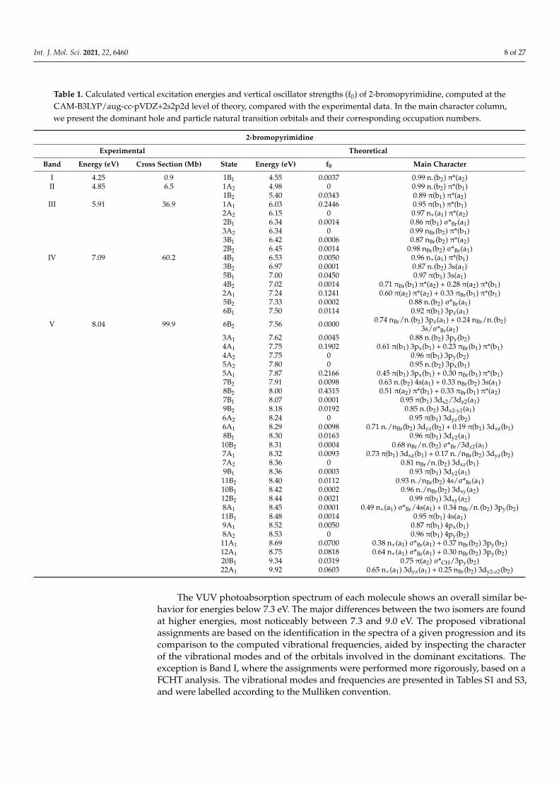
Table 1. Calculated vertical excitation energies and vertical oscillator strengths (f0) of 2-bromopyrimidine, computed at theCAM-B3LYP/aug-cc-pVDZ+2s2p2d level of theory, compared with the experimental data. In the main character column,we present the dominant hole and particle natural transition orbitals and their corresponding occupation numbers.
2-bromopyrimidine
Experimental Theoretical
Band Energy (eV) Cross Section (Mb) State Energy (eV) f0 Main Character
I 4.25 0.9 1B1 4.55 0.0037 0.99 n-(b2) π*(a2)II 4.85 6.5 1A2 4.98 0 0.99 n-(b2) π*(b1)
1B2 5.40 0.0343 0.89 π(b1) π*(a2)III 5.91 36.9 1A1 6.03 0.2446 0.95 π(b1) π*(b1)
2A2 6.15 0 0.97 n+(a1) π*(a2)2B1 6.34 0.0014 0.86 π(b1) σ*Br(a1)3A2 6.34 0 0.99 nBr(b2) π*(b1)3B1 6.42 0.0006 0.87 nBr(b2) π*(a2)2B2 6.45 0.0014 0.98 nBr(b2) σ*Br(a1)
IV 7.09 60.2 4B1 6.53 0.0050 0.96 n+(a1) π*(b1)3B2 6.97 0.0001 0.87 n-(b2) 3s(a1)5B1 7.00 0.0450 0.97 π(b1) 3s(a1)4B2 7.02 0.0014 0.71 πBr(b1) π*(a2) + 0.28 π(a2) π*(b1)2A1 7.24 0.1241 0.60 π(a2) π*(a2) + 0.33 πBr(b1) π*(b1)5B2 7.33 0.0002 0.88 n-(b2) σ*Br(a1)6B1 7.50 0.0114 0.92 π(b1) 3pz(a1)
V 8.04 99.9 6B2 7.56 0.0000 0.74 nBr/n-(b2) 3pz(a1) + 0.24 nBr/n-(b2)3s/σ*Br(a1)
3A1 7.62 0.0045 0.88 n-(b2) 3py(b2)4A1 7.75 0.1902 0.61 π(b1) 3px(b1) + 0.23 πBr(b1) π*(b1)4A2 7.75 0 0.96 π(b1) 3py(b2)5A2 7.80 0 0.95 n-(b2) 3px(b1)5A1 7.87 0.2166 0.45 π(b1) 3px(b1) + 0.30 πBr(b1) π*(b1)7B2 7.91 0.0098 0.63 n-(b2) 4s(a1) + 0.33 nBr(b2) 3s(a1)8B2 8.00 0.4315 0.51 π(a2) π*(b1) + 0.33 πBr(b1) π*(a2)7B1 8.07 0.0001 0.95 π(b1) 3dx2/3dz2(a1)9B2 8.18 0.0192 0.85 n-(b2) 3dx2-y2(a1)6A2 8.24 0 0.95 π(b1) 3dyz(b2)6A1 8.29 0.0098 0.71 n-/nBr(b2) 3dyz(b2) + 0.19 π(b1) 3dxz(b1)8B1 8.30 0.0163 0.96 π(b1) 3dy2(a1)10B2 8.31 0.0004 0.68 nBr/n-(b2) σ*Br/3dz2(a1)7A1 8.32 0.0093 0.73 π(b1) 3dxz(b1) + 0.17 n-/nBr(b2) 3dyz(b2)7A2 8.36 0 0.81 nBr/n-(b2) 3dxz(b1)9B1 8.36 0.0003 0.93 π(b1) 3dy2(a1)11B2 8.40 0.0112 0.93 n-/nBr(b2) 4s/σ*Br(a1)10B1 8.42 0.0002 0.96 n-/nBr(b2) 3dxy(a2)12B2 8.44 0.0021 0.99 π(b1) 3dxy(a2)8A1 8.45 0.0001 0.49 n+(a1) σ*Br/4s(a1) + 0.34 nBr/n-(b2) 3py(b2)11B1 8.48 0.0014 0.95 π(b1) 4s(a1)9A1 8.52 0.0050 0.87 π(b1) 4px(b1)8A2 8.53 0 0.96 π(b1) 4py(b2)
11A1 8.69 0.0700 0.38 n+(a1) σ*Br(a1) + 0.37 nBr(b2) 3py(b2)12A1 8.75 0.0818 0.64 n+(a1) σ*Br(a1) + 0.30 nBr(b2) 3py(b2)20B1 9.34 0.0319 0.75 π(a2) σ*CH/3py(b2)22A1 9.92 0.0603 0.65 n+(a1) 3dyz(a1) + 0.25 nBr(b2) 3dy2-z2(b2)
The VUV photoabsorption spectrum of each molecule shows an overall similar be-havior for energies below 7.3 eV. The major differences between the two isomers are foundat higher energies, most noticeably between 7.3 and 9.0 eV. The proposed vibrationalassignments are based on the identification in the spectra of a given progression and itscomparison to the computed vibrational frequencies, aided by inspecting the characterof the vibrational modes and of the orbitals involved in the dominant excitations. Theexception is Band I, where the assignments were performed more rigorously, based on aFCHT analysis. The vibrational modes and frequencies are presented in Tables S1 and S3,and were labelled according to the Mulliken convention.

Int. J. Mol. Sci. 2021, 22, 6460 9 of 27
Table 2. Calculated vertical excitation energies and vertical oscillator strengths (f0) of 5-bromopyrimidine, computed at theLYP/aug-cc-pVDZ+2s2p2d level of theory, compared with the experimental data. In the main character column, we presentthe dominant hole and particle natural transition orbitals and their corresponding occupation numbers.
5-bromopyrimidine
Experimental Theoretical
Band Energy (eV) Cross Section (Mb) State Energy (eV) f0 Main Character
I 4.21 1.2 1B1 4.48 0.0059 0.99 n-(b2) π*(a2)II 4.80 6.3 1A2 4.89 0 0.99 n-(b2) π*(b1)
1B2 5.35 0.0349 0.89 πr(b1) π*(a2)III 5.98 26.0 2B1 5.89 0.0002 0.99 π(b1) σ*Br(a1)
2A2 6.03 0 0.97 n+(a1) π*(a2)1A1 6.07 0.2129 0.94 π(b1) π*(b1)3B1 6.39 0.0049 0.99 n+(a1) π*(b1)2B2 6.57 0.0003 0.95 nBr(b2) σ*Br(a1)
IV 7.00 45.6 4B1 6.86 0.0020 0.98 nBr(b2) π*(a2)5B1 6.89 0.0162 0.97 π(b1) 3s(a1)3B2 6.98 0.0032 0.74 n-(b2) 3s/σ*Br(a1) + 0.24 nBr(b2) σ*Br(a1)3A2 7.10 0 0.99 nBr(b2) π*(b1)4B2 7.23 0.0474 0.51 π(a2) π*(b1) + 0.46 π(b1) π*(a2)2A1 7.34 0.2217 0.78 π(a2) π*(a2) + 0.10 π(b1) π*(b1)
V 8.10 110.8 5B2 7.36 0.0002 0.83 n-(b2) σ*Br(a1)3A1 7.63 0.0273 0.73 n-(b2) 3py(b2) + 0.14 n+(a1) σ*Br/3s(a1)4A2 7.66 0 0.99 π(b1) 3py(b2)6B2 7.67 0.0686 0.88 n-(b2) 3pz(a1)6B1 7.69 0.0028 0.96 π(b1) 3pz(a1)4A1 7.69 0.0060 0.88 π(b1) 3px(b1)5A2 7.73 0 0.97 n-(b2) 3px(b1)7B2 7.99 0.0056 0.54 nBr/n-(b2) 3s(a1) + 0.25 nBr/n-(b2) 3dx2-z2(a1)7B1 8.06 0.0000 0.96 π(b1) 3dx2(a1)8B2 8.13 0.0107 0.82 n-(b2) 3dx2(a1)9B2 8.18 0.3188 0.43 πBr(b1) π*(a2) + 0.25 π(a2) π*(b1)8B1 8.21 0.0014 0.81 πBr(b1) σ*Br(a1)5A1 8.28 0.1517 0.65 πBr(b1) π*(b1) + 0.25 π(b1) 3dxz(b1)9B1 8.29 0.0063 0.88 π(b1) 3dy2-z2(a1)6A1 8.31 0.0218 0.33 n-(b2) 3dyz(b2) + 0.33 π(b1) 3dxz(b1)10B1 8.36 0.0081 0.98 π(b1) 3dz2(a1)6A2 8.36 0 0.97 π(b1) 3dyz(b2)7A2 8.38 0 0.96 π(a2) σ*Br(a1)7A1 8.38 0.0430 0.45 π(b1) 3dxz(b1) + 0.28 n-(b2)10B2 8.38 0.0249 0.93 π(b1) 3dxy(a2)11B1 8.42 0.0016 0.95 n-(b2) 3dxy(a2)11B2 8.45 0.0261 0.79 n-(b2) 3dy2-z2(a1) + 0.11 nBr(b2) 3s(a1)12B2 8.45 0.0001 0.99 n-(b2) 4s(a1)8A2 8.47 0 0.96 n-(b2) 3dxz(b1)8A1 8.48 0.0460 0.64 n-(b2) 3dyz(b2) + 0.27 n+(a1) σ*Br/3s(a1)9A1 8.51 0.0021 0.92 π(b1) 4px(b1)
11A1 8.81 0.2800 0.62 n+(a1) σ*Br(a1) + 0.31 nBr(b2) 3py(b2)12A1 8.92 0.0683 0.45 nBr(b2) 3py(b2) + 0.37 n+(a1) σ*Br(a1)
Each of the following subsections is devoted to the discussion of a given absorptionband (where energy ranges are loosely defined), while the last subsection concerns Rydbergstates. In particular, the discussion will be focused on the effect of the position of thebromine atom on the VUV photoabsorption spectra, and we also compare with previousresults for the closely related bromobenzene molecule [47], which are reproduced inFigure S4. A detailed comparison with pyrimidine [7] will be presented in a future paper.
2.1. The 3.7–4.6 eV Photon Energy Range (Band I)
In both isomers, the first absorption has an onset around 3.7 eV and extends up to4.6 eV, as shown in Figure 7. Among all the bands discussed, this has the lowest intensities,yet the most fine structure. It is ascribed to the first singlet excited state, a π*(a2)← n-(b2)transition. It involves excitation from the n- non-bonding orbital of the nitrogen atoms, andthus has no parallel with bromobenzene [47].

Int. J. Mol. Sci. 2021, 22, 6460 10 of 27
Figure 7. Comparison between measured and computed cross sections for the absorption Band I of2-bromopyrimidine (top panel) and 5-bromopyrimidine (bottom panel).
This band is similar in both isomers, however in 5BrPyr it is somewhat more intense,slightly redshifted (~0.1 eV), and with more pronounced vibrational structures. The nuclearensemble approach calculations reproduce very well the shape and magnitude of the bands,as well as the main observed differences between the two isomers. Importantly, the goodagreement stems to a large extent from the ensemble averaging of the excitation energy,which decreases by 0.15 eV with respect to the vertical excitation energy, for both molecules.As pointed out before [48–50] this effect should be taken into account when comparingvertical excitation energies with the energy where absorption has a maximum.
In 2BrPyr, the equilibrium geometry of the first singlet excited state (coordinates inthe SI, and normal modes in Table S2) preserves the same symmetry operations as theground state, and thus belongs to the C2v point group. Therefore, there is a reasonableoverlap between ground- and excited-state optimized geometries, and the underlyingapproximations of the FCHT analysis should hold. In Figure 7 we compare the measuredand the computed absorption cross sections for the low energy tail of the band, between3.75 and 4.15 eV. The theoretical curve was redshifted by 0.108 eV, in order to match withthe observed 0–0 transition. In this comparison, we employed an arbitrary linewidthof 2.5 meV (~20.2 cm−1) for the FCHT calculations. Agreement is excellent, given theclear correspondence between most of the structures. This allows us to unequivocallypin down the band origin (the 0–0 transition) to the peak at 3.87 eV, which correspondsto the vibrational ground state of the excited state, thus agreeing with the assignment ofJoshi et al. [27]. A number of low intensity hot bands are observed at lower energies, arising

Int. J. Mol. Sci. 2021, 22, 6460 11 of 27
mostly from initially excited C-Br rocking (υ24) and stretching (υ9) modes. Towards higherenergies, the peaks obtained with the FCHT analysis appear to be gradually blue shiftedwith respect to the measurements, which is an indication of further anharmonic effectsnot considered in the FCHT calculations. In fact, when they are performed without anyanharmonic corrections for the excited state, such deviations are magnified. Eventually,the adiabatic Hessian approximation fails to reproduce the energies and shapes of thefiner structures, especially above ~4.15 eV. Nevertheless, it still manages to capture themain observed vibrational progressions of the whole band, as shown in Figure 8, wherea linewidth of 10 meV (~80.7 cm−1) was employed. A thorough assignment of the finestructures found in the 3.75–4.15 eV range can be found in Table S5. Despite the abovedescribed FCHT limitations, we also present in Table S6 the main transitions that shouldaccount for the main vibrational progressions up to ~4.1 eV. They comprise successiveexcitations of modes of a1 symmetry, mainly ring stretching modes having 700~1200 cm−1
frequencies and the C-Br stretching mode (see Table S6 for the full assignments).
Figure 8. Comparison between measured and computed cross sections for the low energy tail ofabsorption Band I of 2-bromopyrimidine.
For 5BrPyr, the calculations revealed a pair of degenerate equilibrium geometriesfor the first excited state (coordinates in the SI, and normal modes in Table S4), whicharise from symmetry-breaking of the C2 axial symmetry. They still preserve a planarstructure though, and thus belong to the Cs point group. Considering the existence oftwo equilibrium geometries and the smaller overlap between the lower-lying ground- andexcited-state vibrational wave functions, the FCHT analysis is not expected to reproduce thefine structures observed for 5BrPyr. However, just as in the case of 2BrPyr, the calculationscan account for the major vibrational progressions, as also shown in Figure 8 (the mostintense computed transitions are presented in Table S7). Here a linewidth of 10 meV wasemployed, and the computed curve was redshifted by 0.097 eV. The very good comparison

Int. J. Mol. Sci. 2021, 22, 6460 12 of 27
between the measured and the computed spectrum allowed us to identify the band originat 3.79 eV. The main vibrational progressions arise from successive excitations of ringstretching modes presenting frequencies in the 700~1200 cm−1 range, and also of the C-Brstretching mode, which overall share the same characters as those identified for 2BrPyr.
2.2. The 4.6–5.4 eV Photon Energy Range (Band II)
Figure 9 shows the second absorption band, found between 4.6 and 5.4 eV. Theirmagnitude and shape are very similar in both molecules, yet they appear displaced withrespect to each other by around 0.05 eV, being lower in 5BrPyr. We assign this band toexcitation of the third excited state, a π*(a2)← π(b1) dipole allowed transition, though thesecond excited state, a dipole forbidden π*(b1)← n-(b2) transition, contributes to a lesserextent. The band origin 0–0 is tentatively placed at 4.72 eV for 2BrPyr and at 4.68 eV for5BrPyr, whereas the observed vibrational progressions of both molecules are assigned toring stretching (υ7) and Br rocking (υ24) modes (see Tables 3 and 4). The present calculationsoverestimate the cross sections by a factor of two, and place the band too high in energyby around 0.5 eV, for both isomers. This disagreement stands out from the much bettercomparison observed for the other bands. Going from the vertical approximation to theNEA results does not bring any improvement (see Figure S1), and therefore, the problemshould be related to the choice of functional (CAM-B3LYP), while basis set effects provedto have a minor role. We notice, though, that the π(b1) and π*(a2) orbitals also take part inother transitions where the comparison with experiment is more favorable. Thus, it is notclear why the theoretical description of this particular band falls short.
Figure 9. Vibrational progressions in the 4.6–5.4 eV energy range of the absorption Band II of2-bromopyrimidine (top panel) and 5-bromopyrimidine (bottom panel).

Int. J. Mol. Sci. 2021, 22, 6460 13 of 27
Table 3. Proposed vibrational assignments in the 4.6−5.4 eV absorption band of 2BrPyr (Band II).∆E (in eV) represents the energy of one extra quanta in the vibrational progression. The symbol “/”means both modes are involved.
Energy (eV) Assignment ∆E(
υ′7
)∆E
(υ′24
)4.73 00
04.85 71
0 0.124.88 (b) 241
0/710 0.12 0.030
5.97 (b) 720 0.12
5.00 (s) 2410/72
0 0.12 0.0295.09 (b) 73
0 0.125.12 (b) 241
0/730 0.12 0.029
5.24 (b) 2410/74
0 0.124.73 00
04.85 71
0 0.124.88 (b) 241
0/710 0.12 0.030
5.97 (b) 720 0.12
(b) broad structure; (s) shoulder structure.
Table 4. Proposed vibrational assignments in the 4.6−5.4 eV absorption band of 5BrPyr (Band II).∆E (in eV) represents the energy of one extra quanta in the vibrational progression. The symbol “/”means both modes are involved.
Energy (eV) Assignment ∆E(
υ′7
)∆E
(υ′24
)4.68 00
04.70 (b) 241
0 0.034.80 71
0 0.134.83 (b) 242
0/710 0.12 0.03
4.92 720 0.12
4.95 (b) 2430/72
0 0.13 0.035.05 73
0 0.125.07 (b) 244
0/730 0.12 0.03
5.17 (b) 740 0.13
5.20 (b) 2450/74
0 0.13 0.03(b) broad structure.
In bromobenzene, the analogous π*(a2)← π(b1) state was also found in the same energyrange, where it accounts for the first absorption band [47]. There are, however, two keydifferences. First, the cross sections are considerably larger in the bromopyrimidines (havinga maximum of 6–7 Mb) than in bromobenzene (around 1 Mb). Second, the vibrationalstructures are much narrower in the latter, pointing to considerably longer-excited statelifetimes. In this energy range, the photodissociation mechanism of bromobenzene has beensuggested to involve intersystem crossings from the bright singlet state to dissociative tripletstates [51,52]. Assuming a similar mechanism takes place in the bromopyrimidines, signifi-cantly larger intersystem crossing rates from the π*(a2)← π(b1) state would be expected.
2.3. The 5.4–6.5 eV Photon Energy Range (Band III)
The next absorption band lies between 5.4 and 6.5 eV, peaking at 5.91 eV for 2BrPyr andat 5.98 eV for 5BrPyr. In neither are there distinct vibrational structures (see Figure 4). Theband profile is similar in both molecules, though the magnitudes differ from a maximumof 37 Mb in 2BrPyr to 26 Mb in 5BrPyr. The theoretical calculations support that the bulkof the intensity is due to the π*(b1)← π(b1) transition, which have comparable excitationenergies and a somewhat larger oscillator strength in 2BrPyr.
We notice, however, a subtle difference between the two spectra. It is more peakedin 2BrPyr and asymmetric in 5BrPyr, where a shoulder around 5.7 eV can be discerned.Considering that the NEA calculations seem to reproduce this feature, it might have anelectronic origin, rather than a vibrational one. In fact, while other excitation energies

Int. J. Mol. Sci. 2021, 22, 6460 14 of 27
in this range are rather close in both molecules, the σ*Br(a1)← π(b1) transition appears0.45 eV lower in 2BrPyr than in 5BrPyr. Such a big shift would arise from the σ*Br(a1)orbital, as discussed in the context of negative ion states [34–36]. The ensemble averagingfurther decreases the excitation energies by 0.25 eV (in both molecules), besides promotingintensity borrowing from the bright state. The greater sensitivity of this state on the positionof the bromine atom could thus explain the observed difference in the shape of this band.
In the case of bromobenzene, the analogous band emerges in the same energy range,also stemming from a dominant π*(b1)← π(b1) transition [47]. Moreover, the vibrationalprofile observed for bromobenzene is completely lost in the bromopyrimidines. In thelatter, the σ*Br(a1) ← π(b1) excited state (dissociative along the C-Br bond) lies withinthe band, whereas in the former the analogous state arises in the high energy tail of theband. Therefore, more favorable couplings between the optically bright state and thedissociative state are expected in the bromopyrimidines, which could explain the lack ofvibrational structures.
2.4. The 6.5–7.3 eV Photon Energy Range (Band IV)
This energy range accommodates the fourth absorption band (see Figure 10), havinga maximum at 7.09 eV in 2BrPyr, and at 7.00 eV in 5BrPyr. In both, the π*(a2) ← π(a2)transition brings most of the intensity to this band, even though other excitations alsocontribute to some extent, in particular the 3s(a1)← π(b1) transition for 2BrPyr, and theπ*(b1)← π(a2) + π*(a2)← π(b1) transition for 5BrPyr. Differences in the characters andoscillator strengths of such secondary excitations could explain the observed differencesfound in the vibrational progressions of each spectrum. In 5BrPyr the band origin 0–0 istentatively placed at 6.84 eV, and at 6.99 eV for 2BrPyr. The latter develops into two setsof progressions, one has an average energy spacing of 0.13 eV and is tentatively assignedto a ring stretching (υ7) mode. The other has an average energy spacing of 0.03 eV, andalso seems to be present in 5BrPyr, though with an observed spacing of ~0.05 eV. A closeinspection of Tables S1 and S3 reveals that no computed mode matches the observed ones.This may indicate that a significant relaxation of the excited states takes place, resulting inrather different frequencies.
In bromobenzene [47], the most intense band of the VUV spectrum is in the 6.3–7.0 eVrange, being redshifted by around 0.3 eV with respect to the Band IV of the bromopyrim-idines, besides being considerably more intense. As will be clearer in the next section, theintense band of bromobenzene should not only be compared with Band IV of bromopy-rimidines, but also with part of Band V. In bromobenzene, the band has been assigned totwo major excitations, the dominant one with A1 symmetry and π*(a2)← π(a2) + π*(b1)←π(b1) character, followed by the π*(b1)← π(a2) + π*(a2)← π(b1) excitation, of B2 symmetry.Except for the leading term of the former excitation, the other terms are absent in the BandIV of bromopyrimidines, giving rise to the next band, as discussed below.
Int. J. Mol. Sci. 2021, 22, 6460 15 of 27
Figure 10. Vibrational progressions in the 6.5–7.3 eV energy range of the absorption Band IV of2-bromopyrimidine (top panel) and 5-bromopyrimidine (bottom panel).
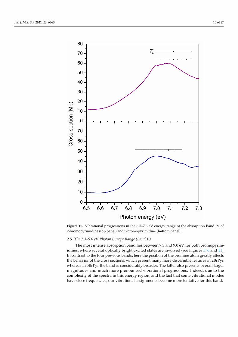
2.5. The 7.3–9.0 eV Photon Energy Range (Band V)
The most intense absorption band lies between 7.3 and 9.0 eV, for both bromopyrim-idines, where several optically bright excited states are involved (see Figures 5, 6 and 11).In contrast to the four previous bands, here the position of the bromine atom greatly affectsthe behavior of the cross sections, which present many more discernible features in 2BrPyr,whereas in 5BrPyr the band is considerably broader. The latter also presents overall largermagnitudes and much more pronounced vibrational progressions. Indeed, due to thecomplexity of the spectra in this energy region, and the fact that some vibrational modeshave close frequencies, our vibrational assignments become more tentative for this band.

Int. J. Mol. Sci. 2021, 22, 6460 16 of 27
Figure 11. Vibrational progressions in the 7.2–9.0 eV energy range of the absorption Band V of2-bromopyrimidine (top panel) and 5-bromopyrimidine (bottom panel).
In 5BrPyr, the band is centered at 8.2 eV, and is assigned to π*(a2)← πBr(b1) + π*(b1)← π(a2) (f0 = 0.32) and π*(b1)← πBr(b1) (f0 = 0.15) excitations, vertically located at 8.18and 8.28 eV according to the calculations. Two vibrational progressions were identifiedand tentatively attributed to excitation of the C–Br stretching (υ9) and ring stretching (υ23)modes (see Table 5). Another progression with an average spacing of 0.180 eV (1452 cm−1)was also identified, and could be related to either υ4 or υ19 ring stretching modes.

Int. J. Mol. Sci. 2021, 22, 6460 17 of 27
Table 5. Proposed vibrational assignments in the 7.3−9.0 eV absorption band of 5BrPyr (Band V).∆E (in eV) represents the energy of one extra quanta in the vibrational progression. The symbol “/”means both modes are involved; the symbol “;” means two alternative assignments.
Energy (eV) Assignment ∆E(
υ′4;υ
′19
)∆E
(υ′9
)∆E
(υ′23
)7.38 00
07.41 91
0 0.037.46 231
0 0.097.50 92
0/2310 0.03 0.09
7.55 2320/41
0; 1910 0.170 0.09
7.59 930/232
0 0.04 0.097.63 233
0 0.087.68 94
0/2330 0.03 0.09
7.72 2340/42
0; 1920 0.170 0.09
7.76 (s) 950/234
0 0.04 0.097.90 (s) 43
0; 1930 0.180
7.99 (s) 000
8.03 910 0.04
8.06 920/231
0/440; 194
0 0.160 0.04 0.078.10 93
0/2310 0.04 0.07
8.14 (w) 940/232
0 0.04 0.088.17 95
0/2320 0.03 0.08
8.22 960/233
0 0.04 0.088.25 97
0/2330/45
0; 1950 0.190 0.04 0.08
8.29 980/234
0 0.05 0.078.32 99
0/2340 0.04 0.07
8.36 9100 /235
0 0.04 0.078.39 911
0 /2350 0.04 0.07
8.43 (s) 9120 /236
0/460; 196
0 0.170 0.04 0.078.47 (s) 913
0 /2360 0.04 0.07
8.53 000
8.57 910 0.04
8.60 920/231
0/470; 197
0 0.190 0.04 0.088.65 93
0/2310 0.05 0.08
8.70 940/232
0 0.05 0.098.73 95
0/2320 0.04 0.09
8.77 960/233
0 0.04 0.088.82 97
0/2330/48
0; 1980 0.210 0.05 0.08
8.85 980/234
0 0.04 0.088.89 99
0/2340 0.04 0.07
8.94 9100 /235
0 0.05 0.098.97 911
0 /2350 0.03 0.08
(s) shoulder structure; (w) weak feature.
The analogous band of 2BrPyr presents a broader peak centered at 7.67 eV, followedby a sharp peak at 8.05 eV. A quick inspection of Table 1 indicates three states (vertically at7.75, 7.87, and 8.00 eV) with large oscillator strengths, having 3px(b1)← π(b1) (f0 = 0.19),π*(b1)← πBr(b1) (f0 = 0.22), and π*(b1)← π(a2) + π*(a2)← πBr(b1) (f0 = 0.43) characters.While these states should account for the two observed features, we cannot be sure whichones contribute to which peak, in view of their close-lying energies. Two vibrationalprogressions were tentatively assigned to ring stretching (υ5, υ6, υ7) and Br rocking (υ24)or C-Br stretching (υ9) modes. At higher energies, a ring deformation (υ23) and a ringstretching (υ8) were also tentatively assigned (see Table 6).
Int. J. Mol. Sci. 2021, 22, 6460 18 of 27
Table 6. Proposed vibrational assignments in the 7.3−9.0 eV absorption band of 2BrPyr (Band V).∆E (in eV) represents the energy of one extra quanta in the vibrational progression. The symbol “/”means both modes are involved; the symbol “;” means two alternative assignments.
Energy (eV) Assignment ∆E(
υ′9;υ′24
)∆E
(υ′23
)∆E
(υ′8
)∆E
(υ′5;υ′6;υ′7
)7.6 00
07.64 (s) 91
0; 2410 0.040
7.67 920; 242
0 0.0357.71 93
0; 2430 0.034
7.74 940; 244
0/540; 64
0; 740 0.034 0.143
7.78 (w) 950; 245
0 0.0347.81 (w) 96
0; 2460 0.034
7.84 (w) 970; 247
0 0.0347.88 (w) 98
0; 2480/54
0; 640; 74
0 0.036 0.1387.92 (w) 99
0; 2490 0.040
8.04 000
8.11 (s) 2310 0.076
8.19 (s) 2320 0.076
8.30 2330 0.080
8.40 000
8.50 (s) 810 0.095
8.59 (s) 820 0.095
8.70 (b) 830 0.095
8.79 (b) 840 0.095
(b) broad structure; (s) shoulder structure; (w) weak feature.
Clearly, the brightest electronic transitions in both of the bromopyrimidine isomersshare the same character, and have similar excitation energies and oscillator strengths. Themain contributions concern excitations of π*(a2)← πBr(b1), π*(b1)← π(a2), and π*(b1)←πBr(b1) characters, which account for the large intensity of Band V. The remaining excitationinvolving these orbitals, π*(a2)← π(a2), is the one responsible for Band IV, as discussedabove. Recalling that the same four excitations give rise to the intense absorption peakaround 6.3–7.0 eV in bromobenzene [47], we may conclude that the analogous band inboth bromopyrimidines is displaced to higher energies, becomes considerably broader, andactually splits into more discernible features. In this sense, the third and most intense bandin bromobenzene would correspond to the combination of Band IV and the first half ofBand V in the bromopyrimidines, extending over the 6.8–8.2 eV energy range in 2BrPyrand the 6.8–8.5 eV range in 5BrPyr.
Embedded into the broad Band V, the distinct peak at 8.41 eV in 2BrPyr as well as thebroad shoulder around 8.7 eV in 5BrPyr arise from σ*Br(a1)← n+(a1) + 3py(b2)← nBr(b2)transitions. The more stable σ*Br(a1) orbital of 2BrPyr explains the observed redshift forthis transition, similarly to what we have discussed for the Band III.
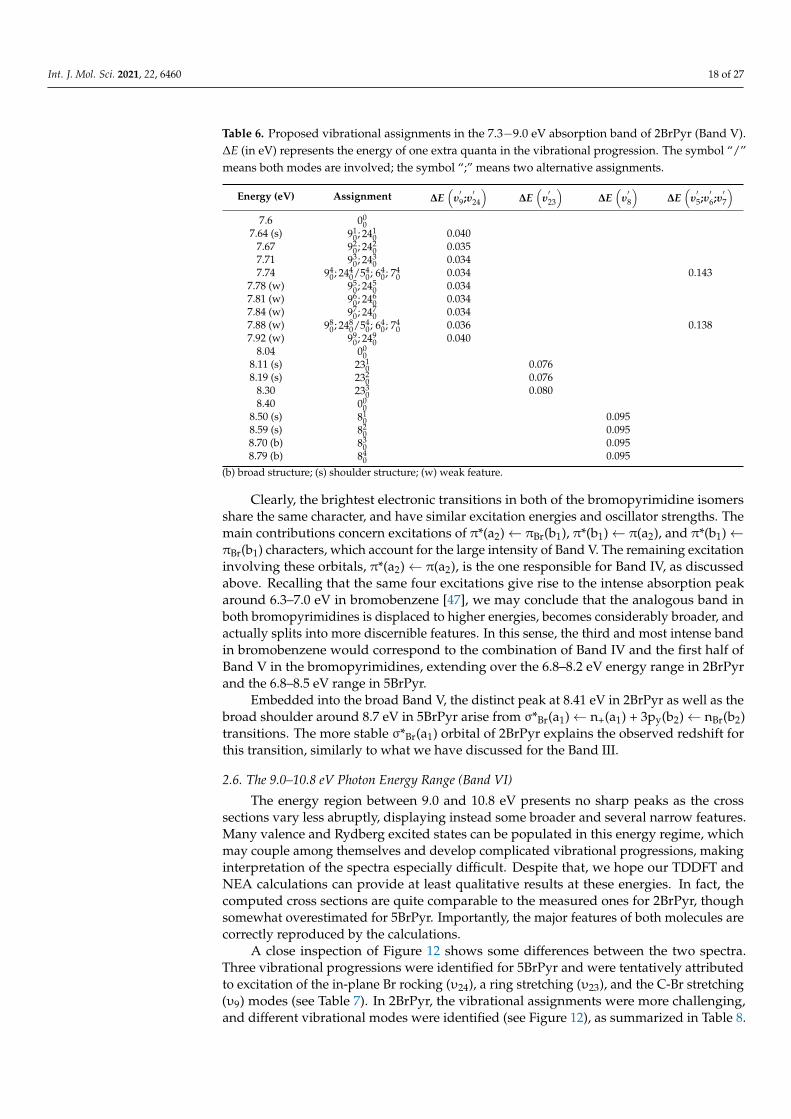
2.6. The 9.0–10.8 eV Photon Energy Range (Band VI)
The energy region between 9.0 and 10.8 eV presents no sharp peaks as the crosssections vary less abruptly, displaying instead some broader and several narrow features.Many valence and Rydberg excited states can be populated in this energy regime, whichmay couple among themselves and develop complicated vibrational progressions, makinginterpretation of the spectra especially difficult. Despite that, we hope our TDDFT andNEA calculations can provide at least qualitative results at these energies. In fact, thecomputed cross sections are quite comparable to the measured ones for 2BrPyr, thoughsomewhat overestimated for 5BrPyr. Importantly, the major features of both molecules arecorrectly reproduced by the calculations.
A close inspection of Figure 12 shows some differences between the two spectra.Three vibrational progressions were identified for 5BrPyr and were tentatively attributedto excitation of the in-plane Br rocking (υ24), a ring stretching (υ23), and the C-Br stretching(υ9) modes (see Table 7). In 2BrPyr, the vibrational assignments were more challenging,and different vibrational modes were identified (see Figure 12), as summarized in Table 8.

Int. J. Mol. Sci. 2021, 22, 6460 19 of 27
Figure 12. Vibrational progressions in the 9.0–10.8 eV energy range of the absorption Band VI of(top panel) and 5-bromopyrimidine (bottom panel).
Table 7. Proposed vibrational assignments in the 9.0−10.8 eV absorption band of 5BrPyr (Band VI).∆E (in eV) represents the energy of one extra quanta in the vibrational progression. The symbol “/”means both modes are involved; the symbol “;” means two alternative assignments.
Energy (eV) Assignment ∆E(
υ′9
)∆E
(υ′23
)∆E
(υ′24
)9.06 00
09.09 241
0 0.039.11 242
0 0.029.14 231
0 0.089.17 231
0/2410 0.08 0.03
9.21 2310/242
0 0.10 0.049.23 232
0 0.099.26 232
0/2410 0.09 0.03
9.29 (s) 2320/242
0 0.08 0.039.32 (s) 233
0 0.099.34 233
0/2410 0.08 0.02
9.37 2330/242
0 0.08 0.039.41 234
0 0.099.43 234
0/2410 0.09 0.02
9.45 2340/242
0 0.08 0.029.49 235
0 0.089.52 235
0/2410 0.09 0.03

Int. J. Mol. Sci. 2021, 22, 6460 20 of 27
Table 7. Cont.
Energy (eV) Assignment ∆E(
υ′9
)∆E
(υ′23
)∆E
(υ′24
)9.54 235
0/2420 0.09 0.02
9.57 2360 0.08
9.60 2360/241
0 0.08 0.039.63 (s) 236
0/2420 0.09 0.03
9.66 2370 0.09
9.69 2370/241
0 0.09 0.039.72 (w) 237
0/2420 0.09 0.03
9.75 2380 0.09
9.79 2380/241
0 0.10 0.049.83 239
0 0.089.86 (s) (b) 239
0/2410 0.07 0.03
9.90 (b) 23100 0.07
9.95 (b) 23100 /241
0 0.07 0.0510.00 (w) 2311
0 0.1010.02 2311
0 /2410 0.09 0.02
10.07 23120 0.07
10.10 23120 /241
0 0.07 0.0310.15 2313
0 0.0810.18 2313
0 /2410 0.08 0.03
10.24 (w) 23140 0.09
10.27 23140 /241
0 0.09 0.0310.32 (w) 2315
0 0.0810.35 (w) 2315
0 /2410 0.08 0.03
10.4110.45 (s) 91
0 0.0410.49 231
0/920 0.04 0.08
10.54 930 0.05
10.57 2320/94
0 0.05 0.0810.61 (w) 95
0 0.0410.67 233
0/960 0.06 0.09
10.71 970 0.05
10.75 2340/98
0 0.06 0.07
(b) broad structure; (s) shoulder structure; (w) weak feature.
Table 8. Proposed vibrational assignments in the 9.0−10.8 eV absorption band of 2BrPyr (Band VI).∆E (in eV) represents the energy of one extra quanta in the vibrational progression. The symbol “/”means both modes are involved; the symbol “;” means two alternative assignments.
Energy (eV) Assignment ∆E(
υ′9;υ′24
)∆E
(υ′7
)∆E
(υ′5;υ′6;υ′7
)9.07 00
09.10 (w) 91
0; 2410 0.033
9.14 (w) 920; 242
0 0.0409.17 93
0; 2430/71
0 0.034 0.1129.21 94
0; 2440 0.036
9.24 950; 245
0 0.0409.28 96
0; 2460/72
0 0.042 0.1109.32 97
0; 2470 0.042
9.36 980; 248
0 0.0429.41 99
0; 2490/73
0 0.037 0.1279.45 910
0 ; 24100 0.033
9.48 (w) 9110 ; 2411
0 0.0359.51 912
0 ; 24120 0.034
9.55 (w) 9130 ; 2413
0 0.0359.58 (s) 914
0 ; 24140 0.035
9.61 9150 ; 2415
0 0.0359.65 916
0 ; 24160 0.035
9.69 (s) 9170 ; 2417
0 0.0359.73 (s) 918
0 ; 24180 0.035
9.77 510; 61
0; 710 0.150
9.93 (s) 520; 62
0; 720 0.150
10.08 530; 63
0; 730 0.158
10.23 (s) 540; 64
0; 740 0.142
10.36 550; 65
0; 750 0.137
10.52 (b) 560; 66
0; 760 0.154
10.65 570; 67
0; 770 0.139
(b) broad structure; (s) shoulder structure; (w) weak feature.

Int. J. Mol. Sci. 2021, 22, 6460 21 of 27
In comparison to bromobenzene [47], the cross sections of both bromopyrimidines aresmaller in this energy range, besides having less pronounced features. Both observationscould be related to the suppression of Rydberg states, discussed in the next section.
2.7. Rydberg States
Rydberg states are characterized by excitation into diffuse (Rydberg) orbitals, whoseenergies follow the well-known Rydberg formula, En = IE − [13.606/(n − δ)2] [eV], wherethe excitation energy En is estimated from the ionization energy IE, the principal quantumnumber n, and the quantum defect δ. For each angular momentum (s, p, d, etc.), and foreach orbital from which the electron is excited, the Rydberg states form a series convergingto the ionization energy associated with this orbital. Such states tend to appear in VUVspectra as sharper peaks than valence excitations, though with varying intensities, andtheir precise assignment is usually performed with the aid of the Rydberg formula.
Our measured spectra do not display pronounced signatures of Rydberg states, foreither 2BrPyr and 5BrPyr, in marked contrast to what was observed for bromobenzene [47].That being said, there is some evidence that weak structures in the VUV spectra wouldarise from the 3s(a1)← π(b1) Rydberg state. In 2BrPyr, the four small peaks observed inBand IV (starting at 6.99 eV) could actually correspond to a vibrational progression of thisRydberg state. The theoretical calculations further support this assignment, as this statewas found at 7.00 eV, with an oscillator strength of 0.045, which, albeit small, is probablysufficiently large to leave a weak mark on the spectrum. The associated quantum defectwould be 0.84, considering the experimental ionization energy of 9.93 eV for the π(b1)orbital [36]. In 5BrPyr, the analogous 3s(a1)← π(b1) Rydberg state could account for theweak shoulder observed at 6.84 eV (a quantum defect of 0.90, given an ionization energy of9.93 eV [35]). Again, this is supported by the calculations, which show an excitation energyof 6.89 eV, and an oscillator strength of 0.016. In particular, the lower oscillator strength in5BrPyr is also consistent with the weaker feature on the spectrum.
According to our calculations, some of the 3p Rydberg states presented strong mixingwith valence states, contributing to Band V. In contrast, purer 3p Rydberg states that showup in the same energy range (7.5–8.0 eV) have reduced oscillator strengths, thus leaving nodistinct signatures in the spectrum. Finally, the 3d Rydberg states would start emerging inthe 8.0–8.5 eV range, most of them with low intensities.
Towards higher energies, a multitude of Rydberg series are expected, though with pro-gressively smaller intensities. In fact, the theoretical calculations (see Tables 1 and 2) indi-cate that Rydberg transitions become more and more prevailing at higher energies, thoughwith small oscillator strengths, particularly for 2BrPyr. For instance, the 4s(a1)← π(b1)state of this isomer would appear at 8.48 eV according to our calculations, with a verylow oscillator strength (0.001). The same applies for the other angular momentum and forexcitations from other high-lying occupied orbitals. Indeed, at higher energies we haveobserved no sharp peaks that could be clearly associated with Rydberg states. Rather,the absorption spectra present a large number of closely-lying weak features. Thus, anyattempt to locate the Rydberg series based on the Rydberg formula and the ionizationenergies would be very tentative and not conclusive. To a great extent, that would be aless pertinent task, considering the overall minor contribution of the higher-lying Rydbergstates to the photoabsorption cross section of bromopyrimidines.
The scenario is quite distinct for bromobenzene, where much more pronouncedstructures have been observed [47]. With the support of high-resolution PES data, thisallowed several Rydberg states to be clearly assigned. We notice that a similar behavior canbe recognized when one compares the VUV spectra of benzene and pyrimidine [7,53]. Suchobservations suggest that going from benzene to pyrimidine, as well as from bromobenzeneto bromopyrimidines, promotes a major suppression of the Rydberg series.

Int. J. Mol. Sci. 2021, 22, 6460 22 of 27
3. Methods3.1. Experimental Methods
The high-resolution VUV photoabsorption measurements were performed at theAU-UV beamline of the ASTRID2 synchrotron facility at Aarhus University in Denmark.A detailed explanation of the experimental apparatus is given by Palmer et al. [54] andan in-depth description of procedures for measurement and calibration can be found inDuflot et al. [55]. In short, the set-up consists of a gas cell placed on the output of themonochromator where a MgF2 window was mounted to separate it from the ultra-highvacuum (UHV) of the beam line. The synchrotron radiation passes through a static gassample and the transmitted light intensity is detected by a photomultiplier tube (PMT). Theabsolute pressure of the effusive molecular gas is measured with a capacitance manometer(Chell CDG100D). The wavelength is selected using a toroidal dispersion grating with2000 lines/mm providing a resolution of 0.075 nm, corresponding to 3 meV at the midpointof the energy range studied. In order to avoid any absorption of O2 from the air inmeasurements below 200 nm (energies above 6.20 eV), the small gap between the PMT andthe MgF2 gas cell exit window is evacuated using a scroll pump. For higher wavelengthmeasurements, air is admitted into the gap at atmospheric pressure to allow O2 to absorbany higher orders of light produced by the monochromator. ASTRID2 operates in “top-up”mode, keeping the stored electron beam current quasi-constant. Absolute photoabsorptioncross sections (σ) were obtained at room temperature (~25 ◦C) by using the Beer−Lambertattenuation law (1),
It = I0 exp(−Nσl), (1)
where It is the light intensity transmitted through the gas sample, I0 is that transmittedthrough the evacuated cell, N is the molecular density, and l is the absorption path length(15.5 cm). The range of pressures used for the measurements of 5BrPyr was 0.01 to0.54 mbar, while for 2BrPyr, which has a lower vapor pressure, the pressure range was 0.02to 0.11 mbar. The accuracy of the cross section is estimated to be ~10%, which is ensured byrecording the VUV spectra in small (5 or 10 nm) sections to allow optimization of pressureaccording to the local cross-sections, with at least 10 points overlap between the adjoiningranges [55,56].
The 2-bromopyrimidine sample was purchased from FluoroChem with a stated purityof ≥97%, while 5-bromopyrimidine was purchased from Sigma Aldrich with a statedpurity of ≥97%. The samples were used with no further purification and degassed beforemeasurement through repeated freeze−pump−thaw cycles.
3.2. Theoretical Methods
The photoabsorption cross sections were computed with the NEA [57,58] in combi-nation with TDDFT calculations. The latter were performed with the Gaussian 16 soft-ware [59], while the managing of the NEA calculations were performed as implemented inthe Newton-X package [60,61].
We employed the CAM-B3LYP functional, which provided excitation energies compa-rable to those observed in the measured VUV spectra. Other range separated functionalswere explored (LC-ωPBE,ωB97X, and LCBLYP), however overall, they did not performas well as CAM-B3LYP for the cases of 2BrPyr and 5BrPyr. We used the aug-cc-pVDZbasis set supplemented with a set of 2s2p2d diffuse functions, centered at the carbon atomclosest to the bromine atom. For each angular momentum, the exponents were generatedby successive divisions of the previous one by four (starting from the most diffuse func-tions of the aug-cc-pVDZ set). The CAM-B3LYP/aug-cc-pVDZ+2s2p2d level of theorywas employed for the geometry optimization, normal mode analysis, and calculation ofanharmonic frequencies (see below).
In the NEA, the configurations are sampled according to the Wigner distribution(Gaussians) of a set of independent harmonic potentials, which represent the normalmodes at the ground state optimized geometry. The corresponding Wigner distribution at298 K (experimental condition) was considered for the sampling. Anharmonic corrections

Int. J. Mol. Sci. 2021, 22, 6460 23 of 27
for the frequencies were accounted for with the importance sampling algorithm [62],though such effects had a marginal impact on the computed cross sections.
Excitation energies ∆E0n(qi) and oscillator strengths f0n(qi) of the n-th excited state arecomputed for each sampled configuration qi. Then, the photoabsorption cross section isobtained as (in atomic units):
σ(E) =1E
Ns
∑n
1Np(n)
Np
∑i
∆E0n(qi) f0n(qi)g[E− ∆E0n(qi), δ], (2)
where the phenomenological line profile g is represented by a normalized Gaussian func-tion centered at each excitation energy ∆E0n(qi). Here we have employed a linewidth ofδ = 0.1 eV, which is large enough to damp the artificial undulations of the cross-sectioncurve, yet small enough to keep its actual overall shape. We have used a decreasingnumber of configurations as we moved higher in energy. For both 2BrPyr and 5BrPyr, thenumber of sampled configurations was Np = 7500 for the first eight singlet excited states(n = 1–8), Np = 5500 for n = 9–16, Np = 3500 for n = 17–40, Np = 2500 for n = 41–60, Np = 1500for n = 61–80, and Np = 1000 for n = 81–160. For 2BrPyr, we used an additional set ofNp = 500 points for n = 161–170. This represents a total of 701,000 computed excitationenergies and oscillator strengths.
While the NEA is able to describe the overall shapes and intensities of the absorptionbands, it cannot describe vibrational progressions, which would require treating the nucleiquantum-mechanically. As discussed in Section 2.1, Band I of both bromopyrimidinespresents very rich vibrational structures, whose interpretation becomes impractical with-out support from theory. Given the limitations of the NEA calculations, we have alsoemployed a second theoretical approach, based on FCHT analysis, as performed withthe time-independent procedure [63–65]. The excited-state potential energy surface isdescribed with the adiabatic Hessian model, where force constants and vibrational fre-quencies are computed at the equilibrium geometry of the excited state. Ground stateanharmonic frequencies were computed with the generalized second-order vibrationalperturbation method [64,66], and from those we also obtained excited state anharmonic fre-quencies by employing a mode-specific scaling factor built from the Duschinsky matrix [67].Temperature effects are accounted for by considering excitations from vibrationally excitedlevels with populations larger than 2%, for a temperature of 298 K, corresponding to thepresent experimental condition. All the calculations required for the FCHT analysis wereperformed with the Gaussian 16 software [59].
4. Conclusions
Absolute photoabsorption cross sections obtained with high resolution VUV syn-chrotron radiation in the 3.7–10.8 eV energy range are reported for 2BrPyr and 5BrPyr.Theoretical cross sections are also presented, as computed within the NEA in combinationwith TDDFT calculations. The good agreement between measurements and calculationsgives strong support for the interpretation and discussion of both obtained VUV spectra.
The present study, together with a previous report on the photoabsorption of fluo-ranil [50] showed that the NEA can be successfully employed to probe photoabsorptionfor considerably larger energies, closer to the ionization threshold. While the improvedaccuracy of the NEA with respect to the simpler vertical approximation is well knownfor the low energy regime, it remained to be demonstrated in practice for higher energies.However, the overall computational cost increases sharply when the NEA is employed forhigher energies, requiring the evaluation of dozens or hundreds of excited states for anextensive set of geometries.
In conclusion, the lowest lying band (3.7–4.6 eV) presents the lowest cross sections,and it was assigned to the first singlet excited state, a π*(a2) ← n-(b2) transition. Withthe support of FCHT calculations, we placed the band origin of 2BrPyr at 3.87 eV andof 5BrPyr at 3.79 eV, whereas a number of low intensity hot bands and main vibrational

Int. J. Mol. Sci. 2021, 22, 6460 24 of 27
progressions could be assigned to C-Br rocking/wagging modes and to totally symmetricring stretching modes found with frequencies in the 700~1200 cm−1 range, respectively.The Band II (4.6–5.4 eV) was attributed to a π*(a2)← π(b1) transition, which is followed bya third band (5.4–6.5 eV) and assigned to a π*(b1)← π(b1) transition, the latter displayingno vibrational structures. For the three lowest-lying bands, the cross sections of both2BrPyr and 5BrPyr are rather similar, the first two being slightly red-shifted in 5BrPyr, andthe third being somewhat more intense in 2BrPyr. Still in Band III, a weak shoulder wasobserved for 5BrPyr, and explained by a larger effect of the bromine atom position on theσ*Br(a1)← πr(b1) transition.
Going up in energy we have the most intense absorption bands, IV (6.5–7.3 eV) andV (7.3–9.0 eV), which should be analyzed together. As a whole, their large intensities stemfrom the four possible excitations from the πBr(b1) and π(a2) occupied orbitals, to the π*(b1)and π*(a2) unoccupied orbitals. The analogous excitations account for the main absorptionbands of the related bromobenzene, pyrimidine, and benzene molecules. Compared withbromobenzene, the band of both bromopyrimidines is displaced to higher energies andbroadens significantly, such that some individual π* ← π transitions become apparent.Even so, the isomer effect is more substantial in this energy region, affecting the behaviorof the individual π*← π excitations and of other, less intense transitions. Finally, Band VI(9.0–10.8 eV) should involve many more valence and Rydberg excited states, but despitethis, broader features could still be interpreted. In this regime, both spectra showed distinctvibrational progressions.
An extensive comparison between the VUV spectra of bromopyrimidines and theavailable data for bromobenzene revealed a series of similarities and differences. Namely,for the bromopyrimidines we observed a new band (Band I), broader (Band II) or a com-plete suppression of vibrational structures (Band III), broadened and blue shifted features(Bands IV and V), and a loss of intensity (Bands II and VI). An additional key distinctionconcerns the Rydberg states, much less pronounced in both bromopyrimidines than inbromobenzene. A similar trend can be noticed when comparing the spectra of pyrimidineand benzene.
This work is the most comprehensive VUV photoabsorption study yet reported for2BrPyr and 5BrPyr. Considering that bromopyrimidines are known for their potential asradiosensitizers, the detailed picture we have provided on their excited states and VUVphotoabsorption spectra could bring important insights on the production of reactiveradicals and on radiation damage in general.
Supplementary Materials: The following are available online at https://www.mdpi.com/article/10.3390/ijms22126460/s1.
Author Contributions: Measurements, F.F.d.S. and N.C.J.; funding acquisition, F.F.d.S.; analysisof results, M.M., A.I.L., F.K., J.P.-d.-S., R.R., J.A.; writing—original draft preparation, M.M., A.I.L.,F.K.; theoretical calculations, F.K.; writing—review, F.F.d.S., N.C.J., S.V.H. All authors have read andagreed to the published version of the manuscript.
Funding: J.P.-d.-S. acknowledges the Portuguese National Funding Agency FCT-MCTES through PhDgrant PD/BD/142768/2018, together with R.R., J.A., M.M. and F.F.d.S. through the researcher grantPTDC/FIS-AQM/31215/2017 and AIL through the researcher grant PTDC/FIS-AQM/31281/2017.This work was also supported by Radiation Biology and Biophysics Doctoral Training Programme(RaBBiT, PD/00193/2012); UIDB/04378/2020 (UCIBIO); and UIDB/00068/2020 (CEFITEC). Theresearch leading to this result has been supported by the project CALIPSOplus under the GrantAgreement 730872 from the EU Framework Programme for Research and Innovation HORIZON 2020.
Data Availability Statement: The data presented in this study are available on request from thecorresponding author.
Conflicts of Interest: The authors declare no conflict of interest.
Int. J. Mol. Sci. 2021, 22, 6460 25 of 27
References1. Alizadeh, E.; Orlando, T.M.; Sanche, L. Biomolecular Damage Induced by Ionizing Radiation: The Direct and Indirect Effects of
Low-Energy Electrons on DNA. Annu. Rev. Phys. Chem. 2015, 66, 379–398. [CrossRef]2. Boudaïffa, B.; Cloutier, P.; Hunting, D.; Huels, M.A.; Sanche, L. Resonant formation of DNA strand breaks by low-energy (3 to
20 eV) electrons. Science 2000, 287, 1658–1660. [CrossRef] [PubMed]3. Huels, M.A.; Boudaïffa, B.; Cloutier, P.; Hunting, D.; Sanche, L. Single, double, and multiple double strand breaks induced in
DNA by 3-100 eV electrons. J. Am. Chem. Soc. 2003, 125, 4467–4477. [CrossRef] [PubMed]4. Wardman, P. Chemical Radiosensitizers for Use in Radiotherapy. Clin. Oncol. 2007, 19, 397–417. [CrossRef] [PubMed]5. Wang, H.; Mu, X.; He, H.; Zhang, X.D. Cancer Radiosensitizers. Trends Pharmacol. Sci. 2018, 39, 24–48. [CrossRef]6. Baccarelli, I.; Bald, I.; Gianturco, F.A.; Illenberger, E.; Kopyra, J. Electron-induced damage of DNA and its components: Experi-
ments and theoretical models. Phys. Rep. 2011, 508, 1–44. [CrossRef]7. Ferreira da Silva, F.; Almeida, D.; Martins, G.; Milosavljevic, A.R.; Marinkovic, B.P.; Hoffmann, S.V.; Mason, N.J.; Nunes, Y.; Garcia,
G.; Limão-Vieira, P. The electronic states of pyrimidine studied by VUV photoabsorption and electron energy-loss spectroscopy.Phys. Chem. Chem. Phys. 2010, 12, 6717–6731. [CrossRef]
8. Ferreira da Silva, F.; Matias, C.; Almeida, D.; García, G.; Ingólfsson, O.; Flosadóttir, H.D.; Ómarsson, B.; Ptasinska, S.; Puschnigg,B.; Scheier, P.; et al. NCO-, a key fragment upon dissociative electron attachment and electron transfer to pyrimidine bases: Siteselectivity for a slow decay process. J. Am. Soc. Mass Spectrom. 2013, 24, 1787–1797. [CrossRef] [PubMed]
9. Mendes, M.; Pamplona, B.; Kumar, S.; Ferreira da Silva, F.; Aguilar, A.; García, G.; Bacchus-Montabonel, M.-C.; Limão-Vieira, P. Ion-pairformation in neutral potassium-neutral pyrimidine collisions: Electron transfer experiments. Front. Chem. 2019, 7, 264. [CrossRef]
10. Almeida, D.; Ferreira Da Silva, F.; García, G.; Limão-Vieira, P. Selective bond cleavage in potassium collisions with pyrimidinebases of DNA. Phys. Rev. Lett. 2013, 110, 1–5. [CrossRef]
11. Itälä, E.; Granroth, S.; Ha, D.T.; Kooser, K.; Levola, H.; Rachlew, E.; Tanzer, K.; Kukk, E. Fragmentation of imidazole, pyrimidineand purine induced by core ionization: Significance of small-scale chemical environment. J. Photochem. Photobiol. A Chem. 2018,356, 283–289. [CrossRef]
12. Pandey, R.; Ryszka, M.; da Fonseca Cunha, T.; Lalande, M.; Dampc, M.; Limão-Vieira, P.; Mason, N.J.; Poully, J.C.; Eden, S. Thresholdbehavior in metastable dissociation of multi-photon ionized thymine and uracil. Chem. Phys. Lett. 2017, 684, 233–238. [CrossRef]
13. Knight, A.E.W.; Lawburgh, C.M.; Parmenter, C.S. n,π* Fluorescence from selected vibronic levels of pyrimidine vapor: Franck-condon factors excited state anharmonic coupling. J. Chem. Phys. 1975, 63, 4336. [CrossRef]
14. Luo, Y.; Ågren, H.; Knuts, S.; Jørgensen, P. The two-photon spectrum of pyrimidine. Role of vibronic coupling. Chem. Phys. Lett.1993, 213, 357–362. [CrossRef]
15. Plekan, O.; Coreno, M.; Feyer, V.; Moise, A.; Richter, R.; de Simone, M.; Sankari, R.; Prince, K.C. Electronic state resolved PEPICOspectroscopy of pyrimidine. Phys. Scr. 2008, 78, 058105. [CrossRef]
16. Opara-Kubinska, Z.; Lorkiewicz, Z.; Szybalski, W. Genetic transformation studies. II. Radiation sensitivity of halogen labeledDNA. Biochem. Biophys. Res. Commun. 1961, 4, 288–291. [CrossRef]
17. Djordjevic, B.; Szybalski, W. Genetics of Human Cell Lines. III. Incorporation of 5-Bromo- and 5-Iododeoxyuridine into theDeoxyribonucleic Acid of Human Cells and Its Effect on Radiation Sensitivity. J. Exp. Med. 1960, 112, 509–531. [CrossRef] [PubMed]
18. Chomicz, L.; Zdrowowicz, M.; Kasprzykowski, F.; Rak, J.; Buonaugurio, A.; Wang, Y.; Bowen, K.H. How to find out whether a5-substituted uracil could be a potential DNA radiosensitizer. J. Phys. Chem. Lett. 2013, 4, 2853–2857. [CrossRef]
19. Ameixa, J.; Arthur-Baidoo, E.; Meißner, R.; Makurat, S.; Kozak, W.; Butowska, K.; Ferreira Da Silva, F.; Rak, J.; Denifl, S. Low-energy electron-induced decomposition of 5-trifluoromethanesulfonyl-uracil: A potential radiosensitizer. J. Chem. Phys. 2018, 149,164307. [CrossRef] [PubMed]
20. Dewey, W.C.; Humphrey, R.M. Increase in Radiosensitivity to Ionizing Radiation Related to Replacement of Thymidine inMammalian Cells with 5-Bromodeoxyuridine. Radiat. Res. 1965, 26, 538–553. [CrossRef]
21. O’Donoghue, J.A.; Wheldon, T.E. Targeted radiotherapy using Auger electron emitters. Phys. Med. Biol. 1996, 41, 1973–1992. [CrossRef]22. Bloomer, W.; Adelstein, S.J. 5-125I-iododeoxyuridine as prototype for radionuclide therapy with Auger emitters. Nature 1977, 265,
620–621. [CrossRef]23. Wang, S.; Zhao, P.; Zhang, C.; Bu, Y. Mechanisms Responsible for High Energy Radiation Induced Damage to Single-Stranded
DNA Modified by Radiosensitizing 5-Halogenated Deoxyuridines. J. Phys. Chem. B 2016, 120, 2649–2657. [CrossRef]24. Berezin, V.I.; Berezin, V.V. Characteristic Vibrations of 5-Monosubstituted Pyrimidines. Chem. Heterocycl. Compd. 1988, 24, 300–303. [CrossRef]25. Berezin, V.I.; Berezin, V.V. Characteristic vibrations of 2-monosubstituted pyrimidines. Chem. Heterocycl. Compd. 1984, 20, 102–106. [CrossRef]26. Ikari, Y.; Sakamoto, H.; Nakama, S.; Nibu, Y.; Shimada, H.; Shimada, R. Normal Vibrations of 5-Chloro-, 5-Bromo-, 5-Methyl-, and
2-Methylpyrimidines. Chem. Soc. Jpn. 1990, 63, 2891–2898. [CrossRef]27. Joshi, A.; Tonannavar, J.; Ayachit, N.H.; Shashidhar, M.A.; Rao, K.S. Infra-red and electronic absorption spectra of 2-
bromopyrimidine. Spectrochim. Acta Part A Mol. Spectrosc. 1987, 43, 1017–1021. [CrossRef]28. Nakama, S.; Shimada, H.; Shimada, R. Polarized Raman and Infrared Spectra of 2-Chloro- and 2-Brimipyrimidines. Chem. Soc.
Jpn. 1984, 57, 2584–2590. [CrossRef]29. Bolognesi, P.; Mattioli, G.; O’Keeffe, P.; Feyer, V.; Plekan, O.; Ovcharenko, Y.; Prince, K.C.; Coreno, M.; Bonapasta, A.A.; Avaldi, L.
Investigation of halogenated pyrimidines by x-ray photoemission spectroscopy and theoretical DFT methods. J. Phys. Chem. A2009, 113, 13593–13600. [CrossRef] [PubMed]

Int. J. Mol. Sci. 2021, 22, 6460 26 of 27
30. Bolognesi, P.; O’Keeffe, P.; Ovcharenko, Y.; Coreno, M.; Avaldi, L.; Feyer, V.; Plekan, O.; Prince, K.C.; Zhang, W.; Carravetta, V.Pyrimidine and halogenated pyrimidines near edge x-ray absorption fine structure spectra at C and N K-edges: Experiment andtheory. J. Chem. Phys. 2010, 133, 034302. [CrossRef]
31. Bolognesi, P.; Kettunen, A.; Cartoni, A.; Richter, R.; Tosic, S.; Maclot, S.; Rousseau, P.; Delaunay, R.; Domaracka, A.; Avaldi, L.Selectivity in the photofragmentation of halo-pyrimidines. J. Phys. Conf. Ser. 2015, 635, 2–3. [CrossRef]
32. Bolognesi, P.; Kettunen, J.A.; Cartoni, A.; Richter, R.; Tosic, S.; Maclot, S.; Rousseau, P.; Delaunay, R.; Avaldi, L. Site- andstate-selected photofragmentation of 2Br-pyrimidine. Phys. Chem. Chem. Phys. 2015, 17, 24063–24069. [CrossRef]
33. Storchi, L.; Tarantelli, F.; Veronesi, S.; Bolognesi, P.; Fainelli, E.; Avaldi, L. The Auger spectroscopy of pyrimidine and halogen-substituted pyrimidines. J. Chem. Phys. 2008, 129. [CrossRef]
34. Castrovilli, M.C.; Bolognesi, P.; Cartoni, A.; Catone, D.; O’Keeffe, P.; Casavola, A.R.; Turchini, S.; Zema, N.; Avaldi, L. Photofrag-mentation of halogenated pyrimidine molecules in the VUV range. J. Am. Soc. Mass Spectrom. 2014, 25, 351–367. [CrossRef]
35. O’Keeffe, P.; Bolognesi, P.; Casavola, A.R.; Catone, D.; Zema, N.; Turchini, S.; Avaldi, L. An experimental and computationalstudy of the valence photoelectron spectra of halogenated pyrimidines. Mol. Phys. 2009, 107, 2025–2037. [CrossRef]
36. Smiałek, M.A.; Szymanska, E.; MacDonald, M.; Zuin, L.; Mason, N.J. Photoelectron spectroscopy of brominated derivative ofpyrimidine: 2-bromopyrimidine. Eur. Phys. J. Spec. Top. 2013, 222, 2361–2366. [CrossRef]
37. Modelli, A.; Bolognesi, P.; Avaldi, L. Temporary anion states of pyrimidine and halopyrimidines. J. Phys. Chem. A 2011, 115,10775–10782. [CrossRef]
38. Barbosa, A.S.; Bettega, M.H.F. Shape resonances in low-energy-electron collisions with halopyrimidines. J. Chem. Phys. 2013, 139,214301. [CrossRef]
39. Cheng, H.Y.; Chen, Y.C.; Lin, C.J.; Liu, W.C.; Hsieh, S.H. Temporary anion states of radiosensitive halopyrimidines: Shape andcore-excited resonances. Comput. Theor. Chem. 2016, 1075, 18–29. [CrossRef]
40. Abouaf, R.; Pommier, J.; Dunet, H. Electronic and vibrational excitation in gas phase thymine and 5-bromouracil by electronimpact. Chem. Phys. Lett. 2003, 381, 486–494. [CrossRef]
41. Kobyłecka, M.; Migani, A.; Asturiol, D.; Rak, J.; Blancafort, L. Benign Decay vs. Photolysis in the Photophysics and Photochemistryof 5-Bromouracil. A Computational Study. J. Phys. Chem. A 2009, 113, 5489–5495. [CrossRef]
42. Abouaf, R.; Pommier, J.; Dunet, H. Negative ions in thymine and 5-bromouracil produced by low energy electrons. Int. J. MassSpectrom. 2003, 226, 397–403. [CrossRef]
43. Abdoul-Carmine, H.; Huels, M.A.; Brüning, F.; Illenberger, E.; Sanche, L. Dissociative electron attachment to gas-phase 5-bromouracil. J. Chem. Phys. 2000, 113, 2517. [CrossRef]
44. Scheer, A.M.; Aflatooni, K.; Gallup, G.A.; Burrow, P.D. Bond Breaking and Temporary Anion States in Uracil and Halouracils:Implications for the DNA Bases. Phys. Rev. Lett. 2004, 92, 3–6. [CrossRef] [PubMed]
45. Kossoski, F.; Varella, M.T.D.N. Negative ion states of 5-bromouracil and 5-iodouracil. Phys. Chem. Chem. Phys. 2015, 17,17271–17278. [CrossRef] [PubMed]
46. Martin, R.L. Natural transition orbitals. J. Chem. Phys. 2003, 118, 4775–4777. [CrossRef]47. Palmer, M.H.; Ridley, T.; Hoffmann, S.V.; Jones, N.C.; Coreno, M.; De Simone, M.; Grazioli, C.; Zhang, T.; Biczysko, M.; Baiardi, A.;
et al. Interpretation of the photoelectron, ultraviolet, and vacuum ultraviolet photoabsorption spectra of bromobenzene by abinitio configuration interaction and DFT computations. J. Chem. Phys. 2015, 143, 164303. [CrossRef]
48. Ferrer, F.J.A.; Cerezo, J.; Stendardo, E.; Improta, R.; Santoro, F. Insights for an accurate comparison of computational data toexperimental absorption and emission spectra: Beyond the vertical transition approximation. J. Chem. Theory Comput. 2013, 9,2072–2082. [CrossRef]
49. Bai, S.; Mansour, R.; Stojanovic, L.; Toldo, J.M.; Barbatti, M. On the origin of the shift between vertical excitation and bandmaximum in molecular photoabsorption. J. Mol. Model. 2020, 26, 107. [CrossRef]
50. Pereira-Da-Silva, J.; Mendes, M.; Kossoski, F.; Lozano, A.I.; Rodrigues, R.; Jones, N.C.; Hoffmann, S.V.; Ferreira da Silva, F.Perfluoro effect on the electronic excited states of para-benzoquinone revealed by experiment and theory. Phys. Chem. Chem. Phys.2021, 23, 2141–2153. [CrossRef]
51. Tang, B.; Zhu, R.; Tang, Y.; Ji, L.; Zhang, B. Photodissociation of bromobenzene at 267 and 234 nm: Experimental and theoreticalinvestigation of the photodissociation mechanism. Chem. Phys. Lett. 2003, 381, 617–622. [CrossRef]
52. Chen, S.F.; Liu, F.Y.; Liu, Y.J. An ab initio investigation of the mechanisms of photodissociation in bromobenzene and iodobenzene.J. Chem. Phys. 2009, 131, 124304. [CrossRef] [PubMed]
53. Dawes, A.; Pascual, N.; Hoffmann, S.V.; Jones, N.C.; Mason, N.J. Vacuum ultraviolet photoabsorption spectroscopy of crystallineand amorphous benzene. Phys. Chem. Chem. Phys. 2017, 19, 27544–27555. [CrossRef] [PubMed]
54. Palmer, M.H.; Ridley, T.; Hoffmann, S.V.; Jones, N.C.; Coreno, M.; De Simone, M.; Grazioli, C.; Biczysko, M.; Baiardi, A.;Limão-Vieira, P. Interpretation of the vacuum ultraviolet photoabsorption spectrum of iodobenzene by ab initio computations. J.Chem. Phys. 2015, 142, 134302. [CrossRef] [PubMed]
55. Duflot, D.; Hoffmann, S.V.; Jones, N.C.; Limão-Vieira, P. Synchrotron Radiation UV-VUV Photoabsorption of Gas Phase Molecules.In Radiation in Bioanalysis: Spectroscopic Techniques and Theoretical Methods, BIOANALYSIS, Volume 8; Pereira, A.S., Tavares, P.,Limão-Vieira, P., Eds.; Springer International Publishing: New York, NY, USA, 2019; pp. 43–81, ISBN 978-3-030-28247-9.
56. Eden, S.; Limão-Vieira, P.; Hoffmann, S.V.; Mason, N.J. VUV photoabsorption in CF3X (X = Cl, Br, I) fluoro-alkanes. Chem. Phys.2006, 323, 313–333. [CrossRef]

Int. J. Mol. Sci. 2021, 22, 6460 27 of 27
57. Crespo-Otero, R.; Barbatti, M. Spectrum simulation and decomposition with nuclear ensemble: Formal derivation and applicationto benzene, furan and 2-phenylfuran. Theor. Chem. Acc. 2012, 131, 1–14. [CrossRef]
58. Bergsma, J.P.; Berens, P.H.; Wilson, K.R.; Fredkin, D.R.; Heller, E.J. Electronic spectra from molecular dynamics: A simpleapproach. J. Phys. Chem. 1984, 88, 612–619. [CrossRef]
59. Frisch, M.J.; Trucks, G.W.; Schlegel, H.B.; Scuseria, G.E.; Robb, M.A.; Cheeseman, J.R.; Scalmani, G.; Barone, V.; Petersson, G.A.;Nakatsuji, H.; et al. Gaussian 16, Revision C.01 2016. Available online: https://gaussian.com/citation/ (accessed on 21 May 2021).
60. Barbatti, M.; Granucci, G.; Ruckenbauer, M.; Plasser, F.; Crespo-Otero, R.; Pittner, J.; Persico, M.; Lischka, H. NEWTON-X:A package for Newtonian dynamics close to the crossing seam. Version 2 2016. Available online: http://www.newtonx.org,(accessed on 1 February 2021).
61. Barbatti, M.; Ruckenbauer, M.; Plasser, F.; Pittner, J.; Granucci, G.; Persico, M.; Lischka, H. Newton-X: A surface-hopping programfor nonadiabatic molecular dynamics. Wiley Interdiscip. Rev. Comput. Mol. Sci. 2014, 4, 26–33. [CrossRef]
62. Kossoski, F.; Barbatti, M. Nuclear Ensemble Approach with Importance Sampling. J. Chem. Theory Comput. 2018, 14, 3173–3183. [CrossRef]63. Barone, V. Anharmonic vibrational properties by a fully automated second-order perturbative approach. J. Chem. Phys. 2005, 122,
014108. [CrossRef]64. Bloino, J.; Biczysko, M.; Santoro, F.; Barone, V. General approach to compute vibrationally resolved one-photon electronic spectra.
J. Chem. Theory Comput. 2010, 6, 1256–1274. [CrossRef]65. Santoro, F.; Lami, A.; Improta, R.; Bloino, J.; Barone, V. Effective method for the computation of optical spectra of large molecules
at finite temperature including the Duschinsky and Herzberg-Teller effect: The Qx band of porphyrin as a case study. J. Chem.Phys. 2008, 128, 224311. [CrossRef] [PubMed]
66. Piccardo, M.; Bloino, J.; Barone, V. Generalized vibrational perturbation theory for rotovibrational energies of linear, symmetricand asymmetric tops: Theory, approximations, and automated approaches to deal with medium-to-large molecular systems. Int.J. Quantum Chem. 2015, 115, 948–982. [CrossRef]
67. Bloino, J.; Biczysko, M.; Crescenzi, O.; Barone, V. Integrated computational approach to vibrationally resolved electronic spectra:Anisole as a test case. J. Chem. Phys. 2008, 128, 244105. [CrossRef]