excretion
DESCRIPTION
TRANSCRIPT

EXCRETION & OSMOREGULATION

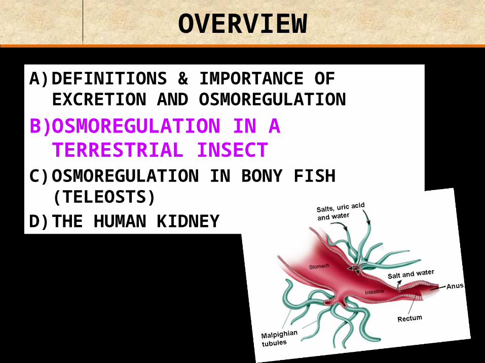
OVERVIEW
A) DEFINITIONS & IMPORTANCE OF EXCRETION AND OSMOREGULATION
B) OSMOREGULATION IN A TERRESTRIAL INSECT
C) OSMOREGULATION IN BONY FISH (TELEOSTS)
D) THE HUMAN KIDNEY

Definitions
Excretion – the elimination of waste metabolic substances
from the body which if permitted to accumulate would prevent the maintenance of a steady state
CO2
urea

Definitions
Egestion - the elimination of waste substances,
mainly undigested food, which have never been involved in the metabolic activities of cells
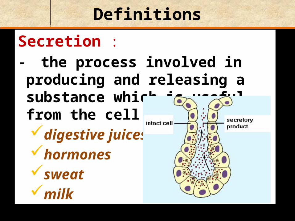
Definitions
Secretion :- the process involved in producing and
releasing a substance which is useful, from the cell e.g. digestive juiceshormonessweatmilk
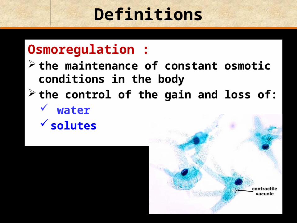
Definitions
Osmoregulation : the maintenance of constant osmotic conditions
in the body the control of the gain and loss of:
water solutes

Importance of excretion and osmoregulation
1) Removal of unwanted by-products of metabolic pathwaysis important to prevent unbalancing
chemical equilibria of reactionse.g. A + B → C + D- constant synthesis of C requires constant
removal of D

Importance of excretion and osmoregulation
2) Removal of toxic wastes e.g. urea, ammonia
Ammonia
Protein in
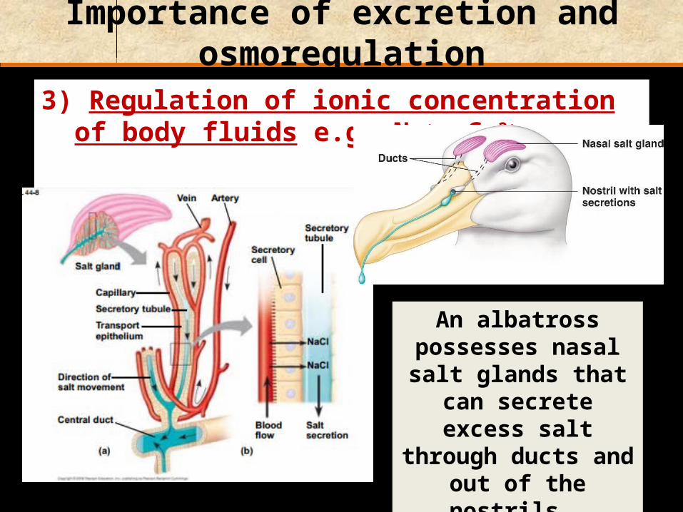
Importance of excretion and osmoregulation
3) Regulation of ionic concentration of body fluids e.g. Na+, Ca2+
An albatross possesses nasal salt glands that can
secrete excess salt through ducts and out of
the nostrils.

Importance of excretion and osmoregulation
4) Regulation of water content of body fluids
5) Regulation of the pH of the body fluids
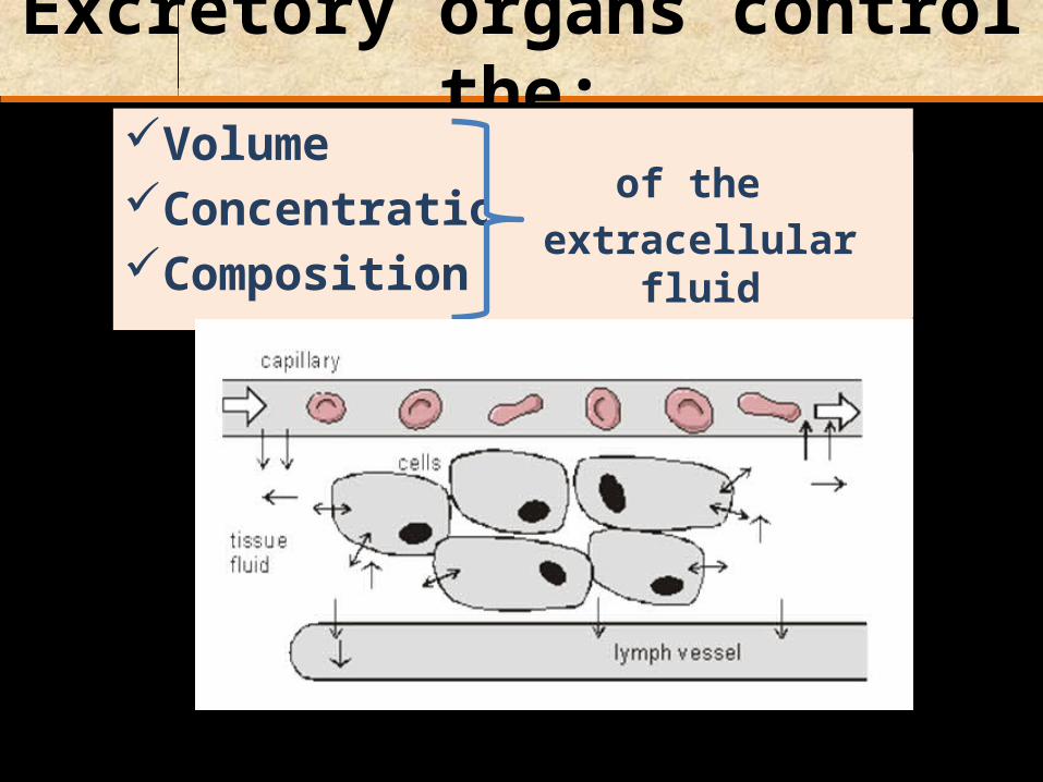
Excretory organs control the:VolumeConcentrationComposition
of the extracellular fluid

Animals in different environments have different excretory functions:
Terrestrial animals must conserve:
Freshwater animals have to: conserve salts excrete excess water
salts + water

Source of the total amount of excreted nitrogen
5%
A variety of animals also excrete small quantities of:
creatine creatinine
95%

Ammonia: is the immediate nitrogenous waste of
deamination
amino acidNH3
ammonia

PROTEINS
Amino acids
NUCLEIC ACIDS
Ammonia Urea Uric acid
Most aquatic animals
Nitrogenous bases
Amino groups
Birds, reptiles, insects
Mammals, most amphibians, sharks,
marine bony fish


Ammonia:Explain why ammonia cannot
be stored in the body.Highly toxic
Large volumes of water are needed for ammonia to be excreted. Why?
To make it less toxic.

Urea is less toxic & soluble than ammonia
Urea is about 100,000 times less toxic than ammonia.
Urea forms during the
Ornithine Cycle

Bird droppings = faeces + nitrogenous waste
Car covered in bird droppings.

Why is uric acid an ideal excretory product for terrestrial organisms
(e.g. insects, reptiles & birds) which produce shelled eggs?
Uric acid can be stored in cells without producing toxic or harmful osmoregulatory effects.

Uric acid is stored in the allantois.
Uric acid: is largely insoluble
in water can be excreted as
a paste with little water loss
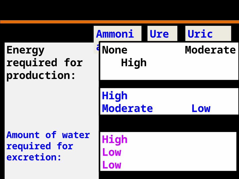
Energy required for production:
Amount of water required for excretion:
Toxicity of waste :
Ammonia Urea Uric acidNone Moderate High
High Moderate Low
High Low Low

Correlation with habitatAmmonia: Freshwater
Uric acid: Terrestrial
Urea: Marine/Terrestrial

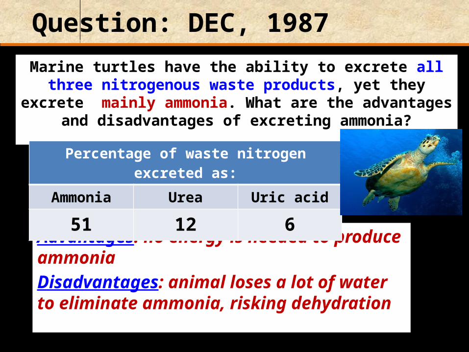
Question: DEC, 1987
The earthworm, although terrestrial, excretes mainly ammonia. How does this affect the
worm’s habitat preferences?
Must inhabit moist environments.
Question: DEC, 1987
Marine turtles have the ability to excrete all three nitrogenous waste products, yet they excrete mainly
ammonia. What are the advantages and disadvantages of excreting ammonia?
Advantages: no energy is needed to produce ammoniaDisadvantages: animal loses a lot of water to eliminate ammonia, risking dehydration
Percentage of waste nitrogen excreted as:Ammonia Urea Uric acid
51 12 6
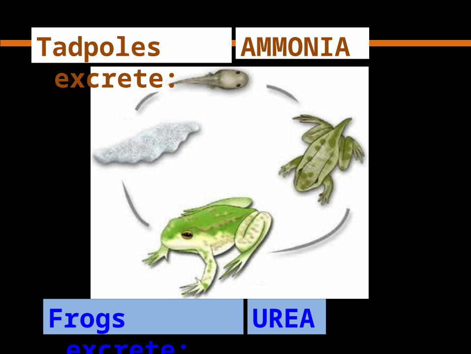
AMMONIATadpoles excrete:
UREAFrogs excrete:

Question: [MAY, 2010]
1. What are nitrogenous wastes? Name a biochemical process that produces nitrogenous waste. (2)Nitrogenous wastes are substances produced as a result of metabolism that are not required by the body and contain nitrogen. Deamination / break down of nucleic acids.
2. Name ONE organism that excretes nitrogenous wastes as urea and
ONE organism that excretes nitrogenous wastes as ammonia: (2)Urea: mammal / marine bony fish / frogAmmonia: protozoans / Amoeba / freshwater bony fish / tadpole
OVERVIEW
A) DEFINITIONS & IMPORTANCE OF EXCRETION AND OSMOREGULATION
B) OSMOREGULATION IN A TERRESTRIAL INSECT
C) OSMOREGULATION IN BONY FISH (TELEOSTS)D) THE HUMAN KIDNEY

How are insects adapted to avoid water loss?
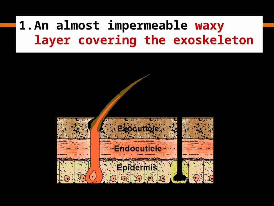
1. An almost impermeable waxy layer covering the exoskeleton

2. Spiracles are the only openings to the body for gaseous exchange
SpiracleSpiracle

3. Valve-like structures and hairs in spiracles reduce water loss
Fully open ant spiracle. A nearly closed grasshopper spiracle; the black areas are the valves.

4. The excretory product is semi-solid
insect droppings
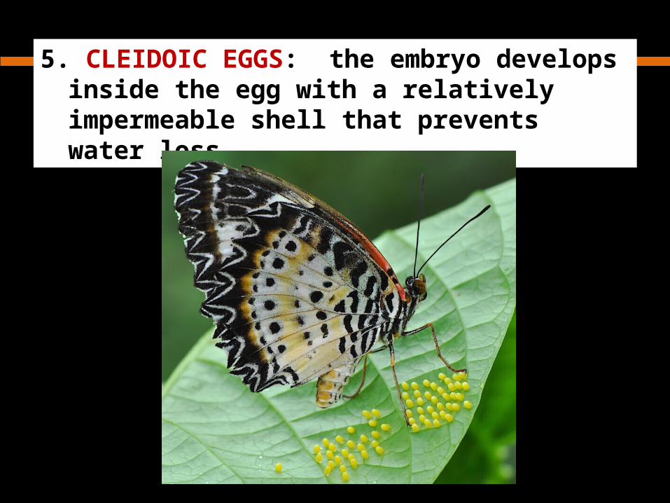
5. CLEIDOIC EGGS: the embryo develops inside the egg with a relatively impermeable shell that prevents water loss

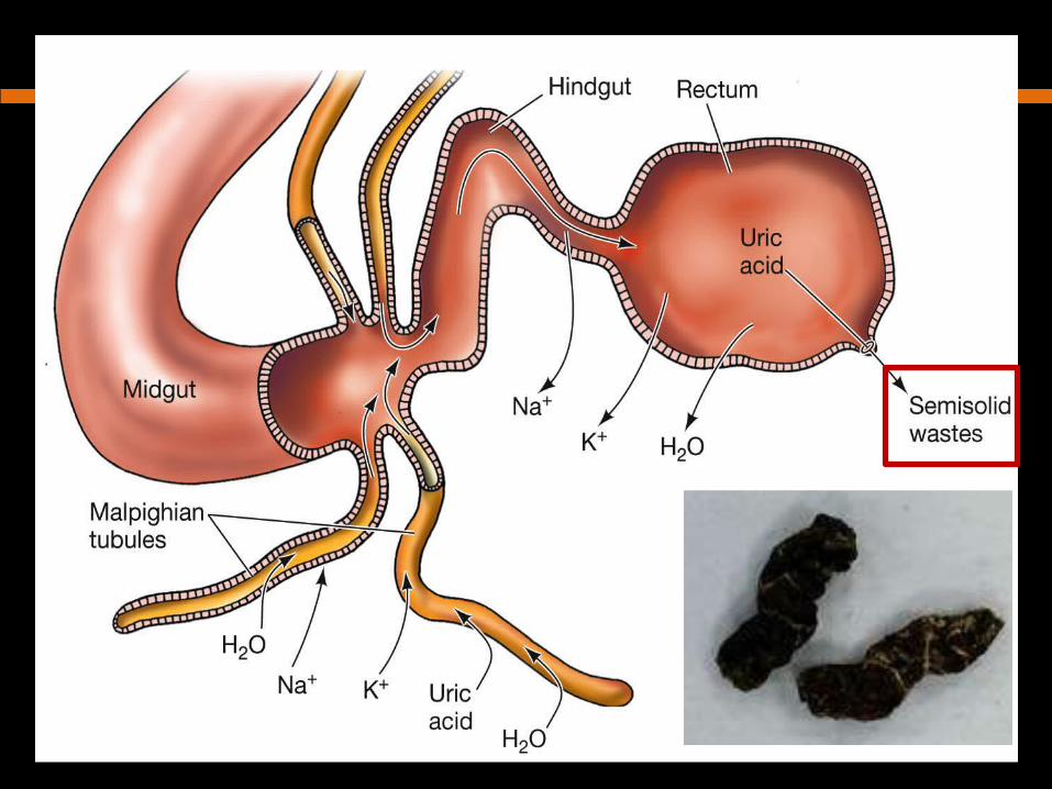
Malpighian Tubules in insects:
Function: excrete uric acid
Location: - lie in the abdomen- open into the
hindgut at its junction with the midgut

Malpighian Tubules are:
blind-ending tubules of the hindgut of insects

Tubules vary in :
Shape:long & slender orshort and compact
Number: a pair - several hundred
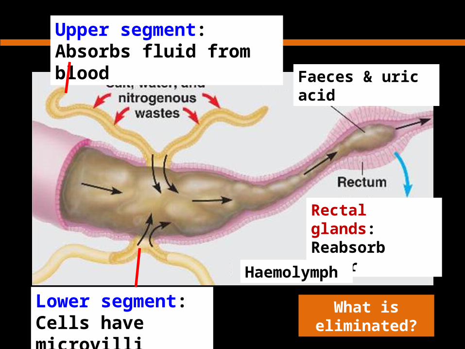
Figure 44.12
Upper segment:Absorbs fluid from blood
Lower segment:Cells have microvilli
Rectal glands:Reabsorb water
Haemolymph
What is eliminated?
Faeces & uric acid

OVERVIEW
A) DEFINITIONS & IMPORTANCE OF EXCRETION AND OSMOREGULATION
B) OSMOREGULATION IN A TERRESTRIAL INSECT
C) OSMOREGULATION IN BONY FISH (TELEOSTS)
D) THE HUMAN KIDNEY

Fish are osmoregulators:control concentration of body fluidsuse energy to regulate
Osmoconformereg. marine
invertebratesosmoregulator
Hypertonic solution
Hypotonic solution
Isotonic solution
osmoregulator

Bony Fish can be:
FRESHWATER Eliminate
ammonia
MARINE Eliminate urea
&Trimethylamine oxide
Trimethylamine oxide = fish odour

Why is the nitrogenous waste product different in the two groups of fish?
FRESHWATER fish afford to lose
water
MARINE fish cannot afford
to lose water

Gills are permeable to:
1. Water2. Ions

Freshwater Bony Fish:
Hypertonic body fluids
Not salty
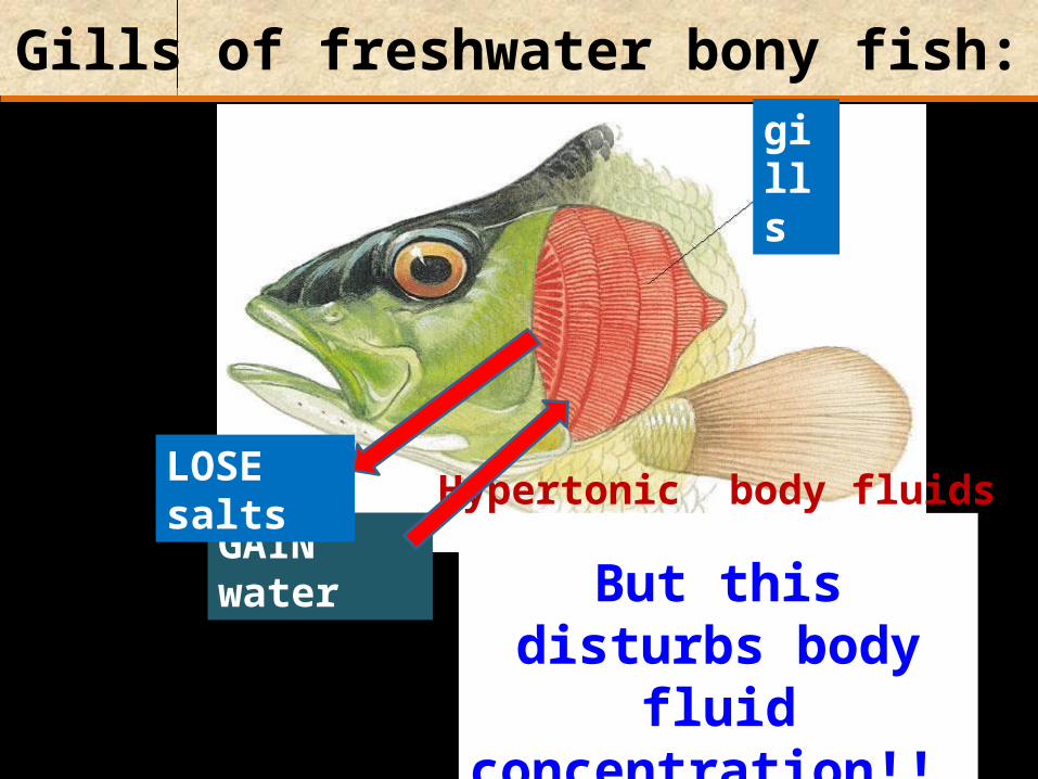
Gills of freshwater bony fish:
Hypertonic body fluids
GAIN water
But this disturbs body fluid concentration!!
LOSE salts
gills
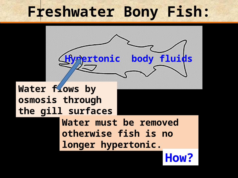
Freshwater Bony Fish:
Hypertonic body fluids
Water flows by osmosis through the gill surfaces
Water must be removed otherwise fish is no longer hypertonic.
How?

Freshwater Bony Fish:
Hypertonic body fluids
The fish discharges copious quantities of very dilute urine, few salts lost
Nitrogenous waste: Ammonia
No drinking

Kidneys of freshwater bony fish:
contain many large Malpighian bodies, with large glomeruli
high rate of filtration produces a large volume of glomerular filtrate

How does the fish remain hypertonic?
GAINS salts:
Selective reabsorption in kidney
Hypertonic body fluids
Selective uptake of Cl- at gills
In food

Marine Fish:
Hypotonic body fluids
SALTY
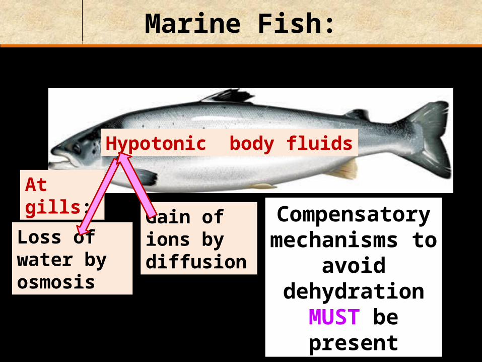
Marine Fish:
Hypotonic body fluids
Loss of water by osmosis
Compensatory mechanisms to
avoid dehydration MUST be present
At gills:
Gain of ions by diffusion

Marine Fish DRINK sea water. WHY?
Hypotonic body fluids
To replace water lost
But this means SALTS are gained too. What happens to salts?
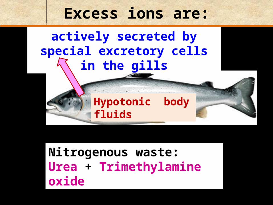
Excess ions are:
Nitrogenous waste:Urea + Trimethylamine oxide
Hypotonic body fluids
actively secreted by special excretory cells in the gills

Some marine bony fish:
have NO glomeruli at all, and so do not filter their blood
urine is isotonic with the body fluids


Question: [SEP, 2010]
Fish do not need to convert ammonia into urea. Suggest ONE reason for this. (1)Freshwater bony fish eliminate ammonia by adding large quantities of water to make it less toxic. As they can afford to lose a lot of water, they eliminate their nitrogenous waste in the form of ammonia rather than urea.

OVERVIEW
A) DEFINITIONS & IMPORTANCE OF EXCRETION AND OSMOREGULATION
B) OSMOREGULATION IN A TERRESTRIAL INSECT
C) OSMOREGULATION IN BONY FISH (TELEOSTS)
D) THE HUMAN KIDNEY

The kidneys contribute to homeostasis
Let us see how:
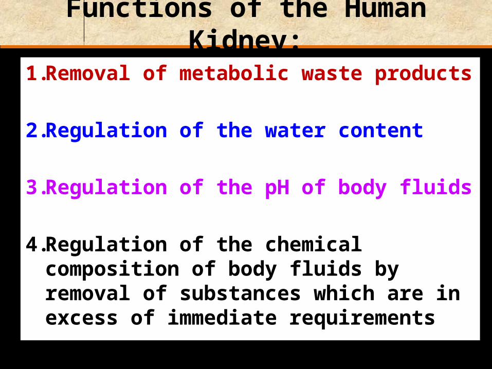
Functions of the Human Kidney:
1. Removal of metabolic waste products
2. Regulation of the water content
3. Regulation of the pH of body fluids
4. Regulation of the chemical composition of body fluids by removal of substances which are in excess of immediate requirements

Position and structure of kidneys

External structure of a Pig Kidney

Kidneys are surrounded by a fibrous capsule:

Kidneys are surrounded by a fibrous capsule:
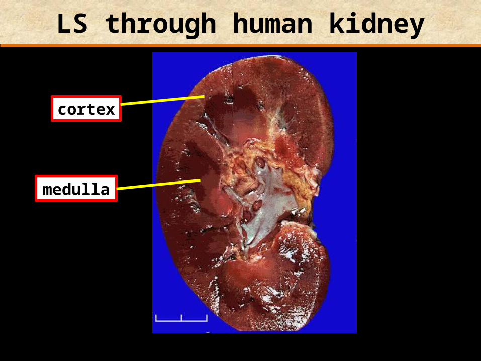
LS through human kidney
medulla
cortex

LS through human kidney
the apex of each pyramid
papilla

The renal artery branches inside kidney
Renal artery
Ureter
Renal vein
Each capillary supplies blood to hundreds of thousands of tiny filtration units called nephrons
Let’s have a look at a nephron!!!

Two types of nephron:
Juxtamedullary nephron
Cortical nephron
Juxta = close to
CORTEX
MEDULLA
Loop of Henle
Collecting duct
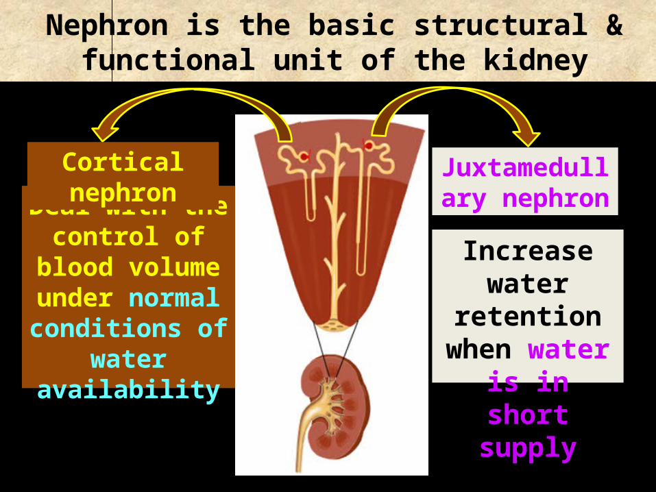
Nephron is the basic structural & functional unit of the kidney
Deal with the control of blood volume under
normal conditions of water
availability
Increase water retention when
water is in short supply
Cortical nephron Juxtamedullary nephron
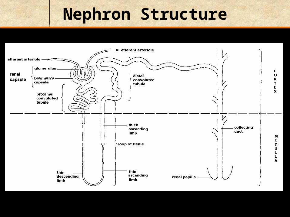
Nephron Structure

vasa recta
Slow blood flow: important to produce a
concentrated urine

The nephron1.5 million per kidney
collectingduct
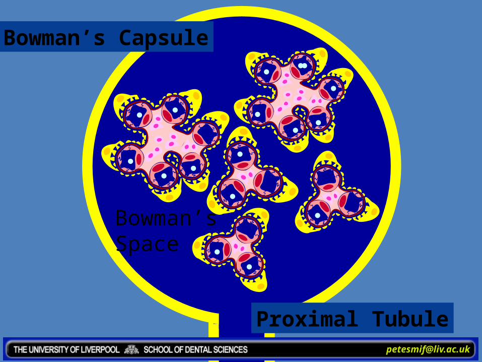
Bowman’scapsule
distaltubule
loop ofHenle
proximaltubule

The nephronblood supply
peritubularcapillaries
VasaRecta
glomerulus
branch of renalartery
afferent arterioles
efferent arterioles
branch of renalvein
The glomerular capillariesdrain into efferent arteriolesnot venules.‘Portal System’

Three key process in urine formation:
Ultrafiltration
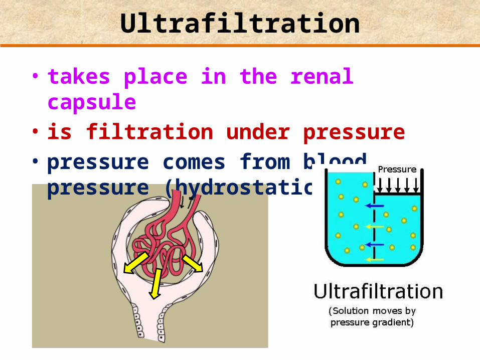
Ultrafiltration
• takes place in the renal capsule• is filtration under pressure• pressure comes from blood pressure
(hydrostatic pressure)

Glomerular Filtrate (GF): is the filtered fluid
chemical composition is similar to blood plasma, containing:-
GlucoseAmino acidsVitaminsIonsNitrogenous wasteSome hormones Water
Glomerular filtrate

Key Words!!
Nephron:structure in the kidney that acts as a microscopic filtration unit
Glomerulus:dense mass of very fine blood capillaries at the nephron that act as a filter

Key Words!!
Bowman’s capsule:cup-shaped part of the nephron that holds a glomerulus and collects the products of filtration from it
Glomerular filtrate:liquid removed from the blood by filtration in the kidney
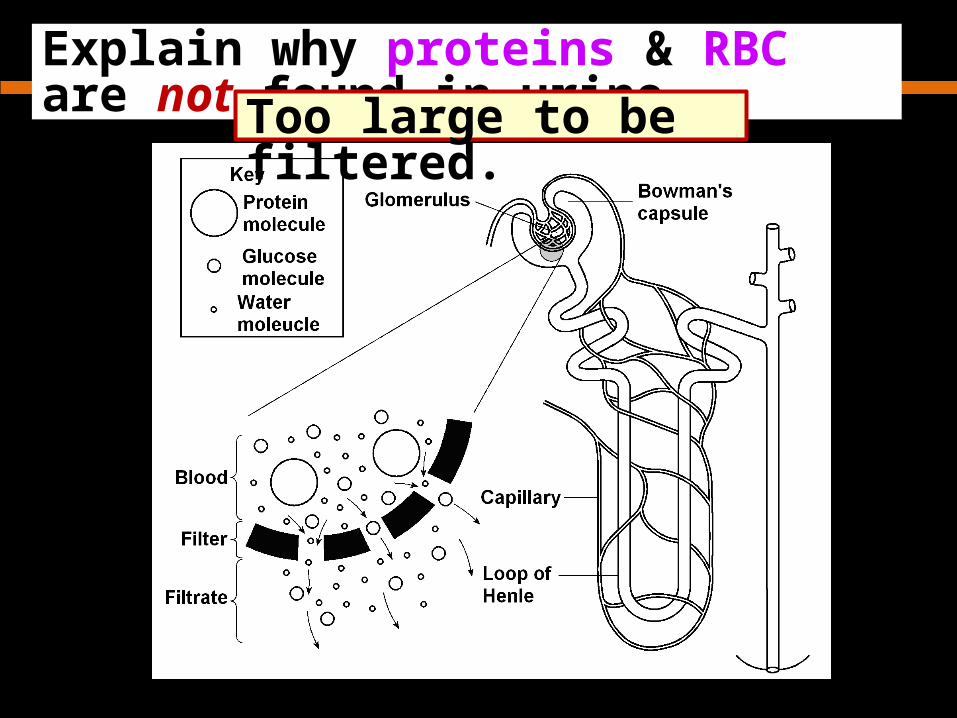
Explain why proteins & RBC are not found in urine. Too large to be filtered.

But can blood ever be detected in urine?
YES. But, this shows that something is
wrong .

Ultrafiltration takes place through three layers:
1) Endothelium of the blood capillary
2) Basement membrane of the blood capillaries
3) Epithelium of the renal capsule

Cells lining the Bowman’s capsule:
Podocyte
Squamous epithelium
Podocytes are highly
modified for filtration
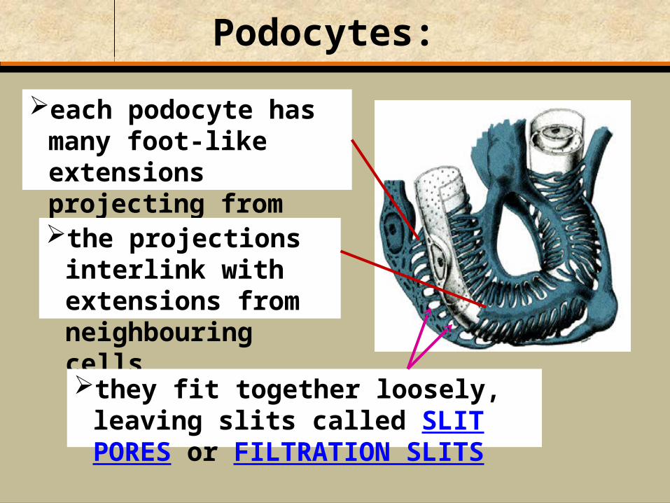
Podocytes:
each podocyte has many foot-like extensions projecting from its surface
the projections interlink with extensions from neighbouring cells
they fit together loosely, leaving slits called SLIT PORES or FILTRATION SLITS
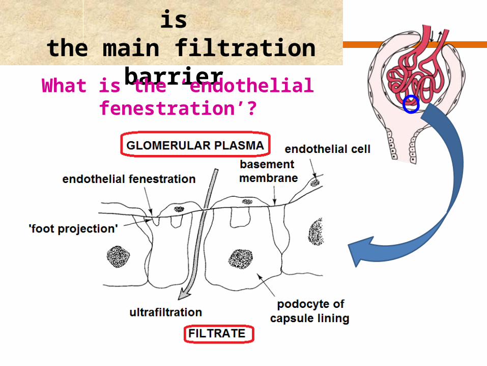
The basement membrane is the main filtration barrier
What is the ‘endothelial fenestration’?
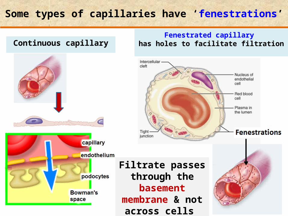
Some types of capillaries have ‘fenestrations’
Fenestrated capillary has holes to facilitate filtrationContinuous capillary
Filtrate passes through the
basement membrane & not across cells

Basementmembrane
Fenestrated capillaries
(capillaries with windows)(capillaries with windows)Permeableto substances< 100 nm
endothelial cell
fenestration
nucleus
Filtration Barrier

mesangial cells
podocyte
slit pore

glucoseamino acids
(basement membrane)
podocyte slit pore
Na+
--
-
-
-
---
--
-
-
-
-
-
-
--
--
- -
-
-
--
--
-
Limited permeability to molecules between
7000 > mwt > 70000 Da4 nm > diameter > 8 nm
Freely permeable tosmall moleculesmwt < 7000 Da
diameter < 4 nm
Not permeable to large moleculesmwt > 70000 Dadiameter > 8 nm
Water Permeablealbumin60000 Da
completelyexcluded…
because of –ve charge
endothelial cellfenestration

Factors affecting the glomerular filtration rate (GFR)
The filtration pressure forcing fluid out of the glomerulus depends on the:
1. hydrostatic pressure of the blood
2. pressure of the glomerular filtrate
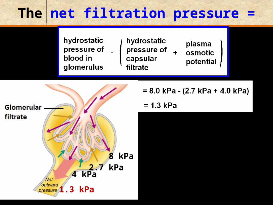
The net filtration pressure =
1.3 kPa
8 kPa2.7 kPa
4 kPa
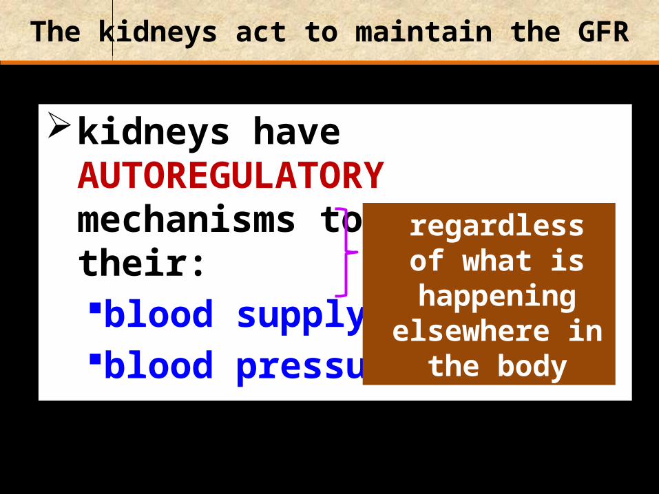
The kidneys act to maintain the GFR
kidneys have AUTOREGULATORY mechanisms to maintain their:blood supply blood pressure
regardless of what is happening elsewhere in the
body

Three ways to increase the filtration rate:
1. Raising blood pressure Efferent arteriole
2. Dilating the afferent arterioles (to decrease the resistance to the flow of blood into the glomerulus)
3. Constricting the efferent arterioles
Afferent arteriole
Filtration pressure
GFR maintained

Afferent arteriole narrow
LOW pressure HIGH pressure
Efferent arteriole
wide
Dilating the afferent arterioles & Constricting the efferent arterioles

BUT when arterial pressure falls too low, however, the kidney fails to produce urine
Arterial pressure 8 kPa
Plasma osmotic pressure 4 kPa
Glomerular capsule pressure 2.7 kPa6.7 kPa

Function of the nephron is to :
further waste substances may be added to the tubules by active secretion from the blood capillaries
selectively reabsorb substances useful to the body
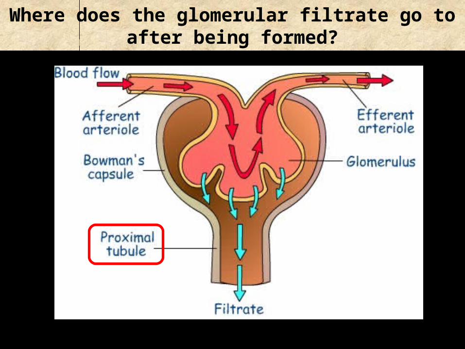
Where does the glomerular filtrate go to after being formed?

Selective reabsorption in the proximal convoluted tubule
In humans:Glomerular filtrate production: 125 cm3 min-1
Urine production: 1 cm3 min-1
24 cm3
100 cm3
Urine 1 cm3
125 cm3

The Proximal Convoluted Tubule longest (14 mm) and widest (60 m) part of the
nephron
carries filtrate from Bowman’s capsule to loop of Henle CORTEX
MEDULLA

Proximal Convoluted Tubule is composed of:
a single layer of cuboidal epithelial cells with extensive microvilli forming a ‘brush border’ on the inside surface of the tubule
Figure 44.9
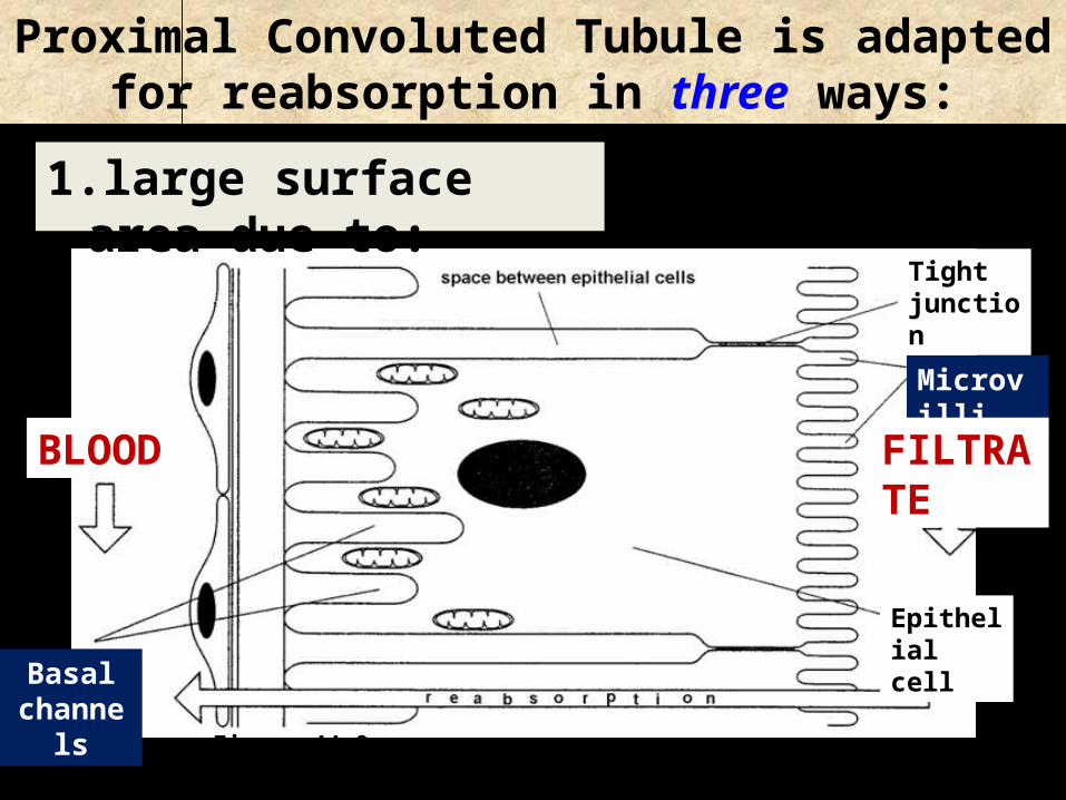
Proximal Convoluted Tubule is adapted for reabsorption in three ways:
1. large surface area due to:
Figure 44.9
Microvilli
Basal channels
BLOOD FILTRATE
Tight junction
Epithelial cell

Proximal Convoluted Tubule is adapted for reabsorption:
Figure 44.9
2. numerous mitochondria (M)

Proximal Convoluted Tubule is adapted for reabsorption:
Figure 44.9
3.closeness of blood capillaries
blood capillaryGlomerular filtrate
MicrovilliCuboidal epithelium
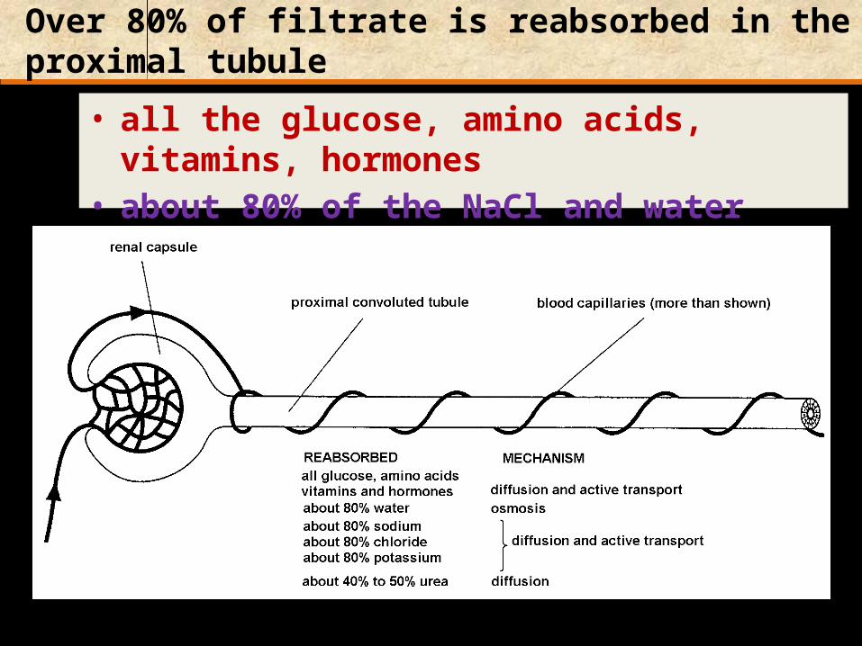
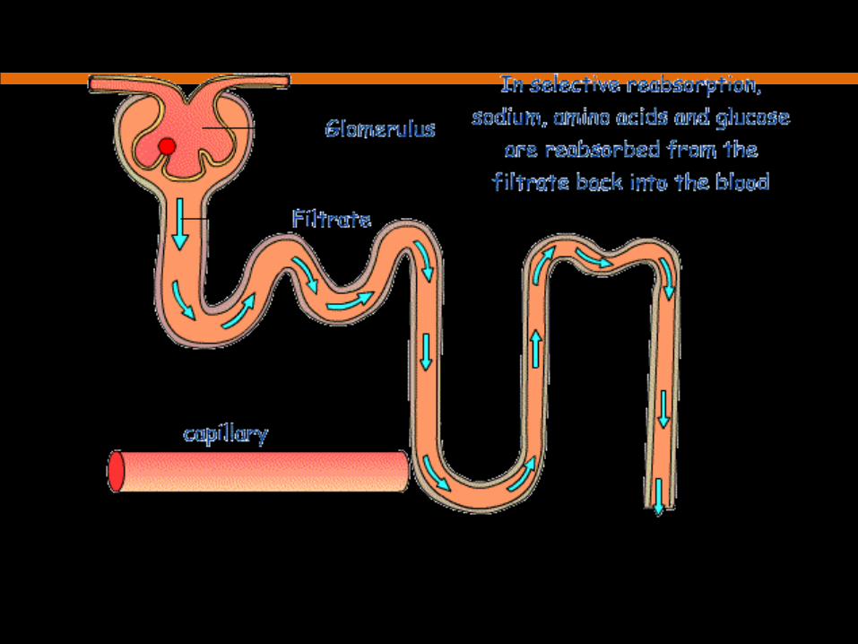
Over 80% of filtrate is reabsorbed in the proximal tubule
• all the glucose, amino acids, vitamins, hormones • about 80% of the NaCl and water
Figure 44.9

Fig. 16 Selective reabsorption of sodium in the proximal convoluted tubule
Figure 44.9
1

Selective reabsorption of glucose in the proximal convoluted tubule
Figure 44.9
Secondary Active
Transport

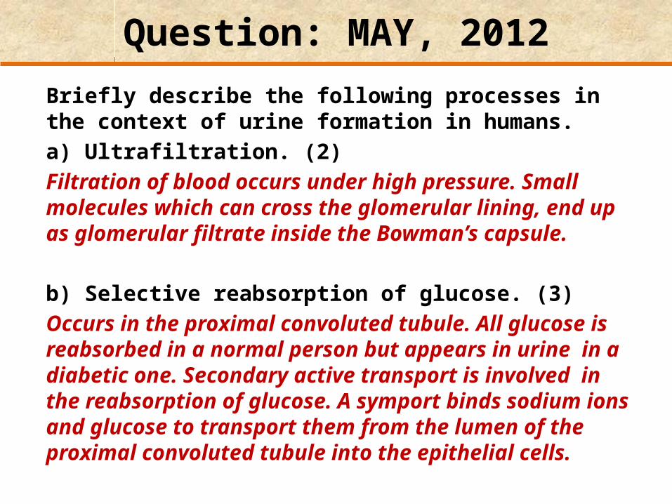
Question: MAY, 2012
Briefly describe the following processes in the context of urine formation in humans. a) Ultrafiltration. (2)Filtration of blood occurs under high pressure. Small molecules which can cross the glomerular lining, end up as glomerular filtrate inside the Bowman’s capsule.
b) Selective reabsorption of glucose. (3) Occurs in the proximal convoluted tubule. All glucose is reabsorbed in a normal person but appears in urine in a diabetic one. Secondary active transport is involved in the reabsorption of glucose. A symport binds sodium ions and glucose to transport them from the lumen of the proximal convoluted tubule into the epithelial cells.

THE LOOP OF HENLE
Function: to conserve water
the concentration of urine produced is directly related to the:length of the loop of Henlethickness of the medulla
relative to the cortex
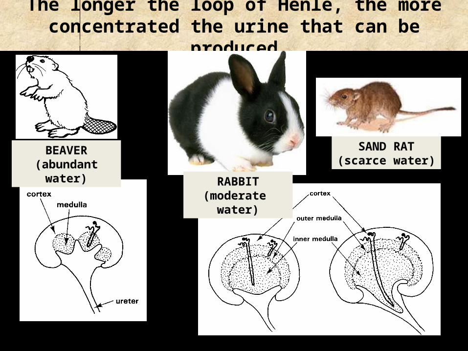
The longer the loop of Henle, the more concentrated the urine that can be produced
BEAVER(abundant water)
RABBIT(moderate water)
SAND RAT(scarce water)

Question: [MAY, 2010]
Use your knowledge of biology to describe the selective advantage of the following adaptation.Desert rats have a long loop of Henle. (5)The loop of Henle acts as a counter-current multiplier. Fluid moves in opposite directions in the descending and ascending limbs. The ascending limb is permeable to salts which contribute towards a concentrated medulla. As water moves down the descending limb, it moves out into the vasa recta.Desert rats need to conserve water. Thus having a long loop of Henle enables them to extract as much water as possible out of the glomerular filtrate as there is more time for reabsorption.

Question: [MAY, 2002]
The table below gives the thickness of the medulla in relation to the rest of the kidney in a number of mammals. The maximum urine concentration for each mammal is also given. The data suggest that maximum urine concentration increases with relative thickness of the medulla.
Mammal Relative thickness of medulla
Maximum urine concentration in arbitrary units
Beaver 1.0 52Human 2.6 140Kangaroo rat
7.8 550
Species X 9.8 940

a) Why is such a relation between urine concentration and the relative thickness of the medulla observed?
(1)The thicker the medulla, the higher the urine concentration produced due to more chance for water reabsorption.
Mammal Relative thickness of medulla
Maximum urine concentration in arbitrary units
Beaver 1.0 52Human 2.6 140Kangaroo rat 7.8 550
Species X 9.8 940

b) What habitat is species X likely to inhabit? (1)
Desert / dry habitat
Mammal Relative thickness of medulla
Maximum urine concentration in arbitrary units
Beaver 1.0 52Human 2.6 140Kangaroo rat 7.8 550
Species X 9.8 940
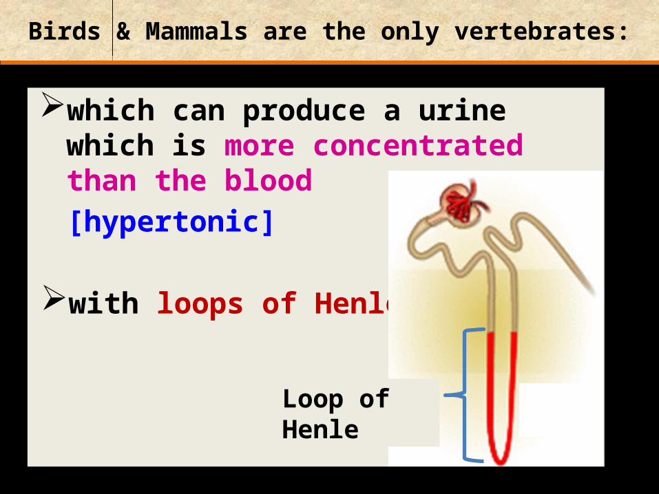
Birds & Mammals are the only vertebrates:
which can produce a urine which is more concentrated than the blood[hypertonic]
with loops of Henle
Loop of Henle
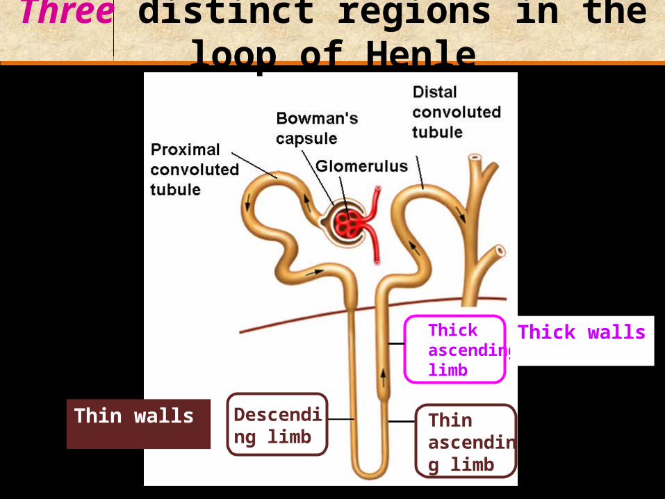
Three distinct regions in the loop of Henle
Thin ascending limb
Descending limb
Thick ascending limb
Thin walls
Thick walls
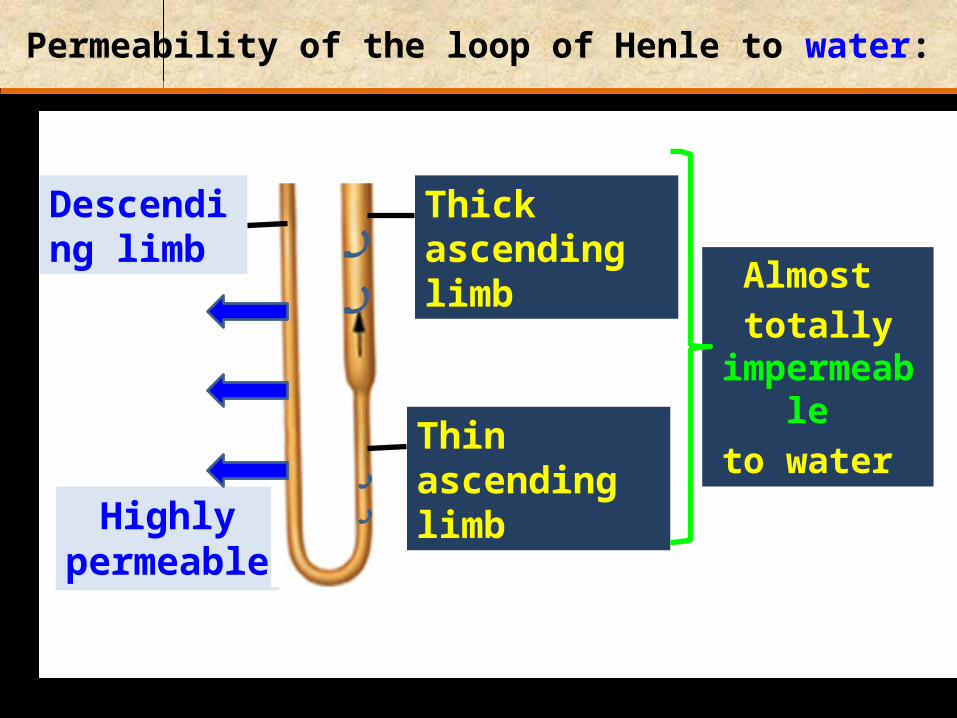
Permeability of the loop of Henle to water:
Highly permeable
Descending limb
Almost totally
impermeable to water Thin ascending
limb
Thick ascending limb
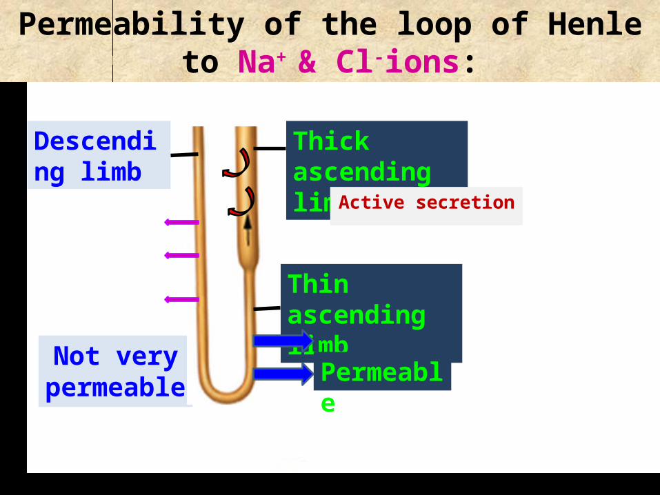
Permeability of the loop of Henle to Na+ & Cl-
ions:
Not very permeable
Descending limb
Thin ascending limb
Thick ascending limb
Permeable
Active secretion

What happens to the concentration of the fluid in the ascending limb as it reaches the
distal convoluted tubule?
The fluid becomes very dilute
Distil convoluted tubule
Reason:IONS are lost
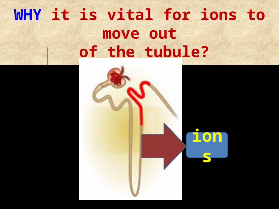
WHY it is vital for ions to move out of the tubule?
ions

To create an Osmotic Gradient From Cortex to Medulla
PelvisMedulla
Cortex
The outer layer of the kidney is isotonic with the blood: ~300 milliosmoles/liter
The innermost layer (medulla) is very hypertonic: ~1200 milliosmoles/liter
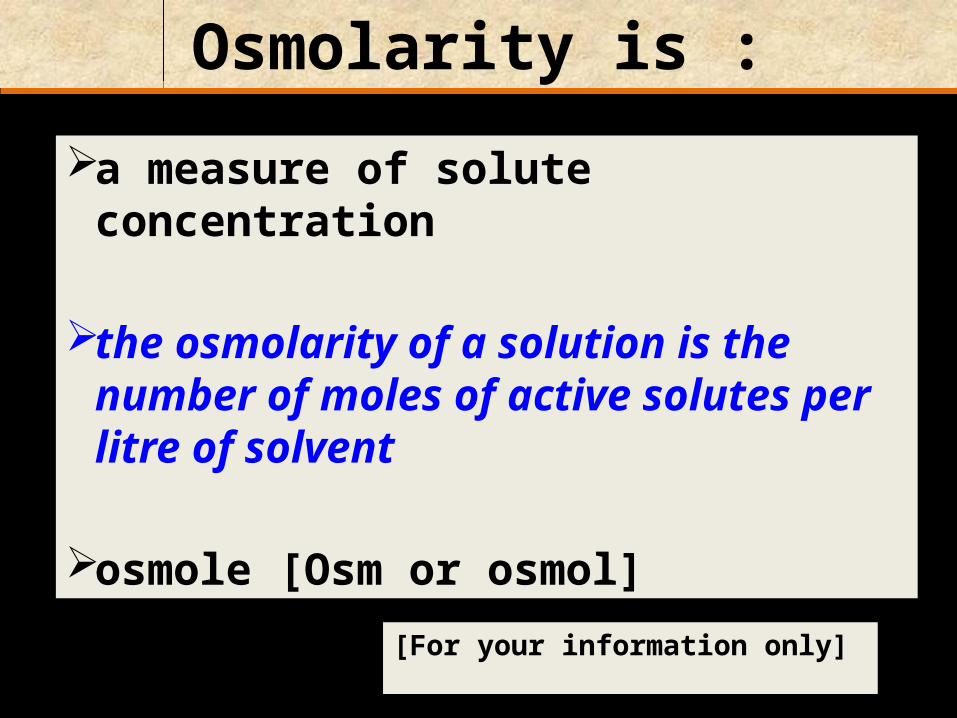
Osmolarity is :
a measure of solute concentration
the osmolarity of a solution is the number of moles of active solutes per litre of solvent
osmole [Osm or osmol][For your information only]
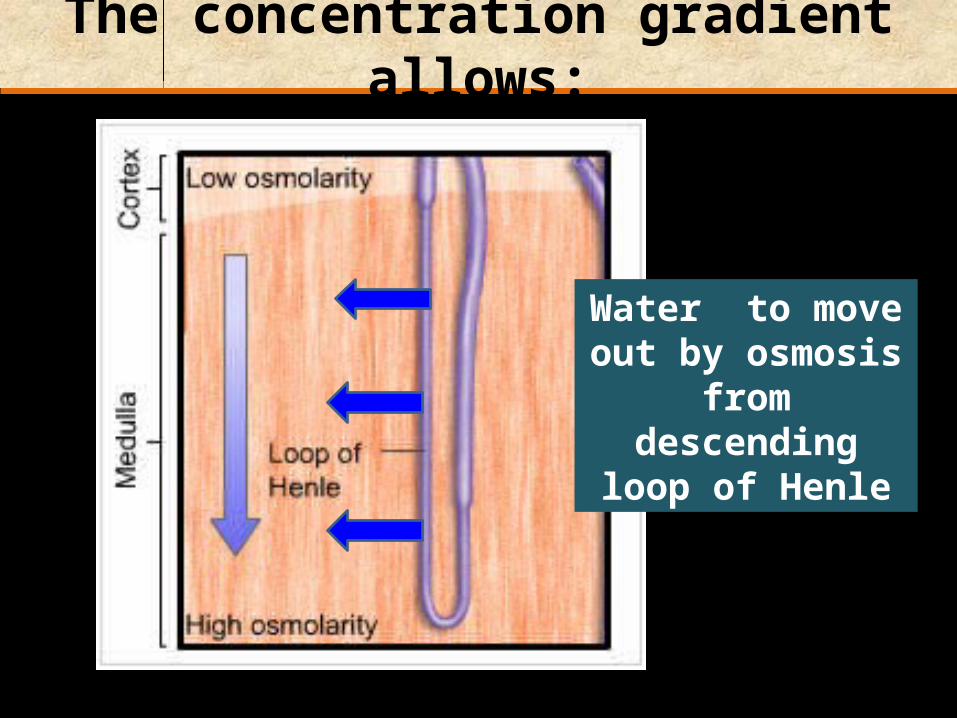
The concentration gradient allows:
Water to move out by osmosis from
descending loop of Henle
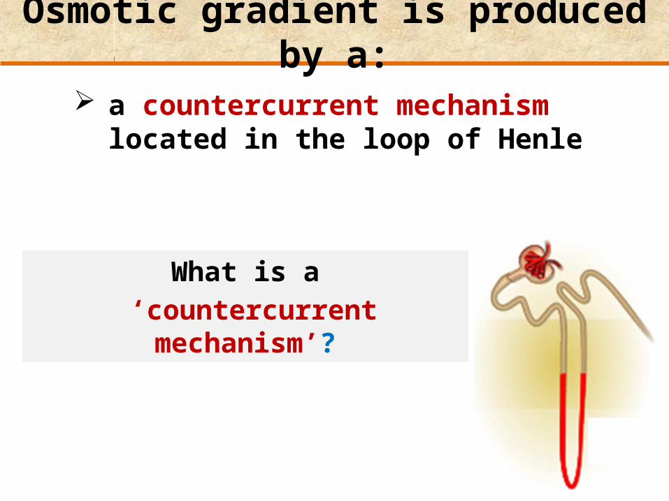
Osmotic gradient is produced by a:
a countercurrent mechanism located in the loop of Henle
What is a ‘countercurrent mechanism’?

Countercurrents exist when :fluids flow in opposite directions in
parallel and adjacent tubes
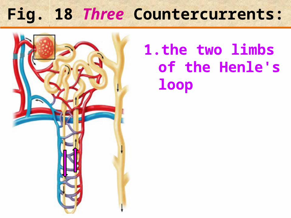
Fig. 18 Three Countercurrents:
1. the two limbs of the Henle's loop

Fig. 18 Three Countercurrents:
1. the two limbs of the Henle's loop
2. the two limbs of the vasa recta
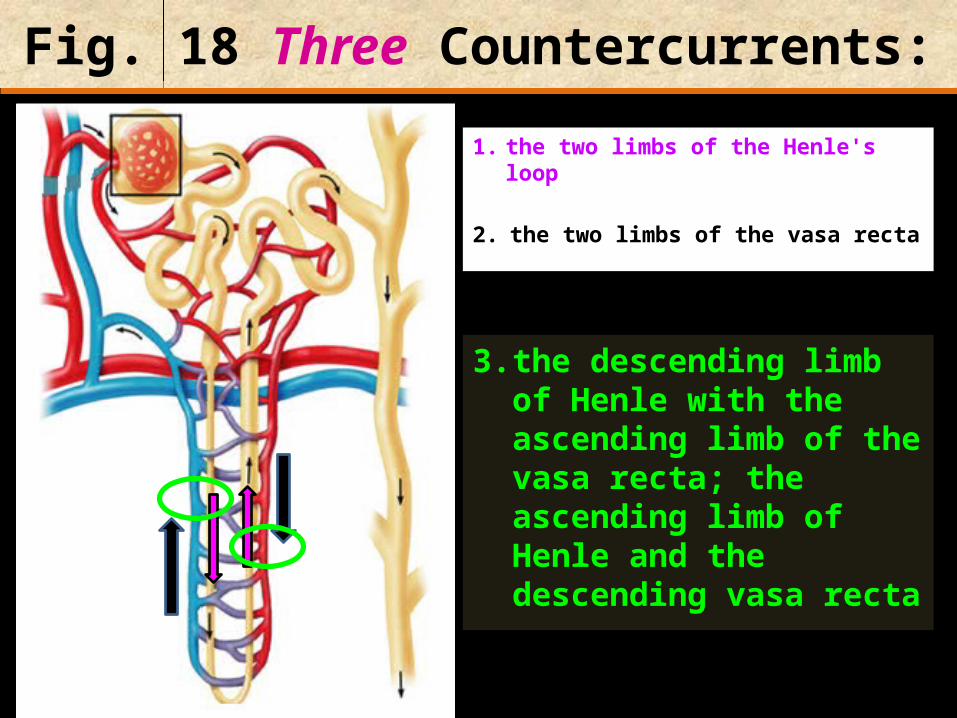
Fig. 18 Three Countercurrents:
1. the two limbs of the Henle's loop
2. the two limbs of the vasa recta
3. the descending limb of Henle with the ascending limb of the vasa recta; the ascending limb of Henle and the descending vasa recta

The loop of Henle as a countercurrent multiplier
countercurrent refers to the direction of urine flow in the descending versus the ascending limbs of the loop
multiplier refers to the ability of this system to create a concentration gradient in the renal medulla


CortexWater leaves - ion concentration in filtrate increases
Filtrate reaches maximum concentration
Chloride ions out (sodium follows) -ion concentration in filtrate decreases
Medulla

To ureter
Collecting duct •Several nephrons empty into one collecting duct.
•The collecting duct passes through the progressively more concentrated medulla, losing water by osmosis. This water is reabsorbed by the capillaries.
•This water is conserved, and a highly concentrated urine is produced.
Water reabsorbed into vasa recta, urine becomes more concentrated
Cortex
Medulla

Question: [SEP, 2009]
Briefly describe the role of each of the following in osmoregulation in humans:
i) The descending limb of the Loop of Henle; (2)
Is permeable to water. Functions towards water conservation.
ii) The ascending limb of the Loop of Henle; (2)Is relatively impermeable to water but permeable to salts. The tissue fluid inside the medulla becomes concentrated as salts move out of the ascending limb. This causes water to be drawn out of the descending limb.

Question: MAY, 2012
The diagram below shows the simplified structure of a human nephron. the loop of Henle
Substance Quantity passing through P
Quantity passing through Q
% reabsorbed
Water 180 dm3 1.5 dm3 99.17%Glucose 180 g 0 g 100%Urea 53 g 25 g 52.8%
The table below represents the quantities of water, glucose and urea passing through P and Q over a period of time, while the last column shows the percentage reabsorption during the same period of time.

Question: MAY, 2012
a) Relate the role of structure R to the filtrate composition as it passes through Q. (5)Structure Q is permeable to water. Water is reabsorbed by the vasa recta as fluid passes through Q. This is possible because the ascending limb creates the ideal concentration gradient within the medulla by losing ions. The thin ascending limb of Structure R is permeable to ions but impermeable to water. The thick ascending limb of Structure R allows ions to move actively out of it and is also impermeable to water. Loss of ions from the whole ascending limb, creates an ever increasing salt concentration on moving deeper into the medulla.

Question: MAY, 2012
Substance Quantity passing through P
Quantity passing through Q
% reabsorbed
Water 180 dm3 1.5 dm3 99.17%Glucose 180 g 0 g 100%Urea 53 g 25 g 52.8%
b) Explain the biological significance of the percentage reabsorption of water and urea. (3)Most of the water is reabsorbed to avoid dehydration.Only half of the urea is reabsorbed so that it contributes to the concentration of solutes in the medulla. A high solute concentration is needed to ensure reabsorption of water from the loop of Henle.

Vasa recta as countercurrent exchangers
• the countercurrent exchange of salt occurs in the vasa recta
1. Blood flowing into the medulla in the descending limb picks up salt from the hypertonic medulla
2. As the surrounding medullary fluid becomes more salty toward the papilla, more salt is picked up by the descending vasa recta limb

Vasa recta as countercurrent exchangers
3. But as the blood heads back up to the cortex in the ascending limb of the vasa recta, the interstitial fluid becomes less and less salty
4. This causes the gradient to reverse and salt diffuses back out of the vasa recta into the medulla

Vasa recta as countercurrent exchangers
3. But as the blood heads back up to the cortex in the ascending limb of the vasa recta, the interstitial fluid becomes less and less salty
4. This causes the gradient to reverse and salt diffuses back out of the vasa recta into the medulla

What is the importance of the vasa recta as an exchanger of salts?
1. to help conserve salt 2. keep the medulla hypertonic
OSMOREGULATION, ADH & URINE FORMATION

In this topic we mention TWO hormones that affect the kidneys:
Urine
ADH(antidiuretic
hormone)
Posterior pituitary
Adrenal cortex
Aldosterone
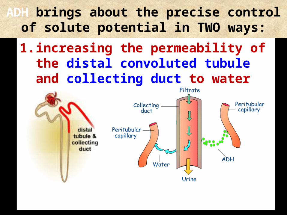
ADH brings about the precise control of solute potential in TWO ways:
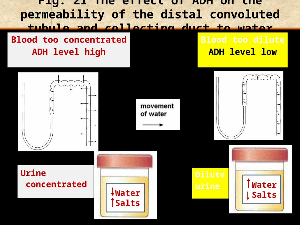
1. increasing the permeability of the distal convoluted tubule and collecting duct to water

2. increasing the permeability of the collecting duct to urea
1. Urea moves into medulla
2. Medulla becomes concentrated
3. Water moves out of descending limb

ADH is released when osmoreceptors:
detect a low level of water in blood
kidney
Water Salts
Fig. 21 The effect of ADH on the permeability of the distal convoluted tubule and collecting duct to water
Blood too concentratedADH level high
Blood too diluteADH level low
Dilute urine
Urine concentrated
Water Salts

Fig. 20 Aquaporins
H2O
H2O
H2O

Release of ADH from the posterior pituitary is inhibited by drinking
alcohol & caffeine.
How would this affect urination?
Increases
ADH

Failure to release sufficient ADH leads to a condition: DIABETES INSIPIDUS large quantities of dilute urine are produced
Water level regulation by negative feedback control

Water content of the blood normal
Water content of the blood HIGH
Water content of the blood LOW
Too much water drunk
Too much salt or sweating
Brain producesMore ADH
Urine output LOW
Brain produces Less ADH
Urine output HIGH
High volume of waterreabsorbed by kidney
Low volume of waterreabsorbed by kidney
(small volume of Concentrated urine)
(large volume of dilute urine)
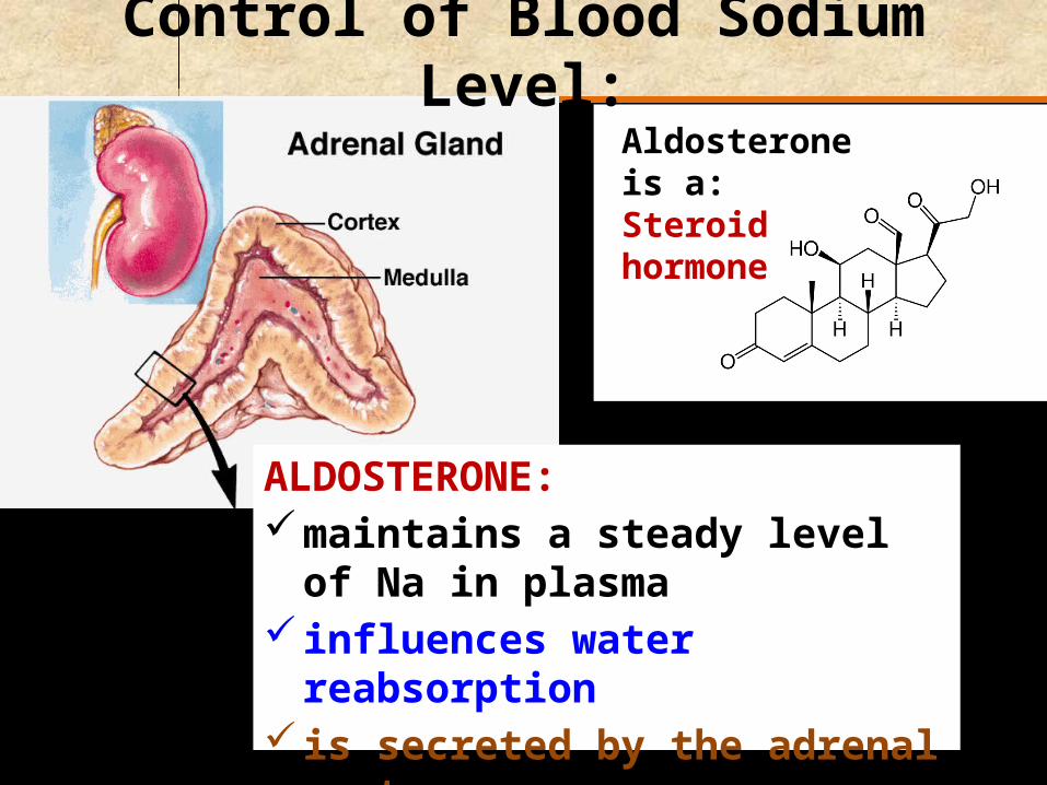
ALDOSTERONE:maintains a steady level of Na in
plasma influences water reabsorption is secreted by the adrenal cortex
Control of Blood Sodium Level:Aldosterone is a:Steroid hormone

A decrease in blood Na leads to a decrease in blood volume. WHY?
Because less water enters the blood by osmosis.
Less water = reduction in blood pressure

The decrease in pressure & volume: stimulates a group of secretory cells,
the juxtaglomerular complex situated between the:
distal convoluted and afferent arteriole
juxtaglomerular complex releases an enzyme called
RENIN

What is the function of renin?angiotensinogen angiotensin
Angiotensin releases aldosterone from the adrenal cortex
Aldosterone: travels in the blood to the distal
convoluted tubule stimulates the Na+/K+ pumps in the cells of
the tubule
Angiotensinogen [a plamsa protein] produced in the liver
Renin
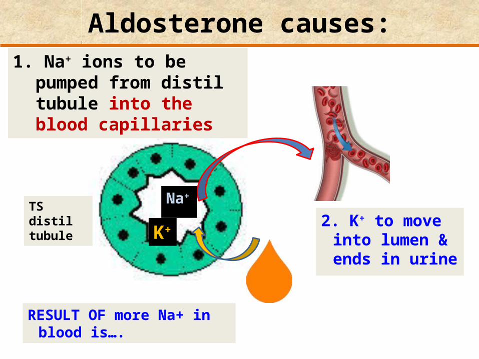
Aldosterone causes:
2. K+ to move into lumen & ends in urine
1. Na+ ions to be pumped from distil tubule into the blood capillaries
TS distil tubule K+
Na+
RESULT OF more Na+ in blood is….
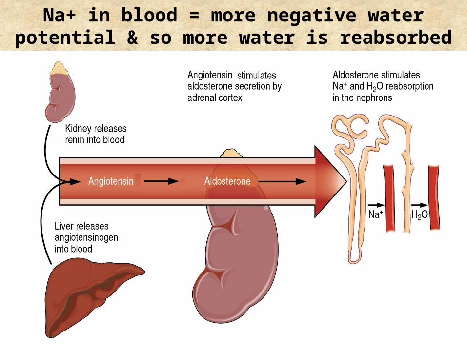
Na+ in blood = more negative water potential & so more water is reabsorbed
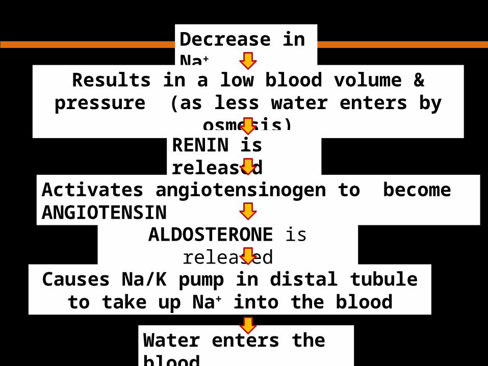
Decrease in Na+
Results in a low blood volume & pressure (as less water enters by osmosis)
Activates angiotensinogen to become ANGIOTENSIN
RENIN is released
ALDOSTERONE is released
Causes Na/K pump in distal tubule to take up Na+ into the blood
Water enters the blood


Question: [MAY, 2011]
1. The human kidney, in association with various hormones, plays a central role in the regulation of the chemical and physical characteristics of blood.
a) List THREE ways through which the human kidney may affect the
chemical composition of blood. (3)1) Through aldosterone, the kidney determines the amount of
sodium and potassium in the blood.
2) Through ADH, the kidney plays a role in the amount of water in the blood.
3) The kidney helps to keep the blood pH constant by secreting H+ or OH-.

b) Complete the table below by filling in the empty spaces with the appropriate answers: (3)
Hormone Site of production
Effect
Antidiuretichormone
Adrenal cortex
Stimulates excretion of potassium ions and reabsorption of sodium ions in the nephron
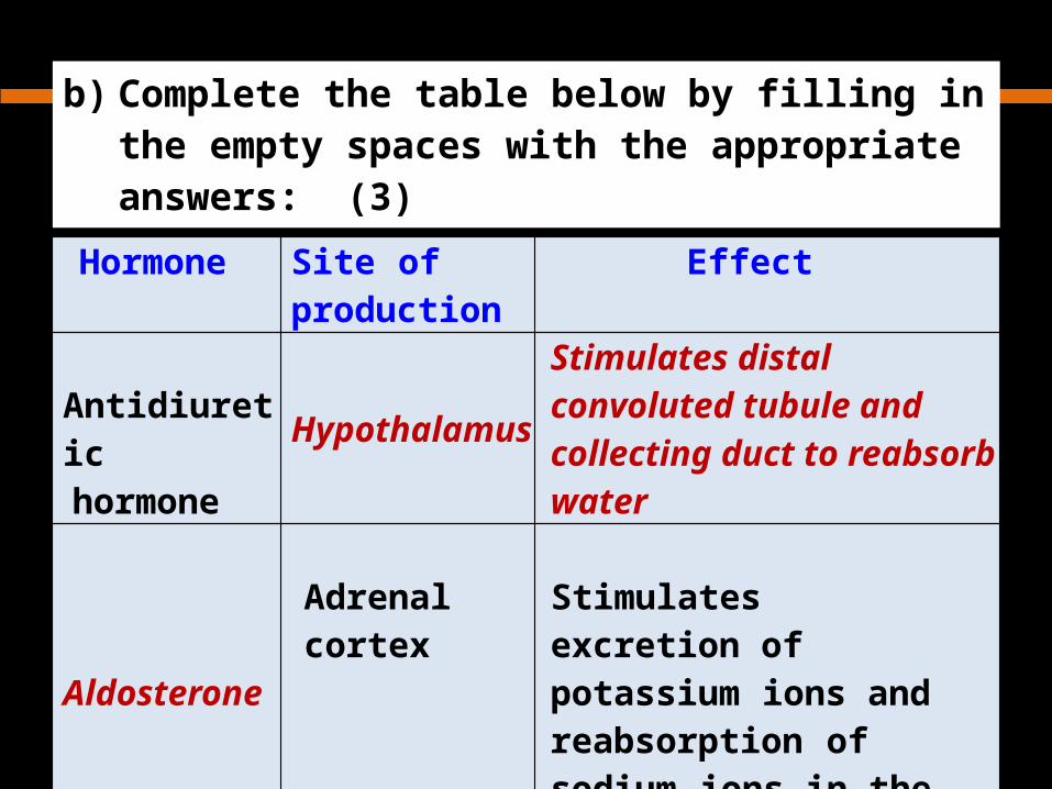
b) Complete the table below by filling in the empty spaces with the appropriate answers: (3)
Hormone Site of production
Effect
Antidiuretichormone
Hypothalamus
Stimulates distal convoluted tubule and collecting duct to reabsorb water
AldosteroneAdrenal cortex
Stimulates excretion of potassium ions and reabsorption of sodium ions in the nephron

Question: [MAY, 2011]
c) Briefly describe how vasoconstriction and vasodilation of blood vessels may affect blood pressure. (4)When blood vessels dilate, the blood pressure is lowered as there is less resistance to blood flow. When blood vessels constrict, the blood pressure becomes higher as cross-sectional area decreases.

Control of Blood pH

Control of Blood pH
pH of blood is maintained at a constant value of 7.4
pH of urine varies: 4.5 - 8.2

Abrupt changes in blood pH:
are prevented by:Plasma proteinsPhosphate Hydrogen carbonate
buffers
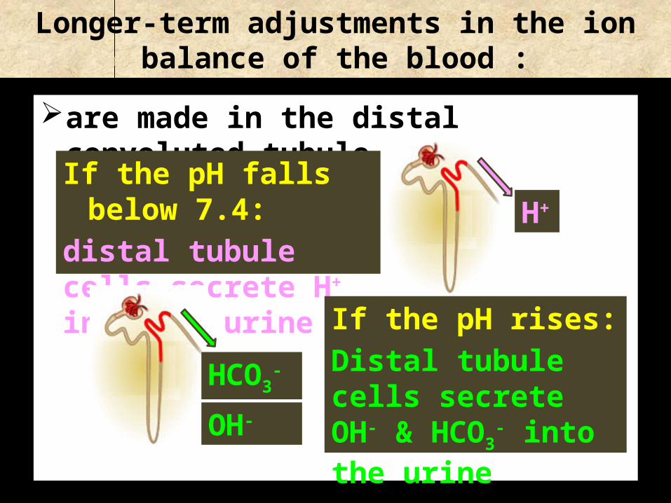
Longer-term adjustments in the ion balance of the blood :
are made in the distal convoluted tubule
If the pH falls below 7.4:distal tubule cells secrete H+ into the urine
If the pH rises:Distal tubule cells secrete OH- & HCO3
- into the urine
H+
HCO3-
OH-

Essay Titles
1. Give an overview of the role of the mammalian kidney in excretion and osmoregulation.
[SEP, 2000] 2. Evaluate the role of the human kidney in excretion
and osmoregulation. [SEP, 2002] 3. The mammalian kidney is a homeostatic organ.
Discuss. [SEP, 2004]

Essay Titles
4. Describe the role of countercurrent flows in biological systems [MAY, 2007]
Gills in bony fish – blood & seawater flow Thermoregulation – blood flow in artery & vein in a
limb Excretion – loop of Henle; vasa recta Pregnant female - blood of embryo & uterus
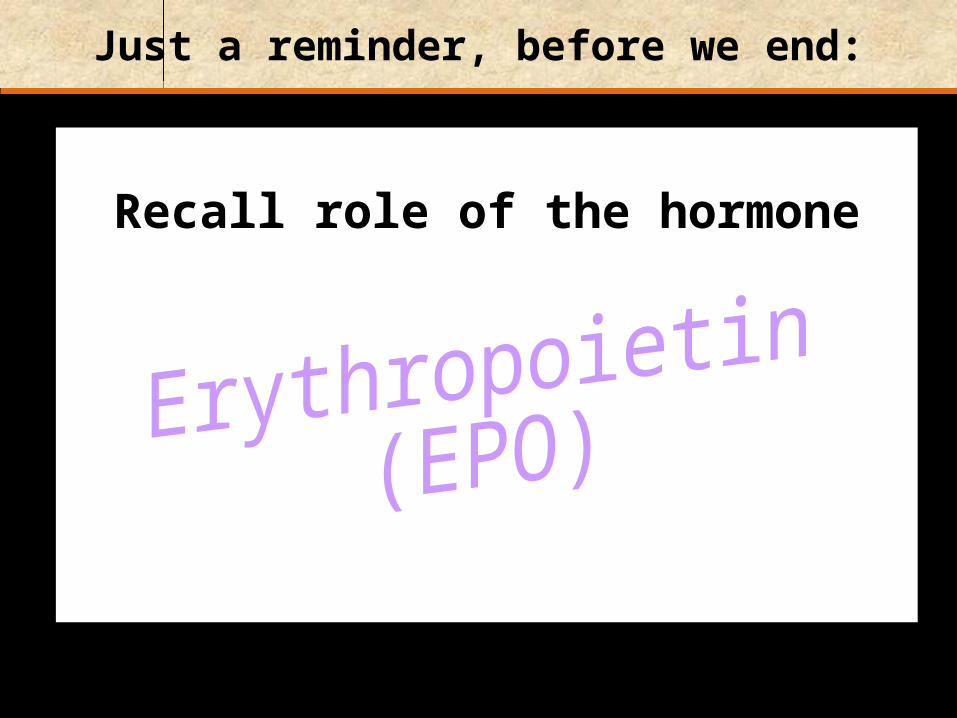
Just a reminder, before we end:
Recall role of the hormone

Erythropoietin :
Made by:Kidneys
Released when:Oxygen levels in blood are low
Causes:RBC formation

Figure 11-61 of 7
Bone marrow
Stemcells
Erythroblasts
Reticulocytes
Increasedmitotic rate
Acceleratedmaturation
Release oferythropoietin
(EPO)
Tissueoxygenlevels
decline
Tissueoxygenlevelsrise
Improvedoxygencontentof blood
Increased numbersof circulating RBCs
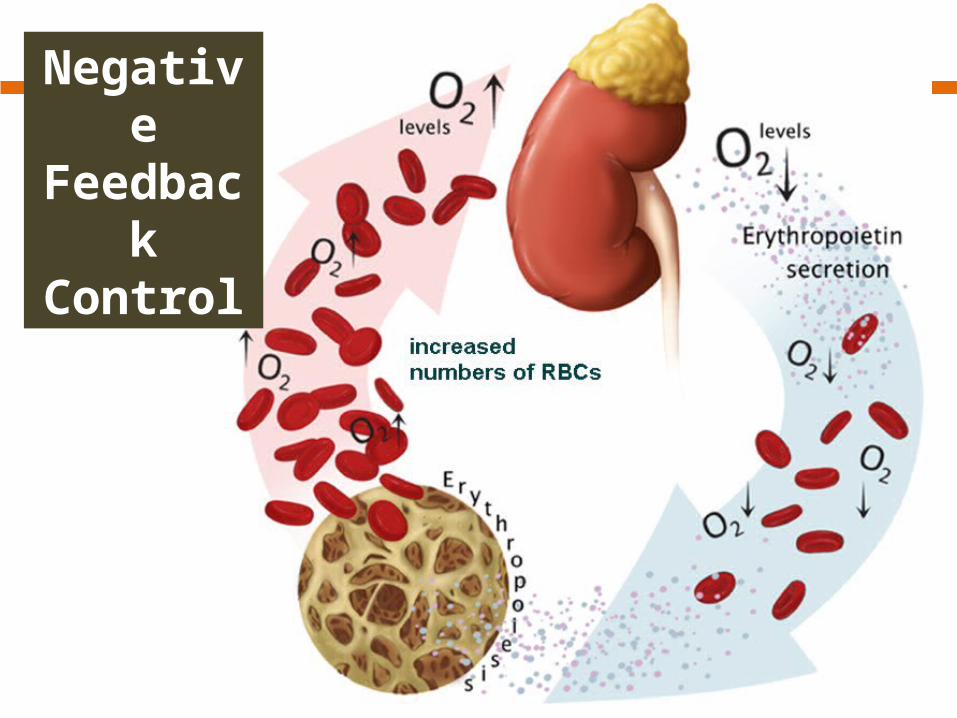
Negative Feedback Control

Manneken Piss [Brussels, Belgium]