exosome-delivered micrornas modulate the · pdf filearticle received 23 sep 2014 | accepted 27...
TRANSCRIPT

ARTICLE
Received 23 Sep 2014 | Accepted 27 Apr 2015 | Published 18 Jun 2015
Exosome-delivered microRNAs modulate theinflammatory response to endotoxinMargaret Alexander1, Ruozhen Hu1, Marah C. Runtsch1, Dominique A. Kagele1, Timothy L. Mosbruger2,
Tanya Tolmachova3, Miguel C. Seabra3, June L. Round1, Diane M. Ward1 & Ryan M. O’Connell1
MicroRNAs regulate gene expression posttranscriptionally and function within the cells in
which they are transcribed. However, recent evidence suggests that microRNAs can be
transferred between cells and mediate target gene repression. We find that endogenous
miR-155 and miR-146a, two critical microRNAs that regulate inflammation, are released from
dendritic cells within exosomes and are subsequently taken up by recipient dendritic cells.
Following uptake, exogenous microRNAs mediate target gene repression and can repro-
gramme the cellular response to endotoxin, where exosome-delivered miR-155 enhances
while miR-146a reduces inflammatory gene expression. We also find that miR-155 and
miR-146a are present in exosomes and pass between immune cells in vivo, as well as
demonstrate that exosomal miR-146a inhibits while miR-155 promotes endotoxin-induced
inflammation in mice. Together, our findings provide strong evidence that endogenous
microRNAs undergo a functional transfer between immune cells and constitute a mechanism
of regulating the inflammatory response.
DOI: 10.1038/ncomms8321 OPEN
1 Division of Microbiology and Immunology, Department of Pathology, University of Utah, 4280 JMRB, 15 North Medical Drive East, Salt Lake City, Utah84112, USA. 2 Huntsman Cancer Institute, University of Utah School of Medicine, Salt Lake City, Utah 84112, USA. 3 Molecular Medicine Section,National Heart and Lung Institute, Imperial College London, London SW7 2AZ, UK. Correspondence and requests for materials should be addressed toR.M.O’C. (email: [email protected]).
NATURE COMMUNICATIONS | 6:7321 | DOI: 10.1038/ncomms8321 | www.nature.com/naturecommunications 1
& 2015 Macmillan Publishers Limited. All rights reserved.

Intercellular communication is essential for immune cells tocoordinate inflammatory responses. Cytokines, chemokinesand cell surface receptors are well-known mediators of this
process. In addition to these classical signalling molecules,emerging evidence suggests that immune cells can signal bysecreting small lipid packages called exosomes, which carry avariety of different molecules that can be taken up by recipientcells1–4. The functional relevance of exosomes in many differentbiological systems, including the immune system, is beginning tobe demonstrated5–11.
MicroRNAs (miRNAs) are important modulators of geneexpression that function by targeting messenger RNAs fordegradation or preventing translation. Typically, miRNAsare thought to function within the cells in which they aremade; however, recently, miRNAs have been observed in secretedexosomes12–14. Immune cells, including antigen-presentingdendritic cells (DCs) and T lymphocytes, can both secrete andtake up exosomal miRNAs, suggesting that exosomal transferof miRNAs could be a novel mechanism for intercellularcommunication12,14,15. Furthermore, recent studies indicatethat the loading of miRNAs into exosomes is a selective processwhere specific motifs in miRNA sequences are recognizedby the RNA-binding protein, hnRNPA2B1 (ref. 16). Otherreports find that miRNA loading into exosomes is dependenton 30-end uridylated isoforms17 and on the levels of miRNAtargets in producer cells18. Consistent with this, exosomalmiRNA signatures do not simply reflect the miRNAcomposition of the parent cell, but are composed of a distinctset of miRNAs16,18–21. This argues that certain miRNAs haveevolved to be packaged into exosomes to carry out their biologicalfunctions.
Exosomally transferred miRNAs are emerging as novelregulators of cellular function. There is evidence in both immunecells and other cell types that transferred miRNAs repress targetmRNAs in recipient cells12–14,22–24. The transfer of miRNAs canalso cause physiological changes in recipient cells5–7, asdemonstrated by miRNAs moving from cancer cells toendothelial cells, which promotes tumour metastasis5. Cancercells can also receive exosomal miRNAs secreted from immunecells, which were shown to have an anti-proliferative effect on thetumour cells7. These data suggest that different cell types cansecrete or receive miRNAs as a form of communication and haveset the stage for investigating the functional roles of transferredmiRNAs in the context of immune responses.
Within the immune system, several specific miRNAs haverecently emerged as important regulators of immune cell function.Among these, miR-155 is a promoter of inflammatory responses,while miR-146a is a mediator of immune suppression25–28.Despite significant progress in our understanding of how thesemiRNAs influence immunity in vivo, there are many aspectsof their regulation and function that remain unclear. In thecurrent study, we investigate whether endogenous miR-155 andmiR-146a are functionally transferred between primary bonemarrow-derived DCs (BMDCs). We find that both of thesemiRNAs are released within exosomes and are taken up byrecipient BMDCs. On uptake, the miRNAs are associated withAgo proteins, knock down their respective targets andreprogramme the response of BMDCs to endotoxin challenge.We also show that miR-155 can be transferred between immunecells in vivo. Finally, we demonstrate that injection of miR-146a-containing exosomes into mice inhibits their inflammatoryresponse to endotoxin, whereas injection of miR-155-containingexosomes promotes inflammation following exposure to thesame inflammatory stimulus. Our study supports a modelwhereby exosomal miRNAs participate in the regulation ofinflammatory responses.
ResultsmiR-155 is found in exosomes and transferred between BMDCs.miR-155 is an immunomodulatory miRNA expressed by manytypes of immune cells including DCs27. We sought to determinewhether miR-155 could be passed between cultured BMDCs. Co-cultures of primary mouse BMDCs derived from CD45.1þ Wtmice and CD45.2þ miR-155� /� mice were set up at a 1:1 ratiowith and without lipopolysaccharide (LPS) treatment (Fig. 1a). Asa control, miR-155� /� BMDCs were also cultured under thesame conditions without Wt cells. After 24 h, the co-cultured Wtand miR-155� /� CD11cþ BMDCs were separated based ontheir differential CD45 markers using fluorescence-activated cellsorting (FACS) (Fig. 1b). Quantitative reversetranscriptase–PCR(qRT–PCR) was performed on RNA isolated from the CD45.2þ
miR-155� /� BMDCs. miR-155 was detected in miR-155� /�BMDCs that were cultured with Wt cells and the signal was clearlyabove background levels established using miR-155� /� BMDCscultured alone (Fig. 1c). When cells were treated with LPS, thetransfer of miR-155 to miR155� /� cells was increased,consistent with previous findings that cellular miR-155concentrations are elevated following LPS stimulation29 (Fig. 1c).
To determine whether cell–cell contact is necessary for thetransfer of miR-155, we used 0.4-mm filters to separatemiR-155� /� and Wt BMDCs that were co-cultured in thepresence or the absence of LPS for 24 h. The 0.4-mm pore sizeallows for small molecules and vesicles such as exosomes to passthrough but prevents cell-contact-mediated exchange of mate-rial23. We detected miR-155 in the miR-155� /� BMDCs thatwere cultured with Wt BMDCs, which was above background(Fig. 1d). Our data indicate that miR-155 is passed between cells,and that cell–cell contact is not necessary for transfer to occurbetween BMDCs.
As miRNAs have recently been shown to be transferredbetween immune cells within exosomes, we investigated whethermiR-155 is contained within these secreted vesicles. To addressthis question, we isolated the exosomal pellet from Wt ormiR-155� /� BMDC conditioned media using differentialcentrifugation. Both electron microscopy (EM) and a CD63western blotting of the isolated vesicles indicated that we hadsuccessfully isolated exosomes (Fig. 1e,f). Using qRT–PCR, wefound that miR-155 was contained in exosomes derived from WtBMDCs but not in exosomes derived from miR-155� /� cells(Fig. 1g). BMDCs treated with LPS enhanced the levels of miR-155 found in the exosomal pellet, consistent with higher levels ofmiR-155 being produced by the activated BMDCs. In addition,we blocked exosome formation by treating donor BMDCs withGW4869, a drug that hinders exosome biogenesis by blockingneutral sphingomyelinase 2 (nSMase2) (refs 13,15). Followingdrug treatment, the pellet contained significantly reducedexosomes as determined by EXOCET quantification (Fig. 1h),CD63 western blotting and EM (Supplementary Fig. 1). Drugtreatment also prevented the detection of miR-155 in theexosomal pellet (Fig. 1i), suggesting that miR-155 is containedwithin exosomes. In addition, we derived BMDCs from Rab27aand Rab27b double-knockout mice (Rab27 DKO), which havebeen previously shown to have decreased release of exosomes23.We found that Rab27 DKO BMDCs had both decreased exosomerelease (Fig. 1j) and a corresponding decrease in miR-155 in theexosomal pellet (Fig. 1k). Together, these data show that miR-155can be passed between BMDCs, and that miR-155 is contained inexosomes produced by BMDCs.
Exosomal transfer of miR-155 is functionally relevant. With theknowledge that miR-155 can be transferred between BMDCs, wewanted to determine whether exosomes are sufficient for this
ARTICLE NATURE COMMUNICATIONS | DOI: 10.1038/ncomms8321
2 NATURE COMMUNICATIONS | 6:7321 | DOI: 10.1038/ncomms8321 | www.nature.com/naturecommunications
& 2015 Macmillan Publishers Limited. All rights reserved.

transfer and whether transfer could result in knockdown oftarget mRNAs. To specifically investigate the impact of exo-somally transferred miRNA without the effects of other factorsthat are released from BMDCs, we purified exosomes awayfrom other components in the conditioned medium using
differential centrifugation and washing. Next, the exosomes werere-suspended in fresh medium and administered to recipientcells. Wt (1� 106) or miR-155� /� BMDCs produced B5� 108
exosomes in 24 h (Supplementary Fig. 2). Exosomes isolated fromthe supernatant of both Wt and miR-155� /� BMDCs treated
± LPS
CD45.1 WTCD45.2miR-155–/–
FACSCD45.2+
qRT-PCRfor miR-155
Rel
ativ
e m
iR-1
55le
vels
Rel
ativ
e m
iR-1
55le
vels
Rel
ativ
e m
iR-1
55le
vels
miR
-155
leve
ls
miR
-155
leve
ls
Exo
som
e nu
mbe
rpe
r m
illio
n B
MD
Cs
Exo
som
e nu
mbe
rpe
r m
illio
n B
MD
Cs
**
*5 6
*
***
****
****
***0.4 μm filter
WT exosomes
SS
C
CD
45.1
CD11c
102
102
103
103
104
104
105
102
103
104
105
105 102 103 104 105
200 nm
CD45.2
4
2
0
4
3
2
1
0155–/–
155–/– 155–/–
NA
Cell
LPS
Co-Cultured
NA WT WT
WT
WTWTWTWT
WT
20101015
10
5
0
109
108
107
Exo. pellet
15
10
5
0Rab27DKO
WT Rab27DKO
8
6
4
2
0WT WTWT
155–/– 155–/– 155–/–
–
– –
–+
+ +
+
155–/–
155–/– 155–/–107
108
109
1010
NA
Cell
LPS
LPS GW4869
GW4869
CellExosome
Exosome
LPS
Exo
LPS
Co-Cultured
NA WT
WTWT
CD63 western
WT
155–/– 155–/– 155–/–
–
–
– –
––
–
–
–
+ +
+++
+
+
+
+
Figure 1 | miR-155 is transferred between BMDCs and is present in exosomes. (a) A schematic of the co-culture experiment. (b) Representative FACS
plots where co-cultured CD45.1þ Wt and CD45.2þ miR-155� /� CD11cþ BMDCs were separated (n¼4). (c) Relative miR-155 levels were quantified via
qRT–PCR from isolated miR-155� /� BMDCs that had been cultured alone or with Wt BMDCs in the presence or the absence of LPS for 24 h (n¼4).
(d) Relative miR-155 levels were measured via qRT–PCR in miR-155� /� BMDCs either cultured alone or with Wt BMDCs separated by a 0.4-mm filter for
24 h with or without LPS (n¼ 3). (e) Cryo-EM of exosomes isolated from Wt BMDCs. Scale bar, 200 nm. Red box is enlarged in the upper left corner.
(f) CD63 protein levels in the exosomal pellet from Wt and miR-155� /� BMDCs treated with or without LPS. (g) Relative levels of miR-155 in exosomes
derived from Wt or miR-155� /� BMDCs treated with or without LPS (n¼ 3). (h) Exosome quantification of Wt BMDCs treated with or without GW4869
(n¼ 3). Limit of detection is 2� 107 exosomes. (i) Relative levels of miR-155 were measured in the exosomal pellet from Wt BMDCs treated with or
without LPS and GW4869 as quantified by qRT–PCR (n¼ 2). (j) Exosome quantification of Wt and Rab27 DKO BMDC-derived exosomes (n¼ 2). Limit of
detection is 2� 107 exosomes. (k) miR-155 levels in exosome pellets from Wt and Rab27 DKO BMDC-conditioned medium (n¼ 2). Data represent two
independent experiments and are presented as the mean±s.d. (error bars). *Po0.05; **Po0.01, Student’s t-test.
NATURE COMMUNICATIONS | DOI: 10.1038/ncomms8321 ARTICLE
NATURE COMMUNICATIONS | 6:7321 | DOI: 10.1038/ncomms8321 | www.nature.com/naturecommunications 3
& 2015 Macmillan Publishers Limited. All rights reserved.

with GW4869 or dimethylsulfoxide vehicle control were trans-ferred to miR-155� /� receipient BMDCs. miR-155� /�recipient BMDCs were incubated with donor exosomes for 24 h,to allow time for miRNA transfer and knockdown of miRNAtargets (Fig. 2a). Using qRT–PCR, we detected increased miR-155levels and decreased mRNA levels of miR-155 targets BACH1
and SHIP1 when cells were treated with Wt exosomes (Fig. 2b–d).These changes were prevented if the exosomes were derived frommiR-155� /� BMDCs, or if the Wt donor cells were pretreatedwith GW4869. SHIP1 protein levels were also decreased inmiR-155� /� BMDCs that received Wt exosomes (Fig. 2e,f).Exosome delivery of miR-155 brought its levels in the
WT
WT
**
*0.1429
*
0
1
2
3 1.5
1.0
0.5
0.0Rel
ativ
e m
iR-1
55 le
vels
Rel
ativ
e m
iR-1
55 le
vels
miR
-155
leve
ls(A
go/Ig
G)
miR
-146
a le
vels
(Ago
/IgG
)
Rel
ativ
e S
HIP
1 le
vels
Rel
ativ
e S
HIP
1le
vels
Rel
ativ
e B
AC
H1
leve
lsR
elat
ive
SH
IP1
prot
ein
leve
ls
Rel
ativ
e H
O1
leve
ls
Rel
ativ
e B
AC
H1
leve
ls
Exosomedonor
Exosomedonor
SHIP1
β-Actin
Recipientcell
GW4869
WT
– –+ +
155–/–
155–/– 155–/–
155–/–155–/–155–/–155–/–
WTExosomedonor
Recipientcell
GW4869
WT
+WT +155–/–
155–/– recipient cells
– –+ +
155–/– 155–/–
155–/–155–/–155–/–155–/–
25 2.5
2.0
1.5
0.0651
0.0662
0.06000.07758
6
4
2
0
1.0
0.5
0.0
20
15
10
5
0
*
WT
WT
Exosomedonor
Exosomedonor
Recipientcell
Recipientcell
GW4869
WT
– –+ +
155–/– 155–/–
155–/–155–/–155–/–
155–/– 155–/–
155–/–
WT
WT WT
Exosomedonor
0
1
2
3 *
Recipientcell
155–/–WT
WT WT
Exosomedonor
Recipientcell
155–/–WT
WT WT
Exosomedonor
Recipientcell
155–/–
WT
WT WT WT
WT WTWTExosome
donorExosome
donor
Recipientcell
Recipientcell
Input0 0
10
2 2
44
6
6
8
8Input
Ago2
β-Actin
Exosomedonor
AGO IP IgG IP
Input αAgo αAgo Input Input αAgo αAgo
155–/–
155–/–155–/–155–/–
155–/– recipient cells
155–/– 155–/–
155–/–
155–/– 155–/–
155–/–155–/–
155–/– 155–/– 155–/–
155–/–
155–/–
1.5
1.0
0.5
0.0
WTExosome
donor
Recipientcell
GW4869
WT
– –+ +
155–/– 155–/–
155–/–155–/–155–/–155–/–
± GW4869
Isolate exosomes viadifferential centrifugation
Recipient 155–/– BMDCs
2.0
*
***
1.5
1.0
0.5
0.0
ARTICLE NATURE COMMUNICATIONS | DOI: 10.1038/ncomms8321
4 NATURE COMMUNICATIONS | 6:7321 | DOI: 10.1038/ncomms8321 | www.nature.com/naturecommunications
& 2015 Macmillan Publishers Limited. All rights reserved.

miR-155� /� recipient cells to B20% of Wt miR-155 levels(Supplementary Fig. 3a,b). Furthermore, we also looked at therelative expression of a separate miRNA, miR-425, which hasbeen previously seen to be released in exosomes30, as a control.The levels of miR-425 increased with exosome delivery and wereroughly the same in Wt and knockout groups (SupplementaryFig. 3c,d).
In addition to using miR-155� /� recipient cells, whichprovide a clean background to clearly detect the transferredmiRNA, we also examined whether transferred miR-155 could bedetected in Wt BMDC recipients. miR-155 levels were increasedon treatment of Wt BMDCs with Wt exosomes but were notincreased when treated with miR-155� /� exosomes (Fig. 2g).In addition, the mRNA levels of both BACH1 and SHIP1 weredecreased in Wt BMDCs receiving exosomal miR-155 (Fig. 2h,i).These data indicate that miR-155 can be transferred between WtBMDCs in exosomes, resulting in the knockdown of known miR-155 targets.
As we have found that miR-155 can knock down its targets inrecipient cells, we next investigated whether the transfer of miR-155 alters downstream factors in recipient cells. We found thatexosomal miR-155 increased the expression of HO1 (Fig. 2j), anoxidative stress response gene that is well known to be repressedby BACH1, a gene we have shown is targeted by transferred miR-155 (ref. 29; Fig. 2c). These results indicate that transferred miR-155 not only represses its putative direct targets but can also affectfactors that are downstream of these targets.
To further characterize the functional transfer of miR-155between BMDCs, we examined whether transferred miR-155 isassociated with AGO proteins that are essential for miRNA-mediated knockdown of targets. Following exosomal transfer ofmiR-155 into miR-155� /� BMDCs, an AGO immunoprecipi-tation (IP) was performed using a pan-AGO antibody andwestern blotting for AGO2 was preformed to verify pulldown wasoccurring. Using qRT–PCR, we found that miR-155 is associatedwith AGO proteins in miR-155� /� recipient cells (Fig. 2k,l).We did not detect miR-155-associated AGO proteins when miR-155� /� BMDCs were treated with miR-155� /� exosomes.Further, AGO2 was not detected via western blotting and miR-155 was not pulled down when an isotype control antibody wasused. As an additional control, we found that another miRNA,miR-146a, was also enriched in the AGO pulldown from bothgroups (Fig. 2m). These data demonstrate that exosomal miR-155is associated with AGO proteins, key components of the RNA-induced silencing complex (RISC) complex, following its uptakeby recipient BMDCs.
Exosomal transfer of miR-146a is functionally relevant.miR-146a is an important anti-inflammatory miRNA involved inDC function27 and plays an opposing role to miR-155 duringinflammatory responses25,31. We wanted to determine whethermiR-146a was also contained in BMDC-derived exosomes; hence,
we isolated exosomes from Wt BMDCs that had been treated withor without GW4869 and/or LPS, and found that miR-146a wascontained in exosomes from untreated BMDCs but was notpresent in the exosomal pellet from BMDCs treated with GW4869(Fig. 3a). miR-146a is marginally increased in exosomes fromBMDCs treated with LPS. In addition, reductions in miR-146awere observed in the extracellular exosomal fraction obtained fromRab27 DKO BMDCs compared with Wt controls (Fig. 3b). Thesedata reveal that miR-146a is contained within exosomes releasedfrom BMDCs.
To test whether miR-146a could be functionally transferredbetween BMDCs, we isolated exosomes from Wt or miR-146a� /�BMDCs and administered them to miR-146a� /� BMDCs(Fig. 3c). Similar to miR-155, we observed that exosomalmiR-146a was taken up by recipient BMDCs (Fig. 3d), and thatmiR-146a targets, IRAK1 and TRAF6, were repressed in recipientBMDCs receiving Wt but not miR-146a� /� exosomes looking atboth the mRNA and protein levels (Fig. 3e–j). In addition, wecalculated miR-146a copy number in Wt and miR-146a� /�exosomes where we found approximately one copy of miR-146a perexosome (Fig. 3k). miR-146a copy number was also calculated in Wtand miR-146a� /� donor BMDCs and BMDCs that receivedeither Wt or miR-146a� /� exosomes (Fig. 3l). We observed anaverage of 370 copies present in recipient BMDCs followingexosomes treatment. It has been suggested that 100–1,000 copies ofmiRNA per cell is likely to be functionally relevant32. As a control,we investigated the relative expression of miR-425, which wassimilar between the genotypes (Supplementary Fig. 3e,f). Our copynumber data along with our observations of target knockdown areconsistent with exosomally transferred miR-146a having functionalrelevance.
Recently, it has been shown in human B-cell lines that miRNAsare selectively packaged into exosomes based on 30-non-templated nucleotide additions (NTAs)17 where 30-uridylationwas enriched in miRNAs contained in exosomes and30-adenylation was enriched in miRNAs retained in cells. Toaddress whether we observe a similar phenomenon, RNAsequencing was performed using RNA from Wt donor BMDCsand miR-155 and miR-146a DKO BMDCs that had receivedWt exosomes. Next, we used a previously reported approach toidentify NTAs in our data set17. However, we did not observe anenrichment of 30-uridylation in transferred miR-155 or miR-146a(Supplementary Fig. 4). This difference from previous findingscould be due to species and cellular differences (mouse primaryBMDCs versus human B-cell lines) or further processing of thetransferred miRNAs in recipient cells. However, we did observesome differences at certain nucleotide positions in each respectivemature miRNA sequence when comparing Wt donor BMDCswith DKO BMDCs that received Wt exosomes (SupplementaryTables 1 and 2). These results are consistent with the idea thatalterations to the mature miRNA sequence may influence miRNAloading into exosomes versus cellular retention.
Figure 2 | Functional transfer of miR-155 via exosomes in vitro. (a) Schematic of the exosome transfer experiment. (b) qRT–PCR was used to
measure relative miR-155 levels in miR-155� /� BMDCs that received either Wt or miR-155� /� exosomes derived from BMDCs treated with or without
GW4869 (n¼ 5). (c,d) mRNA levels of miR-155 targets, BACH1 and SHIP1, from the same experiment shown in b as measured by qRT–PCR (n¼ 5).
(e) Representative western blottings of SHIP1 and b-actin in miR-155� /� BMDCs given either Wt or miR-155� /� exosomes. (f) Protein levels of SHIP1
were quantified using ImageJ software (n¼ 2). (g) Relative miR-155 levels in Wt BMDCs given either Wt or miR-155� /� exosomes as quantified by
qRT–PCR (n¼6). (h,i) BACH1 and SHIP1 mRNA levels were measured in the same experiment shown in g as quantified by qRT–PCR (n¼6). (j) qRT–PCR
was used to quantify HO1 mRNA levels during the experiment in b (n¼ 5). (k) Western blotting for AGO2 and b-actin from miR-155� /� BMDCs given
Wt or miR-155� /� exosomes. On the left is the input (whole-cell lysate), the middle is from the pan-AGO pulldown where one-third of input was used
and the right is the IgG pulldown where one-third of the input was used. (l) Relative miR-155 levels were quantified via qRT–PCR in the same experiment
shown in k. (m) miR-146a levels were quantified using qRT–PCR during the experiment in k. Levels in l,m are plotted as Ago:IgG. Dotted line separates
input from pull-down groups. Data represent two independent experiments and are presented as the mean±s.d. (error bars). *Po0.05; **Po0.01,
Student’s t-test.
NATURE COMMUNICATIONS | DOI: 10.1038/ncomms8321 ARTICLE
NATURE COMMUNICATIONS | 6:7321 | DOI: 10.1038/ncomms8321 | www.nature.com/naturecommunications 5
& 2015 Macmillan Publishers Limited. All rights reserved.

3
1.0 *
0.8
0.6
0.4
0.2
0.0WT Rab27
DKO
2
1
0WT WT WT WT
Exo. pellet WT
Recipient 146a–/– BMDCs
Isolate exosomes viadifferential centrifugation
146a–/–
–
–
–
–
+
+
4*
*
*
3
2
1
0
+
+
Exosome
GW4869
LPS
1.5
1.0
0.5
0.0
Rel
ativ
e m
iR-1
46a
expr
essi
on
Rel
ativ
e IR
AK
1le
vels
WT
1.5
1.0
0.5
0.0
Rel
ativ
e IR
AK
1pr
otei
n le
vels
146a–/–
146a–/–
146a–/–
WT
146a–/–
1.5
1.0
0.5
0.0
WT e
xo
WT d
onor
146a
–/–
exo
146a–/–
146a–/–
3,949
370
6,000
4,000
2,000
0
Cop
y nu
mbe
r of
miR
-146
a pe
r ce
ll
Cop
y nu
mbe
r of
miR
-146
a pe
r ex
osom
e
146a
–/–
dono
r
146a
–/–
+ 14
6a–/
–
146a
–/–
+ W
T exo
s
WT
146a–/–
6
4
2
0
Rel
ativ
e T
RA
F6
prot
ein
leve
ls
146a–/–
146a–/–
WT
WT
TRAF6
β-Actin
+WT +146a–/–
146a–/–
8
6
4
2
0
Rel
ativ
e T
RA
F6
leve
ls
146a–/–
146a–/–
146a–/–
Exosomedonor
Recipientcell
Exosomedonor
Recipientcell
Exosomedonor
Exosomedonor
Recipientcell
WT
146a–/–
146a–/–
146a–/–
Exosome
Recipientcell
Exosomedonor
Exosomedonor
IRAK1
146a–/– Recipient cells
146a–/– Recipient cells
β-Actin
Recipientcell
miR
-146
a le
vels
miR
-146
a le
vels
Figure 3 | Functional transfer of miR-146a via exosomes in vitro. (a) Levels of miR-146a in the exosomal pellet derived from BMDCs that were treated
with or without GW4869 and LPS (n¼ 2). (b) miR-146a levels in Wt and Rab27 DKO BMDC-derived exosomal pellets (n¼ 2). (c) Schematic of miR-146a
exosome-transfer experiment where Wt or miR-146a� /� exosomes were isolated from BMDCs and transferred to recipient miR-146a� /� BMDCs.
RNA was isolated after 24 h and the presence of miR-146a was assayed via qRT–PCR. (d) Relative levels of miR-146a in miR-146a� /� BMDCs given
exosomes derived from Wt or miR-146a� /� BMDCs (n¼4). (e) mRNA levels of miR-146a target, IRAK1, were measured from the same cells as in d via
qRT–PCR (n¼4). (f) Representative western blottings of IRAK1 and b-actin from miR-146a� /� cells given either Wt or miR-146a� /� exosomes.
(g) IRAK1 protein levels were quantified using ImageJ software (n¼ 2). (h) mRNA levels of miR-146a target, TRAF6, were measured in the same cells as in
d via qRT–PCR. (i) Western blottings for TRAF6 and b-actin from miR-146a� /� BMDCs given either Wt or miR-146a� /� exosomes (n¼ 2).
(j) Western blotting results are quantified with ImageJ software. (k) Copy number of miR-146a in Wt and miR-146a� /� exosomes (n¼ 3). Copy number
is calculated based on a standard curve where a known amount of synthetic miR-146a was spiked into miR-146a� /� BMDC-derived exosome pellet
followed by RNA isolation and qRT–PCR. (l) Copy number of miR-146a was measured via qRT–PCR in miR-146a� /� recipient BMDCs that received either
Wt or miR-146a� /� exosomes (146a� /� BMDCþWt exos and 146a� /� BMDCþ 146a� /� exos), as well as in Wt and miR-146a� /� donor
BMDCs (n¼ 3). Average copy number is displayed above. Copy number is calculated based on a standard curve where a known amount of synthetic
miR-146a was spiked into miR-146a� /� BMDC pellet followed by RNA isolation and qRT–PCR. Data represent two independent experiments and are
presented as the mean±s.d. (error bars). *Po0.05, Student’s t-test.
ARTICLE NATURE COMMUNICATIONS | DOI: 10.1038/ncomms8321
6 NATURE COMMUNICATIONS | 6:7321 | DOI: 10.1038/ncomms8321 | www.nature.com/naturecommunications
& 2015 Macmillan Publishers Limited. All rights reserved.

Seed-dependent repression of miRNA targets. We next deter-mined whether miR-155 and miR-146a mimic-loaded exosomeswere sufficient to mediate direct target knockdown in recipientcells. miR-155� /� or miR-146a� /� BMDCs were transfectedwith either corresponding miRNA mimics, scrambled miRNAmimics, or seed mutant miRNA mimics for 24 h, then washedthree times with PBS to remove any mimics that did not make itinto the cells (Fig. 4a). We isolated exosomes from the cells after24 h and transferred them to recipient knockout BMDCs. After24 h, RNA was isolated from the cells and qRT–PCR was per-formed to assay the delivery of mimics and the knockdown oftarget mRNAs. We found that miRNA mimics could be suc-cessfully loaded into exosomes and delivered to recipient cells(Supplementary Figs 5 and 6). The transfer of miRNA mimicscontaining exosomes resulted in knockdown of respective targetmRNAs in recipient BMDCs (Fig. 4b–f). However, exosomes thatdid not carry mimics, or that carried scrambled or seed mutantmimics, caused no change in target mRNA expression in reci-pient cells. These results indicate that exosomal miRNAs areresponsible for direct target repression and are able to comple-ment the target knockdown phenotype.
To further assess whether exosomal miRNA target repressionwas direct, we used 30-untranslated region (UTR) luciferase reporterassays. Knockout BMDCs were transfected with 30-UTR luciferasereporters for 6 h followed by treatment with or without Wtexosomes (Fig. 4g). miR-155� /� BMDCs were transfected witheither a pmiReport empty vector control, BACH1 30-UTR, BACH1miR-155-binding site (bs) mutant 30-UTR, or a miR-155-positivecontrol (2mer). Luciferase activity in cells receiving the BACH130-UTR or 2mer reporter constructs was reduced in response tomiRNAs delivered by exosomes, while the exosomal miRNAs hadlittle impact on luciferase activity in cells receiving the pmiReportempty vector or the BACH1 miR-155 bs mutant 30-UTR reporter(Fig. 4h). In a separate experiment, miR-146a� /� BMDCswere transfected with either a pmiReport empty vector control,TRAF6 30-UTR, or a TRAF6 miR-146a bs mutant 30-UTR. TheBMDCs transfected with the TRAF6 30-UTR had decreasedluciferase activity compared with the pmiReport empty vectorand the TRAF6 miR-146a bs mutant 30-UTR following exosomedelivery of miRNAs (Fig. 4i). These results indicate that exosomallytransferred miRNAs directly repress their targets via direct 30-UTRinteractions.
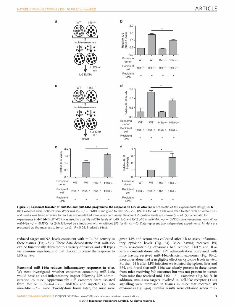
Exosomal miR-155 and miR-146a modulate the response to LPS.To determine whether the transfer of miR-155 via exosomes couldaffect the BMDC response to LPS, exosomes were isolated from Wtor miR-155� /� BMDCs and transferred to miR-155� /�BMDCs. Twenty-four hours later, cells were treated with LPS for6 h (Fig. 5a). Consistent with a previously reported role for miR-155 in promoting interleukin (IL)-6 expression33 and previouslyreported roles of miR-155-regulated responses to endotoxin34, cellsthat were treated with miR-155-containing exosomes producedmore IL-6 on treatment with LPS for 6 h than cells having receivedmiR-155� /� exosomes (Fig. 5b). These findings indicate thatexosomes containing miR-155 can reprogramme recipient BMDCsin a manner that enhances their response to LPS.
miR-146a is known to induce an anti-inflammatory responseto LPS26. Therefore, we wanted to investigate whether exosomallytransferred miR-146a can programme BMDCs to respond in ananti-inflammatory manner, using a similar experimental setup aswe did for miR-155 (Fig. 5c). BMDCs pre-treated with Wtexosomes produced more IL-10, but less IL-6 and IL-12 p40,following LPS stimulation than BMDCs that receivedmiR-146a� /� exosomes (Fig. 5d–f). This gene expressionprofile demonstrates that miR-146a-containing exosomes reduce
the pro-inflammatory response by BMDCs following LPStreatment. Without LPS treatment, there was no significantdifference in IL-10, IL-6 or IL-12 p40 expression by cells receivingWt versus miR-146a� /� exosomes (Fig. 5d–f), indicating thatexosomal miR-146a specifically alters how these cells respond toLPS. Similar to miR-155, our data indicate that miR-146a isfunctionally transferred in exosomes and able to causephysiological changes in recipient cells. However, unlike miR-155, exosomal miR-146a acts to dampen the inflammatoryresponse to LPS. These results are consistent with previousobservations that miR-155 and miR-146a play opposing rolesduring inflammation25,31.
miR-155 is transferred between immune cells in vivo. As weobserved functional transfer of miRNAs in vitro, we wanted todetermine whether miRNAs could be transferred betweenimmune cells in vivo. We first investigated whether exosomeswere present in mouse BM by isolating exosomes directly fromthe BM of Wt and miR-155� /� mice using differential cen-trifugation. We found that both genotypes had exosomes presentin the BM (Fig. 6a), and that miR-155 was expressed in Wt BMexosomes (Fig. 6b), whereas miR-146a was present in exosomesfrom both genotypes (Fig. 6c). We next determined whether miR-155 was passed between immune cells in vivo. To investigate this,miR-155� /� mice were lethally irradiated and reconstitutedwith either an equal mix of CD45.1þ Wt and CD45.2þ miR-155� /� BM or just miR-155� /� BM. After allowing 3months for reconstitution, we injected mice with LPS to stimulateproduction of miR-155 by BM cells (Fig. 6d). BM cells wereisolated 24 h after LPS stimulation and miR-155� /� haemato-poietic cells were sorted via FACS according to their differentCD45 alleles (Fig. 6f). We also further fractionated the miR-155� /� BM into B-cell, myeloid cell and T-cell fractions usingthe surface markers B220, CD11b and CD3, respectively(Fig. 6g,h). Using qRT–PCR, we detected miR-155 expression inmiR-155� /� B cells, T cells and myeloid cells taken from miR-155� /� mice that had been reconstituted with both Wt andmiR-155� /� BM (Fig. 6e). As a control, no miR-155 expressionwas observed in cells from mice reconstituted with only miR-155� /� BM. These data provide evidence that miR-155 islocated within exosomes within the BM and is transferredbetween immune cells in vivo.
To determine whether miRNA-containing exosomes coulddeliver miRNAs to various cell types in vivo, we intraperitoneally(i.p.) injected B109 exosomes derived from miR-155� /� orWt BMDCs into miR-155� /� mice. After multiple injectionsover a week, the spleens of these mice were harvested and CD3þ
T cells, B220þ B cells and CD11bþ myeloid cells were sorted viaFACS. We found that miR-155 was delivered to all three of thesecell types in the spleen (Fig. 6i–k). This indicates that exosomesare able to deliver miRNAs to various immune cell types.
Exosomal miR-155 enhances inflammatory responses in vivo.Our in vitro data suggest that exosomally delivered miR-155 canincrease the BMDC response to LPS (Fig. 5b). Owing to theseobservations, we investigated whether we could see the sameeffect in vivo. Approximately 109 Wt or miR-155� /� BMDC-derived exosomes were i.p. injected into miR-155� /� mice,followed by administration of LPS 24 h later and collection ofserum 2 h after that (Fig. 7a). The injection of Wt exosomesbefore LPS administration resulted in increased tumour necrosisfactor-a (TNFa) and trending elevations in IL-6 serum con-centrations compared with mice pretreated with miR-155� /�exosomes (Fig. 7b,c). In addition, we observed that miR-155 wasdelivered to the spleen, liver and BM, where we also found
NATURE COMMUNICATIONS | DOI: 10.1038/ncomms8321 ARTICLE
NATURE COMMUNICATIONS | 6:7321 | DOI: 10.1038/ncomms8321 | www.nature.com/naturecommunications 7
& 2015 Macmillan Publishers Limited. All rights reserved.

Donor knockoutcells
+No +Speed +Scram
24 h
miR-155 mimic
miR-146a mimic
miR-155 seed mutant
miR-146a seed mutant
miR-155 scramble
miR-146a scramble
24 h
Wash 3X with PBS
Isolate exosomes
Recipient KO cells
0.0743
2.0
1.5
1.0
0.5
0.0
2.5
No
**1.5
1.0
0.5
0.0
Rel
ativ
e B
AC
H1
leve
ls
155–
/–
155–
/–
155–
/–
155–
/–
Seed Scram Mimic
2.0
1.5
1.0
0.5
0.0No Seed Scram Mimic
146a
–/–
146a
–/–
146a
–/–
146a
–/–
146a
–/–
146a
–/–
146a
–/–
146a
–/–
No Seed Scram Mimic
146a–/– Recipient cells
miR-155 miR-146a
**** ****040
* *0.066720
0
–20
–40
–60
–20
–40
Per
cen
t cha
nge
Per
cen
t cha
nge
–60
–80
PmiR
epor
t
PmiR
epor
t
TRAF6
BACH1
TRAF6 m
utan
t
BACH1 m
utan
t2m
er
+No
TRAF6
IRAK1
β-Actin
+Seed +Mimic
0.750.911
1 1 0.79Exosome
donor
Recipientcell
Exosomedonor
Recipientcell
No
155–
/–
155–
/–
155–
/–
155–
/–
Seed Scram MimicExosomedonor
Recipientcell
*
*0.06871.5
1.0
0.5
0.0
Rel
ativ
e S
HIP
1le
vels
Exosomedonor
Recipientcell
Recipient knockout cells
+ 3′ UTR luciferase
6 h
Exosome transfer
24 h
Assay luciferase levels
Rel
ativ
e IR
AK
1le
vels
Rel
ativ
e T
RA
F6
leve
ls
**
*
***
+Mimic
Figure 4 | Seed-dependent repression of miRNA targets by exosome-delivered miR-155 and miR-146a. (a) Schematic for mimic experiment.
(b,c) Relative mRNA levels of the miR-155 targets SHIP1 and BACH1 were measured via qRT–PCR in recipient cells that received exosomes with no mimics
(No) (n¼ 7), miR-155 seed mutant mimics (Seed) (n¼4), with scrambled mimics (Scram) (n¼ 3), or with miR-155-mimics (Mimic) (n¼ 7). (d,e) qRT–
PCR was preformed to assay the mRNA levels of the miR-146a targets, IRAK1 and TRAF6, following treatment with exosomes containing miR-146a mimics
and controls as in b,c. Results are reported normalized to exosomes with no mimics added, which is set as 1. (f) Protein levels of TRAF6, IRAK1 and b-actin
were determined via western blotting using lysates from miR-146a� /� BMDCs that received exosomes containing no mimics, seed mutant mimics or Wt
mimics. Numbers below the blot represent relative protein levels with no mimics set as 1 following normalization to b-actin. (g) Schematic for 30-UTR
luciferase reporter assays in h,i. (h) Results from 30-UTR luciferase reporter assays where miR-155� /� BMDCs were transfected with a pmiReport control
vector, a BACH1 30-UTR vector (BACH1), a BACH1 miR-155-binding site (bs) mutant vector (BACH1 mutant), or a 2mer-positive control vector. Transfected
BMDCs were treated 6 h later with or without Wt exosomes and per cent change in luciferase activity of exosome treated BMDCs compared with no
exosome treatment was calculated after 24 h (n¼4). (i) Results from 30-UTR luciferase reporter assay where miR-146a� /� BMDCs were transfected
with a pmiReport control vector, a TRAF6 30-UTR vector (TRAF6) or TRAF6 miR-146a bs mutant vector (TRAF6 mutant). Six hours later, the BMDCs were
treated with or without Wt exosomes and per cent repression of luciferase activity was calculated 24 h after exosome transfer (n¼4). Results represent
two independent experiments. All data are presented as the mean±s.d. (error bars). *Po0.05; **Po0.01, ****Po0.0001; Student’s t-test.
ARTICLE NATURE COMMUNICATIONS | DOI: 10.1038/ncomms8321
8 NATURE COMMUNICATIONS | 6:7321 | DOI: 10.1038/ncomms8321 | www.nature.com/naturecommunications
& 2015 Macmillan Publishers Limited. All rights reserved.
reduced target mRNA levels consistent with miR-155 activity inthese tissues (Fig. 7d–i). These data demonstrate that miR-155can be functionally delivered to a variety of tissues and cell typesvia exosome injection, and that this can increase the response toLPS in vivo.
Exosomal miR-146a reduces inflammatory responses in vivo.We next investigated whether exosomes containing miR-146awould have an anti-inflammatory impact following LPS admin-istration to mice. Approximately 109 exosomes were isolatedfrom Wt or miR-146a� /� BMDCs and injected i.p. intomiR-146a� /� mice. Twenty-four hours later, the mice were
given LPS and serum was collected after 2 h to assay inflamma-tory cytokine levels (Fig. 8a). Mice having received Wt,miR-146a-containing exosomes had reduced TNFa and IL-6serum concentrations after LPS administration compared withmice having received miR-146a-deficient exosomes (Fig. 8b,c).Exosomes alone had a negligible effect on cytokine levels in vivo.Further, 24 h after LPS injection we isolated the spleen, liver andBM, and found that miR-146a was clearly present in these tissuesfrom mice receiving Wt exosomes but was not present in tissuesfrom mice that received miR-146a� /� exosomes (Fig. 8d–f). Inaddition, miR-146a targets involved in Toll-like receptor (TLR)signalling were repressed in tissues in mice that received Wtexosomes (Fig. 8g–i). Similar results were obtained when miR-
WT 155–/–
WT 146a–/–
Isolate exosomes
0.0
0.0
0.5
1.0
1.5
WT
WT WT
WT
155–/– 155–/– 155–/–
155–/–
146a–/–
146a–/–
146a–/–
146a–/–146a–/–146a–/–
*
*
*
*
155–/–
155–/–
Exosomedonor
Exosomedonor
Recipientcell
Recipientcell
RecipientLPS
RecipientLPS
– –+ +
–– ++
WT WT 146a–/–
146a–/–
146a–/–
146a–/–146a–/–146a–/–
Exosomedonor
Recipientcell
RecipientLPS
–– ++
WT WT 146a–/–
146a–/–
146a–/–
146a–/–146a–/–146a–/–
Exosomedonor
Recipientcell
RecipientLPS
–– ++
0.5
1.0
1.5
2.0
Rel
ativ
e IL
-6pr
otei
n le
vels
Rel
ativ
e IL
-10
leve
ls
Rel
ativ
e IL
-6 le
vels
Rel
ativ
e IL
-12
P40
leve
ls
Isolate exosomes
± LPS for6 h
± LPS for6 h
IL-6 ELISA
qPCR
2.0 4
3
2
1
0
1.5
1.0
0.5
0.0
Figure 5 | Exosomal transfer of miR-155 and miR-146a programme the response to LPS in vitro (a) A schematic of the experimental design for b.
(b) Exosomes were isolated from Wt or miR-155� /� BMDCs and given to miR-155� /� BMDCs for 24 h. Cells were then treated with or without LPS
and media was taken after 6 h for an IL-6 enzyme-linked immunosorbent assay. Relative IL-6 protein levels are shown (n¼4). (c) Schematic for
experiments in d–f. (d–f) qRT–PCR was used to quantify mRNA levels of IL-10, IL-6 and IL-12 p40 in miR-146a� /� BMDCs given exosomes from Wt or
miR-146a� /� BMDCs for 24 h followed by stimulation with or without LPS for 6 h (n¼4). Data represent two independent experiments. All data are
presented as the mean±s.d. (error bars). *Po0.05; Student’s t-test.
NATURE COMMUNICATIONS | DOI: 10.1038/ncomms8321 ARTICLE
NATURE COMMUNICATIONS | 6:7321 | DOI: 10.1038/ncomms8321 | www.nature.com/naturecommunications 9
& 2015 Macmillan Publishers Limited. All rights reserved.

146a-containing exosomes were administered to Wt recipients(Fig. 9a–i). Together, these data demonstrate that exosomal miR-146a can reduce the inflammatory response to LPS in mice.
DiscussionAlthough exosomes have been studied for a number of years, thebiological roles of exosomal miRNAs are just beginning to beinvestigated4–8. Our data demonstrate that miRNAs 155 and 146a
are released from BMDCs in exosomes, are taken up by recipientBMDCs and subsequently mediate target gene repression. Inaddition, we have found that the transfer of miR-155 or miR-146acan alter the ability of recipient cells to respond to inflammatorycues both in vitro and in vivo. The capacity of these transferredmiRNAs to influence the response of BMDCs to a pro-inflammatory stimulus suggests that the transfer of miRNAs isan important mechanism by which immune cells are primed torespond to an imminent encounter with a microbe. As miR-155
CD63 western
BM exosomes
1 2 2
W 155–/–
10 2.0
1.5
1.0
0.5
0.0WT
***20
15
10
Rel
ativ
e m
iR-1
55le
vels
5
0B220 CD3 CD11b
WT + 155–/– BM
CD3
CD
3
CD
11b
CD
45.1
CD45.2
T cells
4 2.5
2.0
1.5
1.0
0.5
0.0
1.5
Myeloid cells
1.0
0.5
0.0
0.0776
* 0.1850
3
Rel
ativ
e m
iR-1
55le
vels
Rel
ativ
e m
iR-1
55le
vels
Rel
ativ
e m
iR-1
55le
vels
2
1
0
Exosomedonor
Recipientmice
Exosomedonor
Recipientmice
WT 155–/–
155–/–155–/–
WT 155–/–
155–/–155–/–
Exosomedonor
Recipientmice
WT 155–/–
155–/–155–/–
B cells
B220
miR + 155–/– mice
+ CD45.1 WTBM and CD45.2miR-155–/– BM
+ CD45.2miR-155–/– BM
After 3 months i.p. inject LPS and isolateB (B220+), T (CD+) and myeloid cells
(CD11b+) via FACS 24 h later
155–/– BM
B220 CD3 CD11b
*
*
155–/–
BM exosomes
Rel
ativ
e m
iR-1
46a
leve
ls
Rel
ativ
e m
iR-1
55le
vels
8
6
4
2
0WT 155–/–
BM exosomes
1
Figure 6 | Transfer of endogenous miR-155 between haematopoietic cells in vivo. (a) CD63 western blotting using exosomes isolated directly from the
BM of Wt or miR-155� /� mice. 1 and 2 stand for two biological replicates. (b,c) Levels of miR-155 and miR-146a in exosomes isolated form Wt
and miR-155� /� mouse BM as measured by qRT–PCR (n¼ 2). (d) Schematic of the in-vivo experiment. (e) qRT–PCR was used to quantify levels of
miR-155 in miR-155� /� CD45.2þ BM cells that were B220þ , CD3þ , or CD11bþ from miR-155� /� mice that were either reconstituted with Wt
(CD45.1þ ) and miR-155� /� BM, or miR-155� /� BM alone as indicated (n¼ 5). (f–h) Representative FACS plots of the cell types in isolated BM shown
in e (n¼ 5). (i–k) miR-155� /� mice were i.p. injected multiple times over a week with either Wt or miR-155� /� exosomes. CD3þ T cells, B220þ
B cells and CD11bþ myeloid cells were sorted from mouse spleens and qRT–PCR was preformed to analyse the delivery of miR-155 to each cell type (n¼ 5).
All data are presented as the mean±s.d. (error bars). *Po0.05; **Po0.01, ***Po0.001, Student’s t-test.
ARTICLE NATURE COMMUNICATIONS | DOI: 10.1038/ncomms8321
10 NATURE COMMUNICATIONS | 6:7321 | DOI: 10.1038/ncomms8321 | www.nature.com/naturecommunications
& 2015 Macmillan Publishers Limited. All rights reserved.

and miR-146a have been shown to regulate inflammation in avariety of contexts, our findings provide novel insights into howand where they function, providing a greater understanding ofhow they regulate mammalian immunity. Furthermore, our studyadds to the growing body of evidence that miRNA transfer withinexosomes is part of the intercellular communication networksthat coordinates complex immune responses8,12,14.
Previous studies have used cell lines, miRNA overexpressionand/or miRNA reporter constructs to study exosomal transferof miRNAs12–14. Although these approaches have providedimportant evidence that miRNAs can be transferred inexosomes, we designed our approach to be as physiologicallyrelevant as possible. miRNAs were produced at endogenous levelsby primary cells and established endogenous miRNA target geneswere used as readouts for miRNA activity in recipient cells.Furthermore, exosomes were purified away from other BMDCfactors, such as cytokines, and miR-155- and miR-146a-deficient
recipient cells were used to confidently track the delivery andspecific effects of the exosomally delivered miRNA both in vitroand in vivo.
A recent report has claimed that the amounts of specificmiRNAs contained within exosomes is less than one copy perexosome35. Our copy number analysis found there to beapproximately one copy of miR-146a per exosome, consistentwith exosomes having low content of individual miRNAs.However, we found that one BMDC produces B500 exosomesafter 24 h of culture, indicating that each cell is able to release atleast hundreds of copies of miR-146a in exosomes to be deliveredto recipient BMDCs and mediate target knockdown. Thus, itseems that the large numbers of exosomes produced per cellallows for the loading of low miRNA numbers per exosome toachieve functional relevance.
It is important to note that exosome populations produced byWt cells contain both miR-155 and miR-146a, which we show
After 24 h i.p. inject LPS
i.p. inject
155–/–
miR-155–/–BMDC-derived
exosomes
WTBMDC-derived
exosomes
Take blood at 2 h and sacrificemice 24 h post LPS injection
15
10
5
0
Serum Serum
Liver
Spleen
Spleen
0.0547 0.0577
0.07870.1172
*
***
*
Liver
BM
BM
150
100
50
0
TN
Fα
(ng
ml–1
)
Rel
ativ
e m
iR-1
55le
vels
Rel
ativ
e m
iR-1
55le
vels
Rel
ativ
e S
HIP
1le
vels
Rel
ativ
e S
HIP
1le
vels
Rel
ativ
e B
AC
H1
leve
ls
Rel
ativ
e m
iR-1
55le
vels
IL-6
(ng
ml–1
)6
4
2
0
2.0
1.5
1.0
0.5
0.0
2.0
2.5
1.5
1.0
0.5
0.0
4
3
2
1
0
4
3
2
1
0
15
10
5
0Exosome
donorWT 155–/–
155–/–155–/–
WT 155–/–
155–/–155–/–Recipientmice
Exosomedonor
WT 155–/–
155–/–155–/–Recipientmice
Exosomedonor
WT 155–/–
155–/–155–/–Recipientmice
Exosomedonor
Recipientmice
Exosomedonor
WT 155–/–
155–/–155–/–
WT 155–/–
155–/–155–/–Recipientmice
Exosomedonor
WT 155–/–
155–/–155–/–Recipientmice
Exosomedonor
WT 155–/–
155–/–155–/–Recipientmice
Exosomedonor
Recipientmice
Figure 7 | miR-155-containing exosomes promote a heightened response to LPS in miR-155� /� mice. (a) Schematic of the experimental design where
miR-155� /� mice were i.p. injected with either Wt or miR-155� /� BMDC-derived exosomes and then challenged with LPS 24 h later. Blood was taken
2 h post LPS injection and the spleen, liver and BM were harvested 24 h post injection. (b,c) Serum TNFa and IL-6 concentrations were analysed via
enzyme-linked immunosorbent assay 2 h after injection of LPS in miR-155� /� mice that had been pretreated with either Wt or miR-155� /� exosomes
(n¼ 5). (d–f) qRT–PCR was preformed using RNA isolated from the spleen, liver and BM, to assay the relative levels of exosomally delivered miR-155
(n¼ 5). (g–i) mRNA levels of the miR-155 targets SHIP1 and BACH1 were measured in the spleen, liver and/or the BM using qRT–PCR (n¼ 5). All data are
presented as the mean±s.d. (error bars). *Po0.05; **Po0.01, Student’s t-test.
NATURE COMMUNICATIONS | DOI: 10.1038/ncomms8321 ARTICLE
NATURE COMMUNICATIONS | 6:7321 | DOI: 10.1038/ncomms8321 | www.nature.com/naturecommunications 11
& 2015 Macmillan Publishers Limited. All rights reserved.

have either pro- or anti-inflammatory effects, respectively. Thereare several possible reasons why exosome populations wouldcontain both of these functionally distinct miRNAs species. First,exosomes could be transferring both pro- and anti-inflammatorymiRNAs together to buffer inflammatory responses by recipientcells, to achieve the optimal magnitude of response. Second, it isplausible that miR-155 and miR-146a are located in separateexosomes that are delivered to different target cell types. A thirdpossibility is that miR-155 and miR-146a release in exosomes is adynamically regulated process where the ratio of miR-155 to miR-146a changes over time. For example, immune cells that havesensed a pathogen could initially release exosomes with highlevels of pro-inflammatory miRNAs such as miR-155 followed bya shift to anti-inflammatory miRNAs such as miR-146a duringthe resolution phase of the response. These possibilities will beexplored in future studies wherein the analysis of single exosomesmay be required.
Exosomes are clearly complex vesicles that contain anassortment of different membrane and soluble proteins, as wellas different types of RNAs, including miRNAs3. Thus, we cannot
formally rule out that exosomes produced by Wt versus miR-155� /� or miR-146a� /� BMDCs may differ in some aspectother than the presence or the absence of the correspondingmiRNA that has been genetically deleted, and that this may alsohave some influence on the inflammatory response by recipientcells. However, we have been able to address this possibility tosome degree by successfully complementing the exosomalmiRNA target gene phenotypes by loading miRNA mimics intomiRNA knockout exosomes. Further, we were also able todemonstrate that target repression is direct through the use ofseed mutant mimics that failed to repress target gene expressionin recipient cells as well as 30-UTR luciferase reporter assayswhere binding-site mutant 30-UTRs were not repressed byexosomally transferred miRNAs. Collectively, these datastrongly support the idea that individual miRNAs in exosomesare transferred between cells in a functionally relevant manner.
We also observed transfer of miRNAs between immune cellsin vivo, indicating that this mechanism of cellular communicationis also occurring in a physiologically relevant setting. Future workwill be needed to isolate and study distinct cell types in the
Serum
BM
Serum Spleen
Spleen
Liver
Liver BM
****
0.0506
After 24 h i.p. inject LPS
i.p. inject
146a–/–
miR-146a–/–BMDC-derived
exosomes
WTBMDC-derived
exosomes
Take blood at 2 h and sacrificemice 24 h post LPS injection
25
20
15
10
5
0
15
10
5
0
2.0
1.5
1.0
0.5
0.0
1.5
1.0
0.5
0.0
1.5
1.0
0.5
0.0
2.0
1.5
1.0
0.5
0.0
1.5
1.0
0.5
0.0
TN
Fα
(ng
ml–1
)
Rel
ativ
e IR
AK
1le
vels
Rel
ativ
e T
RA
F6
leve
ls
Rel
ativ
em
iR-1
46a
leve
ls
Rel
ativ
e T
RA
F6
leve
ls
Rel
ativ
em
iR-1
46a
leve
ls
Rel
ativ
em
iR-1
46a
leve
ls
IL-6
(ng
ml–1
)
200
150
100
50
0Exosome
donorWT 146a–/–
146a–/–146a–/–
WT 146a–/–
146a–/–146a–/–
WT 146a–/–
146a–/–146a–/–
WT 146a–/–
146a–/–146a–/–Recipientmice
Exosomedonor
Recipientmice
Exosomedonor
Recipientmice
Exosomedonor
Recipientmice
Exosomedonor
WT 146a–/–
146a–/–146a–/–
WT 146a–/–
146a–/–146a–/–
WT 146a–/–
146a–/–146a–/–
WT 146a–/–
146a–/–146a–/–Recipientmice
Exosomedonor
Recipientmice
Exosomedonor
Recipientmice
Exosomedonor
Recipientmice
0.0551*****
Figure 8 | miR-146a-containing exosomes reduce inflammatory responses to LPS in miR-146a� /� mice. (a) Schematic of the experimental design
where miR-146a� /� mice were i.p. injected with either Wt or miR-146a� /� BMDC-derived exosomes and then challenged with LPS 24 h later. Blood
was taken 2 h post LPS injection and the spleen, liver and BM were harvested 24 h post injection. (b,c) Serum TNFa and IL-6 were analysed via enzyme-
linked immunosorbent assay 2 h after injection of LPS (n¼ 5). (d–f) qRT–PCR was preformed using RNA isolated from the spleen, liver and BM, to assay the
relative levels of exosomally delivered miR-146a (n¼ 5). (g–i) mRNA levels of the miR-146a targets TRAF6 and IRAK1 were measured in the spleen, liver
and/or the BM using qRT–PCR (n¼ 5). All data are presented as the mean±s.d. (error bars). *Po0.05; **Po0.01, Student’s t-test.
ARTICLE NATURE COMMUNICATIONS | DOI: 10.1038/ncomms8321
12 NATURE COMMUNICATIONS | 6:7321 | DOI: 10.1038/ncomms8321 | www.nature.com/naturecommunications
& 2015 Macmillan Publishers Limited. All rights reserved.

context of exosome production and uptake, as tissues such as thespleen, liver and BM are made up of a heterogeneous populationsof cells that probably differ in their capacity to participate in theseprocesses. Further, we predict that functional miRNA transfer viaexosomes will be most relevant in defined microenvironmentssuch as stem cell niches or within tumours, where exosomeconcentrations might be at their highest.
Future studies will also require reagents where the productionof miRNA-containing exosomes can be specifically blockedin vivo to assess the relevance of this mechanism in distinctinflammatory settings. Rab27 DKO mice will provide one suchreagent despite possible roles for Rab27 a and b in exosome-independent cellular processes. However, it is unclear whether therelease of pro- or anti-inflammatory exosomal miRNAs will havea dominant impact on the inflammatory response in vivo, and wepredict that this will probably be context dependent. However, asRab27 DKO regulatory T cells have recently been shown to be
functionally impaired, it is likely to be that these animals will haveheightened inflammatory responses23. Consequently, therelevance of exosomal miRNA release by distinct immune celltypes may have to be studied using cell-type-specific Rab27 DKOmice.
Exosomal miRNAs are currently being extensively studied asbiomarkers of disease, as their serum levels are altered in a varietyof pathological conditions36–38. Our results, in combination withothers, suggest that these differences have functional consequences.As exosomes appear to be a natural way that cells transfermiRNAs, there is also growing interest in understanding thetherapeutic potential of exosomes as delivery vehicles for specificmiRNAs or their inhibitors39–44. Producing exosomes frompatients’ own cells may serve as an ideal vehicle for autologoustherapies involving miRNA delivery, and our capacity to loadmiRNA mimics suggests that the miRNA content of exosomes canbe manipulated. Further, we clearly demonstrate that injection of
BM
Serum Serum Spleen
After 24 h i.p. inject LPS
i.p. inject
WT
WTBMDC-derived
exosomes
miR-146a–/–BMDC-derived
exosomes
Take blood at 2 h and sacrificemice 24 h post LPS injection
Liver
0.2135*
****
0.0751
Spleen Liver BM
******0.1324
Rel
ativ
em
iR-1
46a
leve
ls
Rel
ativ
e IR
AK
1le
vels
Rel
ativ
e IR
AK
1le
vels
Rel
ativ
e T
RA
F6
leve
lsR
elat
ive
miR
-146
a le
vels
Rel
ativ
em
iR-1
46a
leve
ls
TN
Fα
(ng
ml–1
)
IL-6
(ng
ml–1
)
2.5
2.0
1.5
1.0
0.5
0.0
1.5
1.0
0.5
0.0
2.5
2.0
1.5
1.0
0.5
0.0
6
4
2
0
6
8
4
2
0 0
1
2
3
4
50
40
30
20
10
0
50
40
30
20
10
0
Exosomedonor
Recipientmice
Exosomedonor
Recipientmice
Exosomedonor
Recipientmice
Exosomedonor
Recipientmice
WT WT
WT WTWT WT
146a–/– 146a–/– WT
WT WT
146a–/– WT
WT WT
146a–/–
Exosomedonor
Recipientmice
Exosomedonor
Recipientmice
Exosomedonor
Recipientmice
Exosomedonor
Recipientmice
WT WT
WT WTWT WT
146a–/– 146a–/– WT
WT WT
146a–/– WT
WT WT
146a–/–
Figure 9 | miR-146a-containing exosomes reduce inflammatory response to LPS in Wt mice. (a) Schematic of the experimental design where Wt mice
were i.p. injected with either Wt or miR-146a� /� BMDC-derived exosomes and then challenged with LPS 24 h later. Blood was taken 2 h post LPS
injection and the spleen, liver and BM were harvested 24 h post injection. (b,c) Serum TNFa and IL-6 were analysed via enzyme-linked immunosorbent
assay 2 h after injection of LPS (n¼ 5). (d–f) qRT–PCR using RNA isolated from the spleen, liver and BM was performed to assay the relative levels of
exosomally delivered miR-146a (n¼ 5). (g–i) mRNA levels of the miR-146a targets TRAF6 and IRAK1 were measured in the spleen, liver and/or the BM
using qRT–PCR (n¼ 5). Results represent two independent experiments. All data are presented as the mean±s.d. (error bars). *Po0.05; ****Po0.0001,
Student’s t-test.
NATURE COMMUNICATIONS | DOI: 10.1038/ncomms8321 ARTICLE
NATURE COMMUNICATIONS | 6:7321 | DOI: 10.1038/ncomms8321 | www.nature.com/naturecommunications 13
& 2015 Macmillan Publishers Limited. All rights reserved.

miR-146a- and miR-155-containing exosomes results in delivery ofthese miRNAs to a variety of mouse tissues, repression of targetgenes and an altered inflammatory response in vivo, where miR-155 promoted and miR-146a repressed inflammation in responseto endotoxin. This suggests that exosomal miR-146a could be usedas a prophylaxis or therapy to treat inflammatory diseases, such asbacterial sepsis. Conversely, exosomal miR-155 could be used as anadjuvant to improve vaccine efficacy. However, it is also clear thatthe full spectrum of applications whereby exosomal miRNAs canbe used is potentially quite broad and will require a great deal offuture work. As we refine our understanding of how miRNAs areloaded into exosomes and delivered in a functional manner tospecific recipient cells, such therapeutic approaches may becomefeasible in the clinic.
MethodsMice. miR-155� /� (Allan Bradley Lab, Sanger Institute), miR-146a� /�(David Baltimore Lab, California Institute of Technology), miR-155 and miR-146aDKO (Ryan O’Connell, University of Utah), Wt (Jackson Labs) and CD45.1 Wt(Jackson Labs) are on a C57BL6 genetic background and housed in the animalfacility at the University of Utah. Rab27 DKO (Rab27a ash/ash Rab27b� /� ) mice(Tanya Tolmachova and Miguel C. Seabra, Imperial College London) were housedat the Imperial College London under the UK Home Office animal project license70/7078 and the BM was sent to Utah for experiments together with the BM fromthe Wt animals of similar age, sex and background (C57BL6). Experiments wereapproved by the Institutional Animal Care and Use Committee at the University ofUtah. Mice were age matched and sex matched, and were in the age range of 8–16weeks old. For BM reconstitutions, lethal irradiation (1,000 rads) was deliveredusing an X-ray source. Following irradiation, mice were injected with three millionBM cells via retro-orbital injection. Escherichia coli LPS (Sigma) was administeredthrough i.p. injections at a sub-lethal concentration of 50 mg29. In otherexperiments, exosomes were i.p. injected 24 h before LPS injection of the sameconcentration.
Cells culture. BMDCs were derived from mouse BM by culturing red blood cell-depleted BM in complete RPMI (10% fetal bovine serum, 100 units per ml peni-cillin and 100 units per ml streptomycin, b-mercaptoethanol, glutamate, sodiumpyruvate, HEPES and non-essential amino acids) with 20 ng ml� 1 granulocytemachrophage colony-stimulating factor for 3–4 days at 37 �C with 5% CO2. Thecells were then cultured in 5 ml complete RPMI with 20 ng ml� 1 granulocytemachrophage colony-stimulating factor for an additional 3–4 days for a total of 7days in culture. LPS stimulation was performed at a concentration of 500 ng ml� 1.Cells were separated using a Transwell Permeable Support 0.4 mm PolycarbonateMembrane 24 mm insert six-well plates (Costar).
RNA sequencing. Wt exosomes were transferred to recipient miR-155 and miR-146a DKO BMDCs. Three biological replicates from Wt donor and exosome-recipient DKO BMDCs were submitted to the University of Utah’s HighThroughput Genomic Core for Illumina TrueSeq Small RNA Sample Prep. NTAswere identified and frequencies of A, G, C and U additions were calculated asdescribed previously17 by our bioinformatics core facility. In addition, we analysedeach position in the mature miRNA sequences of miR-155 and miR-146a, andcalculated the percentage of observed bases at each position to determine anychanges in nucleotide composition between miRNAs in donor versus exosome-recipient BMDCs. RNA sequencing data are deposited in GEO with the accessionnumber GSE67946.
Copy-number analysis. miRNA copy number was calculated in Wt and miR-146a� /� donor cells, exosomes and miR-146a� /� BMDCs that received Wtexosomes. Total RNA was isolated (using the miRNeasy kit) from one milliondonor BMDCs and one million recipient BMDCs that were cultured with exosomesfrom one million donor BMDCs collected after 24 h or exosomes isolated from onemillion BMDCs after 24 h. Thirty nanograms of RNA isolated from these sampleswas then used for qRT–PCR analysis. To make a standard curve, 1 ng of syntheticsingle-stranded miR-146a (IDT custom RNA oligo—sequence: 50-UGAGAACUGAAUUCCAUGGGUU-30) was spiked into either one million miR-146a� /�BMDCs or exosomes isolated from one million miR-146a� /� BMDCs, and totalRNA was isolated in the same manner as our experimental samples (miRNeasy).Thirty nanograms of this isolated RNA was used to preform a complementaryDNA reaction to use for standard curves. Standard curves for cells and exosomeswere made with these cDNA samples via serial dilutions and cp values weredetermined via qPCR with miR-146a primers. The BMDC standard curve was thenused to determine copy number in our cellular samples and the BMDC exosomestandard curve was used to determine the copy number of miR-146a in ourexosome samples.
Mimic. miRNA mimics were purchased from Qiagen. Scrambled, seed mutant andmiR-mimic sequences are as follows:
miR-146a scramble (50-ACGAGUUACGUGGUACGUUAAU-30),miR-146a seed mutant (50-UGUCAAGAGAAUUCCAUGGGUU-30),miR-146a mimic (50-UGAGAACUGAAUUCCAUGGGUU-30),miR-155 scramble (50-GGAUGUUAUUGCGUAUAUUAGGA-30),miR-155 seed mutant (50-UUUGCUAAAAUUGUGAUAGGGGU-30) andmiR-155 mimic (50-UUAAUGCUAAUUGUGAUAGGGGU-30). Donor cells
were transfected with 30 ml of the hi-perfect transfection reagent (Qiagen) in 2 mlof serum-free media with 60 ng of each mimic. After 24 h, cells were washed threetimes with PBS and given fresh medium. Exosomes were isolated 24 h after washingand transferred to recipient cells for 24 h.
Luciferase reporter assay. Knockout BMDCs (2.5� 105) were transfected with30-UTR luciferase reporter constructs (for mir-155� /� : pmiReport, Bach1, Bach1155 mutant, 2mer29; (for miR-146� /� : pmiReport, Traf6, Traf6 146a mutant45)using Lonza’s Amaxa Mouse Dendritic Cell Nucleofector Kit, according tomanufacturer’s instructions. After 6 h of nucleofection, BMDCs were treated withor without Wt BMDC-derived exosomes and luciferase activity was measured 24 hlater using a Dual Luciferase Kit (Promega). Luciferase repression of exosome-treated BMDCs compared with no exosome treatment was calculated and graphedas per cent change in luciferase activity. Renilla luciferase was used to normalizefirefly luciferase values. 2 mg of each construct was transfected into BMDCs.
Exosome isolation and procedures. For in-vitro experiments, we isolatedexosomes from approximately one million BMDCs cultured in media for 24 h andtransferred them to the same number of recipient BMDCs. Differentialcentrifugation was performed to isolate exosomes from conditioned medium.Initial spins consisted of a 10-min spin at 1,000g, a 2,000g spin for 10 min and a10,000g spin for 30 min. The supernatant was retained each time. The supernatantwas then spun at 100,000g for 70 min and the pellet was re-suspended in 1� PBS,to dilute remaining soluble factors, followed by another centrifugation at 100,000gfor 70 min. The final pellet contained the exosomes, which were re-suspendedin tissue culture media. This protocol is based on previous exosome isolationmethods46. We used either a Beckman ultracentrifuge with a TI75 fixed angle rotoror a Thermo Scientific Sorvall Lynx 6000 with a T26-8� 50 rotor. GW4869 is aneutral sphingomyelinase 2 inhibitor that has been previously used to preventexosome release13,15. In some experiments, we treated BMDCs with 10 mMGW4869 (Sigma-Aldrich) or vehicle for 24 h.
Exosome numbers for the miR-146a and miR-155 in-vivo experiments weredetermined using the EXOCET Exosome Quantification Assay Kit from SystemBiosciences, according to kit instructions. Three plates of approximately threemillion BMDCs each were cultured in media for 3 days. The supernatant fromthese plates was collected and exosomes were isolated as described above.
Western blotting and enzyme-linked immunosorbent assay. Protein wasisolated with RIPA lysis buffer (RIPA buffer, phenylmethyl sulfonyl fluoride, NaF,NaVO4 and protease inhibitor). Total protein levels were quantified using aBio-Rad protein assay and equal amounts of protein were loaded and separatedusing 12% (TRAF6, Ago2 and CD63) or 8% (SHIP1 and IRAK1) SDS–PAGEfollowed by immunoblotting with appropriate antibodies. Antibodies include thefollowing: a-TRAF6 at 1:500 dilution (EP591Y Abcam, ab33915), a-b-actin anti-body at 1:1,000 dilution (mAbcam 8226, ab8226), a-Ago2/eIF2C2 antibody at1:200 dilution (Abcam, ab32381), a-CD63 (H-193) at 1:200 dilution (Santa CruzBiotechnology, sc-15363), a-SHIP1 (V-19) at 1:250 dilution (Santa Cruz Bio-technology, sc-1963), and a-IRAK1 D5167 at 1:500 Dilution (Cell Signaling, 4504).Western blottings were quantified using ImageJ software. The enzyme-linkedimmunosorbent assay used to quantify mouse IL-6 and TNFa concentrations wereobtained from eBioscience and were performed using the manufacturer’s suggestedprotocol. Images have been cropped for presentation. Full-size images are pre-sented in Supplementary Figs 7–9.
RNA isolation and qRT–PCR. RNA isolation was performed using Qiagen’smiRNeasy kit, according to manufacturer’s instructions. Mature miRNA cDNAwas made with a miRCURY LNA universal RT miRNA PCR kit using 10 ng ofRNA from each sample (Exiqon). qPCR of mature miRNA was performed with themiRCURY LNA universal RT miRNA PCR kit SYBR green master mix (Exiqon)with LNA primers for miR-146a-5p (Exiqon), mmu-miR155-5p (Exiqon), mmu-miR-425-5p (Exiqon) and 5s rRNA (Exiqon). Custom LNA primers were alsomade and designed by Exiqon to detect the miR-155 and miR-146a seed mutantmimics (miR-146a design ID 410833-1) (miR-155 design ID 410829-1). 5s wasused to normalize expression. cDNA from total RNA was made with qScript using30 ng of RNA from each sample (Quanta). qPCR was performed with PromegaGoTaq pPCR master mix. Primer sequences are as follows:
SHIP1-F (50-GAGCGGGATGAATCCAGTGG-30),SHIP1-R (50-GGACCTCGGTTGGCAATGGTA-30),BACH1-F (50-TGAGTGAGAGTGCGGTATTTGC-30),BACH1-R (50-GTCAGTCTGGCCTACGATTCT-30),HO1-F (50-TGACACCTGAGGTCAAGCAC-30),
ARTICLE NATURE COMMUNICATIONS | DOI: 10.1038/ncomms8321
14 NATURE COMMUNICATIONS | 6:7321 | DOI: 10.1038/ncomms8321 | www.nature.com/naturecommunications
& 2015 Macmillan Publishers Limited. All rights reserved.

HO1-R (50-TCCTCTGTCAGCATCACCTG-30),IRAK1-F (50-TGTGCCGCTTCTACAAAGTG-30),IRAK1-R (50-TGTGAACGAGGTCAGCTACG-30),TRAF6-F (50-AAGCCTGCATCATCAAATCC-30),TRAF6-R (50-CTGGCACTTCTGGAAAGGAC-30).L32-F (50-AGCTCCCAAAAATAGACGCAC-30) andL32-R (50-TTCATAGCAGTAGGCACAAAGG-30). L32 levels were used to
normalize mRNA expression levels.
Electron microscopy. EM samples were prepared using differential centrifugationfrom BMDC-conditioned media. Exosomal pellets were re-suspended in PBS andprocessed by the University of Utah’s EM core facility for cryo-EM analysis.
Immunoprecipitations. An anti-pan Ago antibody (clone 2A8, Millipore) was wasused to IP Ago proteins. a-AGO and IgG control coated beads were prepared byincubating magnetic protein G beads (Active motif) with each respective antibodyin IP lysis buffer (0.5% NP40, 150 mM KCl, 1 mM NaF, 25 mM Tris, 2 mM EDTA,protease inhibitor and 0.5 mM dithiothreitol) with rotation overnight at 4 �C. One-third of the protein lysate prepared from BMDCs that had received either Wt ormiR-155� /� exosomes using IP lysis buffer was used for the IP. Bead–antibodymixes were washed three times with lysis buffer with rotation at 4 �C, re-suspendedin lysis buffer and added to the lysates. Lysates were incubated with bead–antibodymix at 4 �C with rotation overnight and then washed six times with IP wash buffer(300 mM NaCl, 50 mM Tris,.01% NP40, 5 mM MgCl2, 129 ml dH2O), with the lastwash done using PBS. Protein was isolated from a fraction of the sample with1� Laemmli diluted in lysis buffer and RNA was isolated from another fractionusing miRNeasy extraction. A western blotting for AGO2 (Abcam) and qRT–PCRanalysis for miR-155 and miR-146a were performed to confirm AGO pulldownand association with these miRNAs.
Flow cytometry. Fluorophore-conjugated monoclonal antibodies specific toCD45.1, CD45.2, B220, CD3, CD11b or CD11c (Biolegend) were used to stain redblood cell-depleted BM and spleen cells. These populations were sorted using aFACS Aria II in the Flow Cytometry Core Facility at the University of Utah.
Statistics. Data were analysed using Student’s t-tests, to determine statisticallysignificant differences between relevant samples. P-values were either listed orrepresented by the following number of asterisks: *Po0.05; **Po0.01;***Po0.001; ****Po0.0001.
References1. Tian, T., Wang, Y., Wang, H., Zhu, Z. & Xiao, Z. Visualizing of the cellular
uptake and intracellular trafficking of exosomes by live-cell microscopy. J. Cell.Biochem. 111, 488–496 (2010).
2. Raposo, G. & Stoorvogel, W. Extracellular vesicles: exosomes, microvesicles,and friends. J. Cell Biol. 200, 373–383 (2013).
3. Thery, C., Zitvogel, L. & Amigorena, S. Exosomes: composition, biogenesis andfunction. Nat. Rev. Immunol. 2, 569–579 (2002).
4. Valadi, H. et al. Exosome-mediated transfer of mRNAs and microRNAs is anovel mechanism of genetic exchange between cells. Nat. Cell Biol. 9, 654–659(2007).
5. Zhou, W. et al. Cancer-secreted miR-105 destroys vascular endothelial barriersto promote metastasis. Cancer Cell 25, 501–515 (2014).
6. Bang, C. et al. Cardiac fibroblast—derived microRNA passenger strand-enriched exosomes mediate cardiomyocyte hypertrophy. J. Clin. Invest. 124,2136–2146 (2014).
7. Aucher, A., Rudnicka, D. & Davis, D. M. MicroRNAs transfer from humanmacrophages to hepato-carcinoma cells and inhibit proliferation. J. Immunol.191, 6250–6260 (2013).
8. Zhang, Y. et al. Microvesicle-mediated transfer of microRNA-150 frommonocytes to endothelial cells promotes angiogenesis. Mol. Cell 288, 23586–23596 (2010).
9. Chairoungdua, A., Smith, D. L., Pochard, P., Hull, M. & Caplan, M. J. Exosomerelease of b-catenin: a novel mechanism that antagonizes Wnt signaling. J. CellBiol. 190, 1079–1091 (2010).
10. Sheldon, H. et al. New mechanism for Notch signaling to endothelium at adistance by delta-like 4 incorporation into exosomes. Blood 116, 2385–2394(2010).
11. Raposo, G. et al. B Lymphocytes secrete antigen-presenting vesicles. J. Exp.Med. 183, 1161–1172 (1996).
12. Montecalvo, A. et al. Mechanism of transfer of functional microRNAs betweenmouse dendritic cells via exosomes. Blood 119, 756–766 (2012).
13. Kosaka, N. et al. Secretory mechanisms and intercellular transfer of microRNAsin living cells. J. Biol. Chem. 285, 17442–17452 (2010).
14. Mittelbrunn, M. et al. Unidirectional transfer of microRNA-loaded exosomesfrom T cells to antigen-presenting cells. Nat. Commun. 2, 282 (2011).
15. Chen, X., Liang, H., Zhang, J., Zen, K. & Zhang, C.-Y. Horizontal transfer ofmicroRNAs: molecular mechanisms and clinical applications. Protein Cell 3,28–37 (2012).
16. Villarroya-Beltri, C. et al. Sumoylated hnRNPA2B1 controls the sorting ofmiRNAs into exosomes through binding to specific motifs. Nat. Commun. 4,2980 (2013).
17. Koppers-Lalic, D. et al. Nontemplated nucleotide additions distinguishthe small RNA composition in cells from exosomes. Cell Rep. 8, 1649–1658(2014).
18. Squadrito, M. L. et al. Endogenous RNAs modulate microRNA sorting toexosomes and transfer to acceptor cells. Cell Rep. 8, 1432–1446 (2014).
19. Villarroya-Beltri, C., Baixauli, F., Gutierrez-Vazquez, C., Sanchez-Madrid, F. &Mittelbrunn, M. Sorting it out: regulation of exosome loading. Semin. CancerBiol. 28, 3–13 (2014).
20. Gibbings, D. J., Ciaudo, C., Erhardt, M. & Voinnet, O. Multivesicular bodiesassociate with components of miRNA effector complexes and modulatemiRNA activity. Nat. Cell Biol. 11, 1143–1149 (2009).
21. Guduric-Fuchs, J. et al. Selective extracellular vesicle-mediated export of anoverlapping set of microRNAs from multiple cell types. BMC Genomics 13, 357(2012).
22. Katakowski, M., Buller, B., Wang, X., Rogers, T. & Chopp, M. FunctionalmicroRNA is transferred between glioma cells. Cancer Res. 70, 8259–8263(2010).
23. Okoye, I. S. et al. MicroRNA-containing T-regulatory-cell-derived exosomessuppress pathogenic T helper 1 cells. Immunity 41, 89–103 (2014).
24. Pegtel, D. M. et al. Functional delivery of viral miRNAs via exosomes. Proc.Natl Acad. Sci. USA 107, 6328–6333 (2010).
25. Huffaker, T. B. et al. Epistasis between microRNAs 155 and 146a duringT cell-mediated antitumor immunity. Cell Rep. 2, 1697–1709 (2012).
26. Boldin, M. P. et al. miR-146a is a significant brake on autoimmunity,myeloproliferation, and cancer in mice. J. Exp. Med. 208, 1189–1201 (2011).
27. Turner, M. L., Schnorfeil, F. M. & Brocker, T. MicroRNAs regulate dendriticcell differentiation and function. J. Immunol. 187, 3911–3917 (2011).
28. O’Connell, R. M., Taganov, K. D., Boldin, M. P., Cheng, G. & Baltimore, D.MicroRNA-155 is induced during the macrophage inflammatory response.Proc. Natl Acad. Sci. USA 104, 1604–1609 (2007).
29. O’Connell, R. M. et al. Sustained expression of microRNA-155 inhematopoietic stem cells causes a myeloproliferative disorder. J. Exp. Med. 205,585–594 (2008).
30. Taylor, D. D. & Gercel-Taylor, C. MicroRNA signatures of tumor-derivedexosomes as diagnostic biomarkers of ovarian cancer. Gynecol. Oncol. 110,13–21 (2008).
31. Hu, R. et al. miR-155 promotes T follicular helper cell accumulation duringchronic, low-grade inflammation. Immunity 41, 605–619 (2014).
32. Mullokandov, G. et al. High-throughput assessment of microRNA activity andfunction using microRNA sensor and decoy libraries. Nat. Methods 9, 840–846(2013).
33. Kurowska-Stolarska, M. et al. MicroRNA-155 as a proinflammatory regulatorin clinical and experimental arthritis. Proc. Natl Acad. Sci. USA 108,11193–11198 (2011).
34. Tili, E. et al. Modulation of miR-155 and miR-125b levels followinglipopolysaccharide/TNFa stimulation and their possible roles in regulating theresponse to endotoxin shock. J. Immunol. 179, 5082–5089 (2007).
35. Chevillet, J. R. et al. Quantitative and stoichiometric analysis of themicroRNA content of exosomes. Proc. Natl Acad. Sci. USA 111, 14888–14893(2014).
36. Mo, M.-H., Chen, L., Fu, Y., Wang, W. & Fu, S. W. Cell-free circulating miRNAbiomarkers in cancer. J. Cancer 3, 432–448 (2012).
37. Kosaka, N., Iguchi, H. & Ochiya, T. Circulating microRNA in body fluid: anew potential biomarker for cancer diagnosis and prognosis. Cancer Sci. 101,2087–2092 (2010).
38. Etheridge, A., Lee, I., Hood, L., Galas, D. & Wang, K. Extracellular microRNA: anew source of biomarkers. Mutat. Res. 717, 85–90 (2011).
39. Kim, S. H. et al. Exosomes derived from genetically modified DC expressingFasL are anti-inflammatory and immunosuppressive. Mol. Ther. 13, 289–300(2006).
40. Bhatnagar, S., Shinagawa, K., Castellino, F. J. & Schorey, J. S. Exosomes releasedfrom macrophages infected with intracellular pathogens stimulate aproinflammatory response in vitro and in vivo. Blood 110, 3234–3244 (2007).
41. Lai, R. C., Chen, T. S. & Lim, S. K. Mesenchymal stem cell exosome: a novelstem cell-based therapy for cardiovascular disease. Regen. Med. 6, 481–492(2011).
42. Ohno, S. et al. Systemically injected exosomes targeted to EGFR deliverantitumor microRNA to breast cancer cells. Mol. Ther. 21, 185–191 (2013).
43. Hu, G., Drescher, K. M. & Chen, X.-M. Exosomal miRNAs: biologicalproperties and therapeutic potential. Front. Genet. 3, 56 (2012).
44. Yang, M. et al. Microvesicles secreted by macrophages shuttle invasion-potentiating microRNAs into breast cancer cells. Mol. Cancer 10, 117 (2011).
NATURE COMMUNICATIONS | DOI: 10.1038/ncomms8321 ARTICLE
NATURE COMMUNICATIONS | 6:7321 | DOI: 10.1038/ncomms8321 | www.nature.com/naturecommunications 15
& 2015 Macmillan Publishers Limited. All rights reserved.

45. Taganov, K. D., Boldin, M. P., Chang, K. & Baltimore, D. NF-kB-dependentinduction of miR-146a, an inhibitor targeted to signaling proteins of innateimmune responses. Proc. Natl Acad. Sci. USA 103, 12481–12486 (2006).
46. Thery, C., Clayton, A., Amigorena, S. & Raposo, G. Isolation andcharacterization of exosomes from cell culture supernatants. Curr. Protoc. CellBiol. doi:10.1002/0471143030.cb0322s30 (2006).
AcknowledgementsThis study was supported by the Sidney Kimmel Foundation (510028810). We also thankthe University of Utah Flow Cytometry core facility for assistance with cell sorting andthe University of Utah EM core for assistance with EM imaging.
Author contributionsM.A. and R.M.O. designed the study. M.A. carried out all experimental work withassistance from R.H., M.C.R. and D.A.K., and guidance from R.M.O., D.M.W. and J.L.R.T.T. and M.C.S. contributed the Rab27 DKO mice. T.L.M. performed bioinformaticanalyses. M.A. and R.M.O. wrote the manuscript with contributions from all authors.
Additional informationSupplementary Information accompanies this paper at http://www.nature.com/naturecommunications
Competing financial interests: The authors declare no competing financial interests.
Reprints and permission information is available online at http://npg.nature.com/reprintsandpermissions/
How to cite this article: Alexander, M. et al. Exosome-delivered microRNAsmodulate the inflammatory response to endotoxin. Nat. Commun. 6:7321doi: 10.1038/ncomms8321 (2015).
This work is licensed under a Creative Commons Attribution 4.0International License. The images or other third party material in this
article are included in the article’s Creative Commons license, unless indicated otherwisein the credit line; if the material is not included under the Creative Commons license,users will need to obtain permission from the license holder to reproduce the material.To view a copy of this license, visit http://creativecommons.org/licenses/by/4.0/
ARTICLE NATURE COMMUNICATIONS | DOI: 10.1038/ncomms8321
16 NATURE COMMUNICATIONS | 6:7321 | DOI: 10.1038/ncomms8321 | www.nature.com/naturecommunications
& 2015 Macmillan Publishers Limited. All rights reserved.