exosomes derived from human adipose mesenchymal stem cells
TRANSCRIPT
RESEARCH Open Access
Exosomes derived from human adiposemesenchymal stem cells attenuatehypertrophic scar fibrosis by miR-192-5p/IL-17RA/Smad axisYan Li1†, Jian Zhang1†, Jihong Shi1†, Kaituo Liu1†, Xujie Wang1, Yanhui Jia1, Ting He1, Kuo Shen1, Yunchuan Wang1,Jiaqi Liu1, Wei Zhang2, Hongtao Wang1*, Zhao Zheng1* and Dahai Hu1*
Abstract
Background: Hypertrophic scar (HS) is a fibro-proliferative disorder of dermis after burn or trauma and usually leadsto esthetic disfiguration and functionary impairment for patients. Emerging evidences demonstrated ADSC-Exocould alleviate the visceral fibrosis, but little attention had been paid to its role in skin fibrosis. In the study, wewould explore the effect of ADSC-Exo on HS and investigated the exact mechanism underlying the properties.
Methods: ADSC-Exo were isolated, identified, and internalized by HS-derived fibroblasts (HSFs). The effect of ADSC-Exo on the proliferation and migration of HSFs were detected by flow cytometry and Ki67 immunofluorescencestaining, or scratch and trans-wells assays, respectively. RT-PCR, immunoblotting, immunofluorescence, andimmunohistochemistry staining were used to evaluate the expression of IL-17RA, Col1, Col3, α-SMA, SIP1, and p-Smad2/p-Smad3 in HSFs stimulated with ADSC-Exo, miR-192-5p mimics, or inhibitors, IL-17RA siRNA and theirnegative controls. Digital morphology, H&E, Masson’s trichrome staining, and immunohistochemistry staining wereperformed to measure the effect of ADSC-Exo and Lv-IL-17RA shRNA on excisional wound of BALB/c mice.
(Continued on next page)
© The Author(s). 2021 Open Access This article is licensed under a Creative Commons Attribution 4.0 International License,which permits use, sharing, adaptation, distribution and reproduction in any medium or format, as long as you giveappropriate credit to the original author(s) and the source, provide a link to the Creative Commons licence, and indicate ifchanges were made. The images or other third party material in this article are included in the article's Creative Commonslicence, unless indicated otherwise in a credit line to the material. If material is not included in the article's Creative Commonslicence and your intended use is not permitted by statutory regulation or exceeds the permitted use, you will need to obtainpermission directly from the copyright holder. To view a copy of this licence, visit http://creativecommons.org/licenses/by/4.0/.The Creative Commons Public Domain Dedication waiver (http://creativecommons.org/publicdomain/zero/1.0/) applies to thedata made available in this article, unless otherwise stated in a credit line to the data.
* Correspondence: [email protected]; [email protected];[email protected]†Yan Li, Jian Zhang, Jihong Shi and Kaituo Liu contributed equally to thiswork.1Department of Burns and Cutaneous Surgery, Xijing Hospital, Fourth MilitaryMedical University, 127 West Chang-le Road, Xi’an 710032, Shaanxi, ChinaFull list of author information is available at the end of the article
Li et al. Stem Cell Research & Therapy (2021) 12:221 https://doi.org/10.1186/s13287-021-02290-0
(Continued from previous page)
Results: The verified ADSC-Exo effectively inhibited the proliferation and migration of HSFs, decreased theexpression of Col1, Col3, α-SMA, IL-17RA, and p-Smad2/p-Smad3 and increased the levels of SIP1 in HSFs. Besides,the mice in ADSC-Exo-treated group demonstrated faster wound healing and less collagen deposition.Furthermore, miR-192-5p was highly expressed in ADSC-Exo and ADSC-Exosomal miR-192-5p amelioratedhypertrophic scar fibrosis. Meanwhile, miR-192-5p targeted the expression of IL-17RA to decrease the pro-fibroticproteins levels. Moreover, IL-17RA was overexpressed in HS and HSFs, and knockdown IL-17RA alleviated theexpression of Col1, Col3, α-SMA, and p-Smad2/p-Smad3 and increased the expression of SIP1 in HSFs. Mostimportantly, IL-17RA silence also facilitated wound healing, attenuated collagen production, and modulated Smadpathway in HSFs.
Conclusions: This study illustrated ADSC-Exo attenuated the deposition of collagen, the trans-differentiation offibroblasts-to-myofibroblasts, and the formation of hypertrophic scar by in vitro and in vivo experiments. ADSC-Exosomal miR-192-5p targeted IL-17RA to regulate Smad pathway in hypertrophic scar fibrosis. ADSC-Exo could bea promising therapeutic strategy for clinical treatment of hypertrophic scar and the anti-fibrotic properties could beachieved by miR-192-5p/IL-17RA/Smad axis.
Keywords: ADSC-Exo, miR-192-5p, IL-17RA, Smad pathway, Hypertrophic scar
BackgroundHypertrophic scar (HS), which generally develops aftersevere burn injury or skin trauma, is a fibro-proliferativedisorder of cutaneous wound healing that manifests asmyofibroblasts activity and collagen deposition. Thetrans-differentiation of fibroblasts to myofibroblasts is acritical procedure in the pathogenesis of scar formation,which is characterized by alpha smooth muscle actin-positive (α-SMA+) fibroblasts that could stimulate colla-gen synthesis, particularly Col1 and Col3 [1]. HS causesesthetic destruction and functional impairment, resultingin the physiological and psychological problems [2].However, the current prophylactic and therapeutic strat-egies of HS are unavailable. Therefore, it is necessary toexplore the novel clinical schedules.Adipose tissue-derived mesenchymal stem cells (ADSCs)
have been widely used as a curative approach in fibrotic dis-eases because of more extensive distribution and lower im-munogenicity [3]. The anti-fibrotic properties of ADSCswere attributed to the paracrine mechanisms [4]. Exosomes,which are secreted from ADSCs and extracted from ADSCsconditioned medium (ADSC-CM), could ameliorate car-diac, liver, and pulmonary fibrosis. It had been reportedthat ADSC-Exo inhibited the bioactivity of keloid fibro-blasts [5–8]. However, little attention had been paid to itsrole in alleviating hypertrophic scar fibrosis and the under-lying mechanism had not been fully understood. Exosomesare natural carrier systems to exert cell-to-cell communica-tion and transport the genetic information (mRNAs, miR-NAs, proteins, and lipids) between donor and recipientcells [9]. Growing evidences had been demonstratedexosome-enriched microRNAs played the crucial role inthe pathogenesis of visceral fibrosis and tissue regeneration[10–12]. Although the effect of miR-192-5p on fibrotic dis-eases was controversial, some investigators found miR-192-
5p exerted the anti-fibrotic properties in renal fibrosis [13,14]. In the study, we mainly focused on the roles of ADSC-Exo-derived miR-192-5p in hypertrophic scar fibrosis.MicroRNAs, endogenous and small noncoding RNAs
(18–25 nucleotides in length), could regulate genes ex-pression by inducing the degradation or translational in-hibition of targeted mRNA. As an epigenetic regulator,miRNAs were closely associated with the process of skinfibrosis [15]. MiR-192-5p was firstly cloned by Lagos-Quintana et al. [16] and further confirmed by Lim et.al[17]. Together with miR-194, human miR-194-2-192clusters are co-transcribed at 11q13.1 and have the sameseed sequence [18, 19]. The predicting result obtainedby bioinformatic analysis indicated there was the bindingsites of complementary pairs between IL-17RA 3′UTRand miR-192-5p. As the receptor of IL-17A, IL-17RA,which was initially identified in 1995 [20], had been re-ported to mainly express in fibroblasts, epithelial cells,smooth muscle cells, and microvascular endothelial cells[21, 22]. In previous literatures, IL-17RA deficiency orblockade was confirmed to inhibit the visceral fibrosis[23–25], but the effect of IL-17RA on scar formationhad not been elucidated. Since Smad pathway has beenrecognized as an important mediator in the fibrotic dis-eases, by which whether IL-17RA could regulate hyper-trophic scar fibrosis? IL-17A/IL-17RA axis was necessaryfor the production of TGF-β, which was known to acti-vate the phosphorylation of Smad2 and Smad3. Then,phosphorylation of Smad3 was constitutively increasedin systemic sclerosis derived fibroblasts [26], andSmad3-deficient mice represented the decrease of colla-gen deposition compared to wild-type mice when giventhe treatment of kidney injury [27]. As noted above, wehypothesized the anti-fibrotic effect of ADSC-Exo wasachieved by miR-192-5p/IL-17RA/Smad axis.
Li et al. Stem Cell Research & Therapy (2021) 12:221 Page 2 of 16
In the study, we identified ADSCs and ADSC-derivedexosome (ADSC-Exo), explored the effect of ADSC-Exoon hypertrophic scar fibrosis through in vitro andin vivo experiments, and then investigated the possiblemechanism involved with the anti-fibrotic properties ofADSC-Exo. The in vitro experiments demonstratedADSC-Exo could inhibit the proliferation, migration,and contraction of HS-derived fibroblasts (HSFs) anddecrease the expression of collagen and α-SMA in HSFs.Simultaneously, the in vivo experiments showed ADSC-Exo facilitated wound healing and attenuated collagendeposition and myofibroblast trans-differentiation in theexcisional model of BALB/c mice. Furthermore, we pro-vided the evidences that miR-192-5p in ADSC-Exo at-tenuated hypertrophic scar fibrosis and miR-192-5ptargeted IL-17RA to regulate Smad signal transductionpathway in scar formation. In a word, our work mightprovide a reasonable explanation for the therapeuticstrategy of ADSC-Exo in hypertrophic scar.
MethodsPatients and ethics approvalAdipose tissues, hypertrophic scar (HS) tissues, and adja-cent full-thickness normal skin (NS) tissues were collectedfrom patients (mean age of 30 years) who underwent plas-tic excision in our department (Xi′an, China). Before sur-gery, all patients were informed of the purpose andprocedures of the study and agreed to offer their excisedtissues. Written informed consent was obtained from allthe participants involved in the experiment, and this studywas approved by the Ethics Committee of Xijing Hospitalaffiliated with Fourth Military Medical University.
The isolation and culture of HSFsBriefly, the dermal portions of hypertrophic scar tissueswere minced and cultured by tissue block explant to iso-late HS-derived fibroblasts (HSFs). HSFs were culturedwith DMEM (Gibco, Grand Island, NY, USA) supple-mented with 10% FBS (Corning, USA), 100 U/ml penicil-lin, and 100 μg/ml streptomycin in a humidifiedincubator containing 5% (v/v) CO2 at 37 °C. Fibroblastsbetween the third and fifth sub-passages were used forthe following experiments. HSFs were cultured in six-well plates at a concentration of 2 × 10⁵ cells/well,starved in serum-free medium overnight when grown to70–80% confluent, and then stimulated with ADSC-Exo(20 μg/ml), miR-192 mimics (100 nM), inhibitors (100nM) and negative control (100 nM), and scramble siRNAor IL-17RA siRNA (100 nM) that transfected with Lipo-fectamine®2000 reagent (Life Technologies Invitrogen,Carlsbad, CA, USA) for approximately 24 h or 48 h todetect the mRNA or protein levels. The lysates wereused to analyze the expression of fibrotic markers (Col1,Col3, and α-SMA).
The isolation and identification of human-derived ADSCsAs previously reported [28], human subcutaneous adi-pose tissues were minced and digested with 1 mg/mltype I collagenase (Gibco, Grand Island, USA,Cat.17100-017) in DMEM at 37 °C. Subsequently, themixers were filtered, centrifuged, and resuspended inhuman ADSC Expansion Media (OriCell medium, Cya-gen, China). ADSCs at 3–5 passages were incubated withfluorescence-conjugated antibodies (CD29-FITC, CD44-PE, CD73-FITC, CD90-FITC, CD34-PE, CD45-FITC)and analyzed by a flow cytometer (BD FACSAria™ IIIsystem; BD Pharmingen). For adipogenic and osteogenicdifferentiation, approximately 80–90% confluent ADSCswere grown in six-well cell culture plates precoated witha 0.1% gelatin solution (Cyagen Bioscience, Inc.,Guangzhou, China). Then, ADSCs were incubated withadipogenic differentiation induction medium for 2 weeksor osteogenic differentiation induction medium for3 weeks, respectively. ADSCs induced by adipogenic andosteogenic differentiation were fixed with 4% parafor-maldehyde and stained with Oil Red O or Alizarin Red Sto detect the results of inducement culture. Images wereobserved under an Olympus IX71 light microscope(Tokyo, Japan).
The isolation, identification, and label of ADSC-derivedexosome (ADSC-Exo)Cell-conditioned medium was collected from approxi-mately 90% confluent adipose tissues derived mesenchy-mal stem cells (ADSCs or ADSCs transfected with miR-192-5p NC and inhibitor) grown in 100-mm cell culturedishes with human ADSC basal medium containing FBSdepleted of bovine serum extracellular vesicles by 16 hultracentrifugation at 100,000×g. Exosomes were isolatedfrom the collected medium by differential ultracentrifu-gation [29]. All centrifugation steps were performed at4 °C. The supernatants were firstly subjected to a centri-fugation step of 300×g for 10 min to pellet and removedcells. Next, the supernatant was spun at 2000×g for 10min to remove debris and apoptotic bodies. Then, thesupernatant was centrifuged at 10,000×g for 30 min,followed by ultracentrifugation at 100,000×g for 70 minusing a Ti70 rotor (Optima XPN-100 Ultracentrifuge,Beckman Coulter, Kraemer Boulevard Brea, USA). Thepelleted exosome was resuspended in 200 μl PBS, themorphology of isolated exosome was immediately visual-ized by transmission electron microscope (TEM), andthe distribution of size was analyzed by nanoparticletracking analysis (NTA; ZetaView®system). Meanwhile,immunoblotting was performed to detect the expressionof known exosomal markers CD9 and CD63. Next, theconcentration of exosomal protein was measured by aBCA protein assay kit and the average level of concen-tration was adjusted to 2 μg/μl. Exosome diluted in
Li et al. Stem Cell Research & Therapy (2021) 12:221 Page 3 of 16
culture medium was passed through a 0.22-μm filter tokeep sterilized before the experiment started. The puri-fied exosome was labeled with red fluorescence dyePKH26 to examine the internalization in HSFs (Sigma-Aldrich, St. Louis, USA). Briefly, 250 μl exosome dilutedin PBS were incubated with a final PKH26 concentrationof 1 × 10− 6 M for 5 min, excess dye was neutralized with1 ml exosome-depleted FBS, the mixture were thenultracentrifuged for 70 min at 4 °C, 100,000×g to removethe supernatant and the pellets were resuspended inPBS. HSFs stimulated with PKH26 labeled-exosome inserum-depleted medium for 24 h were fixed with 4%paraformaldehyde. Cells were washed with PBS threetimes and nuclear was counterstained with DAPI, andthe images were observed by FSX100 (Olympus, Tokyo,Japan).
Real-time quantitative polymerase chain reaction (qRT-PCR)The samples were lysed with TRIzol Reagent (Takara,Japan), and total RNA was extracted and quantified toconfirm the concentration. In total, 500 ng of RNA wasreversely transcribed into cDNA using Prime Script™ RTreagent kit (Takara, Japan). The cDNA was amplifiedwith Ultra SYBR Mixture (CWBIO, Beijing, China) andspecific primers by Bio-Rad IQ5 Real-Time System (Bio-Rad, Hercules, CA, USA). Reaction mixtures weretreated with pre-denaturation at 95 °C for 10 min, ampli-fied for 40 cycles of denaturation at 95 °C for 15 s, andannealed at 60 °C for 1 min followed by melting curvestage. The relative expression was calculated using2−ΔΔCT method. Each reaction was performed in tripli-cate to determine the expression of target genes, whichwere normalized against GAPDH. For miRNA, 800 ng ofRNA was used to transcribe for cDNA with a reversetranscription kit supplied by Clontech (Mir-X™ miRNAFirst-Strand Synthesis). RT-PCR was performed withmiScript SYBR green PCR kit and miRNA-specificprimers, and U6 was recognized as an internal control.All PCR experiments were performed in triplicate. Theprimer pairs used in the study were listed in Table 1 ofsupplementary materials.
Western blottingTo extract cellular and tissular proteins, fibroblasts andskin tissues were collected, washed twice with ice-coldPBS, and solubilized in cell lysis buffer (RIPA, Beyotime)supplemented with proteinase inhibitor (PMSF, Boster,China). Lysed samples were incubated on ice for 30 min.Cell lysates were then centrifuged at 12000 rpm at 4 °Cto remove cellular debris. The protein concentration wasdetermined by BCA kit (Beyotime). Briefly, 50 μg of totalprotein was subjected to 10% SDS-PAGE gels and trans-ferred to PVDF Transfer Membranes (Millipore, Billerica,
MA, USA) at 100 V for 40–100min. Afterwards, mem-branes were blocked for 3 h in 5% non-fat dry milk inTBST at room temperature and incubated with primaryantibodies specific to Col1 (1:1000, Abcam, Cambridge,UK), Col3 (1:1000, Abcam, Cambridge, UK), α-SMA (1:1000, CST, USA), CD9, CD63 (1:1000, Proteintech,China), Smad2/3 Antibody Sampler Kit (1:1000, CST), IL-17RA (1:1000, Abcam, Cambridge, UK), SIP1 (1:1000,Abcam, Cambridge, UK), and β-actin (1:1000, Zhuangzhi,Xi′an) at 4 °C overnight. The next day, the membraneswere incubated with HRP-conjugated anti-rabbit IgG sec-ondary antibodies (1:3000, Boster, Wuhan, China) at 37 °Cfor 1 h. For chemiluminescence detection of proteins, im-munoreactive traces on the membrane were visualizedwith ECL Kit (Millipore, USA) on a FluorChem FC system(Alpha Innotech), and the intensity of protein expressionwas analyzed by ImageJ software and normalized againstβ-actin.
The wound scratch assays and trans-well assaysApproximately 100% confluent HSFs grown in 35-mmcell culture dishes were starved with serum-free mediumfor 12–16 h prior to stimulation with ADSC-Exo (20 μg/ml). Mitomycin C (MMC; 10 μg/ml, Invitrogen, Wal-tham, MA, USA) was supposed to totally inhibit cellproliferation. The monolayer was scratched with a 200 μlsterile pipette tip to create a wound gap, washed withPBS four times, and treated with ADSC-Exo or an equalvolume of PBS. The distance between the scratch bor-ders was measured by Image-Pro Plus 6.0 software at 4points along the scratch after 24 h.The upper chamber of a 24-well trans-well plate with
a 8-μm aperture of the filter membrane (Corning, NY)was filled with 500 μl of complete medium containingFBS depleted of exosomes, and cell suspension of HSFswas seeded at a density of 5 × 10⁴/well. In total, 500 μl ofculture medium supplemented with ADSC-Exo (20 μg/ml) or an equal volume of PBS was added to the lowerchamber and incubated for 24 h. Then, HSFs were fixedwith 4% paraformaldehyde for 30 min and washed withPBS three times. HSFs were dyed with 0.5% crystal violetstaining solution (500 μl, Boster) and incubated for 30min at room temperature. After washed with PBS, thenumber of migrated cells was observed under a micro-scope (FSX100, Olympus, Tokyo, Japan).
The effect of ADSC-Exo on the proliferation of HSFsmeasured by flow cytometryHSFs stimulated with ADSC-Exo or PBS in six-well cellculture plates were digested with 0.25% trypsin and sub-jected to centrifuge at 1000 rpm for 5 min. Then, thepellets were washed with PBS, cautiously added, drop-wise with precooled 75% ethanol to make HSFs be fixeduniformly. HSFs were cryopreserved at − 20 °C at least
Li et al. Stem Cell Research & Therapy (2021) 12:221 Page 4 of 16

for 2 h. Thereafter, HSFs were washed with PBS twice at1500 rpm for 10 min, resuspended with 200 μl of PI/Rnase staining (BD, Biosciences), and incubated in thedark places for 15 min at room temperature. The per-centage of cell cycle in each phase was detected by usingBD Accuri™ C6 flow cytometer.
Immunofluorescence stainingHSFs were cultured on 35-mm culture dishes with 14mmglass diameter to approximately 50% confluence. HSFs ex-posed to ADSC-Exo or IL-17RA siRNA for 24 h werefixed with 4% paraformaldehyde at room temperature for30min. Cells were washed with PBS three times, perme-ated with 0.1% Triton X-100 in PBS for 30min, andblocked in 2% BSA in PBS for 1 h. Primary antibodies (α-SMA, 1:200, CST, ki67, 1:200, CST, IL-17RA 1:200,Abcam) were diluted in 2% BSA and incubated overnightat 4 °C. The next day, HSFs were incubated with the sec-ondary Cy3 antibody anti-rabbit (1:200) for 1 h at 37 °Cand counterstained with DAPI, and the images were ob-tained by an Olympus FSX100 microscope.
Luciferase reporter assayTo ensure that IL-17RA was indeed a direct target ofmiR-192-5p, we obtained luciferase-3′-untranslated re-gion (3′UTR) reporter constructs of IL-17RA mRNA.Co-transfections of wild-type IL-17RA 3′UTR, mutantIL-17RA 3′UTR, or their non-targeting control RNAwith miR-192-5p mimics at a final concentration of 50nM were accomplished with lipofectamine 2000 trans-fection reagent. The samples were harvested after 24 hfor luciferase assays (Promega, WI, USA).
The effect of ADSC-Exo on in vivo experimentSix- to eight-week-old male BABL/c mice were pur-chased from Experimental Animal Center of FourthMilitary Medical University. The animal experimentalprotocols were performed in strict accordance with Ex-perimental Animal Committee of Fourth Military Med-ical University (Xi′an, China). The mice were randomlydivided into two groups: PBS or ADSC-Exo groups(70 μg diluted in 100 μl PBS); EGFP-NC or mIL17-RAshRNA-EGFP groups (HANBIO). In brief, the mice wereanesthetized by isoflurane, 1 × 1 cm2 full-thickness de-fects were created on the dorsal skin. Three days later(day 3), ADSC-Exo or an equal volume of PBS (1 × 10⁹/pfu of Lv-IL-17RA or NC) was administered by subcuta-neous injection into the wound using a 27-gauge needlefor the consecutive 5 days. Digital photographs ofwounds were obtained on days 3, 5, 7, 10, and 14.After 2 weeks, mice were sacrificed and wound tissueswere harvested for the following histological analysis.There were six mice at least in each experimentalgroup (n = 6).
Histopathology analysisThe samples were fixed with 4% paraformaldehyde,dehydrated in graded ethanol, embedded in paraffin, andthen cut into 5-μm-thick sections. H&E and Masson’strichrome staining were used to detect the histologicalchange and collagen deposition. For immunohistochem-istry staining, the sections were immersed in 3% H2O2
after deparaffinization to eliminate the activity of en-dogenous peroxidase at 37 °C for 15 min and blockedwith 5% BSA in PBS for 1 h to exclude the non-specificbinding. Then, the slides were incubated with the pri-mary antibodies against α-SMA and IL-17RA overnightat 4 °C. The next day, the slides were incubated with aPV6000 Histostain™ kit (ZSGB, Beijing, China) andstained with diaminobenzidine (ZSGB, Beijing, China).Images were obtained by FSX100 Bio Imaging Navigator(Olympus, Tokyo, Japan).
Statistical analysisAll data were analyzed using SPSS17.0 software. Everyexperiment was repeated three times at least, and thedata were shown as mean ± standard error of the mean.Student’s t test was used for the comparisons betweentwo groups and analysis of variance (ANOVA) was usedfor multi-group comparisons. p < 0.05 was consideredstatistically significant.
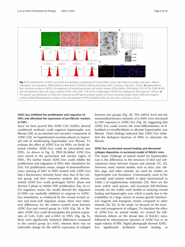
ResultsThe characterization of human ADSCs and ADSC-derivedexosomeADSCs displayed a remarkable fibroblast-like morphologyand had the ability of multiple differentiation potential.Adipogenic differentiation exhibited lipid droplets in thecytoplasm and osteogenic differentiation indicated thepresence of calcium deposition, as evidenced by Oil RedO staining and Alizarin Red S staining, respectively(Fig. 1a). Besides, ADSCs were highly positive for MSC(mesenchymal stem cells) surface markers, includingCD29 (FITC 96.6%), CD44 (PE 98.9%), CD73 (FITC97.7%), and CD90 (FITC 98.3%), but negative for HSC(hematopoietic stem cell) surface markers (CD34 PE 1.4%and CD45 FITC 1.7%) by flow cytometry analysis (Fig. 1b).The features demonstrated the isolated cells were consist-ent with typical ADSC characteristics. Furthermore, wecollected cell-conditioned medium of ADSCs and ex-tracted exosomes. As shown in Fig. 1c, ADSC-Exo pre-sented a cup- or sphere-shaped morphology by TEM,NTA analysis identified the mean diameters of exosomewas 113.6 nm (Fig. 1d), and immunoblotting was per-formed to confirm the presence of known exosomalmarkers (CD63 and CD9, Fig. 1e). The data indicated thatthe nanoparticles were consistent with the definedexosomes.
Li et al. Stem Cell Research & Therapy (2021) 12:221 Page 5 of 16
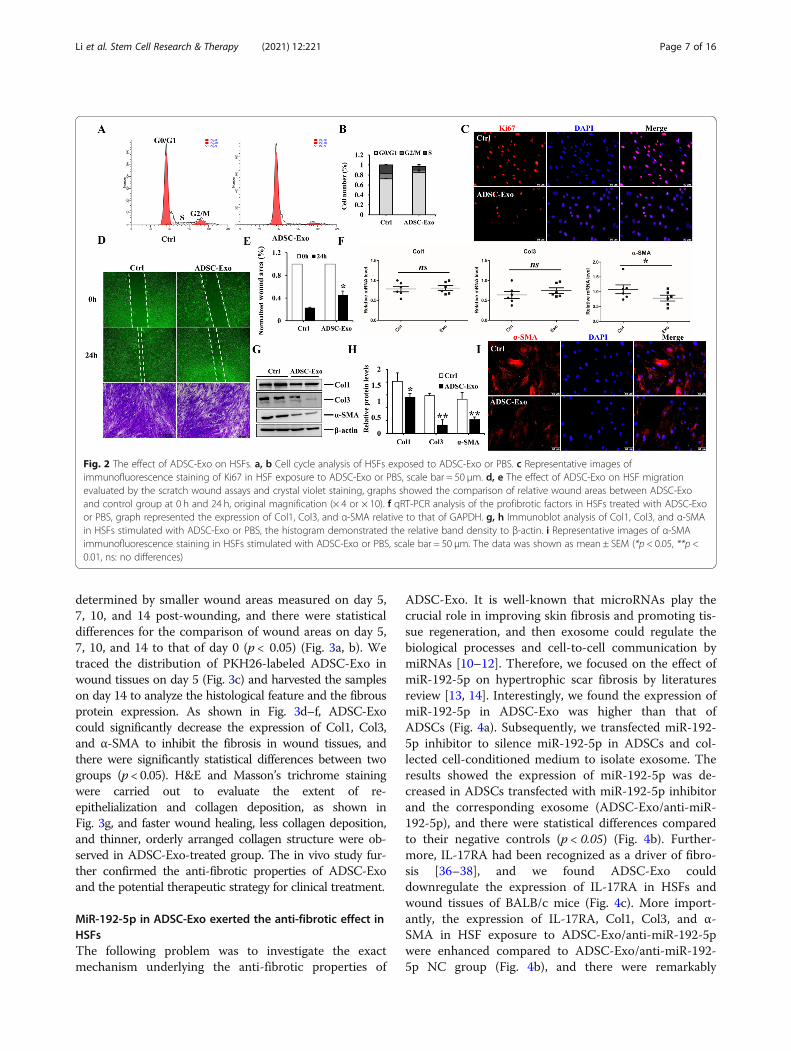
ADSC-Exo inhibited the proliferation and migration ofHSFs and alleviated the expression of pro-fibrotic markersin HSFsSince we have proved that ADSC-CM (ADSCs derivedconditional medium) could suppress hypertrophic scarfibrosis [30], as an essential and executive component ofADSC-CM, we hypothesized exosome played an import-ant role in ameliorating hypertrophic scar fibrosis. Toevaluate the effect of ADSC-Exo on HSFs, we firstly de-tected whether ADSC-Exo could be internalized intoHSFs. As shown in Fig. 1f, PKH-26-labled ADSC-Exowere traced in the perinuclear and nuclear region ofHSFs. We further found ADSC-Exo could inhibit theproliferation and migration of HSFs after stimulation for24 h. For proliferation assays, images of immunofluores-cence staining of Ki67 in HSFs treated with ADSC-Exohad a fluorescence intensity lower than that of the con-trol group, and flow cytometry analysis also demon-strated ADSC-Exo could prolongate G0/G1 phase andshorten S phase to inhibit HSF proliferation (Fig. 2a–c).For migratory assays, the results showed the migrationof HSFs was markedly inhibited in response to ADSC-Exo stimulation, as evidenced by the wound scratch as-says and trans-well migration assays, there were statis-tical differences for the relative scratch areas betweenADSC-Exo and control group (p < 0.05) (Fig. 2d, e). Be-sides, ADSC-Exo could downregulate the protein expres-sion of Col1, Col3, and α-SMA in HSFs (Fig. 2g, h),there were significantly statistical differences comparedto the control group (p < 0.05), whereas there was nonoticeable change for the mRNA expression of collagen
between two groups (Fig. 2f). The mRNA level and theimmunofluorescence intensity of α-SMA were decreasedin HSF exposure to ADSC-Exo (Fig. 2i), suggesting thatADSC-Exo could reverse the trans-differentiation of fi-broblasts to myofibroblasts to alleviate hypertrophic scarfibrosis. These findings indicated that ADSC-Exo inhib-ited the biological function of HSFs to attenuate thefibrosis.
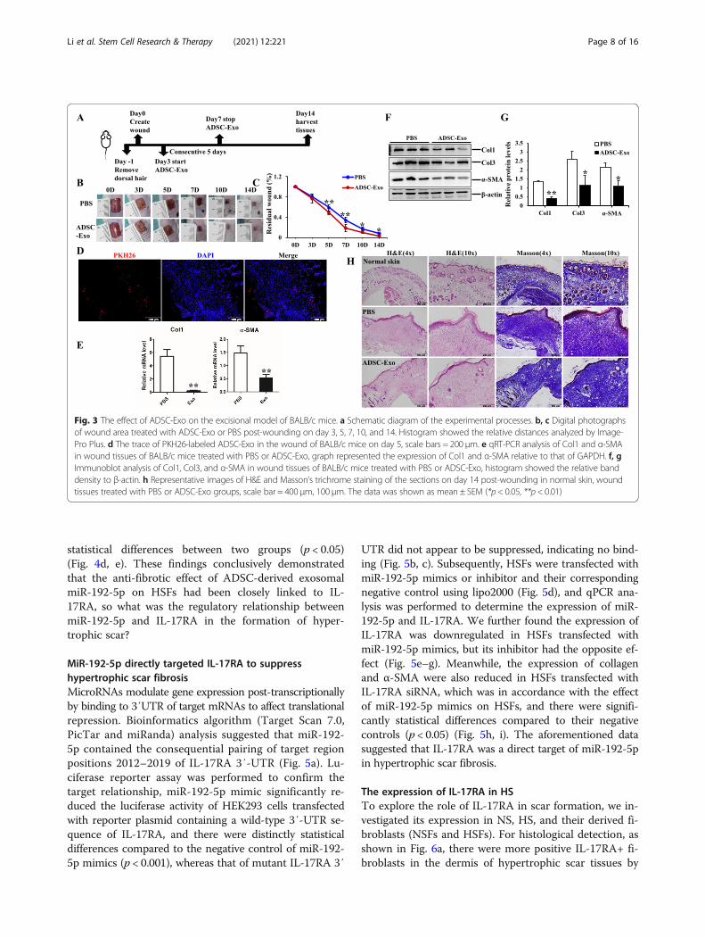
ADSC-Exo accelerated wound healing and decreasedcollagen deposition in excisional model of BALB/c miceThe major challenge of animal model for hypertrophicscar is the differences in the structure of skin and sub-cutaneous tissue between human and animals [31, 32].However, many animal models, such as mice, rats, rab-bits, pigs, and other animals, are used for studies onhypertrophic scar formation. Unfortunately, none of thecurrently used animal models is ideal (summarized inTable 2 of supplementary materials) [33]. Mice are themost widely used species, and excisional full-thicknesswounds are the widely used models in studying woundhealing and hypertrophic scar, which is partly due to theavailability of a large variety of mouse-specific biochem-ical reagents and transgenic strains compared to otheranimals [34, 35]. In the study, we focused on the struc-ture and arrangement of collagen. To elucidate the effectof ADSC-Exo on animal model, we created the full-thickness defects on the dorsal skin of BALB/c mice,followed by subcutaneous injection of ADSC-Exo or anequal volume of PBS. Digital photograph showed ADSC-Exo significantly facilitated wound healing, as
Fig. 1 The identification of ADSCs and ADSC-Exo. a Optical morphology of human ADSCs under light field microscope, scale bars = 400 μm.Adipogenic and osteogenic differentiations measured by Oil Red O staining and Alizarin Red S staining, scale bars = 100 μm. b Representativeflow cytometry analysis of ADSCs, the expression of mesenchymal stem cell surface markers (CD29 96.6%, CD44 98.9%, CD73 97.7%, CD90 98.3%)and hematopoietic stem cell surface markers (CD34 1.4%, CD45 1.7%). c The morphology of ADSC-Exo analyzed by TEM, scale bar = 200 nm. dThe particle size distribution of ADSC-Exo measured by NTA. e Immunoblot analysis of known exosomal markers (CD9, CD63) and negativemarkers (CD68). f Representative images of the internalization of PKH-26-labeled ADSC-Exo into HSFs, scale bars = 25 μm
Li et al. Stem Cell Research & Therapy (2021) 12:221 Page 6 of 16
determined by smaller wound areas measured on day 5,7, 10, and 14 post-wounding, and there were statisticaldifferences for the comparison of wound areas on day 5,7, 10, and 14 to that of day 0 (p < 0.05) (Fig. 3a, b). Wetraced the distribution of PKH26-labeled ADSC-Exo inwound tissues on day 5 (Fig. 3c) and harvested the sampleson day 14 to analyze the histological feature and the fibrousprotein expression. As shown in Fig. 3d–f, ADSC-Exocould significantly decrease the expression of Col1, Col3,and α-SMA to inhibit the fibrosis in wound tissues, andthere were significantly statistical differences between twogroups (p < 0.05). H&E and Masson’s trichrome stainingwere carried out to evaluate the extent of re-epithelialization and collagen deposition, as shown inFig. 3g, and faster wound healing, less collagen deposition,and thinner, orderly arranged collagen structure were ob-served in ADSC-Exo-treated group. The in vivo study fur-ther confirmed the anti-fibrotic properties of ADSC-Exoand the potential therapeutic strategy for clinical treatment.
MiR-192-5p in ADSC-Exo exerted the anti-fibrotic effect inHSFsThe following problem was to investigate the exactmechanism underlying the anti-fibrotic properties of
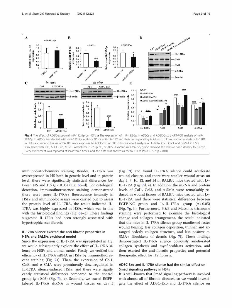
ADSC-Exo. It is well-known that microRNAs play thecrucial role in improving skin fibrosis and promoting tis-sue regeneration, and then exosome could regulate thebiological processes and cell-to-cell communication bymiRNAs [10–12]. Therefore, we focused on the effect ofmiR-192-5p on hypertrophic scar fibrosis by literaturesreview [13, 14]. Interestingly, we found the expression ofmiR-192-5p in ADSC-Exo was higher than that ofADSCs (Fig. 4a). Subsequently, we transfected miR-192-5p inhibitor to silence miR-192-5p in ADSCs and col-lected cell-conditioned medium to isolate exosome. Theresults showed the expression of miR-192-5p was de-creased in ADSCs transfected with miR-192-5p inhibitorand the corresponding exosome (ADSC-Exo/anti-miR-192-5p), and there were statistical differences comparedto their negative controls (p < 0.05) (Fig. 4b). Further-more, IL-17RA had been recognized as a driver of fibro-sis [36–38], and we found ADSC-Exo coulddownregulate the expression of IL-17RA in HSFs andwound tissues of BALB/c mice (Fig. 4c). More import-antly, the expression of IL-17RA, Col1, Col3, and α-SMA in HSF exposure to ADSC-Exo/anti-miR-192-5pwere enhanced compared to ADSC-Exo/anti-miR-192-5p NC group (Fig. 4b), and there were remarkably
Fig. 2 The effect of ADSC-Exo on HSFs. a, b Cell cycle analysis of HSFs exposed to ADSC-Exo or PBS. c Representative images ofimmunofluorescence staining of Ki67 in HSF exposure to ADSC-Exo or PBS, scale bar = 50 μm. d, e The effect of ADSC-Exo on HSF migrationevaluated by the scratch wound assays and crystal violet staining, graphs showed the comparison of relative wound areas between ADSC-Exoand control group at 0 h and 24 h, original magnification (× 4 or × 10). f qRT-PCR analysis of the profibrotic factors in HSFs treated with ADSC-Exoor PBS, graph represented the expression of Col1, Col3, and α-SMA relative to that of GAPDH. g, h Immunoblot analysis of Col1, Col3, and α-SMAin HSFs stimulated with ADSC-Exo or PBS, the histogram demonstrated the relative band density to β-actin. i Representative images of α-SMAimmunofluorescence staining in HSFs stimulated with ADSC-Exo or PBS, scale bar = 50 μm. The data was shown as mean ± SEM (*p < 0.05, **p <0.01, ns: no differences)
Li et al. Stem Cell Research & Therapy (2021) 12:221 Page 7 of 16
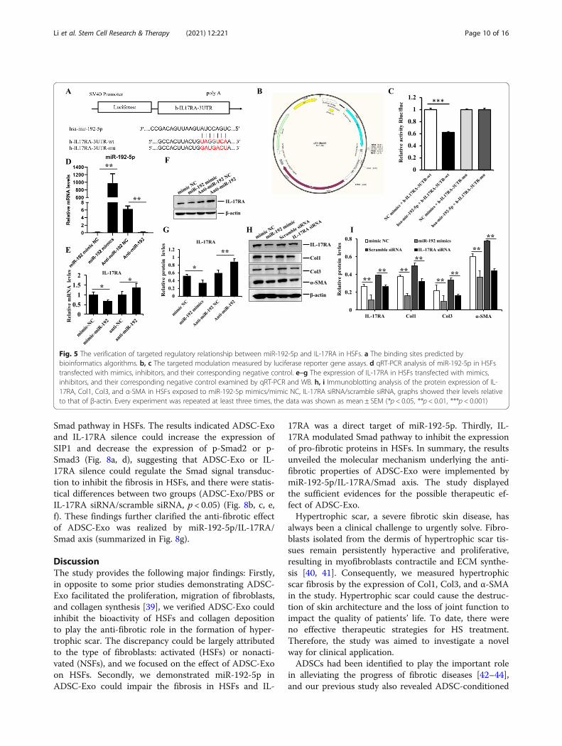
statistical differences between two groups (p < 0.05)(Fig. 4d, e). These findings conclusively demonstratedthat the anti-fibrotic effect of ADSC-derived exosomalmiR-192-5p on HSFs had been closely linked to IL-17RA, so what was the regulatory relationship betweenmiR-192-5p and IL-17RA in the formation of hyper-trophic scar?
MiR-192-5p directly targeted IL-17RA to suppresshypertrophic scar fibrosisMicroRNAs modulate gene expression post-transcriptionallyby binding to 3′UTR of target mRNAs to affect translationalrepression. Bioinformatics algorithm (Target Scan 7.0,PicTar and miRanda) analysis suggested that miR-192-5p contained the consequential pairing of target regionpositions 2012–2019 of IL-17RA 3′-UTR (Fig. 5a). Lu-ciferase reporter assay was performed to confirm thetarget relationship, miR-192-5p mimic significantly re-duced the luciferase activity of HEK293 cells transfectedwith reporter plasmid containing a wild-type 3′-UTR se-quence of IL-17RA, and there were distinctly statisticaldifferences compared to the negative control of miR-192-5p mimics (p < 0.001), whereas that of mutant IL-17RA 3′
UTR did not appear to be suppressed, indicating no bind-ing (Fig. 5b, c). Subsequently, HSFs were transfected withmiR-192-5p mimics or inhibitor and their correspondingnegative control using lipo2000 (Fig. 5d), and qPCR ana-lysis was performed to determine the expression of miR-192-5p and IL-17RA. We further found the expression ofIL-17RA was downregulated in HSFs transfected withmiR-192-5p mimics, but its inhibitor had the opposite ef-fect (Fig. 5e–g). Meanwhile, the expression of collagenand α-SMA were also reduced in HSFs transfected withIL-17RA siRNA, which was in accordance with the effectof miR-192-5p mimics on HSFs, and there were signifi-cantly statistical differences compared to their negativecontrols (p < 0.05) (Fig. 5h, i). The aforementioned datasuggested that IL-17RA was a direct target of miR-192-5pin hypertrophic scar fibrosis.
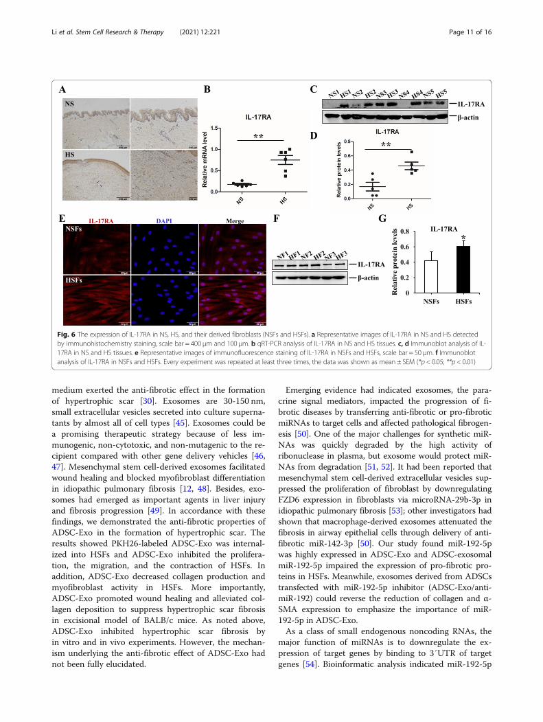
The expression of IL-17RA in HSTo explore the role of IL-17RA in scar formation, we in-vestigated its expression in NS, HS, and their derived fi-broblasts (NSFs and HSFs). For histological detection, asshown in Fig. 6a, there were more positive IL-17RA+ fi-broblasts in the dermis of hypertrophic scar tissues by
Fig. 3 The effect of ADSC-Exo on the excisional model of BALB/c mice. a Schematic diagram of the experimental processes. b, c Digital photographsof wound area treated with ADSC-Exo or PBS post-wounding on day 3, 5, 7, 10, and 14. Histogram showed the relative distances analyzed by Image-Pro Plus. d The trace of PKH26-labeled ADSC-Exo in the wound of BALB/c mice on day 5, scale bars = 200 μm. e qRT-PCR analysis of Col1 and α-SMAin wound tissues of BALB/c mice treated with PBS or ADSC-Exo, graph represented the expression of Col1 and α-SMA relative to that of GAPDH. f, gImmunoblot analysis of Col1, Col3, and α-SMA in wound tissues of BALB/c mice treated with PBS or ADSC-Exo, histogram showed the relative banddensity to β-actin. h Representative images of H&E and Masson’s trichrome staining of the sections on day 14 post-wounding in normal skin, woundtissues treated with PBS or ADSC-Exo groups, scale bar = 400 μm, 100 μm. The data was shown as mean ± SEM (*p< 0.05, **p < 0.01)
Li et al. Stem Cell Research & Therapy (2021) 12:221 Page 8 of 16
immunohistochemistry staining. Besides, IL-17RA wasoverexpressed in HS both in genetic level and in proteinlevel, there were significantly statistical differences be-tween NS and HS (p < 0.05) (Fig. 6b–d). For cytologicaldetection, immunofluorescence staining demonstratedthere were more IL-17RA+ fluorescence intensity inHSFs and immunoblot assays were carried out to assessthe protein level of IL-17RA, the result indicated IL-17RA was highly expressed in HSFs, which was in linewith the histological findings (Fig. 6e–g). These findingssuggested IL-17RA had been strongly associated withhypertrophic scar fibrosis.
IL-17RA silence exerted the anti-fibrotic properties inHSFs and BALB/c excisional modelSince the expression of IL-17RA was upregulated in HS,we would subsequently explore the effect of IL-17RA si-lence on HSFs and animal model. Firstly, we verified theefficiency of IL-17RA siRNA in HSFs by immunofluores-cent staining (Fig. 7a). Then, the expression of Col1,Col3, and α-SMA were prominently downregulated inIL-17RA silence-induced HSFs, and there were signifi-cantly statistical differences compared to the controlgroup (p < 0.05) (Fig. 7b, c). Secondly, we traced EGFP-labeled IL-17RA shRNA in wound tissues on day 5
(Fig. 7f) and found IL-17RA silence could acceleratewound closure, and there were smaller wound areas onday 5, 7, 10, 12, and 14 in BALB/c mice treated with Lv-IL-17RA (Fig. 7d, e). In addition, the mRNA and proteinlevels of Col1, Col3, and α-SMA were remarkably re-duced in wound tissues of BALB/c mice treated with Lv-IL-17RA, and there were statistical differences betweenEGFP-NC group and Lv-IL-17RA group (p < 0.05)(Fig. 7g, h). Furthermore, H&E and Masson’s trichromestaining were performed to examine the histologicalchange and collagen arrangement, the result indicatedthat the mice in IL-17RA silence group manifested fasterwound healing, less collagen deposition, thinner and ar-ranged orderly collagen structure, and less positive α-SMA+ fibroblasts of dermis (Fig. 7i). These findingsdemonstrated IL-17RA silence obviously amelioratedcollagen synthesis and myofibroblasts activation, andthen exerted the anti-fibrotic properties and potentialtherapeutic effect for HS fibrosis.
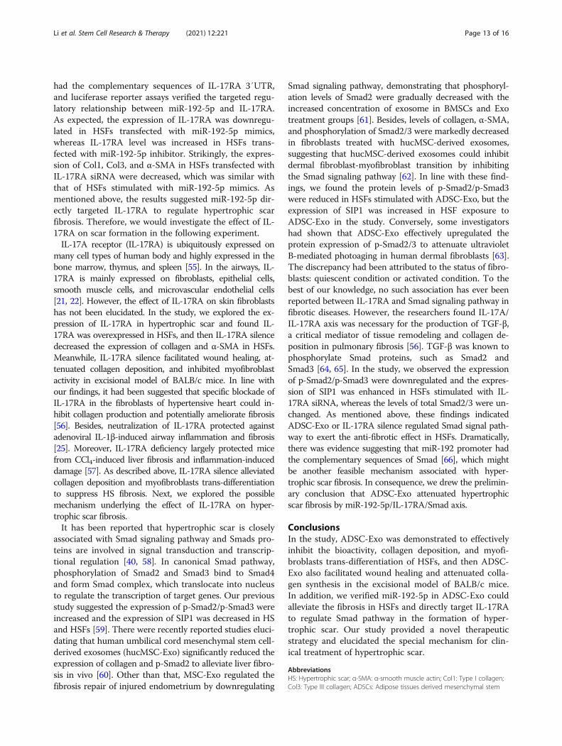
ADSC-Exo and IL-17RA silence had the similar effect onSmad signaling pathway in HSFsIt is well-known that Smad signaling pathway is involvedwith almost all of fibrotic diseases, so we would investi-gate the effect of ADSC-Exo and IL-17RA silence on
Fig. 4 The effect of ADSC-exosomal miR-192-5p on HSFs. a The expression of miR-192-5p in ADSCs and ADSC-Exo. b qRT-PCR analysis of miR-192-5p in ADSCs transfected with miR-192-5p inhibitor NC or anti-miR-192 and their corresponding ADSC-Exo. c Immunoblot analysis of IL-17RAin HSFs and wound tissues of BALB/c mice exposure to ADSC-Exo or PBS. d Immunoblot analysis of IL-17RA, Col1, Col3, and α-SMA in HSFsstimulated with PBS, ADSC-Exo, ADSC-Exo/anti-miR-192-5p NC, or ADSC-Exo/anti-miR-192-5p, graph showed the relative band density to β-actin.Every experiment was repeated at least three times, and the data was shown as mean ± SEM (*p < 0.05; **p < 0.01)
Li et al. Stem Cell Research & Therapy (2021) 12:221 Page 9 of 16
Smad pathway in HSFs. The results indicated ADSC-Exoand IL-17RA silence could increase the expression ofSIP1 and decrease the expression of p-Smad2 or p-Smad3 (Fig. 8a, d), suggesting that ADSC-Exo or IL-17RA silence could regulate the Smad signal transduc-tion to inhibit the fibrosis in HSFs, and there were statis-tical differences between two groups (ADSC-Exo/PBS orIL-17RA siRNA/scramble siRNA, p < 0.05) (Fig. 8b, c, e,f). These findings further clarified the anti-fibrotic effectof ADSC-Exo was realized by miR-192-5p/IL-17RA/Smad axis (summarized in Fig. 8g).
DiscussionThe study provides the following major findings: Firstly,in opposite to some prior studies demonstrating ADSC-Exo facilitated the proliferation, migration of fibroblasts,and collagen synthesis [39], we verified ADSC-Exo couldinhibit the bioactivity of HSFs and collagen depositionto play the anti-fibrotic role in the formation of hyper-trophic scar. The discrepancy could be largely attributedto the type of fibroblasts: activated (HSFs) or nonacti-vated (NSFs), and we focused on the effect of ADSC-Exoon HSFs. Secondly, we demonstrated miR-192-5p inADSC-Exo could impair the fibrosis in HSFs and IL-
17RA was a direct target of miR-192-5p. Thirdly, IL-17RA modulated Smad pathway to inhibit the expressionof pro-fibrotic proteins in HSFs. In summary, the resultsunveiled the molecular mechanism underlying the anti-fibrotic properties of ADSC-Exo were implemented bymiR-192-5p/IL-17RA/Smad axis. The study displayedthe sufficient evidences for the possible therapeutic ef-fect of ADSC-Exo.Hypertrophic scar, a severe fibrotic skin disease, has
always been a clinical challenge to urgently solve. Fibro-blasts isolated from the dermis of hypertrophic scar tis-sues remain persistently hyperactive and proliferative,resulting in myofibroblasts contractile and ECM synthe-sis [40, 41]. Consequently, we measured hypertrophicscar fibrosis by the expression of Col1, Col3, and α-SMAin the study. Hypertrophic scar could cause the destruc-tion of skin architecture and the loss of joint function toimpact the quality of patients’ life. To date, there wereno effective therapeutic strategies for HS treatment.Therefore, the study was aimed to investigate a novelway for clinical application.ADSCs had been identified to play the important role
in alleviating the progress of fibrotic diseases [42–44],and our previous study also revealed ADSC-conditioned
Fig. 5 The verification of targeted regulatory relationship between miR-192-5p and IL-17RA in HSFs. a The binding sites predicted bybioinformatics algorithms. b, c The targeted modulation measured by luciferase reporter gene assays. d qRT-PCR analysis of miR-192-5p in HSFstransfected with mimics, inhibitors, and their corresponding negative control. e–g The expression of IL-17RA in HSFs transfected with mimics,inhibitors, and their corresponding negative control examined by qRT-PCR and WB. h, i Immunoblotting analysis of the protein expression of IL-17RA, Col1, Col3, and α-SMA in HSFs exposed to miR-192-5p mimics/mimic NC, IL-17RA siRNA/scramble siRNA, graphs showed their levels relativeto that of β-actin. Every experiment was repeated at least three times, the data was shown as mean ± SEM (*p < 0.05, **p < 0.01, ***p < 0.001)
Li et al. Stem Cell Research & Therapy (2021) 12:221 Page 10 of 16
medium exerted the anti-fibrotic effect in the formationof hypertrophic scar [30]. Exosomes are 30-150 nm,small extracellular vesicles secreted into culture superna-tants by almost all of cell types [45]. Exosomes could bea promising therapeutic strategy because of less im-munogenic, non-cytotoxic, and non-mutagenic to the re-cipient compared with other gene delivery vehicles [46,47]. Mesenchymal stem cell-derived exosomes facilitatedwound healing and blocked myofibroblast differentiationin idiopathic pulmonary fibrosis [12, 48]. Besides, exo-somes had emerged as important agents in liver injuryand fibrosis progression [49]. In accordance with thesefindings, we demonstrated the anti-fibrotic properties ofADSC-Exo in the formation of hypertrophic scar. Theresults showed PKH26-labeled ADSC-Exo was internal-ized into HSFs and ADSC-Exo inhibited the prolifera-tion, the migration, and the contraction of HSFs. Inaddition, ADSC-Exo decreased collagen production andmyofibroblast activity in HSFs. More importantly,ADSC-Exo promoted wound healing and alleviated col-lagen deposition to suppress hypertrophic scar fibrosisin excisional model of BALB/c mice. As noted above,ADSC-Exo inhibited hypertrophic scar fibrosis byin vitro and in vivo experiments. However, the mechan-ism underlying the anti-fibrotic effect of ADSC-Exo hadnot been fully elucidated.
Emerging evidence had indicated exosomes, the para-crine signal mediators, impacted the progression of fi-brotic diseases by transferring anti-fibrotic or pro-fibroticmiRNAs to target cells and affected pathological fibrogen-esis [50]. One of the major challenges for synthetic miR-NAs was quickly degraded by the high activity ofribonuclease in plasma, but exosome would protect miR-NAs from degradation [51, 52]. It had been reported thatmesenchymal stem cell-derived extracellular vesicles sup-pressed the proliferation of fibroblast by downregulatingFZD6 expression in fibroblasts via microRNA-29b-3p inidiopathic pulmonary fibrosis [53]; other investigators hadshown that macrophage-derived exosomes attenuated thefibrosis in airway epithelial cells through delivery of anti-fibrotic miR-142-3p [50]. Our study found miR-192-5pwas highly expressed in ADSC-Exo and ADSC-exosomalmiR-192-5p impaired the expression of pro-fibrotic pro-teins in HSFs. Meanwhile, exosomes derived from ADSCstransfected with miR-192-5p inhibitor (ADSC-Exo/anti-miR-192) could reverse the reduction of collagen and α-SMA expression to emphasize the importance of miR-192-5p in ADSC-Exo.As a class of small endogenous noncoding RNAs, the
major function of miRNAs is to downregulate the ex-pression of target genes by binding to 3′UTR of targetgenes [54]. Bioinformatic analysis indicated miR-192-5p
Fig. 6 The expression of IL-17RA in NS, HS, and their derived fibroblasts (NSFs and HSFs). a Representative images of IL-17RA in NS and HS detectedby immunohistochemistry staining, scale bar = 400 μm and 100 μm. b qRT-PCR analysis of IL-17RA in NS and HS tissues. c, d Immunoblot analysis of IL-17RA in NS and HS tissues. e Representative images of immunofluorescence staining of IL-17RA in NSFs and HSFs, scale bar = 50 μm. f Immunoblotanalysis of IL-17RA in NSFs and HSFs. Every experiment was repeated at least three times, the data was shown as mean ± SEM (*p < 0.05; **p < 0.01)
Li et al. Stem Cell Research & Therapy (2021) 12:221 Page 11 of 16
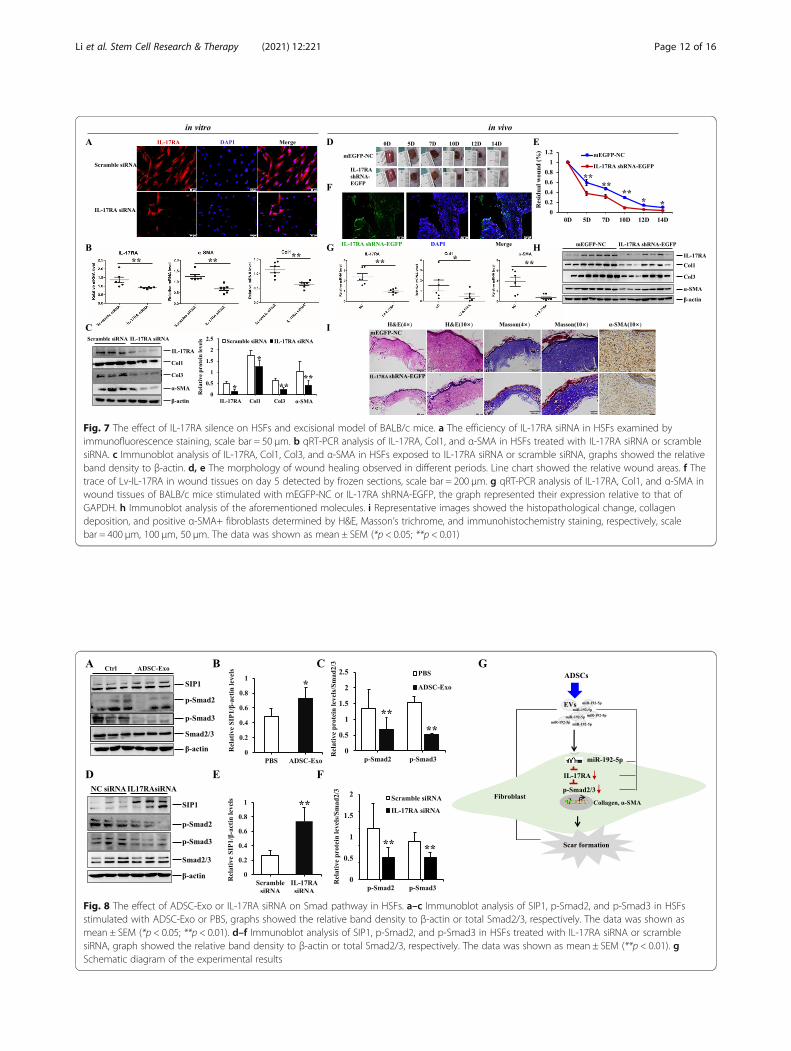
Fig. 7 The effect of IL-17RA silence on HSFs and excisional model of BALB/c mice. a The efficiency of IL-17RA siRNA in HSFs examined byimmunofluorescence staining, scale bar = 50 μm. b qRT-PCR analysis of IL-17RA, Col1, and α-SMA in HSFs treated with IL-17RA siRNA or scramblesiRNA. c Immunoblot analysis of IL-17RA, Col1, Col3, and α-SMA in HSFs exposed to IL-17RA siRNA or scramble siRNA, graphs showed the relativeband density to β-actin. d, e The morphology of wound healing observed in different periods. Line chart showed the relative wound areas. f Thetrace of Lv-IL-17RA in wound tissues on day 5 detected by frozen sections, scale bar = 200 μm. g qRT-PCR analysis of IL-17RA, Col1, and α-SMA inwound tissues of BALB/c mice stimulated with mEGFP-NC or IL-17RA shRNA-EGFP, the graph represented their expression relative to that ofGAPDH. h Immunoblot analysis of the aforementioned molecules. i Representative images showed the histopathological change, collagendeposition, and positive α-SMA+ fibroblasts determined by H&E, Masson’s trichrome, and immunohistochemistry staining, respectively, scalebar = 400 μm, 100 μm, 50 μm. The data was shown as mean ± SEM (*p < 0.05; **p < 0.01)
Fig. 8 The effect of ADSC-Exo or IL-17RA siRNA on Smad pathway in HSFs. a–c Immunoblot analysis of SIP1, p-Smad2, and p-Smad3 in HSFsstimulated with ADSC-Exo or PBS, graphs showed the relative band density to β-actin or total Smad2/3, respectively. The data was shown asmean ± SEM (*p < 0.05; **p < 0.01). d–f Immunoblot analysis of SIP1, p-Smad2, and p-Smad3 in HSFs treated with IL-17RA siRNA or scramblesiRNA, graph showed the relative band density to β-actin or total Smad2/3, respectively. The data was shown as mean ± SEM (**p < 0.01). gSchematic diagram of the experimental results
Li et al. Stem Cell Research & Therapy (2021) 12:221 Page 12 of 16
had the complementary sequences of IL-17RA 3′UTR,and luciferase reporter assays verified the targeted regu-latory relationship between miR-192-5p and IL-17RA.As expected, the expression of IL-17RA was downregu-lated in HSFs transfected with miR-192-5p mimics,whereas IL-17RA level was increased in HSFs trans-fected with miR-192-5p inhibitor. Strikingly, the expres-sion of Col1, Col3, and α-SMA in HSFs transfected withIL-17RA siRNA were decreased, which was similar withthat of HSFs stimulated with miR-192-5p mimics. Asmentioned above, the results suggested miR-192-5p dir-ectly targeted IL-17RA to regulate hypertrophic scarfibrosis. Therefore, we would investigate the effect of IL-17RA on scar formation in the following experiment.IL-17A receptor (IL-17RA) is ubiquitously expressed on
many cell types of human body and highly expressed in thebone marrow, thymus, and spleen [55]. In the airways, IL-17RA is mainly expressed on fibroblasts, epithelial cells,smooth muscle cells, and microvascular endothelial cells[21, 22]. However, the effect of IL-17RA on skin fibroblastshas not been elucidated. In the study, we explored the ex-pression of IL-17RA in hypertrophic scar and found IL-17RA was overexpressed in HSFs, and then IL-17RA silencedecreased the expression of collagen and α-SMA in HSFs.Meanwhile, IL-17RA silence facilitated wound healing, at-tenuated collagen deposition, and inhibited myofibroblastactivity in excisional model of BALB/c mice. In line withour findings, it had been suggested that specific blockade ofIL-17RA in the fibroblasts of hypertensive heart could in-hibit collagen production and potentially ameliorate fibrosis[56]. Besides, neutralization of IL-17RA protected againstadenoviral IL-1β-induced airway inflammation and fibrosis[25]. Moreover, IL-17RA deficiency largely protected micefrom CCl4-induced liver fibrosis and inflammation-induceddamage [57]. As described above, IL-17RA silence alleviatedcollagen deposition and myofibroblasts trans-differentiationto suppress HS fibrosis. Next, we explored the possiblemechanism underlying the effect of IL-17RA on hyper-trophic scar fibrosis.It has been reported that hypertrophic scar is closely
associated with Smad signaling pathway and Smads pro-teins are involved in signal transduction and transcrip-tional regulation [40, 58]. In canonical Smad pathway,phosphorylation of Smad2 and Smad3 bind to Smad4and form Smad complex, which translocate into nucleusto regulate the transcription of target genes. Our previousstudy suggested the expression of p-Smad2/p-Smad3 wereincreased and the expression of SIP1 was decreased in HSand HSFs [59]. There were recently reported studies eluci-dating that human umbilical cord mesenchymal stem cell-derived exosomes (hucMSC-Exo) significantly reduced theexpression of collagen and p-Smad2 to alleviate liver fibro-sis in vivo [60]. Other than that, MSC-Exo regulated thefibrosis repair of injured endometrium by downregulating
Smad signaling pathway, demonstrating that phosphoryl-ation levels of Smad2 were gradually decreased with theincreased concentration of exosome in BMSCs and Exotreatment groups [61]. Besides, levels of collagen, α-SMA,and phosphorylation of Smad2/3 were markedly decreasedin fibroblasts treated with hucMSC-derived exosomes,suggesting that hucMSC-derived exosomes could inhibitdermal fibroblast-myofibroblast transition by inhibitingthe Smad signaling pathway [62]. In line with these find-ings, we found the protein levels of p-Smad2/p-Smad3were reduced in HSFs stimulated with ADSC-Exo, but theexpression of SIP1 was increased in HSF exposure toADSC-Exo in the study. Conversely, some investigatorshad shown that ADSC-Exo effectively upregulated theprotein expression of p-Smad2/3 to attenuate ultravioletB-mediated photoaging in human dermal fibroblasts [63].The discrepancy had been attributed to the status of fibro-blasts: quiescent condition or activated condition. To thebest of our knowledge, no such association has ever beenreported between IL-17RA and Smad signaling pathway infibrotic diseases. However, the researchers found IL-17A/IL-17RA axis was necessary for the production of TGF-β,a critical mediator of tissue remodeling and collagen de-position in pulmonary fibrosis [56]. TGF-β was known tophosphorylate Smad proteins, such as Smad2 andSmad3 [64, 65]. In the study, we observed the expressionof p-Smad2/p-Smad3 were downregulated and the expres-sion of SIP1 was enhanced in HSFs stimulated with IL-17RA siRNA, whereas the levels of total Smad2/3 were un-changed. As mentioned above, these findings indicatedADSC-Exo or IL-17RA silence regulated Smad signal path-way to exert the anti-fibrotic effect in HSFs. Dramatically,there was evidence suggesting that miR-192 promoter hadthe complementary sequences of Smad [66], which mightbe another feasible mechanism associated with hyper-trophic scar fibrosis. In consequence, we drew the prelimin-ary conclusion that ADSC-Exo attenuated hypertrophicscar fibrosis by miR-192-5p/IL-17RA/Smad axis.
ConclusionsIn the study, ADSC-Exo was demonstrated to effectivelyinhibit the bioactivity, collagen deposition, and myofi-broblasts trans-differentiation of HSFs, and then ADSC-Exo also facilitated wound healing and attenuated colla-gen synthesis in the excisional model of BALB/c mice.In addition, we verified miR-192-5p in ADSC-Exo couldalleviate the fibrosis in HSFs and directly target IL-17RAto regulate Smad pathway in the formation of hyper-trophic scar. Our study provided a novel therapeuticstrategy and elucidated the special mechanism for clin-ical treatment of hypertrophic scar.
AbbreviationsHS: Hypertrophic scar; α-SMA: α-smooth muscle actin; Col1: Type I collagen;Col3: Type III collagen; ADSCs: Adipose tissues derived mesenchymal stem
Li et al. Stem Cell Research & Therapy (2021) 12:221 Page 13 of 16
cells; ADSC-CM: ADSC-conditioned medium; ADSC-Exo: ADSC-derivedexosome; DMEM: Dulbecco’s modified Eagle’s medium; FBS: Fetal bovineserum; siRNA: Small interfering RNA; DAPI: 4′,6-Diamidino-2-phenylindole;GAPDH: Glyceraldehyde-3-phosphate dehydrogenase; BSA: Bovine serumalbumin; NS: Normal skin; HSFs: HS-derived fibroblasts; NSFs: NS-derivedfibroblasts; TEM: Transmission electron microscope; NTA: Nanoparticletracking analysis; 3′UTR: 3′-Untranslated regions; SIP1: Smad interactingprotein 1
Supplementary InformationThe online version contains supplementary material available at https://doi.org/10.1186/s13287-021-02290-0.
Additional file 1.
AcknowledgementsWe acknowledge the editors and reviewers for their helpful comments onthis paper.
Authors’ contributionsYL contributed to the research design, performed all experiments, collectedand analyzed data, and wrote the manuscript. ZJ, JS, and KL participated inthe experiments and manuscript drafting. XW and YJ performed experimentsand manuscript drafting. SK and WZ participated in the isolation and cultureof primary cells. TH, YW, and JL provided the clinical samples for theexperiment. TW, ZZ, and DH contributed to the experimental design andmanuscript writing. All authors read and approved the final manuscript.
FundingThis work was supported by National Natural Science Foundation of China(No.81901965, No.81801920, No.81772071, No.81971835), Natural ScienceBasic Research Program of Shaanxi (No.2018JQ8048) and Project of XijingHospital [No. XJZT18MJ37], and Science and Technology Program of ShaanxiProvince (No.2019JM-378).
Availability of data and materialsNot applicable.
Declarations
Ethics approval and consent to participateAll protocols involved with human samples in the study were approved bythe Ethics Committee of Xijing Hospital, First Affiliated Hospital of FourthMilitary Medical University (KY20193244).All animal experimental protocols were approved and performed in strictaccordance with Experimental Animal Committee of Fourth Military MedicalUniversity (Xi′an, China).
Consent for publicationNot applicable.
Competing interestsThe authors declare that they have no competing interests.
Author details1Department of Burns and Cutaneous Surgery, Xijing Hospital, Fourth MilitaryMedical University, 127 West Chang-le Road, Xi’an 710032, Shaanxi, China.2Department of Plastics and Aesthetic Surgery, The First Affiliated Hospital ofXi′an Medical University, No.48 West Fenghao Road, Xi’an 710077, Shaanxi,China.
Received: 16 January 2021 Accepted: 15 March 2021
References1. Perry DM, McGrouther DA, Bayat A. Current tools for noninvasive objective
assessment of skin scars. Plast Reconstr Surg. 2010;126(3):912–23. https://doi.org/10.1097/PRS.0b013e3181e6046b.
2. Coentro JQ, Pugliese E, Hanley G, Raghunath M, Zeugolis DI. Current andupcoming therapies to modulate skin scarring and fibrosis. Adv Drug DelivRev. 2019;146:37–59. https://doi.org/10.1016/j.addr.2018.08.009.
3. Zhou Y, Yuan J, Zhou B, Lee AJ, Lee AJ, Ghawji M Jr, Yoo TJ. The therapeuticefficacy of human adipose tissue-derived mesenchymal stem cells onexperimental autoimmune hearing loss in mice. Immunology. 2011;133(1):133–40. https://doi.org/10.1111/j.1365-2567.2011.03421.x.
4. Li L, Zhang S, Zhang Y, Yu B, Xu Y, Guan Z. Paracrine action mediate theantifibrotic effect of transplanted mesenchymal stem cells in a rat model ofglobal heart failure. Mol Biol Rep. 2009;36(4):725–31. https://doi.org/10.1007/s11033-008-9235-2.
5. Han HS, Lee H, You D, Nguyen VQ, Song DG, Oh BH, Shin S, Choi JS, Kim JD,Pan CH, Jo DG, Cho YW, Choi KY, Park JH. Human adipose stem cell-derivedextracellular nanovesicles for treatment of chronic liver fibrosis. J ControlRelease. 2020;320:328–36. https://doi.org/10.1016/j.jconrel.2020.01.042.
6. Lee TM, Harn HJ, Chiou TW, Chuang MH, Chen CH, Chuang CH, Lin PC, LinSZ. Preconditioned adipose-derived stem cells ameliorate cardiac fibrosis byregulating macrophage polarization in infarcted rat hearts through thePI3K/STAT3 pathway. Lab Invest. 2019;99(5):634–47.
7. Dinh PC, Paudel D, Brochu H, Popowski KD, Gracieux MC, Cores J, Huang K,Hensley MT, Harrell E, Vandergriff AC, et al. Inhalation of lung spheroid cellsecretome and exosomes promotes lung repair in pulmonary fibrosis. NatCommun. 2020;11(1):1064. https://doi.org/10.1038/s41467-020-14344-7.
8. Wang X, Ma Y, Gao Z, Yang J. Human adipose-derived stem cells inhibitbioactivity of keloid fibroblasts. Stem Cell Res Ther. 2018;9(1):40. https://doi.org/10.1186/s13287-018-0786-4.
9. Eguchi A, Kostallari E, Feldstein AE, Shah VH. Extracellular vesicles, the liquidbiopsy of the future. J Hepatol. 2019;70(6):1292–4. https://doi.org/10.1016/j.jhep.2019.01.030.
10. Qu Y, Zhang Q, Cai X, Li F, Ma Z, Xu M, Lu L. Exosomes derived from miR-181-5p-modified adipose-derived mesenchymal stem cells prevent liverfibrosis via autophagy activation. J Cell Mol Med. 2017;21(10):2491–502.https://doi.org/10.1111/jcmm.13170.
11. Deng S, Zhou X, Ge Z, Song Y, Wang H, Liu X, Zhang D. Exosomes fromadipose-derived mesenchymal stem cells ameliorate cardiac damage aftermyocardial infarction by activating S1P/SK1/S1PR1 signaling and promotingmacrophage M2 polarization. Int J Biochem Cell Biol. 2019;114:105564.https://doi.org/10.1016/j.biocel.2019.105564.
12. Fang S, Xu C, Zhang Y, Xue C, Yang C, Bi H, Qian X, Wu M, Ji K, Zhao Y,Wang Y, Liu H, Xing X. Umbilical cord-derived mesenchymal stem cell-derived exosomal microRNAs suppress myofibroblast differentiation byinhibiting the transforming growth factor-beta/SMAD2 pathway duringwound healing. Stem Cells Transl Med. 2016;5(10):1425–39. https://doi.org/10.5966/sctm.2015-0367.
13. Krupa A, Jenkins R, Luo DD, Lewis A, Phillips A, Fraser D. Loss of MicroRNA-192 promotes fibrogenesis in diabetic nephropathy. J Am Soc Nephrol.2010;21(3):438–47. https://doi.org/10.1681/ASN.2009050530.
14. Wang B, Herman-Edelstein M, Koh P, Burns W, Jandeleit-Dahm K, Watson A,Saleem M, Goodall GJ, Twigg SM, Cooper ME, Kantharidis P. E-cadherinexpression is regulated by miR-192/215 by a mechanism that isindependent of the profibrotic effects of transforming growth factor-beta.Diabetes. 2010;59(7):1794–802. https://doi.org/10.2337/db09-1736.
15. Jiang X, Tsitsiou E, Herrick SE, Lindsay MA. MicroRNAs and the regulation offibrosis. FEBS J. 2010;277(9):2015–21. https://doi.org/10.1111/j.1742-4658.2010.07632.x.
16. Lagos-Quintana M, Rauhut R, Meyer J, Borkhardt A, Tuschl T. NewmicroRNAs from mouse and human. Rna. 2003;9(2):175–9. https://doi.org/10.1261/rna.2146903.
17. Lim LP, Glasner ME, Yekta S, Burge CB, Bartel DP. Vertebrate microRNA genes.Science. 2003;299(5612):1540. https://doi.org/10.1126/science.1080372.
18. Hino K, Tsuchiya K, Fukao T, Kiga K, Okamoto R, Kanai T, Watanabe M.Inducible expression of microRNA-194 is regulated by HNF-1alpha duringintestinal epithelial cell differentiation. Rna. 2008;14(7):1433–42. https://doi.org/10.1261/rna.810208.
19. Jenkins RH, Martin J, Phillips AO, Bowen T, Fraser DJ. Pleiotropy ofmicroRNA-192 in the kidney. Biochem Soc Trans. 2012;40(4):762–7. https://doi.org/10.1042/BST20120085.
20. Yao Z, Fanslow WC, Seldin MF, Rousseau AM, Painter SL, Comeau MR,Cohen JI, Spriggs MK. Herpesvirus Saimiri encodes a new cytokine, IL-17,which binds to a novel cytokine receptor. Immunity. 1995;3(6):811–21.https://doi.org/10.1016/1074-7613(95)90070-5.
Li et al. Stem Cell Research & Therapy (2021) 12:221 Page 14 of 16
21. Halwani R, Al-Kufaidy R, Vazquez-Tello A, Pureza MA, BaHammam AS, Al-Jahdali H, Alnassar SA, Hamid Q, Al-Muhsen S. IL-17 enhances chemotaxis ofprimary human B cells during asthma. PLoS One. 2014;9(12):e114604.https://doi.org/10.1371/journal.pone.0114604.
22. Roussel L, Rousseau S. IL-17 primes airway epithelial cells lacking functionalcystic fibrosis transmembrane conductance regulator (CFTR) to increaseNOD1 responses. Biochem Biophys Res Commun. 2010;391(1):505–9. https://doi.org/10.1016/j.bbrc.2009.11.088.
23. Meng F, Wang K, Aoyama T, Grivennikov SI, Paik Y, Scholten D, Cong M,Iwaisako K, Liu X, Zhang M, Österreicher CH, Stickel F, Ley K, Brenner DA,Kisseleva T. Interleukin-17 signaling in inflammatory, Kupffer cells, andhepatic stellate cells exacerbates liver fibrosis in mice. Gastroenterology.2012;143(3):765–76 e763. https://doi.org/10.1053/j.gastro.2012.05.049.
24. Liu W, Wang X, Feng W, Li S, Tian W, Xu T, Song Y, Zhang Z. Lentivirusmediated IL-17R blockade improves diastolic cardiac function inspontaneously hypertensive rats. Exp Mol Pathol. 2011;91(1):362–7. https://doi.org/10.1016/j.yexmp.2011.04.003.
25. Yanagisawa H, Hashimoto M, Minagawa S, Takasaka N, Ma R, Moermans C,Ito S, Araya J, Budelsky A, Goodsell A, Baron JL, Nishimura SL. Role of IL-17Ain murine models of COPD airway disease. Am J Physiol Lung Cell MolPhysiol. 2017;312(1):L122–30. https://doi.org/10.1152/ajplung.00301.2016.
26. Verrecchia F, Mauviel A, Farge D. Transforming growth factor-beta signalingthrough the Smad proteins: role in systemic sclerosis. Autoimmun Rev.2006;5(8):563–9. https://doi.org/10.1016/j.autrev.2006.06.001.
27. Sato M, Muragaki Y, Saika S, Roberts AB, Ooshima A. Targeted disruption ofTGF-beta1/Smad3 signaling protects against renal tubulointerstitial fibrosisinduced by unilateral ureteral obstruction. J Clin Invest. 2003;112(10):1486–94. https://doi.org/10.1172/JCI200319270.
28. Bai X, Yan Y, Song YH, Seidensticker M, Rabinovich B, Metzele R, Bankson JA,Vykoukal D, Alt E. Both cultured and freshly isolated adipose tissue-derivedstem cells enhance cardiac function after acute myocardial infarction. EurHeart J. 2010;31(4):489–501. https://doi.org/10.1093/eurheartj/ehp568.
29. Thery C, Amigorena S, Raposo G, Clayton A. Isolation and characterization ofexosomes from cell culture supernatants and biological fluids. Curr ProtocCell Biol. 2006;Chapter 3:Unit 3 22.
30. Li Y, Zhang W, Gao J, Liu J, Wang H, Li J, Yang X, He T, Guan H, Zheng Z,Han S, Dong M, Han J, Shi J, Hu D. Adipose tissue-derived stem cellssuppress hypertrophic scar fibrosis via the p38/MAPK signaling pathway.Stem Cell Res Ther. 2016;7(1):102. https://doi.org/10.1186/s13287-016-0356-6.
31. Ramos ML, Gragnani A, Ferreira LM. Is there an ideal animal model to studyhypertrophic scarring? J Burn Care Res. 2008;29(2):363–8. https://doi.org/10.1097/BCR.0b013e3181667557.
32. Domergue S, Jorgensen C, Noel D. Advances in research in animal modelsof burn-related hypertrophic scarring. J Burn Care Res. 2015;36(5):e259–66.https://doi.org/10.1097/BCR.0000000000000167.
33. van den Broek LJ, Limandjaja GC, Niessen FB, Gibbs S. Human hypertrophicand keloid scar models: principles, limitations and future challenges from atissue engineering perspective. Exp Dermatol. 2014;23(6):382–6. https://doi.org/10.1111/exd.12419.
34. Canesso MC, Vieira AT, Castro TB, Schirmer BG, Cisalpino D, Martins FS,Rachid MA, Nicoli JR, Teixeira MM, Barcelos LS. Skin wound healing isaccelerated and scarless in the absence of commensal microbiota. JImmunol. 2014;193(10):5171–80. https://doi.org/10.4049/jimmunol.1400625.
35. Willenborg S, Eckes B, Brinckmann J, Krieg T, Waisman A, Hartmann K, RoersA, Eming SA. Genetic ablation of mast cells redefines the role of mast cellsin skin wound healing and bleomycin-induced fibrosis. J Invest Dermatol.2014;134(7):2005–15. https://doi.org/10.1038/jid.2014.12.
36. Zhang J, Wang D, Wang L, Wang S, Roden AC, Zhao H, Li X, Prakash YS,Matteson EL, Tschumperlin DJ, Vassallo R. Profibrotic effect of IL-17A andelevated IL-17RA in idiopathic pulmonary fibrosis and rheumatoid arthritis-associated lung disease support a direct role for IL-17A/IL-17RA in humanfibrotic interstitial lung disease. Am J Physiol Lung Cell Mol Physiol. 2019;316(3):L487–97. https://doi.org/10.1152/ajplung.00301.2018.
37. Liu Y, Zhu H, Su Z, Sun C, Yin J, Yuan H, Sandoghchian S, Jiao Z, Wang S, XuH. IL-17 contributes to cardiac fibrosis following experimental autoimmunemyocarditis by a PKCbeta/Erk1/2/NF-kappaB-dependent signaling pathway.Int Immunol. 2012;24(10):605–12. https://doi.org/10.1093/intimm/dxs056.
38. Xu J, Ma HY, Liu X, Rosenthal S, Baglieri J, McCubbin R, Sun M, Koyama Y,Geoffroy CG, Saijo K, et al. Blockade of IL-17 signaling reverses alcohol-induced liver injury and excessive alcohol drinking in mice. JCI insight. 2020;5(3):e131277. https://doi.org/10.1172/jci.insight.131277.
39. Zhang W, Bai X, Zhao B, Li Y, Zhang Y, Li Z, Wang X, Luo L, Han F, Zhang J,Han S, Cai W, Su L, Tao K, Shi J, Hu D. Cell-free therapy based on adiposetissue stem cell-derived exosomes promotes wound healing via the PI3K/Akt signaling pathway. Exp Cell Res. 2018;370(2):333–42. https://doi.org/10.1016/j.yexcr.2018.06.035.
40. Shao T, Tang W, Li Y, Gao D, Lv K, He P, Song Y, Gao S, Liu M, Chen Y, Yi Z.Research on function and mechanisms of a novel small molecule WG449Efor hypertrophic scar. J Eur Acad Dermatol Venereol. 2020;34(3):608–18.https://doi.org/10.1111/jdv.16028.
41. Liu J, Zhao B, Zhu H, Pan Q, Cai M, Bai X, Li X, Hu X, Zhang M, Shi J, Zheng Z,Yang A, Hu D. Wnt4 negatively regulates the TGF-beta1-induced humandermal fibroblast-to-myofibroblast transition via targeting Smad3 and ERK. CellTissue Res. 2020;379(3):537–48. https://doi.org/10.1007/s00441-019-03110-x.
42. Lam MT, Nauta A, Meyer NP, Wu JC, Longaker MT. Effective delivery of stemcells using an extracellular matrix patch results in increased cell survival andproliferation and reduced scarring in skin wound healing. Tissue Eng A.2013;19(5–6):738–47. https://doi.org/10.1089/ten.tea.2012.0480.
43. Yu LH, Kim MH, Park TH, Cha KS, Kim YD, Quan ML, Rho MS, Seo SY, JungJS. Improvement of cardiac function and remodeling by transplantingadipose tissue-derived stromal cells into a mouse model of acutemyocardial infarction. Int J Cardiol. 2010;139(2):166–72. https://doi.org/10.1016/j.ijcard.2008.10.024.
44. Lee SH, Lee EJ, Lee SY, Kim JH, Shim JJ, Shin C, In KH, Kang KH, Uhm CS,Kim HK, et al. The effect of adipose stem cell therapy on pulmonary fibrosisinduced by repetitive intratracheal bleomycin in mice. Exp Lung Res. 2014;40(3):117–25. https://doi.org/10.3109/01902148.2014.881930.
45. Mathivanan S, Fahner CJ, Reid GE, Simpson RJ. ExoCarta 2012: database ofexosomal proteins, RNA and lipids. Nucleic Acids Res. 2012;40(Databaseissue):D1241–4. https://doi.org/10.1093/nar/gkr828.
46. Lou G, Chen Z, Zheng M, Liu Y. Mesenchymal stem cell-derived exosomesas a new therapeutic strategy for liver diseases. Exp Mol Med. 2017;49(6):e346. https://doi.org/10.1038/emm.2017.63.
47. Zhao AG, Shah K, Cromer B, Sumer H. Mesenchymal stem cell-derivedextracellular vesicles and their therapeutic potential. Stem Cells Int. 2020;2020:8825771.
48. Shentu TP, Huang TS, Cernelc-Kohan M, Chan J, Wong SS, Espinoza CR, TanC, Gramaglia I, van der Heyde H, Chien S, Hagood JS. Thy-1 dependentuptake of mesenchymal stem cell-derived extracellular vesicles blocksmyofibroblastic differentiation. Sci Rep. 2017;7(1):18052. https://doi.org/10.1038/s41598-017-18288-9.
49. Hirsova P, Ibrahim SH, Verma VK, Morton LA, Shah VH, LaRusso NF, Gores GJ,Malhi H. Extracellular vesicles in liver pathobiology: small particles with bigimpact. Hepatology. 2016;64(6):2219–33. https://doi.org/10.1002/hep.28814.
50. Guiot J, Cambier M, Boeckx A, Henket M, Nivelles O, Gester F, Louis E,Malaise M, Dequiedt F, Louis R, Struman I, Njock MS. Macrophage-derivedexosomes attenuate fibrosis in airway epithelial cells through delivery ofantifibrotic miR-142-3p. Thorax. 2020;75(10):870–81. https://doi.org/10.1136/thoraxjnl-2019-214077.
51. Hu C, Meiners S, Lukas C, Stathopoulos GT, Chen J. Role of exosomalmicroRNAs in lung cancer biology and clinical applications. Cell Prolif. 2020;53(6):e12828. https://doi.org/10.1111/cpr.12828.
52. Vignard V, Labbe M, Marec N, Andre-Gregoire G, Jouand N, Fonteneau JF,Labarriere N, Fradin D. MicroRNAs in tumor exosomes drive immune escapein melanoma. Cancer Immunol Res. 2020;8(2):255–67. https://doi.org/10.1158/2326-6066.CIR-19-0522.
53. Wan X, Chen S, Fang Y, Zuo W, Cui J, Xie S. Mesenchymal stem cell-derivedextracellular vesicles suppress the fibroblast proliferation by downregulatingFZD6 expression in fibroblasts via micrRNA-29b-3p in idiopathic pulmonaryfibrosis. J Cell Physiol. 2020;235(11):8613–25. https://doi.org/10.1002/jcp.29706.
54. Baek D, Villen J, Shin C, Camargo FD, Gygi SP, Bartel DP. The impact ofmicroRNAs on protein output. Nature. 2008;455(7209):64–71. https://doi.org/10.1038/nature07242.
55. Zhang SC, Zheng YH, Yu PP, Min TH, Yu FX, Ye C, Xie YK, Zhang QY.Lentiviral vector-mediated down-regulation of IL-17A receptor in hepaticstellate cells results in decreased secretion of IL-6. World J Gastroenterol.2012;18(28):3696–704. https://doi.org/10.3748/wjg.v18.i28.3696.
56. Hasan SA, Eksteen B, Reid D, Paine HV, Alansary A, Johannson K, Gwozd C,Goring KA, Vo T, Proud D, et al. Role of IL-17A and neutrophils in fibrosis inexperimental hypersensitivity pneumonitis. J Allergy Clin Immunol. 2013;131(6):1663–73. https://doi.org/10.1016/j.jaci.2013.01.015.
Li et al. Stem Cell Research & Therapy (2021) 12:221 Page 15 of 16
57. Tan Z, Qian X, Jiang R, Liu Q, Wang Y, Chen C, Wang X, Ryffel B, Sun B. IL-17A plays a critical role in the pathogenesis of liver fibrosis through hepaticstellate cell activation. J Immunol. 2013;191(4):1835–44. https://doi.org/10.4049/jimmunol.1203013.
58. Kretzschmar M, Massague J. SMADs: mediators and regulators of TGF-betasignaling. Curr Opin Genet Dev. 1998;8(1):103–11. https://doi.org/10.1016/S0959-437X(98)80069-5.
59. Fang X, Hu X, Zheng Z, Tao K, Wang H, Guan H, Shi J, Ji P, Cai W, Bai X, ZhuX, Han J, Liu J, Hu D. Smad interacting protein 1 influences transforminggrowth factor-beta1/Smad signaling in extracellular matrix proteinproduction and hypertrophic scar formation. J Mol Histol. 2019;50(6):503–14.https://doi.org/10.1007/s10735-019-09844-w.
60. Li T, Yan Y, Wang B, Qian H, Zhang X, Shen L, Wang M, Zhou Y, Zhu W, LiW, Xu W. Exosomes derived from human umbilical cord mesenchymal stemcells alleviate liver fibrosis. Stem Cells Dev. 2013;22(6):845–54. https://doi.org/10.1089/scd.2012.0395.
61. Gao W, Wang X, Si Y, Pang J, Liu H, Li S, Ding Q, Wang Y. Exosome derivedfrom ADSCs attenuates ultraviolet B-mediated photoaging in human dermalfibroblasts. Photochem Photobiol. 2020. https://doi.org/10.1111/php.13370.
62. Hu J, Chen Y, Huang Y, Su Y. Human umbilical cord mesenchymal stemcell-derived exosomes suppress dermal fibroblasts-myofibroblats transitionvia inhibiting the TGF-beta1/Smad 2/3 signaling pathway. Exp Mol Pathol.2020;115:104468. https://doi.org/10.1016/j.yexmp.2020.104468.
63. Yao Y, Chen R, Wang G, Zhang Y, Liu F. Exosomes derived frommesenchymal stem cells reverse EMT via TGF-beta1/Smad pathway andpromote repair of damaged endometrium. Stem Cell Res Ther. 2019;10(1):225. https://doi.org/10.1186/s13287-019-1332-8.
64. Inazaki K, Kanamaru Y, Kojima Y, Sueyoshi N, Okumura K, Kaneko K,Yamashiro Y, Ogawa H, Nakao A. Smad3 deficiency attenuates renal fibrosis,inflammation,and apoptosis after unilateral ureteral obstruction. Kidney Int.2004;66(2):597–604. https://doi.org/10.1111/j.1523-1755.2004.00779.x.
65. Kim JH, Kim BK, Moon KC, Hong HK, Lee HS. Activation of the TGF-beta/Smad signaling pathway in focal segmental glomerulosclerosis. Kidney Int.2003;64(5):1715–21. https://doi.org/10.1046/j.1523-1755.2003.00288.x.
66. Chung AC, Huang XR, Meng X, Lan HY. miR-192 mediates TGF-beta/Smad3-driven renal fibrosis. J Am Soc Nephrol. 2010;21(8):1317–25. https://doi.org/10.1681/ASN.2010020134.
Publisher’s NoteSpringer Nature remains neutral with regard to jurisdictional claims inpublished maps and institutional affiliations.
Li et al. Stem Cell Research & Therapy (2021) 12:221 Page 16 of 16