experimental infection of dogs with leishmania and saliva ... dj... · figure 2. parasitological...
TRANSCRIPT

Experimental Infection of Dogs with Leishmania andSaliva as a Model to Study Canine Visceral LeishmaniasisDirceu Joaquim Costa1, Rayssa M. de Araujo Carvalho1, Melissa Abbehusen1, Clarissa Teixeira1,
Maiana Pitombo1, Joelma Trigo1, Flavia Nascimento4, Lucilene Amorim4, Ana Lucia Abreu-Silva5,
Maria do Socorro Pires Cruz6, Jose Carlos Miranda1, Kyoshi Fukutani1, Camila I. de Oliveira1,7,
Aldina Barral1,2,7, Manoel Barral-Netto1,2,7, Claudia Brodskyn1,3,6*
1 Centro de Pesquisa Goncalo Moniz, FIOCRUZ-BA, Bahia, Brazil, 2 Faculdade de Medicina, Universidade Federal da Bahia, Bahia, Brazil, 3 Instituto de Ciencias da Saude,
Universidade Federal da Bahia, Bahia, Brazil, 4 Laboratorio de Imunofiosiologia, Departamento de Patologia Universidade Federal do Maranhao, Maranhao, Brazil,
5 Departamento de Patologia, Universidade Estadual do Maranhao, Maranhao, Brazil, 6 Departamento de Morfofisiologia Veterinaria Centro de Ciencias Agrarias,
Universidade Federal do Piauı, Piauı, Brazil, 7 Instituto de Investigacao em Imunologia, Sao Paulo, Brazil
Abstract
Background: Canine Visceral Leishmaniasis (CVL) is a zoonotic disease caused by Leishmania infantum, transmitted by thebite of Lutzomyia longipalpis sand flies. Dogs are the main domestic reservoir of the parasite. The establishment of anexperimental model that partially reproduces natural infection in dogs is very important to test vaccine candidates, mainlyregarding those that use salivary proteins from the vector and new therapeutical approaches.
Methodology/Principal Findings: In this report, we describe an experimental infection in dogs, using intradermal injectionof Leishmania infantum plus salivary gland homogenate (SGH) of Lutzomyia longipalpis. Thirty-five dogs were infected with16107 parasites combined with five pairs of Lutzomyia longipalpis salivary glands and followed for 450 days after infectionand clinical, immunological and parasitological parameters were evaluated. Two hundred and ten days after infection weobserved that 31,4% of dogs did not display detectable levels of anti-Leishmania antibodies but all presented differentnumbers of parasites in the lymph nodes. Animals with a positive xenodiagnosis had at least 3,356105 parasites in theirlymph nodes. An increase of IFN-c and IL-10 levels was detected during infection. Twenty two percent of dogs developedsymptoms of CVL during infection.
Conclusion: The infection model described here shows some degree of similarity when compared with naturally infecteddogs opening new perspectives for the study of CVL using an experimental model that employs the combination ofparasites and sand fly saliva both present during natural transmission.
Citation: Costa DJ, Carvalho RMdA, Abbehusen M, Teixeira C, Pitombo M, et al. (2013) Experimental Infection of Dogs with Leishmania and Saliva as a Model toStudy Canine Visceral Leishmaniasis. PLoS ONE 8(4): e60535. doi:10.1371/journal.pone.0060535
Editor: Simona Stager, INRS - Institut Armand Frappier, Canada
Received December 11, 2012; Accepted February 27, 2013; Published April 5, 2013
Copyright: � 2013 Costa et al. This is an open-access article distributed under the terms of the Creative Commons Attribution License, which permitsunrestricted use, distribution, and reproduction in any medium, provided the original author and source are credited.
Funding: The work was funded by RENORBIO/CNPq, number 554753/2006-5. The funders had no role in study design, data collection and analysis, decision topublish, or preparation of the manuscript.
Competing Interests: The authors have declared that no competing interests exist.
* E-mail: [email protected]
Introduction
Canine visceral leishmaniasis (CVL) is caused by an intracellular
protozoan parasite Leishmania infantum. It is endemic in the
Mediterranean Basin, South America and parts of Asia [1].
Domestic dogs are the main reservoirs and different control
strategies, such as the use of insecticide impregnated collars or
elimination of infected dogs have not been effective to decrease
human incidence of VL [2]. Development of a vaccine for CVL
has been identified as a research priority by WHO/TDR [3] and
mathematical models have highlighted canine vaccination as the
potentially most practical and effective means of impacting disease
control in humans [4]. Also, since dogs present many symptoms
observed in humans, with a long period of asymptomatic infection
followed by wasting, anaemia, enlarged lymph nodes, and fever,
the canine model is important to study VL pathogenesis and for
development of pre clinical trials related to therapy. Although an
experimental canine model for VL is highly desirable previous
attempts to infect dogs have used the inoculation of a high number
of parasites intravenously that in some occasions did not result in
disease development [5,6,7].
Leishmania parasites are transmitted by female sand flies that co-
inject parasites and different products from the vector, including
saliva, in the host’s skin. Saliva of sand flies and of other blood
feeding arthropods contains potent pharmacological components
to facilitate the blood meal. Salivary proteins also play an
important role during pathogen transmission as co-inoculation of
sand fly saliva with the parasite exacerbates parasite infectivity
[8,9,10,11,12,13,14,15]. Although the use of vector saliva and
Leishmania in different experimental models such as mice and
hamsters have been employed, few studies used this experimental
approach in dogs and results are divergent [16,17] Therefore, the
establishment of an experimental model of infection in dogs, using
PLOS ONE | www.plosone.org 1 April 2013 | Volume 8 | Issue 4 | e60535

parasites and saliva, could be very important in the context of
Leishmania natural transmission.
Such model would therefore be useful to test new approaches of
vaccines against CVL and our present research line is to test
potential vaccine candidates employing salivary proteins from the
vector.
Herein, we report that the use of stationary phase promastigotes
of Leishmania infantum and salivary gland homogenate (SGH) of
Lutzomyia longipalpis results in disease development in 100% of the
dogs with different degrees of disease severity. Besides that,
comparing experimentally and naturally infected dogs we noticed
that clinical symptoms as well as inflammatory responses were very
similar suggesting that the currently developed model is appro-
priate for our future objectives, which will test vaccine candidates
using salivary proteins.
Materials and Methods
AnimalsIn this study, we used 35 experimentally infected and eight
naturally infected dogs. We purchased thirty-five beagles of both
genders (eight to ten months old), in a non-endemic area from
Brazil, from a local breeder (Canil Tad’s Henriques, Colombo,
Parana State, Brazil). All procedures performed in experimentally
infected dogs were approved and permitted by the Ethical
Committee for Animals Use (CEUA) from Centro de Pesquisa
Goncalo Moniz/Bahia - FIOCRUZ/Ba, under the number 010/
2009. The study was supported by the Financial Agency from
Estado da Bahia (FAPESB). After quarantine, all dogs received
routine vaccinations and had negative anti-Leishmania and anti-
saliva (Lutzomyia longipalpis) antibody tests. The animals were
housed at the Experimentation Kennel facility in Monte Gordo
(Camacari, Bahia State, Brazil). Eight naturally infected mongrel
dogs of both genders and different ages were obtained at Teresina
(Piauı State, Brazil) where the incidence of CVL is high (3,429
infected dogs in a total population of 18,661) [18]. These naturally
infected dogs were obtained at the Center of Zoonosis Control
(CZC) of Piauı that receive all Leishmania-infected dogs for
euthanasia, since in Brazil according to the law of the Ministry of
Health, all dogs that are positive for Leishmania by detection of
specific antibodies, using ELISA or indirect immunofluorescence,
must be euthanized. Therefore, these eight dogs’ samples were
obtained at CZC and their use was also approved by Centro de
Pesquisa Goncalo Moniz/Bahia- FIOCRUZ/Ba, under the
number 0429/07.
During the experiments, dog stress was minimized by anesthe-
sia, facilitating the animal manipulation and reducing the time of
procedures. After performing the experiments the animals were
followed every day to verify possible inflammatory reactions at the
site of manipulation and in the case of pain after clinical exam, we
used analgesic drugs orally. In the case of a persistent pain,
presence of local edema or even if the animal presented problems
to walk; we used an anti-inflammatory drug, in a therapeuthical
dose, once a day, orally for three days.
The animals were submitted to euthanasia when they presented
severe symptoms that characterize Canine Visceral Leishmaniasis.
The animals were euthanized by intravenous injection of
anesthesia composed of the association of acepromazin/ketamin,
in a higher dose than usually employed for anesthesia. After
injection, death was confirmed by cardiac respiratory arrest. All
dogs were euthanized at the end of the experiments according to
the recommendation of the Ministry of Health. Following
euthanasia, skin samples were processed to investigate the
presence of parasites by immunohistochemistry.
Sand Flies and SGH PreparationLutzomyia longipalpis, Cavunge strain (Cavunge, Bahia) was
reared at the Laboratorio de Imunoparasitologia/CPqGM as
previously described [19]. Salivary glands were dissected from five-
to seven day old females and stored in saline at -70uC. Before use,
salivary glands were sonicated and centrifuged at 8.000 g for five
minutes. The supernatant was collected and used immediately.
Intradermal InfectionFor experimental infection, Leishmania infantum (MCAN/BR/
00/BA262) promastigotes were cultured in Schneider’s medium
(LGC, Brazil) supplemented with 10% of inactivated FBS (fetal
bovine serum), 2 mM L-glutamine, 100 IU/ml penicillin, 1%
streptomycin. Dogs were inoculated by intradermal route, in the
ear with 107 stationary phase promastigotes in the presence of
SGH equivalent to five pairs of glands using a 29-gauge needle in a
volume of 200 ml.
Clinical EvaluationAn independent veterinarian carried clinical examinations of
the dogs monthly after infection looking for signs and symptoms of
CVL. The degree of CVL was defined according to signs such as
nutritional state (loss and variation of weight), skin involvement,
lymphadenomegaly, conjunctivitis, size of nails (onychogryphosis)
and splenomegaly that were assigned a score from 0 to 2 at each
time point, adapted from Manna et al. [20]. At the end of
evaluation a sum of points was obtained and this value was
considered the clinical score of each dog. The same parameters
were applied to naturally infected dogs and a score was attributed
to each animal.
DNA Extraction and Parasite Burden Quantification byReal-time PCR
In experimental infected dogs, after 240 and 450 days of
challenge, popliteal lymph nodes (LN) biopsies and PBMC were
harvested and homogenized in 500 mL of extraction buffer
(0,01 M Tris-HCl, 0,001% EDTA, 0,02% SDS, 8 M urea,
0,3 M NaCl). Later, 500 mL of saturated phenol:chloroform:isoa-
milic alcohol solution (25:24:1) was added, homogenized and
centrifuged at 13,000 rpm for five minutes. Supernatants were
Figure 1. Clinical evaluation of infected dogs. Dog wereintradermally infected with 107 parasites plus five pairs of Lutzomyialongipalpis salivary glands, through the infection (day zero to day 450post infection) according the parameters described in Material andMethods section. The sum of parameters resulted in the clinical scoreshown in the figure.doi:10.1371/journal.pone.0060535.g001
Dogs Experimental VL Infection with Vector Saliva
PLOS ONE | www.plosone.org 2 April 2013 | Volume 8 | Issue 4 | e60535

Figure 2. Parasitological parameters in experimentally and naturally infected dogs. Parasite load in dogs experimentally infected with 107
promastigotes of Leishmania infantum-chagasi plus five pair of Lutzomyia longipalpis salivary glands through intradermal route. Total DNA was
Dogs Experimental VL Infection with Vector Saliva
PLOS ONE | www.plosone.org 3 April 2013 | Volume 8 | Issue 4 | e60535

harvested after new homogenization and centrifuged with 500 mL
of saturated phenol:chloroform:isoamilic alcohol solution. Ammo-
nium acetate at 2 M (45 mL) was added and the volume was
completed to 1.5 mL with cold absolute ethanol. Pellets were
washed with cold ethanol 70%, supernatants discarded and DNA
dried at room temperature. After drying, DNA was eluted in
20 mL of water at 55uC for 10 minutes and stored at 270uC until
use. To determine parasite loads in lymph nodes and PBMC in
experimentally infected dogs, Real-Time PCR using 100 ng of
DNA was performed. Samples were amplified with the ABI Prism
extracted from popliteal lymph node aspirate and used for Leshmania infantum chagasi detection by real time PCR 240 and 450 days after infection(A). Parasite load in PBMC from infected dogs 450 days after infection (B). Skin sections from experimentally infected dogs (C) and naturally infecteddogs (D) (400x). Number of parasites in the skin of experimentally and naturally infected dogs by immunohistochemistry and quantified bymorphometry (E).doi:10.1371/journal.pone.0060535.g002
Figure 3. ROC curve of parasite load threshold levels predicting xenodiagnosis positivity. A ROC curve was built using data regardingparasite load levels in lymph nodes (A) or PBMC (B) against the results of xenodiagnosis from 30 dogs infected with Leishmania infantum-chagasi plusfive pairs of Lutzomyia longipalpis salivary glands, 450 days post infection.doi:10.1371/journal.pone.0060535.g003
Dogs Experimental VL Infection with Vector Saliva
PLOS ONE | www.plosone.org 4 April 2013 | Volume 8 | Issue 4 | e60535

Dogs Experimental VL Infection with Vector Saliva
PLOS ONE | www.plosone.org 5 April 2013 | Volume 8 | Issue 4 | e60535

7500 Sequence Detection System using real-time SYBR-Green
PCR master mix kit (Applied Biosystems) and 250 nM of internal
probes 23F: 59-TCCCAAACTTTTCTGGTCCT-39 and 154R:
59-TTACACCAACCCCCAGTTTC-39 (Gene Bank Identifica-
tion Z35273.1), that target Leishmania kinetoplast DNA [21].
Amplification conditions consisted of an initial pre-incubation at
95uC for 10 min, followed by amplification of the target DNA for
40 cycles of 15 seconds at 95uC and 1 minute at 60uC with the
ABI Prism 7500 Sequence Detection System (The Perkin-Elmer
Corporation, USA), according to the manufacturer’s manual. A
standard curve was generated by amplification of Leishmania
infantum promastigotes DNA, whose first point was 140 ng of DNA
(equivalent to 109 parasites) serially diluted to 1.4 fentograms
(equivalent to 101 parasites) per microliter.
ImmunohistochemistryImmunohistochemistry of skin sections of infected dogs was
performed to verify the presence of parasites. The assay used the
streptavidin peroxidase reaction, which was carried out in
accordance with Tafuri et al. [22] in paraffin-embedded segment
tissue samples. Briefly, skin samples were fixed in 10% formalin
and embedded in paraffin. Sections (5-mm thick) were taken on
poly-L-lysine coated slides for immunohistochemistry. The anti-
sera against Leishmania were obtained from rabbits chronically
infected with Leishmania infantum and were applied on sections at
dilution 1:1,000. Staining was done employing Dako Advanced
HRP kit (Dako, Glostrup, Denmark). Isotype control antibody
(R&D Systems) was used as negative controls. Five fields were
counted for each slide by two independent observers to perform
parasite quantification.
XenodiagnosisTo assess whether infected dogs were able to transmit parasites
to the vector, xenodiagnosis was performed 450 days after
infection. Thirty Lutzomyia longipalpis female sand flies, between
five to seven day old were transferred to a feeder (50 cm
diameter65 cm height) with an open side covered by a fine nylon
mesh placed over the skin on the internal ear surface and covered
with a piece of black fabric for 40 minutes. Sand flies were
transferred to plastic pots with plaster at the base, where they were
kept for five to seven days before dissection. Dissected midguts
were checked under light microscope for the presence of
promastigotes. Genomic DNA was extracted from the midguts
of sand flies that resulted negative by microscopic visualization to
be analyzed by conventional PCR for Leishmania infantum using the
primers 150 (59 TGGGGGAGGGGCGTTCT 39) and 152 (39
AACTGGGGGTTGGTGTAAATT 59) [23]. PCR products
were analyzed by electrophoresis in a 1% agarose gel stained
with ethidium bromide.
Soluble Leishmania Antigens (SLA) PreparationLeishmania infantum SLA was prepared as previously described
[24] with modifications. Briefly, promastigotes were washed six
times in sterile cold 0.8% PBS (pH 7.2) supplemented with 1%
glucose at 3,000 g for 20 min. Leupeptin at 1% and 2 mM EDTA
were added to the pellet after the last wash. The pellet was
resuspended in five ml of sterile water and freeze-thawed 10 to 12
times. The suspension was sonicated, on ice, at 40 hertz, three
times, using a sonic disrupter (Sonifier 250 - Branson Ultrasonics
Corporation, USA). The volume was adjusted to 10–20 mL in
sterile PBS and centrifuged at 12,000 g for 30 minutes. The
supernatant was then harvested, filtered (0.2 mm) and protein
concentration determined by the Bio-Rad Assay kit (Bio-Rad,
Hercules, CA), based on a bovine serum albumin (BSA) standard
curve as previously described [25] and stored at 270uC until use.
Humoral Immune ResponseThe levels of antibodies anti-Leishmania during infection were
determined by conventional enzyme-linked immunosorbent assays
(ELISA), using SLA. Serum samples were added at dilutions of
1:400. Following washing, a rabbit anti-dog IgG alkaline
phosphatase conjugate (1:4,000, Sigma, Missouri, USA) was
added and incubated for one hour. The wells were then re-
washed, substrate and chromogen (p-nitrophenyl phosphate;
Sigma, USA) were added, and absorbance was recorded at
450 nm on a SpectraMax 190 spectrophotometer (Molecular
Devices, USA) automatic microplate reader.
Cell Culture and Cytokine DetectionAt day zero, 90, 180, 270 days after infection, PBMC were
isolated from 10 mL samples of heparinized blood, which was
layered onto three mL of Ficoll-Paque PLUS density gradient
1.077 (GE Healthcare) and centrifuged at 450 g for 30 min at
room temperature. Cells were then washed three times in saline
and finally resuspended in RPMI 1640 medium supplemented
with two mM L-glutamine, penicillin (100 U/ml), gentamicin
(100 mg/ml), and 10% heat-inactivated fetal bovine serum
(HyClone) at 56106 cells/mL. We used a final volume of one
ml per well in the cultures. Cells were then incubated with live
stationary phase Leishmania infantum promastigotes obtained from
Schneider’s culture (ratio parasite:cell 1:1) or concanavalin A at a
final concentration of five mg/mL, at 37uC, 5% CO2 for 24 or
48 h. Supernatants were harvested after 48 and 72 hours and kept
at 220uC until assayed for determination of cytokine levels. Levels
of IL-10 in supernatants were determined by ELISA (assay range
from 31.2 to 2,000 pg/ml) using commercial anti-cytokine
antibody pairs (R&D Systems, USA) according to manufacturer
instructions, IFN-c was determined by ELISA using commercial
Quantikyne Immunoassay (assay range from 62.5 to 4,000 pg/ml)
(R&D Systems, USA).
Statistical AnalysisStatistical analysis was performed using GraphPad Prism 5.0
program (GraphPad Software, USA). Comparisons of cytokine
(pre vs. post stimulation) or IgG levels evaluated during infection
were performed using the paired and non-parametric Wilcoxon
test. Correlation was performed using nonparametric Spearman
test and linear regression parameter. Sensitivity of xenodiagnosis
was calculated by comparing positive sand flies and parasite loads
in lymph nodes and PBMC using the ROC (Receiver Operator
Characteristic) curve taking into account the area under the curve
and the parasite load cut-off displaying the highest likelihood ratio,
sensitivity and specificity. Evaluation of immunohistochemistry
morphometry was performed using non-parametric Mann Whit-
ney test. Differences were considered significant when P values
#0.05.
Figure 4. Serological parameters in experimentally infected dogs. IgG (A), IgG1 (B) and IgG2 (C) antibody levels determined by ELISA in dogsexperimentally infected with Leishmania infantum-chagasi plus five pairs of Lutzomyia longipalpis salivary glands. Dotted line indicates cut-off pointestablished at 2SD above uninfected controls.doi:10.1371/journal.pone.0060535.g004
Dogs Experimental VL Infection with Vector Saliva
PLOS ONE | www.plosone.org 6 April 2013 | Volume 8 | Issue 4 | e60535

Results
Clinical Evaluation of Experimentally Infected DogsWe infected the dogs using intradermal route with 107
stationary promastigotes of Leishmania infantum plus five pairs of
salivary glands of Lutzomyia longipalpis. The amount of salivary
glands utilized in this study is higher compared to others, since we
also increased the number of parasites and in order to obtain an
effect as described in literature about saliva and establishment of
infection, we calculated five pairs of salivary glands as proportional
to the number of parasites used.
Clinical assessment of dogs was performed by evaluating the
severity of clinical signs of Leishmania infantum infection. There was
an increase in the frequency of dogs displaying severe symptoms
during infection, and 450 days after infection, 14% of dogs showed
clinical score up to seven (Figure 1). The most common signs
observed in these dogs were onycogryphosis and lymphadenop-
athy (78.6%), fur changes (50%) and splenomegaly (14%).
According our protocol approved by the Ethical Committee in
Use of Animals, dogs presenting severe disease, with a high clinical
score must be euthanized. Therefore, over the period of
observation, 20% (7/35) of the animals showed severe symptoms
and were euthanized 240 days after infection.
Naturally infected dogs had a similar score, around 9.5, and the
most frequent clinical signs included lymphadenopathy and
splenomegaly (100%), fur changes (77%), nutritional status and
weight loss (67%).
Parasitological ParametersPopliteal lymph nodes aspirate was obtained to measure
parasite load by Real-time PCR, using specific primers for
Leishmania infantum. Although we detected variation in the number
of parasites found in these organs, parasites were detected in all
dogs 240 days after infection. Parasites were still detected 450 days
post-infection with a slight increase comparing to 240 days post-
infection (Figure 2A). Interestingly, we did not detect any parasites
at 240 days after infection in the PBMC, but at the time point of
450 days post infection parasites were detected in this compart-
ment (Figure 2B), but the number observed is lower than those
obtained at lymph node aspirates (median of 102 parasites/106
PBMC). We obtained eight naturally infected dogs at Piauı, and
they were euthanized by Center of Zoonosis Control (CZC).
Unfortunately, we did not have access to the popliteal lymph
nodes or PBMC samples from these dogs. We only obtained skin
biopsies of these animals, besides clinical evaluation at the time of
euthanasia. Therefore, in order to compare some aspects between
naturally and experimentally infected dogs, we evaluated the
presence of parasites in the skin by immunohistochemistry that
were quantified by morfometry (Figure 2C and D). Experimentally
infected dogs presented a significantly higher number of infected
cells than naturally infected dogs (Figure 2E).
Transmissibility to Sand FliesWe evaluated the ability of experimentally infected dogs to
transmit parasites to sand flies through xenodiagnosis. Each dog
was exposed to 30 female sand flies that were allowed to feed for
40 minutes. Five to seven days after blood meal sand flies midguts
were individually dissected to check the presence of promastigotes.
Sand flies that were negative by microscopic visualization were
analyzed by PCR using specific primers for Leishmania infantum to
increase the sensitivity of detection. We found that 16.66% and
10.71% (total of positive xenodiagnosis 27.37%) of infected dogs
presented a positive xenodiagnosis, by light microscopy and PCR,
respectively. Based on these results and lymph node aspirates and
Figure 5. Correlation between parasite load in popliteal lymphnodes with clinical score in experimentally infected dogs.Correlation of Spearman between parasite load at popliteal lymphnodes with clinical score after 450 days of infection (A) and levels ofIgG2 240 and 450 days after infection (B and C).doi:10.1371/journal.pone.0060535.g005
Dogs Experimental VL Infection with Vector Saliva
PLOS ONE | www.plosone.org 7 April 2013 | Volume 8 | Issue 4 | e60535
PBMC parasite loads, we constructed a ROC curve to determine
the cut-off values of parasite burden necessary to provide a positive
result in the xenodiagnosis assay. As shown in Figure 3, the
minimum number of parasites to obtain a positive xenodiagnosis is
3.356105 parasites (p,0,0008) in the lymph nodes and 3.536102
parasites in the PBMC (p,0,001).
Detection of Anti-Leishmania AntibodiesFollowing infection, an increasing frequency of dogs displayed a
positive serology to Leishmania. Ninety days after infection 37.1%
(13/35) of dogs were positive for anti-Leishmania IgG that increased
to 62.9% (22/35) after 180 days. Two hundred and seventy days
after infection, this frequency increased to 77.4% (24/35) and
finally after 450 days of infection 85.14% (18/21) were positive for
IgG anti-Leishmania (Figure 4A). Interestingly, we observed
increased levels of IgG2 subclass and not IgG1 in experimentally
infected dogs (Figures 4B and C). There was a considerable titer
variation among dogs, and even after more than one year of
infection, eight dogs did not show detectable antibody titers.
Correlations between Parasite Load, Clinical Score andLevels of IgG
There was a significant positive correlation (p,0.01 and
r2 = 0.53) between parasite load and clinical score 450 days after
infection (Figure 5A). A similar correlation was also observed
between parasite load and levels of anti-Leishmania IgG or IgG2
where the number of parasites correlates with IgG or IgG2 titers
240 and 450 days after infection (Figures 5B and C).
Cytokine ResponsePBMC from infected dogs were obtained at different time points
after infection (zero, 90, 180 and 270 days post-infection) and were
re-stimulated in vitro with Leishmania infantum promastigotes. After
48 and 72 hours of culture, supernatants were collected and the
levels of IFN-c and IL-10 production determined. At 90, 180 and
270 days post-infection there was an increase in IFN-c production
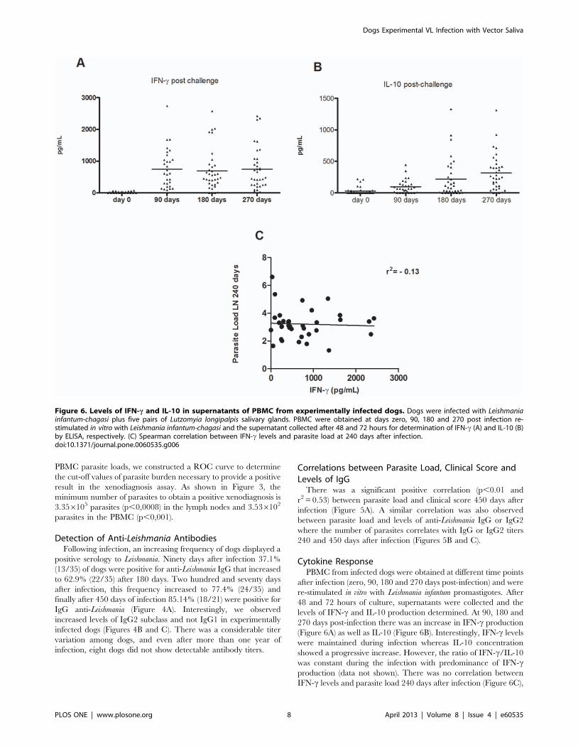
(Figure 6A) as well as IL-10 (Figure 6B). Interestingly, IFN-c levels
were maintained during infection whereas IL-10 concentration
showed a progressive increase. However, the ratio of IFN-c/IL-10
was constant during the infection with predominance of IFN-cproduction (data not shown). There was no correlation between
IFN-c levels and parasite load 240 days after infection (Figure 6C),
Figure 6. Levels of IFN-c and IL-10 in supernatants of PBMC from experimentally infected dogs. Dogs were infected with Leishmaniainfantum-chagasi plus five pairs of Lutzomyia longipalpis salivary glands. PBMC were obtained at days zero, 90, 180 and 270 post infection re-stimulated in vitro with Leishmania infantum-chagasi and the supernatant collected after 48 and 72 hours for determination of IFN-c (A) and IL-10 (B)by ELISA, respectively. (C) Spearman correlation between IFN-c levels and parasite load at 240 days after infection.doi:10.1371/journal.pone.0060535.g006
Dogs Experimental VL Infection with Vector Saliva
PLOS ONE | www.plosone.org 8 April 2013 | Volume 8 | Issue 4 | e60535

suggesting that the levels of this cytokine are not enough to control
parasite growth.
Discussion
To date, there is no strategy able to treat or control canine
visceral leishmaniasis. The establishment of a canine model will
bring opportunities to study new vaccine candidates as well as
different drugs for CVL treatment. In this study, we described an
experimental approach where dogs were infected intradermally
with Leishmania infantum combined with saliva from Lutzomyia
longipalpis. All dogs became infected and several developed
symptomatic infection and for this reason were euthanized 240
days after infection. The clinical scores displayed by these animals
were similar to those observed in naturally infected dogs,
suggesting that injection of parasites plus saliva could be an
important experimental approach, since the ability to perform
natural transmission by sand flies is restricted to few laboratories.
Although the number of parasites as well as the amount of glands
used in this study is higher compared to previous work in the
literature, our objective in the future is to test potential vaccine
candidates using salivary proteins reasserting the importance to
establish a model in dogs that includes vector saliva always present
during transmission in endemic areas.
The majority of animals were asymptomatic surviving more
than one year. This is also observed in endemic areas, where
infected dogs remain asymptomatic or show transient disease after
infection [26].
In the group of experimentally infected dogs living under
controlled conditions, we also observed a range of different clinical
manifestations and parasite load from popliteal lymph nodes and
PBMC, suggesting that factors inherent to the host contribute to
the variability of clinical aspects or outcome of the infection
[27,28,29]. Dogs displaying severe symptoms of CVL showed high
parasite load and they were more infective to sand flies confirmed
by xenodiagnosis where around 21% of the dogs were able to
transmit parasites to uninfected sand flies. In fact, we found out
that a minimum of 3.356105 parasites in the lymph nodes and
3.346102 in PBMC was required to result in a positive
xenodiagnosis.
Interestingly some of the experimentally infected dogs showed a
positive serology but they were non-infective to sand flies. This
information is of highest importance, especially in endemic areas
where culling of dogs that present a positive anti-Leishmania
serology is obligatory. It could be used to improve evaluation of
dogs present in endemic areas considering the selection of those
that are more infective and responsible for the constant VL
transmission. Similar results were found by Travi et al. [17] where
only poly-symptomatic dogs were infective, while the majority of
infected dogs were non-infective to laboratory-reared sand flies
[17]. Other studies have already shown that infectivity to sand flies
correlates with severity of disease [30,31,32].
Interestingly, we observed that the skin of naturally infected
dogs showed significantly lesser parasites than those experimen-
tally infected. The efficiency of vector-mediated transmission of
Leishmania infantum in endemic areas, mainly in dogs, has not been
well established. Vercosa et al. studying the transmissibility of
naturally infected dogs in a CVL endemic area observed that the
presence of parasites in the skin is considered important for
transmission of parasites to the sand fly [33]. Our results also
pointed out the importance of parasite load to result in a positive
xenodiagnosis, but in our findings these dogs also displayed higher
clinical score than those with a negative xenodiagnosis. On the
other hand, Travi et al. [32] observed that there is no correlation
between presence of parasite in the skin and potential transmission
to the vector.
We observed that even though all dogs had parasites detected in
the lymph nodes, some (10/35) still maintained a negative
serology, suggesting that this parameter is not appropriate for
diagnosis of CVL while the detection of parasites seems to be more
accurate. Similar findings were described with naturally infected
dogs where 11% of the animals showing positive splenic cultures
had a negative serology for Leishmania [34]. However, we also
confirm a significant positive correlation between levels of IgG or
IgG2 and parasite load in the lymph node of infected dogs. We
also observed a positive correlation between clinical score and
parasite load that corroborate with the same observation
previously described [17,34]. Although in the mouse model,
IgG1 and IgG2 levels are used as a marker of Th2 and Th1,
respectively, this correlation is not clear in dogs. Several different
studies showed an increase in the levels of IgG2 anti-Leishmania in
infected dogs [35,36,37,38,39,40], whereas the presence of IgG1 is
more controversial [33,39,40]. Different studies have associated
high levels of IgG2 with asymptomatic disease and high IgG1
concentrations with active disease [36,37,40]. In our study, we
observed an increase in the concentration of IgG2, very similar to
total IgG while IgG1 levels showed a greater variation during the
infection. Similar results were previously described comparing
infection of dogs through injection of parasites intradermally or
intravenously and found levels of IgG2, independent of the route
used for infection [17].
Detection of cytokine production in CVL is controversial.
Protective responses in dogs have been associated with the lack of
clinical symptoms, low levels of anti-Leishmania antibodies, parasite
load and a positive DTH. Therefore, a cellular immune response
in CVL is associated with activation of Th1 response, with IFN-c,
IL-2 and TNF-a production [28,41]. On the other hand, Reis et al.
[42] showed that active disease is characterized by a marked
humoral immune response. The clinical symptoms that correlated
with parasite load in different tissues are associated with a specific
immunosupression. In these animals, there is a mixed Th1/Th2
response [41,43]. In our study we observed that at day 90 post-
infection, we detected IFN-c following PBMC stimulation with
Leishmania promastigotes and the levels did not change during
infection. Many reports have shown the importance of IFN-cincrease and IL-10 decrease to obtain a protective response against
Leishmania in different experimental protocols to test vaccine
candidates [26,44,45,46,47,48]. In dogs, low parasite load in the
lymph nodes was also associated with a high production of IFN-cand TNF-a [26]. Travi et al. [17] noticed that expression and
production of IFN-c was detected earlier in dogs intradermally
infected than those infected by intravenous route although the
peak of IFN-c production occurred six months post-infection
independently of the route of infection. They also observed that
most symptomatic dogs produced high levels of IFN-c at the early
stages of infection and the proportion of animals producing this
cytokine increased over time. Our results also suggested that IFN-cwas not sufficient to prevent disease and it could not be considered
a marker of resistance. We also observed a high production of IL-
10 ninety days after infection with a slight increase through time,
but the levels were not significantly different between the time
points evaluated. We did not find any correlation of IL-10 levels
with parasite load in the lymph nodes or clinical symptoms.
However, the frequency of dogs producing high levels of IL-10
increased after 270 days of infection, suggesting that the
production of this cytokine might contribute to infection severity.
Interestingly, few reports in the literature have demonstrated the
role of saliva in the immunopathogenesis of infection by
Dogs Experimental VL Infection with Vector Saliva
PLOS ONE | www.plosone.org 9 April 2013 | Volume 8 | Issue 4 | e60535
Leishmania, mainly in different models of Visceral Leishmaniasis.
Its role is well demonstrated in the establishment of Cutaneous
Leishmaniasis infection, mostly using L. major
[8,9,10,11,12,13,14,15]. However, our results agree with the
findings observed in naturally infected dogs and autochtonous
infected dogs, where increased IL-10 expression is associated with
disease progression [49].
One important aspect of our experimental model is the
inoculation of saliva together with parasites. Although L. infantum
infection can be achieved without co-inoculation with saliva, it is
extremely relevant to recognize that saliva is always present during
natural transmission through the bite of infected sand flies in
endemic areas where ultimately a future vaccine or control
measure would be implemented. Saliva has an important role in
parasite establishment in the host demonstrated in different animal
models [19]. Recently, the possibility to use salivary proteins from
sand flies as components of a vaccine candidate has been
demonstrated in dogs. Immunization with LJM17 and LJL143,
two salivary proteins from Lutzomyia longipalpis, were able to induce
an intense immune response with a high production of IFN-c and
low levels of IL-10 and TGF-b eliciting a DTH response after
challenge of uninfected sand flies with an adverse effect to parasite
infection in vitro [50]. Indeed, Roatt et al. tested a vaccine called
LBSap composed of L. brazilensis antigens plus saponin using
intradermal challenge with L.infantum and saliva of Lutzomyia
longipalpis. Therefore, this experimental model will allow us to test
the efficacy of salivary proteins based vaccine candidates. More
importantly, it also brought relevant new information that could
help in a more adequate evaluation of dogs residing in a VL
endemic area that could be more transmissible to sand flies serving
as potential reservoirs.
Acknowledgments
The authors would like to thank Edvaldo Passos for the technical assistance
with sand flies.
Author Contributions
Responsible for insect colony and dissection of salivary glands: MP JCM.
Conceived and designed the experiments: FN MSPC ALA-S CIO AB MB-
N CB. Performed the experiments: DJC RMAC JT MA MP ALA-S FN
LA MSPC KF. Analyzed the data: DJC MA CT CIO MB-N CB. Wrote
the paper: CT MA CIO MB-N CB.
References
1. Mauricio IL, Stothard JR, Miles MA (2000) The strange case of Leishmania
chagasi. Parasitol Today 16: 188–189.
2. Gramiccia M, Gradoni L (2005) The current status of zoonotic leishmaniasesand approaches to disease control. Int J Parasitol 35: 1169–1180.
3. WHO/TDR website. Available: http://www.who.int/tdr/diseases/leish/
direction.htm. Accessed 2012 Oct 21.
4. Dye C (1996) The logic of visceral leishmaniasis control. Am J Trop Med Hyg
55: 125–130.
5. Campino L, Santos-Gomes G, Rica Capela MJ, Cortes S, Abranches P (2000)Infectivity of promastigotes and amastigotes of Leishmania infantum in a canine
model for leishmaniosis. Vet Parasitol 92: 269–275.
6. Lemesre JL, Holzmuller P, Cavaleyra M, Goncalves RB, Hottin G, et al. (2005)
Protection against experimental visceral leishmaniasis infection in dogsimmunized with purified excreted secreted antigens of Leishmania infantum
promastigotes. Vaccine 23: 2825–2840.
7. Riera C, Valladares JE, Gallego M, Aisa MJ, Castillejo S, et al. (1999)
Serological and parasitological follow-up in dogs experimentally infected withLeishmania infantum and treated with meglumine antimoniate. Vet Parasitol
84: 33–47.
8. Andrade BB, de Oliveira CI, Brodskyn CI, Barral A, Barral-Netto M (2007)Role of sand fly saliva in human and experimental leishmaniasis: current
insights. Scand J Immunol 66: 122–127.
9. Belkaid Y, Kamhawi S, Modi G, Valenzuela J, Noben-Trauth N, et al. (1998)
Development of a natural model of cutaneous leishmaniasis: powerful effects ofvector saliva and saliva preexposure on the long-term outcome of Leishmania
major infection in the mouse ear dermis. J Exp Med 188: 1941–1953.
10. Lima HC, Titus RG (1996) Effects of sand fly vector saliva on development ofcutaneous lesions and the immune response to Leishmania braziliensis in BALB/
c mice. Infect Immun 64: 5442–5445.
11. Ribeiro JM (1995) Blood-feeding arthropods: live syringes or invertebrate
pharmacologists? Infect Agents Dis 4: 143–152.
12. Theodos CM, Ribeiro JM, Titus RG (1991) Analysis of enhancing effect of sandfly saliva on Leishmania infection in mice. Infect Immun 59: 1592–1598.
13. Thiakaki M, Rohousova I, Volfova V, Volf P, Chang KP, et al. (2005) Sand fly
specificity of saliva-mediated protective immunity in Leishmania amazonensis-
BALB/c mouse model. Microbes Infect 7: 760–766.
14. Titus RG, Ribeiro JM (1988) Salivary gland lysates from the sand fly Lutzomyialongipalpis enhance Leishmania infectivity. Science 239: 1306–1308.
15. Zer R, Yaroslavski I, Rosen L, Warburg A (2001) Effect of sand fly saliva on
Leishmania uptake by murine macrophages. Int J Parasitol 31: 810–814.
16. Roatt BM, Aguiar-Soares RD, Vitoriano-Souza J, Coura-Vital W, Braga SL, et
al. (2012) Performance of LBSap vaccine after intradermal challenge with L.infantum and saliva of Lu. longipalpis: immunogenicity and parasitological
evaluation. PLoS One 7: e49780.
17. Travi BL, Osorio EY, Saldarriaga OA, Cadena H, Tabares CJ, et al. (2009)Clinical, parasitologic, and immunologic evolution in dogs experimentally
infected with sand fly-derived Leishmania chagasi promastigotes. Am J Trop
Med Hyg 81: 994–1003.
18. Ativismo.com website. Available: http://www.ativismo.com/site/index.php?option = com_content&view = article&id = 2165:teresina-calazar-
c o n t a m i n o u - m a i s - d e - 3 - m i l - a n i m a i s - s o m e n t e - n o - a n o - d e - 2 0 0 9 -&catid = 33:noticias-em-tempo-real&Itemid = 89. Accessed Aug 21 2010.
19. Gomes R, Teixeira C, Teixeira MJ, Oliveira F, Menezes MJ, et al. (2008)
Immunity to a salivary protein of a sand fly vector protects against the fatal
outcome of visceral leishmaniasis in a hamster model. Proc Natl Acad Sci U S A
105: 7845–7850.
20. Manna L, Reale S, Vitale F, Gravino AE (2009) Evidence for a relationship
between Leishmania load and clinical manifestations. Res Vet Sci 87: 76–78.
21. de Paiva Cavalcanti M, Felinto de Brito ME, de Souza WV, de Miranda Gomes
Y, Abath FG (2009) The development of a real-time PCR assay for the
quantification of Leishmania infantum DNA in canine blood. Vet J 182: 356–
358.
22. Tafuri WL, Santos RL, Arantes RM, Goncalves R, de Melo MN, et al. (2004)
An alternative immunohistochemical method for detecting Leishmania amas-
tigotes in paraffin-embedded canine tissues. J Immunol Methods 292: 17–23.
23. Volpini AC, de Azeredo Passos VM, Romanha AJ (2001) Attempt to
differentiate Leishmania (Leishmania) amazonensis, L. (L.) chagasi, Leishmania
(Viannia) braziliensis and L. (V.) guyanensis using the SSR-PCR technique.
Parasitol Res 87: 1056–1059.
24. Scott P, Pearce E, Natovitz P, Sher A (1987) Vaccination against cutaneous
leishmaniasis in a murine model. I. Induction of protective immunity with a
soluble extract of promastigotes. J Immunol 139: 221–227.
25. Bradford MM (1976) A rapid and sensitive method for the quantitation of
microgram quantities of protein utilizing the principle of protein-dye binding.
Anal Biochem 72: 248–254.
26. Alves CF, de Amorim IF, Moura EP, Ribeiro RR, Michalick MS, et al. (2009)
Expression of IFN-gamma, TNF-alpha, IL-10 and TGF-beta in lymph nodes
associates with parasite load and clinical form of disease in dogs naturally
infected with Leishmania (Leishmania) chagasi. Vet Immunol Immunopathol
128: 349–358.
27. Dye C, Killick-Kendrick R, Vitutia MM, Walton R, Killick-Kendrick M, et al.
(1992) Epidemiology of canine leishmaniasis: prevalence, incidence and basic
reproduction number calculated from a cross-sectional serological survey on the
island of Gozo, Malta. Parasitology 105 (Pt 1): 35–41.
28. Pinelli E, Killick-Kendrick R, Wagenaar J, Bernadina W, del Real G, et al.
(1994) Cellular and humoral immune responses in dogs experimentally and
naturally infected with Leishmania infantum. Infect Immun 62: 229–235.
29. Solano-Gallego L, Llull J, Ramos G, Riera C, Arboix M, et al. (2000) The
Ibizian hound presents a predominantly cellular immune response against
natural Leishmania infection. Vet Parasitol 90: 37–45.
30. Michalsky EM, Rocha MF, da Rocha Lima AC, Franca-Silva JC, Pires MQ, et
al. (2007) Infectivity of seropositive dogs, showing different clinical forms of
leishmaniasis, to Lutzomyia longipalpis phlebotomine sand flies. Vet Parasitol
147: 67–76.
31. Sherlock IA (1996) Ecological interactions of visceral leishmaniasis in the state of
Bahia, Brazil. Mem Inst Oswaldo Cruz 91: 671–683.
32. Travi BL, Tabares CJ, Cadena H, Ferro C, Osorio Y (2001) Canine visceral
leishmaniasis in Colombia: relationship between clinical and parasitologic status
and infectivity for sand flies. Am J Trop Med Hyg 64: 119–124.
33. Vercosa BL, Lemos CM, Mendonca IL, Silva SM, de Carvalho SM, et al. (2008)
Transmission potential, skin inflammatory response, and parasitism of
symptomatic and asymptomatic dogs with visceral leishmaniasis. BMC Vet
Res 4: 45.
Dogs Experimental VL Infection with Vector Saliva
PLOS ONE | www.plosone.org 10 April 2013 | Volume 8 | Issue 4 | e60535

34. Dos-Santos WL, Jesus EE, Paranhos-Silva M, Pereira AM, Santos JC, et al.
(2008) Associations among immunological, parasitological and clinical param-
eters in canine visceral leishmaniasis: Emaciation, spleen parasitism, specific
antibodies and leishmanin skin test reaction. Vet Immunol Immunopathol 123:
251–259.
35. Almeida MA, Jesus EE, Sousa-Atta ML, Alves LC, Berne ME, et al. (2005)
Antileishmanial antibody profile in dogs naturally infected with Leishmania
chagasi. Vet Immunol Immunopathol 106: 151–158.
36. Bourdoiseau G, Bonnefont C, Hoareau E, Boehringer C, Stolle T, et al. (1997)
Specific IgG1 and IgG2 antibody and lymphocyte subset levels in naturally
Leishmania infantum-infected treated and untreated dogs. Vet Immunol
Immunopathol 59: 21–30.
37. Deplazes P, Smith NC, Arnold P, Lutz H, Eckert J (1995) Specific IgG1 and
IgG2 antibody responses of dogs to Leishmania infantum and other parasites.
Parasite Immunol 17: 451–458.
38. Leandro C, Santos-Gomes GM, Campino L, Romao P, Cortes S, et al. (2001)
Cell mediated immunity and specific IgG1 and IgG2 antibody response in
natural and experimental canine leishmaniosis. Vet Immunol Immunopathol 79:
273–284.
39. Nieto CG, Garcia-Alonso M, Requena JM, Miron C, Soto M, et al. (1999)
Analysis of the humoral immune response against total and recombinant
antigens of Leishmania infantum: correlation with disease progression in canine
experimental leishmaniasis. Vet Immunol Immunopathol 67: 117–130.
40. Solano-Gallego L, Llull J, Arboix M, Ferrer L, Alberola J (2001) Evaluation of
the efficacy of two leishmanins in asymptomatic dogs. Vet Parasitol 102: 163–
166.
41. Pinelli E, van der Kaaij SY, Broeren CP, Ruitenberg EJ, Rutten VP (1999)
Measurement of dog cytokines by reverse transcription-quantitative competitive
polymerase chain reaction. Immunogenetics 49: 696–699.
42. Reis AB, Teixeira-Carvalho A, Vale AM, Marques MJ, Giunchetti RC, et al.
(2006) Isotype patterns of immunoglobulins: hallmarks for clinical status and
tissue parasite density in Brazilian dogs naturally infected by Leishmania
(Leishmania) chagasi. Vet Immunol Immunopathol 112: 102–116.43. Santos-Gomes GM, Rosa R, Leandro C, Cortes S, Romao P, et al. (2002)
Cytokine expression during the outcome of canine experimental infection by
Leishmania infantum. Vet Immunol Immunopathol 88: 21–30.44. Belkaid Y, Mendez S, Lira R, Kadambi N, Milon G, et al. (2000) A natural
model of Leishmania major infection reveals a prolonged ‘‘silent’’ phase ofparasite amplification in the skin before the onset of lesion formation and
immunity. J Immunol 165: 969–977.
45. Ferreira JH, Gentil LG, Dias SS, Fedeli CE, Katz S, et al. (2008) Immunizationwith the cysteine proteinase Ldccys1 gene from Leishmania (Leishmania)
chagasi and the recombinant Ldccys1 protein elicits protective immuneresponses in a murine model of visceral leishmaniasis. Vaccine 26: 677–685.
46. Sanchez MA, Diaz NL, Zerpa O, Negron E, Convit J, et al. (2004) Organ-specific immunity in canine visceral leishmaniasis: analysis of symptomatic and
asymptomatic dogs naturally infected with Leishmania chagasi. Am J Trop Med
Hyg 70: 618–624.47. Silvestre R, Cordeiro-Da-Silva A, Santarem N, Vergnes B, Sereno D, et al.
(2007) SIR2-deficient Leishmania infantum induces a defined IFN-gamma/IL-10 pattern that correlates with protection. J Immunol 179: 3161–3170.
48. Tafuri WL, Barbosa AJ, Michalick MS, Genaro O, Franca-Silva JC, et al. (1996)
Histopathology and immunocytochemical study of type 3 and type 4complement receptors in the liver and spleen of dogs naturally and
experimentally infected with Leishmania (Leishmania) chagasi. Rev Inst MedTrop Sao Paulo 38: 81–89.
49. Boggiatto PM, Ramer-Tait AE, Metz K, Kramer EE, Gibson-Corley K, et al.(2010) Immunologic indicators of clinical progression during canine Leishmania
infantum infection. Clin Vaccine Immunol 17: 267–273.
50. Collin N, Gomes R, Teixeira C, Cheng L, Laughinghouse A, et al. (2009) Sandfly salivary proteins induce strong cellular immunity in a natural reservoir of
visceral leishmaniasis with adverse consequences for Leishmania. PLoS Pathog5: e1000441.
Dogs Experimental VL Infection with Vector Saliva
PLOS ONE | www.plosone.org 11 April 2013 | Volume 8 | Issue 4 | e60535