experimental - uclouvain.be · model (subcutaneous implantation for 1 or 3 months, n = 10). fish,...
TRANSCRIPT

Copyright © 2015 American Society of Plastic Surgeons. Unauthorized reproduction of this article is prohibited.
www.PRSJournal.com 279
Despite significant advances in under-standing the pathophysiology of chronic wounds and cellular/molecular mecha-
nisms involved in skin healing, chronic wounds (defined by a failure to achieve complete heal-ing after 3 months)1 remain a major health care
challenge because of the aging population and morbidity related to the dramatic increase in the incidence of diabetes and obesity.2–4 Dermal reconstruction is a key point in wound healing physiology. An unfavorable wound bed with poorly vascularized fibrotic dermis is the major
Disclosure: The authors have no financial interest to declare in relation to the content of this article.
Copyright © 2015 by the American Society of Plastic Surgeons
DOI: 10.1097/PRS.0000000000001437
Aurore Lafosse, M.D.Céline Desmet, M.Sc.
Najima Aouassar, B.Sc.Wivine André, M.Sc.
Marie-Sophie Hanet, M.D.Christophe Beauloye, M.D.,
Ph.D.Romain Vanwijck, M.D.,
Ph.D.Hélène A. Poirel, M.D.,
Ph.D.Bernard Gallez, Ph.D.
Denis Dufrane, M.D., Ph.D.
Brussels, Belgium
Background: Nonhealing wounds are unable to integrate skin autografts by avascular and fibrotic dermal tissue. Adipose-derived stromal cells can improve the local environment of the wound bed by angiogenesis and immunomodula-tion. This work aimed to develop a biological dressing made of adipose-derived stromal cells onto a human acellular collagen matrix.Methods: Adipose-derived stromal cells were isolated from human adipose tissue (n = 8). In vitro, the genetic stability during early and late passages (1, 4, 10, and 16) and vascular endothelial growth factor (VEGF) secretion were assessed. Adipose-derived stromal cell adhesion and spreading on collagen matrix were preliminarily studied. In vivo tumorigenicity, angiogenesis, and tissue oxygenation were assessed after implantation of the construct in nude rats (n = 10). The biological dressing was manufactured and implanted in three patients with chronic wounds.Results: In vitro, aneuploidies, but no clonal transformation, were detected up to late cellular passages. VEGF was secreted more during hypoxia (0.1% oxy-gen) than during normoxia (21% oxygen). Adipose-derived stromal cells can adhere and spread on the scaffold within 18 to 20 days. No tumor development occurred 3 months after implantation in immunocompromised rats. Vessel counts and tissue oxygenation were higher after adipose-derived stromal cell implantation. In patients, granulation tissue was found (276 percent of vessel density), followed by epithelialization or split-thickness skin engraftment up to 22 months after implantation.Conclusions: Implantation of adipose-derived stromal cells seeded onto hu-man acellular collagen matrix (biological dressing) represents a promising therapy for nonhealing wounds, offering improvement in dermal angiogenesis and remodeling. This therapy using autologous stromal cells is safe, without significant genetic alterations after in vitro expansion. (Plast. Reconstr. Surg. 136: 279, 2015.)
From the Endocrine Cell Therapy Unit, Center of Tissue/Cell Therapy, Institut de Recherche Expérimentale et Clinique, Plas-tic and Reconstructive Surgery Unit, Louvain Drug Research Institute, Biomedical Magnetic Resonance Research Unit, Center for Human Genetics and Human Molecular Genetics, de Duve Institute, Cliniques Universitaires Saint-Luc, Cardio-vascular Research Unit, Institut de Recherche Expérimentale et Clinique, Université Catholique de Louvain.Received for publication October 24, 2014; accepted February 12, 2015.Presented at the European Association of Plastic Surgeons Research Council, in Munich, Germany, May 23 through 24, 2012; the 21st Annual Congress of the European
Autologous Adipose Stromal Cells Seeded onto a Human Collagen Matrix for Dermal Regeneration in Chronic Wounds: Clinical Proof of Concept
Association of Tissue Banks, in Vienna, Austria, November 21 through 23, 2012; the Spring Meeting of the Royal Bel-gian Society for Plastic Surgery, in Liège, Belgium, April 27, 2013; and the 11th Annual Meeting of the International Federation for Adipose Therapeutics and Science, in New York, New York, November 21 through 24, 2013.
EXPERIMENTAL

Copyright © 2015 American Society of Plastic Surgeons. Unauthorized reproduction of this article is prohibited.
280
Plastic and Reconstructive Surgery • August 2015
cause of failure of epithelialization or split-thickness skin engraftment.5 Chronic inflamma-tion leads to rupture of the physiologic balance between extracellular matrix synthesis and deg-radation. Excess matrix metalloproteinases lead to nonspecific destruction of growth factors, cells, and extracellular matrix.
Nonhealing wounds remain despite several advances in wound care6–8 (e.g., active dressings, negative-pressure therapy, artificial dermis, topi-cal growth factor application, keratinocyte cell spray). Physiology-based therapy is needed to overcome this unresolved problem. Cell therapy is proposed based on specific physiologic needs
Fig. 1. Study protocol for development of a biological dressing made of human adipose-derived stromal cells (ASC) seeded on a human acellular collagen matrix (HACM). Human adipose-derived stromal cells harvested from eight patients undergoing elective plastic surgery were isolated and cultured until passage 4. The cells were characterized (membrane marker phenotype and differ-entiation capacity). Genetic stability and angiogenic properties were evaluated in vitro (left). The biocompatibility of the scaffold (human acellular collagen matrix) was studied after seeding of the cells onto the scaffold surface (right). Adipose-derived stromal cell adhesion and spreading on the human acellular collagen matrix were followed by histology and confocal microscopy to obtain the biological dressing. The oncologic safety, angiogenesis, and evolution of tissue oxygenation in vivo were tested in a nude rat model (subcutaneous implantation for 1 or 3 months, n = 10). FISH, fluorescent in situ hybridization.
Copyright © 2015 American Society of Plastic Surgeons. Unauthorized reproduction of this article is prohibited.
Volume 136, Number 2 • Biological Dermal Regeneration
281
of chronic wounds, such as angiogenesis, immu-nomodulation, and promotion of regeneration. Although bone marrow was the first clinical source of mesenchymal stem cells,9,10 adipose-derived stem cells (described by Zuk et al. in 200211) present several advantages such as easy harvesting using a minimally invasive procedure and particularly high cellular density (approxi-mately 1 × 10⁶ cells/g of adipose tissue).12 Furthermore, adipose-derived stromal cells dem-onstrated significant advantages in comparison with bone marrow–derived mesenchymal stem cells in terms of proangiogenic properties and immunomodulatory effects.13–15
These considerations led to the development of a therapy based on adipose-derived stromal cells to restore the healing process of the dermis. A biological dressing made of autologous adi-pose-derived stromal cells seeded onto a human acellular collagen matrix16–18 was developed. This graft was proposed to promote homogenous adipose-derived stromal cell engraftment after implantation, to promote growth factor release (in hypoxia) during neoangiogenesis stimula-tion, and to induce adequate tissue remodeling. After the preclinical development of this biologi-cal dressing, this graft was manufactured follow-ing the European Union’s Advanced Therapy Medicinal Product regulations to study its clini-cal potential in patients with nonhealing wounds.
MATERIALS AND METHODSAll procedures were approved by the Ethical
Committee of the Medical Faculty (Université Catholique de Louvain) for tissue procurement and clinical study (B40320108280) (Figs. 1 and 2) and by the local ethics committee for animal care for human and preclinical studies.
Human Adipose-Derived Stromal CellsIsolation and CharacterizationHuman adipose-derived stromal cells were
harvested by lipoaspiration using the Coleman technique19 in eight patients during elective plas-tic surgery [abdominal dermolipectomy (n = 3) or mammaplasty (n = 5), mean of 6.2 g of adipose tis-sue (range, 1.4 to 14.6 g)] and after informed con-sent and serologic screening. The adipose tissue was digested with Good Manufacturing Practices grade collagenase (0.075 g; Serva Electrophoresis GmbH, Heidelberg, Germany) in a water bath at 37°C for 60 minutes. After tissue digestion, cells were collected after centrifugation and main-tained in proliferation medium up to passage 4 (or more for genetic study) after sequential trypsinization, to obtain a pure adipose-derived stromal cell population (>90 percent adipose-derived stromal cells after four passages), charac-terized for membrane marker profiles (i.e., CD44, CD45, CD73, CD90, CD105, CD34, CD14, CD11b,
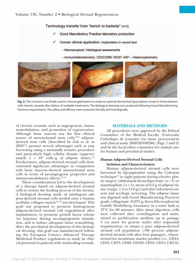
Fig. 2. The construct was finally used in clinical application to create an optimal dermal bed (granulation tissue) in three patients with chronic wounds after failure of available treatments. The biological dressing was produced following Good Manufacturing Practices requirements. The safety and efficacy were assessed clinically and histologically.

Copyright © 2015 American Society of Plastic Surgeons. Unauthorized reproduction of this article is prohibited.
282
Plastic and Reconstructive Surgery • August 2015
CD79α, CD19, and HLA-DR)20 by fluorescence-activated cell sorting (FACScan; BD Biosciences, San Jose, Calif.) and mesenchymal differentiation capacity (i.e., adipogenesis, chondrogenesis, and osteogenesis) (see Fig. 3).14,21–23
Adipose-derived stromal cells were also seeded onto 12-well culture plates for incubation in hypoxic chambers15,24 (Modular Incubator Chamber MIC-101; Billups-Rothenberg, Inc., Del Mar, Calif.) for 72 hours at 0.1% (highly hypoxic environment as seen in necrotic tissues) or 21% oxygen levels (normal culture conditions), respectively. After incubation, cell culture supernatants were harvested individually and stored at −20°C for VEGF quantification (Quan-tikine ELISA kit VEGF; R&D Systems, Minneapolis, Minn.). Cellular viability was assessed by 3-(4,5-dimeth-ylthiazol-2-yl)-5-(3-carboxymethoxyphenyl)-2- (4-sulfophenyl)-2H-tetrazolium solution (Promega, Leiden, The Netherlands).14,15
In Vitro Genetic Stability of Adipose-Derived Stromal Cells
Cytogenetic stability was studied by karyo-type and fluorescent in situ hybridization analyses after different passages to assess the oncogenic safety of the cellular component of the biological dressing (Fig. 4, left). Metaphase chromosomes were obtained from adipose-derived stromal cells of five donors according to standard protocols. Briefly, cultured cells in the exponential growth phase after pas-sages 1, 4, 10, 12, and 16 were treated for 4 hours with 0.02 μg/ml of Colcemid (Invitro-gen, Carlsbad, Calif.). Harvested cells from the flasks after trypsinization were incubated for 30 minutes at 37°C in hypotonic 0.055 M potas-sium chloride and fixed in 3:1 methanol/glacial acetic acid. Chromosome harvesting and meta-phase slide preparation were performed accord-ing to standard procedures.25 Eleven to 20 reverse
Fig. 3. Adipose-derived stromal cell characterization. (Above, left) Membrane marker phenotype. Cell membrane markers characterization by flow cytometry (FACScan). (Above, right) Morphology of adi-pose-derived stromal cells in phase-contrast microscopy (plastic well culture). (Below, left) Osteogenic differentiation capacity of adipose-derived stromal cells demonstrated by alizarin red staining (calcium deposition). (Below, center) Chondrogenic differentiation revealed by Alcian blue staining (glycosamino-glycan deposition). (Below, right) Adipogenic differentiation of adipose-derived stromal cells. Intracel-lular lipid droplets stained with Oil Red. (Original magnification, × 20).

Copyright © 2015 American Society of Plastic Surgeons. Unauthorized reproduction of this article is prohibited.
Volume 136, Number 2 • Biological Dermal Regeneration
283
trypsin Wright G-banded metaphases were ana-lyzed and karyotypes were reported according to the 2013 International System for Human Cytogenetics Nomenclature. Fluorescent in situ hybridization analysis was performed according to standard protocols25 to detect aneuploidy of chromosomes 5, 7, 8, and 18 using TelVysion 5q (SpectrumOrange; Abbot Molecular, Ottig-nies/Louvain-la-Neuve, Belgium), CEP7/D7Z1 (SpectrumGreen or SpectrumOrange; Abbot Molecular), CEP8/D8Z2 (SpectrumOrange or SpectrumGreen), and CEP18/D18Z1 (Spec-trumGreen) probes. Two hundred nuclei were counted for passage 1, passage 4, and passage 16; 120 nuclei were counted for passage 10; and 73 nuclei were counted for passage 12 (the thresh-olds were calculated following the beta law with a confidence interval of 99.9 percent).
Development of the Biological DressingAt passage 4, the capacity of adipose-derived
stromal cells to adhere and spread was compared on the human acellular collagen matrix17,18 (freeze-dried decellularized allogeneic human fascia lata, from the Tissue Bank, University Hospital Saint Luc, Brussels, Belgium) and on plastic wells (as control) (Figs. 4 and 5). Cell adhesion was assessed by confocal laser scan-ning microscopy, as already described.26,27
Briefly, human adipose-derived stromal cells were seeded on the treated fascia lata in 24 wells at a density of 2 × 10⁵ cells/well. Every 2 days until day 15 and every 3 days until day 30 after seeding, human acellular collagen matrix was washed in phosphate-buffered saline to remove the medium and then immersed in 2 μM calcein acetoxymethyl esters (Molecular Probes Europe BV, Leiden, The Netherlands) for approximately 3 hours28 to be
examined by confocal laser scanning microscopy. The total cellular-covered area, cell perimeter, and shape factor [(area/perimeter2) × 4π] were determined using Scion Image Beta 4.02n acquisi-tion and analysis software (Scion Corp., Torrance, Calif.).29
To assess the oncologic safety and efficacy of the biological dressing in vivo, pieces (10 × 10 mm) of the biological dressing and of the scaffold alone were implanted subcutaneously into nude rats (n = 10; Charles River Laborato-ries International, Wilmington, Mass.). Under general anesthesia (isoflurane 3%), a triangular flap was elevated in each paravertebral area to create two subcutaneous pockets, allowing the placement of the grafts (human acellular col-lagen matrix plus adipose-derived stromal cells on the right side and human acellular collagen matrix alone on the left side). A thermic lesion (dermal necrosis) was applied on the inner side of each flap to reproduce the hypoxic wound environment. The biological dressing (human acellular collagen matrix plus adipose-derived stromal cells) was implanted with the cells in contact with the burned dermis on the right side. The flaps were closed with nonabsorbable sutures after the placement of five to eight crys-tals of lithium phthalocyanine to allow further measurement of the postimplantation intratis-sular oxygenation course (Fig. 6, left).
Electron paramagnetic resonance oximetry (EPR spectrometer30; Magnettech, Berlin, Ger-many) was used to follow the intratissular oxy-gen tension course and assess the capacity of the composite graft to improve the tissue oxy-genation in nude rats.31 The oxygen tension in the injured dermis was studied weekly for up to 4 weeks after implantation under gaseous
Table 1. Patient Characteristics*
Patient Age (yr) Sex Wound Cause
Wound Location
(Size)
Duration of Lesions
(mo)Previous Local
TreatmentsPrevious Local
Evolution
1 46 Male Radionecrosis (66 Gy after soft-tissue sarcoma resection and pedicled flap dehiscence)
Pretibial (12 cm²) 13 Active dressings, well-conducted nursing care
Deepening of the wound, strong avascular fibrosis
2 21 Male Drepanocytosis (homozy-gote, poor control of systemic disease)
4 supramalleolar areas (>250 cm²)
46 Active dressings, split-thickness skin autografts ×4, hyperbaric oxygen therapy
Ulcer recurrence after a mean of 23 days after skin graft, hyperalgic lesions
3 41 Female Vasculitis (systemic lupus erythematosus)
4 supramalleolar areas (>200 cm²)
27 Active dressings, ultrasound ther-apy, split-thickness skin autograft ×2
Complete lysis of skin grafts after 6 wk and 2 wk, respectively
*Three patients with nonhealing wounds of different causes were selected. The biological dressing was proposed after a minimum of 1 yr of poor healing evolution after treatment.

Copyright © 2015 American Society of Plastic Surgeons. Unauthorized reproduction of this article is prohibited.
284
Plastic and Reconstructive Surgery • August 2015
anesthesia (isoflurane).32 Results were expressed as percentage of tissue oxygenation at day 6 after implantation. At 1 month (n = 5) or 3 months (n = 5) after implantation, complete excision of the graft plus surrounding tissues was performed for macroscopic and histologic analysis (hematoxy-lin and eosin and Masson trichrome staining for local tumor development and angiogenesis) (Fig. 7).
Clinical Application for Dermal ReconstructionThe biological dressing was proposed for
three patients with nonhealing wounds (Table 1 and Fig. 8, below). Periumbilical fatty tissue (22, 8, and 21 g, respectively) was harvested (using the Coleman technique under local anesthesia) for adipose-derived stromal cell isolation and cul-ture14,15 in line with Good Manufacturing Practices recommendations.
At passage 3, adipose-derived stromal cells were trypsinized, resuspended in Dulbecco’s Modified Eagle Medium, and loaded onto freeze-dried human acellular collagen matrix (corre-sponding to passage 4). The biological dressing was obtained when adipose-derived stromal cells covered more than 90 percent of the human acel-lular collagen matrix. Finally, the composite graft was rinsed with CMRL (Mediatec, Manassas, Va.) and transferred to the operating room for implan-tation (adipose-derived stromal cells loaded onto the upper side of the human acellular collagen matrix).
In these three patients, wound débride-ment was performed by hydrosurgery before implantation to ensure a minimally contami-nated wound bed. The composite graft was cut to an ideal size and oriented with the cell layer directly in contact with the wound surface
Fig. 4. (Left) In vitro adipose-derived stromal cells genetic stability and angiogenesis. In vitro representative fluorescent in situ hybridization analysis for adipose-derived stromal cells at passage 1 (P1), passage 4 (P4), passage 10 (P10), and passage 16 (P16). Karyotypes were all normal (data not shown). Fluorescent in situ hybridization analysis demonstrated borderline percentages of tetrasomy 5 and tetrasomy 18 (passage 1), tetrasomy 7 and tetrasomy 8 (passage 10), and trisomy 7 (passage 4). These were stable when compared with results for passage 1, except for the appearance of a monosomy 7 at passage 16 (15 percent), which was not detected in dividing cells (low proliferative advantage). (Right) VEGF secretion by adipose-derived stromal cells, quantified by enzyme-linked immunosorbent assay assessment, was significantly higher during hypoxia (700.7 ± 466.0 pg/ml versus 494.3 ± 317.4 pg/ml; p < 0.001). Hypoxic stress tests and growth factor quantification were performed in triplicate and in duplicate, respec-tively. ASC, adipose-derived stromal cells; HACM, human acellular collagen matrix.

Copyright © 2015 American Society of Plastic Surgeons. Unauthorized reproduction of this article is prohibited.
Volume 136, Number 2 • Biological Dermal Regeneration
285
and fixed with nonabsorbable sutures (Fig. 8, below). Inflammatory parameters were followed (C-reactive protein, fibrinogen), as were clinical and histologic courses. Petroleum jelly–impreg-nated dressings were applied and changed daily. Biopsies were performed before and after implantation for immunohistochemistry and histomorphometry to study inflammatory reaction, angiogenesis, and tissue remodeling (CD3/CD68, VEGF/factor VIII, and Masson trichrome, respectively).
Statistical AnalysisThe one-sample Kolmogorov-Smirnov test
and Q-Q plots were used to assess the normal dis-tribution of values. Statistically significant differ-ences between groups (with normal distribution) were tested by paired t test and one-way analysis of variance with the Bonferroni post hoc test. Statis-tical tests were performed with Systat version 8.0 (Cranes Software International, Bangalore, India) or PASW 18 (SPSS; IBM Corp., Armonk, N.Y.); val-ues of p < 0.05 were considered significant.
Fig. 5. Biopsies of the construct were performed 3 weeks after seeding of adipose-derived stromal cells onto human acellular collagen matrix to assess the cellular covering and spreading on the scaffold. The cells (arrows) covered more than 90 percent of the human acellular collagen matrix surface (asterisk), as confirmed by 4′,6-diamidino-2-phenylindole fluorescent staining (above, left) and hematoxylin and eosin staining (above, right). Adipose-derived stromal cell spreading and adhesion on human acellular collagen matrix were studied by confocal microscopy. A significantly shorter time was required to obtain a cellular covering of 90 percent of the total surface of the plastic well plate (positive control) in comparison with the same surface of human acellular colla-gen matrix (13 versus 21 days, respectively; p < 0.005) (below, left). The spreading of adipose-derived stromal cells was also delayed on human acellular collagen matrix in comparison with the plastic well (p < 0.005). However, a similar shape factor was obtained at 25 days after seeding for both tested supports (below, right), confirming the capacity of adipose-derived stromal cells to adhere and grow on the collagen matrix. ASC, adipose-derived stromal cells; HACM, human acellular collagen matrix.

Copyright © 2015 American Society of Plastic Surgeons. Unauthorized reproduction of this article is prohibited.
286
Plastic and Reconstructive Surgery • August 2015
RESULTS
In Vitro and In Vivo Safety of Adipose-Derived Stromal Cells plus Human Acellular Collagen Matrix
Adipose-derived stromal cells at passage 4 were characterized by mesenchymal stromal cell sur-face marker profile: CD44+ (>95 percent of cells), CD73+ (>90 percent), CD90+ (>95 percent), CD105+ (>95 percent), CD45− (<5.5 percent), CD34− (<7 percent), CD14− (<7 percent), CD11b− (<7 per-cent), CD79α− (<5.5 percent), CD19− (<5.5 per-cent) and HLA-DR− (<7 percent), and positive markings for mineralization, hyaline deposition and lipid vacuoles (Fig. 3). One day after seed-ing, most adipose-derived stromal cells appeared round (mean shape factor, 0.93 ± 0.13) on both cellular supports, with a mean of 4.8 percent sur-face covering (not significant). Between days 3 and 18 after seeding, a significantly higher area of cellular expansion was found for plastic wells in comparison with human acellular collagen matrix (p < 0.005). The complete surface coverage was found on human acellular collagen matrix 1 week later than on the plastic well. A similar delay was
found for adipose-derived stromal cells spread-ing (shape factor) on human acellular collagen matrix in comparison with the plastic well (p < 0.005) (Fig. 5, below). The covering was delayed but the same cellular growth was reached on the human acellular collagen matrix than on the plastic well.
The in vitro safety study revealed no clonal structural chromosomal aberrations on the karyo-types of adipose-derived stromal cells at passage 1, passage 4, and advanced passages (passages 10, 12, or 16). At all passages, borderline tetrasomies (1.5 to 5.5 percent) were detected for at least two tested chromosomes by fluorescent in situ hybrid-ization analysis on interphase cells (cutoff, 4.5 per-cent) (Fig. 4, left). This technique also revealed a clone with suspected monosomy 7 in 15 percent of interphase cells at passage 16. These aneuploid cells do not seem to have a proliferative advantage because they are not detected in metaphase cells. Macroscopic and microscopic analyses did not find any local tumor development in explanted tissue from immunocompromised rats (months 1 and 3 after implantation) (Fig. 6, right).
Fig. 6. Tumorigenicity in vivo was assessed in nude rats (n = 10). The composite graft (human acellular collagen matrix plus adi-pose-derived stromal cells) and the scaffold alone (human acellular collagen matrix, control) were symmetrically implanted into subcutaneous pockets. Histologic and macroscopic analyses demonstrated the absence of local tumor development at 1 month and 3 months after implantation (Masson trichrome: 1 month, ×2.5; 3 months, ×5).

Copyright © 2015 American Society of Plastic Surgeons. Unauthorized reproduction of this article is prohibited.
Volume 136, Number 2 • Biological Dermal Regeneration
287
Fig. 7. Adipose-derived stromal cell angiogenic properties and subcutaneous oxygenation were studied in vivo (same model, n = 10 nude rats after application of a thermic lesion on the deep dermis) by histology (Masson trichrome), histomorphometry, and electron paramagnetic resonance (EPR). (Right) Histologic aspect (1) of the deep dermis after application of a thermic lesion (circle) at time 0. The burned tissue was characterized by local necrosis. One month after implantation of the dressing (2), an area of high vessel density (asterisk) was found on the site of previous thermic necrosis (where lithium phthalocyanine crystals are visible). This tissue was placed in direct contact with the cell layer of the dressing (implantation with the cell layer on the superficial aspect). Sig-nificant tissue remodeling was found for dermal tissue after implantation of human acellular collagen matrix plus adipose-derived stromal cells. In tissue implanted with human acellular collagen matrix alone (3), fibrotic scar tissue with low vessel density char-acterizes the corresponding area. (Left) Histomorphometry and electron paramagnetic resonance were performed to assess the angiogenic effect of implanted adipose-derived stromal cells (versus scaffold alone). A significantly higher vessel density was found 1 month after graft implantation in the dermis treated with human acellular collagen matrix plus adipose-derived stromal cells (9.5 ± 2.0 vessels/2.56 cm2) in comparison with human acellular collagen matrix alone (6.5 ± 3.0 vessels/2.56 cm2; p = 0.002; five regions of interest were analyzed per slide). In parallel, electron paramagnetic resonance confirmed significantly higher tissue oxygenation of the dermal lesion treated by adipose-derived stromal cells. Results are expressed as a percentage of oxygen tension at day 6 after implantation. The ratio of oxygen tension in the deep dermis (after thermic lesion) was significantly higher with human acellular collagen matrix plus adipose-derived stromal cells than with human acellular collagen matrix alone [161 ± 48 percent versus 96 ± 29 percent (p < 0.05) at day 13; 158 ± 42 percent versus 98 ± 27 percent (p < 0.05) at day 21; and 142 ± 15 percent versus 82 ± 29 percent (p = 0.001) at day 27 after implantation).

Copyright © 2015 American Society of Plastic Surgeons. Unauthorized reproduction of this article is prohibited.
288
Plastic and Reconstructive Surgery • August 2015
In Vitro and In Vivo Efficacy of Adipose-Derived Stromal Cells plus Human Acellular Collagen Matrix (Biological Dressing)
Cell viability after 72 hours of incubation at 0.1% oxygen tension was compared with that at 21% oxygen tension (p = 0.034). Hypoxia had no deleterious impact on adipose-derived stromal cell viability, and a significantly higher
secretion of VEGF was found in hypoxic condi-tions (0.1% oxygen tension) in comparison with 21% oxygen tension (p < 0.001; n = 8) (Fig. 4, right).
Dermal oxygenation at day 6 (for each indi-vidual recipient) was considered as baseline after dermal injury.33 The ratio of oxygen tension in the deep dermis (after thermic lesion) was
Fig. 8. (Above) Timing for the clinical grade production of the biological dressing. A mean of 67 days was needed to expand adipose-derived stromal cells in vitro after isolation to reach a minimum of passage 4. For patient 2, because of the large size of bilateral wounds, a larger expansion was performed before loading the cells onto human acellular collagen matrix (HACM) (97 days, passage 6). After seeding onto the scaffold, a mean of 66 days was needed to obtain more than 90 percent of surface covering by adipose-derived stromal cells. The composite graft with adipose-derived stromal cells loaded onto the upper side of the human acel-lular collagen matrix (above, right) was delivered to the operating room in a sterile culture flask contained in three sterile plastic bags. (Below) Clinical application. Three patients with nonhealing wounds had the biological dressing implanted after failure of other conventional and advanced treatments. Macroscopic aspects of the wound before surgery; at the time of implantation; and after 22, 4, and 2 months of evolution are illustrated. The composite graft was placed with the cell layer in direct contact with the tissue and fixed with nonabsorbable sutures. For patients 2 and 3 (large wounds), a split-thickness skin graft was performed 3 to 4 weeks after implantation of the biological dressing. Results were maintained at 22, 4, and 2 months of follow-up.

Copyright © 2015 American Society of Plastic Surgeons. Unauthorized reproduction of this article is prohibited.
Volume 136, Number 2 • Biological Dermal Regeneration
289
significantly higher with human acellular collagen matrix plus adipose-derived stromal cells than with human acellular collagen matrix alone (p < 0.05 at days 13, 21, and 27 after implantation). Consis-tently significantly higher vessel density was found
in the dermis reconstituted with human acellular collagen matrix plus adipose-derived stromal cells in comparison with the dermis in contact with human acellular collagen matrix alone (p = 0.002) (Fig. 7, left).
Fig. 9. Clinical evolution in patient 1 (radionecrosis). (Above and center) The initial wound was characterized by strong fibrotic tissue without any improvement 13 months after irradiation. An occlusive dressing was maintained for 3 days after implan-tation of the biological dressing. After 13 to 15 days, the scaffold was resorbed pro-gressively until days 28 to 30, and the formation of granulation tissue was observed. (Below) The systemic inflammatory parameters [C-reactive protein (CRP) and fibrino-gen] were followed-up after implantation. A transient inflammatory reaction occurred (peak at day 3 after surgery), with a return to basal values after 28 days.

Copyright © 2015 American Society of Plastic Surgeons. Unauthorized reproduction of this article is prohibited.
290
Plastic and Reconstructive Surgery • August 2015
Fig. 10. Histologic study of the treated wound bed in patient 1. (Above) Parallel views (macrohistology with Masson trichrome staining) of the wound bed tissue at day 0 and day 56 after implantation (asterisk indicates site of biopsy).

Copyright © 2015 American Society of Plastic Surgeons. Unauthorized reproduction of this article is prohibited.
Volume 136, Number 2 • Biological Dermal Regeneration
291
Clinical ApplicationAdipose-derived stromal cells at the end of
passage 3 are trypsinized and then seeded on the human collagen matrix, to obtain the passage 4 on the collagenic scaffold. The implant was obtained when 90 percent of the human acellular collagen matrix surface was covered with adipose-derived stromal cells (5.8 × 10⁵ cells/cm2). The complete manufacturing of the dressing, from cell isolation until delivery, was achieved in a mean of 133 days (Fig. 8, above).
After implantation, initial incorporation of the graft occurred at day 3, followed by progres-sive resorption of the collagen matrix, which was complete at approximately day 28 after implanta-tion, leaving well-vascularized granulation tissue on the wound bed surface (Fig. 9, above).
In patient 1, complete wound closure was achieved at day 60 after implantation and has been maintained (>22 months). In patients 2 and 3, skin autografts were performed on the vascularized granulation tissue 6 weeks after adi-pose-derived stromal cell implantation. The skin autografts were completely integrated after 3 days (first dressing opening) and were associated with pain relief; daily nursing care was discontinued 6 months and 3 months after implantation in patients 2 and 3, respectively (Fig. 8, below). Two patients experienced recurrence of ulcerations, probably because of the systemic causes of wounds (drepanocytosis and vasculitis).
Although a significant increase in C-reactive protein and fibrinogen occurred 1 week after implantation, no chronic inflammation was observed. A decrease to basal values occurred by 1 month after surgery (Fig. 9, below).
Postimplantation biopsy results revealed well-organized, vascularized tissue in comparison with the strong fibrosis before dressing implantation (Fig. 10, above). Significant increases in VEGF (p < 0.001) and factor VIII (p < 0.05) were found in tissues after implantation (Fig. 10, center and below). Significant macrophagic recruitment was found after implanta-tion (p < 0.05), without modification of lymphocyte infiltration (Fig. 10, center and below). Human acel-lular collagen matrix alone (without cells) did not provide beneficial effects in patient 2 (Fig. 11).
DISCUSSIONIn the context of nonhealing wounds, our
study demonstrates that adipose-derived stromal cells on human acellular collagen matrix can sup-port the restoration of physiology for dermal heal-ing; adipose-derived stromal cells can survive in a highly hypoxic environment and release VEGF to promote neoangiogenesis, synthesis of granu-lation tissue, and, consequently, the evolution of healing.
The impact of hostile and desert tissues on stem cell implantation remains underestimated. This environment is characterized mainly by pro-gressive fibrosis and highly hypoxic tissues, lead-ing to the vicious cycle of cellular death. In vitro, adipose-derived stromal cells demonstrated their capacity to survive in hypoxic conditions (0.1% oxygen tension), similar to bone marrow–derived mesenchymal stem cells and placenta-derived mesenchymal stem cells.34,35 These cells can pro-liferate with metabolic activity in intermittent hypoxia, rendering them suitable for therapeutic applications in hypoxic challenges.
However, cellular delivery remains a key ques-tion for stem cell engraftment. Intradermal injec-tions in wounds have several disadvantages for clinical application, such as painful cell implan-tation in fibrotic regions (high-pressure injec-tions), the need for numerous injections for large wounds to ensure homogenous cell distribution, and cell manipulation just before implantation (trypsinization, suspension).36 A bioengineered scaffold with mesenchymal stem cells in animal models of cutaneous wounds recently demon-strated improvement with cell engraftment com-pared with injections.37–39 Therefore, we propose a decellularized collagenic scaffold to improve local adipose-derived stromal cell delivery in fibrotic tissue. This support demonstrated qualities of a favorable scaffold for cellular adhesion and spreading. Moreover, human acellular collagen matrix must be stable enough to deliver the cells
Fig. 10. (Continued ). The avascular fibrotic tissue was replaced by granulation tissue with high vessel density and reorganization of the collagen (green). (Center) Immunohistology for detection of VEGF and factor VIII (angiogenesis), CD3 (lymphocytes), and CD68 (macrophages) for inflammatory infiltrates of the treated tissue before implantation compared with day 56 after implan-tation. (Below) Histomorphometric semiquantitative analysis of VEGF and factor VIII (below, left) and of CD3 and CD68 (below, right) before and after implantation. A significant increase was noted in VEGF-positive cells (176 percent; p < 0.001) and in fac-tor VIII–positive cells (276 percent; p < 0.05). The inflammatory cell count indicated significant macrophagic recruitment fol-lowing implantation (33.75 ± 9.51 and 48.18 ± 12.82 positive cells per 0.16 mm2 before versus after implantation, respec-tively; p < 0.05). No significant variation was observed regarding lymphocyte infiltration. HACM, human acellular collagen matrix; ASCs, adipose-derived stromal cells.

Copyright © 2015 American Society of Plastic Surgeons. Unauthorized reproduction of this article is prohibited.
292
Plastic and Reconstructive Surgery • August 2015
Fig. 11. Comparison of evolution after implantation of conventional autologous skin graft alone, biological dressing followed by autologous skin graft, human acellular collagen matrix (HACM) alone, and human acellular collagen matrix followed by autologous skin graft in patient 2 (drepanocytic ulcers). Before the study, four conventional split-thickness skin grafts (without specific preparation of the dermis (above, left) had been applied between August of 2009 and September of 2010, resulting in complete loss of the grafts after a mean of 31 days (range, 17 to 46 days). The implantation of the human acellular collagen matrix plus adipose-derived stromal cells (above, center) was then proposed to promote wound bed regeneration and integration of the skin autograft (applied 6 weeks later). The wound closure was maintained for 6 months before recurrence (secondary to poor control of the systemic disease). The human acellular collagen matrix alone was finally implanted (above, right) to determine the intrinsic effect of the scaffold. The skin graft applied after the implantation of the human acellular collagen matrix was acutely degraded, and complete recurrence was macroscopically and microscopically confirmed 16 days later. (Center) Histologic examination of the wound bed after the three different treatments (skin autograft alone, human acellular collagen matrix plus adipose-derived stromal cells, human acellular collagen matrix alone) focused on cellular infiltrate, fibrosis, and vessel density (arrows indicate vessels). Radical increase in vessel density and reduction in fibrosis were observed on histologic samples (Masson trichrome) after implanta-tion of the biological dressing, in contrast to conventional skin autograft or human acellular collagen matrix alone. (Below) Wound closure also lasted significantly longer after human acellular collagen matrix plus adipose-derived stromal cells (6 months versus 28 days and 16 days).

Copyright © 2015 American Society of Plastic Surgeons. Unauthorized reproduction of this article is prohibited.
Volume 136, Number 2 • Biological Dermal Regeneration
293
without a significant inflammatory reaction, to obtain the trophic effect of adipose-derived stro-mal cells. It was previously reported that vascular development after cell transplantation begins at day 3 after the initiation of angiogenesis, with the appearance of neocapillaries at day 7 after implan-tation and effective angiogenesis at day 21.15,40,41 The scaffold resorption (as observed in patients) matched the duration of the granulation tissue formation (28 days), without local or systemic pro-longed inflammatory reaction (Fig. 11).
Consistent with the confirmation of higher VEGF secretion by adipose-derived stromal cells in hypoxia (in vitro), implantation of the biological dressing suggests that adipose-derived stromal cells improve angiogenesis and dermal oxygenation in vivo, as seen in animal models (burned dermis) and in patients (chronic wounds). Angiogenesis is only attributable to the effects of adipose-derived stromal cells, because the implantation of human acellular collagen matrix alone did not induce der-mis regeneration or elevation of oxygen tension in the nude rat model or in patient 2.
The major limitation of this therapy, with regard to widespread use in clinical practice, remains the time to complete the manufacturing of the biological bandage. The number of passages (up to passage 4) to obtain a pure population of adipose-derived stromal cells (before seeding onto the human acellular collagen matrix) with optimal angiogenic properties (VEGF secretion in hypoxia) cannot be exceeded. Several alterna-tives of improvement (to obtain passage 4 as quick as possible) can be postulated, such as a larger amount of harvested adipose tissue, improve-ment of cell expansion by the use of platelet lysate,42,43 the use of successive subcultivation,44 and others. Preliminary results (data not shown) demonstrated that the time of the expansion of adipose-derived stromal cells up to passage 4 can be significantly reduced by more than 50 percent.
Although the potential of adipose-derived stro-mal cells for skin regeneration is recognized, their safety must be confirmed to demonstrate the clini-cal potential of this new therapy. Controversy exists concerning the potential of spontaneous transfor-mation of mesenchymal stem cells after prolonged ex vivo culture. However, several studies report that mesenchymal stem cells have limited tendencies to develop tumors.45–48 All genetic analyses revealed minor rates (near the detection threshold) of chromosomal aneuploidy, mainly tetrasomies, sug-gesting tetraploidy as classically observed in cul-tured cells up to passage 16 (cutoff, approximately 4.5 percent). Minor trisomy 7 was also detected
(cutoff, approximately 2 percent), as reported pre-viously by Tarte et al.48 Cells could exhibit recur-ring chromosomal alterations without involving a selective growth advantage in vitro, and mesenchy-mal stem cells with or without chromosomal altera-tions did not induce tumor formation 8 weeks after injection in immunocompromised mice. A higher rate of monosomy 7 (15 percent) was detected in the sample at passage 16. Monosomy 7 is mainly involved in myeloid malignancies45 and is not described in mesenchymal tumors. These aneu-ploid cells do not have a proliferative advantage because they are not detected on the karyotype of metaphase cells. Some studies described karyotype changes in mesenchymal stem cells after 11 to 14 passages (1.5 to 5.95 percent of cells).49,50 Because the adipose-derived stromal cells are implanted at passage 4, this in vitro study demonstrates that adipose-derived stromal cells could grow in the host tissue with a low risk of chromosomal insta-bility and tumor development. In our studies, the in vivo oncologic safety was confirmed by the absence of adverse events in immunodeficient ani-mal recipients (1 or 3 months after implantation) and in patients (up to 22 months after implanta-tion); adipose-derived stromal cell implantation after shorter in vitro culture (passage 4), avoid-ing the selection of tumoral cell clones; and local delivery of adipose-derived stromal cells, allowing direct control of the implanted cells by superficial implantation, in contrast to systemic infusion.
CONCLUSIONSThe biological dressing made of autologous adi-
pose-derived stromal cells and absorbable human acellular collagen matrix can restore a dynamic angiogenesis and tissue remodeling (promoted by the action of adipose-derived stromal cells at low oxygen tension) in the dermal tissue of a non-healing wound. In addition, this therapy has low invasiveness (local anesthesia) and is safe (using autologous cells). However, its clinical indication must be defined in accordance with the pathophys-iology of different causes of chronic wounds. The biological dressing could be improved in terms of cellular density by using a tridimensional scaffold and by a shorter manufacturing period.
Denis Dufrane, M.D., Ph.D.Endocrine Cell Therapy Unit
Center of Tissue/Cell TherapyCliniques Universitaires Saint-Luc
Avenue Hippocrate 10B-1200 Brussels, Belgium

Copyright © 2015 American Society of Plastic Surgeons. Unauthorized reproduction of this article is prohibited.
294
Plastic and Reconstructive Surgery • August 2015
ACKNOWLEDGMENTSThis work was supported by grants from the Fond
pour la Recherche Scientifique, from the Saint-Luc Foun-dation, and by an Action de Recherche Concertée-Fédéra-tion Wallonie Bruxelles grant.
REFERENCES 1. Mustoe TA, O’Shaughnessy K, Kloeters O. Chronic wound
pathogenesis and current treatment strategies: A unifying hypothesis. J Plast Reconstr Surg. 2006;117:35–41.
2. Singer AJ, Clark RA. Cutaneous wound healing. N Engl J Med. 1999;341:738–746.
3. Gottrup F. A specialized wound-healing center concept: Importance of a multidisciplinary department structure and surgical treatment facilities in the treatment of chronic wounds. Am J Surg. 2004;187:38S–43S.
4. Sen CK, Gordillo GM, Roy S, et al. Human skin wounds: A major and snowballing threat to public health and the econ-omy. Wound Repair Regen. 2009;17:763–771.
5. Panuncialman J, Falanga V. The science of wound bed prep-aration. Clin Plast Surg. 2007;34:621–632.
6. Sibbald RG, Orsted H, Schultz GS, et al. Preparing the wound bed 2003: Focus on infection and inflammation. Ostomy Wound Manage. 2003;49:23–51.
7. Menke MN, Menke NB, Boardman CH, et al. Biologic thera-peutics and molecular profiling to optimize wound healing. Gynecol Oncol. 2008;111:S87–S91.
8. Chen SM, Ward SI, Olutoye OO, et al. Ability of chronic wound fluids to degrade peptide growth factors is associated with increased levels of elastase activity and diminished levels of proteinase inhibitors. Wound Repair Regen. 1997;5:23–32.
9. Chen L, Tredget EE, Wu PY, Wu Y. Paracrine factors of mesenchymal stem cells recruit macrophages and endo-thelial lineage cells and enhance wound healing. PLoS One 2008;3:e1886.
10. Friedenstein AJ, Petrakova KV, Kurolesova AI, et al. Heterotopic of bone marrow: Analysis of precursor cells for osteogenic and hematopoietic tissues. Transplantation 1968;6:230–247.
11. Zuk PA, Zhu M, Ashjian P, et al. Human adipose tissue is a source of multipotent stem cells. Mol Biol Cell 2002;13: 4279–4295.
12. Yang XF, He X, He J, et al. High efficient isolation and sys-tematic identification of human adipose-derived mesenchy-mal stem cells. J Biomed Sci. 2011;18:59.
13. Chen JS. Therapeutic potential of bone marrow-derived mesenchymal stem cells for cutaneous wound healing. Front Immunol. 2012;3:192.
14. Schubert T, Xhema D, Vériter S, et al. The enhanced per-formance of bone allografts using osteogenic-differenti-ated adipose-derived mesenchymal stem cells. Biomaterials 2011;32:8880–8891.
15. Vériter S, Aouassar N, Adnet PY, et al. The impact of hyper-glycemia and the presence of encapsulated islets on oxy-genation within a bioartificial pancreas in the presence of mesenchymal stem cells in a diabetic Wistar rat model. Biomaterials 2011;32:5945–5956.
16. Dufrane D, Cornu O, Delloye C, et al. Physical and chemical processing for a human dura mater substitute. Biomaterials 2002;23:2979–2988.
17. Fawzi-Grancher S, Goebbels RM, Bigare E, et al. Human tis-sue allograft processing: Impact on in vitro and in vivo bio-compatibility. J Mater Sci Mater Med. 2009;20:1709–1720.
18. Dufrane D, Mourad M, van Steenberghe M, et al. Regeneration of abdominal wall musculofascial defects by a human acellu-lar collagen matrix. Biomaterials 2008;29:2237–2248.
19. Coleman SR. Structural fat grafts: The ideal filler? Clin Plast Surg. 2001;28:111–119.
20. Dominici M, Le Blanc K, Mueller I, et al. Minimal criteria for defining multipotent mesenchymal stromal cells: The International Society for Cellular Therapy position state-ment. Cytotherapy 2006;8:315–317.
21. Post S, Abdallah BM, Bentzon JF, Kassem M. Demonstration of the presence of independent pre-osteoblastic and pre-adi-pocytic cell populations in bone marrow-derived mesenchy-mal stem cells. Bone 2008;43:32–39.
22. Qu C, Zhang G, Zhang L, et al. Osteogenic and adipogenic potential of porcine adipose mesenchymal stem cells. In Vitro Cell Dev Biol Anim. 2007;43:95–100.
23. Cui L, Wu Y, Cen L, et al. Repair of articular cartilage defect in non-weight bearing areas using adipose derived stem cells loaded polyglycolic acid mesh. Biomaterials 2009;30: 2683–2693.
24. Martinive P, Defresne F, Bouzin C, et al. Preconditioning of the tumor vasculature and tumor cells by intermittent hypoxia: Implications for anticancer therapies. Cancer Res. 2006;66:11736–11744.
25. Duhoux FP, Ameye G, Lambot V, et al. Refinement of 1p36 alterations not involving PRDM16 in myeloid and lymphoid malignancies. PLoS One 2011;6:e26311.
26. White JG, Amos WB, Fordham M. An evaluation of confocal versus conventional imaging of biological structures by fluo-rescence light microscopy. J Cell Biol. 1987;105:41–48.
27. Bacallao R, Stelzer EH. Preservation of biological specimens for observation in a confocal fluorescence microscope and operational principles of confocal fluorescence microscopy. Methods Cell Biol. 1989;31:437–452.
28. Hanthamrongwit M, Wilkinson R, Osborne C, et al. Confocal laser-scanning microscopy for determining the structure of and keratinocyte infiltration through collagen sponges. J Biomed Mater Res. 1996;30:331–339.
29. Sinha RK, Morris F, Shah SA, et al. Surface composition of orthopaedic implant metals regulates cell attachment, spreading, and cytoskeletal organization of primary human osteoblasts in vitro. Clin Orthop Relat Res. 1994;305:258–272.
30. Gallez B, Baudelet C, Jordan BF. Assessment of tumor oxy-genation by electron paramagnetic resonance: Principles and applications. NMR Biomed. 2004;17:240–262.
31. Gallez B, Jordan BF, Baudelet C, et al. Pharmacological modifications of the partial pressure of oxygen in murine tumors: Evaluation using in vivo EPR oximetry. Magn Reson Med. 1999;42:627–630.
32. Baudelet C, Gallez B. Effect of anesthesia on the signal intensity in tumors using BOLD-MRI: Comparison with flow measurements by laser Doppler flowmetry and oxygen measurements by luminescence-based probes. Magn Reson Imaging 2004;22:905–912.
33. Vériter S, Mergen J, Goebbels RM, et al. In vivo selection of biocompatible alginates for islet encapsulation and subcuta-neous transplantation. Tissue Eng Part A 2010;16:1503–1513.
34. Mathew SA, Rajendran S, Gupta PK, Bhonde R. Modulation of physical environment makes placental mesenchymal stro-mal cells suitable for therapy. Cell Biol Int. 2013;37:1197–1204.
35. Tsai CC, Yew TL, Yang DC, Huang WH,Hung SC. Benefits of hypoxic culture on bone marrow multipotent stromal cells. Am J Blood Res. 2012;2:148–159.
36. Rustad KC, Wong VW, Sorkin M, et al. Enhancement of mes-enchymal stem cell angiogenic capacity and stemness by a biomimetic hydrogel scaffold. Biomaterials 2012;33:80–90.

Copyright © 2015 American Society of Plastic Surgeons. Unauthorized reproduction of this article is prohibited.
Volume 136, Number 2 • Biological Dermal Regeneration
295
37. Walker NG, Mistry AR, Smith LE, et al. A chemically defined carrier for the delivery of human mesenchymal stem/stro-mal cells to skin wounds. Tissue Eng Part C Methods 2012;18: 143–155.
38. Wu Y, Chen L, Scott PG, et al. Mesenchymal stem cells enhance wound healing through differentiation and angio-genesis. Stem Cells 2007;25:2648–2659.
39. Chen L, Tredget EE, Liu C, Wu Y. Analysis of allogenicity of mesenchymal stem cells in engraftment and wound healing in mice. PLoS One 2009;4:e7119.
40. Caplan AI, Dennis JE. Mesenchymal stem cells as trophic mediators. J Cell Biochem. 2006;98:1076–1084.
41. Jones GL, Juszczak MT, Hughes SJ, et al. Time course and quantification of pancreatic islet revascularization following intraportal transplantation. Cell Transplant. 2007;16:505–516
42. Naaijkens BA, Niessen HW, Prins HJ, et al. Human platelet lysate as a fetal bovine serum substitute improves human adipose-derived stromal cell culture for future cardiac repair applications. Cell Tissue Res. 2012;348:119–130.
43. Witzeneder K, Lindenmair A, Gabriel C, et al. Human-derived alternatives to fetal bovine serum in cell culture. Transfus Med Hemother. 2013;40:417–423.
44. Di Battista JA, Shebaby W, Kizilay O, et al. Proliferation and differentiation of human adipose-derived mesenchymal stem
cells (ASCs) into osteoblastic lineage are passage dependent. Inflamm Res. 2014;63:907–917.
45. Trobaugh-Lotrario AD, Kletzel M, Quinones RR, et al. Monosomy 7 associated with pediatric acute myeloid leuke-mia (AML) and myelodysplastic syndrome (MDS): Successful management by allogeneic hematopoietic stem cell trans-plant (HSCT). Bone Marrow Transplant. 2005;35:143–149.
46. Meza-Zepeda LA, Noer A, Dahl JA, et al. High-resolution analysis of genetic stability of human adipose tissue stem cells cultured to senescence. J Cell Mol Med. 2008;12:553–563.
47. Bernardo ME, Zaffaroni N, Novara F, et al. Human bone marrow derived mesenchymal stem cells do not undergo transformation after long-term in vitro culture and do not exhibit telomere maintenance mechanisms. Cancer Res. 2007;67:9142–9149.
48. Tarte K, Gaillard J, Lataillade JJ, et al. Clinical-grade production of human mesenchymal stromal cells: Occurrence of aneu-ploidy without transformation. Blood 2010;115:1549–1553.
49. Nikitina VA, Osipova EY, Katosova LD, et al. Study of genetic stability of human bone marrow multipotent mesenchymal stromal cells. Bull Exp Biol Med. 2011;150:627–631.
50. Bochkov NP, Voronina ES, Kosyakova NV, et al. Chromosome variability of human multipotent mesenchymal stromal cells. Bull Exp Biol Med. 2007;143:122–126.