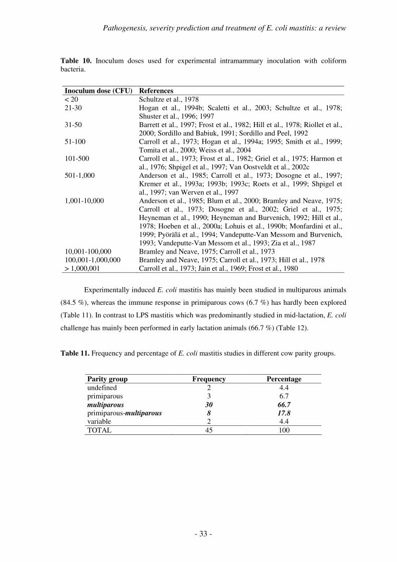
experimentally induced escherichia coli mastitis in...
TRANSCRIPT

Experimentally induced Escherichia coli mastitis in lactating primiparous cows
Frédéric Vangroenweghe
Thesis submitted in fulfillment of the requirements for the degree of Doctor in Veterinary Sciences (PhD), Ghent University, 2004
Promoter: Prof. Dr. C. Burvenich Co-promoter: Dr. P. Rainard
Faculty of Veterinary Medicine Department of Physiology-Biochemistry-Biometrics

ii
The cover of the doctoral thesis was designed by Verzele Roger
This work was printed by DCL Print & Sign B.V.B.A. – Zelzate – www.dclsigns.be
Experimentally induced Escherichia coli mastitis in lactating primiparous cows
F. Vangroenweghe, 2004, Ghent University
ISBN 90-5864-065-5
D/2004/10.412/1

iii
To my Ellen


v
TABLE OF CONTENTS
LIST OF FREQUENTLY USED ABBREVIATIONS
1
PATHOGENESIS, SEVERITY PREDICTION AND TREATMENT OF
ESCHERICHIA COLI MASTITIS: A REVIEW
3
HYPOTHESIS AND OBJECTIVES
63
VALIDATION OF MILK SAMPLE COLLECTION UNDER
ASEPTICAL CONDITIONS
69
MATERIALS AND METHODS EXPERIMENTAL INFECTIONS
97
INFLUENCE OF PARITY ON SEVERITY OF INFLAMMATION
125
MODULATION OF THE INFLAMMATORY REACTION
143
1. Variation of the inoculum dose 145 2. Inhibition of prostaglandin synthesis 167 3. Vaccination against the endotoxin 189
GENERAL DISCUSSION
205
SUMMARY
219
SAMENVATTING
223
DANKWOORD
229
CURRICULUM VITAE
233


- 1 -
LIST OF FREQUENTLY USED ABBREVIATIONS
AOAH acyloxyacyl hydrolase
AUC area under the curve
BHBA beta-hydroxybutyrate
C5a activated complement fragment 5
CD14 cluster of differentiation 14
CFU colony-forming units
CL chemiluminescence
Cl- chlorine
CNF cytotoxic necrotizing factor
CONT-SPL machinal milk sampling technique
COX cyclo-oxygenase
DNA deoxyribonucleic acid
eae attaching and effacing capacity
ELISA enzyme-linked immunosorbent assay
FS forward scatter
HR heart rate
HRP horse-radish peroxidase
IDF International Dairy Federation
IL-8 interleukin-8
K+ potassium
LBP LPS-binding protein
LPS lipopolysaccharide
LT heat-labile toxin
MAN-SPL manual milk sampling technique
mCD14 membrane-associated CD14
NAP neutrophil alkaline phosphatase
Na+ sodium
NSAID non-steroidal anti-inflammatory drug
PAMP pathogen-associated molecular pattern
PBS phosphate-buffered saline

- 2 -
PCR polymerase chain reaction
PCV packed cell volume
PGE2 prostaglandin E2
PI propidium iodide
PIH post-infusion hour
PMA phorbol 12-myristate 13-acetate
PMN polymorphonuclear leukocytes
rbosCD14 recombinant bovine sCD14
ROI region of interest
RR respiration rate
RT rectal temperature
SCC somatic cell count
sCD14 soluble CD14
SEM standard error of the mean
SSC side scatter
ST heat-stabile toxin
STER-SPL sterile milk sampling technique
TLR Toll-like receptor
TMB 3,3’,5,5’-tetramethylbenzidine
TNF tumour-necrosis factor
TXB2 thromboxane B2
vs. versus
WBC white blood cell

- 3 -
PATHOGENESIS, SEVERITY PREDICTION AND
TREATMENT OF ESCHERICHIA COLI MASTITIS:
A REVIEW

- 4 -
CONTENTS
1. Etiology of Coliform Mastitis
1.1. Escherichia coli, a specific environmental pathogen
1.2. Incidence and severity of E. coli mastitis in high-yielding dairy cows
1.3. Economical impact of coliform mastitis
2. Pathogenesis
2.1. Virulence factors of E. coli
2.2. Cow factors that influence the outcome of the disease
2.2.1. Polymorphonuclear leukocytes and their role in mammary defence
2.2.2. Hormonal and metabolic profile during the periparturient period
2.2.3. Severity of experimentally induced E. coli mastitis
2.3. Role of complement, LPS-binding protein and soluble CD14
2.3.1. Complement system and its role in the innate defence
2.3.2. LPS-binding protein and sCD14 recognise and neutralise LPS
2.4. Recurrent intramammary E. coli infections in the bovine
3. Experimental Infection Models
3.1. Lipopolysaccharide model
3.2. Escherichia coli model
4. Diagnosis and Treatment of E. coli Mastitis
4.1. Diagnosis
4.1.1. Clinical and bacteriological diagnosis
4.1.2. Milk SCC and compositional changes
4.1.3. Molecular identification methods
4.2. Treatment
4.2.1. Antimicrobial treatment of E. coli mastitis
4.2.2. Anti-inflammatory treatment of E. coli mastitis
4.2.3. Additional treatments
5. Prediction of the Severity of Experimentally Induced E. coli Mastitis
6. Conclusions

Pathogenesis, severity prediction and treatment of E. coli mastitis: a review
- 5 -
1. ETIOLOGY OF COLIFORM MASTITIS
1.1. Escherichia coli, a specific environmental pathogen
Escherichia coli is a Gram-negative, non-spore-forming rod, which belongs to the family
Enterobacteriaceae. Gram-negative bacteria have a cell wall that typically consists of three
layers, the cytoplasmic membrane, the peptidoglycan layer and the outer membrane (Fig. 1). The
outer cell membrane contains phospholipids, membrane proteins and lipopolysaccharide (LPS).
Lipopolysaccharide comprises lipid-A, the lipopolysaccharide core and repeated polysaccharide
units, called O-antigens (Fig. 2) (Cullor, 1996). Lipid-A is the lipophilic, inner part of LPS,
which causes the toxic effects of LPS, also known as endotoxin (Cullor, 1996; Hogan and Smith,
2003). On the outer surface, bacteria may have fimbriae, which protrude from the cell wall. The
surface may also be covered with a thick polysaccharide layer, called a capsule. Based on the
different structures of O-antigens, K-antigens (capsular) and H-antigens (flagellar), E. coli can
be divided into O:H:K serotypes (Cullor, 1996).
Figure 1. Schematic illustration of the cell wall components of Gram-negative bacteria.
Cytoplasm
Inner cell membrane
Peptidoglycan Lipoprotein
Outer cell membrane
Flagellae
Pili

- 6 -
Escherichia coli is part of the normal intestinal flora of humans and animals, and is the
most common facultative anaerobic bacterial species in the gut. The bacteria are constantly
excreted in the faeces to the environment. Pathogenic E. coli bacteria can cause intestinal and
extra-intestinal infections in mammalian and avian hosts (Cullor, 1996; Nagy and Fekete, 1999).
Infections of the gastrointestinal tract may lead to various kinds of diarrhoeic diseases, which, in
case of Shiga toxin, may even progress to systemic haemolytic uremic syndrome in humans and
oedema disease in pigs (Cullor, 1996). Escherichia coli is the predominant cause of urinary tract
infection in humans, and also causes invasive diseases, such as bacteraemia and meningitis, in
humans and animals (Cullor, 1996).
Figure 2. Schematic diagram of the structure of LPS. Various monosaccharides are present. The number (n) of repeating subunits in the O-antigen is quite variable and may be > 20 (based on Tobias et al., 1999).
Escherichia coli strains involved in acute clinical mastitis have, however, no specific
virulence factors (Lehtolainen, 2004). Over the last decades, several potential virulence factors
have been studied in bovine mastitis isolates from clinical cases of E. coli mastitis. No specific
O-serotypes could be associated with bovine mastitis, and only serum resistance could
consistently be identified as a possible virulence factor of importance, with a prevalence of 64 to
100% depending on the study. Other potential virulence factors, such as adhesins (F17-fimbriae,
S and P fimbriae), toxins (heat-stable toxin (ST), heat-labile toxin (LT), Shiga-like toxins (slt),
cytotoxic necrotising factors (CNF) 1 and 2, Vero-toxin), hemagglutination, colicin V, K-
antigen, invasiveness, presence of capsule, aerobactin, TraT and attaching and effacing capacity
(eae) have extensively been studied, but could not be identified in a consistent number of strains.
Colonies on agar have a smooth or rough appearance. Smooth colonies are characterised
by a shiny surface and an entire edge. They have developed polysaccharide side chains as part of
their LPS outer membrane. In contrast, rough forms appear as dry, wrinkled colonies, which
have lost their polysaccharide side chains by mutation.
n
O-antigen
Polysaccharide
Core
Outer Core Inner Core
Lipid A
Lipid
Monosaccharide Phosphate Fatty Acid

Pathogenesis, severity prediction and treatment of E. coli mastitis: a review
- 7 -
1.2. Incidence and severity of E. coli mastitis in high-yielding dairy cows
Mastitis incidence is the frequency of newly occurring events in a population over a
given time period (Smith, 1999). Throughout the lactation cycle, two distinct periods of
increased mastitis incidence occur, namely the periparturient period, from 2 weeks before
calving until peak lactation (8 weeks), and around drying-off (Natzke, 1981; Burvenich et al.,
2000) (Fig. 3).
Figure 3. Incidence of clinical mastitis throughout the lactation cycle. Two distinct peak moments occur, namely the periparturient period and late lactation – drying-off period (Burvenich et al., 2000, based on results of Natzke, 1981). Time is represented in the X-axis, incidence of clinical mastitis in the Y-axis. The different segments are respectively mammogenesis (�), periparturient period (�), lactation (�) and involution (�). The transition period (�) goes from mammogenesis over colostrogenesis to milk secretion during early lactation.
Acute mastitis is an udder inflammation characterised by its sudden onset with visible
signs of abnormal milk (Smith, 1999). When all cases of acute coliform mastitis during the entire
lactation are considered, a significant percentage occurs before peak lactation (Erskine et al.,
1988). Twenty-five % of the cases of clinical coliform mastitis occur in the first two weeks of
lactation. However, when the first month of lactation is considered, this percentage increases to
45% and to 60% in the period before peak lactation at 8 weeks post-partum (Erskine et al., 1988;
Burvenich et al., 2000) (Fig. 4).
CALVING DRYING-OFF

- 8 -
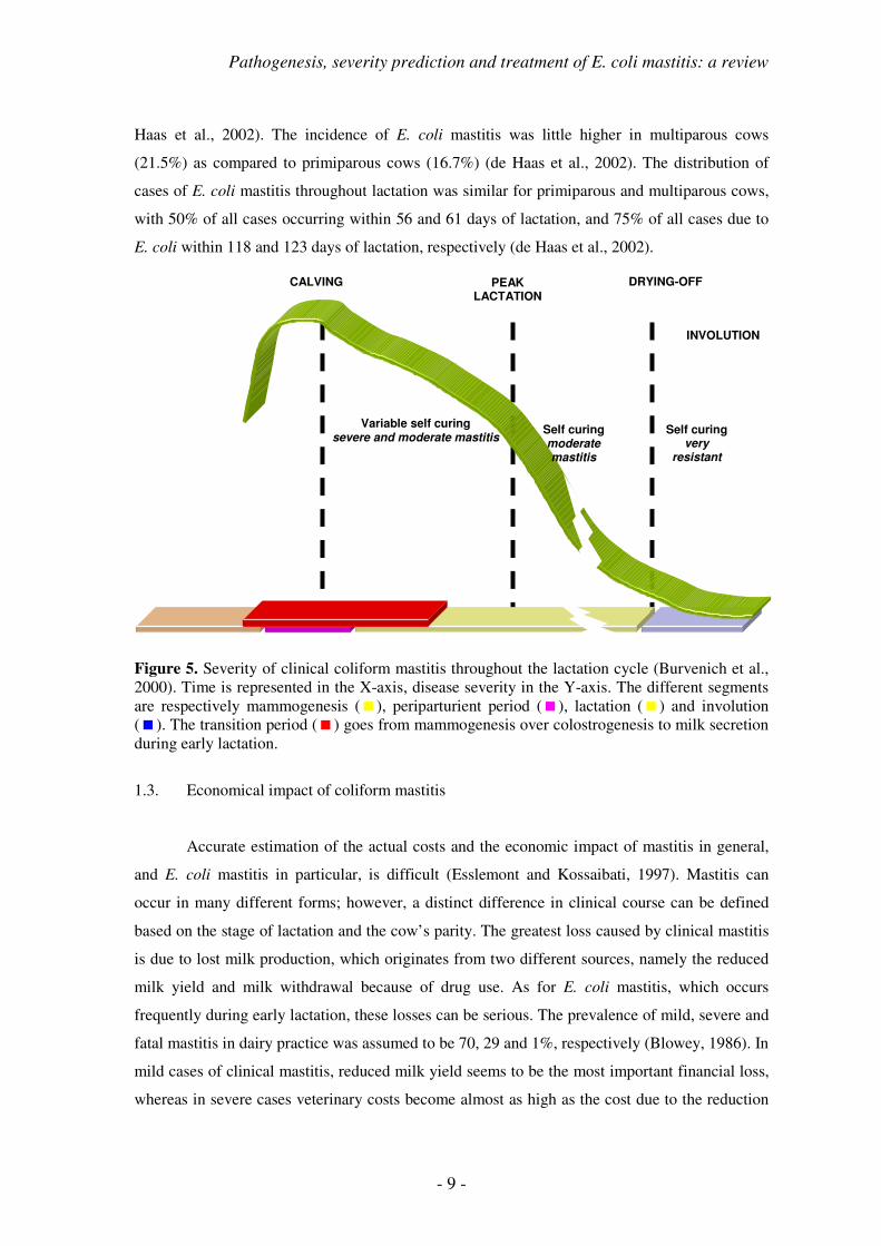
The severity of the disease is, however, quite variable throughout the lactation cycle
(Burvenich et al., 2000). During the transition period and in early lactation (until 8 weeks post-
partum), a variable degree of self-curing can be observed with moderate to severe clinical
responses, whereas during mid- and late lactation, a moderate reaction with a high degree of self-
curing is apparent. Dry cows are very resistant to clinical coliform mastitis (Hill, 1981;
Todhunter et al., 1991a; Vandeputte-Van Messom et al., 1993; Shuster et al., 1996; Burvenich et
al., 2000) (Fig. 5).
Figure 4. Incidence of clinical coliform mastitis during the periparturient period until peak lactation (Burvenich et al., 2000, based on results of Erskine et al., 1988). Twenty-five % of the cases of clinical coliform mastitis already occur in the first 2 weeks of lactation. This increases to 45% after 4 weeks and 60% at peak lactation (8 wks). Time is represented in the X-axis, cumulative percentage of cases of clinical coliform mastitis in the Y-axis. The different segments are respectively mammogenesis (�), periparturient period (�), lactation (�) and involution (�). The transition period (�) goes from mammogenesis over colostrogenesis to milk secretion during early lactation.
Multiple reports on incidence of E. coli mastitis in low SCC herds are available
(Schukken et al., 1989; Barkema et al., 1998; Surayasathaporn et al., 2000; de Haas et al., 2002).
Most research on increased risk for clinical mastitis in low bulk milk SCC has been performed in
The Netherlands. Except for Surayasathaporn et al. (2000), who observed an incidence for E.
coli mastitis of 42.8% in a low bulk milk SCC herd, in most other studies the incidence of
clinical mastitis due to E. coli was around 20% (Schukken et al., 1989; Barkema et al., 1998; de
Weeks post-partum
8
45%
60%
CALVING PEAK LACTATION
2 4
25%
Pathogenesis, severity prediction and treatment of E. coli mastitis: a review
- 9 -
Haas et al., 2002). The incidence of E. coli mastitis was little higher in multiparous cows
(21.5%) as compared to primiparous cows (16.7%) (de Haas et al., 2002). The distribution of
cases of E. coli mastitis throughout lactation was similar for primiparous and multiparous cows,
with 50% of all cases occurring within 56 and 61 days of lactation, and 75% of all cases due to
E. coli within 118 and 123 days of lactation, respectively (de Haas et al., 2002).
Figure 5. Severity of clinical coliform mastitis throughout the lactation cycle (Burvenich et al., 2000). Time is represented in the X-axis, disease severity in the Y-axis. The different segments are respectively mammogenesis (�), periparturient period (�), lactation (�) and involution (�). The transition period (�) goes from mammogenesis over colostrogenesis to milk secretion during early lactation.
1.3. Economical impact of coliform mastitis
Accurate estimation of the actual costs and the economic impact of mastitis in general,
and E. coli mastitis in particular, is difficult (Esslemont and Kossaibati, 1997). Mastitis can
occur in many different forms; however, a distinct difference in clinical course can be defined
based on the stage of lactation and the cow’s parity. The greatest loss caused by clinical mastitis
is due to lost milk production, which originates from two different sources, namely the reduced
milk yield and milk withdrawal because of drug use. As for E. coli mastitis, which occurs
frequently during early lactation, these losses can be serious. The prevalence of mild, severe and
fatal mastitis in dairy practice was assumed to be 70, 29 and 1%, respectively (Blowey, 1986). In
mild cases of clinical mastitis, reduced milk yield seems to be the most important financial loss,
whereas in severe cases veterinary costs become almost as high as the cost due to the reduction
CALVING PEAK LACTATION
DRYING-OFF
INVOLUTION
Self curing moderate mastitis
Variable self curing severe and moderate mastitis
Self curing very
resistant

- 10 -
in milk yield due to the prolonged negative effects of mastitis on the secretory epithelium
(Esslemont and Kossaibati, 1997). In fatal cases of clinical mastitis, major costs are attributed to
replacement costs for dairy heifers (Table 1).
Table 1. Costs of clinical mastitis in dairy cows (adapted from Esslemont and Kossaibati, 1997).
Mild (70%) Severe (29%) Fatal (1%)
Unit Cost (€) Unit Cost (€) Unit Cost (€) Drugs 15.8 39.8 54.8 Herdsman’s time (min) 15 1.9 Discarded milk (l) 80 28.8 120 43.2 Reduced milk yield (l) 247 74.1 450 135.0 Veterinarian’s time (min) 50 94.0 135 244.9 Increased risk of culling (%) 20 231.0 Cost of fatality 3021.9 DIRECT COSTS 120.6 543.0 3321.6
The determining parameter for the total cost of mastitis in a dairy herd is, however, the
number of animals affected by the disease per 100 animals present in the herd. In a British
survey of 90 dairy herds, the average number of cases per 100 cows per year was 37.4, although
a very wide range existed between the best herds in the study (2.8 cases per 100 cows) and the
worst herds (215.4 cases per 100 cows). Taking this into account, mastitis is considered the most
important production disease in terms of reduced profitability with an average yearly cost of €
35.9 per cow on an average herd (Esslemont and Kossaibati, 1997).
2. PATHOGENESIS
2.1. Virulence factors of E. coli
Escherichia coli, involved in bovine coliform mastitis, is part of the normal intestinal
flora of dairy cows. The strains isolated from bovine mastitis are essentially not different from
strains isolated from bovine faeces (Nemeth et al., 1994). This supports the hypothesis that
mastitic E. coli are simply opportunistic pathogens. Nevertheless, several studies have been
performed to identify potential virulence factors of E. coli associated with bovine coliform
mastitis (Linton and Robinson, 1984; Sanchez-Carlo et al., 1984; Barrow and Hill, 1989; Hogan
et al., 1990; Nemeth et al., 1991; 1994; Thomas et al., 1992; Fang et al., 1993; Pohl et al., 1993;
Lipman et al., 1995; Cray et al., 1996; Kaipainen et al., 2002). However, only LPS, the
endotoxin originating from the bacterial outer cell membrane, has been shown to be a consistent
virulence factor in all E. coli strains isolated from bovine coliform mastitis. Lipopolysaccharide

Pathogenesis, severity prediction and treatment of E. coli mastitis: a review
- 11 -
is a potent inducer of inflammatory cytokines (Shuster et al., 1993) and is released by bacteria
during growth and killing (Burvenich, 1983). The second most important virulence characteristic
of bovine mastitis isolates is their serum resistance (Carroll and Jasper, 1977; Sanchez-Carlo et
al., 1984; Valente et al., 1988; Barrow and Hill, 1989; Nemeth et al., 1991; 1994; Fang and
Pyörälä, 1996), and 64 to 100% of the strains were reported to be resistant. Two specific
structures on the outer cell membrane, TraT – a cell surface-exposed lipoprotein – and K1 – a
capsular antigen –, have been assumed to act in concert to inhibit the correct assembly or
membrane insertion of the membrane attack complex of the complement system (Sukupolvi and
O’Connor, 1990). No relation between both TraT and K1, and serum resistance could, however,
be established (Nemeth et al., 1991; Kaipainen et al., 2002).
In order to colonise the mammary gland and induce mastitis, invading bacteria should be
able to proliferate in normal and abnormal mastitis milk (Fang et al., 1993), which means that it
may be necessary for the bacterial strains to adapt to changing growth conditions. Escherichia
coli has improved growth capacity in abnormal mastitis milk, which could be explained through
the presence of growth-enhancing nutrients, although elevated antibacterial activities during
mastitis would rather inhibit bacterial growth (Fang et al., 1993). In vivo adhesion of E. coli to
the epithelial surface of the mammary gland is thought to be unimportant during the initial phase
of infection (Bramley et al., 1979), because in healthy udders collagen or fibronectin are not
exposed. Recent in vitro experiments, using epithelial cell cultures (Döpfer et al., 2000) or tissue
explant cultures (Thomas et al., 1992), have found indications for in vitro adhesion to mammary
epithelial cells. However, strains in one study were isolated from recurrent cases of coliform
mastitis (Döpfer et al., 2000), and a continuous epithelial cell line of MAC-T-cells was used.
Thomas et al. (1992) only observed in vitro adhesion of E. coli when epithelial surface was
damaged and underlying tissues, containing fibronectin and collagen, were displayed.
Although over 100 serotypes of E. coli have been recognised, no specific O-serotypes
have been conclusively related to bovine E. coli mastitis (Linton and Robinson, 1984).
Nevertheless, intramammary challenge with E. coli 487 caused more severe clinical signs of
mastitis than did E. coli 727 (Todhunter et al., 1991b; Hogan et al., 1992; 1995; 1999). In
accordance with E. coli strains causing extra-intestinal diseases in humans, hemagglutination and
hemolysis of erythrocytes have been considered as virulence factors in bovine mastitis (Hogan et
al., 1990), because they enable the bacteria to increase iron availability. However, neither of both
characteristics was related to duration or severity of bovine intramammary infection from which
the bacteria were isolated (Hogan et al., 1990).
Further research related to the presence of F17 fimbriae, genes encoding for enterotoxins
(LT and ST1), verotoxins and cytotoxic necrotising factors (CNF1 and CNF2), attaching and

- 12 -
effacing capacity (eae), Shiga-like toxin (slt) I and II, and heat-stable enterotoxin did not add
substantial evidence on the presence of multiple virulence factors in E. coli involved in bovine
coliform mastitis (Pohl et al., 1993; Lipman et al., 1995; Cray et al., 1996).
2.2. Cow factors that influence the outcome of the disease
2.2.1. Polymorphonuclear leukocytes and their role in mammary defence
Once environmental bacteria, such as E. coli, have passed the first functional barrier
against invasion, formed by the teat canal, they are able to proliferate in the milk at the level of
the teat and udder cistern. However, milk is not the ideal growth medium, mainly due to the
presence of several inhibitory factors, such as lysozyme, lactoferrin, and phagocytic cells,
originating from the blood. The polymorphonuclear leukocyte (PMN) and macrophage are the
functional phagocytic cells of the body. The resident cells in the normal healthy mammary gland
are predominantly macrophages, followed by lymphocytes, whereas only a small portion of
PMN (10-15%) is present (Dulin et al., 1982; Östensson et al., 1988; Leitner et al., 2000; Pillai et
al., 2001).
In the healthy mammary gland, PMN migrate from the blood circulation through the
endothelial gaps in the mammary epithelium to the milk compartment. Directed migration of
PMN into the mammary gland is stimulated by nursing or milking (Paape et al., 1992),
supplying the normal sterile mammary gland with a constant source of fresh PMN (Paape and
Wergin, 1977). Once in the mammary gland, PMN start to phagocytose fat globules and casein,
which induces an activated status of the cells, finally leading to a progressive exhaustion of
cellular functionality, as can be observed with milk PMN chemiluminescence (CL) (Mehrzad et
al., 2001b).
Polymorphonuclear leukocytes have several receptors on their cell membrane, which
serve in the process of directed migration from the blood into the milk compartment, called
diapedesis. Rolling and attachment of PMN to the endothelium is the first step in the recruitment
process and is accomplished by interaction between L-selectin on PMN and its ligand on the
endothelial cells (Kishimoto and Rothlein, 1994). Subsequently, the β2-integrins are responsible
for a strong and sustained attachment, followed by transendothelial PMN migration into the
extracellular matrix and through the mammary gland epithelium. The respective role of the two
subunits, namely CD11b and CD18, in the migration process has recently been studied using an
in vitro diapedesis model (Smits et al., 2000). Migration across the endothelium is almost
completely dependent on CD18 and to a lesser extent on CD11b, whereas the diapedesis across

Pathogenesis, severity prediction and treatment of E. coli mastitis: a review
- 13 -
the mammary epithelial barrier is more dependent on CD11b. Migration across the collagen of
the extracellular matrix was partly dependent on CD18, but completely independent of CD11b
(Smits et al., 2000).
Following diapedesis through the blood-milk barrier, functionality of milk PMN has
been shown to be decreased (Mehrzad et al., 2001b). This was not only attributed to the
ingestion of fat globules and casein from the milk environment (Paape et al., 2003). In vitro
diapedesis showed a reduction in phagocytosis and oxidative burst activity following migration
across a mammary epithelial cell layer (Smits et al., 1999). The migration process also
influenced the appearance of programmed cell death or apoptosis in PMN following migration.
In vitro migration through a collagen-coated membrane induced an apoptotic response, which
was downregulated by the addition of a monolayer of endothelial cells, but negated by a
mammary epithelial cell monolayer (Van Oostveldt et al., 2002a). It was suggested that L-
selectin might play an important role in the PMN apoptosis-inducing effect after in vitro
migration through the collagen-coated membrane inserts (Van Oostveldt et al., 2002a), whereas
CD11b/CD18 could induce an attenuation of the rate of apoptosis after migration through the
blood-milk barrier (Smits et al., 2000).
During the periparturient period, a temporary change in several blood PMN
characteristics and functions has been described: oxidative burst activity (Moreira da Silva et al.,
1998), L-selectin (Monfardini et al., 2002), acyloxyacyl hydrolase (AOAH) (Dosogne et al.,
1998a); whereas β2-integrins (CD11a, CD11b, CD11c and CD18) did not change over time
(Diez-Fraile et al., 2003a).
Acyloxyacyl hydrolase is thought to be one of the mechanisms responsible for the local
intramammary detoxification of released LPS (Dosogne et al., 1998a), besides the recognition
and uptake of LPS by the LPS-binding protein (LBP) / cluster of differentiation (CD) 14 system
(Thomas et al., 2002). Following intramammary endotoxin challenge with high inoculum doses,
clinical signs were less pronounced as compared to intravenous endotoxin challenge (Lohuis et
al., 1988b). This suggests that endotoxemia as such is not the major cause of systemic clinical
signs following LPS or E. coli mastitis. Following E. coli challenge, few peaks of LPS have been
detected in circulation (Dosogne et al., 2002). In contrast, significant differences in circulating
concentrations of TNF-α have been observed between moderate and severe responding animals
(Hoeben et al., 2000a). This suggests that animals with E. coli mastitis rather suffer from
mediator shock than from endotoxemia (Hoeben et al., 2000a; Dosogne et al., 2002). However,
under practical circumstances bacteraemie has been described in a substantial number of cows
with acute clinical mastitis (Wenz et al., 2001). A significant difference in potential bacteraemie

- 14 -
existed between mild-moderate and severe responders, with as much as 42% of the severe
responders having positive bacterial cultures.
Subsequently, endothelial cells of the blood vessel walls in the mammary gland are
activated, leading to an increased margination, attachment and migration into the mammary
gland tissue under the chemotactic guidance of locally produced chemoattractants, such as
activated complement fragment 5 (C5a) and interleukin-8 (IL-8) (Shuster et al., 1997; Rainard,
2003). Meanwhile, an increase in mammary blood flow can also be observed (Dhondt et al.,
1977), providing the mammary gland with a larger amount of fresh reactive blood PMN. Once
the inflammation actively eliminates the invading pathogens from the mammary gland, several
regulatory mechanisms to limit the inflammation and the associated local epithelial damage are
enhanced. During E. coli mastitis, an increase in programmed cell death has been observed (Van
Oostveldt et al., 2002c), whereas the margination decreases through a downregulation of L-
selectin (Monfardini et al., 1999). Moreover, in vitro induction of PMN apoptosis through
addition of LPS or TNF-α resulted in a decreased phagocytic and oxidative burst capacity,
which could also play a role in the resolution of inflammation (Van Oostveldt et al., 2002b).
Nevertheless, functional activity of the viable blood PMN is enhanced during experimental E.
coli mastitis, as the number of circulating PMN with unstimulated respiratory burst activity is
higher (Van Oostveldt et al., 1999). Within the mammary gland, PMN oxidative burst, as
quantified through CL, is higher in severely diseased animals (Mehrzad, 2002), which could
explain the larger, long-lasting decrease in milk production in the affected quarters of these
animals.
At the mammary gland level, activated PMN recognise, phagocytose and kill bacteria
through their oxygen-dependent and oxygen-independent bactericidal mechanisms (Burvenich et
al., 2003). Finally, they become apoptotic and are taken up by macrophages, without release of
their toxic compounds into the surrounding environment (Paape et al., 2003).
2.2.2. Hormonal and metabolic profile during the periparturient period
During the periparturient period, hormonal and metabolic profile of the high-yielding
dairy cow undergoes some tremendous changes which are mainly related to the process of
calving, with its associated hormonal regulation, and the initiation of milk production, the
lactogenesis, which changes metabolic demands quite abruptly. Besides the sudden decrease in
progesterone, there is a rise in oestrogen, and cortisol peaks on the day of calving. Freshly calved
cows meanwhile undergo a decrease in energy balance, characterised by increased blood
concentrations of β-hydroxybutyrate (BHBA) and non-esterified fatty acids (Hoeben et al.,

Pathogenesis, severity prediction and treatment of E. coli mastitis: a review
- 15 -
2000b), in conjunction with a slight and short-lasting dip in glucose (Moreira da Silva et al.,
1998; Hoeben et al., 2000b).
High concentrations of progesterone and to a lesser extent oestrogen have been shown to
decrease blood PMN oxidative burst activity (Moreira da Silva, 1996). β-hydroxybutyric acid
has been shown to have a direct negative effect on circulating blood PMN function (Hoeben et
al., 1997c), but an indirect inhibiting effect on bone marrow progenitor cloning in vitro has also
been reported (Hoeben et al., 1999). Taking these findings into account, the previously described
depression in PMN functions can easily be explained.
However, the described hormonal and metabolic changes all occur within the same
period, which makes a causal interpretation difficult. Using mammectomy, Kimura et al. (1999)
showed two major factors affecting the changes in PMN functionality, namely the process of
calving and its related hormonal changes on the one hand, and the onset of lactation, the
lactogenesis, on the other hand. Eliminating the second factor, decreased PMN functionality
could still be demonstrated as an effect of parturition alone (Kimura et al., 1999).
2.2.3. Severity of experimentally induced E. coli mastitis
A clear distinction must be made between risk factors for severe clinical mastitis and
severity determining factors. Risk factors are parameters or characteristics which have a
causative relation with the occurrence of severe clinical mastitis. There are, however, no
possibilities to manipulate these factors. In contrast, severity determining factors are mechanisms
or parameters which can actively be changed or manipulated, resulting in a different outcome of
the course of clinical E. coli mastitis.
Several studies have identified the number of PMN in blood immediately prior to
infection as an important risk factor in the pathogenesis of mastitis (Hill, 1981; Kremer et al.,
1993c; Dosogne et al., 1997). It has been shown that a decreased number and function of blood
PMN predisposes cows to a severe clinical response to intramammary E. coli challenge
(Burvenich et al., 1994). A large pool of circulating PMN is apparently required for an effective
resistance against intramammary infections (Heyneman et al., 1990; Sordillo and Peel, 1992;
Kremer et al., 1993c; van Werven et al., 1999), and considerable difference can be observed in
the number of blood PMN or white blood cells (WBC) before infection between moderate and
severe responding animals following E. coli challenge (Heyneman et al., 1990; Sordillo and
Peel, 1992; Kremer et al., 1993c; van Werven et al., 1997) (Table 2).
Many of these functions are associated with the maturity of the circulating PMN
(Moreira da Silva et al., 1998; Van Merris et al., 2002), although the dramatic hormonal and

- 16 -
metabolic changes that occur around parturition and at onset of lactation can also influence these
functions (Dosogne et al., 1999; Hoeben et al., 1999). The circulating number of PMN represents
a dynamic balance between cells that disappear from the circulation through margination and
diapedesis on the one hand, and the rate at which cells are introduced from the bone marrow or
by demargination. Within an individual animal, the number of blood PMN seems to be relatively
constant. High coefficients of correlation (r = 0.68 to 0.83) could be observed between blood
PMN number during 5 consecutive days before experimental challenge (van Werven et al.,
1997). Moreover, older cows (> 4 parities), which have been shown to be more susceptible to
coliform mastitis, had significantly lower numbers of circulating PMN on the day of challenge as
compared to young animals (2nd parity). This discrepancy was confirmed by the course of the
bacterial counts in the infected quarters, where younger animals had a much lower peak bacterial
count (≈ 105 CFU/ml) in comparison with the older animals (≈ 108 CFU/ml) (van Werven et al.,
1997). However, no data are available on the inflammatory response in primiparous cows.
Table 2. Number of WBC or PMN in blood (x 106/ml) of severe and moderate responding cows immediately before infection.
Reference Cell Type Severe Moderate
Heyneman et al., 1990 PMN 2.3 3.2 Kremer et al., 1993c WBC 5.7 7.8 Sordillo and Peel, 1992 WBC 6.1 7.8 van Werven et al., 1997 WBC 10.5 12.5
Modulation of the number of circulating PMN available for intramammary defence
against invading pathogens has been performed (Jain et al., 1971; Paape et al., 1986).
Administration of an equine anti-bovine leukocyte serum resulted in neutropenia in all treated
cows. Following intramammary Aerobacter aerogenes challenge, neutropenic cows only
developed slight udder swelling and little leukocytosis in milk, resulting in a massive
intramammary proliferation of A. aerogenes within 30 h post-infusion. Subsequently, a large
amount of endotoxin was generated, which resulted in an extreme inflammatory reaction,
leading to necrosis and irreversible tissue damage (Jain et al., 1971). In contrast, the insertion of
an intramammary device induced a permanent moderate recruitment of PMN into the mammary
gland, resulting in significant higher milk SCC. The continuous PMN activation in the mammary
gland resulted in a lower number of acute clinical mastitis cases as compared to the control
animals (Paape et al., 1986).
In contrast to the number of circulating PMN, the pre-infection adhesion molecule
expression is not a good predictor for the ability to recruit leukocytes to an intramammary
infection during the periparturient period (Burvenich et al., 1994). Nevertheless, cows with a

Pathogenesis, severity prediction and treatment of E. coli mastitis: a review
- 17 -
higher CD11c/CD18 and a lower CD11b/CD18 expression on their blood PMN before infection
typically showed less severe disease symptoms (van Werven et al., 1997).
During intramammary infection, it is of importance that a high number of circulating
PMN can reach the site of infection within reasonable time, and therefore, an optimal
chemotaxis and diapedesis of these cells is the determining step for the final outcome of coliform
mastitis during early lactation (Burvenich et al., 1994). Pre-infection in vitro PMN chemotaxis
was higher in moderately diseased animals than in severely diseased cows (Kremer et al., 1993a;
van Werven et al., 1997). The same difference was observed by Shuster et al. (1996) when
comparing the inflammatory response of early and mid-lactating dairy cows. In vivo, rapid influx
of PMN into the infected quarters occurred in moderate responders, resulting in efficient
suppression of bacterial growth, whereas in severe responders excessive bacterial growth
appeared to be related to a delayed diapedesis of PMN into the glands (Vandeputte-Van Messom
et al., 1993). Therefore, it can be concluded that one of the most important risk factors for a
severe clinical response is a slow migration of PMN from the blood into the infected mammary
gland (Table 3).
Table 3. Chemotactic differential (ratio of chemotactic versus (vs.) random migration) of blood PMN of severe and moderate responding cows immediately before infection.
Reference Lactation Stage Severe Moderate
Kremer et al., 1993a early 3.5 6.4 Shuster et al., 1996 early / mid 0.5 0.7 van Werven et al., 1997 early 2.9 3.2
In contrast to the previously described functions and characteristics of blood PMN,
phagocytosis does not seem to be critically reduced during early lactation, and therefore, no
significant correlation with severity of E. coli mastitis could be observed (Dosogne et al., 1997).
However, a significant correlation between the number of PMN (number of circulating PMN *
% phagocytic PMN) and the severity of clinical mastitis existed in that study, which confirms
the importance of a high number of immunologically active cells in the circulation for an
effective resistance against intramammary infections by E. coli during the periparturient period
(Burvenich et al., 1994).
The oxidative burst activity of blood PMN following phagocytosis is, however, an
important predictive parameter for the clinical outcome of experimentally induced E. coli
mastitis (Heyneman et al., 1990). The competence of PMN to generate reactive oxygen species
following stimulation with opsonised particles prior to infection was negatively correlated with
severity of subsequently induced E. coli mastitis.

- 18 -
Binding and detoxification of LPS is strictly controlled upon entrance in the body: after
binding to the serum-derived LBP, the complex is recognised by CD14 on macrophages and
PMN, and subsequently internalised for further metabolisation by enzymes, such as
phosphatases and hydrolases. In milk, the number of PMN expressing CD14 and their receptor
density is significantly higher as compared to blood PMN (Paape et al., 1996), and the
expression of CD14 molecules on PMN was not significantly reduced during the periparturient
period (Dosogne et al., 1998a). The average AOAH activity, however, was reduced during this
period (Dosogne et al., 1998a). No relation could be observed between pre-infection blood PMN
AOAH activity and severity following experimental E. coli challenge (Dosogne et al., 2000). It
can therefore be concluded that AOAH activity is not a good marker to predict the final outcome
of clinical coliform mastitis.
Neutrophil alkaline phosphatase (NAP) activity increased during experimentally induced
E. coli mastitis, which may suggest this enzyme plays a role in the pathogenesis of the disease.
During mastitis, NAP activity was much higher in severe responders (van Werven et al., 1998),
and was associated with an increased percentage of immature PMN, suggesting a higher
production in these cells (Heyneman and Burvenich, 1992; van Werven et al., 1998). Despite the
association of NAP activity and severity during infection, no relation between pre-infection NAP
activity and outcome of clinical E. coli mastitis could be observed (van Werven et al., 1998).
Besides its role as a parameter for milk quality and udder hygiene, milk SCC can also be
considered as a potential risk factor for mastitis, as a high SCC in milk of healthy cows during
early lactation protected against severe clinical symptoms during subsequent experimental E.
coli challenge (van Werven, 1999). In a comparative study between early and mid-lactating
cows, Shuster et al. (1996) also observed lower SCC in the cows during early lactation.
Moreover, within early lactating cows, a significant lower pre-infection SCC has been reported
in severe responders (Hirvonen et al., 1999; Vandeputte-Van Messom et al., 1993) (Table 4).
Nevertheless, a high degree of variation can be observed in pre-infection milk SCC between
different studies.
Table 4. Somatic cell count (cells/ml) of severe and moderate responding cows immediately before infection.
Reference Lactation Stage Severe Moderate
Hirvonen et al., 1999 early 10,000 35,000 Shuster et al., 1996 early / mid 20,000 63,000 Vandeputte-Van Messom et al., 1993 early 99,630 132,720

Pathogenesis, severity prediction and treatment of E. coli mastitis: a review
- 19 -
In conclusion, several parameters associated with number and function of blood PMN
have shown predictive capacity in relation to the outcome (moderate/severe) of experimentally
induced E. coli mastitis. Besides the absolute number of circulating PMN, their chemotactic
migratory capacity, diapedesis and oxidative burst activity have a significant impact on
subsequent outcome of intramammary infection.
2.3. Role of complement, LPS-binding protein and soluble CD14
2.3.1. Complement system and its role in the innate defense
The complement system plays an important role in the innate immunity against
microorganisms through its various functions. Proteins of the complement system are not only
able to lyse micro-organisms and erythrocytes, as initially thought; they also play a role in
recognition and ingestion of micro-organisms by phagocytes. Complement can contribute at
three pivotal steps of phagocytosis, which is an essential defence mechanism against mastitis
(Craven and Williams, 1985; Burvenich et al., 1994), namely the opsonisation of bacteria
through deposition of complement fragments at the bacterial surface, which are recognised by
phagocyte receptors; chemotactic attraction of phagocytes to the site of inflammation; and
priming or activation of ingestion and/or intracellular killing of pathogens (Rainard, 2003).
Moreover, the elements of the complement system are important operators in the initiation and
control of inflammation (Frank and Fries, 1991). The concentrations of complement components
in milk in different physiological situations have amply been studied. Complement component
C1q is known to be relatively deficient, whereas C3 is present in relative abundance. Until now,
the origin of the complement components found in bovine milk is essentially a matter of
speculation (Rainard, 2003). Although transudation of complement proteins is likely to
contribute to the total amount of complement present in normal healthy milk, this route of supply
is probably limited due to the relative impermeability of the mammary epithelium. This may be
one of the main reasons for the lack of C1q, the largest component (900 kDa) in normal milk
(Rainard and Poutrel, 1995). During mastitis, the selective blood-milk barrier is damaged, which
enables complement components to temporarily exudate to the affected mammary glands
together with other plasma proteins, such as serum albumin and fibrinogen. The concentration of
C5 in milk can be highly variable between cows during inflammation (Rainard and Poutrel,
2000), although plasma C5 concentrations in these animals were quite comparable. In milk from
uninflamed, uninfected quarters, generated C5a concentrations range from 0.6 ng/ml to 20 ng/ml
(Rainard and Poutrel, 2000), whereas in milk from quarters affected by clinical E. coli mastitis

- 20 -
C5a concentrations can exceed 50 ng/ml (Shuster et al., 1997). In normal healthy milk, little
complement-dependent bactericidal activity is present. The low level of the bactericidal activity,
possibly due to low concentrations of immunoglobulins for most mastitis pathogens in this milk,
can be increased by systemic immunisation of cows against a defined pathogen (Korhonen et al.,
2000). In contrast to normal milk, milk derived from mastitis affected quarters exhibits
pronounced bactericidal and haemolytic complement-mediated activity (Rainard, 2003), which is
highly correlated with the magnitude of inflammation. Following injury, damage or infection,
complement activation results in the production of pro-inflammatory mediators C4a, C3a and
C5a, of which C5a is the most biologically relevant peptide in relation to PMN (Rainard, 2003).
Complement fragment C5a has various biological functions: increased vascular permeability,
potent chemoattractant for PMN, basophils, macrophages and lymphocyte subpopulations,
modulation of phagocyte receptor for opsonins, increased oxidative metabolism, release of
eicosanoids and degradative enzymes, and stimulation of cytokine synthesis (Damereau, 1987;
Frank and Fries, 1991; Tomlinson, 1993).
2.3.2. LPS-binding protein and sCD14 recognise and neutralise LPS
The early innate immune response is considered to be the first line of defence against
infectious diseases. The principal challenge of the host is to detect the pathogen within a
reasonable time; and rapidly mount a defensive response to limit the pathogen growth in order to
obtain a total elimination of the pathogen, if possible. Within the first line of defence, the
leukocytes, such as macrophages and PMN, are the predominant cell types. They can
phagocytose and kill the pathogens and concurrently co-ordinate additional host responses
through the synthesis of a wide range of inflammatory mediators and cytokines (Aderem and
Underhill, 1999). An important aspect of the innate defence system is the ability to recognise a
large number of potential pathogens with a limited number of available receptors. An additional
problem is the tendency of pathogens to mutate. However, the host has tried to overcome this by
the development of receptors, recognising conserved motifs on pathogens. These motifs have
essential roles in the biology of the invading agents, and are therefore not subjected to high
mutation rates (Aderem and Ulevitch, 2000). Janeway and Medzhitov (1998) have defined these
patterns as pathogen-associated molecular patterns (PAMP’s), and their respective binding sites
on the phagocytes as pattern-recognition receptors.
In Gram-negative bacteria, LPS is the most important PAMP, and the most essential
structural feature governing interactions with the innate immune system is known as lipid A.
Lipid A is composed of a diglucosamine backbone, containing ester-linked and amide-linked

Pathogenesis, severity prediction and treatment of E. coli mastitis: a review
- 21 -
long-chain fatty acids. Lipopolysaccharide is released from the outer membrane of the Gram-
negative cell membrane by actively growing, damaged and dead bacteria (Petsch and Anspach,
2000). The pattern-recognition receptors on the phagocytes for this PAMP are LBP and CD14,
which enhance the inflammatory response mediated by Toll-like receptors (TLR); and TLR-4
and TLR-2, which recognise LPS and initiate the inflammatory response (Aderem and Ulevitch,
2000).
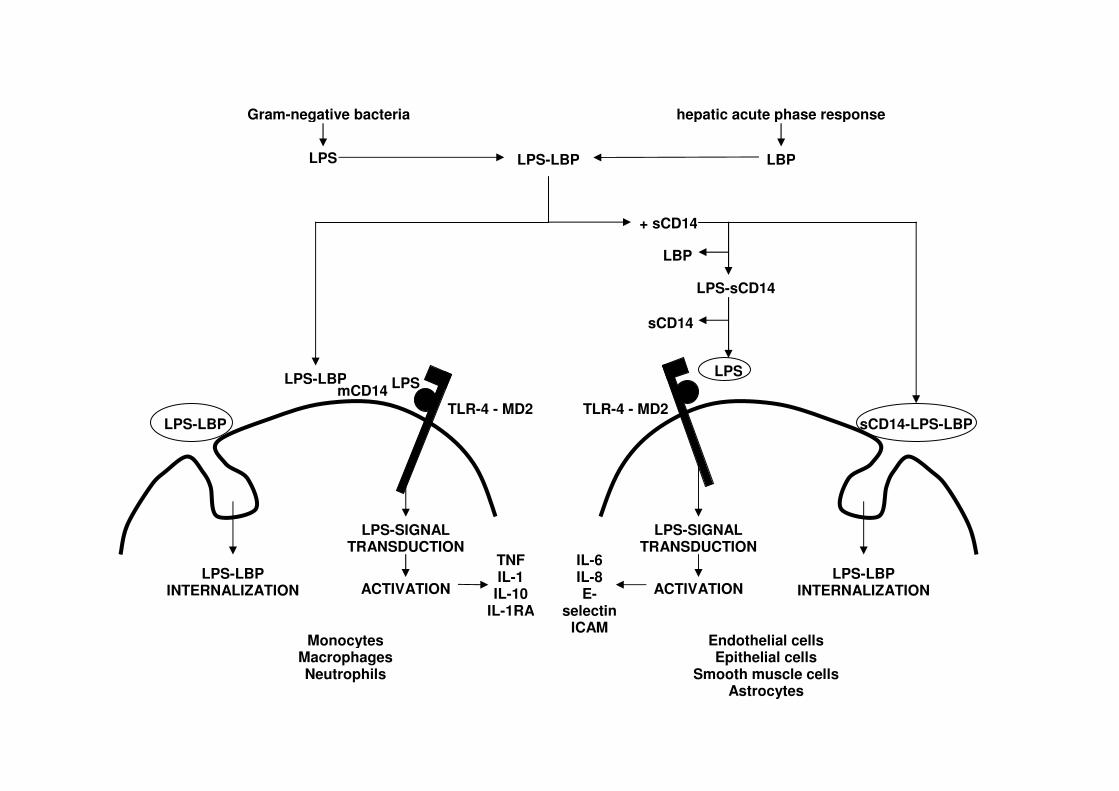
Lipopolysaccharide, a constituent of the outer membrane of Gram-negative bacteria, is
apparently one of the major toxins responsible for initiating the pathophysiological cascade
resulting in sepsis or a systemic inflammatory response (Rietschel et al., 1996). LPS-binding
protein recognises bacterial LPS and transfers it to CD14, thereby enhancing internalisation of
LPS or host cell stimulation. Therefore, LBP can be considered as an opsonin, whereas CD14 is
an opsonic receptor for complexes of LBP-LPS or LPS-containing particles, such as Gram-
negative bacteria. Lipopolysaccharide-mediated stimulation of CD14-positive cells, such as
monocytes, macrophages and PMN, is enhanced 100 to 1,000-fold when LBP is added to a
serum-free system. Although CD14 alone can efficiently interact with LPS, the presence of LBP
has been demonstrated to increase not only the association rate, but also the association constant
of LPS with CD14 by three orders of magnitude (Thomas et al., 2002). In addition, LBP can
catalytically transfer monomeric LPS from LPS aggregates onto membrane-associated CD14
(mCD14) or soluble CD14 (sCD14) molecules (Tobias et al., 1993; Hailman et al., 1994), which
has been suggested a major advantage compared to binding proteins, such as
bactericidal/permeability-increasing protein (Tobias et al., 1997).
Therefore, LBP participates in the pathogenesis of sepsis (Schumann et al., 1990;
Mathison et al., 1992), although it is unclear why the host would release large quantities of LBP
during acute inflammation, thereby further enhancing LPS-induced cytokine secretion. However,
it has been demonstrated that the acute phase LBP has a protective effect against LPS and
bacterial infection; and may represent a physiologic defence mechanism against Gram-negative
infection (Lamping et al., 1998). LPS-binding protein not only mediates binding of LPS to
CD14, but also the binding of whole Gram-negative bacteria to CD14 leading to phagocytosis
and subsequent clearance of bacteria (Lengacher et al., 1996). This LBP-mediated CD14-
dependent phagocytic uptake of bacteria could be a mechanism for improved survival following
bacterial infection.
In vitro, high concentrations of LBP have been shown to block the LPS-induced
stimulation of a murine macrophage cell line, both in the absence and in the presence of murine
serum (Lamping et al., 1998). The LBP dependency in this cell culture system was of bipolar
nature. Addition of murine LBP at concentrations corresponding to the constitutive murine LBP

- 22 -
levels resulted in an increased secretion of TNF-α in vitro, whereas high LBP concentrations,
simulating the acute phase rise of LBP, resulted in a pronounced decrease in TNF-α response
(Lamping et al., 1998). A somehow bipolar nature of biological activity has been described for
CD14, another protein involved in LPS recognition. In vitro, intermediate concentrations of
sCD14 increased the LBP dependent stimulation of PMN, whereas high concentrations of
sCD14 inhibited the PMN activation (Hailman et al., 1996), and protected against the toxic
properties of LPS (Haziot et al., 1995). It can be concluded that LBP and sCD14 in vivo both
seem to protect from LPS toxicity and bacteraemie if elevated, although in vitro both proteins
seem to contribute to LPS recognition and LPS-mediated cell stimulation (Lamping et al., 1998).
This bipolar physiologic defence mechanism could have potential applications in therapeutic
intervention strategies during sepsis or Gram-negative infections, using a natural host defence
mechanism against overstimulation by bacterial products (Lamping et al., 1998) (Fig. 6).
In the bovine, only a few studies have been performed in order to elucidate the kinetics
and the role of sCD14 and LBP during LPS-induced or Gram-negative mastitis (Bannerman et
al., 2003; Lee et al., 2003a). Lee et al. (2003a) followed sCD14 concentrations throughout
lactation and observed the highest concentrations (11.4 ± 0.17 µg/ml) of milk sCD14 during the
transitional period. Intermediate concentrations (± 5.5 µg/ml) were present during early (5-100 d
post-partum) and late lactation (> 200 d post-partum), and lowest concentrations (4.6 ± 0.27
µg/ml) were found in mid-lactating animals. Increased sCD14 concentration was associated with
higher SCC, although sCD14 concentration in subclinically infected quarters did not differ from
sCD14 concentration in healthy mammary glands. Upon induction of acute LPS mastitis, milk
sCD14 rapidly increased to reach maximal levels between 12 and 24 h post-challenge (Lee et al.,
2003a). Possible sources of sCD14 in milk were postulated to be blood serum leakage through
the damaged blood-milk barrier or cells present in the mammary gland. Milk sCD14 appeared
much later than the initial breakdown of the blood-milk barrier, as indicated by the increase in
serum albumin concentrations, ruling out the first possibility. Bovine PMN have been
demonstrated capable of releasing sCD14 into milk upon contact with LPS. Moreover, the
increase in sCD14 paralleled the increase in SCC following LPS challenge. It is therefore most
likely that the PMN influx into the inflamed mammary gland induces the increase in milk sCD14
(Lee et al., 2003a).
Figure 6. An overview of the association of LPS with cells mediated by (LBP) and CD14 (based on Tobias et al., 1999).
Gram-negative bacteria hepatic acute phase response
LPS LBP LPS-LBP
+ sCD14
LBP
LPS-sCD14
sCD14
sCD14-LPS-LBP
LPS-LBP INTERNALIZATION
Endothelial cells Epithelial cells
Smooth muscle cells Astrocytes
TLR-4 - MD2
LPS
LPS-SIGNAL TRANSDUCTION
ACTIVATION
Monocytes Macrophages Neutrophils
LPS-LBP INTERNALIZATION
LPS-SIGNAL TRANSDUCTION
ACTIVATION
mCD14
LPS-LBP
LPS-LBP
TNF IL-1 IL-10
IL-1RA
IL-6 IL-8 E-
selectin ICAM
LPS
TLR-4 - MD2

- 24 -
Following intramammary LPS challenge, peak fever, a marker of the acute phase
response, preceded LBP increase both in blood and milk (Bannerman et al., 2003). The increase
in milk LBP paralleled increments of sCD14 levels. From a host perspective, the simultaneous
increase of both molecules should be considered advantageous, because they act in concert to
facilitate activation of the host defence mechanisms by presenting LPS to the transmembrane
LPS receptor, TLR-4 (Bannerman and Goldblum, 2003). However, maximal levels of IL-8 were
already observed before increases in either LBP or sCD14, which suggests that initial host cell
activation can take place in the presence of only basal levels of sCD14 and LBP (Bannerman et
al., 2003). In contrast to sCD14, LBP increases in the affected glands occur coincidently with the
disruption of the blood-milk barrier, as indicated by the rise in serum albumin concentration
(Bannerman et al., 2003), which is in accordance with the fact that LBP is of hepatic origin and
is already present in plasma at high concentrations before the onset of increases in milk.
During the initial phase of inflammation, sCD14 mediates the activation of non-CD14-
bearing cells, including epithelial cells (Frey et al., 1992). Low concentrations of LPS-
recombinant bovine sCD14 (rbosCD14) have been demonstrated to induce IL-8 transcription in
vitro in mammary ductal epithelial cell cultures (Wang et al., 2002). Interleukin-8 is an
important early chemotactic factor, inducing a massive influx of leukocytes, predominantly
PMN into the infected mammary gland (Shuster et al., 1997). A delay in leukocyte recruitment
after intramammary coliform infections resulted in 10 times more bacteria (Erskine et al., 1989),
indicating the importance of a rapid and early inflammatory reaction in protecting the host from
overwhelming bacterial infections. Indeed, delayed leukocyte recruitment was not only
associated with overwhelming bacterial growth, but could also be related with increased severity
of the inflammatory reaction (Vandeputte-Van Messom et al., 1993). Therefore, the signalling
role of sCD14 during the early stage of infection, inducing activation of mammary epithelial
cells with the production of IL-8, is an essential step in innate host defence (Wang et al., 2002).
Recently, biological functionality of rbosCD14 has been demonstrated in a mouse mastitis model
using experimental E. coli challenge (Lee et al., 2003b). Intramammary administration of
rbosCD14 reduced TNF-α production by phagocytes in milk, possibly by competition with
mCD14 for LPS. Moreover, rbosCD14 administration reduced the number of E. coli, most likely
through an early infiltration of PMN, induced by activation of epithelial cells with the rapid
production of IL-8 upon infection (Lee et al., 2003b).

Pathogenesis, severity prediction and treatment of E. coli mastitis: a review
- 25 -
2.4. Recurrent intramammary E. coli infections in the bovine
Intramammary infections caused by E. coli are commonly considered to be limited in
duration. Sometimes, the micro-organisms may even be eliminated before or shortly after the
onset of clinical symptoms (Hogan et al., 1989). Therefore, the host defence system appears to
eliminate E. coli efficiently (Hill et al., 1978), especially when the intramammary infection
occurs late in lactation (Hill and Shears, 1979). Recurrent clinical mastitis caused by E. coli in a
cow that experiences persistent intramammary infection is known to exist, but is considered to
be exceptional (Hogan et al., 1989; Lam et al., 1996). It has been observed that quarters with
recurrent clinical episodes of mastitis caused by E. coli were infected by the same E. coli strain,
as confirmed by serotyping and deoxyribonucleic acid (DNA) polymorphism patterns with REP
(= repetitive extragenic palindromic) and ERIC (= enterobacterial repetitive intergenic
consensus) primers (Lipman et al., 1994). The estimated occurrence of episodes of clinical
mastitis during persistent intramammary infection caused by E. coli within the same quarter
ranged from 4.77% (Döpfer et al., 1999), over 7.5% (Hogan et al., 1989) to 9.1% (Lam et al.,
1996), depending on the study design and confirmation techniques used. The E. coli bacteria
causing persistent intramammary infection may survive intracellularly after invading the udder
epithelial cells (Döpfer et al., 1999), which is, however, in contrast with the general perception
that strains of E. coli from mastitis cases are non-invasive for the udder epithelial cells (Linton et
al., 1979; Sanchez-Carlo et al., 1984; Valente et al., 1988) and mainly opportunistic
environmental pathogens (Nemeth et al., 1994). A limited number of E. coli strains from
recurrent mastitis has been studied with respect to adhesion and invasion mechanisms towards
mammary epithelial cells (Döpfer et al., 2000; 2001). Besides occurrence of persistent
intramammary infection in the same quarter, multiple cases of recurrent mastitis in different
quarters of the same cow with the same genotype of E. coli occurred (Döpfer et al., 1999).
Possible explanations could be that 1) cows were re-infected with the same strain from the
environment, 2) quarters were infected simultaneously but showed clinical symptoms of mastitis
at different times, or 3) transmission of E. coli from one quarter to another occurred (Döpfer et
al., 1999).
Recently, it was observed that quarters infected with an enterobacterial organism during
the last third of the dry period were more likely to develop mastitis due to the same pathogen
than were uninfected quarters in the subsequent lactation cycle (Bradley and Green, 2000).
Previously, the ability of infections acquired during the dry period to remain quiescent within the
udder until calving, subsequently causing clinical mastitis in early lactation, has been illustrated
(McDonald and Anderson, 1981). The dry period has been implicated as a crucial period for

- 26 -
acquisition of new coliform intramammary infections (Todhunter et al., 1991a), with more than
60% of all new intramammary infections occurring at that time. Although, it has been suggested
that the dry gland is highly resistant to enterobacterial infection as a result of the high levels of
lactoferrin; more recent research, applying polymerase chain reaction (PCR) DNA fingerprinting
techniques, has shown that the mammary gland is not resistant to the acquisition of new
enterobacterial intramammary infections (Bradley and Green, 2000). Multiple aspects should be
taken into account concerning this new phenomenon, such as potential pathogen adaptation and
changed host susceptibility (Bradley and Green, 2001a). Few intramammary enterobacterial
infections appeared to persist throughout the entire dry period (Bradley and Green, 2001b).
3. EXPERIMENTAL INFECTION MODELS
Several experimental infection models for the induction of intramammary E. coli infection
have been explored throughout the years in an attempt to mimic naturally occurring E. coli
mastitis. During natural intramammary infection, bacteria have to overcome the physical barrier
formed by the teat canal and its keratinisation before they can enter the teat sinus and start to
proliferate in the milk. External contamination of the teat ends with a bacterial suspension of E.
coli did, however, not induce acute clinical mastitis (Vandeputte-Van Messom et al., 1992). In
order to obtain an intramammary infection following bacterial exposure, researchers passed the
physical barrier of the teat canal by direct infusion of the bacterial suspension or the bacterial
cell wall compounds, such as endotoxin, into the teat and gland sinus.
3.1. Lipopolysaccharide model
Lipopolysaccharide (endotoxin) is a component of the Gram-negative cell wall, and is
composed of three basic subunits: 1) the O-polysaccharide, providing serospecificity for Gram-
negative bacteria, 2) the lipid moiety, generally called lipid A, which is considered to be the
toxic component of the cell wall, and 3) the R core, consisting of hexoses, hexamines, and
heptose, which acts as a bridge between the O-polysaccharide and the lipid (Westphal, 1975).
The extraction of LPS from Gram-negative bacteria can be performed through different methods
(Burvenich, 1983): 1) trichloracetic acid (Boivin-extraction), 2) diethylene glycol, 3) phenol /
water (Westphal-extraction), 4) water / ether, 5) phenol / chloroform / petroleumether and
butanol. Most commercially available endotoxin preparations are prepared by the Boivin- or
Westphal-extraction (Burvenich, 1983). Boivin-extracted LPS contains some protein and
Pathogenesis, severity prediction and treatment of E. coli mastitis: a review
- 27 -
possesses therefore much stronger antigenic properties. In contrast, Westphal-extracted LPS
contains some peptides, nucleic acids and sugar residues.
The toxicity of LPS is determined by two of its subunits. Lipid A, the lipid moiety of
LPS, is identical for LPS originating from different Gram-negative bacterial species. Primary
toxicity is due to the lipid A fraction of the LPS, whereas secondary toxicity is related to
antigen-antibody reactions, mainly directed against the sugar residues on the O-polysaccharide,
which are weakly antigenic. Host hypersensitivity determines the impact of secondary LPS
toxicity. A longer O-polysaccharide chain with more sugar residues has higher immunogenic
properties and can therefore result in more pronounced secondary toxicity (Burvenich, 1983).
Intramammary LPS challenge is frequently used to study inflammation in the bovine
mammary gland, mainly due to its resemblance to E. coli challenge (Lohuis et al., 1988b;
1988c). Nevertheless, some fundamental differences exist between both challenge models. In the
LPS model, peak fever is reached within 6 h post-challenge, depending on the infused dose of
LPS, and is accompanied by acute local and systemic clinical symptoms (Lohuis et al., 1988b),
although no depression in reticulorumen motility could be observed following intramammary
LPS challenge. Duration of clinical symptoms and general illness in the LPS model is limited,
and therefore, quarter milk production is only temporarily depressed and rapidly returns to pre-
infection values (Hoeben et al., 2000a; Mehrzad et al., 2001a). In contrast to earlier assumptions,
general clinical symptoms following intramammary LPS challenge are not due to LPS resorption
into circulation, but mediated through locally produced and systemically active inflammatory
cytokines (Dosogne et al., 2002).
Throughout the years, a wide range of LPS inoculum doses, varying from 0.0001 (van
der Vliet et al., 1989) to 20,000 µg (Frost et al., 1984), has been applied, although currently used
doses appear in a more narrow range of 100 to 1,000 µg per quarter (Table 5). The tendency to
use a specific LPS inoculum dose or range of doses also seems to be research group-related. In
contrast to the wide variety of applied inoculum doses, only a small selection of strains was used
for the extraction and preparation of LPS (Table 6).
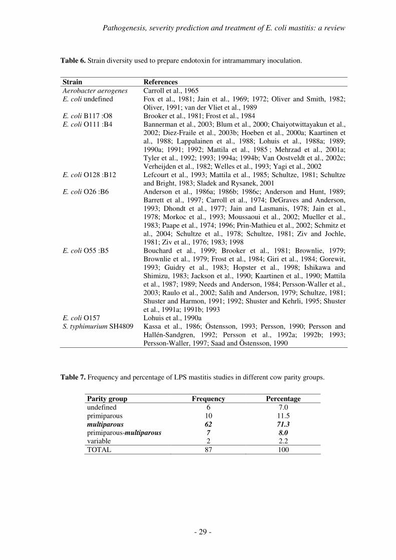
A general trend in LPS-induced mastitis is the use of multiparous cows (78.3 %) during
the mid-lactation stage (Table 7-8). Although in many, predominantly older studies, neither
parity group nor stage of lactation are defined, the most recent research reports on endotoxin-
induced mastitis provide more detailed information on the experimental animals and conditions
applied in the studies. Taking into account that cow factors may determine the outcome of
mastitis (Burvenich et al., 2003), the trend towards a more standardised choice of experimental
animals is positive and should be encouraged at all levels.

- 28 -
Table 5. LPS inoculum dose for intramammary endotoxin challenge.
LPS inoculum dose References
100 pg van der Vliet et al., 1989 1 ng Persson et al., 1993 10 ng Persson et al., 1993; Schultze, 1981; Verheijden et al., 1982 100 ng Mattila et al., 1985; Persson et al., 1993; Schultze, 1981 1 µg Needs and Anderson, 1984; Persson et al., 1993; Persson-Waller,
1997; Schultze et al., 1978; Schultze, 1981; Schultze and Bright, 1983; Verheijden et al., 1982
2 µg Hopster et al., 1998; Salih and Anderson, 1979 5 µg Giri et al., 1984; Persson, 1990; Persson et al., 1992a; Sladek and
Rysanek, 2001 10 µg Anderson et al., 1986a; 1986b; 1986c; Anderson and Hunt, 1989;
Barrett et al., 1997; Brownlie, 1979; Brownlie et al., 1979; Giri et al., 1984; Guidry et al., 1983; Ishikawa and Shimizu, 1983; Lohuis et al., 1989; Mattila et al., 1987; Mattila and Frost, 1989; Morkoc et al., 1993; Moussaoui et al., 2002; Needs and Anderson, 1984; Paape et al., 1996; Prin-Mathieu et al., 2002; Raulo et al., 2002; Shuster and Harmon, 1992; Shuster et al., 1991a; 1991b; 1993; Verheijden et al., 1982
12.5 µg Gorewit, 1993 15 µg Bouchard et al., 1999 20 µg Kaartinen et al., 1990; Persson and Hallén-Sandgren, 1992; Persson
et al., 1992a; Persson-Waller, 1997 25 µg Giri et al., 1984; Gorewit, 1993 50 µg Carroll et al., 1974; Jain and Lasmanis, 1978; Jain et al., 1978;
Mueller et al., 1983; Oliver, 1991; Östensson, 1993; Persson-Waller et al., 2003; Saad and Östensson, 1990
100 µg Bannerman et al., 2003; Brooker et al., 1981; Chaiyotwittayakun et al., 2002; Frost et al., 1984; Giri et al., 1984; Guidry et al., 1983; Jackson et al., 1990; Kaartinen et al., 1988; Lappalainen et al., 1988; Lohuis et al., 1988a; 1990b; 1991; Mattila et al., 1985; Schmitz et al., 2004; Shuster and Harmon, 1991; Verheijden et al., 1982; Ziv et al., 1998
200 µg Carroll et al., 1965; Jain et al., 1972 500 µg Blum et al., 2000; Carroll et al., 1974; Diez-Fraile et al., 2003b;
Dosogne et al., 2002; Hoeben et al., 2000a; Lefcourt et al., 1993; Mehrzad et al., 2001a; Paape et al., 1974; Van Oostveldt et al., 2002c; Yagi et al., 2002
750 µg Jain et al., 1972 900 µg Brooker et al., 1981 1 mg Brooker et al., 1981; DeGraves and Anderson, 1993; Dhondt et al.,
1977; Frost et al., 1984; Kassa et al., 1986; Tyler et al., 1992; 1993; 1994a; 1994b; Verheijden et al., 1982; Welles et al., 1993; Ziv et al., 1983
3 mg Dhondt et al., 1977 5 mg Brooker et al., 1981; Carroll et al., 1974; Frost et al., 1984; Jain et
al., 1969 10 mg Verheijden et al., 1982; Ziv et al., 1976 15 mg Ziv and Jochle, 1981 20 mg Carroll et al., 1965
Pathogenesis, severity prediction and treatment of E. coli mastitis: a review
- 29 -
Table 6. Strain diversity used to prepare endotoxin for intramammary inoculation.
Strain References
Aerobacter aerogenes Carroll et al., 1965 E. coli undefined Fox et al., 1981; Jain et al., 1969; 1972; Oliver and Smith, 1982;
Oliver, 1991; van der Vliet et al., 1989 E. coli B117 :O8 Brooker et al., 1981; Frost et al., 1984 E. coli O111 :B4 Bannerman et al., 2003; Blum et al., 2000; Chaiyotwittayakun et al.,
2002; Diez-Fraile et al., 2003b; Hoeben et al., 2000a; Kaartinen et al., 1988; Lappalainen et al., 1988; Lohuis et al., 1988a; 1989; 1990a; 1991; 1992; Mattila et al., 1985 ; Mehrzad et al., 2001a; Tyler et al., 1992; 1993; 1994a; 1994b; Van Oostveldt et al., 2002c; Verheijden et al., 1982; Welles et al., 1993; Yagi et al., 2002
E. coli O128 :B12 Lefcourt et al., 1993; Mattila et al., 1985; Schultze, 1981; Schultze and Bright, 1983; Sladek and Rysanek, 2001
E. coli O26 :B6 Anderson et al., 1986a; 1986b; 1986c; Anderson and Hunt, 1989; Barrett et al., 1997; Carroll et al., 1974; DeGraves and Anderson, 1993; Dhondt et al., 1977; Jain and Lasmanis, 1978; Jain et al., 1978; Morkoc et al., 1993; Moussaoui et al., 2002; Mueller et al., 1983; Paape et al., 1974; 1996; Prin-Mathieu et al., 2002; Schmitz et al., 2004; Schultze et al., 1978; Schultze, 1981; Ziv and Jochle, 1981; Ziv et al., 1976; 1983; 1998
E. coli O55 :B5 Bouchard et al., 1999; Brooker et al., 1981; Brownlie, 1979; Brownlie et al., 1979; Frost et al., 1984; Giri et al., 1984; Gorewit, 1993; Guidry et al., 1983; Hopster et al., 1998; Ishikawa and Shimizu, 1983; Jackson et al., 1990; Kaartinen et al., 1990; Mattila et al., 1987; 1989; Needs and Anderson, 1984; Persson-Waller et al., 2003; Raulo et al., 2002; Salih and Anderson, 1979; Schultze, 1981; Shuster and Harmon, 1991; 1992; Shuster and Kehrli, 1995; Shuster et al., 1991a; 1991b; 1993
E. coli O157 Lohuis et al., 1990a S. typhimurium SH4809 Kassa et al., 1986; Östensson, 1993; Persson, 1990; Persson and
Hallén-Sandgren, 1992; Persson et al., 1992a; 1992b; 1993; Persson-Waller, 1997; Saad and Östensson, 1990
Table 7. Frequency and percentage of LPS mastitis studies in different cow parity groups.
Parity group Frequency Percentage
undefined 6 7.0 primiparous 10 11.5 multiparous 62 71.3
primiparous-multiparous 7 8.0 variable 2 2.2 TOTAL 87 100

- 30 -
Table 8. Frequency and percentage of LPS mastitis studies in different stages of lactation.
Parity group Frequency Percentage
undefined 19 22.0 early 13 15.0 mid 23 26.4 late 4 4.6 dry 7 8.0 variable 7 8.0 early-mid 4 4.6
mid-late 8 9.2 late-dry 1 1.1 early-mid-late 1 1.1 TOTAL 87 100
Most frequently, 1 or 2 quarters are challenged with LPS (86.6 %) (Fig. 7).
Figure 7. Percentage of LPS mastitis studies inoculating 1, 2, 3 or 4 quarters of each dairy cow.
In conclusion, experimental endotoxin mastitis is mostly induced through a single dose
administration. This is in contrast with the more dynamic release of LPS in the E. coli mastitis
model, where LPS is released during growth and subsequent killing of the inoculated bacteria
over a much longer time interval (Petsch and Anspach, 2000). Therefore, the model is suitable in
the study of inflammatory kinetics, without major health risk for the experimental animals with
respect to persistent clinical disease, as is occasionally observed following E. coli challenge.
0
10
20
30
40
50
60
1 2 3 4
number of quarters
%

Pathogenesis, severity prediction and treatment of E. coli mastitis: a review
- 31 -
3.2. Escherichia coli model
The E. coli mastitis model is the more realistic approach for an experimental
intramammary infection, as it can be observed in field cases. The most important difference with
the previously described LPS mastitis model is the occurrence of a continuous release of LPS
during bacterial growth and killing (Burvenich, 1983; Petsch and Anspach, 2000), although an
initial delay in this release can occur. Intramammary inoculation is performed in one or more
quarters, leaving the other quarters as negative controls during inflammation (Fig. 8). The
distribution of the number of inoculated quarters with live E. coli bacteria is comparable to the
previously discussed LPS model.
Figure 8. Percentage of E. coli mastitis studies inoculating 1, 2, 3 or 4 quarters of each dairy cow.
Inflammatory characteristics and kinetics related to experimentally induced E. coli
mastitis are mainly influenced by the initial inoculum dose. When low numbers of bacteria are
inoculated, several hours of bacterial growth are needed before initiation of an inflammatory
response (Shuster et al., 1996; Riollet et al., 2000), whereas inoculation of high numbers of
bacteria rapidly induce local and systemic clinical symptoms (Vandeputte-Van Messom et al.,
1993; Hoeben et al., 2000a; Dosogne et al., 2002). Therefore, it is not easy to clearly define the
exact time point when peak fever and maximal systemic and local clinical symptoms are
reached. A major clinical difference between the LPS and the E. coli mastitis model is the
pronounced suppression of reticulorumen motility, which can only be observed in the E. coli
challenge model (Verheijden et al., 1982; Lohuis et al., 1988b).
For induction of intramammary challenge, a great variety of strains has been used,
however, only a few have regularly been applied (Table 9).
0
10
20
30
40
50
60
1 2 3 4
number of quarters
%

- 32 -
Table 9. Strain diversity of coliform bacteria used for intramammary challenge.
Strain References
Aerobacter aerogenes Jain et al., 1969 A. aerogenes 2414-1 Carroll et al., 1973; Zia et al., 1987 A. aerogenes 2413-2 Carroll et al., 1973 Klebsiella K644 Carroll et al., 1973 Klebsiella K-6 Carroll et al., 1973 K. pneumoniae Bramley and Neave, 1975 E. coli 12795 Carroll et al., 1973 E. coli Harmon et al., 1976 E. coli S-16 Carroll et al., 1973 E. coli Lilly Carroll et al., 1973 E. coli 1128 Schultze et al., 1978 E. coli McDonald 487 Hogan et al., 1994b; Shuster et al., 1996; 1997 E. coli 727 Barrett et al., 1997; Hogan et al., 1994a; 1995; Scaletti et al., 2003;
Smith et al., 1999; Tomita et al., 2000; Weiss et al., 2004 E. coli B117:O8 Frost et al., 1980; 1982 E. coli FT238 Pyörälä et al., 1994 E. coli O157 Dosogne et al., 1997; Kremer et al., 1993a; 1993b; 1993c; Lohuis et
al., 1990b; Roets et al., 1999; van Werven et al., 1997 E. coli O5 Griel et al., 1975 E. coli P4:O32 Anderson et al., 1985; Blum et al., 2000; Dosogne et al., 2002;
Heyneman et al., 1990; Heyneman and Burvenich, 1992; Hill et al., 1978; Hoeben et al., 2000a; Monfardini et al., 1999; Riollet et al., 2000; Shpigel et al., 1997; Van Oostveldt et al., 2002c; Vandeputte-Van Messom and Burvenich, 1993; Vandeputte-Van Messom et al., 1993
E. coli Saskatchewan Sordillo and Babiuk, 1991; Sordillo and Peel, 1992
Throughout the years, different inoculum doses have been used from 5 CFU (Schultze et
al., 1988) up to 1.8 x 10¹⁰ CFU (Frost et al., 1980), although currently used inoculum doses were
2 x 10² to 1 x 10⁴ CFU (Carroll et al., 1973; Fox et al., 1981; Frost et al., 1982; Heyneman et al.,
1992; Vandeputte-Van Messom et al., 1993; Hoeben et al., 2000a) (Table 10). Inoculum doses
above 1 x 10⁴ CFU per quarter have mainly been used in older studies. Another remarkable
observation is that American mastitis research groups predominantly inoculate low numbers (<
100 CFU) of E. coli into the mammary gland, whereas European investigators prefer to use
higher numbers ( > 100 CFU). Therefore, care should be taken when study results in terms of
inflammatory kinetics are compared between both types of experimental design for reasons
discussed earlier.
Pathogenesis, severity prediction and treatment of E. coli mastitis: a review
- 33 -
Table 10. Inoculum doses used for experimental intramammary inoculation with coliform bacteria.
Inoculum dose (CFU) References
< 20 Schultze et al., 1978 21-30 Hogan et al., 1994b; Scaletti et al., 2003; Schultze et al., 1978;
Shuster et al., 1996; 1997 31-50 Barrett et al., 1997; Frost et al., 1982; Hill et al., 1978; Riollet et al.,
2000; Sordillo and Babiuk, 1991; Sordillo and Peel, 1992 51-100 Carroll et al., 1973; Hogan et al., 1994a; 1995; Smith et al., 1999;
Tomita et al., 2000; Weiss et al., 2004 101-500 Carroll et al., 1973; Frost et al., 1982; Griel et al., 1975; Harmon et
al., 1976; Shpigel et al., 1997; Van Oostveldt et al., 2002c 501-1,000 Anderson et al., 1985; Carroll et al., 1973; Dosogne et al., 1997;
Kremer et al., 1993a; 1993b; 1993c; Roets et al., 1999; Shpigel et al., 1997; van Werven et al., 1997
1,001-10,000 Anderson et al., 1985; Blum et al., 2000; Bramley and Neave, 1975; Carroll et al., 1973; Dosogne et al., 2002; Griel et al., 1975; Heyneman et al., 1990; Heyneman and Burvenich, 1992; Hill et al., 1978; Hoeben et al., 2000a; Lohuis et al., 1990b; Monfardini et al., 1999; Pyörälä et al., 1994; Vandeputte-Van Messom and Burvenich, 1993; Vandeputte-Van Messom et al., 1993; Zia et al., 1987
10,001-100,000 Bramley and Neave, 1975; Carroll et al., 1973 100,001-1,000,000 Bramley and Neave, 1975; Carroll et al., 1973; Hill et al., 1978 > 1,000,001 Carroll et al., 1973; Jain et al., 1969; Frost et al., 1980
Experimentally induced E. coli mastitis has mainly been studied in multiparous animals
(84.5 %), whereas the immune response in primiparous cows (6.7 %) has hardly been explored
(Table 11). In contrast to LPS mastitis which was predominantly studied in mid-lactation, E. coli
challenge has mainly been performed in early lactation animals (66.7 %) (Table 12).
Table 11. Frequency and percentage of E. coli mastitis studies in different cow parity groups.
Parity group Frequency Percentage
undefined 2 4.4 primiparous 3 6.7 multiparous 30 66.7 primiparous-multiparous 8 17.8 variable 2 4.4 TOTAL 45 100

- 34 -
Table 12. Frequency and percentage of E. coli mastitis studies in different stages of lactation.
Parity group Frequency Percentage
undefined 4 8.9 early 28 62.3 mid 3 6.7 late 4 8.9 dry 0 0 variable 2 4.4 early-mid 1 2.2 mid-late 2 4.4 late-dry 0 0 early-mid-late 1 2.2
TOTAL 45 100
4. DIAGNOSIS AND TREATMENT OF E. COLI MASTITIS
4.1. Diagnosis
Coliform mastitis affects the quality of milk, may induce partial or total loss of milk
production capacity in the affected quarters or can even result in a life-threatening situation for
the diseased animal. Therefore, rapid and correct diagnosis of the etiological agent of clinical
mastitis should be performed. Knowledge on risk factors and severity determining factors may
assist in the recognition of a clinical case as being due to E. coli.
4.1.1. Clinical and bacteriological diagnosis
Clinical diagnosis, consisting of inspection and visual examination of the diseased
animal, the affected quarter and the changes in milk composition, does not systematically result
in a correct etiological diagnosis. Appearance of mastitis with acute or peracute clinical signs
and watery mammary secretions with flakes is indicative of an intramammary E. coli infection,
although bacteriological sampling of clinical mastitis cases only resulted in 16.2% E. coli
cultures (Schukken et al., 1989). If the absence of bacteria in culture would also be considered as
E. coli, a total of 39.1% of the clinical cases would be due to E. coli (Schukken et al., 1989).
Therefore, etiological diagnosis solely based on clinical parameters is assumed to be difficult to
take the appropriate therapeutic measures to combat acute clinical mastitis. It is clear that further
steps should be taken to identify the etiological agent through bacteriological examination or
molecular identification methods.
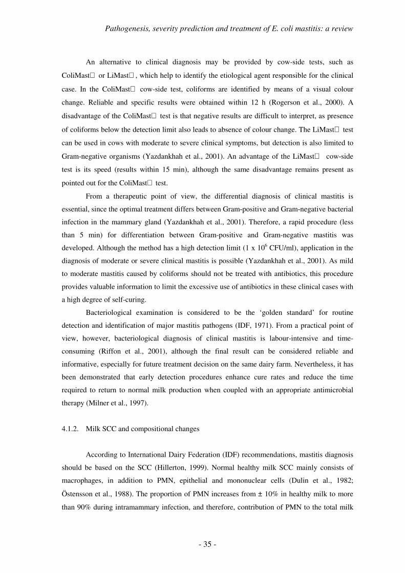
Pathogenesis, severity prediction and treatment of E. coli mastitis: a review
- 35 -
An alternative to clinical diagnosis may be provided by cow-side tests, such as
ColiMast or LiMast, which help to identify the etiological agent responsible for the clinical
case. In the ColiMast cow-side test, coliforms are identified by means of a visual colour
change. Reliable and specific results were obtained within 12 h (Rogerson et al., 2000). A
disadvantage of the ColiMast test is that negative results are difficult to interpret, as presence
of coliforms below the detection limit also leads to absence of colour change. The LiMast test
can be used in cows with moderate to severe clinical symptoms, but detection is also limited to
Gram-negative organisms (Yazdankhah et al., 2001). An advantage of the LiMast cow-side
test is its speed (results within 15 min), although the same disadvantage remains present as
pointed out for the ColiMast test.
From a therapeutic point of view, the differential diagnosis of clinical mastitis is
essential, since the optimal treatment differs between Gram-positive and Gram-negative bacterial
infection in the mammary gland (Yazdankhah et al., 2001). Therefore, a rapid procedure (less
than 5 min) for differentiation between Gram-positive and Gram-negative mastitis was
developed. Although the method has a high detection limit (1 x 10⁶ CFU/ml), application in the
diagnosis of moderate or severe clinical mastitis is possible (Yazdankhah et al., 2001). As mild
to moderate mastitis caused by coliforms should not be treated with antibiotics, this procedure
provides valuable information to limit the excessive use of antibiotics in these clinical cases with
a high degree of self-curing.
Bacteriological examination is considered to be the ‘golden standard’ for routine
detection and identification of major mastitis pathogens (IDF, 1971). From a practical point of
view, however, bacteriological diagnosis of clinical mastitis is labour-intensive and time-
consuming (Riffon et al., 2001), although the final result can be considered reliable and
informative, especially for future treatment decision on the same dairy farm. Nevertheless, it has
been demonstrated that early detection procedures enhance cure rates and reduce the time
required to return to normal milk production when coupled with an appropriate antimicrobial
therapy (Milner et al., 1997).
4.1.2. Milk SCC and compositional changes
According to International Dairy Federation (IDF) recommendations, mastitis diagnosis
should be based on the SCC (Hillerton, 1999). Normal healthy milk SCC mainly consists of
macrophages, in addition to PMN, epithelial and mononuclear cells (Dulin et al., 1982;
Östensson et al., 1988). The proportion of PMN increases from ± 10% in healthy milk to more
than 90% during intramammary infection, and therefore, contribution of PMN to the total milk

- 36 -
SCC has been proposed as an indicator of mastitis (Kelly et al., 2000). This approach would
provide more information on the health status of the mammary quarters (Östensson et al., 1988),
but further applications did not occur mainly due to practical limitations.
Milk SCC threshold for the diagnosis of mastitis has been reduced from 500,000 to
200,000 cells/ml, and recently a threshold of 100,000 cells/ml was even suggested (Hillerton,
1999) for quarters to be considered as clinically healthy. This seems to be in agreement with
SCC results recently obtained through meta-analysis (Djabri et al., 2002), suggesting an average
SCC of 68,000 cells/ml in bacteriologically negative quarters. Minor pathogens only result in a
slight increase of quarter SCC (110,000 to 150,000 cells/ml), whereas major pathogens induced
more pronounced increases (350,000 cells/ml) (Djabri et al., 2002). Recently, SCC patterns have
been associated with pathogen-specific cases of clinical mastitis. In that study, clinical E. coli
mastitis was significantly associated with the presence of a short peak in SCC (de Haas et al.,
2004).
Besides SCC, several compositional changes occur in milk during clinical mastitis
(Kitchen, 1981; Östensson et al., 1988; Hamann and Krömker, 1997), such as decreased quarter
milk yield, lactose, fat, long-chained fatty acids, total casein, α-lactalbumin, β-lactoglobulin,
Ca2+, Mg2+, P2+, Zn2+ and potassium (K+), and increased whey proteins, serum albumin,
immunoglobulins, proteose peptone, free fatty acids, short-chained fatty acids, sodium (Na+),
chlorine (Cl-), lactate, lipase, lysozyme, N-acetyl-β-D-glucosaminidase, β-glucuronidase and
plasmin activity (Pyörälä, 2003).
4.1.3. Molecular identification methods
Recently, a PCR technique was developed for simultaneous detection of several major
mastitis pathogens (Riffon et al., 2001). The advantage of PCR, compared to bacteriology, lays
in the possibility to use only nanograms of nucleic acid samples, allowing the elimination of
bacterial culture, combined with rapidity (within 24 h results) and easy analysis. The detection
limit of the assay is set at 5 x 10³ CFU/ml of milk in the absence of a pre-PCR enzymatic step,
which should be sensitive enough to be used as a diagnostic tool in bovine mastitis.
4.2. Treatment
Various preventive measures and management practices have been shown effective to
decrease the incidence of coliform mastitis over the years. Nevertheless, a great need exists for
therapeutic measures following the occurrence of acute or peracute coliform mastitis. Currently,

Pathogenesis, severity prediction and treatment of E. coli mastitis: a review
- 37 -
these measures include the use of antimicrobials, anti-inflammatory agents (steroidal and non-
steroidal) and additional treatments (frequent milk-out, oxytocin administration and fluid
therapy) (Ziv, 1992).
Until now, differential therapies, depending on the risk factors and severity determining
factors associated with the specific case of acute mastitis, have not been established. The
importance of severity estimation should be stressed in relation to subsequent outcome of the
clinical disease. Early lactation and ketonemia should be considered important severity
determining factors, in combination with higher parity number (Gilbert et al., 1993; Kremer et
al., 1993c; Hoeben et al., 1997c; 2000b; van Werven et al., 1997).
4.2.1. Antimicrobial treatment of E. coli mastitis
Local and systemic antibiotics are frequently used in the treatment of clinical E. coli
mastitis in the field. Mastitis in general is currently the most frequent reason for antibiotic use in
lactating dairy cattle (Gardner et al., 1990; Meek et al., 1986; Guterbock, 1995). Even though
antibiotic treatment of mastitis has been performed for decades, our current knowledge about
their actual efficacy is still very scarce. Conclusive data on mastitis treatment efficacy through
antibiotics should not only be based on clinical field trials, but also consist of in vitro studies and
experimentally induced mastitis studies (Shpigel, 2001). Antimicrobial drugs are assumed to
exert their beneficial therapeutic effect via bactericidal or bacteriostatic action.
In vitro studies on the potential effects of local and systemic antimicrobials on bovine
blood and milk PMN functionality have demonstrated severe suppressive effects when PMN
were exposed to relatively high concentrations of several antimicrobials (Hoeben et al., 1997a;
1997b; 1998a; Dosogne et al., 1998b). In practice, these high doses could only be reached by
prolonged multiple dose treatment schedules, mainly through a local administration route
(Hoeben et al., 1998a). In these studies a wide range of antimicrobials (Table 13) was tested at
different subtherapeutic, therapeutic and supratherapeutic concentrations. Several products had
the spectrum required for treatment of infections with Gram-negative bacteria, such as E. coli
mastitis (Hoeben et al., 1997a) (Fig. 9). All antibiotics, except sulphadiazin and enrofloxacin
decreased CL at the highest concentration. The stimulatory effect of enrofloxacin might be due
to a stimulation of the production of H2O2 (Hoeben et al., 1997a) (Fig. 10).
Intravenous administration of enrofloxacin following experimentally induced E. coli
mastitis in early lactating dairy cows did not result in an improved clinical condition (Hoeben et
al., 2000a), as 2 animals in the treated group responded severely following intramammary
challenge. Nevertheless, a significantly decreased number of bacteria (1:100) in the infected

- 38 -
quarters could be observed following systemic antimicrobial treatment at 10 h post-challenge
(Monfardini et al., 1999). It is known that the administration of a bactericidal antibiotic during
intramammary bacterial infection is a double-edged sword, on the one hand decreasing the
number of bacteria in the affected quarters, on the other hand inducing a major release of LPS
from the destroyed bacteria into the mammary gland compartment (Hoeben et al., 2000a).
In a clinical field trial evaluating the efficacy of parenteral administration of procaine
penicillin G, spiramycin and enrofloxacin, bacteriological cure rate in cows with mastitis caused
by E. coli was 74% for those treated with penicillin G and 71% for the non-treated controls
(Pyörälä and Pyörälä, 1998). Following experimentally induced E. coli mastitis, treatment with
trimethoprim-sulphadiazin or colistin sulphate were apparently no more beneficial than no
treatment (Pyörälä et al., 1994). Another study reported beneficial effects of cefquinome
treatment on bacteriological cure rate following experimentally induced E. coli mastitis, whereas
no beneficial effect on bacteriological cure rate was observed for ampicillin or cloxacillin
administration (Shpigel et al., 1997).
Table 13. Antimicrobial products tested in vitro for their influence on blood or milk PMN functionality, more specifically phagocytosis, oxidative burst activity or bactericidal activity (Hoeben et al., 1997a; 1997b; 1998a; Dosogne et al., 1998b).
Antimicrobial Antibiotic group Local or systemic treatment
ampicillin aminopenicillins S cephapirin 1st generation cephalosporins L / S chloramphenicol - S cloxacillin penicillinase-resistant penicillins L danofloxacin fluoroquinolones S dihydrostreptomycin macrolides S doxycyclin tetracyclins S enrofloxacin fluoroquinolones S erythromycin macrolides S lincomycin lincosamides S mecillinam β-lactam antibiotics L Na+-ceftiofur 3rd generation cephalosporins L / S neomycin macrolides S oleandomycin macrolides S oxytetracyclin tetracyclins S penicillin natural penicillins L / S spiramycin macrolides S sulphadiazin sulphonamides S

Pathogenesis, severity prediction and treatment of E. coli mastitis: a review
- 39 -
Figure 9. Chemiluminescence index (%) of isolated blood PMN incubated with some currently used antibiotics for the treatment of mastitis (Hoeben et al., 1998a).
Figure 10. Chemiluminescence index (%) of isolated blood PMN incubated with some currently used antibiotics for the treatment of mastitis (Hoeben et al., 1997a; 1997b).
0
20
40
60
80
100
120
140
160
-2 -1 0 1 2 3
concentration (ug/ml)
CL
in
de
x (
%)
chloramphenicol
danofloxacin
erythromycin
oxytetracyclin
spiramycin
Na-ceftiofur
penicillin
enrofloxacin
sulphadiazin
0
20
40
60
80
100
120
2 x 10E-6 2 x 10E-5 2 x 10E-4 2 x 10E-3
concentration (M)
CL
in
dex (
%)
ampicillin
cloxacillin
dihydrostreptomycin
doxycyclin
lincomycin
neomycin
oleandomycin

- 40 -
4.2.2. Anti-inflammatory treatment of E. coli mastitis
Glucocorticosteroids are very well known and widely used anti-inflammatory agents in
the treatment of a variety of inflammatory processes, such as acute E. coli mastitis in the
periparturient cow. Local and systemic administration of glucocorticosteroids is possible. Local
administration reduces swelling and oedema in the affected tissue, whereas systemic
administration can combat the symptoms of endotoxin shock (Phillips et al., 1987). However,
both endogenous and exogenous glucocorticosteroids in high concentrations have been shown to
suppress functions of circulating PMN (Hoeben et al., 1998b), although no adverse effects on in
vitro PMN CL could be shown at therapeutic concentrations. Attention has to be paid to possible
immunodepressive effects of glucocorticosteroids after repeated and local administration. It is
therefore advisable to avoid the regular use of these drugs during inflammation in vivo and to
limit potential administration under controlled circumstances (Hoeben et al., 1998b).
Furthermore, inhibitory effects of glucocorticosteroids on bone marrow progenitor cells were
recently reported (Hoeben et al., 1999). The inhibitory effect on in vitro cultured bovine bone
marrow progenitors not only appeared at supratherapeutic, but already at physiological
concentrations.
Non-steroidal anti-inflammatory drugs (NSAID’s) are known for many years for their
anti-inflammatory, antipyretic and analgesic effects. They generally prevent the formation of
inflammatory mediators by cyclo-oxygenase (COX) inhibition and reduction of prostaglandin E2
(PGE2) production in the brain. Under experimental conditions, NSAID’s have been shown to
have some beneficial effects (reduced rectal temperature, decrease in quarter inflammation,…),
combined with a reduced concentration of inflammatory mediators in milk and plasma
(Anderson et al., 1986a; 1986b; Lohuis et al., 1989; Ziv, 1992). A major disadvantage of most
experimental studies evaluating NSAID efficacy is the use of an LPS model, in which animals
rapidly return to normal milk production, therefore lacking to show any beneficial effects on
milk production, rate of return to production or survival (Shpigel, 2001).
Recently, NSAID efficacy was evaluated in the field (Shpigel et al., 1994; 1996).
Ketoprofen significantly improved recovery in clinical mastitis, increasing the odds ratio for
recovery by as much as 7 (Shpigel et al., 1994). In contrast, phenylbutazone and dipyrone only
increased odds ratio to 2.42 and 1.71, respectively (Shpigel et al., 1996). Therefore, ketoprofen
can be recommended as an adjunctive therapy in the treatment of Gram-negative clinical mastitis
in dairy cows (Shpigel et al., 1994).

Pathogenesis, severity prediction and treatment of E. coli mastitis: a review
- 41 -
4.2.3. Additional treatments
Several non-antibiotic methods have been suggested for the treatment of clinical
mastitis, such as frequent milk-out with or without oxytocin administration, fluid therapy,
hydrotherapy, intramammary infusions of saline, antihistamines, diuretics, hot and cold packing,
hypertonic saline infusion and ultrasonic therapy (Roberson et al., 2004). Only two of the most
relevant and practical supportive treatments will be discussed here.
Frequent milk-out was encouraged as a possible treatment for mastitis as early as 1869
by Sloan (1869), who claimed that the bad milk should be drawn three to four times a day, for by
remaining in the bag, it tends to increase the inflammation. Since then, the practice of frequent
milk-out, often facilitated by the administration of oxytocin, has been encouraged, although no
scientific reports on potential beneficial effects are available (Leininger et al., 2003).
Theoretically, frequent milk-out should have beneficial effects on clinical and bacteriological
cure of E. coli mastitis, based on the fact that released and accumulated endotoxin in the affected
mammary gland would more frequently be removed (Morin et al., 1998; Roberson et al., 2004).
Therefore, from a practical point of view, this strategy was frequently recommended in the past
(Eberhart et al., 1987). Recently, two studies compared non-treated controls to cows treated by
frequent milk-out with oxytocin administration in field cases (Roberson et al., 2004) or
experimentally induced E. coli challenge (Leininger et al., 2003). Neither of both studies could
show beneficial effects of frequent milk-out on the clinical or bacteriological cure rate of the
affected animals. This is, however, not surprising as frequent milk-out is rather difficult during
acute E. coli mastitis due to severe swelling of the affected quarter.
Fluid therapy is widely recommended for the treatment of severe clinical E. coli mastitis,
although very little published information assessing its efficacy is available (Green, 1998). In
practice, several problems can arise when deciding upon a fluid therapy regimen: 1) oral fluid
administration alone is probably of little benefit as gastro-intestinal mobility and function are
depressed during severe clinical mastitis, resulting in a poor absorption of the administered
fluids, 2) no field data are available which indicate improved survival rates following a fluid
therapy regimen. Even though no direct clinical benefit would be proven, fluid therapy may
nevertheless support the general clinical condition of the treated animal (Green, 1998).
Essentially, two types of intravenous fluid therapy with practical application are
available: isotonic and hypertonic fluid therapy. Isotonic solutions contain 0.9% NaCl and are
administered in large quantities (50 to 100 ml/kg body weight per 24 h), with a fast rate of
administration in the first hours of therapy (50 ml/kg). In contrast, hypertonic solutions contain
much higher concentrations of NaCl (7.2%) and are administered concomitantly with an ad

- 42 -
libitum provision of water. The principal effect of hypertonic saline is thought to be an increase
in preventricular load, and hence an increased plasma volume and blood pressure, leading to a
general improvement in tissue perfusion (Green, 1998).
5. PREDICTION OF THE SEVERITY OF EXPERIMENTALLY INDUCED E.
COLI MASTITIS
The variability in the severity of clinical signs of acute coliform mastitis is due to the
physiological quality of the PMN. The quality seems to be determined by factors acting at the
level of the bone marrow, the blood and the mammary gland. It is therefore reasonable and
logical to assume that it could be possible to predict the severity of acute mastitis during early
lactation through measurement of circulating PMN activity (Burvenich et al., 1999).
The predictability of the number of circulating PMN as a marker for mastitis
susceptibility was limited to the moment immediately before infection (Kremer et al., 1993b)
(Table 14), although the correlation coefficient between the number of circulating leukocytes
immediately before infection (d0) was +0.81 with d-1, +0.83 with d-2, and +0.74 with d-5 (van
Werven et al., 1997).
Table 14. Correlation between pre-infection parameters determined immediately prior to inoculation (d0), and at d+1, 2, 5, and 6 before inoculation and severity of E. coli mastitis characterised by bacterial growth in infected quarters (AUC) (Kremer et al., 1993b).
Pre-infection parameter days before infection r P
number of PMN in blood 0 -0.71 * 1 -0.61 2 -0.61 5 -0.22 6 . chemotactic differential of WBC 0 -0.83 *** 1 -0.90 *** 2 -0.88 *** 5 -0.55 6 -0.16 chemotactic differential of PMN 0 -0.60 1 -0.72 * 2 -0.74 ** 5 -0.92 *** 6 0.13
Besides a high number of circulating PMN, a fast migration of these cells into the
affected mammary gland upon bacterial infection is critical for the outcome of coliform mastitis

Pathogenesis, severity prediction and treatment of E. coli mastitis: a review
- 43 -
during early lactation (Hill, 1981). Chemotaxis of PMN isolated from blood was negatively
correlated with bacterial growth in the milk of the infected quarters (Lohuis et al., 1990b).
Moreover, PMN isolated from blood of cows with severe clinical signs of mastitis had a
significantly lower pre-infection chemotactic activity than cows with moderate symptoms of
mastitis (Kremer et al., 1993a). During 2 consecutive days before infection, the chemotactic
activity in severely diseased animals was only about 50% of the normal chemotactic activity, as
measured in moderate responders. This resulted in a significant predictive capacity of the
chemotactic differential towards severe clinical mastitis (Table 14).
In another study (van Werven et al., 1997), the predictive value of PMN chemotaxis,
phagocytosis, oxidative burst and CD11/CD18 adhesion molecules for severity of acute clinical
mastitis was evaluated (Table 15). Of all the investigated markers in the circulation, PMN
chemotaxis predicted best the outcome of experimentally induced E. coli mastitis.
Table 15. Correlation between assessments immediately prior to inoculation (d0), and at d+1, 2, 3, 4 5, and 6 before inoculation for values of chemotaxis, phagocytosis, oxidative burst, and CD11/CD18 molecule expression on PMN isolated from blood (van Werven et al., 1997).
Pre-infection PMN functionality and characteristics
day chemotaxis phagocytosis oxidative
burst
CD11a
expression
CD11b
expression
CD11c
expression
-6 0.53 . 0.30 0.73 0.69 0.24 -5 0.75 0.50 . . . . -4 0.66 0.86 0.70 0.78 0.89 0.33 -3 0.59 . . . . . -2 0.35 0.84 0.85 0.47 0.80 0.68 -1 0.05 0.77 0.84 0.46 0.31 0.91
Besides these risk factors for a severe clinical mastitis following intramammary E. coli
challenge, predictive capacity could also be attributed to several severity determining factors. It
is obvious from previously presented data that the incidence of severe clinical mastitis is much
higher during early lactation, compared to the other stages of lactation and the dry period
(Erskine et al., 1988; Burvenich et al., 2000). Moreover, severe reactions have mainly been
observed in older animals in negative energy balance (Kremer et al., 1993c; Vandeputte-Van
Messom et al., 1993; van Werven et al., 1997), whereas little is known on severity of the clinical
response in primiparous cows.
- 44 -
6. CONCLUSIONS
In conclusion, the knowledge on the pathogenesis of E. coli mastitis has expanded
greatly during the last 20 years. In contrast to infectious mastitis, the incidence and severity of E.
coli mastitis is mainly due to cow factors, whereas bacterial virulence factors only play a minor
role in the disease. Moreover, the circumstances under which moderate and severe clinical
responses appear have extensively been studied in experimental E. coli mastitis models. In
practice, however, the etiological diagnosis of E. coli mastitis is still problematic, and therefore,
the availability of a practical severity scoring should help the clinician to estimate the cow’s
clinical response and the related treatment prognosis. An attitude change is needed in order to
successfully establish this innovative approach towards treatment of acute coliform mastitis.
However, to be able to make a reliable prognosis, several severity determining factors
remain unexplored, such as the parity or age of the affected animals. Until now, older cows are
supposed to be more susceptible to severe clinical coliform mastitis, but data on primiparous
cows remain scarce. Taking into account the rapid turnover of dairy cows in our dairy herds,
knowledge on resistance against or severity during intramammary E. coli infection of a large
proportion of animals in the dairy herd, the primiparous cows, remains insufficient.
If an age-related resistance would exist, further questions towards the efficacy of
therapeutic or prophylactic strategies remain unanswered. It would therefore be of interest to
evaluate some of these strategies in an experimental E. coli model.

Pathogenesis, severity prediction and treatment of E. coli mastitis: a review
- 45 -
REFERENCES
Aderem, A., and D.M. Underhill. 1999. Mechanisms of phagocytosis in macrophages. Annu. Rev.
Immunol. 17:593-623.
Aderem, A., and R.J. Ulevitch. 2000. Toll-like receptors in the induction of the innate immune response.
Nature 406:782-787.
Anderson, K.L., H. Kindahl, A. Petroni, A.R. Smith, and B.K. Gustafsson. 1985. Arachidonic acid
metabolites in milk of cows during acute coliform mastitis. Am. J. Vet. Res. 46:1573-1577.
Anderson, K.L., H. Kindahl, A.R. Smith, L.E. Davis, and B.K. Gustafsson. 1986a. Endotoxin-induced
bovine mastitis: arachidonic acid metabolites in milk and plasma and effect of flunixin meglumine. Am.
J. Vet. Res. 47:1373-1377.
Anderson, K.L., A.R. Smith, R.D. Shanks, L.E. Davis, and B.K. Gustafsson. 1986b. Efficacy of flunixin
meglumine for the treatment of endotoxin-induced bovine mastitis. Am. J. Vet. Res. 47:1366-1372.
Anderson, K.L., A.R. Smith, R.D. Shanks, H.L. Whitmore, L.E. Davis, and B.K. Gustafsson. 1986c.
Endotoxin-induced bovine mastitis: immunoglobulins, phagocytosis, and effect of flunixin meglumine.
Am. J. Vet. Res. 47:2405-2410.
Anderson, K.L., and E. Hunt. 1989. Anti-inflammatory therapy in acute endotoxin-induced bovine
mastitis. Vet. Res. Commun. 13:17-26.
Bannerman, D.D., and S.E. Goldblum. 2003. Mechanism of bacterial lipopolysaccharide-induced
endothelial apoptosis. Am. J. Physiol. Long Cell. Mol. Physiol. 284:L899-L914.
Bannerman, D.D., M.J. Paape, W.R. Hare, and E.J. Sohn. 2003. Increased levels of LPS-binding protein in
bovine blood and milk following bacterial lipopolysaccharide challenge. J. Dairy Sci. 86:3128-3137.
Barkema, H.W., Y.H. Schukken, T.J. Lam, M.L. Beiboer, H. Wilmink, G. Benedictus, and A. Brand.
1998. Incidence of clinical mastitis in dairy herds grouped in three categories by bulk milk somatic cell
counts. J. Dairy Sci. 81:411-419.
Barrett, J.J., J.S. Hogan, W.P. Weiss, K.L. Smith, and L.M. Sordillo. 1997. Concentrations of alpha-
tocopherol after intramammary infusion of Escherichia coli or lipopolysaccharide. J. Dairy Sci. 80:2826-
2832.
Barrow, P.A., and A.W. Hill. 1989. The virulence characteristics of strains of Escherichia coli isolated
from cases of bovine mastitis in England and Wales. Vet. Microbiol. 20:35-48.
Blowey, R.W. 1986. An assessment of the economic benefit of a mastitis control scheme. Vet. Rec.
119:551-553.
Blum, J.W., H. Dosogne, D. Hoeben, F. Vangroenweghe, H.M. Hammon, R.M. Bruckmaier, and C.
Burvenich. 2000. Tumor necrosis factor-alpha and nitrite/nitrate responses during acute mastitis induced
by Escherichia coli infection and endotoxin in dairy cows. Domest. Anim. Endocrinol. 19:223-235.
Bouchard, L., S. Blais, C. Desrosiers, X. Zhao, and P. Lacasse. 1999. Nitric oxide production during
endotoxin-induced mastitis in the cow. J. Dairy Sci. 82:2574-2581.
- 46 -
Bradley, A.J., and M.J. Green. 2000. A study of the incidence and significance of intramammary
enterobacterial infections acquired during the dry period. J. Dairy Sci. 83:1957-1965.
Bradley, A.J., and M.J. Green. 2001a. Adaptation of Escherichia coli to the bovine mammary gland. J.
Clin. Microbiol. 39:1845-1849.
Bradley, A.J., and M.J. Green. 2001b. An investigation of the impact of intramammary antibiotic dry cow
therapy on clinical coliform mastitis. J. Dairy Sci. 84:1632-1639.
Bramley, A.J., and F.K. Neave. 1975. Studies on the control of coliform mastitis in dairy cows. Br. Vet. J.
131:160-169.
Bramley, A.J., J.S. King, T.M. Higgs, and F.K. Neave. 1979. Colonisation of the bovine duct following
inoculation with Staphylococcus aureus and Escherichia coli organisms at or into the teat duct. Br. Vet.
J. 135:149-162.
Brooker, B.E., A.J. Frost, and A.W. Hill. 1981. At least two toxins are involved in Escherichia coli
mastitis. Experientia 37:290-292.
Brownlie, J. 1979. The effect of an intramammary infusion of endotoxin on the establishment of
experimental mastitis by Streptococcus agalactiae in the cow. J. Hyg. 83:103-109.
Brownlie, J., C.J. Howard, and R.N. Gourlay. 1979. The effect of an intramammary infusion of endotoxin
on experimentally induced mycoplasmal mastitis. J. Hyg. 83:501-505.
Burvenich, C. 1983. Mammary blood flow in conscious lactating goats in various physiological and
pathological (mastitis) conditions. Ph.D. Thesis, Ghent University, Belgium.
Burvenich, C., M.J. Paape, A.W. Hill, A.J. Guidry, R.H. Miller, R. Heyneman, W.D.J. Kremer, and A.
Brand. 1994. Role of the neutrophil leukocyte in the local and systemic reactions during experimentally
induced E. coli mastitis in cows immediately after calving. Vet. Q. 16:45-50.
Burvenich, C., H. Dosogne, J. Detilleux, and T. van Werven. 1999. Is it possible to predict the severity of
acute mastitis during early lactation through measurement of the activity of circulating
polymorphonuclear cells? Journée Nationale GTV-INRA, Toulouse, France. p. 43-52.
Burvenich, C., J. Detilleux, M.J. Paape, and A.M. Massart-Leën. 2000. Physiological and genetic factors
that influence the cows resistance to mastitis especially during early lactation. IDF-Meeting:
International Symposium on Immunology of Ruminant Mammary Gland, 11-14 June 2000, Stresa, Italy.
p. 9-20.
Burvenich, C., V. Van Merris, J. Mehrzad, A. Diez-Fraile, and L. Duchateau. 2003. Severity of E. coli
mastitis is mainly determined by cow factors. Vet. Res. 34:521-564.
Carroll, E.J., O.W. Schalm, and J. Lasmanis. 1965. The use of a synthetic corticoid on experimental
coliform (Aerobacter aerogenes) mastitis in cattle: the effects of intramammary and intramuscular
administration on the inflammatory response. Am. J. Vet. Res. 26:858-864.
Carroll, E.J., N.C Jain, O.W. Schalm, and J. Lasmanis. 1973. Experimentally induced coliform mastitis:
inoculation of udders with serum-sensitive and serum-resistant organisms. Am. J. Vet. Res. 34:1143-
1146.
Carroll, E.J., J. Lasmanis, N.C. Jain, and O.W. Schalm. 1974. Use of dimethyl sulfoxide-flumethasone
combination for treatment of endotoxin-induced bovine mastitis. Am. J. Vet. Res. 35:781-785.

Pathogenesis, severity prediction and treatment of E. coli mastitis: a review
- 47 -
Carroll, E.J., and D.E. Jasper. 1977. Bactericidal activity of standard bovine serum against coliform
bacteria isolated from udders and the environment of dairy cows. Am. J. Vet. Res. 38:2019-2022.
Chaiyotwittayakun, A., R.J. Erskine, P.C. Bartlett, T.H. Herd, P.M. Sears, and R.J. Harmont. 2002. The
effect of ascorbic acid and L-histidine therapy on acute mammary inflammation in dairy cattle. J. Dairy
Sci. 85:60-67.
Craven, N., and M.R. Williams. 1985. Defences of the bovine mammary gland against infection and
prospects for their enhancement. Vet. Immunol. Immunopathol. 10:71-127.
Cray, W.C., Jr., L.A. Thomas, R.A. Schneider, and H.W. Moon. 1996. Virulence attributes of Escherichia
coli isolated from dairy heifer feces. Vet. Microbiol. 53:369-374.
Cullor, J. 1996. Endotoxin and disease in food animals. Comp. Cont. Ed. Food Animal. 18:31-38.
Damereau, B. 1987. Biological activities of complement-derived peptides. Rev. Physiol. Biochem.
Pharmacol. 108:151-206.
de Haas, Y., H.W. Barkema, and R.F. Veerkamp. 2002. Effect of pathogen-specific clinical mastitis on the
lactation curve for somatic cell count. J. Dairy Sci. 85:1314-1323.
de Haas, Y., R.F. Veerkamp, H.W. Barkema, Y.T. Gröhn, and Y.H. Schukken. 2004. Associations
between pathogen-specific cases of clinical mastitis and somatic cell count patterns. J. Dairy Sci. 87:95-
105.
DeGraves, F.J., and K.L. Anderson. 1993. Ibuprofen treatment of endotoxin-induced mastitis in cows.
Am. J. Vet. Res. 54:1128-1132.
Dhondt, G., C. Burvenich, and G. Peeters. 1977. Mammary blood flow during experimental Escherichia
coli endotoxin induced mastitis in goats and cows. J. Dairy Res. 44:433-440.
Diez-Fraile, A., L. Duchateau, E. Meyer, and C. Burvenich. 2003a. Expression of beta-2 integrin on
monocytes and polymorphonuclear leukocytes in the periparturient period in dairy cows. Can. J. Vet.
Res. 67:235-238.
Diez-Fraile, A., E. Meyer, L. Duchateau, and C. Burvenich. 2003b. L-selectin and beta-2 integrin
expression on circulating bovine polymorphonuclear leukocytes during endotoxin mastitis. J. Dairy Sci.
86:2334-2342.
Djabri, B., N. Bareille, F. Beaudeau, and H. Seegers. 2002. Quarter milk somatic cell count in infected
dairy cows: a meta-analysis. Vet. Res. 33:335-357.
Döpfer, D., H.W. Barkema, T.J.G.M. Lam, Y.H. Schukken, and W. Gaastra. 1999. Recurrent clinical
mastitis by Escherichia coli in dairy cows. J. Dairy Sci. 82:80-85.
Döpfer, D., R.A. Almeida, T.J.G.M. Lam, H. Nederbragt, S.P. Oliver, and W. Gaastra. 2000. Adhesion
and invasion of Escherichia coli from single and recurrent cases of bovine mastitis in vitro. Vet.
Microbiol. 74:331-343.
Döpfer, D., H. Nederbragt, R.A. Almeida, and W. Gaastra. 2001. Studies about the mechanism of
internalisation by mammary epithelial cells of Escherichia coli isolated from persistent bovine mastitis.
Vet. Microbiol. 80:285-296.
Dosogne, H., C. Burvenich, T. van Werven, E. Roets, E.N. Noordhuizen-Stassen, and B. Goddeeris. 1997.
Increased surface expression of CD11b receptors on polymorphonuclear leukocytes is not sufficient to

- 48 -
sustain phagocytosis during Escherichia coli mastitis in early postpartum dairy cows. Vet. Immunol.
Immunopathol. 60:47-59.
Dosogne, H., A.V. Capuco, M.J. Paape, E. Roets, C. Burvenich, and B. Fenwick. 1998a. Reduction of
acyloxyacyl hydrolase activity in circulating neutrophils from cows after parturition. J. Dairy Sci.
81:672-677.
Dosogne, H., D. Hoeben, C. Burvenich, and J.A. Lohuis. 1998b. Effect of cephapirin and mecillinam on
the phagocytic and respiratory burst activity of neutrophil leukocytes isolated from bovine blood. J. Vet.
Pharmacol. Ther. 21:421-427.
Dosogne, H., C. Burvenich, and J.A. Lohuis. 1999. Acyloxyacyl hydrolase activity of neutrophil
leukocytes in normal early postpartum dairy cows and in cows with retained placenta. Theriogenol.
51:867-874.
Dosogne, H., J. Mehrzad, A.M. Massart-Leën, and C. Burvenich. 2000. Acyloxyacyl hydrolase activity of
blood neutrophils in early lactation dairy cows during Escherichia coli and endotoxin mastitis. IDF-
Meeting: International Symposium on Immunology of Ruminant Mammary Gland, 11-14 June 2000,
Stresa, Italy. p. 64-66.
Dosogne, H., E. Meyer, A. Sturk, J. van Loon, A.M. Massart-Leën, and C. Burvenich. 2002. Effect of
enrofloxacin treatment on plasma endotoxin during bovine Escherichia coli mastitis. Inflamm. Res.
51:201-205.
Dulin, A.M., M.J. Paape, and B.T. Weinland. 1982. Cytospin centrifuge in differential counts of milk
somatic cells. J. Dairy Sci. 65:1247-1251.
Eberhart, R.J., R.J. Harmon, D.E. Jasper, R.P. Natzke, S.C. Nickerson, J.K. Reneau, E.H. Row, K.L.
Smith, and S.B. Spencer. 1987. Current concepts of bovine mastitis. NMC, Arlington, USA.
Erskine, R.J., R.J. Eberhart, L.J. Hutchinson, S.B. Spencer, and M.A. Campbell. 1988. Incidence and types
of clinical mastitis in dairy herds with high and low somatic cell counts. JAVMA 192:761-765.
Erskine, R.J., R.J. Eberhart, P.J. Grasso, and R.W. Scholz. 1989. Induction of Escherichia coli mastitis in
cows fed selenium-deficient or selenium-supplemented diets. Am. J. Vet. Res. 50:2093-2100.
Esslemont, M.A., and R.J. Kossaibati. 1997. The costs of production diseases in dairy herds in England.
Vet. J. 154:41-51.
Fang, W., M. Shi, L. Huang, Q. Shao, and J. Chen. 1993. Growth of Lactobacilli, Staphylococcus aureus
and Escherichia coli in normal and mastitic milk and whey. Vet. Microbiol. 37:115-125.
Fang, C.J., and S. Pyörälä. 1996. Mastitis-causing Escherichia coli: serum sensitivity and susceptibility to
selected antibacterials in milk. J. Dairy Sci. 79:76-82.
Fox, L.K., C.W. Heald, F.C. Gwazdauskas, and W.E. Vinson. 1981. Concentrations of glucocorticoids,
bovine serum albumin, and somatic cells in mastitic milk. J. Dairy Sci. 64:2258-2261.
Frank, M.M., and L.F. Fries. 1991. The role of complement in inflammation and phagocytosis. Immunol.
Today 12:322-325.
Frey, E.A., D.S. Miller, T.G. Jahr, A. Sundan, V. Bazil, T. Espevik, B.B. Finlay, and S.D. Wright. 1992.
Soluble CD14 participates in the response of cells to lipopolysaccharide. J. Exp. Med. 176:1665-1671.

Pathogenesis, severity prediction and treatment of E. coli mastitis: a review
- 49 -
Frost, A.J., A.W. Hill, and B.E. Brooker. 1980. The early pathogenesis of bovine mastitis due to
Escherichia coli. Proc. R. Soc. Lond. Biol. Sci. 209: 431-439.
Frost, A.J., A.W. Hill, and B.E. Brooker. 1982. Pathogenesis of experimental bovine mastitis following a
small inoculum of Escherichia coli. Res. Vet. Sci. 33:105-112.
Frost, A.J., B.E. Brooker, and A.W. Hill. 1984. The effect of Escherichia coli endotoxin and culture
filtrate on the lactating bovine mammary gland. Aust. Vet. J. 61:77-82.
Gardner, I.A., D.W. Hird, W.W. Utterback, C. Danaye-Elmi, B.R. Heron, K.H. Christiansen, and W.M.
Sischo. 1990. Mortality, morbidity, case-fatality, and culling-rates for California dairy cattle as evaluated
by the National Animal Health Monitoring System, 1986-1987. Prev. Vet. Med. 8:157-169.
Giri, S.N., Z. Chen, E.J. Carroll, R. Mueller, M.J. Schiedt, and L. Panico. 1984. Role of prostaglandins in
pathogenesis of bovine mastitis induced by Escherichia coli endotoxin. Am. J. Vet. Res. 45:586-591.
Gorewith, R.C. 1993. Effects of intramammary endotoxin infusion on milking-induced oxytocin release. J.
Dairy Sci. 76:722-727.
Green, M. 1998. Toxic mastitis in cattle. In Practice 21:128-133.
Griel, L.C., Jr., A. Zarkower, and R.J. Eberhart. 1975. Clinical and clinico-pathological effects of
Escherichia coli endotoxin in mature cattle. Can. J. Comp. Med. 39:1-6.
Guidry, A.J., M. Ost, I.H. Mather, W.E. Shainline, and B.T. Weinland. 1983. Sequential response of milk
leukocytes, albumin, immunoglobulins, monovalent ions, citrate, and lactose in cows given infusions
with Escherichia coli endotoxin. Am. J. Vet. Res. 44:2262-2267.
Guterbock, W.M. 1995. Rational treatment of clinical mastitis. Proceedings 2nd Western Large Herd Dairy
Management Conference, Las Vegas, USA. p. 49-59.
Hailman, E., H.S. Lichenstein, M.M. Wurfel, D.S. Miller, D.A. Johnson, M. Kelley, L.A. Busse, M.M
Zukowski, and S.D. Wright. 1994. Lipopolysaccharide (LPS)-binding protein accelerates the binding of
LPS to CD14. J. Exp. Med. 179:269-277.
Hamann, J., and V. Krömker. 1997. Potential of specific milk composition variables for cow health
management. Livest. Prod. Sci. 48:201-208.
Harmon, R.J., F.L. Schanbacher, L.C. Ferguson, and K.L. Smith. 1976. Changes in lactoferrin,
immunoglobulin G, bovine serum albumin, and alpha-lactalbumin during acute experimental and natural
coliform mastitis in cows. Infect. Immun. 13:533-542.
Haziot, A., G. Rong, X. Lin, J. Silver, and S.M. Goyert. 1995. Recombinant soluble CD14 prevents
mortality in mice treated with endotoxin (lipopolysaccharide). J. Immunol. 154:6529-6532.
Heyneman, R., C. Burvenich, and R. Vercauteren. 1990. Interaction between the respiratory burst activity
of neutrophil leukocytes and experimentally induced Escherichia coli mastitis in cows. J. Dairy Sci.
73:985-994.
Heyneman, R., and C. Burvenich. 1992. Kinetics and characteristics of bovine neutrophil alkaline
phosphatase during acute Escherichia coli mastitis. J. Dairy Sci. 75:1826-1834.
Hill, A.W., A.L. Shears, and K.G. Hibbitt. 1978. The elimination of serum-resistant Escherichia coli from
experimentally infected single mammary glands of healthy cows. Res. Vet. Sci. 25:89-93.
Hill, A.W., and A.L. Shears. 1979. Recurrent coliform mastitis in the dairy cows. Vet. Rec. 105:299-301.

- 50 -
Hill, A.W. 1981. Factors influencing the outcome of Escherichia coli mastitis in the dairy cow. Res. Vet.
Sci. 31:107-112.
Hillerton, J.E. 1999. Redefining mastitis based on somatic cell count. IDF Bulletin 345:4-6.
Hirvonen, J., K. Eklund, A.M. Teppo, G. Huszenicza, M. Kulcsar, H. Saloniemi, and S. Pyörälä. 1999.
Acute phase response in dairy cows with experimentally induced Escherichia coli mastitis. Acta Vet.
Scand. 40:35-46.
Hoeben, D., C. Burvenich, and R. Heyneman. 1997a. Influence of antimicrobial agents on bactericidal
activity of bovine milk polymorphonuclear leukocytes. Vet. Immunol. Immunopathol. 56:271-282.
Hoeben, D., H. Dosogne, R. Heyneman, and C. Burvenich. 1997b. Effect of antibiotics on the
phagocytotic and respiratory burst activity of bovine granulocytes. Eur. J. Pharmacol. 332:289-297.
Hoeben, D., R. Heyneman, and C. Burvenich. 1997c. Elevated levels of beta-hydroxybutyric acid in
periparturient cows and in vitro effect on respiratory burst activity of bovine neutrophils. Vet. Immunol.
Immunopathol. 58:165-170.
Hoeben, D., C. Burvenich, and R. Heyneman. 1998a. Antibiotics commonly used to treat mastitis and
respiratory burst of bovine polymorphonuclear leukocytes. J. Dairy Sci. 81:403-410.
Hoeben, D., C. Burvenich, and A.M. Massart-Leën. 1998b. Glucocorticosteroids and in vitro effects on
chemiluminescence of isolated bovine blood granulocytes. Eur. J. Pharmacol. 354:197-203.
Hoeben, D., Burvenich C., A.M. Massart-Leën, M. Lenjou, G. Nijs, D. Van Bockstaele, and J.F. Beckers.
1999. In vitro effect of ketone bodies, glucocorticosteroids and bovine pregnancy-associated
glycoprotein on cultures of bone marrow progenitor cells of cows and calves. Vet. Immunol.
Immunopathol. 68:229-240.
Hoeben, D., C. Burvenich, E. Trevisi, G. Bertoni, J. Hamann, R.M. Bruckmaier, and J.W. Blum. 2000a.
Role of endotoxin and TNF-alpha in the pathogenesis of experimentally induced coliform mastitis in
periparturient cows. J. Dairy Res. 67:503-514.
Hoeben, D., E. Monfardini, G. Opsomer, C. Burvenich, H. Dosogne, A. de Kruif, and J.F. Beckers. 2000b.
Chemiluminescence of bovine polymorphonuclear leukocytes during the periparturient period and
relation with metabolic markers and bovine pregnancy-associated glycoprotein. J. Dairy Res. 67:249-
259.
Hogan, J.S., D.A. Todhunter, K.L. Smith, and P.S. Schoenberger. 1989. Serum susceptibility of coliforms
isolated from bovine intramammary infections. J. Dairy Sci. 72:1893-1899.
Hogan, J.S., D.A. Todhunter, K.L. Smith, and P.S. Schoenberger. 1990. Hemagglutination and hemolysis
by Escherichia coli isolated from bovine intramammary infections. J. Dairy Sci. 73:3126-3131.
Hogan, J.S., W.P. Weiss, D.A. Todhunter, K.L. Smith, and P.S. Schoenberger. 1992. Efficacy of an
Escherichia coli J5 mastitis vaccine in an experimental challenge trial. J. Dairy Sci. 75:415-422.
Hogan, J.S., K.L. Smith, D.A. Todhunter, and P.S. Schoenberger. 1994a. Therapy of experimentally
induced coliform mastitis with a Propionibacterium acnes product. J. Dairy Sci. 77:462-467.
Hogan, J.S., W.P. Weiss, K.L. Smith, D.A. Todhunter, P.S. Schoenberger, and L.M. Sordillo. 1994b.
Effects of an Escherichia coli J5 vaccine on mild clinical coliform mastitis. J. Dairy Sci. 78:285-290.

Pathogenesis, severity prediction and treatment of E. coli mastitis: a review
- 51 -
Hogan, J.S., W.P. Weiss, D.A. Todhunter, K.L. Smith, and P.S. Schoenberger. 1995. Effects of an
Escherichia coli J5 vaccine on mild clinical coliform mastitis. J. Dairy Sci. 78:285-290.
Hogan, J.S., V.L. Bogacz, M. Aslam, and K.L. Smith. 1999. Efficacy of an Escherichia coli J5 bacterin
administered to primigravid primiparous cows. J. Dairy Sci. 82:939-943.
Hogan, J.S., and K.L. Smith. 2003. Coliform mastitis. Vet. Res. 34:507-519.
Hopster, H., J.T. van der Werf, and H.J. Blokhuis. 1998. Stress enhanced reduction in peripheral blood
lymphocyte numbers in dairy cows during endotoxin-induced mastitis. Vet. Immunol. Immunopathol.
66:83-97.
IDF. 1971. A monograph of bovine mastitis.
Ishikawa, H., and T. Shimizu. 1983. Depression of B-lymphocytes by mastitis and treatment with
levamisole. J. Dairy Sci. 66:556-561.
Jackson, J.A., D.E. Shuster, W.J. Silvia, and R.J. Harmon. 1990. Physiological responses to intramammary
or intravenous treatment with endotoxin in lactating dairy cows. J. Dairy Sci. 73:627-632.
Jain, N.C., O.W. Schalm, and J. Lasmanis. 1969. Comparison of normal and leukopenic cows of
experimental mastitis due to Aerobacter aerogenes or Escherichia coli endotoxin. Am. J. Vet. Res.
30:715-724.
Jain, N.C., O.W. Schalm, and J. Lasmanis. 1971. Experimentally induced coliform (Aerobacter
aerogenes) mastitis in normal cows and in cows made neutropenic by an equine anti-bovine leukocyte
serum. Am. J. Vet. Res. 32:1929-1935.
Jain, N.C., O.W. Schalm, E.J. Carroll, and J. Lasmanis. 1972. Leukocytes and tissue factors in the
pathogenesis of bovine mastitis. Am. J. Vet. Res. 33:1137-1145.
Jain, N.C., and J. Lasmanis. 1978. Leukocyte changes in cows given intravenous injections of Escherichia
coli endotoxin. Res. Vet. Sci. 24:386-387.
Jain, N.C., O.W. Schalm, and J. Lasmanis. 1978. Neutrophil kinetics in endotoxin-induced mastitis. Am. J.
Vet. Res. 39:1662-1667.
Janeway, C.A., Jr., and R. Medzhitov. 1998. Introduction: the role of innate immunity in the adaptive
immune response. Semin. Immunol. 10:349-350.
Kaartinen, L., K. Veijalainen, P.L. Kuosa, S. Pyörälä, and M. Sandholm. 1988. Endotoxin-induced
mastitis. Inhibition of casein synthesis and activation of the caseinolytic system. Zentralbl. Veterinarmed
B. 35:353-360.
Kaartinen, L., T. Mattila, A. Frost, and M. Sandholm. 1990. Sequestration of N-acetyl-beta-D-
glucosaminidase in somatic cells during experimental bovine mastitis induced by endotoxin,
Staphylococcus aureus or Streptococcus agalactiae. Res. Vet. Sci. 48:306-309.
Kaipainen, T., T. Pohjanvirta, N.Y. Shpigel, A. Schwimmer, S. Pyörälä, and S. Pelkonen. 2002. Virulence
factors of Escherichia coli isolated from bovine clinical mastitis. Vet. Microbiol. 85:37-46.
Kassa, T., K.A. Ahlin, K. Larsson, and H. Kindahl. 1986. The effect of mastitis on milk progesterone
concentration in dairy cows. Nord. Vet. Med. 38:352-359.

- 52 -
Kelly, A.L., D. Tiernan, C. O'Sullivan, and P. Joyce. 2000. Correlation between bovine milk somatic cell
count and polymorphonuclear leukocyte level for samples of bulk milk and milk from individual cows.
J. Dairy Sci. 83:300-304.
Kimura, K., J.P. Goff, and M.E. Kehrli. 1999. Effects of the presence of the mammary gland on
expression of neutrophil adhesion molecules and myeloperoxidase activity in periparturient dairy cows.
J. Dairy Sci. 82:2385-2392.
Kishimoto, T.K., and R. Rothlein. 1994. Integrins, ICAMs, and selectins: role and regulation of adhesion
molecules in neutrophil recruitment to inflammatory sites. Adv. Pharmacol. 25:117-169.
Kitchen, B. 1981. Review of the progress of dairy science: bovine mastitis: milk compositional changes
and related diagnostic tests. J. Dairy Sci. 48:167-188.
Korhonen, H., P. Marnila, and H.S. Gill. 2000. Milk immunoglobulins and complement factors. Br. J.
Nutr. 84 Suppl 1:S75-S80.
Kremer, W.D., E.N. Noordhuizen-Stassen, F.J. Grommers, A.J. Daemen, A. Brand, and C. Burvenich.
1993a. Blood polymorphonuclear leukocyte chemotaxis during experimental Escherichia coli bovine
mastitis. 76:2613-2618.
Kremer, W.D., E.N. Noordhuizen-Stassen, F.J. Grommers, A.J. Daemen, P.A. Henricks, A. Brand, and C.
Burvenich. 1993b. Pre-infection chemotactic response of blood polymorphonuclear leukocytes to predict
severity of Escherichia coli mastitis. J. Dairy Sci. 76:1568-1574.
Kremer, W.D., E.N. Noordhuizen-Stassen, F.J. Grommers, Y.H. Schukken, R. Heerlinga, A. Brand, and
C. Burvenich. 1993c. Severity of experimental Escherichia coli mastitis in ketonemic and nonketonemic
dairy cows. J. Dairy Sci. 76:3428-3436.
Lam, T.J.G.M., L.J. Lipman, Y.H. Schukken, W. Gaastra, and A. Brand. 1996. Epidemiological
characteristics of bovine clinical mastitis caused by Staphylococcus aureus and Escherichia coli by
DNA fingerprinting. Am. J. Vet. Res. 57:39-42.
Lamping, N., R. Dettmer, N.W.J. Schröder, D. Pfeil, W. Hallatschek, R. Burger, and R.R. Schumann.
1998. LPS-binding protein protects mice from septic shock caused by LPS or Gram-negative bacteria. J.
Clin. Invest. 10:2065-2071.
Lappalainen, R., L. Kaartinen, K. Veijalainen, P.L. Kuosa, S. Sankari, S. Pyörälä, and M. Sandholm.
1988. Sequential changes of mineral and trace elements in milk during the course of endotoxin-induced
mastitis as analysed by particle induced X-ray (PIXE), gamma-ray emission (PIGE) and ion-selective
electrodes. Zentralbl. Veterinarmed B. 35:664-676.
Lee, J.-W., M.J. Paape, T.H. Elsasser, and X. Zhao. 2003a. Elevated milk soluble CD14 in bovine
mammary glands challenged with Escherichia coli lipopolysaccharide. J. Dairy Sci. 86:2382-2389.
Lee, J.-W., M.J. Paape, and X. Zhao. 2003b. Recombinant bovine soluble CD14 reduces severity of
experimental Escherichia coli mastitis in mice. Vet. Res. 34:307-316.
Lefcourt, A.M., J. Bitman, D.L. Wood, B. Stroud, and W.D. Schultze. 1993. Radiotelemetry temperature
responses of mammary gland and body to intramammary injection of Escherichia coli endotoxin or
Streptococcus agalactiae in lactating dairy cows. Am. J. Vet. Res. 54:798-804.
Pathogenesis, severity prediction and treatment of E. coli mastitis: a review
- 53 -
Lehtolainen, T. 2004. Escherichia coli mastitis: bacterial factors and host response. Ph.D. Thesis,
University of Helsinki, Finland.
Leininger, D.J., J.R. Roberson, F. Elvinger, D. Ward, and R.M. Ackers. 2003. Evaluation of frequent
milkout for treatment of cows with experimentally induced Escherichia coli mastitis. JAVMA 222:63-
66.
Leitner, G., E. Shoshani, O. Krifucks, M. Chaffer, and A. Saran. 2000. Milk leukocyte population patterns
in bovine udder infection of different aetiology. J. Vet. Med. B 47:581-589.
Lengacher, S., C.V. Jongeneel, D. Le Roy, J.D. Lee, C. Kravchenko, R.J. Ulevitch, M.P. Glauser, and D.
Heuman. 1996. Reactivity of murine and human recombinant LPS binding protein (LBP) with LPS and
Gram negative bacteria. J. Inflamm. 47:165-172.
Linton, A.H., K. Howe, W.J. Sojka, and C. Wray. 1979. A note on the range of Escherichia coli O-
serotypes causing clinical bovine mastitis and their antibiotic resistance spectra. J. Appl. Bacteriol.
46:585-590.
Linton, A.H., and T.C. Robinson. 1984. Studies on the association of Escherichia coli with bovine
mastitis. Br. Vet. J. 140:368-373.
Lipman, L.J.A., A. de Nijs, T.J.G.M. Lam, and W. Gaastra. 1994. Identification of Escherichia coli strains
from cows with clinical mastitis by serotyping and DNA polymorphism patterns with REP and ERIC
primers. Vet. Microbiol. 43:13-19.
Lipman, L.J.A., A. de Nijs, and W. Gaastra. 1995. Isolation and identification of fimbriae and toxin
production by Escherichia coli strains from cows with clinical mastitis. Vet. Microbiol. 47:1-7.
Lohuis, J.A., W. van Leeuwen, J.H. Verheijden, J.A. Smit, A. Brand, and A.S. van Miert. 1988a. Growth
of Escherichia coli in whole and skim milk from endotoxin-induced mastitic quarters: in vitro effects of
deferoxamine, zinc and iron supplementation. J. Dairy Sci. 71:2772-2781.
Lohuis, J.A., J.H. Verheijden, C. Burvenich, and A.S. van Miert. 1988b. Pathophysiological effects of
endotoxins in ruminants. 1. Changes in body temperature and reticulo-rumen motility, and the effect of
repeated administration. Vet. Q. 10:109-116.
Lohuis, J.A., J.H. Verheijden, C. Burvenich, and A.S. van Miert. 1988c. Pathophysiological effects of
endotoxins in ruminants. 2. Metabolic aspects. Vet. Q. 10:117-125.
Lohuis, J.A., W. van Leeuwen, J.H. Verheijden, A. Brand, and A.S. van Miert. 1989. Effect of steroidal
anti-inflammatory drugs on Escherichia coli endotoxin-induced mastitis in the cow. J. Dairy Sci. 72:241-
249.
Lohuis, J.A., W. Kremer, Y.H. Schukken, J.A. Smit, J.H. Verheijden, A. Brand, and A.S. van Miert.
1990a. Growth of Escherichia coli in milk from endotoxin-induced mastitic quarters and the course of
subsequent experimental Escherichia coli mastitis in the cow. J. Dairy Sci. 73:1508-1514.
Lohuis, J.A., Y.H. Schukken, P.A. Henricks, R. Heyneman, C. Burvenich, J.H. Verheijden, A.S. van
Miert, and A. Brand. 1990b. Pre-infection functions of blood polymorphonuclear leukocytes and the
outcome of experimental Escherichia coli mastitis in the cow. J. Dairy Sci. 73:342-350.
Lohuis, J.A., T. van Werven, A. Brand, A.S. van Miert, E. Rohde, B. Ludwig, P. Heizman, and W.F.
Rehm. 1991. Pharmacodynamics and pharmacokinetics of carprofen, a non-steroidal anti-inflammatory

- 54 -
drug, in healthy cows with Escherichia coli endotoxin-induced mastitis. J. Vet. Pharmacol. Ther. 14:219-
229.
Lohuis, J.A., H.M. Sutter, T. Graser, B. Ludwig, A.S. van Miert, W.F. Rehm, E. Rohde, B. Schneider, M.
Wanner, and T. van Werven. 1992. Effects of endotoxin-induced mastitis on the pharmacokinetic
properties of aditoprim in dairy cows. Am. J. Vet. Res. 53:2311-2314.
Mathison, J.C., P.S. Tobias, E. Wolfson, and R.J. Ulevitch. 1992. Plasma lipopolysaccharide (LPS)-
binding protein. A key component in macrophage recognition of Gram-negative LPS. J. Immunol.
149:200-206.
Mattila, T., T. Honkanen-Buzalski, and M. Sandholm. 1985. Depressed bacterial growth in whey during
endotoxin-induced mastitis. Zentralbl. Veterinarmed B. 32:85-92.
Mattila, T., D. O’Boyle, and A.J. Frost. 1987. The effect of a Staphylococcal cell wall factor on endotoxin
mastitis in dairy cows. Zentralbl. Veterinarmed B. 34:743-748.
Mattila, T., and A.J. Frost. 1989. Induction by endotoxin of the inflammatory response in the lactating and
dry bovine mammary gland. Res. Vet. Sci. 46:238-240.
McDonald, J.S., and J.S. Anderson. 1981. Experimental IMI of the dairy cow with Escherichia coli during
the nonlactating period. Am. J. Vet. Res. 42:229-231.
Meek, A.H., S.W. Martin, J.B. Stone, I. McMillan, J.B. Britney, and D.G. Grieve. 1986. The relationship
among current management systems, production, disease and drug usage on Ontario dairy farms. Can. J.
Vet. Res. 50:7-19.
Mehrzad, J., H. Dosogne, E. Meyer, and C. Burvenich. 2001a. Local and systemic effects of endotoxin
mastitis on the chemiluminescence of milk and blood neutrophils in dairy cows. Vet. Res. 32:131-144.
Mehrzad, J., H. Dosogne, E. Meyer, R. Heyneman, and C. Burvenich. 2001b. Respiratory burst activity of
blood and milk neutrophils in dairy cows during different stages of lactation. J. Dairy Res. 68:399-415.
Mehrzad, J. 2002. Respiratory burst activity and viability of bovine blood and milk neutrophils during
different stages of lactation and mastitis. Ph.D. Thesis, Ghent University, Belgium.
Mehrzad, J., L. Duchateau, S. Pyörälä, and C. Burvenich. 2002. Blood and milk neutrophil
chemiluminescence and viability in primiparous and pluriparous cows during late pregnancy, around
parturition and early lactation. J. Dairy Sci. 85:3268-3276.
Milner, P., K.L. Page, and J.E. Hillerton. 1997. The effects of early antibiotic treatment following
diagnosis of mastitis detected by a change in the electrical conductivity of milk. J. Dairy Sci. 80:859-
863.
Monfardini, E., C. Burvenich, A.M. Massart-Leën, E. Smits, and M.J. Paape. 1999. Effect of antibiotic
induced bacterial clearance in the udder on L-selectin shedding of blood neutrophils in cows with
Escherichia coli mastitis. Vet. Immunol. Immunopathol. 67:373-384.
Monfardini, E., M.J. Paape, Y. Wang, A.V. Capuco, M. Husheem, L. Wood, and C. Burvenich. 2002.
Evaluation of L-selectin expression and assessment of protein tyrosine phosphorylation in bovine
polymorphonuclear neutrophil leukocytes around parturition. Vet. Res. 33:271-281.
Moreira da Silva, F. 1996. Studies on reproduction and immunity: effect of the reproductive hormones on
the oxidative burst activity of bovine neutrophils. Ph.D. Thesis, Ghent University, Belgium.

Pathogenesis, severity prediction and treatment of E. coli mastitis: a review
- 55 -
Moreira da Silva, F., C. Burvenich, A.M. Massart-Leën, and L. Brossé. 1998. Assessment of blood
neutrophil oxidative burst activity in dairy cows during the period of parturition. Anim. Sci. 67:421-426.
Morin, D.E., R.D. Shanks, and G.C. McCoy. 1998. Comparison of antibiotic administration in conjunction
with supportive measures versus supportive measures alone for treatment of dairy cows with clinical
mastitis. JAVMA 215:676-683.
Morkoc, A.C., W.L. Hurley, H.L. Whitmore, and B.K. Gustafsson. 1993. Bovine acute mastitis: effects of
intravenous sodium salicylate on endotoxin-induced intramammary inflammation. J. Dairy Sci. 76:2579-
2588.
Moussaoui, F., I. Michelutti, Y. Le Roux, and F. Laurent. 2002. Mechanisms involved in milk endogenous
proteolysis induced by a lipopolysaccharide experimental mastitis. J. Dairy Sci. 85:2562-2570.
Mueller, R., E.J. Carroll, and L. Panico. 1983. Hemolytic complement titers and complement C3 levels in
endotoxin-induced mastitis. Am. J. Vet. Res. 44:1442-1445.
Nagy, B., and P.Z. Fekete. 1999. Enterotoxigenic Escherichia coli (ETEC) in farm animals. Vet. Res.
30:259-284.
Natzke, R.P. 1981. Elements in mastitis control. J. Dairy Sci. 64:1431-1442.
Needs, E.C., and M. Anderson. 1984. Lipid composition of milks from cows with experimentally induced
mastitis. J. Dairy Res. 51:239-249.
Nemeth, J., C. Anne-Muckle, and R.Y.C. Lo. 1991. Serum resistance and the traT gene in bovine mastitis-
causing Escherichia coli. Vet. Microbiol. 28:343-351.
Nemeth, J., C.A. Muckle, and C.L. Gyles. 1994. In vitro comparison of bovine mastitis and fecal
Escherichia coli isolates. Vet. Microbiol. 40:231-238.
Oliver, S.P., and K.L. Smith. 1982. Non-antibiotic approach in control of bovine mastitis during dry
period. J. Dairy Sci. 65:2119-2124.
Oliver, S.P. 1991. Growth of Staphylococcus aureus and Streptococcus species in bovine mammary
secretions during the nonlactating and peripartum periods following intramammary infusion of
lipopolysaccharide at cessation of milking. J. Vet. Med. B 38:538-544.
Östensson, K., M. Hageltorn, and G. Astrom. 1988. Differential cell counting in fraction-collected milk
from dairy cows. Acta Vet. Scand. 29:493-500.
Östensson, K. 1993. Total and differential leukocyte counts, N-acetyl-beta-D-glucosaminidase activity,
and serum albumin content in foremilk and residual milk during endotoxin-induced mastitis in cows.
Am. J. Vet. Res. 54:231-238.
Paape, M.J., W.D. Schultze, C. Desjardins, and R.H. Miller. 1974. Plasma corticoid, circulating leukocyte
and milk somatic cell responses to Escherichia coli endotoxin-induced mastitis. Proc. Soc. Exp. Biol.
Med. 145:553-559.
Paape, M.J., and W.P. Wergin. 1977. The leukocyte as a defense mechanism. JAVMA 170:1214-1223.
Paape, M.J., G. Ziv, R.H. Miller, and W.D. Schultze. 1986. Update on the use of intramammary devices in
the control of mastitis. Proceedings of 25th Annual Meeting of the National Mastitis Council. Columbus,
Ohio, USA. 9-12 February. p. 87-103.

- 56 -
Paape, M.J., R.H. Miller, M.D. Young, and R.R. Peters. 1992. Influence of involution on intramammary
phagocytic defense mechanisms. J. Dairy Sci. 75:1849-1856.
Paape, M.J., E.M. Lilius, P.A. Wiitanen, M.P. Kontio, and R.H. Miller. 1996. Intramammary defense
against infections induced by Escherichia coli in cows. Am. J. Vet. Res. 57:477-482.
Paape, M.J., D.D. Bannerman, X. Zhao, and J.-W. Lee. 2003. The bovine neutrophil: structure and
function in blood and milk. Vet. Res. 34:597-627.
Persson, K. 1990. Inflammatory reactions in the teat and udder of the dry cow. J. Vet. Med. B 37:599-610.
Persson, K., and C. Hallén-Sandgren. 1992. A study of the development of endotoxin-induced
inflammation in the bovine teat. Acta Vet. Scand. 33:283-295.
Persson, K., A. Carlsson, C. Hambleton, and A.J. Guidry. 1992a. Immunoglobulins, lysozyme and
lactoferrin in the teat and udder of the dry cow during endotoxin-induced inflammation. J. Vet. Med. B
39:165-174.
Persson, K., C. Hallén-Sandgren, and H. Rodriguez-Martinez. 1992b. Studies of endotoxin-induced
neutrophil migration in bovine teat tissues, using indium-111-labeled neutrophils and biopsies. Am. J.
Vet. Res. 53:2235-2240.
Persson, K., I. Larsson, and C. Hallén-Sandgren. 1993. Effects of certain inflammatory mediators on
bovine neutrophil migration in vivo and in vitro. Vet. Immunol. Immunopathol. 37:99-112.
Persson-Waller, K. 1997. Modulation of endotoxin-induced inflammation in the bovine teat using
antagonists/inhibitors to leukotrienes, platelet activating factor and interleukin 1 beta. Vet. Immunol.
Immunopathol. 57:239-251.
Persson-Waller, K., I.G. Colditz, S. Lun, and K. Östensson. 2003. Cytokines in mammary lymph and milk
during endotoxin-induced bovine mastitis. Res. Vet. Sci. 74:31-36.
Petsch, D., and F.B. Anspach. 2000. Endotoxin removal from protein solutions. J. Biotechnol. 76:97-119.
Phillips, T.R., W.C. Yang, and R.D. Schultze. 1987. The effects of glucocorticosteroids on the
chemiluminescence response of bovine phagocytic cells. Vet. Immunol. Immunopathol. 14:245-256.
Pillai, S.R., E. Kunze, L.M. Sordillo, and B.M. Jayarao. 2001. Application of differential inflammatory
cell counts as a tool to monitor udder health. J. Dairy Sci. 84:1413-1420.
Pohl, P., E. Oswald, K. Van Muylem, E. Jacquemin, P. Linterman, and J. Mainil. 1993. Escherichia coli
producing CNF1 and CNF2 cytotoxins in animals with different disorders. Vet. Res. 24:311-315.
Prin-Mathieu, C., Y. Le Roux, G.C. Faure, F. Laurent, M.C. Bene, and F. Moussaoui. 2002. Enzymatic
activities of bovine peripheral blood leukocytes and milk polymorphonuclear neutrophils during
intramammary inflammation caused by lipopolysaccharide. Clin. Diagn. Lab. Immunol. 9:812-817.
Pyörälä, S., L. Kaartinen, H. Kack, and V. Rainio. 1994. Efficacy of two therapy regimens for treatment of
experimentally induced Escherichia coli mastitis in cows. J. Dairy Sci. 77:453-461.
Pyörälä, S.H., and E.O. Pyörälä. 1998. Efficacy of parenteral administration of three antimicrobial agents
and treatment of clinical mastitis in lactating cows: 487 cases (1989-1995). JAVMA 212:407-412.
Pyörälä, S. 2003. Indicators of inflammation in the diagnosis of mastitis. Vet. Res. 34:565-578.
Rainard, P., and B. Poutrel. 1995. Deposition of complement components on Streptococcus agalactiae in
bovine milk in the absence of inflammation. Infect. Immun. 63:3422-3427.

Pathogenesis, severity prediction and treatment of E. coli mastitis: a review
- 57 -
Rainard, P., and B. Poutrel. 2000. Generation of complement fragments C5a in milk is variable among
cows. J. Dairy Sci. 83:945-951.
Rainard, P. 2003. The complement in milk and defense of the bovine mammary gland against infections.
Vet. Res. 34:647-670.
Raulo, S.M., T. Sorsa, T. Tervahartiala, T. Latvanen, E. Pirila, J. Hirvonen, and P. Maisi. 2002. Increase in
milk metalloproteinase activity and vascular permeability in bovine endotoxin-induced and naturally
occurring Escherichia coli mastitis. Vet. Immunol. Immunopathol. 85:137-145.
Rietschel, E.T., H. Brade, O. Holst, L. Brade, L.S. Müller, U. Mamat, U. Zähringer, F. Beckmann, U.
Seydel, and K. Brandenburg. 1996. Bacterial endotoxin: chemical constitution, biological recognition,
host response, and immunological detoxification. Curr. Top. Microbiol. Immunol. 216:39-81.
Riffon, R., K. Sayasith, H. Khalil, P. Dubreuil, M. Drolet, and J. Lagacé. 2001. Development of a rapid
and sensitive test for identification of major pathogens in bovine mastitis by PCR. J. Clin. Microbiol.
39:2584-2589.
Riollet, C., P. Rainard, and B. Poutrel. 2000. Differential induction of complement fragment C5a and
inflammatory cytokines during intramammary infections with Escherichia coli and Staphylococcus
aureus. Clin. Diagn. Lab. Immunol. 7:161-167.
Roberson, J.R., L.D. Warnick, and G. Moore. 2004. Mild to moderate clinical mastitis: efficacy of
intramammary amoxicillin, frequent milk-out, a combined intramammary amoxicillin, and frequent
milk-out treatment versus no treatment. J. Dairy Sci. 87:583-592.
Roets, E., C. Burvenich, A. Diez-Fraile, and E.N. Noordhuizen-Stassen. 1999. Evaluation of the role of
endotoxin and cortisol on modulation of CD18 adhesion receptors in cows with mastitis caused by
Escherichia coli. Am. J. Vet. Res. 60:534-540.
Rogerson, A., N. Ilic, and R.K.C. Sharpin. 2000. Development of ColiMast - a rapid, simple method for
detection of coliform mastitis in dairy cows. IDF-Meeting: International Symposium on Immunology of
Ruminant Mammary Gland, 11-14 June 2000, Stresa, Italy. p. 285-286.
Saad, A.M., and K. Östensson. 1990. Flow cytofluorometric studies on the alteration of leukocyte
populations in blood and milk during endotoxin-induced mastitis in cows. Am. J. Vet. Res. 51:1603-
1607.
Salih, A.M., and M. Anderson. 1979. Observations of the influence of high cell count on lipolysis in
bovine milk. J. Dairy Res. 46:453-462.
Sanchez-Carlo, V., R.A. McDonald, and R.A. Packer. 1984. Virulence factors of Escherichia coli isolated
from cows with acute mastitis. Am. J. Vet. Res. 45:1775-1777.
Scaletti, R.W., D.S. Trammell, B.A. Smith, and R.J. Harmon. 2003. Role of dietary copper in enhancing
resistance to Escherichia coli mastitis. J. Dairy Sci. 86:1240-1249.
Schmitz, S., M.W. Pfaffl, H.H. Meyer, and R.M. Bruckmaier. 2004. Short-term changes of mRNA
expression of various inflammatory factors and milk proteins in mammary tissue during LPS-induced
mastitis. Dom. Anim. Endocrinol. 26:111-126.
Schukken, Y.H., F.J. Grommers, D. van de Geer, and A. Brand. 1989. Incidence of clinical mastitis on
farms with low somatic cell count in bulk milk. Vet. Rec. 125:60-63.

- 58 -
Schultze, W.D., P.D. Thompson, and S.A. Bright. 1978. Inflammatory response of the bovine mammary
gland to an irritant in the streak canal. Am. J. Vet. Res. 39:785-790.
Schultze, W.D. 1981. Assay of penetrability of bovine papillary duct implanted with Escherichia coli
endotoxin. Am. J. Vet. Res. 42:1993-1998.
Schultze, W.D., and S.C. Bright. 1983. Changes in penetrability of bovine papillary duct to endotoxin after
milking. Am. J. Vet. Res. 44:2373-2375.
Schumann, R.R., S.R. Leong, G.W. Flaggs, P.W. Gray, S.D. Wright, J.C. Mathison, P.S. Tobias, and R.J.
Ulevitch. 1990. Structure and function of lipopolysaccharide binding protein. Science 249:1429-1431.
Shpigel, N.Y., R. Chen, M. Winkler, A. Saran, G. Ziv, and F. Longo. 1994. Anti-inflammatory ketoprofen
in the treatment of field cases of bovine mastitis. Res. Vet. Sci. 56:62-68.
Shpigel, N.Y., M. Winkler, A. Saran, and G. Ziv. 1996. The anti-inflammatory drugs phenylbutazone and
dipyrone in the treatment of field cases of bovine mastitis. J. Vet. Med. A 43:331-336.
Shpigel, N.Y., D. Levin, M. Winkler, A. Saran, G. Ziv, and A. Bottner. 1997. Efficacy of cefquinome for
treatment of cows with mastitis experimentally induced by using Escherichia coli. J. Dairy Sci. 80:318-
323.
Shpigel, N.Y. 2001. Clinical and therapeutic aspects of coliform mastitis in dairy cows under intensive
management. Ph.D. Thesis, University of Helsinki, Finland.
Shuster, D.E., and R.J. Harmon. 1991. Lactating cows become partially refractory to frequent
intramammary endotoxin infusions: recovery of milk yield despite a persistently high somatic cell count.
Res. Vet. Sci. 51:272-277.
Shuster, D.E., R.J. Harmon, J.A. Jackson, and R.W. Hemken. 1991a. Endotoxin mastitis in cows milked
four times daily. J. Dairy Sci. 74:1527-1538.
Shuster, D.E., R.J. Harmon, J.A. Jackson, and R.W. Hemken. 1991b. Suppression of milk production
during endotoxin-mastitis. J. Dairy Sci. 74:3763-3774.
Shuster, D.E., and R.J. Harmon. 1992. High cortisol concentrations and mediation of the hypogalactia
during endotoxin-induced mastitis. J. Dairy Sci. 75:739-746.
Shuster, D.E., M.E. Kehrli, Jr., and M.G. Stevens. 1993. Cytokine production during endotoxin-induced
mastitis in lactating dairy cows. Am. J. Vet. Res. 54:80-85.
Shuster, D.E., and M.E. Kehrli, Jr. 1995. Administration of recombinant human interleukin-1 receptor
antagonist during endotoxin-induced mastitis in cows. Am. J. Vet. Res. 56:313-320.
Shuster, D.E., E.K. Lee, and M.E. Kehrli, Jr. 1996. Bacterial growth, inflammatory cytokine production,
and neutrophil recruitment during coliform mastitis in cows within ten days after calving, compared with
cows at midlactation. Am. J. Vet. Res. 57:1569-1575.
Shuster, D.E., M.E. Kehrli, Jr., P. Rainard, and M.J. Paape. 1997. Complement fragment C5a and
inflammatory cytokines in neutrophil recruitment during intramammary infection with Escherichia coli.
Infect. Immun. 65:3286-3292.
Sladek, Z., and D. Rysanek. 2001. Neutrophil apoptosis during the resolution of bovine mammary gland
injury. Res. Vet. Sci. 70:41-46.

Pathogenesis, severity prediction and treatment of E. coli mastitis: a review
- 59 -
Sloan, W.B. 1869. Sloan’s complete farrier and cattle doctor: containing full and complete directions for
choosing, breeding, rearing and general management of horses, cattle, sheep, swine and poultry. 5th
edition. Walker & Taylor, Chicago, USA.
Smith, J.L., J.S. Hogan, and K.L. Smith. 1999. Efficacy of intramammary immunization with an
Escherichia coli J5 bacterin. J. Dairy Sci. 82:2582-2588.
Smith, K.L. 1999. Glossary of mastitis terminology. In: Bulletin of the International Dairy Federation.
Smith K.L. (Ed.). IDF, Brussels, Belgium.
Smits, E., C. Burvenich, A.J. Guidry, R. Heyneman, and A.M. Massart-Leën. 1999. Diapedesis across
mammary epithelium reduces phagocytic and oxidative burst of bovine neutrophils. Vet. Immunol.
Immunopathol. 68:169-176.
Smits, E., C. Burvenich, A.J. Guidry, and A.M. Massart-Leën. 2000. Adhesion receptor CD11b/CD18
contributes to neutrophil diapedesis across the bovine blood-milk barrier. Vet. Immunol. Immunopathol.
73:255-265.
Sordillo, L.M., and L.A. Babiuk. 1991. Controlling acute Escherichia coli mastitis during the
periparturient period with recombinant bovine interferon gamma. Vet. Microbiol. 28:189-198.
Sordillo, L.M., and J.E. Peel. 1992. Effect of interferon-gamma on the production of tumor necrosis factor
during acute Escherichia coli mastitis. J. Dairy Sci. 75:2119-2125.
Sukupolvi, S., and C.D. O’Connor. 1990. TraT lipoprotein, a plasmid-specified mediator of interactions
between Gram-negative bacteria and environment. Microbiol. Rev. 17:331-341.
Surayasathaporn, W., Y.H. Schukken, M. Nielen, and A. Brand. 2000. Low somatic cell count: a risk
factor for subsequent clinical mastitis in a dairy herd. J. Dairy Sci. 83:1248-1255.
Thomas, L.H., J.A. Leigh, A.P. Bland, and R.S. Cook. 1992. Adherence and colonization by bacterial
pathogens in explant cultures of bovine mammary tissue. Vet. Res. Comm. 16:87-96.
Thomas, C.J., M. Kapoor, S. Sharma, H. Bausinger, U. Zyilan, D. Lipsker, D. Hanau, and A. Surolia.
2002. Evidence of a trimolecular complex involving LPS, LPS binding protein and soluble CD14 as an
effector of LPS response. FEBS Lett. 531:84-188.
Tobias, P.S., K. Soldau, L. Kline, J.D. Lee, K. Kato, T.P. Martin, and R.J. Ulevitch. 1993. Cross-linking of
lipopolysaccharide (LPS) to CD14 on THP-1 cells mediated by LPS-binding protein. J. Immunol.
150:3011-3021.
Tobias, P.S., K. Soldau, N.M. Iovine, P. Elsbach, and J. Weiss. 1997. Lipopolysaccharide (LPS)-binding
proteins BPI and LBP form different types of complexes with LPS. J. Biol. Chem. 272:18682-18685.
Tobias, P.S., R.I. Tapping, and J.A. Gegner. 1999. Endotoxin interactions with lipopolysaccharide-
responsive cells. Clin. Infect. Dis. 28:476-481.
Todhunter, D.A., K.L. Smith, J.S. Hogan, and P.S. Schoenberger. 1991a. Gram-negative bacterial
infections of the mammary gland in cows. Am. J. Vet. Res. 52:184-188.
Todhunter, D.A., K.L. Smith, J.S. Hogan, and P.S. Schoenberger. 1991b. Intramammary challenge with
Escherichia coli following immunization with a curli-producing Escherichia coli. J. Dairy Sci. 74:819-
825.

- 60 -
Tomita, G.M., and C.H. Ray, S.C. Nickerson, W.E. Owens, and G.F. Gallo. 2000. A comparison of two
commercially available Escherichia coli J5 vaccines against E. coli intramammary challenge. J. Dairy
Sci. 83:2276-2281.
Tomlinson, S. 1993. Complement defense mechanisms. Curr. Opin. Immunol. 5:83-89.
Tyler, J.W., J.S. Cullor, R.J. Erskine, W.L. Smith, J. Dellinger, and K. McClure. 1992. Milk antimicrobial
drug residue assay results in cattle with experimental endotoxin-induced mastitis. JAVMA 201:1378-
1384.
Tyler, J.W., E.G. Welles, D.C. Sorjonen, J.S. Spano, J.T. Gaslin, and H. Spears. 1993. Cerebrospinal fluid
composition of cattle with endotoxin-induced mastitis treated with isotonic (0.9%) or hypertonic (7.5%)
sodium chloride. J. Vet. Intern. Med. 7:91-94.
Tyler, J.W., F.J. DeGraves, R.J. Erskine, M.G. Riddell, H.C. Lin, and J.H. Kirk. 1994a. Milk production in
cows with endotoxin-induced mastitis treated with isotonic or hypertonic sodium chloride solution.
JAVMA 204:1949-1952.
Tyler, J.W., E.G. Welles, R.J. Erskine, H.C. Lin, M.A. Williams, J.S. Spano, J.T. Gaslin, and K.A.
McClure. 1994b. Clinical and clinicopathological changes in cows with endotoxin-induced mastitis
treated with small volumes of isotonic or hypertonic sodium chloride administered intravenously. Am. J.
Vet. Res. 55:278-287.
Valente, C., P. Cardaras, A. Ciorba, and B. Tesei. 1988. Studies on virulence factors of Escherichia coli
isolated from cows with acute mastitis. Arch. Vet. Ital. 39:254-260.
van der Vliet, H., P.A. Henricks, F.J. Grommers, D. van de Geer, and F.P. Nijkamp. 1989. The
relationship between phagocytic activity of polymorphonuclear leukocytes and mastitis in cattle. Agents
Actions 26:211-212.
Van Merris, V., E. Meyer, and C. Burvenich. 2002. Functional maturation during bovine granulopoiesis. J.
Dairy Sci. 85:2859-2868.
Van Oostveldt, K., C. Burvenich, F. Moreira da Silva, and A.M. Massart-Leën. 1999. Respiratory burst
activity in activated and unstimulated isolated bovine blood neutrophils during experimentally induced
Escherichia coli mastitis. J. Dairy Res. 66:375-383.
Van Oostveldt, K., M.J. Paape, and C. Burvenich. 2002a. Apoptosis of bovine neutrophils following
diapedesis through a monolayer of endothelial and mammary epithelial cells. J. Dairy Sci. 85:139-147.
Van Oostveldt, K., M.J. Paape, H. Dosogne, and C. Burvenich. 2002b. Effect of apoptosis on
phagocytosis, respiratory burst and CD18 adhesion receptor expression of bovine neutrophils. Domest.
Anim. Endocrinol. 22:37-50.
Van Oostveldt, K., G.M. Tomita, M.J. Paape, A.V. Capuco, and C. Burvenich. 2002c. Apoptosis of bovine
neutrophils during mastitis experimentally induced with Escherichia coli or endotoxin. Am. J. Vet. Res.
63:448-453.
van Werven, T., E.N. Noordhuizen-Stassen, A.J. Daemen, Y.H. Schukken, A. Brand, and C. Burvenich.
1997. Pre-infection in vitro chemotaxis, phagocytosis, oxidative burst, and expression of CD11/CD18
receptors and their predictive capacity on the outcome of mastitis induced in dairy cows with
Escherichia coli. J. Dairy Sci. 80:67-74.

Pathogenesis, severity prediction and treatment of E. coli mastitis: a review
- 61 -
van Werven, T., C. Burvenich, K. Piens, J. van den Broek, R. Heyneman, E.N. Noordhuizen-Stassen, Y.H.
Schukken, and A. Brand. 1998. Flow cytometric measurement of neutrophil alkaline phosphatase before
and during initiation of an induced Escherichia coli mastitis in cattle. Vet. Immunol. Immunopathol.
62:235-244.
van Werven, T. 1999. The role of leukocytes in bovine Escherichia coli mastitis. Ph.D. Thesis, University
of Utrecht, The Netherlands.
Vandeputte-Van Messom, G., C. Burvenich, and A.M. Massart-Leën. 1992. Failure to induce acute
mastitis by external contamination of teat end with Escherichia coli. Arch. Int. Physiol. Biochim.
100:13.
Vandeputte-Van Messom, G., and C. Burvenich. 1993. Effect of somatotropin on changes in milk
production and composition during coliform mastitis in periparturient cows. J. Dairy Sci. 76:3727-3741.
Vandeputte-Van Messom, G., C. Burvenich, E. Roets, A.M. Massart-Leën, R. Heyneman, W.D.J. Kremer,
and A. Brand. 1993. Classification of newly calved cows into moderate and severe responders to
experimentally induced Escherichia coli mastitis. J. Dairy Res. 60:19-29.
Verheijden, J.H., A.S. van Miert, A.J. Schotman, and C.T. van Duin. 1982. Plasma zinc and iron
concentrations as measurements for evaluating the influence of endotoxin-neutralizing agents in
Escherichia coli endotoxin-induced mastitis. Am. J. Vet. Res. 43:724-728.
Wang, Y., D.S. Zarlenga, M.J. Paape, and G.E. Dahl. 2002. Recombinant bovine soluble CD14 sensitizes
the mammary gland to lipopolysaccharide. Vet. Immunol. Immunopathol. 86:115-124.
Weiss, W.P., J.S. Hogan, and K.L. Smith. 2004. Changes in vitamin C concentrations in plasma and milk
from dairy cows after an intramammary infusion of Escherichia coli. J. Dairy Sci. 87:32-37.
Welles, E.G., M.A. Williams, J.W. Tyler, and H.C. Lin. 1993. Hemostasis in cows with endotoxin-
induced mastitis. Am. J. Vet. Res. 54:1230-1234.
Wenz, J.R., G.M. Barrington, F.B. Garry, K.D. McSweeney, R.P. Dinsmore, G. Goodell, and R.J. Callan.
2001. Bacteremia associated with naturally occurring acute coliform mastitis in dairy cows. JAVMA
219:976-981.
Westphal, O. 1975. Bacterial endotoxins. Int. Arch. Allergy Appl. Immun. 49:1-43.
Yagi, Y., H. Shiono, T. Shibahara, Y. Chikayama, I. Nakamura, and A. Ohnuma. 2002. Increase in
apoptotic polymorphonuclear neutrophils in peripheral blood after intramammary infusion of
Escherichia coli lipopolysaccharide. Vet. Immunol. Immunopathol. 28:115-125.
Yazdankhah, S.P., H. Sφrum, H.J.S. Larsen, and G. Gogstad. 2001. Rapid method for detection of Gram-
positive and -negative bacteria in milk from cows with moderate or severe clinical mastitis. J. Clin.
Microbiol. 39:3228-3233.
Zia, S., S.N. Giri, J. Cullor, P. Emau, B.I. Osburn, and R.B. Bushnell. 1987. Role of eicosanoids,
histamine, and serotonin in the pathogenesis of Klebsiella pneumoniae-induced bovine mastitis. Am. J.
Vet. Res. 48:1617-1625.
Ziv, G., I. Hartman, E. Bogin, J. Abidar, and A. Saran. 1976. Endotoxin in blood and milk and enzymes of
cows during experimental Escherichia coli endotoxin mastitis. Theriogenol. 6:343-347.
- 62 -
Ziv, G., and W. Jochle. 1981. Effect of solcoseryl on the clinical course of experimental Escherichia coli-
endotoxin mastitis. J. Vet. Pharmacol. Ther. 4:249-252.
Ziv, G. 1992. Treatment of peracute and acute mastitis. Vet. Clin. North Am. Food Anim. Pract. 8:1-15.
Ziv, G., S. Soback, and A. Bor. 1983. Concentrations of methicillin in blood, normal milk and mastitic
milk of cows after intramuscular injection of methicillin and tamethicillin. J. Vet. Pharmacol. Ther. 6:41-
47.
Ziv, G., M. Shem-Tov, and F. Ascher. 1998. Combined effect of ampicillin, colistin and dexamethasone
administered intramuscularly to dairy cows on the clinico-pathological course of E. coli-endotoxin
mastitis. Vet. Res. 29:89-98.

- 63 -
HYPOTHESIS AND OBJECTIVES


Hypothesis and objectives
- 65 -
Review of literature clearly indicates the important role of PMN in the pathogenesis of
E. coli mastitis, particularly in the elimination of E. coli itself and the released LPS. Therefore, it
is obvious that small changes in PMN functionality, e.g. diapedesis and oxidative burst, could
have a tremendous impact on the pathogenesis of subsequent E. coli mastitis (Heyneman et al.,
1990; Vandeputte-Van Messom et al., 1993). From this perspective, the experimental
investigation of these factors is of interest to quantify the inflammatory reaction. However, in the
past, PMN functionality during different stages of lactation and experimentally induced
challenge has mainly been studied in the blood, although the site of infection is situated at the
level of the mammary gland. Epidemiological studies have shown an association between low
bulk milk SCC and an increased incidence of coliform mastitis (Schukken et al., 1989a; 1989b;
Barkema et al., 1999). The role of the resident cells in the healthy bovine mammary gland with a
low SCC and the effect of external bacterial contamination during milk sample collection on the
PMN functions remain unclear.
Until now, practical treatment of E. coli mastitis remains problematic. Standard therapy
includes antibiotics, although efficacy and treatment success have never been fully established
(Shpigel, 2001). Of major importance is the fact that intramammary infections with Gram-
negative bacteria only occur as clinical mastitis, whereas Gram-positive bacteria can have a
clinical, subclinical or chronic course of infection. Therefore, there is an urgent need for
improved prognosis of the course of clinical disease, mainly to discriminate between moderate
cases, which have a high degree of self-curing and do not require treatment, and severe
responders, which require rapid and efficient intervention with general supportive therapy and
anti-inflammatory drugs. Risk factors for the development of severe clinical E. coli mastitis are
limited to the stage of lactation (Burvenich et al., 1994; 2003), whereas parity or age of the cow
have hardly been studied (Gilbert et al., 1993; van Werven et al., 1997). Nevertheless, some
indications for differences in susceptibility between primiparous and multiparous cows have
recently been found (Mehrzad et al., 2002). In the context of modern farm management, related
with a relatively high turn-over of the dairy cow population, no information is available on
resistance against clinical E. coli mastitis for a significant proportion of the farm population,
namely the primiparous cows.
Using an experimental intramammary E. coli challenge model to obtain information on
inflammatory reaction kinetics in primiparous cows, it could also be of interest to evaluate
possible modulatory effects of inoculum dose, NSAID treatment and prophylactic vaccination
against the endotoxin. General perception would link an increased inoculum dose to more
pronounced disease symptoms, whereas practically used therapeutic or prophylactic measures
are usually thought to accelerate resolution or to protect from severe clinical symptoms.

- 66 -
Figure 1. Schematic drawing of the hypothesis and objectives of this thesis. Left pathway: study of the effect of external bacterial contamination on cellular response of resident cells in milk (*) (p. 69). Right pathway: following intramammary E. coli infection, bacterial growth and lysis leads to the release of LPS, which interacts with the resident cells in milk. Binding of LPS by LBP/sCD14 induces a cellular response, which changes resident milk cell characteristics and elicits an inflammatory response resulting in changes in milk secretion. (**) Effect of parity is studied through intramammary challenge of primiparous cows (p. 125), (***) the modulatory effects of a variation in inoculum dose (p. 145), inhibition of prostaglandin synthesis (p. 167) and vaccination against the endotoxin (p. 189) on the inflammatory reaction (mild-moderate-severe) in primiparous cows are subsequently studied.
Intramammary E. coli infection
Growth and lysis
Milk sample collection (effect of external bacterial contamination)
???
LPS release
Recognition (e.g. LBP/sCD14)
Cellular response
??? Modulation * inoculum dose * prostaglandin synthesis * vaccination against LPS
Inflammatory response
Parity ???
*
** ***
Resident cells
mild moderate severe Changes in milk secretion
Viability Phagocytosis
Chemiluminescence Intracellular killing
???

Hypothesis and objectives
- 67 -
In this thesis, the effect of external bacterial contamination through milk sampling on
several pre-infection milk related parameters was studied. Moreover, the inflammatory reaction
kinetics of primiparous cows were evaluated in an intramammary E. coli challenge model and
possible modulatory effects were studied. Specific objectives were:
1. to evaluate the effect of milk sampling technique on cellular response of resident
cells in low SCC milk of high-yielding dairy cows.
2. to determine the influence of lactation number on the inflammatory reaction
following intramammary E. coli challenge.
3. to study modulation of a moderate inflammatory reaction through a variation in
inoculum dose, inhibition of prostaglandin synthesis and vaccination against the
endotoxin in primiparous cows following intramammary E. coli challenge.

- 68 -
REFERENCES
Barkema, H.W., Y.H. Schukken, T.J.G.M. Lam, M.L. Beiboer, G. Benedictus, and A. Brand. 1999.
Management practices associated with the incidence rate of clinical mastitis. J. Dairy Sci. 82:1643-1654.
Burvenich, C., M.J. Paape, A.W. Hill, A.J. Guidry, R.H. Miller, R. Heyneman, W.D.J. Kremer, and A.
Brand. 1994. Role of the neutrophil leukocyte in the local and systemic reactions during experimentally
induced E. coli mastitis in cows immediately after calving. Vet. Q. 16:45-50.
Burvenich, C., V. Van Merris, J. Mehrzad, A. Diez-Fraile, and L. Duchateau. 2003. Severity of E. coli
mastitis is mainly determined by cow factors. Vet. Res. 34:521-564.
Gilbert, R.O., Y.T. Gröhn, P.M. Miller, and D.J. Hoffman. 1993. Effect of parity on periparturient
neutrophil function in dairy cows. Vet. Immunol. Immunopathol. 36:75-82.
Heyneman, R., C. Burvenich, and R. Vercauteren. 1990. Interaction between the respiratory burst activity
of neutrophil leukocytes and experimentally induced Escherichia coli mastitis in cows. J. Dairy Sci.
73:985-994.
Mehrzad, J., L. Duchateau, S. Pyörälä, and C. Burvenich. 2002. Blood and milk neutrophil
chemiluminescence and viability in primiparous and pluriparous cows during late pregnancy, around
parturition and early lactation. J. Dairy Sci. 85:3268-3276.
Schukken, Y.H., F.J. Grommers, D. van de Geer, and A. Brand. 1989a. Incidence of clinical mastitis on
farms with low somatic cell counts in bulk milk. Vet. Rec. 125:60-63.
Schukken, Y.H., D. van de Geer, F.J. Grommers, J.A.H. Smit, and A. Brand. 1989b. Intramammary
infections and risk factors for clinical mastitis in herds with low somatic cell counts in bulk milk. Vet.
Rec. 125:393-396.
Shpigel, N.Y. 2001. Clinical and therapeutic aspects of coliform mastitis in dairy cows under intensive
management. Ph.D. Thesis, University of Helsinki, Finland.
van Werven, T., E.N. Noordhuizen-Stassen, A.J.J.M. Daemen, Y.H. Schukken, A. Brand, and C.
Burvenich. 1997. Pre-infection in vitro chemotaxis, phagocytosis, oxidative burst, and expression of
CD11/CD18 receptors and their predictive capacity on the outcome of mastitis induced in dairy cows. J.
Dairy Sci. 80:67-74.
Vandeputte-Van Messom, G., C. Burvenich, E. Roets, A.M. Massart-Leën, R. Heyneman, W.D.J. Kremer,
and A. Brand. 1993. Classification of newly calved cows into moderate and severe responders to
experimentally induced Escherichia coli mastitis. J. Dairy Res. 60:19-29.

- 69 -
VALIDATION OF MILK SAMPLE COLLECTION
UNDER ASEPTICAL CONDITIONS
Based on:
Vangroenweghe F., H. Dosogne, J. Mehrzad, and C. Burvenich. 2001. Effect of milk sampling
techniques on milk composition, bacterial contamination, viability and functions of resident cells
in milk. Vet. Res. 32:565-579.
Dosogne H., F. Vangroenweghe, J. Mehrzad, A.-M. Massart-Leën, and C. Burvenich. 2003.
Differential leukocyte count method for bovine low somatic cell count milk. J. Dairy Sci.
86:828-834.

Validation of milk sample collection under aseptical conditions
- 71 -
INTRODUCTION
Milk SCC is generally considered to be an important parameter for mastitis detection
because inflammation of the mammary gland results in an influx of somatic cells, predominantly
PMN, from the blood into the mammary gland (Burvenich et al., 1994). It is of importance that
evaluation of animals, based on pre-infection milk parameters, can be performed under
standardised conditions. Cows with low milk SCC have been suggested to be more susceptible to
intramammary infection (Kehrli and Shuster, 1994). Although the important role of blood PMN
in the pathogenesis of mastitis is well described, the contribution of resident milk cells to the
intramammary defence mechanism is not well characterised. In healthy cows, the number,
differential count and function of leukocytes within the mammary gland could contribute to the
defence against invading pathogens. Both artificially induced (Vandeputte-Van Messom et al.,
1993; Shuster et al., 1996) as well as naturally occurring (Nickerson et al., 1990) increased SCC
have been shown to exert a protective effect against severe inflammatory response to
intramammary infections.
Bacterial contamination during sampling could influence various cell functions and may
interfere with the interpretation of physiological effects on cell function. Bacteria or yeast have
been used in assays of phagocytosis, intracellular killing or oxidative burst of stimulated cells
(Paape and Pearson, 1979; Saad and Hageltorn, 1985; Saad, 1987; Hallén-Sandgren et al., 1991;
Stevens et al., 1991; Smits et al., 1997). Paape and Pearson (1979) studied the influence of the
isolation procedure of PMN in the milk on the variation of phagocytosis. They reported that the
variation in milk PMN isolation technique accounted for only 1.5% of the total variation. The
variability between duplicate determinations was 4.4% of the total variation.
Not only milk cell function, but also the distribution of the different cell populations in
milk has recently been considered a major point of interest. Although several flow cytometric
differential leukocyte count methods for bovine milk have been described (Hageltorn and Saad,
1986; Östensson et al., 1988; Redelman et al., 1988; Saad and Östensson, 1990), these methods
have predominantly been developed for high SCC milk samples. In addition, little attention has
been paid to the standardisation of sample preparation procedures. Milk sample processing
varies from the use of centrifuged whole milk samples (Redelman et al., 1988) to dilution with a
hypotonic buffer (Hageltorn and Saad, 1986; Östensson et al., 1988); and the temperature of
milk sample collection, storage and processing have never been described in detail. Flow
cytometric identification of bovine milk cells based on forward scatter (FS) and side scatter
(SSC) is difficult because phagocytosis of milk components may alter both size and intracellular

- 72 -
granularity and cellular debris may interfere with the scatter pattern of normal cells. Therefore,
most flow cytometric differential milk leukocyte count techniques are based on fluorescent
labelling with monoclonal antibodies against CD molecules or DNA labelling (Hageltorn and
Saad, 1986; Kelly et al., 2000; Leitner et al., 2000; Pillai et al., 2001). The cells in milk are end-
stage cells which have been activated by diapedesis and phagocytosis of milk fat and protein.
Therefore, they are particularly sensitive to permeabilising agents, causing drastic alterations of
the FS and SSC pattern.
The objectives of the present study are the validation of milk sample collection under
aseptical conditions and the evaluation of the effect of bacterial contamination on pre-infection
PMN functionality in vitro. Moreover, a differential leukocyte count method with minimal
effects on milk cell morphology was developed in order to approach the original cellular
composition of milk as close as possible. Therefore, alteration of cellular morphology was
avoided by using a live cell permeable DNA labelling dye and optimising the collection,
processing, storage and labelling conditions.

Validation of milk sample collection under aseptical conditions
- 73 -
MATERIALS AND METHODS
Experimental Animals
All cows were free of intramammary infection. Mean milk SCC from each quarter was
determined to be < 2 x 10⁵ cells/ml. In part 1 of the study, 10 Holstein-Friesian cows were
selected from the Ghent University dairy herd (Biocentrum Agri-Vet, Melle, Belgium). The
parity of the cows ranged between 1 and 5. The cows were in early lactation (9 ± 3 days in
lactation). In part 2 of the study, 13 healthy Holstein-Friesian cows were selected from the same
dairy farm. For method development, cows (n = 6) in mid-lactation (150-210 days in lactation)
were used. For method application, cows (n = 7) were in early lactation (22 ± 4 days in
lactation). The parity of the cows ranged between 1 and 6.
Milk Sampling
In part 1 of the study, 3 different milk sampling techniques were compared: 1) a sterile
teat cannula infusion apparatus (STER-SPL), 2) manual hand milking (MAN-SPL) and 3)
quarter machine milking (CONT-SPL). Because of practical restrictions, the experiment was
divided into 2 separate trials. In trial 1, STER-SPL was compared with MAN-SPL (n = 5). In
trial 2, STER-SPL was compared with CONT-SPL (n = 5). In all three sampling techniques, the
teat ends were thoroughly disinfected with 70% ethanol containing 0.5% chlorhexidin before
sample collection. The same quarter was used to perform the different sampling techniques.
During both trials, milk was first collected through STER-SPL, followed by one of both other
techniques (MAN-SPL or CONT-SPL). For MAN-SPL, a volume of 1,000 ml was milked into a
sterile pyrex container (volume 1,500 ml, diameter opening 50 mm) by manual hand milking.
For STER-SPL, a sterile and pyrogen-free cannula (L 7 cm, Ø 2 mm; Me.Ve.Mat, Deinze,
Belgium) was inserted into the teat canal, which was then connected to the free end of an
infusion set attached to a 2,000 ml sterile collection bag (Uripac; Vycon, Brussels, Belgium)
(Fig. 1). For CONT-SPL, the same fraction of milk was collected using a quarter milking
machine. All different milk samples were collected during morning milking at 0800 a.m.
Transport of Milk Samples
Milk samples were stored in a coolbox with active cooling immediately after sampling.
The time interval between sampling and the arrival at the laboratory was kept as short as
possible (max. 1 h).
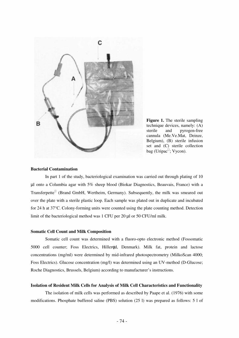
- 74 -
Figure 1. The sterile sampling technique devices, namely: (A) sterile and pyrogen-free cannula (Me.Ve.Mat, Deinze, Belgium), (B) sterile infusion set and (C) sterile collection bag (Uripac; Vycon).
Bacterial Contamination
In part 1 of the study, bacteriological examination was carried out through plating of 10
µl onto a Columbia agar with 5% sheep blood (Biokar Diagnostics, Beauvais, France) with a
Transferpette (Brand GmbH, Wertheim, Germany). Subsequently, the milk was smeared out
over the plate with a sterile plastic loop. Each sample was plated out in duplicate and incubated
for 24 h at 37°C. Colony-forming units were counted using the plate counting method. Detection
limit of the bacteriological method was 1 CFU per 20 µl or 50 CFU/ml milk.
Somatic Cell Count and Milk Composition
Somatic cell count was determined with a fluoro-opto electronic method (Fossomatic
5000 cell counter; Foss Electrics, Hillerφd, Denmark). Milk fat, protein and lactose
concentrations (mg/ml) were determined by mid-infrared photospectrometry (MilkoScan 4000;
Foss Electrics). Glucose concentration (mg/l) was determined using an UV-method (D-Glucose;
Roche Diagnostics, Brussels, Belgium) according to manufacturer’s instructions.
Isolation of Resident Milk Cells for Analysis of Milk Cell Characteristics and Functionality
The isolation of milk cells was performed as described by Paape et al. (1976) with some
modifications. Phosphate buffered saline (PBS) solution (25 l) was prepared as follows: 5 l of

Validation of milk sample collection under aseptical conditions
- 75 -
stock solution (5.5842 g KH2PO4, 37.2438 g Na2HPO4, 40.9 g NaCl, distilled H2O; pH adjusted
to 7.5) was diluted to 25 l with distilled H2O and 180 g NaCl was added. The PBS solution was
sterilised through filtration with a cellulose acetate filter (pore size 0.2 µm; Whatman,
Maidstone, UK). Milk, diluted 2/1 with sterile PBS, was centrifuged (1,000 x g, 15 min, 4°C)
and the cream was removed after the first centrifugation. The cell pellet was washed twice in
PBS and the cells were finally resuspended in Hanks balanced salt solution (HBSS; Gibco Life
Technologies, Paisley, UK) containing Ca2+ and Mg2+ supplemented with 0.1% bovine serum
albumin (BSA, low endotoxin; Sigma Chemicals, St. Louis, MO, USA) and 25 mM HEPES
buffer (Sigma).
During isolation, the cells were maintained on melting ice (1°C). Isolated cells were
counted using an electronic particle counter (Coulter Counter Z2; Coulter Electronics Ltd.,
Luton, England) and viability was determined. The milk cells were resuspended to a
concentration of 5 x 10⁶ viable milk cells/ml with HBSS containing Ca2+ and Mg2+ supplemented
with 0.1% BSA and 25 mM HEPES buffer.
Viability, Apoptosis and Differential Counts of Isolated Milk Cells
Milk PMN viability was determined by propidium iodide (PI) exclusion. Viable PMN
having an intact cell membrane, are impermeable to PI (PI-negative); whereas necrotic PMN
having a deficient cell membrane, are permeable to PI (PI-positive). Ten µl of PI (50 µg/ml
PBS) was added to the cell suspension (400 µl) and analysed by flow cytometry (FACScan;
Becton Dickinson Immunocytometry Systems, San José, USA). The PMN were gated, based on
their FS and SSC, in accordance with the settings found through flow cytometric analysis of pure
PMN populations (Mehrzad et al., 2001) (Fig. 2).

- 76 -
M1
Figure 2. Flow cytometric analysis of isolated bovine milk PMN gated in the FS-SSC dot plot (A). Green fluorescence (FL-1) of PMN labelled with a monoclonal antibody specific against bovine granulocytes and with a secondary FITC-labelled antibody (B). Red fluorescence of PI-incubated PMN selectively gated in the FS-SSC dot plot (C). Gate M1 is applied to determine the percentage of dead PMN (for the quantification of the viability).
Milk PMN apoptosis was assessed by addition of annexin-V-FITC and PI according to
Van Oostveldt et al. (1999) with slight modifications. Briefly, 50 µl of annexin-V-FITC/PI was
added to the cell suspension and incubated for 10 min. After incubation, 300 µl of RPMI 1640
(Gibco Life Technologies) were added and apoptosis was quantified through flow cytometry
PMN
A
B
C

Validation of milk sample collection under aseptical conditions
- 77 -
(FACScan). Polymorphonuclear leukocytes were again gated, based on their FS/SSC. Apoptotic
PMN (programmed cell death) are also PI-negative (viable), but can be distinguished within the
viable PMN population by their expression of phospholipid phosphatidyl-serine, binding with
annexin-V-FITC, on their outer cell membrane (Fig. 3).
Figure 3. Flow cytometric analysis of isolated bovine milk PMN gated in the FS-SSC dot plot (A). Green fluorescence (FL-1) of annexin-V-FITC in the X-axis and red fluorescence (FL-2) from PI in the Y-axis of the flow cytometric analysis program (B). Cells in the lower left quadrant are negative for both fluorescent markers and are classified as living PMN. Cells in the lower right quadrant are positive for annexin-V-FITC (apoptosis marker), though negative for PI and are classified as apoptotic. Cells in the upper right quadrant are positive for both annexin-V-FITC and PI and are classified as dead (necrotic).
Differential counts of isolated milk cells were carried out as described by Dulin et al.
(1988). Two hundred cells were counted on each of the smears and the percentages of
lymphocytes-monocytes (LM), macrophages (MΦ) and PMN were determined. These data were
used to correct intracellular killing and CL for the percentage of PMN in the isolated milk cells.
PMN
A
B

- 78 -
Neutrophil Function Tests
Phagocytosis and intracellular killing of Staphylococcus aureus Newbould 305 was
evaluated in a bacteriological assay according to Barta (1993) and Barrio et al. (2000) with few
modifications for milk. The assay was run in duplicate in Eppendorf tubes (Netheler-Hinz
GmbH, Hamburg, Germany) in a final volume of 1 ml with the following composition: 500 µl
isolated cells (5 x 10⁶ viable cells/ml), 100 µl S. aureus Newbould 305 suspension (1 x 10⁸
CFU/ml) and 400 µl pooled bovine serum diluted to a final concentration (v/v) of 5%
(complement-inactivated, 56°C, 30 min). Control samples contained bacteria, HBSS and serum
without PMN. The bacteria to PMN ratio was between 4:1 and 5:1.
The results from the bacteriological assay were expressed as the percentages of killed (%
killing) and phagocytosed (% phagocytosis) bacteria and were corrected for the percentage of
PMN and MΦ in the samples of isolated milk cells as described by Dosogne et al. (2001).
Luminol-enhanced PMA (phorbol 12-myristate 13-acetate) –stimulated cellular CL of
milk PMN was quantified on isolated milk cells as described by Mehrzad et al. (1999). Non-
stimulated CL was compared with PMA-stimulated CL. The area under the curve (AUC) was
calculated for the registrated impulse rate over a 30-min period. The CL response was corrected
for 1,000 viable PMN.
SYTO13 Labelling
A stock of SYTO13 (5 mM, Molecular Probes, Eugene, USA), a live cell permeable
DNA labelling dye, in DMSO was stored at −18°C and diluted 1:40 with RPMI 1640 (Gibco
Life Technologies) immediately before labelling, according to the manufacturer’s prescriptions.
This corresponds with a final concentration of 200 nM in the samples. Two other dilutions
(1:200 and 1:400; 40 and 20 nM final concentrations, respectively) were also evaluated with low
SCC milk samples from 5 cows, but 1:40 was used for the standard procedure. Four hundred and
ninety µl RPMI was added to the cell pellets (1 x 10⁵ – 4 x 10⁶ cells/ml), followed by 10 µl
diluted SYTO13 solution. This mixture was incubated during 0, 5, 10, 20 and 30 min. A 10-min
incubation period was necessary for optimal labelling of all cells (Table 5) and was further used
for the standard differential leukocyte count procedure.
Flow Cytometry and Cell Sorting
A flow cytometer (FACScan) equipped with an argon ion laser was used for
measurement of differential leukocyte count. The excitation wavelength of the laser was 488 nm.
The FS signal was amplified with a factor 1.98, whereas SSC and fluorescence signals were not
amplified. The voltage applied on the SSC detector was 381 V. Forward scatter and SSC were

Validation of milk sample collection under aseptical conditions
- 79 -
measured on a linear scale. Four hundred and thirty V was applied on the photomultipliers for
both green and red fluorescence. Green fluorescence was measured using a 530-nm band-pass
filter and registered on a log scale, whereas red fluorescence emission was measured using a 650
nm long-pass filter and also registered on a log scale. The data were analysed using the
CellQuest (Becton Dickinson) software. Flow cytometric differential leukocyte count of milk
after SYTO13 staining was quantified using the SSC - green fluorescence (FL-1) dot plot. Flow
cytometric cell sorting of SYTO13-labeled milk cells was performed using a FACSVantage
cell sorter (Becton Dickinson).
Confocal Laser Scanning Microscopy
A lambda (wavelength) scan was performed on cytospin smears of SYTO13-labeled
milk cells using an inverted confocal laser scanning microscope (DMIRBE HCB fluoro
microscope with a TCS SP2 confocal system; Leica Microsystems, Wetzlar, Germany). Four
different regions of interest (ROI) were defined around 4 different cell types based on size and
shape. The fluorescence emission intensity at wavelengths between 498 and 644 nm was
registered for these 4 ROI’s.
Milk Sample Preparation for Differential Leukocyte Count
All milk samples were gently mixed before dilution and further processing. For the flow
cytometric cell sorting procedure, cells were isolated from milk according to Hoeben et al.
(1997) and finally resuspended to a concentration of 5 x 10⁶/ml after counting with an electronic
particle counter (Coulter Counter Z2). For the differential leukocyte count procedure developed
in this study, 1.5 ml milk samples were layered on 3.5 ml PBS into 12 x 75 mm polystyrene flow
cytometry tubes (Vel, Leuven, Belgium) and centrifuged during 10 min at 180 x g. Fat was
removed, the supernatant was discarded and the tubes were kept upside down until they were
carefully cleaned with a cotton swab to remove all fat adhering to the test tube wall. The pellet
was then resuspended with 490 µl RPMI for flow cytometric differential leukocyte count or with
1 ml tryptic soy broth (TSB; Sigma Chemicals) for preparation of microscope slides.
Different dilution buffers were tested with low SCC milk samples from 6 cows: PBS;
RPMI 1640 supplemented with 1% bovine serum albumin (Sigma) and 0.2% sodium azide
(Sigma); and PBS supplemented with 0.1% paraformaldehyde (Sigma).
The following dilutions of milk samples were evaluated: 0.5, 1.0, 1.5 and 2.0 ml milk +
4.5, 4.0, 3.5 and 3 ml PBS, respectively, using low SCC milk samples from 6 cows. In order to
evaluate the effect of temperature on the differential leukocyte count of milk, low SCC milk
samples from 6 cows were maintained at 4, 20 and 37°C from immediately after collection, over

- 80 -
transportation and processing until flow cytometric analysis. The optimal dilution (1.5 ml milk
with 3.5 ml PBS) and transportation and processing temperature (20°C) were further used for
validation of the standard differential leukocyte count procedure.
Variability of Differential Leukocyte Count Method
The variability of the flow cytometric measurement of differential leukocyte count was
tested by performing 6 repeated measurements on one sample. Variability of the complete
differential cell count method was evaluated by performing 5 repetitions of the identification
procedure for low SCC milk samples from 6 different cows. Significant differences in
identification between the milk samples were evaluated for 2, 3, 4 and 5 repeated differential cell
counts. The variability of the method was also evaluated at 500; 1,000; 2,000 and 3,000 analysed
cells, with 6 repeated measurements for each number of cells analysed in a single sample. In the
standard procedure, 3,000 cells per sample were analysed (Table 5).
Preparation of Microscope Slides and Comparison Between Flow Cytometric and
Microscopic Differential Leukocyte Count
A described procedure for preparing microscope slides was used (Dulin et al., 1982).
Slides were prepared in a cytocentrifuge (Shandon, Astmoor, UK) using 200 µl cell suspension
with a concentration between 1 and 5 x 10⁵ cells/ml. The suspensions were centrifuged during 10
min at 113 x g. The smears were stained with May-Grünwald’s eosin-methylene blue (Merck
Diagnostica, Darmstadt, Germany). At least 200 cells were identified per sample, at a
magnification of 1,000. Polymorphonuclear leukocytes were identified based on their multi-
lobed dark purple stained nucleus and lightly blue stained cytoplasm. Polymorphonuclear
leukocytes were characterised for apoptosis based on the morphologic features of pycnotic
nuclei. Mature MΦ were large, with a light purple stained round nucleus and a vacuolated
cytoplasm. Milk LM had the same appearance as their counterparts in the blood. Epithelial cells
were large, mostly without nucleus, irregularly shaped and homogeneously light blue stained.
For comparison of the flow cytometric differential leukocyte count method with microscopy,
low SCC milk samples from 7 cows in early lactation were prepared in duplicate using the
sample preparation method described above. One sample was used for flow cytometry and from
the other one, slides were prepared in duplicate. Correlation between the % LM, PMN and
mature MΦ obtained with the 2 methods was calculated.

Validation of milk sample collection under aseptical conditions
- 81 -
Statistical Analysis
In part 1, differences between sampling techniques for SCC, milk composition, bacterial
contamination, viability, phagocytosis, intracellular killing, PMA-stimulated and non-stimulated
CL were evaluated by least squares ANOVA with Bonferroni's correction (Statistix, Analytical
Software, Tallahassee, FL, USA). Significant differences were determined at P < 0.05*, P <
0.01**, and P < 0.001***.
In part 2, the effects of sample dilution, temperature and storage time at 4°C were
evaluated using ANOVA. For evaluation of the method variability, the results were subjected to
a Wilk Shapiro rankit plot to examine the normality of the distribution of repeated differential
cell count analyses.

- 82 -
RESULTS
Somatic Cell Count and Milk Composition
No significant difference in SCC, fat, protein, lactose and glucose content was found
between STER-SPL and MAN-SPL, nor between STER-SPL and CONT-SPL milk sampling
(Table 1).
Table 1. SCC and milk composition of STER-SPL vs. MAN-SPL and STER-SPL vs. CONT-SPL milk samples during early lactation. Data are means (± SEM).
Milk Sampling Technique
STER-SPL MAN-SPL CONT-SPL
SCC (log10/ml) 4.4 ± 4.1 4.5 ± 3.9
4.4 ± 4.0 4.9 ± 4.7
FAT (mg/ml) 24.2 ± 5.2 22.3 ± 4.6 25.5 ± 5.8 42.8 ± 7.0 PROTEIN (mg/ml) 39.5 ± 2.6 40.1 ± 2.8 39.0 ± 3.7 37.3 ± 3.6 LACTOSE (mg/ml) 49.3 ± 1.5 50.1 ± 1.6 49.3 ± 2.1 46.2 ± 2.7 GLUCOSE (mg/l) 93.6 ± 11.2 102.0 ± 0.8 106.9 ± 12.9 94.0 ± 9.7
Bacterial Contamination
Bacterial contamination was not significantly different between STER-SPL and MAN-
SPL milk sampling, whereas it was significantly (P < 0.001) higher in CONT-SPL compared to
STER-SPL milk sampling (Table 2).
Table 2. Bacterial contamination (CFU/ml) from STER-SPL vs. MAN-SPL and STER-SPL vs. CONT-SPL milk samples during early lactation. Data are means (± SEM). Significant difference: a STER-SPL vs. CONT-SPL (P < 0.001).
Milk Sampling Technique
STER-SPL MAN-SPL CONT-SPL
bacterial count (CFU/ml) 0 660 ± 413 0 a 6,620 ± 2,339 a

Validation of milk sample collection under aseptical conditions
- 83 -
Resident Milk Cell Viability
No significant difference in viability was found between STER-SPL and MAN-SPL, nor
between STER-SPL and CONT-SPL milk sampling (Fig. 4). During early lactation, milk PMN
viability was on average 38.6 ± 6.83 %.
Figure 4. Viability of resident PMN: (a) trial 1: STER-SPL � vs. MAN-SPL � milk samples, and (b) trial 2: STER-SPL � vs. CONT-SPL � milk samples during early lactation. Data (expressed as %) are means (± SEM).
Phagocytosis and Intracellular Killing of Staphylococcus aureus
Neither of both PMN functionality tests were significantly influenced by the different
milk sampling techniques (Fig. 5). Phagocytosis and intracellular killing during early lactation
were on average 83.4 ± 2.63 % and 14.8 ± 7.33 %, respectively.
Figure 5. Phagocytosis (A) and intracellular killing (B) of S. aureus: (a) trial 1: STER-SPL � vs. MAN-SPL � milk samples, and (b) trial 2: STER-SPL � vs. CONT-SPL � milk samples during early lactation. Data (expressed as %) are means (± SEM).
Non-Stimulated and PMA-Stimulated Chemiluminescence
Non-stimulated and PMA-stimulated CL were not significantly different between STER-
SPL and MAN-SPL, nor between STER-SPL and CONT-SPL milk sampling (Fig. 6). During
early lactation, non-stimulated and PMA-stimulated CL were 1,150 ± 220 AUC and 2,087 ± 351
AUC, respectively.
0
25
50
75
100
1 2
trial number
% V
iab
ilit
y
0
25
50
75
100
1 2
trial number
% P
hag
ocyto
sis
0
25
50
75
100
1 2
trial number
% In
tracellu
lar
killin
g
A B

- 84 -
Figure 6. Non-stimulated chemiluminescence (A) and PMA-stimulated chemiluminescence (B): (a) trial 1: STER-SPL � vs. MAN-SPL � milk samples, and (b) trial 2: STER-SPL � vs. CONT-SPL � milk samples during early lactation. Data (expressed as AUC) are means (± SEM).
Flow Cytometry and Cell Sorting was Performed Based on Green Fluorescence (FL-1) and
Side Scatter (SSC)
Both red (FL-3) and green (FL-1) fluorescence emission of bovine milk cells was
observed following SYTO13 labelling. Because the best resolution between the populations
was obtained with FL-1 and SSC, these parameters were used for the differential leukocyte count
procedure. Red fluorescence was reduced at lower SYTO13 concentrations, but this also
resulted in decreased resolution in the green fluorescence (Table 3). In the FL-1 – SSC dot plot
of isolated cells, 5 different leukocyte populations were identified (Fig. 7A). Following flow
cytometric cell sorting of SYTO13-labeled milk cells using the FL-1 − SSC dot plot (Fig. 7A),
the different cell populations were identified as: 1) SSClow – FL-1high: high FL-1 LM (R1); 2)
SSClow – FL-1intermediate: intermediate FL-1 LM (R1); 3) SSClow – FL-1low: cells with apoptotic
features (R2); 4) SSChigh – FL-1high: mature MΦ (R4) and 5) SSChigh – FL-1intermediate: PMN (R5)
(Fig. 7B). Uptake of SYTO13 did not occur immediately and differed between the cell types
(Table 4). Saturation of the fluorescence intensity was obtained in most cell types after a 10-min
incubation and remained nearly constant during 20 min. The highest green fluorescence intensity
was observed in mature MΦ and viable PMN. Lower fluorescence intensity values were found in
cells with apoptotic features, FL-1low LM and debris. For the standard procedure, a 10-min
incubation time of cells with SYTO13 was used.
0
1250
2500
3750
5000
1 2
trial number
AU
C C
L
0
1250
2500
3750
5000
1 2
trial number
AU
C C
L
A B

Validation of milk sample collection under aseptical conditions
- 85 -
Table 3. Green (FL-1) and red (FL-3) fluorescence emission intensity (mean fluorescence intensity) of different cell types at different SYTO13 concentrations, measured by flow cytometry. Values are means (± SEM) of 5 cows.
SYTO13 concentration (nM)
20 40 200
FL-1 FL-3 FL-1 FL-3 FL-1 FL-3
Lymphocytes 20 ± 4 2 ± 0 53 ± 21 4 ± 1 176 ± 41 16 ± 3 Macrophages 286 ± 34 46 ± 24 1,816 ± 125 148 ± 34 2,825 ± 156 243 ± 28 PMN 37 ± 4 11 ± 8 378 ± 21 30 ± 3 479 ± 31 57 ± 5
Table 4. Effect of incubation time of bovine milk cells with SYTO13 on green fluorescence emission intensity (mean fluorescence intensity). Values are the average of 3 repeated identification assays.
Incubation time (min)
0 5 10 20 30
Lymphocytes-monocytes 10 556 1,199 1,829 2,140 Macrophages 177 3,279 3,365 3,626 3,802 PMN 162 3,116 3,302 3,628 3,668 Cells with apoptotic features 4 158 410 831 1,056 Debris 24 132 140 147 155
Confocal Laser Scanning Microscopy Could Confirm and Identify the Different Cell Types
Sorted by Flow Cytometry
Using a lambda scan of SYTO13-labeled milk cells with confocal laser scanning
microscopy, a different morphology could be associated with different fluorescence emission
characteristics (Fig. 8A-B). The cells in ROI1 had an intermediate size, were round-shaped with
a round nucleus and had a maximal fluorescence emission at 514 nm. This is consistent with the
characteristics of LM. The cells in ROI2 were large, irregularly shaped and also had a maximal
fluorescence emission at 514 nm but their fluorescence intensity was much higher than any of
the other cell types. These cells conform to the characteristics of mature MΦ. The cells in ROI3
had a multi-lobed nucleus and a maximal but low fluorescence intensity at 498 nm, which are
typical features of PMN. Finally, the cells in ROI4 had similar fluorescence characteristics as the
cells in ROI3 but the fluorescence intensity was higher, especially in the higher emission
wavelength region. These cells contain all characteristics of high FL-1 LM.

- 86 -
Figure 7. Flow cytometric analysis (A) of SYTO13-labeled bovine milk cells: green fluorescence (FL-1) – side scatter (SSC) dot plot of isolated milk cells used for flow cytometric cell sorting and subsequent microscopic identification (B). R1: LM; R2: apoptotic cells; R3: cellular debris; R4: MΦ; and R5: PMN.
R1
R2
R3
R4
R5
0
20
40
60
80
100
R1 R2 R3 R4 R5
Sorted Region (R)
Dif
fere
nti
al L
eu
ko
cyte
Co
un
t (%
)
macrophages apoptotic cells PMN
lymphocytes epithelial cells debris
A
B

Validation of milk sample collection under aseptical conditions
- 87 -
Figure 8. Confocal laser scanning microscopic analysis of fluorescence emission of different SYTO13-labeled bovine milk cells using a wavelength scan. A) identification of different regions of interest (ROI); B) fluorescence emission characteristics of the different ROI’s identified in A). ROI1: LM (high FL-1); ROI2: MΦ; ROI3: PMN; and ROI4: LM (low FL-1).
0
10
20
30
40
50
60
70
498
505
513
521
529
537
545
553
561
569
576
584
592
600
608
616
624
632
640
648
Emission Wavelength (nm)
Inte
nsit
y
ROI 1 ROI 2 ROI 3 ROI 4
A
B

- 88 -
Milk Sample Preparation and Effect of Storage
Optimal conditions for milk sample preparation were the following: 1) milk sample
dilution with PBS or RPMI-BSA; 2) 30% (v/v) dilution (i.e. 1.5 ml milk + 3.5 ml PBS) (Fig. 9);
3) a sample collection, transportation and processing temperature of 20°C (Fig. 10). Both the
SCC and the differential leukocyte count remained constant throughout a 48-h storage period at
7°C (Fig. 11).
Figure 9. Effect of sample dilution on the differential leukocyte count of SYTO13-labeled bovine milk cells in low SCC (SCC < 200,000 cells/ml) milk samples. The following dilutions were performed: 0.5, 1.0, 1.5, and 2.0 ml milk with 4.5, 4.0, 3.5 and 3.0 ml PBS, respectively. Average values for 6 cows are presented. Different cell types analysed from bottom to top are: debris, cells with apoptotic features, PMN, mature MΦ and LM.
Figure 10. Effect of sample collection, transportation, processing and labelling temperature on the differential leukocyte count and on % debris. Average values of 6 cows are presented. Different cell types analysed from bottom to top are: debris, cells with apoptotic features, PMN, mature MΦ and LM.
0%
20%
40%
60%
80%
100%
10 20 30 40
Dilution (%)
Iden
tifi
cati
on
0%
20%
40%
60%
80%
100%
4 20 37
Temperature (°C)
Iden
tifi
cati
on

Validation of milk sample collection under aseptical conditions
- 89 -
Figure 11. Effect of sample storage at 4°C during 24 and 48 h on the differential leukocyte count of bovine low SCC (SCC < 200,000 cells/ml) milk samples. Average values for 6 cows are presented. Different cell types analysed from bottom to top are: debris, PMN, mature MΦ and LM.
Variability of the Differential Leukocyte Count Method
The coefficient of variation for repeated measurements in identical cell suspensions was
1.5% for LM, 1.4% for mature MΦ, 0.7% for PMN and 0.8% for cells with apoptotic features.
Average coefficients of variation for the complete differential leukocyte count method including
sample dilution, resuspension and SYTO13 labelling of milk samples from 6 cows were 2.5%
for LM, 15.8% for mature MΦ, 11.8% for PMN and 10.7 % for cells with apoptotic features
(Fig. 12). According to the Wilk-Shapiro rankit plot against the ordered data, which resulted in a
straight line, the samples conformed to a normal distribution. Significant differences between the
samples were obtained at P < 0.05 for duplicate differential leukocyte count assays and at P <
0.01 for 3 or more repeated assays. The highest variability between repeated measurements was
observed with 500 analysed cells. The variability further decreased from 1,000 over 2,000 to
3,000 cells (Table 5). The average % cells in the different regions was not dependent on the
number of analysed cells.
Table 5. Variability of the differential leukocyte count method at different numbers of analysed cells. Values are the average percentage of each cell type (± SEM) for 6 repeated measurements.
Number of analysed cells
500 1,000 2,000 3,000
Lymphocytes 69 ± 14 68 ± 4 67 ± 3 67 ± 1 Macrophages 11 ± 3 11 ± 1 12 ± 1 12 ± 1 PMN 9 ± 3 9 ± 1 9 ± 1 9 ± 1 Cells with apoptotic features 11 ± 1 12 ± 1 12 ± 1 12 ± 1
0%
20%
40%
60%
80%
100%
0 24 48
Storage time (h)
Iden
tifi
cati
on
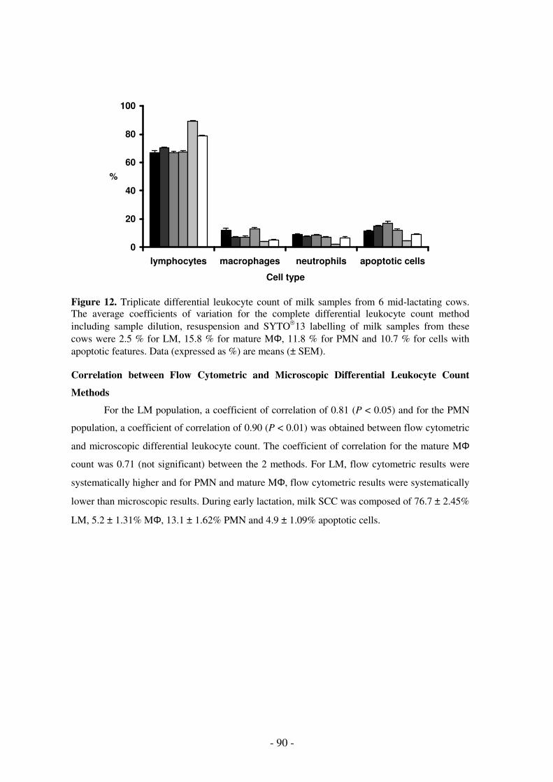
- 90 -
Figure 12. Triplicate differential leukocyte count of milk samples from 6 mid-lactating cows. The average coefficients of variation for the complete differential leukocyte count method including sample dilution, resuspension and SYTO13 labelling of milk samples from these cows were 2.5 % for LM, 15.8 % for mature MΦ, 11.8 % for PMN and 10.7 % for cells with apoptotic features. Data (expressed as %) are means (± SEM). Correlation between Flow Cytometric and Microscopic Differential Leukocyte Count
Methods
For the LM population, a coefficient of correlation of 0.81 (P < 0.05) and for the PMN
population, a coefficient of correlation of 0.90 (P < 0.01) was obtained between flow cytometric
and microscopic differential leukocyte count. The coefficient of correlation for the mature MΦ
count was 0.71 (not significant) between the 2 methods. For LM, flow cytometric results were
systematically higher and for PMN and mature MΦ, flow cytometric results were systematically
lower than microscopic results. During early lactation, milk SCC was composed of 76.7 ± 2.45%
LM, 5.2 ± 1.31% MΦ, 13.1 ± 1.62% PMN and 4.9 ± 1.09% apoptotic cells.
0
20
40
60
80
100
lymphocytes macrophages neutrophils apoptotic cells
Cell type
%

Validation of milk sample collection under aseptical conditions
- 91 -
DISCUSSION
In the present study, milk sample collection was validated and the effect of external
bacterial contamination on viability and functions of milk cells was studied in healthy mammary
glands with low SCC. Therefore, normal milk was obtained without stimulation of the mammary
gland and all milk samples had low SCC (< 2 x 10⁵ cells/ml). In previous studies, PMN
recruitment into the mammary gland was stimulated by either the insertion of an intramammary
device (Paape et al., 1976; Saad and Hageltorn, 1985), or by intramammary infusion of E. coli
endotoxin (Dulin et al., 1988).
A new sterile milk sampling technique (STER-SPL) was introduced as reference
technique, which allowed milk sample collection under aseptical conditions without external
bacterial contamination. Although manual milk sampling (MAN-SPL) could increase the risk of
external contamination, bacterial counts observed with this sampling technique were
unexpectedly low. In this milk, no change in viability and different cell functions of isolated
milk cells was observed. In contrast, machine milking (CONT-SPL) had a significantly higher
contamination level as compared to STER-SPL. Despite this bacterial contamination, no
significant differences in viability and different cell functions of isolated milk cells were found.
It seems that the bacterial contamination present in CONT-SPL milk samples could not
significantly activate the isolated resident milk cells. However, it is important to mention that the
whole study was performed on milk, which was slowly cooled down to 1°C. Under these
conditions, cell functions are inhibited and cells do not react when challenged by these
contaminating bacteria. Nevertheless, milk cells did not loose their reaction potential, since they
performed CL, intracellular killing and phagocytosis when collected and maintained under the
specified conditions. It can be concluded that all three milk sampling techniques are equally
appropriate for the study of cellular and non-cellular qualities of bovine milk and resident cells.
The reference technique (sterile cannula) is the best for eliminating bacterial contamination,
although for practical routine milk sample collection manual milk sampling (MAN-SPL) can be
used.
In the present study, a simple, accurate and reproducible flow cytometric differential
leukocyte count method for low SCC milk from healthy cows was developed. The method is
complementary to flow cytometric differential leukocyte count methods for high SCC milk
(Redelman et al., 1988; Pillai et al., 2001). The low amount of cells in milk from healthy cows (<
105 cells/ml; Laevens et al., 1997), makes the identification of leukocytes more difficult than in
high SCC milk (109 cells/ml during coliform mastitis; Dosogne et al., 1997). A maximal amount

- 92 -
of identifiable cells in low SCC milk was obtained with 1) a 30 % (v/v) dilution of milk samples
(which corresponds with large-scale isolations; Hoeben et al., 1997); 2) centrifugation and
removal of the fat layer; 3) resuspension of the pellet with RPMI 1640; 4) a sample collection,
storage and processing temperature of 20 °C. Although the initial SCC of the milk samples was
low, a sufficient amount of cells was obtained from 1.5 ml samples. Thus, our method is
applicable for small volumes of low SCC milk samples. Another advantage of the method
described here is that milk samples can be stored for 2 days at 7°C without any effect on the
differential leukocyte count. The method was also shown to be reproducible, even at a very low
number of cells analysed: variability was not significantly different between 1,000; 2,000 or
3,000 analysed cells and the average values for differential milk leukocyte count were not
dependent on the number of analysed cells.
Four populations with different fluorescence emission characteristics were obtained
upon SYTO13 staining of milk leukocytes. The high fluorescence of mature MΦ is consistent
with previous results obtained with acridine orange labelling (Hageltorn and Saad, 1986) and
could be explained by a higher degree of mitochondrial RNA staining. Indeed, green
fluorescence obtained with the SYTO dyes is not only governed by stoichiometric DNA
binding, but also by cytoplasmic mitochondrial labelling (Poot et al., 1997). The emission
wavelength maximum of SYTO13-labeled RNA (514 nm) is similar to DNA (509 nm)
(Molecular probes, SYTO green fluorescent nucleic acid stains product information sheet).
Mitochondrial labelling could also explain the high green fluorescence of some LM and is
indicative of a high metabolic rate. Polymorphonuclear leukocytes and LM had similar
fluorescence characteristics but could be distinguished on the basis of cytoplasmic granularity,
which was typically higher for PMN than for LM.
CONCLUSION
Following validation of milk sample collection under aseptical conditions, no effect of
limited bacterial contamination on PMN viability and functions could be observed. Manual milk
sample collection will be preferred for further use during experimental challenge, because the
technique results in a small bacterial contamination and is easy to perform during repeated
sample collection. During early lactation, pre-infection milk SCC is mainly composed of
lymphocytes, whereas only small percentages of monocytes, PMN and apoptotic cells are
present.
Validation of milk sample collection under aseptical conditions
- 93 -
REFERENCES
Barrio, B., F. Vangroenweghe, H. Dosogne, and C. Burvenich. 2000. Decreased neutrophil bactericidal
activity during phagocytosis of a slime-producing Staphylococcus aureus strain. Vet. Res. 31:603-609.
Barta, O. 1993. Neutrophil function tests. Chapter B-IV. Phagocytosis and killing of bacteria monitored by
sample cultivation. In: Veterinary clinical immunology laboratory, Barta O. (Ed.), Bar-Lab. Inc.,
Blacksburg, USA. pp. B3.12-B3.17.
Burvenich, C., M.J. Paape., A.W. Hill, A.J. Guidry, R.H. Miller, R. Heyneman, W.D.J. Kremer, and A.
Brand. 1994. Role of the neutrophil leukocyte in the local and systemic reactions during experimentally
induced E. coli mastitis in cows immediately after calving. Vet. Quart. 16:45-49.
Dosogne, H., C. Burvenich, T. van Werven, E. Roets, E.N. Noordhuizen-Stassen, and B. Goddeeris. 1997.
Increased surface expression of CD11b receptors on polymorphonuclear leukocytes is not sufficient to
sustain phagocytosis during Escherichia coli mastitis in early postpartum dairy cows. Vet. Immunol.
Immunopathol. 60:47-59.
Dosogne, H., F. Vangroenweghe, B. Barrio, P. Rainard, and C. Burvenich. 2001. Decreased number and
bactericidal activity against S. aureus of the resident cells in milk of dairy cows during early lactation. J.
Dairy Res. 68:539-549.
Dulin, A.M., M.J. Paape, and B.T. Weinland. 1982. Cytospin centrifuge in differential counts of milk
somatic cells. J. Dairy Sci. 65:1247-1251.
Dulin, A.M., M.J. Paape, and S.C. Nickerson. 1988. Comparison of phagocytosis and chemiluminescence
by blood and mammary gland neutrophils from multiparous and nulliparous cows. Am. J. Vet. Res.
49:172-177.
Hageltorn, M., and M.A. Saad. 1986. Flow cytofluorometric characterization of bovine blood and milk
leukocytes. Am. J. Vet. Res. 47:2012-2016.
Hallén-Sandgren, C., K. Nordling, and I. Björk. 1991. Isolation and phagocytic properties of neutrophils
and other phagocytes from nonmastitic bovine milk J. Dairy Sci. 74:2965-2975.
Hoeben, D., C. Burvenich, and R. Heyneman. 1997. Influence of antimicrobial agents on bactericidal
activity of bovine milk polymorphonuclear leukocytes. Vet. Immunol. Immunopathol. 56:271-282.
Kehrli, M.E., Jr., and D.E. Shuster. 1994. Factors affecting milk somatic cells and their role in health of
the bovine mammary gland. J. Dairy Sci. 77:619-627.
Kelly, A.L., D. Tiernan, C. O’Sullivan, and P. Joyce. 2000. Correlation between bovine milk somatic cell
count and polymorphonuclear leukocyte level for samples of bulk milk and milk from individual cows.
J. Dairy Sci. 83:300-304.
Laevens, H., H. Deluyker, Y.H. Schukken, L. De Meulemeester, R. Vandermeersch, E. De Meulenaere,
and A. de Kruif. 1997. Influence of parity and stage of lactation on the somatic cell count in
bacteriologically negative dairy cows. J. Dairy Sci. 80:3219-3226.
- 94 -
Leitner, G., E. Shoshani, O. Krifucks, M. Chaffer, and A. Saran. 2000. Milk leukocyte population patterns
in bovine udder infection of different aetiology. J. Vet. Med. B, 47:581-589.
Mehrzad, J., H. Dosogne, E. Meyer, and C. Burvenich. 1999. Respiratory burst activity of bovine milk
neutrophils during early and mid-lactation. Pflügers Archiv - Eur. J. Physiol. 438:R31.
Mehrzad, J., H. Dosogne, E. Meyer, R. Heyneman, and C. Burvenich. 2001. Respiratory burst activity of
blood and milk neutrophils in dairy cows during different stages of lactation. J. Dairy Res. 68:399-415.
Nickerson, S.C., R.L. Boddie, W.E. Owens, and J.L. Watts. 1990. Effects of novel intramammary device
models on incidence of mastitis after experimental challenge. J. Dairy Sci. 73:2774-2784.
Östensson, K., M. Hageltorn, and G. Astrom. 1988. Differential cell counting in fraction-collected milk
from dairy cows. Acta vet. Scand. 29:493-500.
Paape, M.J., R.E. Pearson, W.P. Wergin, and A.J. Guidry. 1976. Enhancement of chemotactic response of
polymorphonuclear leukocytes into the mammary gland and isolation from milk. J. Dairy Sci. 60:53-62.
Paape, M.J., and R.E. Pearson. 1979. Sources of variation in in vitro phagocytosis assays. Am. J. Vet. Res.
40:630-635.
Pillai, S.R., E. Kunze, L.M. Sordillo, and B.M. Jayarao. 2001. Application of differential inflammatory
cell count as a tool to monitor udder health. J. Dairy Sci. 84:1413-1420.
Poot, M., L.L. Gibson, and V. Singer. 1997. Detection of apoptosis in live cells by Mitotracker Red
CMXRos and SYTO dye flow cytometry. Cytometry 27:358-364.
Redelman, D., S. Butler, J. Robison, and D. Garner. 1988. Identification of inflammatory cells in bovine
milk by flow cytometry. Cytometry 9:463-468.
Saad, A., and M. Hageltorn. 1985. Flow cytometric characterisation of bovine blood neutrophil
phagocytosis of fluorescent bacteria and zymosan particles. Acta Vet. Scand. 26:289-307.
Saad, A.M. 1987. Flow cytometric measurement of bovine milk neutrophil phagocytosis. Acta Vet. Scand.
28:333-342.
Saad, A.M., and K. Östensson. 1990. Flow cytofluorometric studies on the alteration of leukocyte
populations in blood and milk during endotoxin-induced mastitis in cows. Am. J. Vet. Res. 51:1603-
1607.
Shuster, D.E., E.K. Lee, and M.E. Kehrli, Jr. 1996. Bacterial growth, inflammatory cytokine production,
and neutrophil recruitment during coliform mastitis in cows within ten days after calving, compared with
cows at midlactation. Am. J. Vet. Res. 57:1569-1575.
Smits, E., C. Burvenich, and R. Heyneman. 1997. Simultaneous flow cytometric measurement of
phagocytotic and oxidative burst activity of polymorphonuclear leukocytes in whole bovine blood. Vet.
Immunol. Immunopathol. 56:259-269.
Stevens, M.G., M.E. Kehrli, Jr., and P.C. Canning. 1991. A colorimetric assay for quantitating bovine
neutrophil bactericidal activity. Vet. Immunol. Immunopathol. 28:45-56.
Van Oostveldt, K., H. Dosogne, C. Burvenich, M.J. Paape, V. Brochez, and E. Van den Eeckhout. 1999.
Flow cytometric procedure to detect apoptosis of bovine polymorphonuclear leukocytes in whole blood.
Vet. Immunol. Immunopathol. 70:125-133.

Validation of milk sample collection under aseptical conditions
- 95 -
Vandeputte-Van Messom, G., C. Burvenich, E. Roets, A.-M. Massart-Leën, R. Heyneman, W.D.J.
Kremer, and A. Brand. 1993. Classification of newly calved cows into moderate and severe responders
to experimentally induced Escherichia coli mastitis. J. Dairy Res. 60:19-29.


- 97 -
MATERIALS AND METHODS EXPERIMENTAL
INFECTIONS

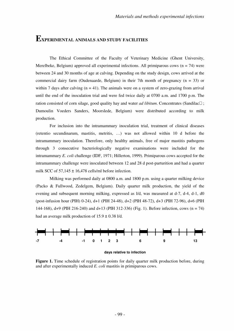
Materials and methods experimental infections
- 99 -
EXPERIMENTAL ANIMALS AND STUDY FACILITIES
The Ethical Committee of the Faculty of Veterinary Medicine (Ghent University,
Merelbeke, Belgium) approved all experimental infections. All primiparous cows (n = 74) were
between 24 and 30 months of age at calving. Depending on the study design, cows arrived at the
commercial dairy farm (Oudenaarde, Belgium) in their 7th month of pregnancy (n = 33) or
within 7 days after calving (n = 41). The animals were on a system of zero-grazing from arrival
until the end of the inoculation trial and were fed twice daily at 0700 a.m. and 1700 p.m. The
ration consisted of corn silage, good quality hay and water ad libitum. Concentrates (Sandilac;
Dumoulin Voeders Sanders, Moorslede, Belgium) were distributed according to milk
production.
For inclusion into the intramammary inoculation trial, treatment of clinical diseases
(retentio secundinarum, mastitis, metritis, …) was not allowed within 10 d before the
intramammary inoculation. Therefore, only healthy animals, free of major mastitis pathogens
through 3 consecutive bacteriologically negative examinations were included for the
intramammary E. coli challenge (IDF, 1971; Hillerton, 1999). Primiparous cows accepted for the
intramammary challenge were inoculated between 12 and 28 d post-parturition and had a quarter
milk SCC of 57,145 ± 16,478 cells/ml before infection.
Milking was performed daily at 0800 a.m. and 1800 p.m. using a quarter milking device
(Packo & Fullwood, Zedelgem, Belgium). Daily quarter milk production, the yield of the
evening and subsequent morning milking, expressed as l/d, was measured at d-7, d-4, d-1, d0
(post-infusion hour (PIH) 0-24), d+1 (PIH 24-48), d+2 (PIH 48-72), d+3 (PIH 72-96), d+6 (PIH
144-168), d+9 (PIH 216-240) and d+13 (PIH 312-336) (Fig. 1). Before infection, cows (n = 74)
had an average milk production of 15.9 ± 0.38 l/d.
Figure 1. Time schedule of registration points for daily quarter milk production before, during and after experimentally induced E. coli mastitis in primiparous cows.
-7
-4 -1 0 1 2 3 6 9 13
days relative to infection

- 100 -
INOCULATION DOSE
The generation time of E. coli in mammary secretions can be as short as 20 min (Petsch
and Anspach, 2000; Burvenich et al., 2003). Because we were interested in bacterial elimination
rather than bacterial growth in the affected mammary glands, a high inoculum dose was used in
all the experiments. Practically, two different high inoculum doses were applied, depending on
the experiment: 1 x 10⁴ and 1 x 10⁶ CFU.
During in vitro bacterial growth in a batch culture system, in which nothing is added to
or removed from the environment once the medium is inoculated with the living cells, several
distinct phases can be identified (Fig. 2). Due to the closed nature of a batch culture system, cell
multiplication can only be supported for a limited time and during bacterial growth, progressive
changes in the original growth medium occur.
Figure 2. Idealised normal growth cycle for a bacterial population (expressed as log10 of the number of viable bacteria) in a batch culture system. Time is expressed in relative units.
Growth proceeds through a lag phase, during which cell numbers do not increase. This is
followed by a growth phase, which is usually characterised by an exponential increase in
Time
Log
Lag phase
Growth phase
Maximum stationary phase
Death phase

Materials and methods experimental infections
- 101 -
bacterial numbers. Ultimately, changes in the chemical and/or physical environment of the
medium result in a phase of no net increase in bacterial numbers, the maximum stationary phase.
Cells in stationary phase still require an energy source for viability maintenance. By definition,
the availability of an energy source is limited in a batch culture, and hence a death phase follows,
often characterised by an exponential decrease in the number of living cells.
Following overnight culture, bacteria harvested for intramammary challenge were in
maximum stationary phase at the time of inoculation. Although a change in growth medium (i.e.
healthy mammary gland) should enhance their growth, it is clear from several previous
experiments that intramammary bacterial kinetics are variable depending on the initial inoculum
dose. When a low number of bacteria is infused, an exponential growth may occur (Shuster et
al., 1996), which is followed by subsequent clearance of the bacteria after peak numbers are
reached (Fig. 3a). In contrast, stationary phase is immediately reached when a high number of
bacteria is inoculated (Hoeben et al., 2000a), which is followed by clearance of bacteria (Fig.
3b).
Figure 3. Intramammary growth of inoculated E. coli bacteria starting from a) a low inoculum dose (~ 30 CFU; Shuster et al., 1996) or b) a high inoculum dose (~ 10,000 CFU; Hoeben et al., 2000a).
INTRAMAMMARY INOCULATION PROCEDURE
Inoculation was performed as described before (Hoeben et al., 2000a; 2000b). Briefly, E.
coli P4:O32 (H37, β-glucuronidase +, haemolysin -), maintained as a stock in lyophilisation
medium at –20°C, was subcultured in brain-heart infusion broth (CM225; Oxoid, Nepean, USA)
at 37°C during 3 consecutive days and subsequently washed 3 times with pyrogen-free PBS, and
resuspended in PBS. Just before inoculation, the suspension was diluted in pyrogen-free PBS to
a final concentration of 1 x 10⁴ CFU/ml or 1 x 10⁶ CFU/ml, depending on the inoculum dose
required in each specific study. On d0, 30 min after morning milking (1.5 h after feeding), the
0
1
2
3
4
5
6
7
0 6
12
24
48
72
14
4
PIH
Lo
g1
0/m
l
a b
0
1
2
3
4
5
6
70
10
18
48
14
4
PIH
Lo
g1
0/m
l

- 102 -
cows were inoculated in the left front and rear quarters with a total volume of 10 ml, containing
1 ml of inoculum and 9 ml of pyrogen-free saline solution (NaCl 0.9%; Baxter N.V., Lessines,
Belgium) per quarter. All bacterial suspensions were infused into the teat cistern using a sterile,
pyrogen-free teat cannula (L 7 cm, Ø 2 mm; Me.Ve.Mat, Deinze, Belgium). Before challenge,
the teat ends were disinfected with 70% ethanol containing 0.5% chlorhexidin. After infusion,
the bacterial suspension was thoroughly distributed into the udder cistern through a 30 s massage
(Fig. 4).
Following inoculation, a control sample of the inoculum was diluted, plated out on
Columbia agar with 5% sheep blood (Biokar Diagnostics, Beauvois, France) and incubated for
24 h at 37°C to check for correct inoculum preparation and adequate inoculum dose
administration at intramammary E. coli challenge.
Figure 4. Intramammary inoculation in the left rear quarter (picture of MMRC, Merelbeke, Belgium).
EXPERIMENTAL DESIGN
To study the influence of lactation number on severity of inflammation and kinetic
response of the innate immunity, two different high inoculum doses were used, namely 1 x 10⁴
and 1 x 10⁶ CFU per quarter.
Materials and methods experimental infections
- 103 -
For modulation of moderate inflammatory reaction through variation of the inoculum
dose, both previously used doses were randomly assigned to animals during the experimental
trial. Modulation of moderate inflammatory reaction through inhibition of prostaglandin
synthesis was performed following inoculation of 1 x 10⁴ CFU in the two left quarters of all
animals. In the study on effect of vaccination against the endotoxin, 6 animals were inoculated
with 1 x 10⁴ CFU, the others (n = 17) received 1 x 10⁶ CFU in both left quarters.
TREATMENT PROTOCOLS
The modulation of moderate inflammatory reaction in primiparous cows has been
performed through variation of the inoculum dose, inhibition of prostaglandin synthesis and
vaccination against the endotoxin.
In the first study, no specific treatments were performed following the random
assignment of the two inoculum doses to the experimental animals. In this study, clinical
response in animals receiving the usual inoculum dose of 1 x 10⁴ CFU was compared to animals
inoculated with a higher dose of 1 x 10⁶ CFU (study p. 145).
In the second study, the inducible COX-2 enzyme was inhibited using a NSAID,
carprofen, administered at PIH 9, when acute clinical symptoms were already present. Carprofen
(1.4 mg/kg, Rimadyl; Pfizer Animal Health, Sandwich, UK) was administered according to
body weight (2.9 ml/100 kg). Control animals received saline solution (0.9% NaCl; Baxter N.V.,
Lessines, Belgium) according to body weight (2.9 ml/100 kg) (study p. 167)
In the third study, vaccination against the endotoxin was performed following a tight
vaccination schedule. Animals received three 2 ml doses and each animal received the same
treatment (vaccine or saline) for all three injections. The first dose was administered at 56 (±7) d
prior to expected calving, followed by a second dose at 28 d after the first vaccination. The third
dose was administered at 7 (±7) d post-parturition. The active ingredients of the J5 vaccine
(Enviracor; Pfizer Animal Health, Sandwich, UK) were the E. coli J5 mutant, mixed with a
proprietary adjuvant. Controls received saline solution (0.9% NaCl; Tomlinson Ltd., Corby,
UK). The vaccine and saline were injected subcutaneously behind the shoulder and one hand’s
width away from it. The first injection for all animals was made on the left side, the second
injection on the right side and the third again on the left side (study p. 189).

- 104 -
SAMPLING PROCEDURE
Blood and milk samples were collected at d-7, d-4, d-1, d0, d+1, d+2, d+3, d+6, d+9, and d+13
relative to the day of challenge. On the day of challenge, blood and milk samples were collected
at PIH 3, 6, 9, 12, 15, 18 and 21 (Fig. 5).
Figure 5. Schedule for blood and milk sampling before, during and after experimentally induced E. coli mastitis in primiparous cows.
Blood samples were drawn aseptically from the external jugular vein of each cow by
venipuncture in evacuated tubes (Table 1).
Foremilk (5 ml) was aseptically collected for diagnostic bacteriology on major
pathogens before inoculation and for quantification of the population of E. coli (CFU) from PIH
3 onward. Furthermore, milk samples (100 ml) were collected manually, using a procedure
which was validated previously, for determination of SCC, milk composition and the preparation
of milk whey through centrifugation. All samples were kept on melting ice (1°C) during
transport and at the laboratory until analysis was performed or aliquots were prepared for
freezing (−20°C).
-7 -4 -1 0 1 2 3 6 9 13
0 3 6 9 12 15 18 21 24
PIH
days relative to infection

Materials and methods experimental infections
- 105 -
Table 1. Evacuated tubes for venous blood collection and subsequent analytical possibilities.
Type Evacuated tube Analysis
Unclotted blood - plasma BD vacutainer LH 143 I.U Blood leukocyte count Differentiation blood smears Packed cell volume PGE2
TXB2
Clotted blood - serum BD vacutainer SST LBP BHBA Unclotted blood - NaF BD vacutainer FX 12.5 mg 10 mg Glucose
CLINICAL EXAMINATION
Classical clinical parameters were examined. Rectal temperature (RT), heart rate (HR),
respiration rate (RR), reticulorumen motility, skin turgor, fecal appearance, appetite, general
attitude, body condition score (Edmondson et al., 1989) and aspects of the mammary gland
(abnormal milk, swelling, temperature, pain, teat relaxation and milk leakage (Massart-Leën et
al., 1988)) were recorded at each blood and milk sampling. Normal values of different clinical
parameters before infection were calculated (Table 2).
Table 2. Normal values of different clinical parameters obtained during clinical examination before intramammary E. coli inoculation (n = 74).
Clinical parameter Mean ±±±± SEM
Rectal temperature (°C) 38.5 ± 0.04 Heart rate (beats/min) 71 ± 0.9 Respiration rate (/min) 22 ± 0.6 Body condition score (0-5) 2.8 ± 0.03

- 106 -
SEVERITY DETERMINATION AND CLINICAL SEVERITY SCORE
Throughout the years, severity prediction following experimentally induced E. coli
mastitis has been performed using different clinical and laboratory parameters, such as number
of E. coli in the infected quarters (Kremer et al., 1993; van Werven et al., 1997), capacity of
reactive oxygen species production (Heyneman et al., 1990) and quarter milk production
(Burvenich, 1983; Burvenich and Peeters, 1983; Heyneman et al., 1990; Vandeputte-Van
Messom et al., 1993; Dosogne et al., 1997).
Burvenich (1983) first quantified severity of an intramammary E. coli inflammation
through the reduction in quarter milk production of the uninfected quarters. This severity
classification was subsequently used in many experimental E. coli mastitis studies (Heyneman et
al., 1990; Vandeputte-Van Messom and Burvenich, 1993; Vandeputte-Van Messom et al., 1993;
Dosogne et al., 1997). Two mechanisms might be involved in production losses during mastitis.
The first is a direct effect on milk synthesis by local alteration of the mammary epithelial
activity, which is most pronounced in the infected glands. The second mechanism causing a
decrease in milk production is rather indirect. During mastitis, milk secretion may be suppressed
as a result of general illness in the animals, thereby reducing the availability of milk precursors.
This mechanism is responsible for losses in production in the uninfected quarters, since no
indication of severe alteration of the blood-milk barrier could be demonstrated in these quarters
(Vandeputte-Van Messom et al., 1993). A striking difference in degree and onset of different
phenomena of inflammatory parameters could be observed between moderate and severe
responders. Besides the pronounced reduction in quarter milk production in the uninfected
quarters, blood PMN count was severely reduced and a drastic shift in circulating cell types was
established in the days following experimental E. coli challenge (Heyneman et al., 1990;
Vandeputte-Van Messom et al., 1993). Moreover, a significant correlation existed between
reactive oxygen species production on d-1 and the reduction in quarter milk production
(Heyneman et al., 1990; Vandeputte-Van Messom et al., 1993). Significant differences in several
blood and milk constituents already existed before intramammary challenge (Vandeputte-Van
Messom et al., 1993).
The severity of E. coli mastitis was determined based on quarter milk production in the
uninfected quarters at d+2 post-infusion. Animals with a quarter milk production in the
uninfected quarters at d+2 higher than 50% of the quarter milk production at d-1 in the same
quarters were scored as moderate responders, whereas animals with a quarter milk production at

Materials and methods experimental infections
- 107 -
0
20
40
60
80
100
120
% p
rod
uc
tio
n
8 4 11 12 1 9 7 3 2 5 6 10 13
cow n°
d+2 lower than 50% were scored as severe responders (Vandeputte-Van Messom et al., 1993;
Dosogne et al., 1997; 1999) (Fig. 6).
Figure 6. Classification of newly calved cows into moderate and severe responders following intramammary E. coli challenge, based on quarter milk production in the uninfected quarters at d+2 post-infection. The dashed line indicates the border between severe (under line, < 50 %) and moderate (above line, > 50 %) responders (Dosogne et al., 1997; 1999).
The assessment of disease severity using systemic disease signs in dairy cows with acute
coliform mastitis was introduced by Wenz et al. (2001). We developed a classification scheme
based on readily observable systemic disease signs, which used rectal temperature, hydration
status, rumen contraction rate and attitude as major parameters. In contrast to Wenz et al. (2001),
hydration status (assessed through degree of enophtalmus) and attitude (assessment of signs of
depression) were scored using another scale (Vangroenweghe et al., 2004). Therefore, severity in
our studies was scored based on Wenz et al. (2001) with some slight modifications. Clinical data
(RT, skin turgor, reticulorumen motility and general attitude) obtained from PIH 9 to 48 were
scored as described in Table 3 and based on their total score, primiparous cows were classified
into mild, moderate and severe responders.

- 108 -
Table 3. Severity scoring system, based on systemic disease signs, for the classification of primiparous cows, following an experimental intramammary inoculation with E. coli P4:O32 (according to Wenz et al., 2001; with slight modifications). Briefly, the 4 parameters are scored, total score is calculated and compared to respective ranges for classification into mild, moderate or severe disease.
Variable Criteria Score
Rectal temperature (°C) 37.80 - 39.25 0 39.30 – 39.80 1 > 39.80 or < 37.80 2 Skin turgor regains normal shape in < 5 sec 0 > 5 sec to regain normal shape 1 Rumen motility rate 3 x / 2 min 0 (contractions/min) 1 – 2 x / 2 min 1 0 x / 2 min 2 General attitude Alert 0 (signs of depression) Lethargic 1 depressed – unable to stand 2 extremely sick – recumbent 3 Total score Mild disease 0 - 2
Moderate disease 3 - 5
Severe disease 6 - 8
SOMATIC CELL COUNT AND MILK COMPOSITION
The terminology ‘somatic cell count’ was first introduced by Paape et al. (1963),
because of the presence of epithelial cells in milk, and has become the internationally accepted
IDF terminology. Somatic cell count was determined using a fluoro-opto electronic method
(Fossomatic 5000 cell counter; Foss Electrics, Hillerφd, Denmark) by the Milk Control Centre
(MCC, Lier, Belgium) under accreditation circumstances (ISO 13366-2; Ubben et al., 1997).
Briefly, whole milk samples are heated to 42 ± 2°C, thoroughly homogenised by gentle mixing
and analysed within 25 min. Nuclear DNA of the somatic cells present in the milk is stained with
a fluorescent probe (ethidiumbromide) and followed by flow cytometric analysis and
quantification (cells/ml). Somatic cell count values range from 10,000 to 9,999,000 cells/ml.
Control of SCC analysis is performed at three subsequent levels. The first level control includes
blank sample analysis every 400 determinations and standard control sample analysis every 35
determinations. Second level control is performed by governmental officials, whereas third level

Materials and methods experimental infections
- 109 -
control includes participation in organised national (Belgian) and international (IDF
collaborative test) ring test analyses.
Fat, protein and lactose concentration (mg/ml) were determined using mid-infrared-
photospectrometry (MilkoScan 4000; Foss Electrics) (Fig. 7) by the Milk Control Centre (MCC)
under accreditation circumstances (IDF standard 141; Trossat and Leray, 1996).
Figure 7. MilkoScan 4000 mid-infrared-photospectrometer (Foss Electrics) for the quantitative analysis of fat, protein and lactose in whole milk samples under accreditation circumstances (picture of MCC, Lier, Belgium).
Briefly, whole milk samples are prepared as previously described for SCC
determination. Fat, protein and lactose each contain specific chemical structures with different
wavelength absorption spectra in the mid-infrared spectrum. The electromagnetic absorption
measured at these wavelengths enables quantitative determination (mg/ml) of the different
components. Whole milk determination for fat, protein and lactose ranges from 20-70 mg/ml,
25-50 mg/l and 20-70 mg/ml, respectively. Control of milk composition analysis is also
performed at three subsequent levels as described for SCC analysis.
Milk samples for the determination of serum albumin (mg/dl), sodium (Na+), chlorine
(Cl-) and potassium (K+) concentration (mmol/l) were centrifuged at 1,000 x g (30 min, 4°C). Fat
was removed and samples of skim milk were taken and immediately frozen at –80°C until
analysis. After thawing, serum albumin was quantified using a radial immunodiffusion kit
(bovine low level albumin, Bethyl VET-RID; Bethyl Laboratories, Montgomery, TX, USA).
Briefly, 10 µl of milk whey was added to each well and incubated at 20°C for 24 h before the
diameter of the radial immunodiffusion reaction was assessed. Squared diameters (d2) were
compared with a constructed standard curve of known amounts of serum albumin. Ion
concentration was analysed using an ion-selective electrode analyser (Ilyte; Instrumentation
Laboratories, Milan, Italy). Briefly, milk samples are brought into contact with specific
electrodes consisting of a particular glass composition, which are sensitive to one of the analysed
ions, namely Na+, Cl- or K+. This leads to potential differences, which can be measured and
- 110 -
transformed, into ion concentration values. Average concentrations of different milk components
before intramammary challenge are given in Table 4.
Table 4. Normal values of different parameters for milk composition obtained before intramammary E. coli inoculation.
Milk component Mean ±±±± SEM N
Fat (mg/ml) 18.7 ± 1.27 130 Protein (mg/ml) 30.7 ± 0.27 114 Lactose (mg/ml) 49.8 ± 0.22 130 Serum albumin (mg/dl) 17.0 ± 1.37 104 Na+ (mmol/l) 28.5 ± 1.11 111 K+ (mmol/l) 43.5 ± 0.91 111 Cl- (mmol/l) 33.2 ± 1.14 111
PACKED CELL VOLUME, BLOOD LEUKOCYTE COUNT AND
DIFFERENTIATION OF BLOOD SMEARS
Blood packed cell volume (PCV) (%) was determined in hematocrit-capillaries (60 µl/75
mm; Hirschmann Laborgeräte, Eberstadt, Germany) using a micro-hematocrit centrifuge
(Hawksley, London, UK). Normal values before inoculation were 29 ± 0.4% (n = 66). Blood
leukocyte count (log10/ml) was determined using an electronic particle counter (Coulter Counter
Z2; Coulter Electronics Ltd., Luton, UK). Briefly, 25 µl of whole heparinised blood was added
to 20 ml of isotonic solution (Coulter ISOTONII Dilution; Beckman Coulter Diagnostics
GmbH; Krefeld, Germany). Following addition of 3 drops of zapoglobin (ZAP-OGLOBINII
Lytic Reagent; Beckman Coulter Diagnostics GmbH), the sample was gently mixed and
duplicate measurements were performed. Settings for the electronic particle counter were at gain
256, current 0.707 and lower threshold 30 fl. Following assessment, concentration per ml was
automatically calculated (x 40) and displayed as such. For statistical analysis, results were log10-
transformed. Blood leukocyte count was 8.5 ± 0.24 x 10⁶/ml (6.8 ± 0.04 log10/ml) before
infection (n = 74).
Differential counts were performed on blood smears. Briefly, 10 µl of homogenised
whole blood was added onto a microscope slide and very thin smears were prepared, following
drying and staining as described previously (Mehrzad et al., 2001). Differential counts of the
blood smears were carried out according to Wilde (1964). Two hundred cells were counted on
each smear and percentages of PMN (myelocytes-metamyelocytes, band cells and mature PMN),
monocytes, lymphocytes and eosinophils were determined (Fig. 8). Myelocytes-metamyelocytes
Materials and methods experimental infections
- 111 -
were characterised by their round-oval to elongated-arciform nuclei with yellowish cytoplasm.
Band cells had longer, circular formed nuclei with more condensed chromatin, situated near the
periphery of the cell. Mature PMN had clearly defined nuclei with 3 to 6 lobes and typical
neutral granules. Monocytes were round to irregular in shape containing greyish-blue cytoplasm,
with foamy appearance in which fine red granules could be found. The nuclear substance had a
fine reticulate structure with evenly distributed chromatin. Lymphocytes had compact nuclei,
surrounded by cytoplasm varying from a narrow band to a wider, more irregular mass.
Eosinophils had their specific eosinophilic granules in the cytoplasm and nuclei with 2 to 3
lobes. Normal values of different cell types before infection were calculated (Table 5).
Table 5. Normal values (expressed as %) of different cell types before intramammary E. coli inoculation (n = 33).
Cell type Mean ±±±± SEM
Myelocytes-metamyelocytes 9.6 ± 0.78 Band cells 2.1 ± 0.26 Mature PMN 26.5 ± 2.17 Macrophages 8.8 ± 0.88 Lymphocytes 51.2 ± 2.36 Eosinophils 1.9 ± 0.28

- 112 -
Figure 8. Differential leukocyte count of blood smears: a) myelocyte, b) metamyelocyte, c) band cell, d) mature PMN, e) lymphocyte, f) monocyte, and g) eosinophil (pictures of MMRC, Merelbeke, Belgium). indicates the specified cell type.
a
c
f
e
d
g
b

Materials and methods experimental infections
- 113 -
BACTERIOLOGY ON MAJOR PATHOGENS
Diagnostic bacteriology on major pathogens (i.e. Escherichia coli, Streptococcus uberis,
Staphylococcus aureus, Corynebacterium bovis, Streptococcus dysgalactiae, Streptococcus
agalactiae) was carried out at the laboratory of Dierengezondheidszorg-Vlaanderen (Drongen,
Belgium) under accreditation circumstances. Briefly, quarter foremilk samples were plated out
onto Columbia agar with 5% sheep blood (Biokar Diagnostics) and incubated at 37°C for 24 h.
Suspect colonies were tentatively identified based on colony morphology and growth
characteristics, and subsequently typed using specific laboratory identification standards. Results
of diagnostic laboratory analysis were reported as negative or positive for major pathogens with
specifications of the isolated bacterial species.
COLONY-FORMING UNITS IN THE INOCULATED QUARTERS
The number of E. coli (CFU/ml) after experimental inoculation was determined by
appropriate 10-fold dilutions of each milk sample in PBS. Ten µl of these dilutions was plated
out on Columbia agar with 5% sheep blood (Biokar Diagnostics). All dilutions were performed
in duplicate. Colonies were counted after a 24 h incubation at 37°C. The colony count was
converted to CFU/ml based on the factor of dilution and finally expressed as log10/ml for
statistical analysis.
Typical colony morphology of inoculated E. coli was rough with a predominantly grey
colour (Fig. 9a). Occasionally, mutation to smooth colonies could be observed during
intramammary passage (Fig. 9b).
Figure 9. Colony morphology of inoculated E. coli following a 24 h incubation at 37°C on Columbia agar with 5% sheep blood. A. Rough mutant, B. Smooth mutant (pictures of MMRC).
A B

- 114 -
ββββ-HYDROXYBUTYRATE
On arrival at the laboratory, clotted blood was incubated for 2 h at 37°C to neutralise the
cryoglobulins present in bovine serum. After centrifugation (1,000 x g, 20 min, 20°C), serum
was aliquoted and stored at –80°C until analysis of BHBA.
β-hydroxybutyrate (mmol/l) was determined twice in the week before challenge to check
the status of the cow’s energy balance. For analysis, the method with acetone oxidation,
described by Williamson and Mellanby (1970), was used. Briefly, 1 ml of HClO4 (0.9 M) was
mixed with 1 ml of plasma and subsequently centrifuged (1,000 x g, 10 min). Four ml KOH was
added to 1 ml supernatant fluid (pH 7.0) and mixed. Subsequently, 2 ml of the mixture was
transferred into photospectrometry cups and 1 ml of Tris-hydrazin-EDTA buffer (pH 8.5 in
NaOH) and 50 µl NAD-solution were added. After gently mixing, OD was determined at 340
nm. After the first measurement (OD1), 5 µl of β-hydroxybutyrate dehydrogenase was added,
followed by incubation at 4°C for 90 min before the second measurement (OD2). β-
hydroxybutyrate concentration in the sample was calculated as following:
ββββ-hydroxybutyrate = (OD2 – OD1) x 2.89
GLUCOSE
NaF-collected blood was centrifuged (1,000 x g, 20 min, 20°C) and plasma was
collected and stored at –20°C until analysis of glucose. Glucose concentration (mmol D-
glucose/l) was determined using an UV-method (D-Glucose; Roche Diagnostics, Brussels,
Belgium). Briefly, 1 ml HClO3 was added to 100 µl plasma and centrifuged at 1,000 x g for 10
min to purify the samples. Subsequently, D-glucose was enzymatically transformed to D-
glucose-6-phosphate by addition of hexokinase and ATP. Then, D-glucose-6-phosphate was
oxidised by glucose-6-phosphate-dehydrogenase into D-gluconate-6-phosphate, with a
simultaneous reduction of NADP+ into NADPH. The produced amount of NADPH is a measure
for the quantity of D-glucose in the initial plasma sample and is determined by
spectrophotometry (Beckman DU 640D; Beckman Coulter Diagnostics GmbH) at 340 nm
wavelength. D-glucose concentration is calculated according to manufacturer’s instructions,
taking into account dilution effects, molecular weight, and extinction coefficient of the analysed
solution. Normal glucose concentration before challenge was 3.3 ± 0.20 mmol/l (n = 23).

Materials and methods experimental infections
- 115 -
a
b
c
d
e
DETERMINATION OF IL-8
Milk IL-8 concentration was determined from undiluted whey samples assayed with a
commercially available human IL-8 enzyme-linked immunosorbent assay (ELISA) kit (R&D
Systems, Inc., Minneapolis, USA) (Fig. 10). The range of the IL-8 kit was 31.2 to 2,000 pg/ml.
The antibody pairs used in this kit have previously been shown to cross-react with bovine IL-8
(Shuster et al., 1996; 1997).
Figure 10. Schematic principle of direct sandwich ELISA for the determination of IL-8 in bovine milk whey samples. (a) mouse monoclonal IL-8 antibody, (b) milk whey sample or standard, (c) polyclonal HRP-conjugated anti-IL-8 antibody, (d) TMB substrate solution resulting in a blue product colour, and (e) H2SO4 stop solution.
Briefly, 100 µl of milk whey sample was added to flat-bottomed microtiter plates, pre-
coated with mouse monoclonal anti-IL-8 antibodies, and incubated for 1 h at 20°C. Following
washing, polyclonal anti-IL-8 antibodies conjugated to horse-radish peroxidase (HRP) were
added and incubated (1 h, 20°C). The substrate solution (3,3’,5,5’-tetramethylbenzidine (TMB))
was freshly prepared and added to each well following washing of the wells. After a 30 min
incubation at room temperature, the reaction was stopped with 2 N H2SO4. The optical density at
450 nm and a correction wavelength of 550 nm were measured on an automated microplate
reader (Bio-Kinetics Reader; Bio-Tek Instruments, Winooski, USA). Values expressed in
picograms per millilitre were extrapolated using linear regression from a standard curve of
known amounts of human IL-8. Normal concentration of IL-8 before challenge was 1.9 ± 0.21
pg/ml (n = 82).

- 116 -
DETERMINATION OF C5a
Milk C5a was quantified by ELISA as previously described (Rainard et al., 1998) (Fig.
11). The range of the C5a ELISA test was 20 to 5,000 pg/ml.
Figure 11. Schematic principle of indirect sandwich ELISA for the determination of C5a in bovine milk whey samples. (a) goat anti-mouse IgG, (b) monoclonal anti-C5a antibody, (c) milk whey sample or standard, (d) rabbit anti-bovine C5a/C5 antibody, (e) goat anti-rabbit HRP-conjugated antibody, and (f) ABTS substrate solution resulting in a green product colour.
Briefly, flat-bottomed microtiter plates (Immulon 1; Dynatech, Chantilly, USA) were
coated with 100 µl of goat anti-mouse IgG (Jackson Immunoresearch Laboratories, West Grove,
USA) diluted to 2 µg/ml in 0.1 M carbonate bicarbonate buffer pH 9.6 for 1.5 h at 39°C. After
each incubation, the plates were washed five times with PBS supplemented with 0.1% (v/v)
Tween 20 (PBST). Unsaturated binding sites were blocked with a solution of 0.5% (w/v) gelatin.
The sequence of incubation steps with 100 µl reagents diluted with PBST plus 0.1% gelatin
(PBSTG) was as follows: (i) a 1/10,000 dilution of the anti-C5a monoclonal antibody (mAb)
6G4 for 1 h; (ii) twofold dilutions of purified C5adesArg for the calibration curve, or appropriate
dilutions of the milk whey sample under test, diluted in PBSTG containing 1 mM EDTA for 1.5
h; (iii) a 1/5,000 dilution of rabbit anti-bovine C5a/C5 for 30 min; (iv) a 1/10,000 dilution of
HRP-conjugated goat anti-rabbit IgG (Jackson Immunoresearch Laboratories) for 30 min; (v) 52
mM 2,2’-azido-di-(3-ethylbenzthiazoline-6-sulphonic acid) (ABTS; Sigma) in 0.1 M citrate
buffer pH 4.2 with 7.5 mM hydrogen peroxide (Sigma). The absorbance at 415 nm was read
after about 30 min with an automated microplate reader (Bio-Kinetics Reader; Bio-Tek
a
b
c
d
e
f

Materials and methods experimental infections
- 117 -
Instruments). Values expressed in picograms per millilitre were extrapolated using linear
regression from a standard curve of known amounts of C5adesArg (Rainard et al., 1998).
Concentration of C5adesArg before intramammary E. coli inoculation was 210.1 ± 22.08 pg/ml (n
= 82).
DETERMINATION OF sCD14
A sandwich ELISA was used to quantify sCD14 levels in milk whey as described by
Bannerman et al. (2003) (Fig. 12). The range of the sCD14 ELISA test was 780 to 50,000 ng/ml.
Figure 12. Schematic principle of direct sandwich ELISA for the determination of sCD14 in bovine milk whey samples. (a) monoclonal mouse anti-bovine CD14 antibody, (b) milk whey sample or standard, (c) polyclonal HRP-conjugated mouse anti-bovine CD14 antibody, (d) TMB substrate solution resulting in a blue product colour, and (e) H2SO4 stop solution.
Briefly, flat-bottom 96-well plates were coated overnight with 5 µg/ml of mouse anti-
bovine CD14 monoclonal antibody (CAM36A; VMRD, Inc., Pullman, USA) diluted in 0.05 M
sodium carbonate-bicarbonate (Sigma), pH 9.6 at 4°C. The plates were washed x4 with 0.05%
Tween 20 diluted in 50 mM Trizma-buffered saline (TBS; Sigma), pH 8.0, and subsequently
blocked with 2% fish skin gelatin (Sigma) for 1 h at room temperature. Plates were washed and
100 µl of diluted whey samples (1:10) were added to each well in duplicate. Plates were
incubated for 1 h at room temperature and subsequently washed as above. Mouse anti-bovine
CD14 antibody (MM61A clone, VMRD, Inc.) was conjugated to HRP using a commercially
available kit (EZ-Link Plus Activated Peroxidase Kit; Pierce Chemical Co., Rockford, USA)
and used as the detection antibody. This HRP-conjugated anti-bovine CD14 antibody was
diluted 1:1,000 in TBS wash buffer containing 2% gelatin, and 100 µl of the resulting solution
a
b
c
d
e
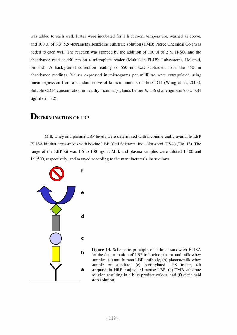
- 118 -
was added to each well. Plates were incubated for 1 h at room temperature, washed as above,
and 100 µl of 3,3’,5,5’-tetramethylbenzidine substrate solution (TMB; Pierce Chemical Co.) was
added to each well. The reaction was stopped by the addition of 100 µl of 2 M H2SO4 and the
absorbance read at 450 nm on a microplate reader (Multiskan PLUS; Labsystems, Helsinki,
Finland). A background correction reading of 550 nm was subtracted from the 450-nm
absorbance readings. Values expressed in micrograms per millilitre were extrapolated using
linear regression from a standard curve of known amounts of rbosCD14 (Wang et al., 2002).
Soluble CD14 concentration in healthy mammary glands before E. coli challenge was 7.0 ± 0.84
µg/ml (n = 82).
DETERMINATION OF LBP
Milk whey and plasma LBP levels were determined with a commercially available LBP
ELISA kit that cross-reacts with bovine LBP (Cell Sciences, Inc., Norwood, USA) (Fig. 13). The
range of the LBP kit was 1.6 to 100 ng/ml. Milk and plasma samples were diluted 1:400 and
1:1,500, respectively, and assayed according to the manufacturer’s instructions.
Figure 13. Schematic principle of indirect sandwich ELISA for the determination of LBP in bovine plasma and milk whey samples. (a) anti-human LBP antibody, (b) plasma/milk whey sample or standard, (c) biotinylated LPS tracer, (d) streptavidin HRP-conjugated mouse LBP, (e) TMB substrate solution resulting in a blue product colour, and (f) citric acid stop solution.
a
b
c
d
e
f

Materials and methods experimental infections
- 119 -
Briefly, diluted milk whey or plasma samples were added to flat-bottomed microtiter
plates, pre-coated with solid bound antibodies recognising LBP of a wide variety of species, and
incubated at room temperature for 1 h. The plates were washed 3 times and biotinylated LPS-
tracer molecules were added. Following incubation (1 h, 20°C) and three washings, streptavidin-
peroxidase conjugated LBP was added (1 h, 20°C). After washing the non-bound conjugated
LBP, freshly prepared substrate solution (TMB) was added and the plates were incubated for 30
min (20°C). The enzymatic reaction was stopped by addition of citric acid. The optical density at
450 nm and a correction wavelength of 550 nm were measured on a microplate reader
(Multiskan PLUS; Labsystems). The concentration of LBP (expressed in micrograms per
millilitre) was calculated by extrapolation using linear regression from a standard curve of
known amounts of human LBP. LPS-binding protein concentration in milk whey and plasma
before intramammary E. coli mastitis were 17.2 ± 2.04 µg/ml (n = 82) and 65.4 ± 7.24 µg/ml (n
= 41), respectively.
DETERMINATION OF PGE2
Milk whey and plasma PGE2 concentrations were determined with a commercially
available PGE2 competitive ELISA kit (Neogen, Lexington, USA) according to manufacturer’s
instructions (Fig. 14). The range of the PGE2 kit was 100 to 4,000 pg/ml.
Figure 14. Schematic principle of competitive ELISA for the determination of PGE2 in bovine plasma and milk whey samples. (a) monoclonal mouse anti-PGE2 antibody, (b) plasma/milk whey sample or standard, (c) HRP-conjugated PGE2, and (d) TMB substrate solution resulting in a blue product colour.
Briefly, quarter milk samples from acute E. coli mastitis were filtered with a 70 µm cell
strainer (Becton Dickinson; Erembodegem, Belgium) in order to discard cell clusters. One ml of
milk or plasma sample was diluted with 1 ml of distilled water, and 1 ml of the mixture was
loaded on a 100 mg C18 column (Varian, St.-Katelijne-Waver, Belgium) after conditioning with
2 ml of distilled water followed by 2 ml of methanol. The column was subsequently washed with
a
b
c
d
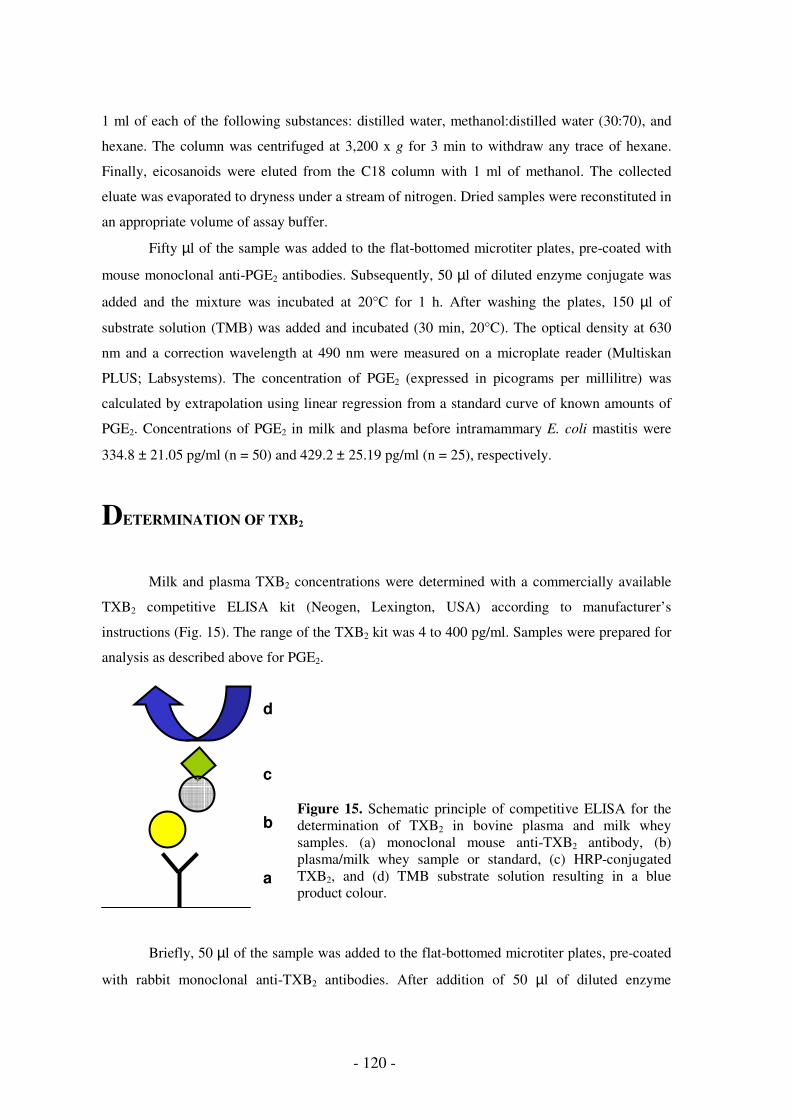
- 120 -
1 ml of each of the following substances: distilled water, methanol:distilled water (30:70), and
hexane. The column was centrifuged at 3,200 x g for 3 min to withdraw any trace of hexane.
Finally, eicosanoids were eluted from the C18 column with 1 ml of methanol. The collected
eluate was evaporated to dryness under a stream of nitrogen. Dried samples were reconstituted in
an appropriate volume of assay buffer.
Fifty µl of the sample was added to the flat-bottomed microtiter plates, pre-coated with
mouse monoclonal anti-PGE2 antibodies. Subsequently, 50 µl of diluted enzyme conjugate was
added and the mixture was incubated at 20°C for 1 h. After washing the plates, 150 µl of
substrate solution (TMB) was added and incubated (30 min, 20°C). The optical density at 630
nm and a correction wavelength at 490 nm were measured on a microplate reader (Multiskan
PLUS; Labsystems). The concentration of PGE2 (expressed in picograms per millilitre) was
calculated by extrapolation using linear regression from a standard curve of known amounts of
PGE2. Concentrations of PGE2 in milk and plasma before intramammary E. coli mastitis were
334.8 ± 21.05 pg/ml (n = 50) and 429.2 ± 25.19 pg/ml (n = 25), respectively.
DETERMINATION OF TXB2
Milk and plasma TXB2 concentrations were determined with a commercially available
TXB2 competitive ELISA kit (Neogen, Lexington, USA) according to manufacturer’s
instructions (Fig. 15). The range of the TXB2 kit was 4 to 400 pg/ml. Samples were prepared for
analysis as described above for PGE2.
Figure 15. Schematic principle of competitive ELISA for the determination of TXB2 in bovine plasma and milk whey samples. (a) monoclonal mouse anti-TXB2 antibody, (b) plasma/milk whey sample or standard, (c) HRP-conjugated TXB2, and (d) TMB substrate solution resulting in a blue product colour.
Briefly, 50 µl of the sample was added to the flat-bottomed microtiter plates, pre-coated
with rabbit monoclonal anti-TXB2 antibodies. After addition of 50 µl of diluted enzyme
a
b
c
d

Materials and methods experimental infections
- 121 -
conjugate, the mixture was incubated at 20°C for 1 h. Following washing, 150 µl of substrate
(TMB) was added and incubated (30 min, 20°C). The optical density at 630 nm and a correction
wavelength at 490 nm were measured on a microplate reader (Multiskan PLUS; Labsystems).
The concentration of TXB2 (expressed in picograms per millilitre) was calculated by
extrapolation using linear regression from a standard curve of known amounts of TXB2.
Concentrations of TXB2 in milk and plasma before intramammary E. coli mastitis were 84.3 ±
5.07 pg/ml (n = 50) and 180.4 ± 26.50 pg/ml (n = 25), respectively.
STATISTICAL ANALYSIS
The general linear model is a commonly used statistical tool in agricultural research to
study the relationship between a normally distributed dependent variable and one or more
independent variables. The independent variables can be either continuous or discrete. If an
independent variable is discrete, it only takes a restricted number of values and the effect of each
level of the discrete variable generates a separate parameter in the model. If an independent
variable is, on the other hand, continuous, an underlying functional relationship has to be
assumed between the independent variable and the response variable. The number of model
parameters corresponds to the number of parameters in the function describing the underlying
relationship (Duchateau et al., 1998). The model parameters arising from either a continuous or a
discrete independent variable can be fixed or random effects. The mixed model methodology
was specifically developed to describe and analyse experiments, which contain both random and
fixed effects factors (Duchateau et al., 1998). A major advantage of a mixed model framework is
that complex data structures can be described and analysed in a natural way (Duchateau, 1997).
The mixed model was used to analyse the data of the experimental infection randomised
trials because animals were measured repeatedly over time during the infection and these
repeated measures are not independent. By introducing cow as a random effect in the model, a
compound symmetry structure was imposed on the repeated measures, thus accounting for the
correlation between the repeated measures.

- 122 -
REFERENCES
Bannerman, D.D., M.J. Paape, W.R. Hare, and E.J. Sohn. 2003. Increased levels of LPS-binding protein in
bovine blood and milk following bacterial lipopolysaccharide challenge. J. Dairy Sci. 86:3128-3137.
Burvenich, C. 1983. Mammary blood flow in conscious lactating goats in various physiological and
pathological (mastitis) conditions. Ph.D. Thesis, Ghent University, Belgium.
Burvenich, C., and G. Peeters. 1983. Effect of experimentally induced mastitis on blood flow in the
heterolateral gland in lactating goats. Zbl. Vet. Med. A 30:459-469.
Burvenich, C., V. Van Merris, J. Mehrzad, A. Diez-Fraile, and L. Duchateau. 2003. Severity of E. coli
mastitis is mainly determined by cow factors. Vet. Res. 34:521-564.
Dosogne, H., C. Burvenich, T. van Werven, E. Roets, E.N. Noordhuizen-Stassen, and B. Goddeeris. 1997.
Increased surface expression of CD11b receptors on polymorphonuclear leukocytes is not sufficient to
sustain phagocytosis during Escherichia coli mastitis in early postpartum dairy cows. Vet. Immunol.
Immunopathol. 60:47-59.
Dosogne, H., K. Van Oostveldt, A. Diez-Fraile, E. Monfardini, and C. Burvenich. 1999. Verminderde
aspecifieke afweer van melkkoeien tijdens de vroege post-partum periode tegen mastitis.
Synthesebrochure Ministerie van Middenstand en Landbouw. ISBN 90-74070-07-8. p. 71.
Duchateau, L. 1997. Comparing the mixed model and the fixed effects model: do the advantages justify
the costs? In: Proceedings of the 5th Scientific Conference of the East, Central and Southern Africa
Network of the International Biometrics Society. Duchateau L., and H.G. Mwambi (Eds.). Mombasa,
Kenya. p. 39-43.
Duchateau, L., P. Janssen, and G.J. Rowlands. 1998. Linear mixed models. An introduction with
applications in veterinary research. ILRI, Nairobi, Kenya. (ISBN 92-9146-038-9). 159 pages.
Edmondson, A.J., I.J. Lean, I.D. Weaver, T. Farver, and G. Webster. 1989. A body condition scoring chart
for Holstein dairy cows. J. Dairy Sci. 72:68-78.
Heyneman, R., C. Burvenich, and R. Vercauteren. 1990. Interaction between the respiratory burst activity
of neutrophil leukocytes and experimentally induced Escherichia coli mastitis in cows. J. Dairy Sci.
73:985-994.
Hillerton, J.E. 1999. Redefining mastitis based on somatic cell count. IDF Bull. 345:4-6.
Hoeben, D., C. Burvenich, J.A. Leigh, and J. Hamann. 2000a. Chemiluminescence of circulating
polymorphonuclear leukocytes and clinical symptoms during Escherichia coli, Streptococcus uberis and
endotoxin mastitis in cows. J. Dairy Res. 67:249-259.
Hoeben, D., E. Monfardini, C. Burvenich, and J. Hamann. 2000b. Treatment of acute Escherichia coli
mastitis in cows with enrofloxacin: effect on clinical signs and chemiluminescence of circulating
neutrophils. J. Dairy Sci. 67:485-502.
IDF. 1971. A monograph on bovine mastitis.
Materials and methods experimental infections
- 123 -
IDF Standard 141 Whole milk – Determination of milk fat, protein and lactose content – Guidance on the
operation of mid-infrared instruments.
ISO 13366-2 Milk – Enumeration of somatic cells – part 2: guidance on the operation of fluoro-opto-
electronic counters.
Kremer, W.D.J., E.N. Noordhuizen-Stassen, F.J. Grommers, A.J.J.M. Daemen, and A. Brand. 1993. Blood
polymorphonuclear leukocyte chemotaxis during experimental Escherichia coli bovine mastitis. J. Dairy
Sci. 76:2613-2618.
Massart-Leën, A.M., G. Vandeputte-Van Messom, and C. Burvenich. 1988. Oxytocin release during
experimentally induced fever and inflammation in lactating ruminants. Arch. Int. Physiol. Biochim.
96:43-44.
Mehrzad, J., H. Dosogne, E. Meyer, R. Heyneman, and C. Burvenich. 2001. Respiratory burst activity of
blood and milk neutrophils in dairy cows during different stages of lactation. J. Dairy Res. 68:399-415.
Paape, M.J., H.D. Hafs, and W.W. Snyder. 1963. Variation of estimated number of milk somatic cells
stained with Wright’s stain or pyronin Y-methyl green stain. J. Dairy Sci. 46:1211-1216.
Petsch, D., and F.B. Anspach. 2000. Endotoxin removal from protein solutions. J. Biotechnol. 76:97-119.
Rainard, P., P. Sarradin, M. J. Paape, and B. Poutrel. 1998. Quantification of C5a/C5adesArg in bovine
plasma, serum and milk. Vet. Res. 29:73-88.
Shuster, D.E., E.K. Lee, and M.E. Kehrli, Jr. 1996. Bacterial growth, inflammatory cytokine production,
and neutrophil recruitment during coliform mastitis in cows within ten days after calving, compared with
cows at midlactation. Am. J. Vet. Res. 57:1569-1575.
Shuster, D.E., M.E. Kehrli, Jr., P. Rainard, and M.J. Paape. 1997. Complement fragment C5a and
inflammatory cytokines in neutrophil recruitment during intramammary infection with Escherichia coli.
Infect. Immun. 65:3286-3292.
Trossat, P., and O. Leray. 1996. Rapport d’évaluation du MilkoScan 4000. CecaLait.
Ubben, E.H., W. Heeschen, and J. Reichmuth. 1997. Somatic cell counting in milk: flow cytometric
measurement with Fossomatic 5000. Milchwissenschaft 52:487-491.
van Werven, T., E.N. Noordhuizen-Stassen, A.J.J.M. Daemen, Y.H. Schukken, A. Brand, and C.
Burvenich. 1997. Pre-infection in vitro chemotaxis, phagocytosis, oxidative burst, and expression of
CD11/CD18 receptors and their predictive capacity on the outcome of mastitis induced in dairy cows
with Escherichia coli. J. Dairy Sci. 80:67-74.
Vandeputte-Van Messom, G., and C. Burvenich. 1993. Effect of somatotropin on changes in milk
production and composition during coliform mastitis in periparturient cows. J. Dairy Sci. 76:3727-3741.
Vandeputte-Van Messom, G., C. Burvenich, E. Roets, A.-M. Massart-Leën, R. Heyneman, W.D.J.
Kremer, and A. Brand. 1993. Classification of newly calved cows into moderate and severe responders
to experimentally induced Escherichia coli mastitis. J. Dairy Res. 60:19-29.
Vangroenweghe, F., L. Duchateau, and C. Burvenich. 2004. Moderate inflammatory response during
experimental E. coli mastitis in primiparous cows. J. Dairy Sci. 87:886-895.
Wang, Y., D.S. Zarlenga, M.J. Paape, and G.E. Dahl. 2002. Recombinant bovine soluble CD14 sensitizes
the mammary gland to lipopolysaccharide. Vet. Immunol. Immunopathol. 86:115-124.

- 124 -
Wenz, J.R., G.M. Barrington, F.B. Garry, K.D. McSweeney, R.P. Dinsmore, G. Goodell, and R.J. Callan.
2001. Bacteremia associated with naturally occurring acute coliform mastitis in dairy cows. JAVMA
219:976-981.
Wilde, J.K.H. 1964. The cellular elements of the bovine bone marrow. Res. Vet. Sci. 5:213-227.
Williamson, D.H., and J. Mellanby. 1970. D-(-)-3-Hydroxybutyrat. In: Methoden der enzymatischen
analyse. 2. Auflage – Band II. Bergmeyer H.U. (Ed.). Verlag Chemie, Weinheim, Germany. pp. 1772-
1775.

- 125 -
INFLUENCE OF PARITY ON SEVERITY OF
INFLAMMATION
Adapted from:
Vangroenweghe F., L. Duchateau, and C. Burvenich. 2004. Moderate inflammatory reaction
during experimental E. coli mastitis in primiparous cows. J. Dairy Sci. 87:886-895.


Influence of parity on severity of inflammation
- 127 -
INTRODUCTION
Several factors have been shown to play a role in the clinical outcome of E. coli mastitis,
namely farm management (Jackson and Bramley, 1983; Oz et al., 1985; Smith et al., 1985;
Jones, 1986; Pankey et al., 1987; Schukken et al., 1989a; 1989b; Oliver et al., 1990; Lam et al.,
1995; Barkema et al., 1999), bacterial factors (Frost et al., 1980; Hill, 1981; Linton and
Robinson, 1984; Sanchez-Carlo et al., 1984a; 1984b; Todhunter et al., 1991; Hogan et al., 1992;
1995; 1999; Cross et al., 1993; Nemeth et al., 1994; Nagy and Fekete, 1999) and physiological
factors (Heyneman et al., 1990; Gilbert et al., 1993; Kremer et al., 1993a; 1993b; Vandeputte-
Van Messom et al., 1993; Dosogne et al., 1997; 2001; van Werven et al., 1997; Mehrzad et al.,
2001; 2002; Vangroenweghe et al., 2001; Burton and Erskine, 2003).
Preventive measures, that are known to be efficient against contagious mastitis, such as
post-milking teat disinfection (Schukken et al., 1989a; 1989b; Barkema et al., 1999) have been
shown to be inefficient in the control of E. coli mastitis (Smith et al., 1985; Oliver et al., 1990;
Lam et al., 1995). In contrast, a few studies (Pankey et al., 1987; Oliver et al., 1993) showed that
pre-milking teat disinfection may prevent infections with environmental pathogens. Several
epidemiological studies have demonstrated a negative correlation between SCC and the
incidence of E. coli mastitis. Therefore, SCC is thought to be a farm related risk factor for the
susceptibility to E. coli mastitis (Schukken et al., 1989a; 1989b; Barkema et al., 1999).
Significant management factors that are related to a higher incidence of clinical coliform mastitis
are: infection pressure from the environment (Jackson and Bramley, 1983), the use of saw dust
as bedding material (Oz et al., 1985) and permanent indoor housing during winter (Jackson and
Bramley, 1983). Prevention of cows from laying down after milking is often advised to omit
contact with bedding material (Jones, 1986).
Various bacterial virulence factors have been studied during E. coli mastitis (Sanchez-
Carlo et al., 1984a; 1984b), however, only a few have been found to substantially influence the
eventual outcome of the disease. Escherichia coli is of environmental origin (Nemeth et al.,
1994). Although over 100 serotypes have yet been recognised, no specific O-serotypes have
been related to bovine E. coli mastitis (Linton and Robinson, 1984). Nevertheless,
intramammary challenge with E. coli 487 caused more severe clinical signs of mastitis than did
E. coli 727 (Todhunter et al., 1991; Hogan et al., 1992; 1995; 1999). Moreover, a capsulated E.
coli B117 strain appeared to cause more severe clinical symptoms of mastitis, because it was
more difficult to opsonise than a non-capsulated E. coli P4 strain (Hill, 1981). Clinical
expression after challenge with low doses of E. coli is comparable with those after inoculation of
- 128 -
high doses (Frost et al., 1980). In vivo adhesion of E. coli to the epithelial surface of the
mammary gland is thought to be unimportant during the initial phase of infection (Bramley et al.,
1979), because in healthy udders collagen or fibronectin are not exposed. Recently, Döpfer et al.
(2000) found some indications for in vitro adhesion to mammary epithelial cells (MAC-T cells).
However, these strains were isolated from recurrent cases of coliform mastitis, a rare disease
(Döpfer et al., 2000). The adhesion factors present in enterotoxigenic E. coli, which are essential
during the initial steps of adhesion in the pathogenesis, do not play a role in the pathogenesis of
E. coli mastitis (Nagy and Fekete, 1999). A bacterial factor, that plays an important role in the
pathogenesis of E. coli mastitis, is endotoxin or LPS. Lipopolysaccharide is a potent inducer of
inflammatory cytokines (Shuster et al., 1993) during growth and killing (Burvenich, 1983;
Petsch and Anspach, 2000). Clinical signs following experimentally induced E. coli mastitis are
rather due to mediator shock than to endotoxin shock, because endotoxin mainly plays a local
role (Hoeben et al., 2000; Dosogne et al., 2002). The capability of some E. coli strains to resist
the bactericidal activity of serum has also been demonstrated as a virulence factor (Barrow and
Hill, 1989).
Physiological factors have a big impact on the clinical outcome of E. coli mastitis
(Burvenich et al., 2003), far more than farm management and bacterial characteristics that were
cited above. Several markers to predict the clinical outcome of E. coli mastitis have been
studied, such as: the number of circulating leukocytes, production of reactive oxygen species by
neutrophils (Heyneman et al., 1990; Vandeputte-Van Messom et al., 1993) and chemotactic
activity of PMN (Kremer et al., 1993a; 1993b; Dosogne et al., 1997; van Werven et al., 1997).
However, predictability of clinical outcome is only significant during 1 to 2 days pre-challenge
and does not explain any causal relationship. These markers were mainly studied in multiparous
cows, varying between 2nd lactation (Heyneman et al., 1990; Vandeputte-Van Messom et al.,
1993; Dosogne et al., 1997) to 2nd – 6th lactation (Kremer et al., 1993a; 1993b; van Werven et al.,
1997). Because BHBA alters both PMN chemotactic activity and reactive oxygen species
production (Heyneman et al., 1990; Kremer et al., 1993a; 1993b; Vandeputte-Van Messom et al.,
1993), many E. coli challenges that were done at Ghent University and the University of Utrecht
were performed on cows with a serum BHBA concentration < 1.4 mmol/l (Kremer et al., 1993b).
Early lactating cows, infected with E. coli, are much more severely affected than cows
after peak lactation (Hill, 1981). This is mainly due to the impairment of early lactation
leukocyte function, as observed by many research groups (Burton and Erskine, 2003). During
this period a decrease in cell function of the PMN, resident in the healthy mammary gland
(Dosogne et al., 2001; Mehrzad et al., 2001; Vangroenweghe et al., 2001) has been observed.
The decrease in cell function was mainly related to the decrease in viability, oxidative burst and
Influence of parity on severity of inflammation
- 129 -
intracellular killing by PMN (Dosogne et al., 2001; Mehrzad et al., 2001; Vangroenweghe et al.,
2001).
Besides energy balance and stage of lactation, cow parity was also found to be an
important physiological factor that influences severity of clinical mastitis (Gilbert et al., 1993;
van Werven et al., 1997; Mehrzad et al., 2002). Blood PMN function was higher in younger
animals than in cows after their 4th parturition (Gilbert et al., 1993; van Werven et al., 1997).
Moreover, viability and oxidative burst have been found to be significantly different between
primiparous cows and multiparous cows during the periparturient period (Mehrzad et al., 2002).
In conclusion, many studies indicate that physiological factors determine the clinical
outcome of E. coli mastitis. In this study, the clinical outcome of E. coli mastitis was studied in
primiparous cows using the same criteria for severity as in other studies using multiparous cows
(Heyneman et al., 1990; Vandeputte-Van Messom et al., 1993; Dosogne et al., 1997; van
Werven et al., 1997; Hoeben et al., 2000).
The purpose of the present study was to evaluate the outcome of intramammary E. coli
inoculation in primiparous cows under identical conditions as described before with multiparous
cows (Heyneman et al., 1990; Vandeputte-Van Messom et al., 1993; Dosogne et al., 1997;
Hoeben et al., 2000; Dosogne et al., 2002). Moreover, the relation between pre-infection
parameters and the outcome of infection in term of severity was assessed. Finally, two high
inoculum doses, with a 100-fold difference (1 x 10⁴ and 1 x 10⁶ colony-forming units (CFU)),
were used because the amount of LPS produced is related to the number of E. coli bacteria
(Burvenich, 1983; Cross et al., 1993; Monfardini et al., 1999). The primiparous cows in this
study were inoculated with the same strain as described before (Vandeputte-Van Messom et al.,
1993; Hoeben et al., 2000; Dosogne et al., 2002).

- 130 -
MATERIALS AND METHODS
Experimental Animals and Study Facilities
All primiparous cows (n = 19) were in their 7th month of pregnancy on arrival at the
commercial dairy farm. At calving, cows were between 24 and 30 months of age.
Experimental Design
Animals were challenged with E. coli P4:O32 on 4 different trial days. The 1 x 10⁴ CFU
inoculum group (group A) was challenged in a group of 4 and 5 animals, the 1 x 10⁶ CFU
inoculum group (group B) in two groups of 5 animals.
Sampling Procedure
Blood and milk samples were collected at d-4, d-1, d0, d+1, d+2 and d+3 relative to the
day of challenge. On the day of challenge, blood and milk samples were collected at PIH 3, 6, 9,
12, 15, 18 and 21.
SCC and Milk Composition
In this study, SCC and milk composition were determined. The daily production of fat,
protein and lactose (g/24 h per quarter) was calculated based on daily quarter milk production
and concentrations of these parameters.
Statistical Analysis
The two inoculum groups were not formally compared because inoculum dose was not
randomly assigned to animals. Pre-infection values of blood and milk constituents of the
inoculum groups were compared just before intramammary E. coli inoculation using a paired
two-sided t-test, assuming unequal variances (Statistix; Analytical Software, Tallahassee, USA)
(Table 1). The relationship between pre-infection concentrations of blood and milk constituents
and the milk production reduction in the uninfected quarters at d+2 (PIH 48-72) was assessed by
linear regression with an adjustment for inoculum size.
For Further Details see chapter 'Materials and Methods Experimental Infections', p. 97.

Influence of parity on severity of inflammation
- 131 -
RESULTS
Relation Between Pre-infection Blood and Milk Constituents and Percentage Reduction in
Milk Production at d+2 Post-Infection
No significant differences were observed in pre-infection levels of blood and milk
constituents (Table 1).
Table 1. Pre-infection levels of blood and milk constituents (expressed as means ± SEM) in primiparous cows infused with 1 x 10⁴ (group A; n = 9) and 1 x 10⁶ (group B; n = 10) CFU E.
coli P4:O32.
(Values are means ± SEM)
Group A Group B
(n = 9) (n = 10) Blood constituents
β-hydroxybutyric acid (mmol/l) 0.89 ± 0.20 0.66 ± 0.05 Glucose (mmol/l) 3.8 ± 0.3 3.2 ± 0.2 Blood leukocyte count (log10/ml) 6.942 ± 0.024 6.903 ± 0.027 Milk constituents SCC (log10/ml) 4.46 ± 0.10 4.18 ± 0.07 Albumin (mg/dl) 23.4 ± 3.3 27.8 ± 3.1 Milk production (l/24 h per quarter) 3.4 ± 0.4 3.8 ± 0.2 Fat (g/24 h per quarter) 155 ± 22 164 ± 29 Protein (g/24 h per quarter) 101 ± 5 110 ± 8 Lactose (g/24 h per quarter) 169 ± 18 186 ± 12
No significant relationships between pre-infection blood and milk constituents and the
reduction in milk production at d+2 post-infection could be demonstrated (Table 2).
Clinical and Laboratory Results of Primiparous Cows in Group A Inoculated with 1 x 10⁴
CFU
All primiparous cows in group A had a moderate clinical outcome of E. coli mastitis,
based on their quarter milk production of the uninfected quarters on d+2 post-infection. Milk
production in the uninfected quarters returned to pre-infection level on d+2 post-infection (Fig.
1).
Rectal temperature increased from PIH 9 onward, with peak fever reached at PIH 12
(Fig. 2a). Heart rate and RR peaked at PIH 12 (Fig. 2b-c). Rumen motility was depressed from
PIH 9 onward, resulting in a decrease in appetite. Primiparous cows returned to normal appetite
and reticulorumen motility around PIH 21. Udder parameters, such as swelling and elevated

- 132 -
quarter temperature, appeared at PIH 9 in the infected quarters. Teat relaxation, milk leakage and
diarrhoea, considered as indicators for severe clinical illness, only appeared in a small number of
animals. Changes to abnormal milk with clots, flakes and a watery appearance were maximally
present in all infected animals at PIH 18 (results not shown). Body condition score slightly
decreased from 3.5 to 2.5 between calving and the end of the experimental challenge period.
Table 2. Relationship between pre-infection blood and milk constituents and the reduction in quarter milk production at d+2 (PIH 48-72) in the uninfected quarters of primiparous cows from both groups, assessed by linear regression with correction for inoculum dose. The slope value indicates the change in milk production reduction for one unit change in the blood and milk constituents.
Slope Standard error P-value
Blood constituents
β-hydroxybutyric acid (mmol/l) -24 14 0.110
Glucose (mmol/l) -11 6 0.089
Blood leukocyte count (log10/ml) -44 64 0.497
Milk constituents
SCC (log10/ml) +12 20 0.529
Albumin (mg/ml) -0.8 0.5 0.126
Milk production (l/24 h per quarter) +0.013 0.014 0.120
Fat (g/24 h per quarter) +0.14 0.07 0.060
Protein (g/24h per quarter) +0.37 0.20 0.089
Lactose (g/24 h per quarter) +0.23 0.12 0.067
Based on the severity scoring adapted from Wenz et al. (2001), primiparous cows in
group A predominantly reacted with a mild response (n = 7) at PIH 9. At PIH 12, the clinical
symptoms progressed mostly to a moderate response (n = 7). Thereafter, only one primiparous
cow with a moderate response was observed at PIH 15. No severe responses were observed at
any time point between PIH 9 and 48 in group A, which received the 1 x 10⁴ CFU E. coli
inoculum dose.
Milk production in the infected quarters maximally decreased (± 55%) on the day of
challenge. At d+1 post-infection, milk production gradually increased and almost totally
recovered by d+3 post-infusion (Fig. 2d). The number of E. coli in the infected quarters peaked
at PIH 6, followed by a rapid elimination until PIH 15. At PIH 72, only 5 animals still had low
numbers of E. coli in the infected quarters (Fig. 2e). Somatic cell count rapidly increased from
PIH 6, reached a maximum at PIH 12, and remained high until the end of the experiment at PIH
72 (Fig. 2f).
Blood leukocyte number decreased to nadir at PIH 12 and recovered to pre-infection
level at PIH 48 (Fig. 2j). The presence of early immature PMN (myelocytes-metamyelocytes) in

Influence of parity on severity of inflammation
- 133 -
circulation increased at PIH 6, reaching peak levels at PIH 21 (Fig. 2g). Similarly, maximal
percentages of late immature PMN (band cells) were reached at PIH 21 (Fig. 2h).
Concomitantly, circulating mature PMN decreased at PIH 6 with nadir at PIH 12 (Fig. 2i).
Figure 1. Milk production (l/d) of the uninfected right quarters from d-4 until d+3 relative to infusion from primiparous cows infused with 1 x 10⁴ (; group A; n = 9) and 1 x 10⁶ (------; group B; n = 10) CFU E. coli P4:O32. Data are means (± SEM).
The concentration of lactose in the infected quarters decreased from PIH 9 with a
minimum at PIH 21-24 (Fig. 2k). Serum albumin in the infected quarters increased from PIH 9,
and peaked at PIH 15 (Fig. 2l). Sodium and chlorine concentrations peaked at PIH 12 (Fig. 2m-
n), whereas potassium concentration decreased from PIH 9, and reached its greatest reduction at
PIH 12 (Fig. 2o). All indicators for the presence of mastitis gradually recovered to pre-infection
levels at PIH 72, after reaching their peak values (Fig. 2k-o).
Time relative to infusion
l/d
Right Quarter milk production
d-4 d-1 d0 d1 d2 d3
12
34
56

- 134 -
Figure 2. Rectal temperature (a), heart rate (b), respiration rate (c), left quarter milk production (d), number of CFU E. coli P4:O32 (e), SCC (f), % myelocytes-metamyelocytes (g), % band cells (h), % mature neutrophils (i), blood leukocyte count (j), lactose (k), serum albumin (l), milk sodium (m), milk chlorine (n) and milk potassium (o) from PIH –96 until PIH 72 in infected quarters or blood, respectively, from primiparous cows infused with 1 x 10⁴ (; group A; n = 9) and 1 x 10⁶ (------; group B; n = 10) CFU E. coli P4:O32. Data are means.
Time relative to infection (hours)
°C
aRectal temperature
-96 0 3 6 9 12 18 24 72
38
.53
9.5
40
.5
Time relative to infection (hours)
be
ats
/min
bHeart rate
-96 0 3 6 9 12 18 24 72
65
70
75
80
85
90
95
Time relative to infection (hours)
co
un
ts/m
in
cRespiration rate
-96 0 3 6 9 12 18 24 72
20
25
30
35
Time relative to infection (days)
l/d
Left Quarter milk productiond
d-4 d-1 d0 d1 d2 d3
1.5
2.5
3.5
Time relative to infection (hours)
log
10
(CF
U/m
l)
eColony-forming units E. Coli
-96 0 3 6 9 12 18 24 72
01
23
4
Time relative to infection (hours)
log
10
(scc/m
l)
f
Somatic cell count
-96 0 3 6 9 12 18 24 72
4.5
5.5
6.5
Time relative to infection (hours)
%
gMyelocytes-metamyelocytes
-96 0 3 6 9 12 18 24 72
10
15
20
25
30
35
Time relative to infection (hours)
%
hBand cells
-96 0 3 6 9 12 18 24 72
24
68
10
12
14
Time relative to infection (hours)
%
iMature neutrophils
-96 0 3 6 9 12 18 24 72
15
20
25
30
Time relative to infection (hours)
log
10
(BL
C/m
l)
Blood Leucocyte countj
-96 0 3 6 9 12 18 24 72
46
81
0
Time relative to infection (hours)
g/l
kLactose
-96 0 3 6 9 12 18 24 72
25
30
35
40
45
50
Time relative to infection (hours)
mg
/dl
lSerum albumin
-96 0 3 6 9 12 18 24 72
50
15
02
50
Time relative to infection (hours)
mm
ol/l
mSodium
-96 0 3 6 9 12 18 24 72
30
40
50
60
Time relative to infection (hours)
mm
ol/l
nChlorine
-96 0 3 6 9 12 18 24 72
40
50
60
Time relative to infection (hours)
mm
ol/l
oPotassium
-96 0 3 6 9 12 18 24 72
35
40
45
50
Influence of parity on severity of inflammation
- 135 -
Clinical and Laboratory Results of Primiparous Cows in Group B Inoculated with 1 x 10⁶
CFU
Clinical reaction and changes in laboratory parameters were similar to the above
described results for group A. However, the changes during inflammation generally appeared
more rapidly (approximately 3 h) in primiparous cows from group B, infused with the 1 x 10⁶
CFU E. coli inoculum dose, compared to group A, infused with the 1 x 10⁴ CFU E. coli
inoculum dose.
All primiparous cows in this group reacted with a moderate clinical response to
intramammary E. coli infusion. Milk production in the uninfected quarters was only decreased
until d+1 post-infection (Fig. 1). Rectal temperature increased from PIH 6, peaked at PIH 9 and
gradually decreased to pre-infection values at PIH 18 (Fig. 2a). Peak tachycardia was reached at
PIH 9 (Fig. 2b), whereas maximal RR was reached at PIH 12 (Fig. 2c). Reticulorumen motility
and all other clinical parameters seemed to change some 3 h earlier in group B. The same trend
in BCS could be observed as in group A.
Clinical scores, adapted from Wenz et al. (2001), showed an earlier reaction (approx. 3
h) in group B compared with group A. At PIH 9, most of the primiparous cows reacted
moderately (n = 8), gradually decreasing at PIH 12 (n = 5). Only one animal in group B had a
prolonged moderate response until PIH 24. No severe responses were present in this group at
any time point between PIH 9 and 48.
Milk production reduction in the infected quarters was maximal (± 65%) on the day of
infusion and recovered rapidly to pre-infection production at d+3 post-infection (Fig. 2d). The
number of E. coli in the infected quarters peaked at PIH 3 (Fig. 2e). A rapid influx of cells into
the infected quarters was observed from PIH 3, with a peak at PIH 9 (Fig. 5f).
The number of circulating blood leukocytes decreased from PIH 6, peaking at PIH 9. A
pronounced rebound effect was observed between PIH 15 and 48 (Fig. 2j). The increase in
circulating early immature cells was pronounced and peaked at PIH 15 (Fig. 2g). Band cells
increased at PIH 9, and peaked at PIH 18, thereafter gradually decreasing to reach pre-challenge
levels at PIH 72 (Fig. 2h). The greatest reduction in circulating mature PMN was reached at PIH
3, gradually increasing to pre-infection values at PIH 21 (Fig. 2i).
The concentrations of lactose, serum albumin, sodium, chlorine and potassium followed
similar kinetics as in group A, although the initial changes appeared approximately 3 h earlier
(Fig. 5k-o).

- 136 -
DISCUSSION
In this study, the same strain and range of high inoculum doses of E. coli was used to
induce E. coli mastitis as previously described (Heyneman et al., 1990; Vandeputte-Van Messom
et al., 1993; Hoeben et al., 2000; Dosogne et al., 2002). In contrast to previous studies,
primiparous cows were used. Similar clinical symptoms, such as quarter inflammation, fever,
depression of reticulorumen motility, loss of appetite and general discomfort were observed as
expected. In previous studies, however, the clinical responses showed large variations from
mild-moderate to severe; whereas in the present study, the variation in clinical response was
quite narrow. Quarter inflammation was associated with a temporary loss of milk production,
combined with the secretion of abnormal milk from the infected glands. Maximal decrease in
milk production in the infected and uninfected quarters occurred on the day of challenge, in
contrast to previous observations where maximal decrease in milk production was observed later
(Heyneman et al., 1990; Hoeben et al., 2000). In the present study, none of the animals reacted
severely following intramammary E. coli challenge (Fig. 3). In both infected and uninfected
quarters, milk production seemed to recover more rapidly on d+1 post-infection in primiparous
cows from group B, which received the 1 x 10⁶ CFU E. coli inoculum dose. Animals were
scored as previously described (Vandeputte-Van Messom et al., 1993) and compared with the
clinical score used by Wenz et al. (2001). This resulted in a similar classification of mild and
moderate responses. The only difference that could be observed between group A and B was the
time of latency that was shorter when higher inoculum doses were used.
Body condition score and the concentration of BHBA and glucose indicated that the
primiparous cows in this study were not ketotic at the time of challenge (Kremer et al., 1993b),
although their quarter milk production was comparable to the multiparous cows previously
challenged (Heyneman et al., 1990; Vandeputte-Van Messom et al., 1993).
The number of circulating PMN, a marker to predict the clinical outcome of the disease,
was similar to the levels previously observed in moderate responders (Vandeputte-Van Messom
et al., 1993). Primiparous cows in this study could therefore be expected to react with a moderate
clinical response. Although no PMN functionality was determined in this study, it is expected
that PMN reactive oxygen species production was high in these primiparous cows. Mehrzad et
al. (2002) recently found that in the period from 5 weeks before until 5 weeks post-parturition
the CL activity of PMN was higher in primiparous cows than in multiparous cows.
Polymorphonuclear leukocyte reactive oxygen species production and chemotactic activity were
inversely correlated to the clinical outcome of E. coli mastitis (Heyneman et al., 1990; Kremer et

Influence of parity on severity of inflammation
- 137 -
al., 1993a; 1993b). No significant relationship with the reduction in quarter milk production on
d+2 post-infection was observed, possibly due to the limited number of animals in this study.
Fig. 3. Percentage of the initial quarter milk production in the uninfected quarters on d+2 (PIH 48-72) relative to infection. A. Primiparous cows infused with 1 x 10⁴ (group A; n = 9) or 1 x 10⁶ (group B; n = 10) CFU E. coli P4:O32 in both left quarters (present study). B. Multiparous cows infused with 1 x 10³ CFU E. coli O157 in both left quarters and scored into moderate (n = 7) and severe (n = 5) clinical response (Dosogne et al., 1997; historical control). Means (•) and standard deviation (I) of percentages of the initial milk production in the uninfected quarters were given for each group. Line (……) at 50% of the initial milk production arbitrarily indicates the difference between moderate and severe responders (Vandeputte-Van Messom et al., 1993).
The onset of local clinical signs of mastitis, characterised by quarter swelling, coincided
with the influx of PMN to the infected quarters. Somatic cell count increased rapidly in both
groups, which is in accordance with earlier observations, where moderate responders had a
rapidly occurring leukocytosis in the infected glands (Vandeputte-Van Messom et al., 1993). The
extraction of mature PMN from the blood to the infected glands is known to result in early and
late immature PMN recruitment from the bone marrow to restore the number of circulating
PMN. In this study the recruitment of immature PMN was of short duration, compared to
Heyneman et al. (1990), who observed immature forms in circulation for at least 3 d in moderate
responders and for almost 10 d in severe responders.
%re
duction
020
40
60
80
100
120
10 CFU4
Primiparous
10 CFU6
Primiparous
A
%re
duction
020
40
60
80
100
120
10 CFU3
Multiparous
B

- 138 -
The rapid influx of PMN into the infected glands was associated with fast elimination of
bacteria from the quarters. In this study, high inoculum doses were used for experimental
induction of E. coli mastitis, because we were mainly interested in bacterial elimination rather
than bacterial growth in the mammary gland. In contrast to an earlier study with the same
inoculum dose in multiparous animals (Vandeputte-Van Messom et al., 1993), peak bacterial
numbers were already reached around PIH 3 to 6. This peak number of bacteria was followed
by a rapid elimination from the affected glands. Contrary to the induction of E. coli mastitis with
low inoculum doses, where elimination is preceded by excessive bacterial growth, in this study,
peak numbers were reached within 6 h post-infection and followed by a subsequent bacterial
elimination. Therefore, PMN influx is thought to be fast and strong enough to rapidly clear the
bacteria from the affected glands. It can be presumed that the bactericidal capacity of the PMN
migrated to the infected quarters is high, because more efficient PMN functionality was reported
in primiparous cows recently (Mehrzad et al., 2002).
Several indicators (lactose, serum albumin, sodium, chlorine and potassium) for the
presence of mastitis were determined in this study. Generally, the changes could be observed
approximately 3 h earlier in group B, although peak levels were almost identical for both groups,
which coincides with all other data, indicating that the animals reacted with a mild to moderate
response and little variation in the clinical response was present in this study.
From this study, it appears that the inflammatory response in primiparous cows from
group B has an earlier onset compared to group A. One possible explanation for this observation
could be the 100-fold difference in the number of E. coli infused into the mammary glands,
because the amount of LPS produced is related to the number of E. coli bacteria (Burvenich,
1983; Cross et al., 1993; Monfardini et al., 1999). A direct effect of LPS present in the inoculum
can be excluded, because the bacterial cultures were washed three times in pyrogen-free PBS
before further dilutions were made. Lipopolysaccharide, known as a potent inducer of
inflammatory cytokines (Shuster et al., 1993), can be produced quite rapidly during bacterial
growth following intramammary E. coli infusion. Therefore, a sufficient amount of
inflammatory cytokines should be produced early during inflammation in both groups, resulting
in the rapid attraction of PMN from the blood into the mammary gland, with a subsequent
pronounced increase of SCC in the infected glands.
Following E. coli mastitis, treatment with a bactericidal antibiotic at PIH 10 has been
shown inefficient to alter local and systemic symptoms already present, although the number of
bacteria in the infected quarters decreased 100-fold (Monfardini et al., 1999). Therefore, it can
be suggested that early inflammatory events (first 3 h) could play an important role in the further
regulation of the inflammatory response to combat the invading organisms. Experimental design

Influence of parity on severity of inflammation
- 139 -
in this study, particularly in relation to the number and time of samplings, was however not
suitable to unravel these elements of early acute phase response.
Nevertheless, the moderate inflammation model with primiparous cows in the present
study clearly indicates that a mild to moderate response following intramammary challenge with
E. coli is sufficient to combat the induced mastitis with a maximal resolution of mammary gland
functionality.
CONCLUSION
In conclusion, despite the use of relative high inoculum doses, primiparous cows react
with a moderate inflammatory response following intramammary infusion. This moderate
response was evident from the pre-infection number of circulating leukocytes, the concentration
of BHBA and glucose, the prompt clinical response, the rapid influx of PMN into the infected
quarters, the efficient bacterial elimination of the affected glands and the fast recovery of milk
production in both infected and uninfected glands. To follow-up the bacterial elimination from
the affected glands, the use of a high inoculum dose was of particular interest. In contrast to
previous studies, pre-infection parameters were not significantly related to the clinical outcome
of the disease in terms of severity. The absence of a significant relation between these
parameters could be due to the narrow variation in clinical response in the present study,
especially compared to the large variation, ranging from mild-moderate to severe, observed in
previous studies. The difference in time of latency between both inoculum doses could also be
considered as an interesting observation. However, to further elucidate the effect of inoculum
dose, a completely randomised study should be designed for this purpose. The moderate
inflammation model with primiparous cows presented here, clearly indicates that a mild to
moderate response following intramammary challenge with E. coli is sufficient to combat the
induced mastitis with a maximal resolution of mammary gland functionality.

- 140 -
REFERENCES
Barkema, H.W., Y.H. Schukken, T.J.G.M. Lam, M.L. Beiboer, G. Benedictus, and A. Brand. 1999.
Management practices associated with the incidence rate of clinical mastitis. J. Dairy Sci. 82:1643-1654.
Barrow, P.A., and A.W. Hill. 1989. The virulence characteristics of strains of Escherichia coli isolated
from cases of bovine mastitis in England and Wales. Vet. Microbiol. 20:35-48.
Bramley, A.J., J.S. King, T.M. Higgs, and F.K. Neave. 1979. Colonization of the bovine duct following
inoculation with Staphylococcus aureus and Escherichia coli organisms at or into the teat duct. Br. Vet.
J. 135:149-162.
Burton, J.L., and R.J. Erskine. 2003. Immunity and mastitis – some new ideas for an old disease. Vet.
Clin. N. Am. – Food A. 19:1-45.
Burvenich, C. 1983. Mammary blood flow in conscious lactating goats in various physiological and
pathological (mastitis) conditions. Ph.D. Thesis, Ghent University, Belgium.
Burvenich, C., V. Van Merris, J. Mehrzad, A. Diez-Fraile, and L. Duchateau. 2003. Severity of E. coli
mastitis is mainly determined by cow factors. Vet. Res. 34:521-564.
Cross, A.S., S.M. Opal, J.C. Sadoff, and P. Gemski. 1993. Choice of bacteria in animal models of sepsis.
Infect. Immun. 61:2741-2747.
Döpfer, D., R.A. Almeida, T.J.G.M. Lam, H. Nederbragt, S.P. Oliver, and W. Gaastra. 2000. Adhesion
and invasion of Escherichia coli from single and recurrent clinical cases of bovine mastitis in vitro. Vet.
Microbiol. 74:331-343.
Dosogne, H., C. Burvenich, T. van Werven, E. Roets, E.N. Noordhuizen-Stassen, and B. Goddeeris. 1997.
Increased surface expression of CD11b receptors on polymorphonuclear leukocytes is not sufficient to
sustain phagocytosis during Escherichia coli mastitis in early postpartum dairy cows. Vet. Immunol.
Immunopathol. 60:47-59.
Dosogne, H., F. Vangroenweghe, B. Barrio, P. Rainard, and C. Burvenich. 2001. Decreased bactericidal
activity of bovine blood and milk neutrophils against Staphylococcus aureus in dairy cows shortly after
parturition. J. Dairy Res. 68:539-549.
Dosogne, H., E. Meyer, A. Sturk, J. van Loon, A.-M. Massart-Leën, and C. Burvenich. 2002. Effect of
enrofloxacin treatment on plasma endotoxin during bovine Escherichia coli mastitis. Inflamm. Res.
51:201-205.
Frost, A.J., A.W. Hill, and B.E. Brooker. 1980. The early pathogenesis of bovine mastitis due to
Escherichia coli. Proc. R. Soc. Lond. B 209:431-439.
Gilbert, R.O., Y.T. Gröhn, P.M. Miller, and D.J. Hoffman. 1993. Effect of parity on periparturient
neutrophil function in dairy cows. Vet. Immunol. Immunopathol. 36:75-82.
Heyneman, R., C. Burvenich, and R. Vercauteren. 1990. Interaction between the respiratory burst activity
of neutrophil leukocytes and experimentally induced Escherichia coli mastitis in cows. J. Dairy Sci.
73:985-994.

Influence of parity on severity of inflammation
- 141 -
Hill, A.W. 1981. Factors influencing the outcome of Escherichia coli mastitis in the dairy cow. Res. Vet.
Sci. 31:107-112.
Hogan, J.S., W.P. Weiss, D.A. Todhunter, K.L. Smith, and P.S. Schoenberger. 1992. Efficacy of an
Escherichia coli J5 mastitis vaccine in an experimental challenge trial. J. Dairy Sci. 75:415-422.
Hogan, J.S., W.P. Weiss, D.A. Todhunter, K.L. Smith, and P.S. Schoenberger. 1995. Effects of an
Escherichia coli J5 vaccine on mild clinical coliform mastitis. J. Dairy Sci. 78:285-290.
Hogan, J.S., V.L. Bogacz, M. Aslam, and K.L. Smith. 1999. Efficacy of an Escherichia coli J5 bacterin
administered to primigravid primiparous cows. J. Dairy Sci. 82:939-943.
Hoeben, D., E. Monfardini, C. Burvenich, and J. Hamann. 2000. Treatment of acute Escherichia coli
mastitis in cows with enrofloxacin: effect on clinical signs and chemiluminescence of circulating
neutrophils. J. Dairy Sci. 67:485-502.
Jackson, E., and J. Bramley. 1983. Coliform mastitis. In Practice 7:135-146.
Jones, T.O. 1986. A review of teat factors in bovine E. coli mastitis. Vet. Rec. 118:507-509.
Kremer, W.D.J., E.N. Noordhuizen-Stassen, F.J. Grommers, A.J.J.M. Daemen, P.A.J. Henricks, C.
Burvenich, and A. Brand. 1993a. Pre-infection chemotactic response of blood polymorphonuclear
leukocytes to predict severity of Escherichia coli bovine mastitis. J. Dairy Sci. 76:1568-1574.
Kremer, W.D.J., E.N. Noordhuizen-Stassen, F.J. Grommers, Y.H. Schukken, R. Heeringa, and A. Brand.
1993b. Severity of experimental Escherichia coli mastitis in ketonemic and nonketonemic dairy cows. J.
Dairy Sci. 76:3428-3436.
Lam, T.J.G.M., J.H. van Vliet, and Y.H. Schukken. 1995. Tepeldesinfectie en mastitis bij het rund: een
literatuuroverzicht. Tijdschrift voor Diergeneeskunde 120:392-399.
Linton, A.H., and T.C. Robinson. 1984. Studies on the association of Escherichia coli with bovine
mastitis. Br. Vet. J. 140:368-373.
Mehrzad, J., H. Dosogne, E. Meyer, R. Heyneman, and C. Burvenich. 2001. Respiratory burst activity of
blood and milk neutrophils in dairy cows during different stages of lactation. J. Dairy Res. 68:399-415.
Mehrzad, J., L. Duchateau, S. Pyörälä, and C. Burvenich. 2002. Blood and milk neutrophil
chemiluminescence and viability in primiparous and pluriparous dairy cows during late pregnancy,
around parturition and early lactation. J. Dairy Sci. 85:3268-3276.
Monfardini, E., C. Burvenich, A.-M. Massart-Leën, E. Smits, and M.J. Paape. 1999. Effect of antibiotic
induced bacterial clearance in the udder on L-selectin shedding of blood neutrophils in cows with
Escherichia coli mastitis. Vet. Immunol. Immunopathol. 67:373-384.
Nagy, B., and P.Z. Fekete. 1999. Enterotoxigenic Escherichia coli (ETEC) in farm animals. Vet. Res.
30:259-284.
Nemeth, J., C.A. Muckle, and C.L. Gyles. 1994. In vitro comparison of bovine mastitis and fecal
Escherichia coli isolates. Vet. Microbiol. 40:231-238.
Oliver, S.P., S.H. King, M.J. Lewis, P.M. Torre, K.R. Matthews, and H.H. Dowlen. 1990. Efficacy of
chlorhexidine as a postmilking teat disinfectant for the prevention of bovine mastitis during lactation. J.
Dairy Sci. 73:2230-2235.

- 142 -
Oliver, S.P., M.J. Lewis, T.L. Ingle, B.E. Gillespie, and K.R. Matthews. 1993. Prevention of bovine
mastitis by a premilking teat disinfectant containing chlorous acid and chloride dioxide. J. Dairy Sci.
76:287-292.
Oz, H.H., R.J. Farnsworth, and V.L. Larson. 1985. Environmental mastitis. Vet. Bull. 55:829-840.
Pankey, J.W., E.E. Wildman, P.A. Drechsler, and J.S. Hogan. 1987. Field trial evaluation of premilking
teat disinfection. J. Dairy Sci. 70:867-872.
Petsch, D., and F.B. Anspach. 2000. Endotoxin removal from protein solutions. J. Biotechnol. 76:97-119.
Sanchez-Carlo, V., R.A. Wilson, J.S. McDonald, and R.A. Packer. 1984a. Biochemical and serological
properties of Escherichia coli isolated from cows with acute mastitis. Am. J. Vet. Res. 45:1771-1774.
Sanchez-Carlo, V., J.S. McDonald, and R.A. Packer. 1984b. Virulence factors of Escherichia coli isolated
from cows with acute mastitis. Am. J. Vet. Res. 45:1775-1777.
Schukken, Y.H., F.J. Grommers, D. van de Geer, and A. Brand. 1989a. Incidence of clinical mastitis on
farms with low somatic cell counts in bulk milk. Vet. Rec. 125:60-63.
Schukken, Y.H., D. van de Geer, F.J. Grommers, J.A.H. Smit, and A. Brand. 1989b. Intramammary
infections and risk factors for clinical mastitis in herds with low somatic cell counts in bulk milk. Vet.
Rec. 125:393-396.
Shuster, D.E., M.E. Kehrli, Jr., and M.G. Stevens. 1993. Cytokine production during endotoxin-induced
mastitis in lactating dairy cows. Am. J. Vet. Res. 54:80-85.
Smith, K.L., D.A. Todhunter, and P.S. Schoenberger. 1985. Environmental pathogens and intramammary
infection during the dry period. J. Dairy Sci. 68:402-417.
Todhunter, D.A., K.L. Smith, J.S. Hogan, and P.S. Schoenberger. 1991. Intramammary challenge with
Escherichia coli following immunization with a curli-producing Escherichia coli. J. Dairy Sci. 74:819-
825.
van Werven, T., E.N. Noordhuizen-Stassen, A.J.J.M. Daemen, Y.H. Schukken, A. Brand, and C.
Burvenich. 1997. Pre-infection in vitro chemotaxis, phagocytosis, oxidative burst, and expression of
CD11/CD18 receptors and their predictive capacity on the outcome of mastitis induced in dairy cows
with Escherichia coli. J. Dairy Sci. 80:67-74.
Vandeputte-Van Messom, G., C. Burvenich, E. Roets, A.-M. Massart-Leën, R. Heyneman, W.D.J.
Kremer, and A. Brand. 1993. Classification of newly calved cows into moderate and severe responders
to experimentally induced Escherichia coli mastitis. J. Dairy Res. 60:19-29.
Vangroenweghe, F., H. Dosogne, J. Mehrzad, and C. Burvenich. 2001. Effect of milk sampling and stage
of lactation on milk composition, viability and functions of resident cells in milk. Vet. Res. 32:565-579.
Wenz, J.R., G.M. Barrington, F.B. Garry, K.D. McSweeney, R.P. Dinsmore, G. Goodell, and R.J. Callan.
2001. Bacteremia associated with naturally occurring acute coliform mastitis in dairy cows. JAVMA
219:976-981.
- 143 -
MODULATION OF THE INFLAMMATORY
REACTION
1. Variation of the inoculum dose
2. Inhibition of prostaglandin synthesis
3. Vaccination against the endotoxin

- 145 -
1.
VARIATION OF THE INOCULUM DOSE
Adapted from:
Vangroenweghe F., P. Rainard, M.J. Paape, L. Duchateau, and C. Burvenich. 2004. Increase of
Escherichia coli inoculum doses induce faster innate immune response in primiparous cows. J.
Dairy Sci. 87:4132-4144.


Variation of the inoculum dose
- 147 -
INTRODUCTION
The importance of innate immunity in recognising microbial pathogens and mounting a
response against them is now widely recognised. The immediate, innate immune response is
mediated largely by white blood cells such as PMN and macrophages, cells that phagocytose and
kill the pathogens, and that concurrently co-ordinate additional host responses by synthesising a
wide range of inflammatory mediators and cytokines (Aderem and Ulevitch, 2000). Several
species of Gram-negative bacteria, including E. coli, Klebsiella pneumoniae, and various species
of Enterobacter, are common mastitis pathogens and are all characterised by the presence of
endotoxin or LPS in their outer membrane. Lipopolysaccharide is a pro-inflammatory molecule
that is shed from the bacterial surface during bacterial replication or death (Burvenich, 1983;
Petsch and Anspach, 2000; Burvenich et al., 2003). Clinical signs following experimentally
induced E. coli mastitis contributed to mediator shock rather than to endotoxin shock, because
endotoxin mainly plays a local role (Hoeben et al., 2000; Dosogne et al., 2002).
Several of the pro-inflammatory cytokines that mediate the localised and systemic
response to Gram-negative mastitis, including IL-1β, IL-6, IL-8, and TNF-α are upregulated by
LPS (Shuster et al., 1993; Guha and Mackman, 2001). The upregulation of these cytokines is
mediated by the interaction of LPS with the accessory proteins, LBP and CD14 (Guha and
Mackman, 2001). LPS-binding protein, an acute phase protein binding with circulating LPS,
facilitates the transfer of LPS to mCD14, found on PMN and cells of the monocytic lineage
(Wright et al., 1990). Membrane-associated CD14 is a glycosyl phosphatidylinositol-anchored
protein that lacks an intracellular cytoplasmic domain, rendering it incapable of signal
transduction across the cell membrane. In cells lacking mCD14, such as epithelial and
endothelial cells, activation is dependent on cellular recognition of LPS-LBP complexes bound
to circulating sCD14, which is derived from the shedding of mCD14 (Tapping and Tobias, 1997)
from CD14-bearing cells. Toll-like receptor-4 has been identified in both cells of the monocytic
lineage and non-mCD14-bearing cells as a LPS transmembrane receptor capable of activating
cells (Chow et al., 1999; Faure et al., 2000). Recently, it was shown that LBP in addition to
transferring LPS to CD14 also forms an integral part of a trimolecular LPS-LBP-sCD14
complex. Monocytes can therefore detect the presence of LPS at concentrations as low as 10
pg/ml (Thomas et al., 2002). Transmembrane signalling and cell activation in cells lacking
mCD14 is thought to be associated with TLR-4, through a cell surface assembly of a multi-
protein recognition complex consisting of CD14, MD-2, and TLR-4 (Akashi et al., 2000).

- 148 -
Activation and transmembrane signal transduction through the TLR-4 complex activates several
NF-κB controlled genes such as IL-8 in endothelial cells (Aderem and Ulevitch, 2000).
A role for sCD14 and LBP in mediating bovine host responses to intramammary LPS or
E. coli challenge have recently been demonstrated (Wang et al., 2002; Bannerman et al., 2003;
Lee et al., 2003a; 2003b). Following intramammary LPS infusion, sCD14 increases in milk
(Bannerman et al., 2003; Lee et al., 2003a) paralleled by an increase in LBP (Bannerman et al.,
2003). Moreover, sCD14 has been shown to sensitise the mammary gland to LPS (Wang et al.,
2002) and to reduce the severity of experimental E. coli mastitis in mice (Lee et al., 2003c) and
cows (Lee et al., 2003b). Interestingly, maximal levels of the chemoattractant IL-8 were
observed before increases in either milk LBP or sCD14. This suggests that initial host cell
activation can occur in the presence of basal levels of sCD14 and LBP (Bannerman et al., 2003).
Furthermore, PMN influx as determined by SCC, was similarly elevated before increases in
sCD14 and LBP, indicating that heightened levels of these molecules were not required for
immediate host innate immune responses (Bannerman et al., 2003).
Several physiological factors impact on the clinical outcome of E. coli mastitis
(Burvenich et al., 2003). Early lactating cows, infected with E. coli, are more severely affected
than cows after peak lactation (Hill, 1981). This is mainly due to the impairment of early
lactation leukocyte function which begins a few weeks before parturition and only recovers
several weeks post-partum (Kehrli et al., 1989; Sordillo and Babiuk, 1991a; 1991b; Sordillo and
Peel, 1992; Detilleux et al., 1995; Dosogne et al., 1999; Mehrzad et al., 2002). This pronounced
immunosuppresion is not only related to parturition itself (Kimura et al., 1999), but is influenced
by several periparturient diseases (Kehrli and Goff, 1989) and has a consequence on many other
diseases, such as retained placenta (Kimura et al., 2002). In addition to stage of lactation, parity
was also reported to be an important physiological factor that influences severity of clinical
mastitis (Gilbert et al., 1993; van Werven et al., 1997; Mehrzad et al., 2002; Vangroenweghe et
al., 2004). Blood PMN function was higher in younger animals than in cows after their 4th
parturition (Gilbert et al., 1993; van Werven et al., 1997). Moreover, PMN viability and
oxidative burst activity have been found to be significantly different between primiparous cows
and multiparous cows during the periparturient period (Mehrzad et al., 2002). In a non-
randomised intramammary challenge study using large numbers of E. coli, primiparous cows
reacted as moderate responders based on their quarter milk production in the uninfected quarters
on d+2 post-infection. Based on clinical severity, all of the primiparous animals were scored as
mild to moderate in their clinical response throughout the entire experimental challenge period
(Vangroenweghe et al., 2004).

Variation of the inoculum dose
- 149 -
The purpose of the present study was to quantify several inflammatory mediators and
cytokines and to evaluate the outcome of intramammary E. coli inoculation in primiparous cows
under identical conditions as described previously (Vangroenweghe et al., 2004) with a fully
randomised study design, using two high inoculum doses, with a 100-fold difference (1 x 10⁴
and 1 x 10⁶) in CFU. This difference in inoculum dose was based on the amount of LPS
produced related to the number of E. coli bacteria injected (Burvenich, 1983; Monfardini et al.,
1999). The hypothesis of the present study is that the application of 2 different inoculum doses
elicits differences of the innate immune response. An effect of 2 different inoculum doses in
highly resistant primiparous cows has never been studied before, and is important to have a
better insight into the mechanism of innate immune response in these animals.
- 150 -
MATERIALS AND METHODS
Experimental animals and study facilities
All primiparous cows (n = 16) calved within 3 days before arrival at the commercial
dairy farm.
Experimental Design
Animals were randomly assigned to one of both treatment groups (1 x 10⁴ CFU (group
A) or 1 x 10⁶ CFU (group B)) and challenged with E. coli P4:O32 on 2 different trial days (8
cows per challenge day, 4 cows per treatment group).
Sampling Procedure
Blood and milk samples were collected at d-4, d-1, d0, d+1, d+2 and d+3 relative to the
day of challenge. On the day of challenge, blood and milk samples were collected at PIH 3, 6, 9,
12, 15, 18 and 21.
Determination of cytokines in milk and plasma
The following cytokines were determined in milk from the affected quarters: IL-8, C5a,
sCD14 and LBP. In plasma, only LBP was quantified.
Statistical Analysis
In order to compare the two inoculum groups with respect to the various parameters
analysed in blood and milk, a mixed model was used with cow as random effect and time,
inoculum and their interaction as categorical fixed effects. Furthermore, for the SCC, CFU, RT,
IL-8, C5a, sCD14 and LBP, the two inoculum doses were compared at PIH 6, 9 and 12, using
Bonferroni’s multiple comparisons procedure with an overall type I error equal to 5%. The effect
of the inoculum size on the clinical score was tested by the Fisher Exact test.
For Further Details see chapter 'Materials and Methods Experimental Infections', p. 97.

Variation of the inoculum dose
- 151 -
RESULTS
Local and Systemic Inflammatory Response
Rectal temperature rapidly increased after the intramammary E. coli inoculation in both
infusion groups and was significantly higher (P < 0.002) at PIH 6 and 9 in group B compared to
group A. The fever peak appeared 3 h earlier and was highest in group B, which received the 1 x
106 CFU inoculum (Fig. 1a). As indicated by the significant interaction between time and
inoculum (P < 0.0001), the kinetics of the RT curve significantly differed between the infusion
groups. Heart rate and RR followed identical kinetics as RT (results not shown).
Figure 1. Rectal temperature (a), number of CFU E. coli P4:O32 (b), lactose (c), sodium (d), potassium (e) and chlorine (f) from PIH 0 until PIH 48 in the infected quarters, respectively, from primiparous cows infused with 1 x 10⁴ (; group A; n = 8) and 1 x 10⁶ (-----; group B; n = 8) CFU E. coli P4:O32. Data are means (± SEM).
Rumen motility was reduced in both infusion groups from PIH 9 onwards. However, the
depression of reticulorumen motility was less pronounced in group A with only 3 animals
showing a slight decrease in motility (1-2 contractions/2 min) and 1 animal with a complete
PIH
RT
(C
)
Rectal temperature
38
39
40
41
0 3 6 9 12 18 24 48
a
PIH
log
(CF
U)/
ml
01
23
4
0 3 6 9 12 18 24 48
log(CFU)b
PIH
La
cto
se
(m
g/m
l)
Lactose
02
04
06
0
0 3 6 9 12 18 24 48
c
PIH
Na
+ (
mm
ol/m
l)
Na+
50
10
01
50
20
0
0 3 6 9 12 18 24 48
d
PIH
K+
(m
mo
l/m
l)
K+
20
30
40
50
60
0 3 6 9 12 18 24 48
e
PIH
Cl-
(m
mo
l/m
l)
Cl-
40
60
80
0 3 6 9 12 18 24 48
f
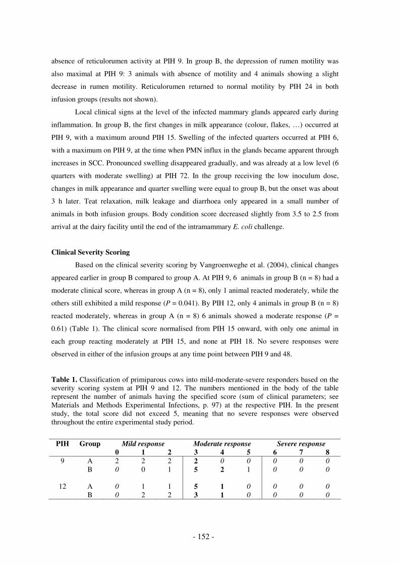
- 152 -
absence of reticulorumen activity at PIH 9. In group B, the depression of rumen motility was
also maximal at PIH 9: 3 animals with absence of motility and 4 animals showing a slight
decrease in rumen motility. Reticulorumen returned to normal motility by PIH 24 in both
infusion groups (results not shown).
Local clinical signs at the level of the infected mammary glands appeared early during
inflammation. In group B, the first changes in milk appearance (colour, flakes, …) occurred at
PIH 9, with a maximum around PIH 15. Swelling of the infected quarters occurred at PIH 6,
with a maximum on PIH 9, at the time when PMN influx in the glands became apparent through
increases in SCC. Pronounced swelling disappeared gradually, and was already at a low level (6
quarters with moderate swelling) at PIH 72. In the group receiving the low inoculum dose,
changes in milk appearance and quarter swelling were equal to group B, but the onset was about
3 h later. Teat relaxation, milk leakage and diarrhoea only appeared in a small number of
animals in both infusion groups. Body condition score decreased slightly from 3.5 to 2.5 from
arrival at the dairy facility until the end of the intramammary E. coli challenge.
Clinical Severity Scoring
Based on the clinical severity scoring by Vangroenweghe et al. (2004), clinical changes
appeared earlier in group B compared to group A. At PIH 9, 6 animals in group B (n = 8) had a
moderate clinical score, whereas in group A (n = 8), only 1 animal reacted moderately, while the
others still exhibited a mild response (P = 0.041). By PIH 12, only 4 animals in group B (n = 8)
reacted moderately, whereas in group A (n = 8) 6 animals showed a moderate response (P =
0.61) (Table 1). The clinical score normalised from PIH 15 onward, with only one animal in
each group reacting moderately at PIH 15, and none at PIH 18. No severe responses were
observed in either of the infusion groups at any time point between PIH 9 and 48.
Table 1. Classification of primiparous cows into mild-moderate-severe responders based on the severity scoring system at PIH 9 and 12. The numbers mentioned in the body of the table represent the number of animals having the specified score (sum of clinical parameters; see Materials and Methods Experimental Infections, p. 97) at the respective PIH. In the present study, the total score did not exceed 5, meaning that no severe responses were observed throughout the entire experimental study period.
PIH Group Mild response Moderate response Severe response
0 1 2 3 4 5 6 7 8
9 A 2 2 2 2 0 0 0 0 0
B 0 0 1 5 2 1 0 0 0
12 A 0 1 1 5 1 0 0 0 0
B 0 2 2 3 1 0 0 0 0

Variation of the inoculum dose
- 153 -
Quarter Milk Production
Milk production in the infected quarters decreased (P < 0.0001) on d0, the day of
intramammary inoculation. The maximal decrease of 60-70% appeared already on the day of
infection (Fig. 2a). Milk production decrease (68%) was slightly more pronounced in group B,
with a slightly more rapid recovery on d+1, but no significant differences between the two doses
were observed. On d+3, the recovery in the infected quarters was still incomplete and averaged
86 and 76% of the pre-infection milk production in group A and B, respectively.
The milk production decrease in the contralateral uninfected quarters is considered to be
an indicator of general systemic illness due to intramammary infection in the left quarters. A
moderate and short-termed decrease in the milk production of the contralateral quarters was
present in both infusion groups (Fig. 2b). Based on quarter milk production at d+2, no severe
responders were observed in the two groups. At d+3, relative to the day of infusion, milk
production of these quarters was at 97 and 99% of the pre-infection milk production in group A
and B, respectively.
Figure 2. Milk production of the infected left (a) and uninfected right quarters (b) from d-1 until d+3 from primiparous cows infused with 1 x 10⁴ (; group A; n = 8) and 1 x 10⁶ (-----; group B; n = 8) CFU E. coli P4:O32. Data are means (± SEM).
Colony-Forming Units, Somatic Cell Count and Blood Leukocyte Count
The number of E. coli increased to peak values (3.96 and 3.20 log10(CFU)/ml) at PIH 3
and 6 in the high and low dose infusion group, respectively. On average, the number of E. coli
did not significantly differ between both infusion groups. Furthermore, no significant interaction
between time and inoculum dose was found, indicating that the number of E. coli changed over
time in a similar fashion for the two inoculum doses (Fig. 1b).
Somatic cell count rapidly increased following intramammary E. coli infection and was
significantly (P < 0.0001) higher at PIH 6 in the high dose infusion group compared to the group
receiving the low dose. At PIH 9, both infusion groups reached a plateau level of 106.57 cells/ml
(Fig. 3f). Peak SCC levels were reached at PIH 15 and 18 in group B and A, respectively.
Somatic cell count kinetics significantly differed (P = 0.0005) between the inoculum groups,
Day relative to challenge
milk
pro
du
ctio
n (
ml)
10
00
30
00
-1 0 1 2 3
Milk Infected Quartersa
Day relative to challenge
milk
pro
du
ctio
n (
ml)
25
00
35
00
45
00
-1 0 1 2 3
Milk Uninfected Quartersb
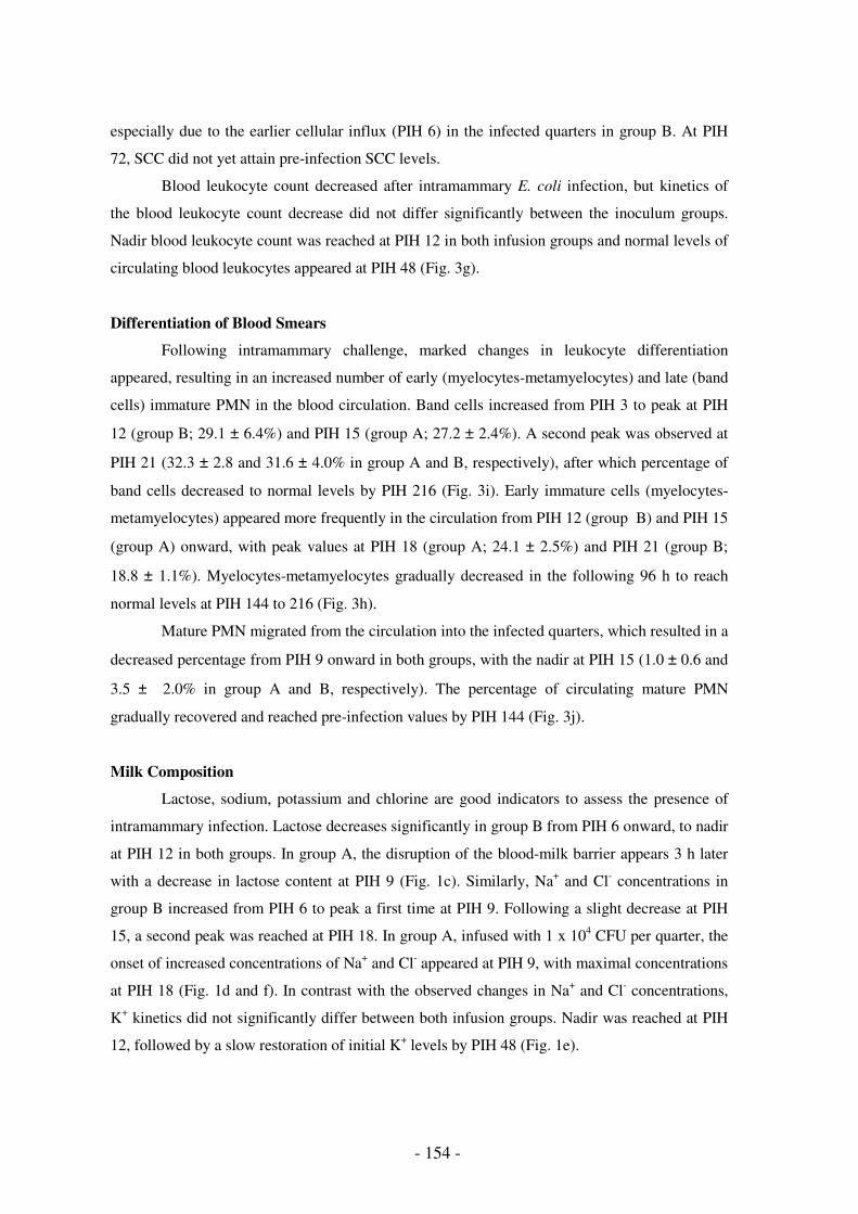
- 154 -
especially due to the earlier cellular influx (PIH 6) in the infected quarters in group B. At PIH
72, SCC did not yet attain pre-infection SCC levels.
Blood leukocyte count decreased after intramammary E. coli infection, but kinetics of
the blood leukocyte count decrease did not differ significantly between the inoculum groups.
Nadir blood leukocyte count was reached at PIH 12 in both infusion groups and normal levels of
circulating blood leukocytes appeared at PIH 48 (Fig. 3g).
Differentiation of Blood Smears
Following intramammary challenge, marked changes in leukocyte differentiation
appeared, resulting in an increased number of early (myelocytes-metamyelocytes) and late (band
cells) immature PMN in the blood circulation. Band cells increased from PIH 3 to peak at PIH
12 (group B; 29.1 ± 6.4%) and PIH 15 (group A; 27.2 ± 2.4%). A second peak was observed at
PIH 21 (32.3 ± 2.8 and 31.6 ± 4.0% in group A and B, respectively), after which percentage of
band cells decreased to normal levels by PIH 216 (Fig. 3i). Early immature cells (myelocytes-
metamyelocytes) appeared more frequently in the circulation from PIH 12 (group B) and PIH 15
(group A) onward, with peak values at PIH 18 (group A; 24.1 ± 2.5%) and PIH 21 (group B;
18.8 ± 1.1%). Myelocytes-metamyelocytes gradually decreased in the following 96 h to reach
normal levels at PIH 144 to 216 (Fig. 3h).
Mature PMN migrated from the circulation into the infected quarters, which resulted in a
decreased percentage from PIH 9 onward in both groups, with the nadir at PIH 15 (1.0 ± 0.6 and
3.5 ± 2.0% in group A and B, respectively). The percentage of circulating mature PMN
gradually recovered and reached pre-infection values by PIH 144 (Fig. 3j).
Milk Composition
Lactose, sodium, potassium and chlorine are good indicators to assess the presence of
intramammary infection. Lactose decreases significantly in group B from PIH 6 onward, to nadir
at PIH 12 in both groups. In group A, the disruption of the blood-milk barrier appears 3 h later
with a decrease in lactose content at PIH 9 (Fig. 1c). Similarly, Na+ and Cl- concentrations in
group B increased from PIH 6 to peak a first time at PIH 9. Following a slight decrease at PIH
15, a second peak was reached at PIH 18. In group A, infused with 1 x 10⁴ CFU per quarter, the
onset of increased concentrations of Na+ and Cl- appeared at PIH 9, with maximal concentrations
at PIH 18 (Fig. 1d and f). In contrast with the observed changes in Na+ and Cl- concentrations,
K+ kinetics did not significantly differ between both infusion groups. Nadir was reached at PIH
12, followed by a slow restoration of initial K+ levels by PIH 48 (Fig. 1e).

Variation of the inoculum dose
- 155 -
Milk IL-8 and C5a
Before challenge, IL-8 was very low (2.19 ± 0.93 and 1.89 ± 0.84 pg/ml in group A and
B, respectively) in the quarters that were to be infused with E. coli. Interleukin-8 kinetics of the
two infusion groups differed significantly from each other. In group B, a significant increase in
IL-8 appeared as early as PIH 6 and reached a peak of 305 ± 36 pg/ml at PIH 12 (Fig. 3a). At
PIH 6 and 9, the concentration of IL-8 in the inflamed quarters was significantly (P < 0.05)
higher in group B, receiving the 1 x 10⁶ CFU inoculum dose. Following infusion of 1 x 10⁴ CFU
(group A), significant increases in IL-8 occurred at PIH 9 and reached peak levels of 240 ± 40
pg/ml at PIH 12. Elevated levels of IL-8 persisted until PIH 24 in both groups, after which the
levels returned to baseline.
The complement component C5a had similar kinetics in both groups. The initial increase
in C5a following intramammary challenge appeared at PIH 6 (± 1.5 ng/ml), and rapidly reached
maximal values of 37.1 ± 16.0 ng/ml and 30.6 ± 12.4 ng/ml at PIH 12 for group A and B,
respectively. Elevated levels of C5a persisted until PIH 24 in both groups, after which the levels
returned to baseline (Fig. 3b).
Milk sCD14
To determine whether E. coli could alter mammary gland levels of sCD14, a sandwich
ELISA was used to quantitate milk sCD14 (Fig. 3c). Before challenge, sCD14 in mammary
quarters (7.7 ± 2.7 µg/ml and 10.4 ± 3.1 µg/ml for group A and B, respectively) was in the range
previously described (Lee et al., 2003a) for early lactating uninfected glands (5.46 to 6.90
µg/ml). At PIH 6, a significant increase in milk sCD14 was observed in group B, whereas in
group A, the increase in milk sCD14 only appeared at PIH 9. Concentrations of sCD14 were
significantly (P < 0.0001) different between the two groups at PIH 12. Milk sCD14 peaked at
PIH 15 in both groups, although the level of sCD14 was significantly (P = 0.0323) higher (205 ±
44 µg/ml) in group B compared to group A (151 ± 54 µg/ml) (Fig. 3c).

- 156 -
PIH
log
(BL
C)/
ml
Blood Leukocyte Count
46
81
0
0 3 6 9 12 18 24 48
g
PIH
Mye
locyte
s-m
eta
mye
locyte
s (
%) Myelocytes-metamyelocytes
05
10
20
0 3 6 9 12 18 24 48
h
PIH
Ba
nd
(%
)
Band cells
01
02
03
0
0 3 6 9 12 18 24 48
i
PIH
Ma
ture
(%
)
Mature PMN
01
02
03
04
0
0 3 6 9 12 18 24 48
j
Figure 3. Interleukin-8 (a), C5a (b), sCD14 (c), plasma LBP (d), milk LBP (e), SCC (f), blood leukocyte count (g), % myelocytes-metamyelocytes (h), % band cells (i) and % mature PMN (j) from PIH 0 until PIH 48 in the infected quarters or blood, respectively, from primiparous cows infused with 1 x 10⁴ (; group A; n = 8) and 1 x 10⁶ (-----; group B; n = 8) CFU E. coli P4:O32. Data are means (± SEM).
PIH
IL8
(p
g/m
l)
01
00
30
0
0 3 6 9 12 18 24 48
IL8a
PIH
c5
a (
pg
/ml)
02
00
00
40
00
0
0 3 6 9 12 18 24 48
C5ab
PIH
sC
D1
4 (
µg
/ml)
05
01
50
0 3 6 9 12 18 24 48
sCD14c
PIH
LB
P (
µg
/ml)
30
50
70
0 3 6 9 12 18 24 48
LBP Bloodd
PIH
LB
Pm
(µ
g/m
l)
02
04
06
08
0
0 3 6 9 12 18 24 48
LBP milke
PIH
log
(SC
C)/
ml
45
67
8
0 3 6 9 12 18 24 48
log(SCC)f

Variation of the inoculum dose
- 157 -
Plasma and Milk LBP
It has been shown previously (Bannerman et al., 2003) that elevated levels of sCD14 are
associated with similar increases in milk LBP, thereby providing an environment for optimal
host recognition of LPS, originating from infused E. coli bacteria. Plasma LBP was also assayed
as it is known that hepatic synthesis of this protein increases during the acute phase response,
mainly due to hepatic cell stimulation by IL-1β and IL-6 (Tobias et al., 1999). Under basal
conditions, LBP was detected in bovine blood at concentrations of 29.8 ± 7.5 µg/ml and 41.9 ±
5.4 µg/ml for group A and B, respectively; whereas the concentration of LBP in milk was lower
with a 8.5 ± 5.1 µg/ml and 14.5 ± 11.5 µg/ml for group A and B, respectively. At PIH 9, plasma
LBP in group B increased and this increase persisted throughout the entire study period (Fig.
3d). Plasma LBP levels reached maximal levels of 70.1 ± 1.5 and 68.8 ± 1.1 µg/ml at PIH 18,
after which they slightly declined to plateau until the end of the study. In quarters inoculated
with E. coli, significant elevation of milk LBP was observed as early as PIH 9, resulting in a first
peak at PIH 15 (44.4 ± 10.6 and 40.4 ± 8.7 µg/ml in group A and B, respectively). Milk LPB
was significantly (P = 0.0431) higher in group B at PIH 12. A second peak (61.6 ± 12.3 and 61.2
± 12.0 µg/ml in group A and B, respectively) was reached at PIH 24, after which milk LBP
levels remained high until the end of the study (Fig. 3e). Peak levels of milk LBP were observed
6 h later than the maximal elevation of plasma LBP levels.
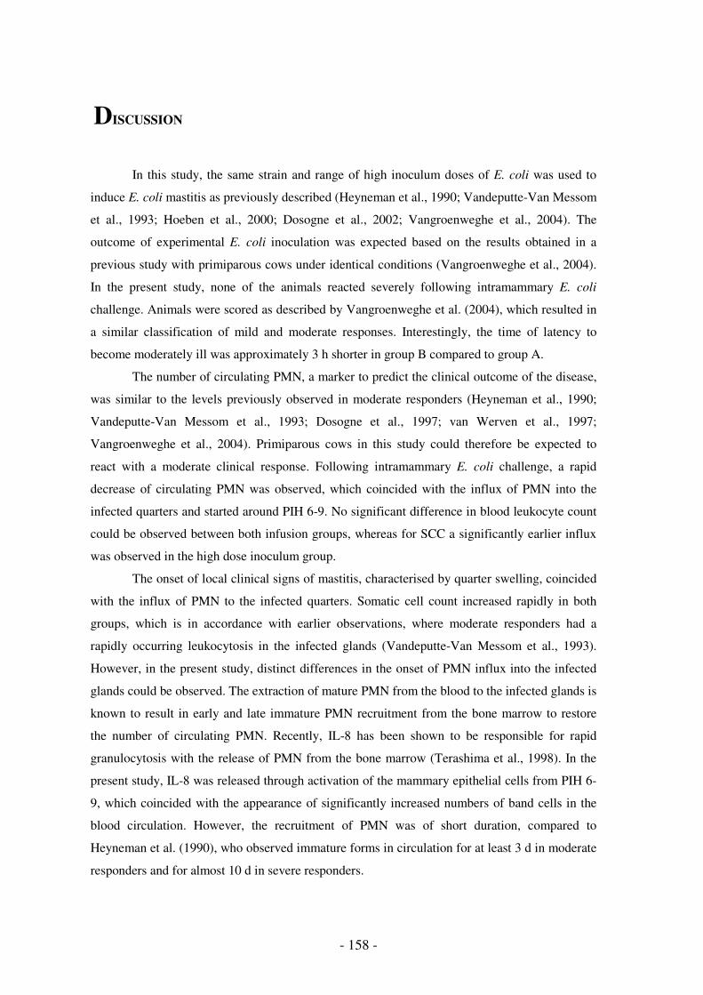
- 158 -
DISCUSSION
In this study, the same strain and range of high inoculum doses of E. coli was used to
induce E. coli mastitis as previously described (Heyneman et al., 1990; Vandeputte-Van Messom
et al., 1993; Hoeben et al., 2000; Dosogne et al., 2002; Vangroenweghe et al., 2004). The
outcome of experimental E. coli inoculation was expected based on the results obtained in a
previous study with primiparous cows under identical conditions (Vangroenweghe et al., 2004).
In the present study, none of the animals reacted severely following intramammary E. coli
challenge. Animals were scored as described by Vangroenweghe et al. (2004), which resulted in
a similar classification of mild and moderate responses. Interestingly, the time of latency to
become moderately ill was approximately 3 h shorter in group B compared to group A.
The number of circulating PMN, a marker to predict the clinical outcome of the disease,
was similar to the levels previously observed in moderate responders (Heyneman et al., 1990;
Vandeputte-Van Messom et al., 1993; Dosogne et al., 1997; van Werven et al., 1997;
Vangroenweghe et al., 2004). Primiparous cows in this study could therefore be expected to
react with a moderate clinical response. Following intramammary E. coli challenge, a rapid
decrease of circulating PMN was observed, which coincided with the influx of PMN into the
infected quarters and started around PIH 6-9. No significant difference in blood leukocyte count
could be observed between both infusion groups, whereas for SCC a significantly earlier influx
was observed in the high dose inoculum group.
The onset of local clinical signs of mastitis, characterised by quarter swelling, coincided
with the influx of PMN to the infected quarters. Somatic cell count increased rapidly in both
groups, which is in accordance with earlier observations, where moderate responders had a
rapidly occurring leukocytosis in the infected glands (Vandeputte-Van Messom et al., 1993).
However, in the present study, distinct differences in the onset of PMN influx into the infected
glands could be observed. The extraction of mature PMN from the blood to the infected glands is
known to result in early and late immature PMN recruitment from the bone marrow to restore
the number of circulating PMN. Recently, IL-8 has been shown to be responsible for rapid
granulocytosis with the release of PMN from the bone marrow (Terashima et al., 1998). In the
present study, IL-8 was released through activation of the mammary epithelial cells from PIH 6-
9, which coincided with the appearance of significantly increased numbers of band cells in the
blood circulation. However, the recruitment of PMN was of short duration, compared to
Heyneman et al. (1990), who observed immature forms in circulation for at least 3 d in moderate
responders and for almost 10 d in severe responders.

Variation of the inoculum dose
- 159 -
The rapid influx of PMN into the infected glands was associated with rapid elimination
of bacteria from the quarters. In the present study, high inoculum doses were used for
experimental induction of E. coli mastitis, because we were mainly interested in bacterial
elimination rather than bacterial growth in the mammary gland. In contrast to an earlier study
with the same inoculum dose in multiparous animals (Vandeputte-Van Messom et al., 1993),
peak bacterial numbers were already reached around PIH 3 to 6, which was in accordance with a
previous study in primiparous cows (Vangroenweghe et al., 2004). This peak number of bacteria
was followed by a rapid elimination from the infected glands. Contrary to the induction of E.
coli mastitis with low inoculum doses, where elimination is preceded by excessive bacterial
growth (Shuster et al., 1996; 1997; Riollet et al., 2000; Scaletti et al., 2003), in this study, peak
numbers were reached within 6 h post-infection and followed by a subsequent bacterial
elimination. Therefore, PMN influx rapidly cleared the bacteria from the affected glands. It can
be presumed that the bactericidal capacity of the PMN that migrated to the infected quarters was
high, because more efficient PMN functionality has recently been reported in primiparous cows
(Mehrzad et al., 2002).
Several indicators (lactose, sodium, chlorine and potassium) for the presence of mastitis
were determined in this study. The changes appeared significantly earlier (approximately 3 h) in
group B, receiving the 1 x 10⁶ CFU inoculum dose, although peak levels were almost identical
for both groups, which coincides with all other data, indicating that the animals reacted with a
mild to moderate response and little variation in clinical response was present in this study.
Complement component C5a and its derivative C5adesArg play a potential role in the
inflammatory response accompanying mastitis (Rainard et al., 1998). The level of C5a/C5adesArg
on the day of challenge was within the range previously described by Rainard et al. (1998),
taking into account that the average SCC immediately before intramammary infusion was a little
higher (approximately 90,000 cells/ml vs. 25,000 cells/ml). In the present study, increased levels
of C5a/C5adesArg could be observed from PIH 3 onwards, which is in accordance with an earlier
study (Rainard et al., 1998), although in this study the induction of inflammatory symptoms was
performed with E. coli endotoxin and not with live bacteria. The assessment of C5adesArg
concentrations in milk following infusion of E. coli endotoxin or live bacteria showed that
biologically significant amounts of C5a/C5adesArg are present in mastitis milk. The bulk of
complement-derived components in mastitis milk are likely to have their origin in blood plasma,
exudating through the damaged blood-milk barrier following inflammation of the mammary
gland (Rainard et al., 1998). However, milk concentrations of C5a/C5adesArg are probably not a
reliable indicator of exudation, as C5a/C5adesArg is rapidly taken up by several cell types present
in inflamed milk, in particular PMN (Rainard et al., 1998). Major elimination mechanism of this

- 160 -
inflammatory mediator is believed to rely on the binding of C5a/C5adesArg to cell surface
receptors. As milk from inflamed quarters contained high concentrations of cells, mainly PMN,
it can be put forward that the concentrations of C5a/C5adesArg measured in milk were
underestimates of the total amounts of C5a/C5adesArg which had originally been generated.
Complement component C5a generated in milk could well contribute to the activation of the
recruited phagocytic cells, with the consequence of improving their bactericidal activity
(Rainard, 2003).
Interleukin-8 is considered to play an important role in PMN recruitment to the inflamed
quarters (Baggniolini and Clark-Lewis, 1992; Barber and Yang, 1998). In contrast to Shuster et
al. (1997), who suggested the importance of C5a to be greater than IL-8 during the early
inflammatory response mainly due to earlier peak maxima, in the present study, peak levels of
IL-8 appeared 6 h earlier than C5a. The increase of IL-8 chemotactic activity appears coincident
with the increment in SCC at the level of the infected quarters. The increase in C5a also
appeared as early as PIH 3-6, but its peak maximum was only reached at PIH 18. The induction
of IL-8 production and release is known to be independent of the presence of IL-1β and TNF-α
(Persson-Waller et al., 2003). Induction of IL-8 is rather associated with the formation of the
tripartite LPS-LBP-sCD14 complex, that subsequently activates the mammary gland epithelial
cells through a cell surface assembly of a multi-protein recognition complex consisting of CD14,
TLR-4 and MD-2 (Akashi et al., 2000), resulting in the activation of the NF-κB controlled IL-8
gene (Aderem and Ulevitch, 2000).
Detectable increases in milk LBP were observed after initial increases in blood LBP, and
maximal levels of LBP in milk were observed at PIH 24, some 6 h after the peak levels of
plasma LBP. In accordance to Bannerman et al. (2003), the increases in milk LBP paralleled
increments in sCD14 levels. From a host perspective, the simultaneous increase in both LBP and
sCD14 levels would be expected to be advantageous as both molecules act in concert to facilitate
activation of host defence mechanisms by presenting LPS, released during bacterial growth and
death, to the transmembrane LPS receptor, TLR-4 (Bannerman and Goldblum, 2003).
Interestingly, the increase in IL-8 already started at PIH 6 in group B, receiving the 1 x 10⁶ CFU
inoculum dose, whereas significant increases of sCD14 and LBP only appeared from PIH 9,
suggesting that initial host cell activation can take place in the presence of basal levels of sCD14
and LBP. Although the increase in sCD14 was earlier and significantly higher in group B, PMN
influx as determined by SCC in the infected quarters was similarly elevated before pronounced
increment of SCD14. It was proposed that heightened levels of these molecules are not required
for immediate host innate immune responses (Bannerman et al., 2003). However, peak levels of
SCC were not observed until PIH 15-18, at a time when levels of both sCD14 and LBP in milk

Variation of the inoculum dose
- 161 -
were elevated. Whether increments in sCD14 and LBP are necessary for maximal recruitment of
PMN to the inflamed mammary glands remains unknown.
From this study, it appears that the inflammatory response in primiparous cows from
group B has an earlier onset compared to group A. One possible explanation for this observation
could be the 100-fold difference in the number of E. coli infused into the mammary glands,
because the amount of LPS produced is related to the number of E. coli bacteria (Burvenich,
1983; Monfardini et al., 1999). Because the bacterial cultures were washed three times in
pyrogen-free PBS before further dilutions were made, a direct effect of LPS present in the
inoculum can be excluded. Lipopolysaccharide, known as a potent inducer of inflammatory
cytokines (Shuster et al., 1993), can be produced quite rapidly during bacterial growth following
intramammary infusion. In the present study, the increase in sCD14 and LBP, which are known
to bind to LPS, facilitated the release of IL-8 from mammary endothelial cells in the infected
quarters. Moreover, the infused bacteria rapidly activated the complement system through the
alternative pathway, resulting in an early abundant production of C5a. The combined effects of
these events resulted in a rapid attraction of PMN from the blood into the mammary gland, with
a subsequent pronounced increase of SCC in the infected glands.
The novelty of the present study is the fact that primiparous cows, recently shown to
react as mild to moderate responders (Vangroenweghe et al., 2004), are responding faster
following an 100-fold increase in the inoculum dose. This quicker response is related to an
earlier activation of innate host immunity.
Following E. coli mastitis, treatment with antibiotics at PIH 10 did not alter local and
systemic symptoms despite a 100-fold decrease in CFU (Monfardini et al., 1999). From the
present study, it is evident that at the set time point, antibiotic treatment is unable to alter the
production or release of various inflammatory mediators. Therefore, it can be suggested that
early inflammatory events (first 3 h) could play an major role in the further regulation of the
inflammatory response to combat the invading pathogens.
CONCLUSIONS
Despite the use of relatively high inoculum doses, primiparous cows react with a
moderate inflammatory response following intramammary E. coli infusion. This moderate
response was evident from the pre-infection number of circulating leukocytes, the prompt
clinical response, the rapid influx of PMN into the infected quarters, the efficient bacterial
clearance of the affected glands and the fast recovery of milk production in both infected and

- 162 -
uninfected glands. In the present study, the difference in time of latency between both inoculum
doses could be confirmed and documented with kinetics of various inflammatory mediators such
as sCD14, LBP, IL-8 and C5a. The early increase in IL-8 following activation of the mammary
gland epithelium appeared before increases in sCD14 or LBP, indicating that innate host cell
activation can occur in the presence of basal levels of sCD14 and LBP. Although C5a increased
during early innate host immune response, maximal levels were reached after IL-8 had peaked.
In conclusion, primiparous cows were able to efficiently clear an intramammary E. coli infection
and the increase in inoculum dose induced a more rapid clinical response, mainly due to the
earlier activation of the innate host immune response. To further elucidate the regulation of early
events, mammary gland biopsies and milk sample collection during the early phase of
inflammation should be performed.

Variation of the inoculum dose
- 163 -
REFERENCES
Aderem, A., and R.J. Ulevitch. 2000. Toll-like receptors in the induction of the innate immune response.
Nature 460:782-787.
Akashi, S., H. Ogata, F. Kirikae, T. Kirikae, K. Kawasaki, M. Nishijima, R. Shimazu, Y. Nagai, K.
Fukudome, M. Kimoto, and K. Miyake. 2000. Regulatory roles for CD14 and phosphatidylinositol in the
signalling via Toll-like receptor 4 – MD-2. Biochem. Biophys. Res. Comm. 268:172-177.
Baggniolini, M., and I. Clark-Lewis. 1992. Interleukin-8, a chemotactic and inflammatory cytokine. FEBS
Lett. 307:97-101.
Bannerman, D.D., and S.E. Goldblum. 2003. Mechanisms of bacterial lipopolysaccharide-induced
endothelial apoptosis. Am. J. Physiol. Lung Cell. Mol. Physiol. 284:L899-L914.
Bannerman, D.D., M.J. Paape, W.R. Hare, and E.J. Sohn. 2003. Increased levels of LPS-binding protein in
bovine blood and milk following bacterial lipopolysaccharide challenge. J. Dairy Sci. 86:3128-3137.
Barber, M.R., and T.J. Yang. 1998. Chemotactic activities in nonmastitic and mastitic mammary
secretions: presence of interleukin-8 in mastitis but not in non-mastitic secretions. Clin. Diagn. Lab.
Immunol. 5:82-86.
Burvenich, C. 1983. Mammary blood flow in conscious lactating goats in various physiological and
pathological (mastitis) conditions. Ph.D. Thesis, Ghent University, Belgium.
Burvenich, C., V. Van Merris, J. Mehrzad, A. Diez-Fraile, and L. Duchateau. 2003. Severity of E. coli
mastitis is mainly determined by cow factors. Vet. Res. 34:521-564.
Chow, J.C., D.W. Young, D.T. Golenbock, W.J. Christ, and F. Gusovsky. 1999. Toll-like receptor-4
mediates lipopolysaccharide-induced signal transduction. J. Biol. Chem. 274:10689-10692.
Detilleux, J.C., M.E. Kehrli, Jr., J.R. Stabel, A.E. Freeman, and D.H. Kelley. 1995. Study of
immunological dysfunction in periparturient Holstein cattle selected for high and average milk
production. Vet. Immunol. Immunopathol. 44:251-267.
Dosogne, H., C. Burvenich, T. van Werven, E. Roets, E.N. Noordhuizen-Stassen, and B. Goddeeris. 1997.
Increased surface expression of CD11b receptors on polymorphonuclear leukocytes is not sufficient to
sustain phagocytosis during Escherichia coli mastitis in early postpartum dairy cows. Vet. Immunol.
Immunopathol. 60:47-59.
Dosogne, H., C. Burvenich, A.E. Freeman, M. E. Kehrli, Jr., J.C. Detilleux, J. Sulon, J.-F. Beckers, and D.
Hoeben. 1999. Association between pregnancy-associated glycoprotein and decreased
polymorphonuclear leukocyte function in early postpartum dairy cows. Vet. Immunol. Immunopathol.
67:47-54.
Dosogne, H., E. Meyer, A. Sturk, J. van Loon, A.-M. Massart-Leën, and C. Burvenich. 2002. Effect of
enrofloxacin treatment on plasma endotoxin during bovine Escherichia coli mastitis. Inflamm. Res.
51:201-205.

- 164 -
Faure, E., J.W. Equils, P.A. Sieling, L. Thomas, F.X. Zhang, C.J. Krischning, N. Polentarutti, M. Muzio,
and M. Arditi. 2000. Bacterial lipopolysaccharide activates NF-kappaB through Toll-like receptor 4
(TLR-4) in cultured human dermal endothelial cells. Differential expression of TLR-4 and TLR-2 in
endothelial cells. J. Biol. Chem. 275:11058-11063.
Gilbert, R.O., Y.T. Gröhn, P.M. Miller, and D.J. Hoffman. 1993. Effect of parity on periparturient
neutrophil function in dairy cows. Vet. Immunol. Immunopathol. 36:75-82.
Guha, M., and N. Mackman. 2001. LPS induction of gene expression in human monocytes. Cell. Signal.
13:85-94.
Heyneman, R., C. Burvenich, and R. Vercauteren. 1990. Interaction between the respiratory burst activity
of neutrophil leukocytes and experimentally induced Escherichia coli mastitis in cows. J. Dairy Sci.
73:985-994.
Hill, A.W. 1981. Factors influencing the outcome of Escherichia coli mastitis in the dairy cow. Res. Vet.
Sci. 73:985-994.
Hoeben, D., C. Burvenich, E. Trevisi, G. Bertoni, J. Hamann, R. M. Bruckmaier, and J. W. Blum. 2000.
Role of endotoxin and TNF-α in the pathogenesis of experimentally induced coliform mastitis in
periparturient cows. J. Dairy Res. 67:503-514.
Kehrli, M. E., Jr. and J. P. Goff. 1989. Periparturient hypocalcemia in cows: effects on peripheral blood
neutrophil and lymphocyte function. J. Dairy Sci. 72:1188-1196.
Kehrli, M. E., Jr., B. J. Nonnecke, and J. A. Roth. 1989. Alterations in bovine neutrophil function during
the periparturient period. Am. J. Vet. Res. 50:207-214.
Kimura, K., J.P. Goff, and M.E. Kehrli, Jr. 1999. Effects of the presence of the mammary gland on
expression of neutrophil adhesion molecules and myeloperoxidase activity in periparturient dairy cows.
J. Dairy Sci. 82:2385-2392.
Kimura, K., J.P. Goff, M.E. Kehrli, Jr., and T.A. Reinhardt. 2002. Decreased neutrophil function as a
cause of retained placenta in dairy cattle. J. Dairy Sci. 85:544-550.
Lee, J.-W., M.J. Paape, T.H. Elsasser, and X. Zhao. 2003a. Elevated milk soluble CD14 in bovine
mammary glands challenged with Escherichia coli lipopolysaccharide. J. Dairy Sci. 86:2382-2389.
Lee, J.-W., M.J. Paape, T.H. Elsasser, and X. Zhao. 2003b. Recombinant soluble CD14 reduces severity
of intramammary infection by Escherichia coli. Infect. Immun. 71:4034-4039.
Lee, J.-W., M.J. Paape, and X. Zhao. 2003c. Recombinant bovine soluble CD14 reduces severity of
experimental Escherichia coli mastitis in mice. Vet. Res. 34:307-316.
Mehrzad, J., L. Duchateau, S. Pyörälä, and C. Burvenich. 2002. Blood and milk neutrophil
chemiluminescence and viability in primiparous and pluriparous dairy cows during late pregnancy
around parturition and early lactation. J. Dairy Sci. 85:3268-3276.
Monfardini, E., C. Burvenich, A.-M. Massart-Leën, E. Smits, and M.J. Paape. 1999. Effect of antibiotic
induced bacterial clearance in the udder on L-selectin shedding of blood neutrophils in cows with
Escherichia coli mastitis. Vet. Immunol. Immunopathol. 67:373-384.
Persson-Waller, K., I.G. Colditz, S. Lun, and K. Östensson. 2003. Cytokines in mammary lymph and milk
during endotoxin-induced bovine mastitis. Res. Vet. Sci. 74:31-36.

Variation of the inoculum dose
- 165 -
Petsch, D., and F.B. Anspach. 2000. Endotoxin removal from protein solutions. J. Biotechnol. 76:97-119.
Rainard, P., P. Sarradin, M.J. Paape, and B. Poutrel. 1998. Quantification of C5a/C5adesArg in bovine
plasma, serum and milk. Vet. Res. 29:73-88.
Rainard, P. 2003. The complement in milk and defence of the bovine mammary gland against infection.
Vet. Res. 34:647-670.
Riollet, C., P. Rainard, and B. Poutrel. 2000. Differential induction of complement fragment C5a and
inflammatory cytokines during intramammary infections with Escherichia coli and Staphylococcus
aureus. Clin. Diagn. Lab. Immunol. 7:161-167.
Scaletti, R.W., D.S. Trammell, B.A. Smith, and R.J. Harmon. 2003. Role of dietary copper in enhancing
resistance to Escherichia coli mastitis. J. Dairy Sci. 86:1240-1249.
Shuster, D.E., M.E. Kehrli, Jr., and M.G. Stevens. 1993. Cytokine production during endotoxin-induced
mastitis in lactating dairy cows. Am. J. Vet. Res. 54:80-85.
Shuster, D.E., E.K. Lee, and M.E. Kehrli, Jr. 1996. Bacterial growth, inflammatory cytokine production,
and neutrophil recruitment during coliform mastitis in cows within ten days after calving, compared with
cows at midlactation. Am. J. Vet. Res. 57:1569-1575.
Shuster, D.E., M.E. Kehrli, Jr., P. Rainard, and M.J. Paape. 1997. Complement fragment C5a and
inflammatory cytokines in neutrophil recruitment during intramammary infection with Escherichia coli.
Infect. Immun. 65:3286-3292.
Sordillo, L.M., and L.A. Babiuk. 1991a. Controlling acute Escherichia coli mastitis during the
periparturient period with recombinant bovine interferon gamma. Vet. Microbiol. 28:189-198.
Sordillo, L.M., and L.A. Babiuk. 1991b. Modulation of bovine mammary neutrophil function during the
periparturient period following in vitro exposure to recombinant bovine interferon gamma. Vet.
Immunol. Immunopathol. 27:393-402.
Sordillo, L.M. and J.E. Peel. 1992. Effect of interferon-g on the production of tumor necrosis factor during
acute Escherichia coli mastitis. J. Dairy Sci. 75:2119-2125.
Tapping, R.L., and P.S. Tobias. 1997. Cellular binding of soluble CD14 requires lipopolysaccharide (LPS)
and LPS-binding protein. J. Biol. Chem. 272:23157-23164.
Terashima, T., D. English, J.C. Hogg, and S.F. van Eeden. 1998. Release of polymorphonuclear
leukocytes from the bone marrow by interleukin-8. Blood 92:1062-1069.
Thomas, C.J., M. Kapoor, S. Sharma, H. Bausinger, U. Zyilan, D. Lipsker, D. Hanau, and A. Surolia.
2002. Evidence of a trimolecular complex involving LPS, LPS binding protein and soluble CD14 as an
effector of LPS response. FEBS Lett. 531:184-188.
Tobias, P.S., R.L. Tapping, and J.A. Gegner. 1999. Endotoxin interactions with lipopolysaccharide-
responsive cells. Clin. Infect. Dis. 28:476-481.
Vandeputte-Van Messom, G., C. Burvenich, E. Roets, A.-M. Massart-Leën, R. Heyneman, W.D.J.
Kremer, and A. Brand. 1993. Classification of newly calved cows into moderate and severe responders
to experimentally induced Escherichia coli mastitis. J. Dairy Res. 60:19-29.
Vangroenweghe, F., L. Duchateau, and C. Burvenich. 2004. Moderate inflammatory response during
experimental E. coli mastitis in primiparous cows. J. Dairy Sci. 87:886-895.
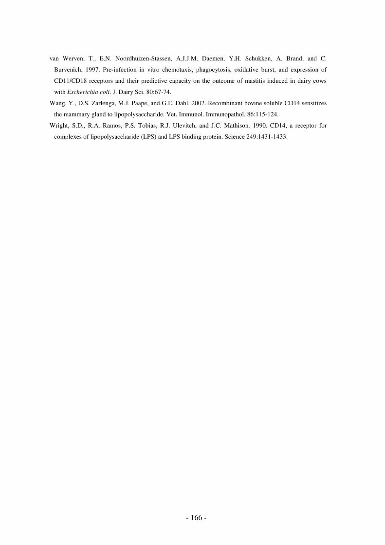
- 166 -
van Werven, T., E.N. Noordhuizen-Stassen, A.J.J.M. Daemen, Y.H. Schukken, A. Brand, and C.
Burvenich. 1997. Pre-infection in vitro chemotaxis, phagocytosis, oxidative burst, and expression of
CD11/CD18 receptors and their predictive capacity on the outcome of mastitis induced in dairy cows
with Escherichia coli. J. Dairy Sci. 80:67-74.
Wang, Y., D.S. Zarlenga, M.J. Paape, and G.E. Dahl. 2002. Recombinant bovine soluble CD14 sensitizes
the mammary gland to lipopolysaccharide. Vet. Immunol. Immunopathol. 86:115-124.
Wright, S.D., R.A. Ramos, P.S. Tobias, R.J. Ulevitch, and J.C. Mathison. 1990. CD14, a receptor for
complexes of lipopolysaccharide (LPS) and LPS binding protein. Science 249:1431-1433.

- 167 -
2.
INHIBITION OF PROSTAGLANDIN SYNTHESIS


Inhibition of prostaglandin synthesis
- 169 -
INTRODUCTION
Production costs attributed to mastitis are the largest economic loss to the dairy industry.
Mastitis caused by coliform bacteria is responsible for a major portion of these losses,
predominantly as a result of acute disease. Coliform mastitis in cattle may be associated with
systemic clinical disease, which occurs predominantly in the period immediately after calving
(Hill, 1981; Wilesmith et al., 1986; Burvenich et al., 2003). Systemic signs include general
depression, fever, tachycardia, inhibition of reticulorumen motility, and many non-specific
responses, such as neutropenia, followed by leukocytosis (Verheijden et al., 1983). Considerable
losses in milk production may occur in cows suffering from E. coli mastitis (Hill, 1981; Lohuis
et al., 1989; Dosogne et al., 1997; Hoeben et al., 2000). Inhibition of reticulorumen motility is a
frequent complication of E. coli mastitis (Verheijden et al., 1983) and as a result the microbial
degradation of ingested feed is disturbed and the milk yield is reduced. Moreover, marked
decreases in plasma tyrosine levels occur (Vandeputte-Van Messom et al., 1987), which are not
directly related to peak fever.
The pathophysiology of E. coli mastitis is characterised by the presence of endotoxin or
LPS in the outer membrane of the etiological bacteria. Lipopolysaccharide is a pro-inflammatory
molecule that is shed from the bacterial surface during bacterial replication or death (Burvenich
et al., 2003). Clinical signs following experimentally induced E. coli mastitis are attributed to
mediator shock rather than to endotoxin shock, because endotoxin mainly plays a local role
(Hoeben et al., 2000; Dosogne et al., 2002). Several of the pro-inflammatory cytokines that
mediate the localised and systemic response to Gram-negative mastitis, including IL-1β, IL-6,
IL-8, and TNF-α, are upregulated by LPS (Shuster et al., 1993; Guha and Mackman, 2001).
Binding of LPS to cell membranes also activates the membrane-bound enzyme, phospholipase
A2, liberating arachidonic acid which can be metabolised by two major enzyme systems, COX
and lipoxygenase. The prostaglandins, prostacyclin and thromboxanes are the products of the
cyclo-oxygenase pathway, whereas the leukotrienes are synthesised following lipoxygenase
enzyme activity. The eicosanoids are important mediators and modulators of inflammation, and
play a role in the mediation of acute inflammation (Anderson, 1989; Rose et al., 1989).
Intracerebroventricular injection of 50 to 200 µg PGE2 in lactating goats elicited fever in normal
lactating goats, the increment and decrement stage being accompanied respectively by a decrease
and an increase of mammary blood flow (Burvenich et al., 1982). Pre-treatment with
flurbiprofen, administered through intracerebroventricular cannulation, inhibited fever and
changes in HR following experimentally induced LPS mastitis, but had little effect on the Cl-
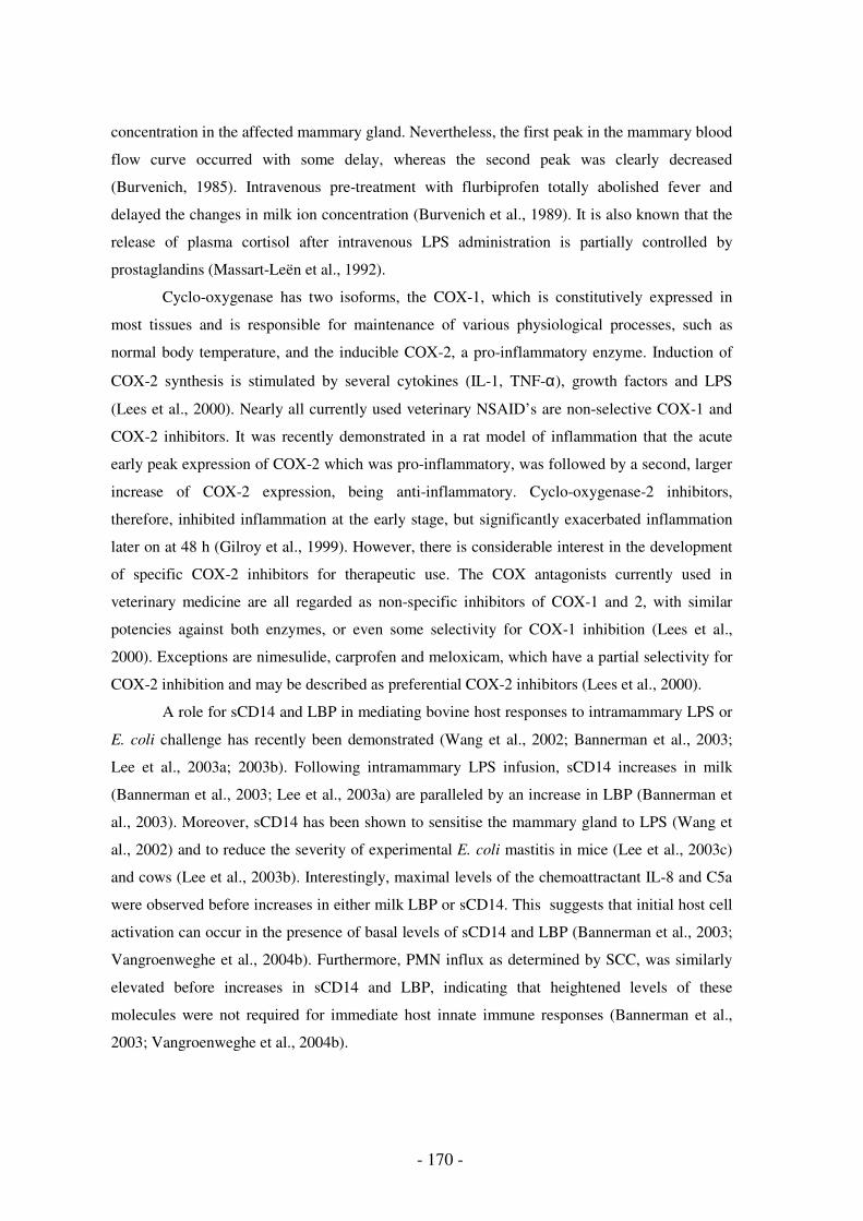
- 170 -
concentration in the affected mammary gland. Nevertheless, the first peak in the mammary blood
flow curve occurred with some delay, whereas the second peak was clearly decreased
(Burvenich, 1985). Intravenous pre-treatment with flurbiprofen totally abolished fever and
delayed the changes in milk ion concentration (Burvenich et al., 1989). It is also known that the
release of plasma cortisol after intravenous LPS administration is partially controlled by
prostaglandins (Massart-Leën et al., 1992).
Cyclo-oxygenase has two isoforms, the COX-1, which is constitutively expressed in
most tissues and is responsible for maintenance of various physiological processes, such as
normal body temperature, and the inducible COX-2, a pro-inflammatory enzyme. Induction of
COX-2 synthesis is stimulated by several cytokines (IL-1, TNF-α), growth factors and LPS
(Lees et al., 2000). Nearly all currently used veterinary NSAID’s are non-selective COX-1 and
COX-2 inhibitors. It was recently demonstrated in a rat model of inflammation that the acute
early peak expression of COX-2 which was pro-inflammatory, was followed by a second, larger
increase of COX-2 expression, being anti-inflammatory. Cyclo-oxygenase-2 inhibitors,
therefore, inhibited inflammation at the early stage, but significantly exacerbated inflammation
later on at 48 h (Gilroy et al., 1999). However, there is considerable interest in the development
of specific COX-2 inhibitors for therapeutic use. The COX antagonists currently used in
veterinary medicine are all regarded as non-specific inhibitors of COX-1 and 2, with similar
potencies against both enzymes, or even some selectivity for COX-1 inhibition (Lees et al.,
2000). Exceptions are nimesulide, carprofen and meloxicam, which have a partial selectivity for
COX-2 inhibition and may be described as preferential COX-2 inhibitors (Lees et al., 2000).
A role for sCD14 and LBP in mediating bovine host responses to intramammary LPS or
E. coli challenge has recently been demonstrated (Wang et al., 2002; Bannerman et al., 2003;
Lee et al., 2003a; 2003b). Following intramammary LPS infusion, sCD14 increases in milk
(Bannerman et al., 2003; Lee et al., 2003a) are paralleled by an increase in LBP (Bannerman et
al., 2003). Moreover, sCD14 has been shown to sensitise the mammary gland to LPS (Wang et
al., 2002) and to reduce the severity of experimental E. coli mastitis in mice (Lee et al., 2003c)
and cows (Lee et al., 2003b). Interestingly, maximal levels of the chemoattractant IL-8 and C5a
were observed before increases in either milk LBP or sCD14. This suggests that initial host cell
activation can occur in the presence of basal levels of sCD14 and LBP (Bannerman et al., 2003;
Vangroenweghe et al., 2004b). Furthermore, PMN influx as determined by SCC, was similarly
elevated before increases in sCD14 and LBP, indicating that heightened levels of these
molecules were not required for immediate host innate immune responses (Bannerman et al.,
2003; Vangroenweghe et al., 2004b).
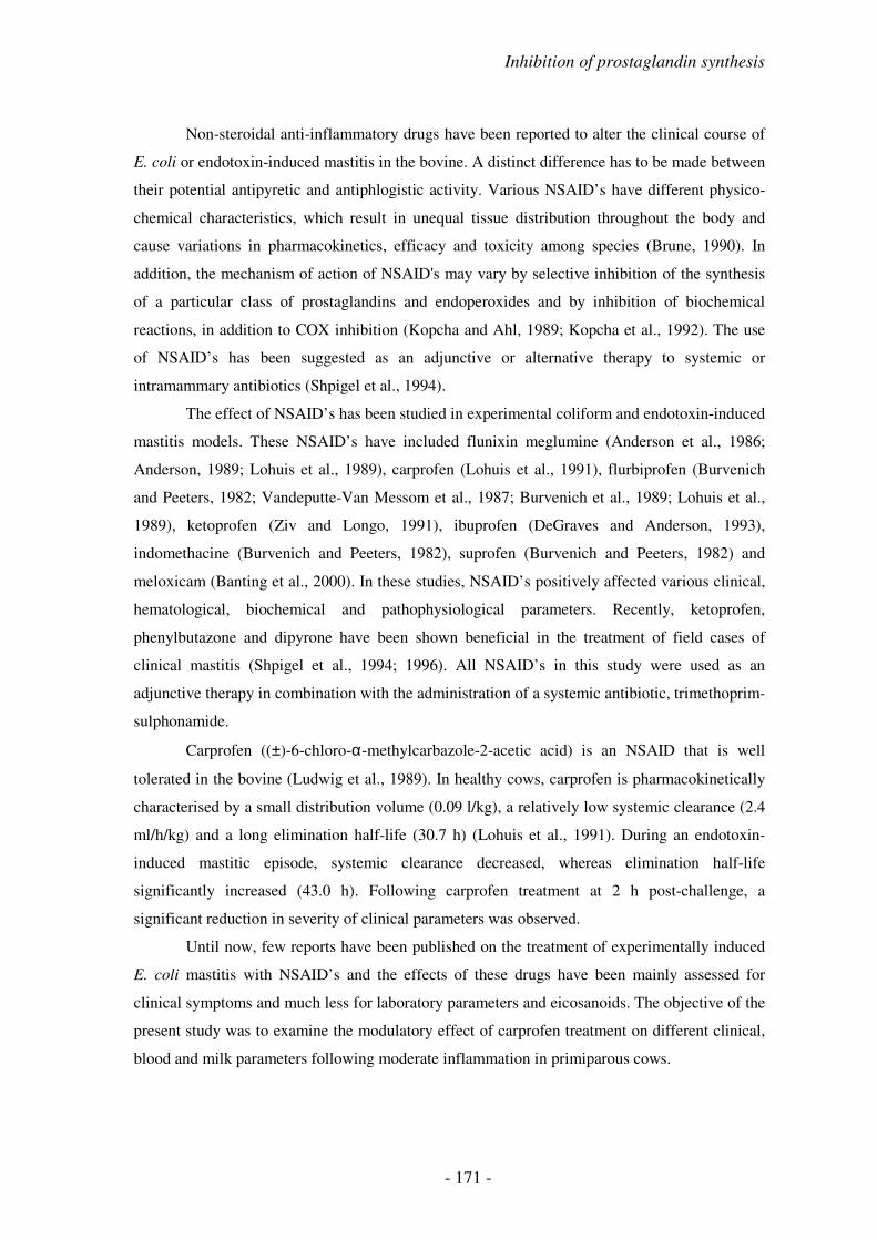
Inhibition of prostaglandin synthesis
- 171 -
Non-steroidal anti-inflammatory drugs have been reported to alter the clinical course of
E. coli or endotoxin-induced mastitis in the bovine. A distinct difference has to be made between
their potential antipyretic and antiphlogistic activity. Various NSAID’s have different physico-
chemical characteristics, which result in unequal tissue distribution throughout the body and
cause variations in pharmacokinetics, efficacy and toxicity among species (Brune, 1990). In
addition, the mechanism of action of NSAID's may vary by selective inhibition of the synthesis
of a particular class of prostaglandins and endoperoxides and by inhibition of biochemical
reactions, in addition to COX inhibition (Kopcha and Ahl, 1989; Kopcha et al., 1992). The use
of NSAID’s has been suggested as an adjunctive or alternative therapy to systemic or
intramammary antibiotics (Shpigel et al., 1994).
The effect of NSAID’s has been studied in experimental coliform and endotoxin-induced
mastitis models. These NSAID’s have included flunixin meglumine (Anderson et al., 1986;
Anderson, 1989; Lohuis et al., 1989), carprofen (Lohuis et al., 1991), flurbiprofen (Burvenich
and Peeters, 1982; Vandeputte-Van Messom et al., 1987; Burvenich et al., 1989; Lohuis et al.,
1989), ketoprofen (Ziv and Longo, 1991), ibuprofen (DeGraves and Anderson, 1993),
indomethacine (Burvenich and Peeters, 1982), suprofen (Burvenich and Peeters, 1982) and
meloxicam (Banting et al., 2000). In these studies, NSAID’s positively affected various clinical,
hematological, biochemical and pathophysiological parameters. Recently, ketoprofen,
phenylbutazone and dipyrone have been shown beneficial in the treatment of field cases of
clinical mastitis (Shpigel et al., 1994; 1996). All NSAID’s in this study were used as an
adjunctive therapy in combination with the administration of a systemic antibiotic, trimethoprim-
sulphonamide.
Carprofen ((±)-6-chloro-α-methylcarbazole-2-acetic acid) is an NSAID that is well
tolerated in the bovine (Ludwig et al., 1989). In healthy cows, carprofen is pharmacokinetically
characterised by a small distribution volume (0.09 l/kg), a relatively low systemic clearance (2.4
ml/h/kg) and a long elimination half-life (30.7 h) (Lohuis et al., 1991). During an endotoxin-
induced mastitic episode, systemic clearance decreased, whereas elimination half-life
significantly increased (43.0 h). Following carprofen treatment at 2 h post-challenge, a
significant reduction in severity of clinical parameters was observed.
Until now, few reports have been published on the treatment of experimentally induced
E. coli mastitis with NSAID’s and the effects of these drugs have been mainly assessed for
clinical symptoms and much less for laboratory parameters and eicosanoids. The objective of the
present study was to examine the modulatory effect of carprofen treatment on different clinical,
blood and milk parameters following moderate inflammation in primiparous cows.

- 172 -
MATERIALS AND METHODS
Experimental Animals and Study Facilities
All primiparous cows (n = 25) calved within 7 days before arrival at the commercial
dairy farm.
Inoculation Dose
Primiparous cows were inoculated with 1 x 10⁴ CFU E. coli P4:O32 in both left quarters.
Sampling Procedure
Blood and milk samples were collected at d-4, d-1, d0, d+1, d+2, d+3 and d+6 relative to
the day of challenge. On the day of challenge, blood and milk samples were collected at PIH 3,
6, 9, 12, 15, 18 and 21.
Determination of Inflammatory Cytokines and Eicosanoids
Two specific eicosanoids, PGE2 and TXB4, were quantified besides IL-8, C5a, sCD14
and LBP in order to assess for the effect of NSAID treatment following intramammary E. coli
challenge.
Statistical Analysis
In order to compare the two treatment groups with respect to the various parameters
analysed in blood and milk, a mixed model was used with cow as random effect and treatment,
time and their interaction as categorical fixed effects. Furthermore, the two treatment groups
were compared at PIH 12 for all analysed parameters and some determined parameters were
tested at post-treatment intervals PIH 12-18 (RT, HR, blood leukocyte number and PCV), PIH
12-48 (IL-8, C5a, sCD14, LBP, PGE2 and TXB2) and PIH 12-72 (SCC, CFU, lactose, Na+, K+,
Cl- and serum albumin), respectively, for significant differences between both treatment groups.
Bonferroni’s multiple comparisons procedure with an overall type I error equal to 5% was used
to adjust for multiple comparison. Quarter milk production in both infected and uninfected
quarters were tested for significant differences between treatments at d+1 and the interval d+1 -
d+3. The effect of treatment on the local aspects of the mammary gland was tested by the
Wilcoxon rank sum test.
For Further Details see chapter 'Materials and Methods Experimental Infections', p. 97.

Inhibition of prostaglandin synthesis
- 173 -
RESULTS
Local and Systemic Inflammatory Response
Following intramammary E. coli inoculation, RT rapidly increased from PIH 9 onward,
to reach its maximum at PIH 12 in the saline treated group. Carprofen administration
immediately reduced RT significantly (P < 0.0001) at 3 and 6 h post-treatment, and RT
normalised at PIH 15 (6 h post-treatment). No relapses of RT increase were observed in the
carprofen treated group, whereas in the saline treated group, RT increased above 39°C again at
PIH 24 (Fig. 1a).
Heart rate followed almost identical kinetics as described for RT. In the carprofen
treated group, HR was significantly (P < 0.05) lower at PIH 12, 15 and 18. From 9 h post-
treatment onward, HR in the carprofen treated animals remained lower, although not significant,
until the end of the observation period (Fig. 1b).
Carprofen treatment following intramammary E. coli inoculation had a beneficial effect
on the duration of reticulorumen motility depression. Although reticulorumen motility was
equally depressed in both treatment groups at PIH 9 (time of treatment), the motility increased
following carprofen treatment (P < 0.01), whereas in the saline treated group, depression of
reticulorumen motility reached its maximum at PIH 12 (Fig 1c).
Local clinical symptoms at the level of the mammary gland were also assessed during
clinical examination. Local swelling appeared at PIH 6 and reached its maximum at PIH 12 in
both treatment groups. In the carprofen treated animals, quarter swelling decreased from PIH 18
and normal quarter consistency was present at PIH 144. Quarter swelling disappeared more
slowly in control animals, although normal quarter consistency at palpation was also reached by
PIH 144. Maximal quarter swelling score was significantly lower (P < 0.05) in the carprofen
treated animals. Milk appearance, quarter milk leakage scores and maximum milk leakage scores
were significantly (P < 0.05) higher in the carprofen treated group at PIH 12. Quarter pain and
temperature did not differ between both treatment groups (results not shown).
Clinical Severity Scoring
Based on the clinical severity scoring by Vangroenweghe et al. (2004a), clinical scores
increased to a maximum at PIH 9 and 12 in the carprofen and saline treated animals,
respectively. The clinical severity score in the carprofen treated group decreased more rapidly
from 3 h post-treatment onwards. At PIH 12 (P < 0.0001) and 15 (P < 0.01), clinical severity
score was significantly lower in the carprofen treated group compared to the saline treated group.

- 174 -
At PIH 9, the time of treatment, approximately equal numbers of animals in both
treatment groups displayed a mild and moderate response. Following carprofen administration,
all animals (n = 12) responded mildly (score 0-2), whereas in the saline treated group, 6 animals
reacted with a moderate response (score 3-5) and only 7 animals had a mild response. By 6 h
post-treatment, 10 animals in the carprofen treated group had a score 0, whereas in the other
group, only 3 animals had score 0 and the others respectively scored 1 (n = 4), 2 (n = 5) or 3 (n =
1). During the following observations, the clinical severity score in both groups seemed to
normalise with only a slight flare-up of 3 animals (score 2) in the saline treated group at PIH 24.
Figure 1. Rectal temperature (a), heart rate (b), reticulorumen motility (c), packed cell volume (d), blood leukocyte count (e), plasma LPB (f), plasma PGE2 (g) and plasma TXB2 (h) from PIH 0 until PIH 144 from primiparous cows infused with 1 x 10⁴ CFU E. coli P4:O32 and treated at PIH 9 (dashed vertical line) with carprofen (; n = 12) or saline (-----; n = 13). Data are means (± SEM).
Quarter Milk Production
Milk production in the infected quarters decreased equally on d0, the day of
intramammary E. coli challenge, in both treatment groups. No significant effect of carprofen
treatment on the recovery of milk production in the infected quarters could be observed (Fig. 2a).
In the uninfected control quarters, no significant differences in milk production could be
observed throughout the entire study period (Fig. 2b). As expected, none of the animals in both
PIH
RT
(°C
)
38.5
41.0
0 3 6 9
12
15
18
21
24
48
72
144
a
PIHH
R (
beats
/min
)
70
90
110
0 3 6 9
12
15
18
21
24
48
72
144
b
PIH
Rum
en (
contr
./2m
in)
2.0
3.0
0 3 6 9
12
15
18
21
24
48
72
144
c
PIH
PC
V (
%)
26
30
0 3 6 9
12
15
18
21
24
48
72
144
d
PIH
Log(B
LC
)/m
l
6.0
6.6
0 3 6 9
12
15
18
21
24
48
72
144
e
PIH
LB
P (
µg/m
l)
50
150
0 3 6 9
12
15
18
21
24
48
72
144
f
PIH
PG
E2 (
pg/m
l)
0300
0 3 6 9
12
15
18
21
24
48
72
144
g
PIH
TX
B2 (
pg/m
l)
0200
0 3 6 9
12
15
18
21
24
48
72
144
h
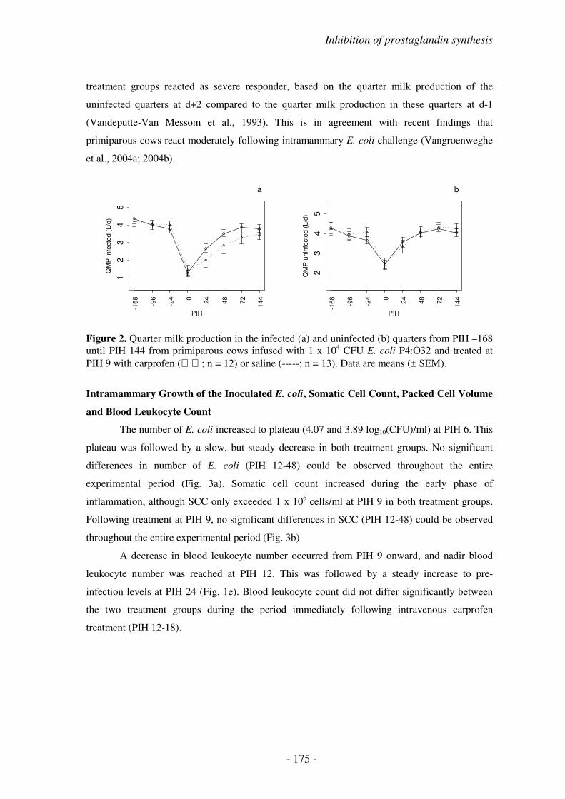
Inhibition of prostaglandin synthesis
- 175 -
treatment groups reacted as severe responder, based on the quarter milk production of the
uninfected quarters at d+2 compared to the quarter milk production in these quarters at d-1
(Vandeputte-Van Messom et al., 1993). This is in agreement with recent findings that
primiparous cows react moderately following intramammary E. coli challenge (Vangroenweghe
et al., 2004a; 2004b).
Figure 2. Quarter milk production in the infected (a) and uninfected (b) quarters from PIH –168 until PIH 144 from primiparous cows infused with 1 x 10⁴ CFU E. coli P4:O32 and treated at PIH 9 with carprofen (; n = 12) or saline (-----; n = 13). Data are means (± SEM).
Intramammary Growth of the Inoculated E. coli, Somatic Cell Count, Packed Cell Volume
and Blood Leukocyte Count
The number of E. coli increased to plateau (4.07 and 3.89 log10(CFU)/ml) at PIH 6. This
plateau was followed by a slow, but steady decrease in both treatment groups. No significant
differences in number of E. coli (PIH 12-48) could be observed throughout the entire
experimental period (Fig. 3a). Somatic cell count increased during the early phase of
inflammation, although SCC only exceeded 1 x 10⁶ cells/ml at PIH 9 in both treatment groups.
Following treatment at PIH 9, no significant differences in SCC (PIH 12-48) could be observed
throughout the entire experimental period (Fig. 3b)
A decrease in blood leukocyte number occurred from PIH 9 onward, and nadir blood
leukocyte number was reached at PIH 12. This was followed by a steady increase to pre-
infection levels at PIH 24 (Fig. 1e). Blood leukocyte count did not differ significantly between
the two treatment groups during the period immediately following intravenous carprofen
treatment (PIH 12-18).
PIH
QM
P infe
cte
d (
L/d
)
12
34
5
-168
-96
-24 0
24
48
72
144
a
PIH
QM
P u
nin
fecte
d (
L/d
)
23
45
-168
-96
-24 0
24
48
72
144
b

- 176 -
PIH
Log(C
FU
)/m
l
02
4
0 3 6 9
12
15
18
21
24
48
72
144
a
PIH
Log(S
CC
)/m
l
45
67
0 3 6 9
12
15
18
21
24
48
72
144
b
PIH
Lacto
se (
mg/m
l)
30
40
50
0 3 6 9
12
15
18
21
24
48
72
144
c
PIHSeru
m a
lbum
in (
mg/d
l)
0200
0 3 6 9
12
15
18
21
24
48
72
144
d
PIH
Na+
(m
mol/m
l)
0100
0 3 6 9
12
15
18
21
24
48
72
144
e
PIH
K+
(m
mol/m
l)
20
40
0 3 6 9
12
15
18
21
24
48
72
144
f
PIH
Cl- (
mm
ol/m
l)
20
60
0 3 6 9
12
15
18
21
24
48
72
144
g
Figure 3. Colony-forming units of E. coli (a), SCC (b), lactose (c), serum albumin (d), sodium (e), potassium (f) and chlorine (g) in the infected quarters from PIH 0 until PIH 144 from primiparous cows infused with 1 x 10⁴ CFU E. coli P4:O32 and treated at PIH 9 (dashed vertical line) with carprofen (; n = 12) or saline (-----; n = 13). Data are means (± SEM).
Milk Composition
Lactose, serum albumin, sodium, potassium and chlorine are indicators of changes in
milk composition following mastitis (Burvenich, 1983). Following E. coli challenge, the lactose
concentration started to decrease at PIH 9, reaching the nadir at PIH 12 (3 h post-treatment) in
both treatment groups. No significant differences (PIH 12-48) in lactose concentration were
observed following carprofen treatment (Fig. 3c). Maximal concentrations of serum albumin in
milk were reached at PIH 15. In the interval PIH 12-48, serum albumin concentration showed a
significant interaction between time and treatment (P = 0.013), meaning that serum albumin
kinetics were different between both groups. At PIH 21, carprofen treated animals had
significant (P = 0.0099) lower concentrations of serum albumin in the milk of the affected
quarters (Fig. 3d).
The Na+ concentration was significantly lower in the carprofen treated animals at PIH 21
(P = 0.005) and PIH 24 (P = 0.007), whereas the K+ concentration was significantly higher in the
carprofen treated animals at PIH 21 (P = 0.002) and PIH 24 (P = 0.005) (Fig. 3e and f). The Cl-

Inhibition of prostaglandin synthesis
- 177 -
concentration just failed to be significantly different at PIH 21 (P = 0.013 compared to
Bonferroni adjusted comparisonwise significance level of 0.05/6 = 0.0083) (Fig. 3g).
Milk IL-8, C5a and sCD14
Before challenge, IL-8 was very low (1.66 ± 0.23 and 2.11 ± 0.35 pg/ml in the saline and
carprofen treated groups, respectively) in the quarters that were to be infused with E. coli. Until
PIH 9, the time of treatment, IL-8 kinetics did not differ between both treatment groups. Peak
concentrations (471 ± 50 pg/ml and 389 ± 61 pg/ml for the saline and carprofen treated group,
respectively) were reached in the period PIH 12-15. No significant differences between both
treatments could be observed throughout the study period (Fig. 4a).
The complement component C5a had similar kinetics in both treatment groups until PIH
18, 9 h post-treatment. From PIH 21, C5a concentration decreased in the carprofen treated
animals, whereas animals in the saline treated groups reached peak C5a levels at PIH 24,
followed by a decrease at PIH 48, but no significant differences between the two groups were
observed. At PIH 72, C5a levels in both groups reached similar values (Fig. 4b).
To determine whether carprofen treatment following intramammary E. coli challenge
could alter mammary gland levels of sCD14, an ELISA was used to quantitate milk sCD14.
Before challenge, sCD14 in mammary quarters (7.33 ± 1.72 µg/ml and 3.77 ± 0.70 µg/ml in
saline and carprofen treated animals, respectively) was in the range previously described (Lee et
al., 2003a) for early lactating uninfected glands (5.46 to 6.90 µg/ml). In both treatment groups, a
first peak was observed at PIH 12 (11 ± 2 µg/ml and 13 ± 2 µg/ml, respectively) followed by a
second higher peak at PIH 24 (27 ± 4 µg/ml and 31 ± 6 µg/ml, respectively). Following peak
concentrations, sCD14 decreased from PIH 48 onwards to reach normal pre-infection values by
PIH 144. No significant differences in sCD14 concentrations were observed throughout the
entire experimental period (Fig. 4c).

- 178 -
Figure 4. IL-8 (a), C5a (b), sCD14 (c), LBP (d), PGE2 (e) and TXB2 (f) in the infected quarters from PIH 0 until PIH 144 from primiparous cows infused with 1 x 10⁴ CFU E. coli P4:O32 and treated at PIH 9 (dashed vertical line) with carprofen (; n = 12) or saline (-----; n = 13). Data are means (± SEM).
Plasma and Milk LBP
It has been shown previously (Bannerman et al., 2003) that elevated levels of sCD14
were associated with similar increases in milk LBP, thereby providing an environment for
optimal host recognition of LPS, originating from inoculated E. coli bacteria. Besides the
quantification of milk LBP to assess possible effects of carprofen treatment on the levels of LBP
in the infected quarters, plasma LBP was assayed, as it is known that hepatic synthesis of this
protein increases during the acute phase response, mainly due to hepatic cell stimulation by IL-
1β and IL-6 (Tobias et al., 1999), which could be influenced by NSAID treatment through the
intermediate eicosanoid products, PGE2 and TXB2. Under basal conditions, LBP was detected in
bovine blood at concentrations of 71.3 ± 10.9 µg/ml and 98.3 ± 16.2 µg/ml for saline and
carprofen treated animals, respectively; whereas the concentration of LBP in milk was lower
with 18.1 ± 2.3 µg/ml and 23.7 ± 2.2 µg/ml for the treatment groups, respectively. Plasma LBP
increased from PIH 15 onward to reach maximal values at PIH 24 (162 ± 10 µg/ml and 167 ± 14
µg/ml for saline and carprofen treated animals, respectively). Thereafter, concentrations of
PIH
IL-8
(pg/m
l)
-100
200
500
0 3 6 9
12
15
18
21
24
48
72
144
a
PIH
C5a (
pg/m
l)
-2000
6000
0 3 6 9
12
15
18
21
24
48
72
144
b
PIH
sC
D14 (
µg/m
l)
020
40
0 3 6 9
12
15
18
21
24
48
72
144
c
PIH
LB
P (
µg/m
l)
20
40
60
0 3 6 9
12
15
18
21
24
48
72
144
d
PIH
PG
E2 (
pg/m
l)
02000
0 3 6 9
12
15
18
21
24
48
72
144
e
PIH
TX
B2 (
pg/m
l)
0500
1500
0 3 6 9
12
15
18
21
24
48
72
144
f

Inhibition of prostaglandin synthesis
- 179 -
plasma LBP declined to reach pre-infection levels at PIH 144 (Fig. 1f). No significant difference
in plasma LBP was present between the treatment groups throughout the experimental period. In
quarters inoculated with E. coli, significant elevation of milk LBP appeared from PIH 9 onward,
with maxima (58 ± 1 µg/ml and 61 ± 1 µg/ml in saline and carprofen treated animals,
respectively) at PIH 21. From PIH 48 on, milk LBP concentrations declined (Fig. 4d). A
significant difference (P = 0.0001) in milk LBP was observed between the carprofen and saline
treated animals at PIH 12.
Plasma and Milk Prostaglandin E2 and Thromboxane B2
Plasma PGE2 concentration was 429.2 ± 25.2 pg/ml at the time of inoculation in both
treatment groups. Prostaglandin E2 started to decrease in plasma as early as PIH 9 and reached
nadir at PIH 15 in both treatment groups. Plasma PGE2 concentrations did not differ between
carprofen and saline treated animals (Fig. 1g). Thromboxane B2 concentration in plasma was as
high as 180.4 ± 26.5 pg/ml at the time of infection. Throughout the inflammatory episode, no
changes over time and no differences between treatment groups were observed (Fig. 1h).
In milk, PGE2 concentration was lower than in plasma and was 334.8 ± 21.1 pg/ml at the
time of intramammary inoculation. Prostaglandin E2 subsequently increased from PIH 9 onward
in both treatment groups. No significant difference in milk PGE2 concentration was observed
between the carprofen and the saline treated animals (Fig. 4e). Initial milk TXB2 concentrations
(84.3 ± 5.1 pg/ml) were also markedly lower than concentrations observed in plasma.
Thromboxane B2 increases occurred at PIH 9, which was also the maximal concentration in the
carprofen treated animals. In saline treated animals, TXB2 further increased and reached its
maximum at PIH 15. The milk TXB2 concentrations continued to be higher in the saline treated
groups as compared to the carprofen treated animals until PIH 72.

- 180 -
DISCUSSION
The aim of the present study was to evaluate the potential modulatory effects of
treatment with carprofen, a PG synthetase inhibitor through COX-2 inhibition, on a moderate
inflammatory reaction following E. coli challenge. Therefore, the study design included
carprofen administration following the appearance of initial clinical symptoms, which was
mainly based on practical field data (Shpigel et al., 1994) and inflammatory dynamics of the
moderate inflammatory model used (Vangroenweghe et al., 2004a; 2004b). It is known that pre-
treatment far more effectively inhibits the inducible COX-2 enzyme (Burvenich, 1985;
Burvenich et al., 1989), however, from a practical point of view, the earliest occasion at which
field cases of clinical E. coli mastitis can be diagnosed and subsequently treated, occurs at the
milking following the infection of the mammary quarter (Shpigel et al., 1994; 1996). The first
clinical signs in the present experimental model, using a 1 x 10⁴ CFU inoculum dose, have been
shown to appear around PIH 9. Therefore, the choice for a carprofen administration at PIH 9 was
considered most suitable, as clinical signs would be present at that time, although maximal RT
was not yet reached (Vangroenweghe et al., 2004a; 2004b).
In the present study, the same strain and high inoculum dose of E. coli was used to
induce a moderate inflammatory reaction in primiparous cows as described before
(Vangroenweghe et al., 2004a; 2004b). The clinical course and changes in different laboratory
parameters following intramammary E. coli challenge in the control animals were similar to
animals receiving the same dose (1 x 10⁴ CFU) in previous experiments (Vangroenweghe et al.,
2004a; 2004b). Carprofen treated animals elicited an immediate and significant decrease in RT
at 3 h post-treatment, whereas pyrexia continued in the control animals with a peak fever at PIH
12. Single dose carprofen administration in the present study resulted in a more pronounced and
prolonged antipyretic effect compared to meloxicam treatment in an E. coli endotoxin model
(Banting et al., 2000), where peak fever was reached 2 h post-treatment in both groups.
Reticulorumen motility was equally depressed in both treatment groups at the time of carprofen
administration. However, 3 h post-treatment, reticulorumen motility in the carprofen treated
animals restored to normal activity, whereas a maximal depression was reached in the control
group at PIH 12. In contrast, in an LPS model, no reticulorumen motility depression occurs, and
therefore beneficial effects of NSAID treatment on the reticulorumen motility depression can not
be quantified.
Improvement of local clinical signs at the level of the affected mammary quarters by
carprofen treatment was limited to swelling. There are fewer beneficial effects than observed by

Inhibition of prostaglandin synthesis
- 181 -
Anderson et al. (1986), who reported significant improvement of quarter temperature, oedema,
pain and size following flunixin meglumine treatment of cows suffering from endotoxin-induced
mastitis. However, the administration of flunixin meglumine in that study was performed much
earlier (PIH 2) as compared to our study, where carprofen was only administered at appearance
of the first clinical symptoms.
Clinical scores, combining several clinical parameters, have been described (Wenz et al.,
2001; Friton et al., 2002; Vangroenweghe et al., 2004a). Using the clinical severity score
described by Vangroenweghe et al. (2004a), carprofen treated animals had a significant lower
clinical score at PIH 12 and 15 as compared to the saline treated group. Although, as expected
from previous trials, all animals in both groups responded mild to moderate following E. coli
challenge, the carprofen treated animals generally showed a lower clinical severity score upon
NSAID administration. This was mainly due to the rapid restoration of reticulorumen motility
and the immediate antipyretic activity following the NSAID administration.
Quarter inflammation was associated with a temporary loss of MP, combined with
secretion of abnormal milk from the infected glands. Maximal depression in MP in the infected
and uninfected quarters occurred on the day of challenge (d0), and was followed by a rapid
recovery during subsequent days. Carprofen treatment did not exert any beneficial effect on milk
yield in the infected quarters following intramammary E. coli challenge. The absence of any
effect of carprofen on the milk production in the uninfected right quarters can mainly be
attributed to the moderate inflammatory signs of the mastitis model (Vangroenweghe et al.,
2004a).
In the present study, no significant effect of carprofen treatment on bacterial elimination
occurred, which could be expected as carprofen treatment was administered alone and not as an
adjunctive therapy to intramammary or systemic antibiotics. Enrofloxacin is known to enhance
bacterial clearance during experimentally induced E. coli mastitis (Monfardini et al., 1999).
Rantala et al. (2002) reported a more rapid decline in bacterial number in the infected quarters
following combined flunixin meglumine and enrofloxacin treatment, which could probably be
attributed to the enrofloxacin treatment and not the adjunctive NSAID therapy.
The leukocyte influx into the infected glands following E. coli challenge was not
significantly affected by carprofen treatment. This observation is in accordance with previous
results obtained using flurbiprofen in experimentally induced LPS mastitis in goats (Burvenich,
1985; Burvenich et al., 1989). Comparable results were obtained following ibuprofen treatment
at 2 h post-endotoxin infusion (DeGraves and Anderson, 1993). Although the dose of carprofen
(1.4 mg/kg BW) administered intravenously in the present study was twice as high as the dose
(0.7 mg/kg BW) used by Lohuis et al. (1991), oedema and leukocyte infiltration in the affected

- 182 -
glands could not be prevented. Nevertheless, the applied treatment was potent enough to reduce
eicosanoid generation in the affected mammary glands in the carprofen treated animals. In the
blood, similar kinetics could be observed in blood leukocyte number between both treatment
group. Nadir blood leukocyte number was reached at PIH 12, 3 h post-treatment in both
treatment groups, and was followed by progressive recovery of blood leukocyte number to pre-
infection levels by PIH 24.
Dehydration is often assessed during acute coliform mastitis as an easy tool to judge
clinical severity. The assessment of dehydration can be performed by skin turgor estimation. In
the present study, no difference in skin turgor assessment could be observed throughout the
entire study period. This could have been expected based on the mild to moderate responses
usually obtained using the experimental infection model. Packed cell volume did not differ
among treatments throughout the study either.
Lactose, serum albumin, sodium, potassium and chlorine concentrations in milk of the
infected quarters are often determined to assess the severity and duration of the intramammary
inflammation. Serum albumin and all ions (Na+, K+ and Cl-) showed similar kinetics, although a
significant interaction between time and treatment was only present for serum albumin from 12 h
post-treatment (PIH 21) onward in the carprofen treated animals. At PIH 21 and 24, both Na+
and K+ concentration significantly improved in the carprofen treated animals. These results
indicate that carprofen induces a more rapid recovery of normal milk composition in animals
intramammarily challenged with E. coli. Previous studies using ibuprofen in an endotoxin model
were unable to demonstrate any effect on milk composition (DeGraves and Anderson, 1993).
The elimination half-life of carprofen in healthy cows has been calculated 30.7 h, which is
shorter than the half-life of phenylbutazone (31.2 – 82.1 h), but considerably longer than flunixin
meglumine (8.1 h) (Lohuis et al., 1991). Therefore, it is not surprising that a single dose of
carprofen had prolonged beneficial effects on clinical parameters compared to the control group.
Following intramammary E. coli challenge, significant increases in PGE2 (Peter et al.,
1990; Zia et al., 1987) and TXB2 (Anderson et al., 1985; 1986; Zia et al., 1987) have been
observed in milk. Moreover, a significant effect of flunixin meglumine administration on TXB2
concentrations was reported (Anderson et al., 1986). Even in the uninfected quarter a slight
increase in PGF2α and TXB2 concentration was reported (Anderson et al., 1985), although this
increase disappeared more rapidly than in the infected quarters. Following flunixin meglumine
treatment, TXB2 concentrations remained at baseline levels, whereas in the saline treated
animals, a pronounced peak value was observed at PIH 8 in the endotoxin challenged animals
(Anderson et al., 1986).

Inhibition of prostaglandin synthesis
- 183 -
Although the effect of NSAID’s on eicosanoids production following inflammation is
well documented in literature (Anderson et al., 1986), no reports are available on the possible
effect on other immunological inflammatory parameters, such as the chemotactic agents IL-8
and C5a or the early innate immune molecules sCD14 and LBP. Interleukin-8 kinetics in the
present study were similar with previous reports using the same inoculum dose (Vangroenweghe
et al., 2004b). Local IL-8 production started at PIH 6, although major increases only occurred
from PIH 9 onward. Because IL-8 is a well-known chemotactic agent, it is obvious from these
results that the initial leukocyte influx into the infected quarters, which occurred around PIH 9,
was not affected by the decline of IL-8 occurring much later during inflammation. The
appearance and evolution of C5a concentrations in the affected quarters did not differ between
both groups until PIH 21, 12 h post-treatment. In the control group, C5a increased until maximal
values at PIH 24, which is 6 h later than previously observed (Vangroenweghe et al., 2004b).
The marked difference in the onset of carprofen effects on chemotactic agents IL-8 and C5a
could be explained by the location of mediator production. Interleukin-8 is directly produced by
the epithelial cells in the mammary gland (Baggniolini and Clark-Lewis, 1992; Barber and Yang,
1998), whereas C5a precursors are obtained from the blood through leakage of the disintegrated
blood-milk barrier (Rainard et al., 1998; Rainard, 2003).
In accordance with previous reports (Bannerman et al., 2003; Vangroenweghe et al.,
2004b), the increases in milk LBP paralleled increments in sCD14 levels. From a host
perspective, the simultaneous increase in both LBP and sCD14 levels would be expected to be
advantageous as both molecules act in concert to facilitate activation of host defence
mechanisms by presenting LPS, released during bacterial growth and death, to the
transmembrane LPS receptor, TLR-4 (Bannerman and Goldblum, 2003). Carprofen treatment
did not affect either parameter.
The present study showed that carprofen treatment following experimental E. coli
challenge in a moderate inflammation model with primiparous cows has only slight modulatory
effects, which are mainly limited to RT and reticulorumen motility. Administration of carprofen
at PIH 9, when first clinical signs occurred, showed beneficial effects on general clinical
condition, recovery of milk composition, and reduced production of eicosanoids. Mediators of
early innate immune response, IL-8, C5a, sC14 and LBP, were not affected by NSAID
treatment.

- 184 -
CONCLUSIONS
Although carprofen treatment was administered late during the acute phase reaction,
when first clinical signs appeared, the NSAID had some modulatory effects on clinical,
production and immunological parameters in a moderate inflammation model with primiparous
cows. The main modulatory potential occurred at the level of improved clinical condition,
mainly due to the antipyretic effects and the ability of carprofen to improve reticulorumen
motility. Milk production could not efficiently be modulated through the administration of
carprofen at PIH 9. Milk composition (serum albumin, Na+ and K+) was significantly affected by
carprofen treatment at PIH 21 and 24, but no further effects on milk composition occurred.
Carprofen treatment did not result in a decrease of chemotactic inflammatory mediators, IL-8
and C5a, and no effect was observed for early innate immune molecules, such as sCD14 and
LBP. Carprofen treatment did not affect PGE2 and TXB2 in plasma or milk, although there were
trends for decreased concentrations of both eicosanoids in milk of the affected quarters. In
conclusion, the inflammatory model using primiparous cows during the periparturient period is a
minimal and moderate state of inflammation, necessary to eliminate the invading pathogens from
the affected mammary quarters. Major modulatory effects from NSAID administration were
therefore not observed in this model.

Inhibition of prostaglandin synthesis
- 185 -
REFERENCES
Anderson, K.L., H. Kindahl, A. Petroni, A.R. Smith, and B.K. Gustafsson. 1985. Arachidonic acid
metabolites in milk of cows during acute coliform mastitis. Am. J. Vet. Res. 46:1573-1577.
Anderson, K.L., H. Kindahl, A.R. Smith, L.E. Davis, and B.K. Gustafsson. 1986. Endotoxin-induced
bovine mastitis: arachidonic acid metabolites in milk and plasma and effects of flunixin meglumine. Am.
J. Vet. Res. 47:1373-1377.
Anderson, K.L. 1989. Therapy for acute coliform mastitis. Comp. Cont. Ed. Prac. Vet. 11:1125-1133.
Baggniolini, M., and I. Clark-Lewis. 1992. Interleukin-8, a chemotactic and inflammatory cytokine. FEBS
Lett. 307:97-101.
Bannerman, D.D., and S.E. Goldblum. 2003. Mechanisms of bacterial lipopolysaccharide-induced
endothelial apoptosis. Am. J. Physiol. Lung Cell. Mol. Physiol. 284:L899-L914.
Bannerman, D.D., M.J. Paape, W.R. Hare, and E.J. Sohn. 2003. Increased levels of LPS-binding protein in
bovine blood and milk following bacterial lipopolysaccharide challenge. J. Dairy Sci. 86:3128-3137.
Banting, A., H. Schmidt, and S. Banting. 2000. Efficacy of meloxicam in lactating cows with E. coli
endotoxin induced acute mastitis. 8th International Congress EAVPT, Jerusalem, Israel. 30 July – 3
August. Abstract N° E4.
Barber, M.R., and T. J. Yang. 1998. Chemotactic activities in nonmastitic and mastitic mammary
secretions: presence of interleukin-8 in mastitis but not in non-mastitic secretions. Clin. Diagn. Lab.
Immunol. 5:82-86.
Brune, K. 1990. Is there a rational basis for the different spectra of adverse effects of nonsteroidal anti-
inflammatory drugs (NSAID's)? Drugs 40 Supplement 5:12-15.
Burvenich, C., and G. Peeters. 1982. Effect of prostaglandin synthetase inhibitors on mammary blood flow
during experimentally induced mastitis in lactating goats. Arch. Int. Pharmacodyn. Ther. 258:128-137.
Burvenich, C., G. Vandeputte-Van Messom, and G. Peeters. 1982. Effect of experimentally induced fever
on mammary blood flow in lactating goats. Arch. Int. Pharmacodyn. Therap. 259:119-133.
Burvenich, C. 1983. Mammary blood flow in conscious lactating goats in various physiological and
pathological (mastitis) conditions. Ph.D. Thesis, Ghent University, Belgium.
Burvenich, C. 1985. Beinvloeding van het bifasische bloedvloeipatroon in de ontstoken melkklier door
I.V. and I.C.V. toediening van een antipyreticum. Kon. Acad. Geneesk. Belg. 5:341-383.
Burvenich, C., G. Vandeputte-Van Messom, E. Roets, A.-M. Massart-Leën, and R. Heyneman. 1989.
Effects of antagonists of inflammatory mediators on endotoxin induced mastitis in lactating goats. Arch.
Int. Physiol. Biochim. 97:47-51.
Burvenich, C., V. Van Merris, J. Mehrzad, A. Diez-Fraile, and L. Duchateau. 2003. Severity of E. coli
mastitis is mainly determined by cow factors. Vet. Res. 34:521-564.
DeGraves, F.J., and K.L. Anderson. 1993. Ibuprofen treatment of endotoxin-induced mastitis in cows.
Am. J. Vet. Res. 54:1128-1132.

- 186 -
Dosogne, H., C. Burvenich, T. van Werven, E. Roets, E.N. Noordhuizen-Stassen, and B. Goddeeris. 1997.
Increased surface expression of CD11b receptors on polymorphonuclear leukocytes is not sufficient to
sustain phagocytosis during Escherichia coli mastitis in early postpartum dairy cows. Vet. Immunol.
Immunopathol. 60:47-59.
Dosogne, H., E. Meyer, A. Sturk, J. van Loon, A.-M. Massart-Leën, and C. Burvenich. 2002. Effect of
enrofloxacin treatment on plasma endotoxin during bovine Escherichia coli mastitis. Inflamm. Res.
51:201-205.
Friton, G., H. Phillip, and R. Kleemann. 2002. Efficacy of Meloxicam (Metacam) in lactating cows with
acute mastitis. Proceedings of XXII World Buiatrics Congress. 18-23 August, Hannover, Germany.
Gilroy, D.W., P.R. Colville-Nash, D. Willis, J. Chevers, M.J. Paul-Clark, and D.A. Willoughby. 1999.
Inducible cyclo-oxygenase may have anti-inflammatory properties. Nature Medicine 5:698-701.
Guha, M., and N. Mackman. 2001. LPS induction of gene expression in human monocytes. Cell. Signal.
13:85-94.
Hill, A.W. 1981. Factors influencing the outcome of Escherichia coli mastitis in the dairy cow. Res. Vet.
Sci. 73:985-994.
Hoeben, D., C. Burvenich, E. Trevisi, G. Bertoni, J. Hamann, R. M. Bruckmaier, and J. W. Blum. 2000.
Role of endotoxin and TNF-α in the pathogenesis of experimentally induced coliform mastitis in
periparturient cows. J. Dairy Res. 67:503-514.
Kopcha, M.C., and A.S. Ahl. 1989. Experimental uses of flunixin meglumine and phenylbutazone in food-
producing animals. JAVMA 194:45-49.
Kopcha, M., J.B. Kaneene, M.E. Shea, R. Miller, and A.S. Ahl. 1992. Use of non-steroidal anti-
inflammatory drugs in food animal practice. JAVMA 12:1868-1872.
Lee, J.-W., M.J. Paape, T.H. Elsasser, and X. Zhao. 2003a. Elevated milk soluble CD14 in bovine
mammary glands challenged with Escherichia coli lipopolysaccharide. J. Dairy Sci. 86:2382-2389.
Lee, J.-W., M.J. Paape, T.H. Elsasser, and X. Zhao. 2003b. Recombinant soluble CD14 reduces severity
of intramammary infection by Escherichia coli. Infect. Immun. 71:4034-4039.
Lee, J.-W., M.J. Paape, and X. Zhao. 2003c. Recombinant bovine soluble CD14 reduces severity of
experimental Escherichia coli mastitis in mice. Vet. Res. 34:307-316.
Lees, P., M.F. Landoni, S. Armstrong, and S. Frean. 2000. New insights into inflammation with particular
reference to the role of COX enzymes. 8th International Congress EAVPT, Jerusalem, Israel. 30 July – 3
August. Abstract N° A5.
Lohuis, J.A.C.M., W. van Leeuwen, J.H.M. Verheijden, A. Brand, and A.S.J.P.A.M. van Miert. 1989.
Flunixin meglumine and flurbiprofen in cows with experimental Escherichia coli mastitis. Vet. Rec.
124:305-308.
Lohuis, J.A.C.M., T. van Werven, A. Brand, A.S.J.P.A.M. van Miert, B. Ludwig, E. Rohde, P.
Heinzmann, and W.F. Rehm. 1991. Pharmacodynamics and pharmacokinetics of carprofen, a non-
steroidal anti-inflammatory drug, in healthy cows and cows with Escherichia coli endotoxin-induced
mastitis. J. Vet. Pharmacol. Therap. 14:219-229.

Inhibition of prostaglandin synthesis
- 187 -
Ludwig, B., J.C. Jordan, W.F. Rehm, and R. Thun. 1989. Carprofen in veterinary medicine. I. Plasma
disposition, milk excretion and tolerance in milk-producing cows. Schweizer Archiv für Tierheilkunde
131:99-106.
Massart-Leën, A.M., C. Burvenich, G. Vandeputte-Van Messom, and H. Hilderson. 1992. Partial
prostaglandin-mediated mechanism controlling the release of cortisol in plasma after intravenous
administration of endotoxins. Dom. Anim. Endocrinol. 9:273-283.
Monfardini, E., C. Burvenich, A.-M. Massart-Leën, E. Smits, and M.J. Paape. 1999. Effect of antibiotic
induced bacterial clearance in the udder on L-selectin shedding of blood neutrophils in cows with
Escherichia coli mastitis. Vet. Immunol. Immunopathol. 67:373-384.
Peter, A.T., P.W. Clark, D.E. van Roekel, C.W. Luker, J.D. Gaines, and W.T. Bosu. 1990. Temporal
changes in metabolites of prostanoids in milk of heifers after intramammary infusion of Escherichia coli
organisms. Prostaglandins 39:451-457.
Rainard, P., P. Sarradin, M. J. Paape, and B. Poutrel. 1998. Quantification of C5a/C5adesArg in bovine
plasma, serum and milk. Vet. Res. 29:73-88.
Rainard, P. 2003. The complement in milk and defence of the bovine mammary gland against infection.
Vet. Res. 34:647-670.
Rantala, M., L. Kaartinen, E. Välimäki, M. Stryrman, M. Hiekkaranta, A. Niemi, L. Saari, and S. Pyörälä.
2002. Efficacy and pharmacokinetics of enrofloxacin and flunixin meglumine for treatment of cows with
experimentally induced Escherichia coli mastitis. J. Vet. Pharmacol. Therap. 25:251-258.
Rose, D.M., S.N. Giri, S.J. Wood, and J.S. Cullor. 1989. Role of leukotriene B4 in the pathogenesis of
Klebsiella pneumoniae-induced bovine mastitis. Am. J. Vet. Res. 50:915-918.
Shpigel, N.Y., R. Chen, M. Winkler, A. Saran, G. Ziv, and F. Longo. 1994. Anti-inflammatory ketoprofen
in the treatment of field cases of bovine mastitis. Res. Vet. Sci. 56:62-68.
Shpigel, N.Y., M. Winkler, A. Saran, and G. Ziv. 1996. The anti-inflammatory drugs fenylbutazone and
dipyrone in the treatment of field cases of bovine mastitis. J. Vet. Med. A 43:331-336.
Shuster, D.E., M.E. Kehrli, Jr., and M.G. Stevens. 1993. Cytokine production during endotoxin-induced
mastitis in lactating dairy cows. Am. J. Vet. Res. 54:80-85.
Tobias, P.S., R.I. Tapping, and J.A. Gegner. 1999. Endotoxin interactions with lipopolysaccharide-
responsive cells. Clin. Infect. Dis. 28:476-481.
Vandeputte-Van Messom, G., R. Reynaert, C. Burvenich, and A.M. Massart-Leën. 1987. Effects of
flurbiprofen on endotoxin-induced changes of plasma tyrosine in lactating goats. Arch. Int.
Pharmacodyn. Therap. 290:159-160.
Vandeputte-Van Messom, G., C. Burvenich, E. Roets, A.-M. Massart-Leën, R. Heyneman, W.D.J.
Kremer, and A. Brand. 1993. Classification of newly calved cows into moderate and severe responders
to experimentally induced Escherichia coli mastitis. J. Dairy Res. 60:19-29.
Vangroenweghe, F., L. Duchateau, and C. Burvenich. 2004a. Moderate inflammatory response during
experimental E. coli mastitis in primiparous cows. J. Dairy Sci. 87:886-895.

- 188 -
Vangroenweghe, F., P. Rainard, M.J. Paape, L. Duchateau, and C. Burvenich. 2004b. Increase of
Escherichia coli inoculum doses induce faster innate immune response in primiparous cows. J. Dairy
Sci. 87:4132-4144.
Verheijden, J.H.M., A.J.H. Schotman, A.S.J.P.A.M. van Miert, and C.T.M. van Duin. 1983.
Pathophysiological aspects of Escherichia coli mastitis in ruminants. Vet. Res. Commun. 7:229-236.
Wang, Y., D.S. Zarlenga, M.J. Paape, and G.E. Dahl. 2002. Recombinant bovine soluble CD14 sensitizes
the mammary gland to lipopolysaccharide. Vet. Immunol. Immunopathol. 86:115-124.
Wenz, J.R., G.M. Barrington, F.B. Garry, K.D. McSweeney, R.P. Dinsmore, G. Goodell, and R.J. Callan.
2001. Bacteremia associated with naturally occurring acute coliform mastitis in dairy cows. JAVMA
219:976-981.
Wilesmith, J.W., P.G. Francis, and C.D. Wilson. 1986. Incidence of clinical mastitis in a cohort of British
dairy herds. Vet. Rec. 22:199-204.
Zia, S., S.N. Giri, J. Cullor, P. Emau, B.I. Osburn, and R.B. Bushnell. 1987. Role of eicosanoids,
histamine, and serotonin in the pathogenesis of Klebsiella pneumoniae-induced bovine mastitis. Am. J.
Vet. Res. 48:1617-1625.
Ziv, G., and F. Longo. 1991. Comparative clinical efficacy of ketoprofen and flunixin in the treatment of
induced E. coli endotoxin mastitis in lactating dairy cows. Mammites des vaches laitières, Société
Française de Buiatrie, Paris, France. p. 207-208.

- 189 -
3.
VACCINATION AGAINST THE ENDOTOXIN


Vaccination against the endotoxin
- 191 -
INTRODUCTION
It is estimated that 20% of all cases of clinical mastitis in some dairy herds are caused by
coliform bacteria, particularly in herds where contagious pathogens are well controlled
(Schukken et al., 1989a; 1989b; Barkema et al., 1999). The most common of these organisms are
E. coli, Enterobacter aerogenes, Klebsiella pneumoniae and K. oxytoca (Eberhart et al., 1977;
Jain, 1979; Smith et al., 1985), which are all known as coliforms. Since most of these organisms
are either normal gut inhabitants (Eberhart et al., 1977; Smith et al., 1985) or are found in
bedding materials (Oz et al., 1985), there is an almost constant exposure of the mammary gland
to these environmental bacteria (Jackson and Bramley, 1983).
The clinical manifestation of an intramammary E. coli infection is acute, sometimes
peracute and associated with high fever and toxaemia (Hill, 1981). Chronic infections
characterised by quiescent periods and periodic acute flare-ups may also occur (Bradley and
Green, 2001). A factor, that plays an important role in the pathogenesis of E. coli mastitis, is
endotoxin or LPS. Lipopolysaccharide may cause release of pre-formed inflammatory
compounds, resulting in a mediator shock (Hoeben et al., 2000; Dosogne et al., 2002), which
may sometimes progress to fatal disease. The effects of these mediators reflect their normal
biological activities and include smooth muscle contraction, increased vascular permeability
resulting in oedema, and an early increase in vascular resistance, which may be followed by
vascular collapse if sufficiently high concentrations of these mediators are present. Uncorrected,
the clinical signs of mediator shock are followed by metabolic acidosis, cyanosis, changed
vascular resistance and cardiac output, coma and eventually death (Hill, 1981).
Treatment of acute and peracute clinical coliform mastitis can be difficult and is quite
costly to the dairy producer. Moreover, antibiotic treatment immediately following clinical
diagnosis has been shown to have little effect on clinical signs present, although the number of
E. coli in the challenged quarters decreased 100-fold (Monfardini et al., 1999). Therefore,
prevention of the disease is considered to be the best option for control. Methods of prevention
include decreasing the exposure of the teat end to coliforms from the environment (Smith et al.,
1985; Oliver et al., 1990) and increasing the animal’s resistance to infection. Maintaining a good
sanitation program and practising good milking procedures should reduce the amount of
exposure to coliform organisms (Schukken et al., 1989a; 1989b). Taking these control measures
into account, however, dairy cattle exposed to the organisms may not possess the appropriate
immune status to resist the wide variety of coliform organisms present in the environment,
especially as it is known that no specific O-serotypes have been related to bovine E. coli mastitis

- 192 -
(Linton and Robinson, 1984). Pre-parturient cows and cows in high production and under
nutritional stress during early lactation are particularly susceptible to coliform mastitis, due to a
decrease in the effectiveness of their non-specific resistance mechanisms at the mammary gland
resident milk cell level (Dosogne et al., 2001; Mehrzad et al., 2001; Vangroenweghe et al.,
2001).
The severity of the inflammation, following an intramammary infection with E. coli,
may be reduced by the use of an effective immunogen (Gonzales et al., 1989). Immunisation of
dairy cattle with J5 E. coli bacterins has been shown to reduce the occurrence of clinical
coliform mastitis under field conditions (Gonzales et al., 1989). The vaccine was developed with
the concept of the exposure of the core antigen common to Gram-negative organisms in the
mutant J5 strain. Vaccination should result in the development of cross-reactive antibodies, able
to enhance cow’s immunity to Gram-negative pathogens that cause coliform mastitis (Tomita et
al., 2000).
Recently, primiparous dairy cows have been shown to be more resistant to severe
clinical E. coli mastitis, resulting in a moderate inflammatory reaction with complete resolution
(Vangroenweghe et al., 2004a; 2004b). The purpose of the present study was to evaluate if a
bacterin formulation made with a J5 mutant strain of E. coli used to immunise the animals
against the endotoxin (Enviracor; Pfizer Animal Health, Sandwich, UK) could modulate the
moderate inflammation in primiparous dairy cows challenged intramammarily with an E. coli.

Vaccination against the endotoxin
- 193 -
MATERIALS AND METHODS
Experimental Animals and Study Facilities
All primiparous cows (n = 23) were in their 7th month of pregnancy on arrival at a
commercial dairy farm.
Inoculation Dose
Primiparous cows were inoculated with 1 x 10⁴ (n = 6) or 1 x 10⁶ CFU (n = 17) E. coli
P4:O32 in both left quarters. Control animals were predominantly inoculated with 1 x 10⁶ CFU
(10 out of 11 animals), whereas inoculum doses were equally distributed in animals vaccinated
against the endotoxin (n = 5 for 1 x 10⁴ CFU; n = 7 for 1 x 10⁶ CFU).
Sampling Procedure
Blood and milk samples were collected at d-7, d-4, d-1, d0, d+1, d+2, d+3, d+6, d+9 and
d+13 relative to the day of challenge. On the day of challenge, blood and milk samples were
collected at PIH 3, 6, 9, 12, 15, 18 and 21.
Glucose Serum
Glucose was determined in NaF-plasma.
Statistical Analysis
In order to compare the two treatment groups with respect to the various parameters
analysed in blood and milk, a mixed model was used with cow as random effect and treatment,
time and their interaction as categorical fixed effects. Pairwise comparisons were adjusted using
Bonferroni’s multiple comparisons procedure with an overall type I error equal to 5%. Quarter
milk production in both infected and uninfected quarters was tested for significant differences
between treatments at d+1 and the interval d+1 − d+3. The effect of treatment on the local
aspects of the mammary gland was tested by the Wilcoxon rank sum test.
For Further Details see chapter 'Materials and Methods Experimental Infections', p. 97.

- 194 -
RESULTS
Local and Systemic Clinical Signs
Following intramammary challenge, RT (Fig. 1a) and HR (Fig. 1b) increased from PIH
6 onward and reached a peak at PIH 9 in both treatment groups. Rectal temperature and HR
rapidly decreased following peak values and normal values were reached from PIH 18 on in both
treatment groups. Although on average RT did not differ between the two treatment groups from
0 to 48 hours (P = 0.43), RT evolved differently over time in the two treatment groups (P =
0.01). Rectal temperature was significantly higher at PIH 6 (P = 0.007), but significantly lower
at PIH 12 (P = 0.009) in the vaccinated group. No significant differences were found for HR or
RR (Fig. 1c). Maximal RR appeared between PIH 9 and 12 in both treatment groups.
Following intramammary E. coli inoculation, typical local clinical symptoms occurred at
the level of the affected quarters with swelling and abnormal milk (flakes and clots). During the
acute phase of inflammation (PIH 0-48), no significant differences in local swelling and milk
appearance were observed.
Figure 1. Rectal temperature (a), heart rate (b), and respiration rate (c) from PIH -168 until PIH 312 from primiparous cows vaccinated against the endotoxin (; n = 12) or the placebo (-----; n = 11) and intramammarily challenged with E. coli P4:O32. Data are means (± SEM).
Quarter Milk Production
Following E. coli challenge, milk production decreased in the infected and uninfected
quarters (Fig. 2a-b). During the days following experimental challenge, milk production rapidly
recovered and almost reached pre-infection values in the uninfected quarters (Fig. 2b). In the
infected quarters, production at d+13 (PIH 312) was still slightly lower than pre-infection in both
PIH
RT
(°C
)
38
39
40
41
-168
-96
-24 0 3 6 9
12
15
18
21
24
48
72
144
216
312
a
PIH
HR
(beats
/min
)
60
80
100
-168
-96
-24 0 3 6 9
12
15
18
21
24
48
72
144
216
312
b
PIH
RR
(cycle
s/m
in)
15
25
35
-168
-96
-24 0 3 6 9
12
15
18
21
24
48
72
144
216
312
c
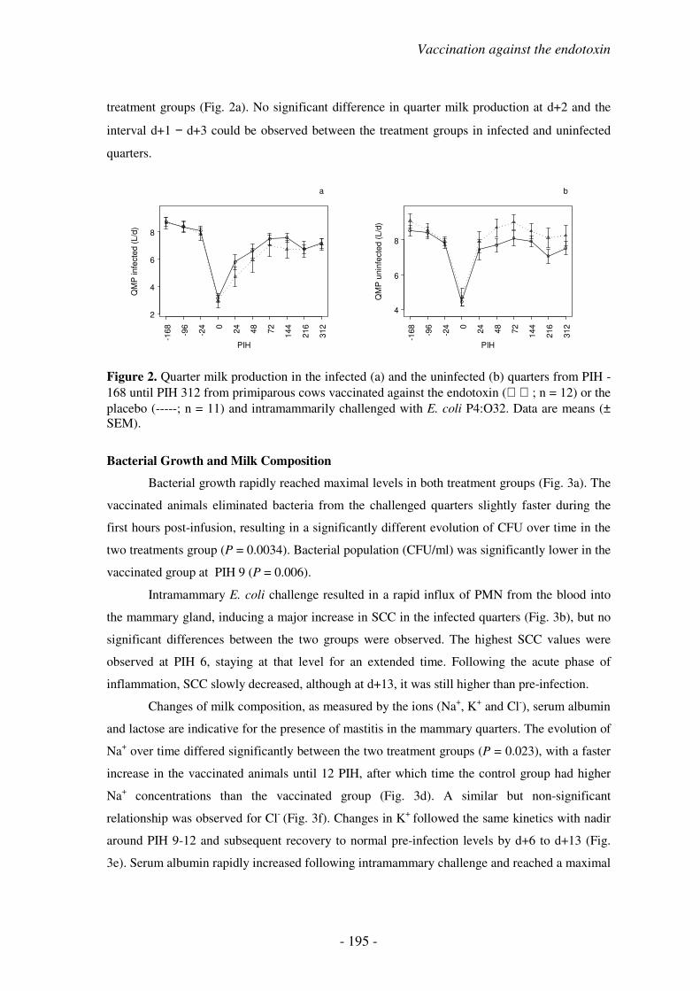
Vaccination against the endotoxin
- 195 -
treatment groups (Fig. 2a). No significant difference in quarter milk production at d+2 and the
interval d+1 − d+3 could be observed between the treatment groups in infected and uninfected
quarters.
Figure 2. Quarter milk production in the infected (a) and the uninfected (b) quarters from PIH -168 until PIH 312 from primiparous cows vaccinated against the endotoxin (; n = 12) or the placebo (-----; n = 11) and intramammarily challenged with E. coli P4:O32. Data are means (± SEM).
Bacterial Growth and Milk Composition
Bacterial growth rapidly reached maximal levels in both treatment groups (Fig. 3a). The
vaccinated animals eliminated bacteria from the challenged quarters slightly faster during the
first hours post-infusion, resulting in a significantly different evolution of CFU over time in the
two treatments group (P = 0.0034). Bacterial population (CFU/ml) was significantly lower in the
vaccinated group at PIH 9 (P = 0.006).
Intramammary E. coli challenge resulted in a rapid influx of PMN from the blood into
the mammary gland, inducing a major increase in SCC in the infected quarters (Fig. 3b), but no
significant differences between the two groups were observed. The highest SCC values were
observed at PIH 6, staying at that level for an extended time. Following the acute phase of
inflammation, SCC slowly decreased, although at d+13, it was still higher than pre-infection.
Changes of milk composition, as measured by the ions (Na+, K+ and Cl-), serum albumin
and lactose are indicative for the presence of mastitis in the mammary quarters. The evolution of
Na+ over time differed significantly between the two treatment groups (P = 0.023), with a faster
increase in the vaccinated animals until 12 PIH, after which time the control group had higher
Na+ concentrations than the vaccinated group (Fig. 3d). A similar but non-significant
relationship was observed for Cl- (Fig. 3f). Changes in K+ followed the same kinetics with nadir
around PIH 9-12 and subsequent recovery to normal pre-infection levels by d+6 to d+13 (Fig.
3e). Serum albumin rapidly increased following intramammary challenge and reached a maximal
PIH
QM
P infe
cte
d (
L/d
)
2
4
6
8
-168
-96
-24 0
24
48
72
144
216
312
a
PIH
QM
P u
nin
fecte
d (
L/d
)
4
6
8
-168
-96
-24 0
24
48
72
144
216
312
b

- 196 -
concentration at PIH 15 in both treatment groups (Fig. 3g), subsequently followed by
normalisation from PIH 48 onward. No significant difference was observed between the two
treatment groups.
Lactose concentration decreased in both groups from PIH 9 onward to reach nadir at PIH
15 in vaccinated animals and at PIH 21 in placebo animals (Fig. 3c). The evolution of lactose
over time differed significantly in the two groups (P = 0.0004) with the vaccinated animals
having a more pronounced decrease of lactose during the acute phase of intramammary E. coli
challenge.
Figure 3. Colony-forming units of E. coli (a), SCC (b), lactose (c), sodium (d), potassium (e), chlorine (f), serum albumin (g) in the affected quarters from PIH -168 until PIH 312 from primiparous cows vaccinated against the endotoxin (; n = 12) or the placebo (-----; n = 11) and intramammarily challenged with E. coli P4:O32. Data are means (± SEM).
Hematology
Packed cell volume did not significantly differ between both treatment groups
throughout the experimental challenge (Fig. 4a). Following E. coli challenge, blood leukocyte
number rapidly decreased and reached nadir at PIH 9 and 12 in the vaccinated and placebo
animals, respectively (Fig. 4b). Blood leukocyte count was on average (marginally) significantly
PIH
Log(C
FU
)/m
l
02
4
-168
-96
-24 0 3 6 9
12
15
18
21
24
48
72
144
216
312
a
PIH
Log(S
CC
)/m
l
35
7
-168
-96
-24 0 3 6 9
12
15
18
21
24
48
72
144
216
312
b
PIH
Lacto
se (
mg/m
l)
10
30
50
-168
-96
-24 0 3 6 9
12
15
18
21
24
48
72
144
216
312
c
PIH
Na+
(m
mol/m
l)
20
60
-168
-96
-24 0 3 6 9
12
15
18
21
24
48
72
144
216
312
d
PIH
K+
(m
mol/m
l)
30
50
-168
-96
-24 0 3 6 9
12
15
18
21
24
48
72
144
216
312
e
PIH
Cl- (
mm
ol/m
l)
30
50
70
-168
-96
-24 0 3 6 9
12
15
18
21
24
48
72
144
216
312
f
PIHSeru
m a
lbum
in (
mg/d
l)
-100
300
-168
-96
-24 0 3 6 9
12
15
18
21
24
48
72
144
216
312
g

Vaccination against the endotoxin
- 197 -
higher in the vaccinated group (P = 0.054). In the placebo group, blood leukocyte number
remained lower than in vaccinated animals until PIH 144.
Leukocyte differentiation did not significantly differ between both treatment groups in
terms of mature PMN (Fig. 4c), early (myelocytes-metamyelocytes) immature PMN (Fig. 4e),
lymphocytes (Fig. 4f), and monocytes (Fig. 4g). Overall, a significantly lower percentage of late
(band cells) immature cells (P = 0.052) was, however, observed in the vaccinated group (Fig. 4d)
as compared to the placebo group.
Figure 4. Packed cell volume (a), blood leukocyte count (b), percentage of mature PMN (c), band cell (d), myelocytes-metamyelocytes (e), lymphocytes (f), monocytes (g), and glucose (h) in the blood from PIH -168 until PIH 312 from primiparous cows vaccinated against the endotoxin (; n = 12) or the placebo (-----; n = 11) and intramammarily challenged with E.
coli P4:O32. Data are means (± SEM).
Glucose
The response of blood glucose to intramammary E. coli challenge was mainly
characterised by two peak concentrations and a dip during the first 24 h post-challenge. In the
vaccinated animals, the glucose concentration evolved differently (P = 0.045) with a significant
lower glucose concentration during the acute phase of inflammation (Fig. 4h).
PIH
PC
V (
%)
25
28
-168
-96
-24 0 3 6 9
12
15
18
21
24
48
72
144
216
312
a
PIH
Log(B
LC
)/m
l
6.4
6.8
-168
-96
-24 0 3 6 9
12
15
18
21
24
48
72
144
216
312
b
PIH
Matu
re P
MN
(%
)
515
30
-168
-96
-24 0 3 6 9
12
15
18
21
24
48
72
144
216
312
c
PIH
Band c
ells
(%
)
05
10
-168
-96
-24 0 3 6 9
12
15
18
21
24
48
72
144
216
312
d
PIH
M+
M*
(%)
020
-168
-96
-24 0 3 6 9
12
15
18
21
24
48
72
144
216
312
e
PIH
Lym
phocyte
s (
%)
40
60
-168
-96
-24 0 3 6 9
12
15
18
21
24
48
72
144
216
312
f
PIH
Monocyte
s (
%)
01
23
-168
-96
-24 0 3 6 9
12
15
18
21
24
48
72
144
216
312
g
PIH
Glu
cose (
mm
ol/m
l)
2.5
4.0
-168
-96
-24 0 3 6 9
12
15
18
21
24
48
72
144
216
312
h
- 198 -
DISCUSSION
Peak E. coli concentrations in the infected quarters have been reported to be indicative
for the severity of experimental E. coli mastitis (Vandeputte-Van Messom et al., 1993), and have
been shown to be both higher and prolonged in severe cases (1 x 10¹⁰ CFU/ml) than in moderate
(1 x 10⁷ CFU/ml) or mild (1 x 10⁴-5 CFU/ml) cases (Shuster et al., 1996; van Werven et al.,
1997; Hirvonen et al., 1999; Vangroenweghe et al., 2004a; 2004b). Moreover, mild cases of
clinical coliform mastitis showed a more rapid elimination of the bacteria from the infected
quarters compared to moderate and severe clinical cases. The AUC0-126 of CFU/ml has
repeatedly been shown to be a significant indicator of severity of clinical E. coli mastitis (Lohuis
et al., 1990; Kremer et al., 1993; van Werven et al., 1997). These data, obtained during
experimentally induced coliform mastitis, are consistent with field observations by Wenz et al.
(2001), who reported higher proportions of cases with > 1 x 10⁵ CFU/ml E. coli concentrations
in severe (77%) than in moderate (51%) and in mild (28%) cases.
Hogan et al. (1992) conducted an experimental challenge comparing cows vaccinated
with the E. coli J5 strain, using formulations different from Enviracor, to a placebo group and
showed favourable results for the J5 vaccinates when the infection model resulted in a rather
high CFU/ml in milk. The mean log10 CFU/ml in the control group at the time point with the
highest value was about 4 log10 CFU/ml. Hence, the CFU challenge obtained in the present study
should be considered as mild. With the exception of Hogan et al. (1992), most other published
challenge studies resulted in peak CFU/ml that were lower than those cited above. In such
studies, few modulatory effects on the course of inflammation were observed following
vaccination against the endotoxin (Hogan et al., 1995; Smith et al., 1999). This is also the case in
the present study.
In cows with E. coli mastitis, the milk production can be greatly affected in severe
responders (Heyneman et al., 1990; Vandeputte-Van Messom et al., 1993; Shuster et al., 1996;
Dosogne et al., 1997; van Werven et al., 1997) in which it does not normalise rapidly (Hirvonen
et al., 1999). Although milk yield has been measured in intramammary challenge studies,
evaluating the effect of J5 vaccination, none of these studies (Hogan et al., 1992; 1995; 1999;
Smith et al., 1999) showed a significant improvement in milk yield loss of the J5 vaccinates over
placebo animals. Milk yield loss in infected and uninfected quarters has been found to be highly
correlated with other indicators of severity, in particular AUC of E. coli CFU/ml (Lohuis et al.,
1990; Kremer et al., 1993). As expected, milk yield recovered fast in the presently used
moderate inflammation model (Vangroenweghe et al., 2004a; 2004b) and vaccination against the

Vaccination against the endotoxin
- 199 -
endotoxin could not accelerate this phenomenon.
Increases in RT and HR have been found to be higher in animals with a severe than with
a moderate clinical response (Heyneman et al., 1990; Vandeputte-Van Messom et al., 1993;
Shuster et al., 1996; Ohtsuka et al., 2001). In moderately reacting primiparous cows, RT was
normalised around PIH 18 (Vangroenweghe et al., 2004a; 2004b). Rectal temperature and HR
have been shown to correlate significantly positive to both E. coli CFU/ml (Kremer et al., 1993)
and milk production in the infected and uninfected quarters (Lohuis et al., 1990). Hence, it is not
surprising that, based on the results of quarter milk production and CFU, no significant
differences in HR were observed between the vaccinated group and the placebo animals.
However, significant differences in RT could be observed during the acute phase of
inflammation. The significant difference between both groups in RT at PIH 6 and 12 could not
be explained by the unequal distribution of the inoculum dose in both treatment groups, as
placebo animals predominantly (10/11) received the high inoculum dose (1 x 10⁶ CFU per
quarter), which should induce an earlier and more rapid fever response (Vangroenweghe et al.,
2004a; 2004b).
Rapid and efficient increase in SCC appears to be the SCC characteristic associated with
a moderate inflammatory reaction (Vangroenweghe et al., 2004a; 2004b). None of the previously
reported experimental challenges comparing cows vaccinated with the E. coli J5 strain to a
placebo group showed a significant difference in SCC between both treatment groups (Hogan et
al., 1992; 1995; 1999; Smith et al., 1999). A significant lower SCC in the J5 vaccinates was only
reported in one experimental challenge study, however, this difference in SCC was at a single
time point and at 7 d post-infusion (Hogan et al., 1995). In the present study with moderately
reacting animals, the same observations concerning the changes in SCC during the acute phase
of inflammation were present.
The milk composition variables determined in the present study are all indicators of the
presence of mastitis in the affected glands. In a moderate inflammation model, changes in the
concentrations of lactose, potassium, sodium, chlorine and serum albumin were similar to
previous studies (Vangroenweghe et al., 2004a; 2004b). Serum albumin has been found to be
significantly less increased in J5 vaccinates (Hogan et al., 1995). In the present study, no
treatment effect was observed for chlorine, potassium and serum albumin, whereas an earlier
change was present for sodium and lactose in the vaccinated animals.
Only one study (Vandeputte-Van Messom et al., 1993) showed a significant lower PCV
in severe than in moderate responders. In the present study, being a moderate inflammatory
model, no significant difference in PCV was observed between both treatment groups. Following
E. coli mastitis challenge, blood leukocyte number will first decrease and subsequently increase,

- 200 -
when compared to baseline values (Heyneman and Burvenich, 1992; van Werven et al., 1997;
Ohtsuka et al., 2001). Both the initial decrease and the subsequent increase are larger in severe
than in moderate responders (Heyneman et al., 1990; Heyneman and Burvenich, 1992; Sordillo
and Peel, 1992; Ohtsuka et al., 2001). This typical pattern of blood leukocyte changes has been
shown to also occur with endotoxin challenge (Tennant et al., 1975; Morris et al., 1986). From
the present study, it is clear that although no significant difference in SCC was present between
both treatment groups during the first 72 h post-challenge, significant differences in the blood
leukocyte patterns were present. Blood leukocyte number was significantly higher throughout
the experiment in the vaccinated animals as compared to the placebo animals. However,
significant mobilisation of late immature PMN (band cells) from the bone marrow could only be
observed in the placebo group.
The presently used E. coli mastitis challenge model consistently results in clinical
mastitis and has, therefore, frequently been used to study risk factors influencing the severity of
the response to challenge, rather than the occurrence of clinical coliform mastitis (Heyneman et
al., 1990; Kremer et al., 1993; Vandeputte-Van Messom et al., 1993; Dosogne et al., 1997; van
Werven et al., 1997; Hoeben et al., 2000; Dosogne et al., 2002). However, using this model in J5
vaccination studies, it has often been difficult to produce a severe response. A first possible
explanation for this observation is that, due to the need for vaccination from dry-off onward,
these vaccination studies are often much longer than the classical experimental E. coli mastitis
challenges. This means that these cattle have remained in the same familiar environment for a
longer period prior to challenge. In a classical challenge study, the observation period to
challenge can be much shorter, mostly 1 to 2 weeks (Dosogne et al., 1997; Blum et al., 2000 ;
Hoeben et al., 2000), meaning that these animals had much less time to adjust to the study
settings (experimental facilities and personnel) as well as to the extra interventions (blood and
milk sampling, clinical examination), all of which cause extra stress. In this regard, it has been
shown that even 1 h of isolation of a cow from her herd mates caused enough stress to result in
an increase of leakiness of the tight-junctions between the endothelial cells in the mammary
gland, thereby allowing an influx of serum components (serum albumin, chlorine, sodium) into
the milk and a loss of milk components (lactose) to the serum (Stelwagen et al., 2000). Another
major limitation to obtain a severe clinical response is the fact that in most of the cited J5
vaccine studies the challenge did not take place within days after calving, the period when
animals are most likely to develop severe coliform mastitis. A third aspect is that primiparous
cows are less susceptible to severe coliform mastitis than older animals (van Werven et al., 1997;
Vangroenweghe et al., 2004a; 2004b).

Vaccination against the endotoxin
- 201 -
CONCLUSION
In the present study, pre-calving vaccination against the endotoxin through a J5 vaccine
administration could slightly alter the clinical course of a moderate inflammatory reaction.
Although vaccinated animals had favourable RT and blood leukocyte kinetics, no major clinical
improvement could be observed following a moderate inflammatory reaction during E. coli
challenge. Therefore, primiparous cows seem to have an optimal inflammatory reaction
following intramammary E. coli inoculation, characterised by a rapid and efficient influx of
PMN into the affected mammary quarters, resulting in a fast and short-lasting inflammation,
finally leading to resolution of mammary gland functionality.

- 202 -
REFERENCES
Barkema, H.W., Y.H. Schukken, T.J.G.M. Lam, M.L. Beiboer, G. Benedictus, and A. Brand. 1999.
Management practices associated with the incidence rate of clinical mastitis. J. Dairy Sci. 82:1643-1654.
Blum, J.W., H. Dosogne, D. Hoeben, F. Vangroenweghe, H.M. Hammon, R.M. Bruckmaier, and C.
Burvenich. 2000. Tumor necrosis factor-α and nitrite/nitrate responses during acute mastitis induced by
Escherichia coli infection and endotoxin in dairy cows. Dom. Anim. Endocrinol. 19:223-235.
Bradley, A.J., and M.J. Green. 2001. Aetiology of clinical mastitis in six Somerset dairy herds. Vet. Rec.
148:683-686.
Dosogne, H., C. Burvenich, T. van Werven, E. Roets, E.N. Noordhuizen-Stassen, and B. Goddeeris.
1997. Increased surface expression of CD11b receptors on polymorphonuclear leukocytes is not
sufficient to sustain phagocytosis during Escherichia coli mastitis in early postpartum dairy cows. Vet.
Immunol. Immunopathol. 60:47-59.
Dosogne, H., F. Vangroenweghe, B. Barrio, P. Rainard, and C. Burvenich. 2001. Decreased number and
bactericidal activity against Staphylococcus aureus of the resident cells in milk of dairy cows during
early lactation. J. Dairy Res. 68:539-549.
Dosogne, H., E. Meyer, A. Sturk, J. van Loon, A.-M. Massart-Leën, and C. Burvenich. 2002. Effect of
enrofloxacin treatment on plasma endotoxin during bovine Escherichia coli mastitis. Inflamm. Res.
51:201-205.
Eberhart, R.J., R.P. Natzke, R.S.H. Newbould, B. Nonnecke, and P. Thompson. 1977. Coliform mastitis.
A review. J. Dairy Sci. 62:1-22.
Gonzales, R.N., J.S. Cullor, D.E. Jasper, T.B. Farver, R.B. Bushnell, and M.N. Oliver. 1989. Prevention of
clinical coliform mastitis in dairy cows by a mutant Escherichia coli vaccine. Can. J. Vet. Res. 53:301-
307.
Heyneman, R., C. Burvenich, and R. Vercauteren. 1990. Interaction between the respiratory burst activity
of neutrophil leukocytes and experimentally induced Escherichia coli mastitis cows. J. Dairy Sci.
73:985-994.
Heyneman, R., and C. Burvenich. 1992. Kinetics and characteristics of bovine neutrophils alkaline
phosphatase during acute Escherichia coli mastitis. J. Dairy Sci. 75:1826-1834.
Hill, A.W. 1981. Factors influencing the outcome of Escherichia coli mastitis in the dairy cow. Res. Vet.
Sci. 31:107-112.
Hirvonen, J., K. Eklund, A.M. Teppo, G. Huszenicza, M. Kulcsar, H. Saloniemi, and S. Pyörälä. 1999.
Acute phase response in dairy cows with experimentally induced Escherichia coli mastitis. Acta Vet.
Scand. 40:35-46.
Hoeben, D., C. Burvenich, E. Trevisi, G. Bertoni, J. Hamann, R.M. Bruckmaier, and J.W. Blum. 2000.
Role of endotoxin and TNF-α in the pathogenesis of experimentally induced coliform mastitis in
periparturient cows. J. Dairy Res. 67:503-514.

Vaccination against the endotoxin
- 203 -
Hogan, J.S., W.P. Weiss, D.A. Todhunter, K.L. Smith, and P.S. Schoenberger. 1992. Efficacy of an
Escherichia coli J5 mastitis vaccine in an experimental challenge trial. J. Dairy Sci. 75:415-422.
Hogan, J.S., W.P. Weiss, K.L. Smith, D.A. Todhunter, and P.S. Schoenberger. 1995. Effects of an
Escherichia coli J5 vaccine on mild clinical coliform mastitis. J. Dairy Sci. 78:285-290.
Hogan, J.S., V.L. Bogacz, M. Aslam, and K.L. Smith. 1999. Efficacy of an Escherichia coli J5 bacterin
administered to primigravid heifers. J. Dairy Sci. 82:939-943.
Jackson, E., and J. Bramley. 1983. Coliform mastitis. In Practice 7:135-146.
Jain, N.C. 1979. Common mammary pathogens and factors of infection in mastitis. J. Dairy Sci. 62:128-
134.
Kremer, W.D.J., E.N. Noordhuizen-Stassen, F.J. Grommers, Y.H. Schukken, R. Heeringa, A. Brand, and
A. Brand. 1993. Severity of experimental Escherichia coli mastitis in ketonemic and nonketonemic dairy
cows. J. Dairy Sci. 76:3428-3436.
Linton, A.H., and T.C. Robinson. 1984. Studies on the association of Escherichia coli with bovine
mastitis. Br. Vet. J. 140:368-373.
Lohuis, J.A.C.M., Y.H. Schukken, J.H.M. Verheijden, A. Brand, A.S.J.P.A.M. Van Miert. 1990. Effect of
severity of systemic signs during the acute phase of experimentally induced Escherichia coli mastitis on
milk production losses. J. Dairy Sci. 73:333-341.
Mehrzad, J., H. Dosogne, E. Meyer, R. Heyneman, and C. Burvenich. 2001. Respiratory burst activity of
blood and milk neutrophils in dairy cows during different stages of lactation. J. Dairy Res. 68:399-415.
Monfardini, E., C. Burvenich, A.-M. Massart-Leën, E. Smits, and M.J. Paape. 1999. Effect of antibiotic
induced bacterial clearance in the udder on L-selectin shedding of blood neutrophils in cows with
Escherichia coli mastitis. Vet. Immunol. Immunopathol. 67:373-384.
Morris, D.D., J.S. Cullor, R.H. Whitlock, M. Wickstrom, and L.B. Corbeil. 1986. Endotoxemia in
neonatal calves given antiserum to a mutant Escherichia coli (J-5). Am. J. Vet. Res. 47:2554-2565.
Ohtsuka, H., K. Kudo, K. Mori, F. Nagai, A. Hatsugaya, M. Tajima, K. Tamura, F. Hoshi, M. Koiwa, and
S. Kawamura. 2001. Acute phase response in naturally occurring coliform mastitis. J. Vet. Med. Sci.
63:675-678.
Oliver, S.P., S.H. King, M.J. Lewis, P.M. Torne, K.R. Matthews, and H.H. Dowlen. 1990. Efficacy of
chlorhexidine as a postmilking teat disinfectant for the prevention of bovine mastitis during lactation. J.
Dairy Sci. 73:2230-2235.
Oz, H.H., R.J. Farnsworth, and V.L. Larson. 1985. Environmental mastitis. Vet. Bull. 55:829-840.
Schukken, Y.H., F.J. Grommers, D. van de Geer, and A. Brand. 1989a. Incidence of clinical mastitis on
farms with low somatic cell counts in bulk milk. Vet. Rec. 125:60-63.
Schukken, Y.H., D. van de Geer, F.J. Grommers, J.A.H. Smit, and A. Brand. 1989b. Intramammary
infections and risk factors for clinical mastitis in herds with low somatic cell counts in bulk milk. Vet.
Rec. 125:393-396.
Shuster, D.E., E.K. Lee, and M.E. Kehrli, Jr. 1996. Bacterial growth, inflammatory cytokine production,
and neutrophil recruitment during coliform mastitis in cows within ten days after calving, compared with
cows at midlactation. Am. J. Vet. Res. 57:1569-1575.

- 204 -
Smith, K.L., D.A. Todhunter, and P.S. Schoenberger. 1985. Environmental mastitis: cause, prevalence,
prevention. J. Dairy Sci. 68:1531-1553.
Smith, J.L., J.S. Hogan, and K.L. Smith. 1999. Efficacy of intramammary immunization with an
Escherichia coli J5 bacterin. J. Dairy Sci. 82:2582-2588.
Sordillo, L.M., and J.E. Peel. 1992. Effect of interferon-γ on the production of tumor necrosis factor
during acute Escherichia coli mastitis. J. Dairy Sci. 75:2119-2125.
Stelwagen, K., H. Hopster, J.T.N. Van Der Werf, and H.J. Blokhuis. 2000. Short communication: Effects
of isolation stress on mammary tight junctions in lactating dairy cows. J. Dairy Sci. 83:48-51.
Tennant, B., D. Harrold, and M. Guerra-Reina. 1975. Hematology of neonatal calf. II. Response
associated with acute enteric infections, Gram-negative septicemia, and experimental endotoxemia.
Cornell Vet. 65:457-475.
Tomita, G.M., C.H. Ray, S.C. Nickerson, W.E. Owens, and G.F. Gallo. 2000. A comparison of two
commercially available Escherichia coli J5 vaccines against E. coli intramammary challenge. J. Dairy
Sci. 83:2276-2281.
van Werven, T., E.N. Noordhuizen-Stassen, A.J. Daemen, Y.H. Schukken, A. Brand, and C. Burvenich.
1997. Pre-infection in vitro chemotaxis, phagocytosis, oxidase burst, and expression of CD11/CD18
receptors and their predictive capacity on the outcome of mastitis induced in dairy cows with
Escherichia coli. J. Dairy Sci. 80:67-74.
Vandeputte-Van Messom, G., C. Burvenich, E. Roets, A.-M. Massart-Leën, R. Heyneman, W.D.J.
Kremer, and A. Brand. 1993. Classification of newly calved cows into moderate and severe responders
to experimentally induced Escherichia coli mastitis. J. Dairy Res. 60:19-29.
Vangroenweghe, F., H. Dosogne, J. Mehrzad, and C. Burvenich. 2001. Effect of milk sampling and stage
of lactation on milk composition, viability and functions of resident cells in milk. Vet. Res. 32:565-579.
Vangroenweghe, F., L. Duchateau, and C. Burvenich. 2004a. Moderate inflammatory reaction during
experimental E. coli mastitis in primiparous cows. J. Dairy Sci. 87:886-895.
Vangroenweghe, F., P. Rainard, M.J. Paape, L. Duchateau, and C. Burvenich. 2004b. Increase in
Escherichia coli inoculum doses induce faster innate immune response in primiparous cows. J. Dairy
Sci. 87:4132-4144.
Wenz, J.R., G.M. Barrington, F.B. Garry, R.P. Dinsmore, and R.J. Callan. 2001. Use of systemic disease
signs to assess disease severity in dairy cows with acute coliform mastitis. JAVMA 218:567-572.

- 205 -
GENERAL DISCUSSION


General discussion
- 207 -
Escherichia coli, involved in bovine coliform mastitis, is part of the normal intestinal
flora of animals. The strains isolated from bovine mastitis are essentially not different from
strains isolated from bovine faeces. The etiological bacteria do not possess specific virulence
factors that could be attributed to disease severity (Lehtolainen, 2004). In contrast, cow factors
such as stage of lactation, PMN functionality and metabolic status are associated with disease
severity (Burvenich et al., 2003; 2004). Concerning E. coli, numerous epidemiological data are
available (Schukken et al., 1989a; 1989b; Barkema et al., 1999), in addition to the results
obtained from experimental infection models, which can be used to generate practical
conclusions. However, the specific experimental model used, i.e. E. coli vs. endotoxin, has a
tremendous effect on the results obtained from pathogenesis studies. The inflammatory reaction
in multiparous dairy cows has extensively been studied throughout the last decade (Heyneman
and Burvenich, 1992; Vandeputte-Van Messom et al., 1993; Kremer et al., 1993a; 1993b; 1993c;
van Werven et al., 1997; Dosogne et al., 1997; Hoeben et al., 2000). However, taking into
account the present turn-over of high-yielding dairy cows (approximately 25 to 30% per year) in
modern dairy herds, a relatively large population of primiparous cows is present, from which no
data on resistance or susceptibility towards E. coli mastitis are available.
Although the influence of stage of lactation has extensively been studied, until now,
little information on the effect of parity is available. Younger cows (2nd parity) have been
postulated to be more resistant to intramammary E. coli infection, mainly based on the lower
bacterial count and the higher number of peripheral circulating leukocytes during experimentally
induced E. coli mastitis (van Werven et al., 1997). Evaluation of impairment of periparturient
neutrophil function revealed a significantly lower superoxide anion production in cows in their
4th or greater lactation than in cows in the first three lactations (Gilbert et al., 1993). Superoxide
formation plays an important role in the intracellular killing of E. coli inside the PMN. A
longitudinal study of PMN viability and functions in primiparous and multiparous dairy cows
around calving showed significant differences between both groups (Mehrzad et al., 2002).
Although a periparturient decrease in PMN viability occurred in both groups, overall PMN
viability was much higher in primiparous cows (Mehrzad et al., 2002). It seems logic that
animals in their first lactation have markedly different pre-inflammatory parameters, which may
result in differences in inflammatory kinetics following intramammary E. coli challenge (Gilbert
et al., 1993; van Werven et al., 1997; Mehrzad et al., 2002).
Normal milk contains a heterogeneous cell population, with its composition dependent
on the stage of lactation. The normal proportion of PMN in the clinically healthy mammary
gland is low (< 15%). During inflammation, due to for instance mastitis, the proportion of PMN
increases to more than 90%. Therefore, inflammation should be considered a PMN related

- 208 -
condition with a well-controlled cell functionality. In this thesis, milk sample collection under
pre-infection conditions was validated in relation to composition of the cell population and PMN
functionality in milk, collected under different aseptical and non-aseptical conditions, because
external bacterial contamination was thought to activate the PMN functionality in vitro. In low
SCC milk, the number of PMN was only 13% and the average cell viability was decreased
(40%). This observation was in accordance to Mehrzad et al. (2001), which also reported
decreased PMN viability and functionality during early lactation. Validation of aseptical milk
sample collection revealed no significant effect of bacterial contamination on milk PMN
functionality under basal pre-infection conditions, and therefore, manual milk sampling was
preferred to perform milk sample collection in the subsequent experimental E. coli challenges.
Number and composition of the somatic cell population could be considered as an
important determining factor in the immunological status of the mammary gland. Numerous
studies have been performed in order to identify the various cell populations present in the
mammary gland, however, most of these studies have focused on bovine high SCC milk
(Hageltorn and Saad, 1986; Östensson et al., 1988; Redelman et al., 1988; Saad and Östensson,
1990). Moreover, most techniques were complicated and labour-intensive, and therefore without
practical applicability. In epidemiological studies, low SCC has been demonstrated as an
important risk factor for the occurrence of clinical coliform mastitis (Schukken et al., 1989a;
1989b; Barkema et al., 1999). In this thesis, a rapid, simple and accurate bovine low SCC milk
differentiation technique was developed using flow-cytometry (Dosogne et al., 2003). With this
technique, PMN, macrophages, lymphocytes and apoptotic cells could easily be distinguished.
Questions can arise to what extent the isolation method could influence the proportional
composition of SCC, but this problem occurs in all published differentiation techniques. The
present method could limit preparative manipulations to a minimum, but obligatory
centrifugation for the removal of fat, interfering with flow cytometric analysis, could in fact bias
the proportional distribution of cell types obtained in differentiation. However, compared to
previous differentiation procedures, milk sample collection and preparation, cell staining and
flow cytometric analysis were validated.
As proposed in the study objectives, inflammatory kinetics and severity of the
experimentally induced intramammary E. coli challenge were described using an inoculum dose
of 1 x 10⁴ and 1 x 10⁶ CFU of E. coli P4:O32 per quarter (Vangroenweghe et al., 2004a).
Primiparous cows generally appeared to react as moderate responders, based on their quarter
milk production in the right uninfected quarters on d+2 post-infection. Moderate responders had
a higher number of circulating PMN (Heyneman et al., 1990; Sordillo and Peel, 1992; Kremer et
al., 1993c; van Werven et al., 1997), and the functionality of circulating blood PMN pre-

General discussion
- 209 -
infection, such as chemotactic differential (Kremer et al., 1993a; van Werven et al., 1997) and
oxidative burst activity (Heyneman et al., 1990), was clearly increased. Some of these aspects,
such as circulating number of PMN, were also observed in this thesis (Vangroenweghe et al.,
2004a). Therefore, an effect of parity or age can be concluded from the present experiment.
Comparison of historical data of quarter milk production in the uninfected quarters at
d+2 with data obtained in this thesis showed that primiparous cows had a remarkably higher
average milk production in their uninfected quarters following intramammary challenge as
compared to multiparous cows. Several animals already exceeded their pre-infection milk
production at d+2 post-infection (Vangroenweghe et al., 2004a). Following E. coli challenge,
primiparous cows exhibited a moderate inflammatory reaction, which could not be altered
through an increase (1 x 10⁴ → 1 x 10⁶ CFU per quarter) in intramammary inoculum dose. The
inflammatory reaction was characterised by a rapid onset of clinical symptoms, such as rectal
temperature increase, udder swelling, and changes in milk composition, which also disappeared
rather fast, resulting in a complete resolution of the functionality of the affected mammary
quarters. To better diversify between different clinical responses, a severity scoring system, used
to score cases of clinical mastitis in practice (Wenz et al., 2001), was adapted for use in the
experimental infection model. Evaluation of clinical responses through this system neither
revealed any severe responses in the challenged primiparous cows (Vangroenweghe et al.,
2004a). The severity scoring system could therefore be used as an additional scoring tool,
besides the established measurement of quarter milk production in the uninfected quarters at d+2
post-infection.
Literature data revealed a wide diversity of inoculum doses and strains used to provoke
experimental E. coli mastitis in the bovine. However, strain diversity could not be held
responsible for variations in clinical outcome of E. coli mastitis, because several researchers
were unable to characterise specific virulence factors, which might be associated with severity or
virulence following intramammary E. coli challenge (Lehtolainen, 2004). Differences in
inoculum dose have been shown to result in variable inflammatory kinetics. Following
inoculation of low doses (~ 30 CFU), bacterial growth preceded subsequent elimination (Shuster
et al., 1996). Inoculation of high doses (~ 10,000 CFU) resulted in rapid bacterial elimination
(Hoeben et al., 2000). However, studies in which inoculum doses were randomised have rarely
been performed (Jain et al., 1969; Carroll et al., 1973; Bramley and Neave, 1975; Frost et al.,
1980). The variation in inoculum dose in primiparous cows, performed in this thesis, could
modulate the inflammatory response, which was characterised by an earlier onset (approximately
3 h) of clinical signs and chemotactic activity in the animals inoculated with the highest
inoculum dose (1 x 10⁶ CFU E. coli P4:O32) (Vangroenweghe et al., 2004b). These results seem

- 210 -
to confirm that a minimal number of bacteria should be present in the mammary gland to trigger
the innate immune response in its activation of several defence mechanisms, such as activation
of the mammary gland epithelium to produce chemotactic factors (IL-8) or macrophage
activation with inflammatory cytokine production (Bannerman et al., 2003).
Therapeutic and prophylactic strategies are claimed to modulate the course of an
inflammatory reaction. Administration of an inhibitor of prostaglandin synthesis before or during
experimentally induced LPS mastitis has been shown beneficial in reducing the clinical episode,
characterised by the absence of fever or a shorter or lower fever peak (Burvenich and Peeters,
1982; Anderson et al., 1986; Lohuis et al., 1989; 1991; Banting et al., 2000), mainly through the
interaction with fever inducing mechanisms, such as central PGE2 production in the
thermoregulatory centre (Burvenich and Peeters, 1982). In primiparous cows, the modulatory
effect of carprofen treatment following experimentally induced E. coli mastitis was limited to a
reduced peak fever and a shorter episode of reticulorumen motility depression. Moreover,
eicosanoid (PGE2 and TXB2) production in the affected quarters decreased following carprofen
treatment. The inflammatory reaction mounted in these animals, therefore, seems to be on the
edge of clinically detectable inflammation, especially as increased rectal temperature is only
present for 12 h (from PIH 6 until PIH 18). Furthermore, mammary gland functionality, as
quantified by quarter milk production, is rapidly resolved.
From a prophylactic point of view, vaccination against the endotoxin, using a J5 vaccine,
has not been able to reduce the incidence of clinical coliform mastitis (Tyler et al., 1993),
although a shift in clinical response from severe to moderate has been observed (Gonzales et al.,
1989; Tyler et al., 1993). To show efficacy in an experimental model, it is known that a severe
inflammatory reaction is needed (Hogan et al., 1992). The present study with primiparous cows
resulted in a homogenous population of moderate responders (Vangroenweghe et al., 2004a;
2004b), and it is therefore not surprising that little modulatory effects of vaccination against the
endotoxin were observed.
Prognostic evaluation of clinical severity before treatment, combined with rapid
etiological diagnosis, is difficult to achieve under practical conditions. However, based on
experimentally induced intramammary E. coli challenge, several severity determining and risk
factors have been identified, which could be associated with the potential occurrence of a severe
inflammatory response (Burvenich et al., 2003). One of these factors could be age or parity of
the affected animal (Gilbert et al., 1993; van Werven et al., 1997). The inflammatory reaction in
primiparous cows following intramammary E. coli challenge has been shown mild to moderate
based on clinical severity scoring (Vangroenweghe et al., 2004a). Therefore, primiparous cows
with acute clinical mastitis during the early post-partum period should be considered low risk

General discussion
- 211 -
candidates for a severe clinical response due to E. coli. Moreover, prophylactic and therapeutic
interventions have been shown unable to modulate the already moderate inflammatory reaction,
resulting in a maximal resolution of original functionality of the affected quarters. In case of
clinical mastitis in primiparous cows, antibiotic treatment should be focused on combating
Gram-positive infections, because infection with Gram-negative bacteria does not seem to cause
a potential risk for severe inflammatory reactions. In contrast, high-yielding multiparous cows in
negative energy balance during the early post-partum period are striking potential candidates for
a severe clinical response (Kremer et al., 1993c; Hoeben et al., 1997; van Werven et al., 1997),
and treatment should therefore be focused on these animals, providing them antimicrobial and
anti-inflammatory therapy, eventually in combination with other additional treatments.
CONCLUSION
In conclusion, validation of milk sample collection under aseptical conditions to
establish basal pre-infection values has revealed no effect of minor bacterial contamination on
the milk PMN functionality in vitro. Therefore, manual milk sample collection was performed in
the subsequent experimental E. coli challenges. Moreover, flow cytometric differentiation of the
milk cell population present in bovine low SCC milk was validated. Following intramammary E.
coli challenge during the periparturient period, primiparous cows reacted as moderate responders
with a rapid and efficient inflammatory reaction and short-lasting depression in milk production.
Variation of the inoculum dose could modulate the inflammatory reaction and a higher inoculum
dose (1 x 10⁶ CFU per quarter) resulted in a faster innate immune response. Inhibition of the
prostaglandin synthesis through NSAID administration at PIH 9, when clinical symptoms were
present, improved general clinical condition by rapid defeverescence and restoration of
depressed reticulorumen motility. In addition, eicosanoid production (PGE2 and TXB2)
decreased following NSAID treatment. Pre-calving prophylactic vaccination against the
endotoxin could slightly alter the clinical course of the moderate inflammatory reaction in
primiparous cows. Although vaccinated animals had favourable RT and blood leukocyte
kinetics, no major clinical improvement could be observed following intramammary E. coli
challenge. Therefore, primiparous cows can be considered low risk candidates for severe clinical
mastitis following experimental intramammary E. coli challenge. Care should be taken with
respect to generalisation of the obtained results of experimentally induced mastitis to the field
situation, especially due to some major differences, such as the inoculum dose, which exist
between experimental models used in research and field cases.

- 212 -
Figure 1. Schematic drawing of the hypothesis and objectives of this thesis. Left pathway: external bacterial contamination has no effect on cellular response of resident cells in milk (*) (p. 69). Right pathway: following intramammary E. coli infection, bacterial growth and lysis leads to the release of LPS, which interacts with the resident cells in milk. Binding of LPS by LBP/sCD14 induces a cellular response, which changes resident milk cell characteristics and elicits an inflammatory response resulting in changes in milk secretion. (**) Primiparous cows mainly react as moderate responders following intramammary E. coli challenge (p. 125), (***) inoculum dose can modulate the kinetics of the moderate inflammatory reaction in primiparous cows (p. 145), whereas inhibition of prostaglandin synthesis only affects general clinical condition and eicosanoid production in milk (p. 167), vaccination against the endotoxin could slightly alter the clinical course of the moderate inflammation in primiparous cows (p. 189).
Intramammary E. coli infection
Growth and lysis
Milk sample collection (effect of external bacterial contamination)
↓↓↓↓↓↓↓↓↓↓↓↓
LPS release
Recognition (e.g. LBP/sCD14)
Cellular response
Modulation * inoculum dose ++++++++++++
* prostaglandin synthesis ++++ * vaccination against LPS ±±±±
Inflammatory response
Parity ↓↓↓↓↓↓↓↓↓↓↓↓
*
** ***
Resident cells
mild moderate severe Changes in milk secretion
Viability Phagocytosis
Chemiluminescence Intracellular killing
General discussion
- 213 -
PRACTICAL IMPLICATIONS AND FUTURE RESEARCH
1. Until now, milk quality and immune status of the mammary gland are assessed through
quantification of the cells in milk (SCC measurement). However, in the future,
functional examination of the resident cells in milk will become an additional tool in the
study of mammary gland immunity and milk quality. In this thesis, manual milk
collection has been shown sufficient for these purposes.
2. Questions will arise concerning the future approach of E. coli mastitis, especially in
relation to treatment or prevention/prophylaxis. Focusing on prevention, one or more
stable immune parameters should be available that can be measured throughout the
transition period, in order to obtain quantitative feedback about the currently performed
farm management practices. Therefore, the immune status of the dairy cows should be
measured through the assessment of specific markers in the population. Besides the
preventive approach, treatment of clinical mastitis will still be performed. The present
work contributes, together with other data of our laboratory and world-wide mastitis
research groups, to the knowledge of the physiological limits and the scientific basis of
disease severity during clinical E. coli mastitis. This knowledge helps to answer the
question: “which animals should not be treated ?” or rather: “should we treat with broad
spectrum antibiotics or antibiotics with a Gram-positive spectrum ?”. Due to the high
degree of self-curing, it is clear that all animals in mid-lactation and the primiparous
cows during early lactation are low risk candidates for a severe clinical response due to
E. coli mastitis and should therefore not be treated. Moreover, EU directives will also
determine use of antimicrobials in the near future. However, until now, rapid etiological
diagnosis is still difficult. Therefore, mild to moderate clinical mastitis occurring in
primiparous cows during early lactation, which could be caused by Gram-positive
bacteria, should be treated with antimicrobials with a unique Gram-positive spectrum. In
contrast, primiparous cows exhibiting a severe clinical response following naturally
occurring mastitis can be treated with an antibiotic with broad spectrum.
3. Under practical circumstances, the use of a severity scoring system will help to obtain a
good prognostic evaluation in animals suffering from clinical mastitis. A mild or
moderate response in primiparous cows during early lactation may be due to E. coli or
coagulase-negative staphylococci, which do both not require antimicrobial treatment. In
practice, severe cases of clinical mastitis can occur in primiparous cows during the
periparturient period. Under these circumstances, bacteriological diagnosis is an

- 214 -
essential tool in etiological diagnosis to differentiate between E. coli, which is
predominantly self-curing, and S. aureus, which should efficiently be treated to omit
chronic pathogen survival into the mammary gland. Moreover, differences in the clinical
picture can occur between experimentally induced E. coli mastitis and naturally
occurring cases of E. coli, which are observed under field circumstances.
4. Clinical and subclinical mastitis should clearly be distinguished. Primiparous cows are
susceptible to coagulase-negative staphylococci, whereas they react mild to moderate
following intramammary E. coli challenge. However, clinical E. coli mastitis is mainly
determined by the host (Burvenich et al., 2003), whereas subclinical mastitis by
coagulase-negative staphylococci is rather a problem of the bacteria (De Vliegher,
2004). Veterinary clinicians should therefore be forced to perform an etiological
diagnosis of mastitis, especially because the term 'mastitis' is rather broad and more
specifications should be made in terms of 'environmental mastitis' or 'contagious
mastitis' to diversify between the two major groups of etiological bacteria. The more
correct description of mastitis problematics does not discharge the veterinary clinician
from his duty to perform an etiological diagnosis, because inventarisation of various
bacteria, causing mastitis, present at the farm is advisable in good dairy farm
management.
5. Primiparous cows suffering from mild to moderate clinical mastitis due to E. coli should
not be treated with antimicrobials or anti-inflammatory drugs, but care should be taken
towards the potential risk to develop a chronic E. coli mastitis problem, although this is a
rare disease. The presence of multiple cases of clinical E. coli mastitis in primiparous
cows may be indicative for more fundamental problems in relation to a decreased
immunological status of the cows during the early lactation period. This can eventually
spread to the population of multiparous cows on the farm, which are far more susceptible
to severe clinical coliform mastitis.
General discussion
- 215 -
REFERENCES
Anderson, K.L., A.R. Smith, R.D. Shanks, L.E. Davis, and B.K. Gustafsson. 1986. Efficacy of flunixin
meglumine for the treatment of endotoxin-induced bovine mastitis. Am. J. Vet. Res. 47:1366-1372.
Bannerman, D.D., M.J. Paape, W.R. Hare, and E.J. Sohn. 2003. Increased levels of LPS-binding protein in
bovine blood and milk following bacterial lipopolysaccharide challenge. J. Dairy Sci. 86:3128-3137.
Banting, A., H. Schmidt, and S. Banting. 2000. Efficacy of meloxicam in lactating cows with E. coli
endotoxin induced acute mastitis. 8th International Congress EAVPT, Jerusalem, Israel. 30 July – 3
August. Abstract N° E4.
Barkema, H.W., Y.H. Schukken, T.J.G.M. Lam, M.L. Beiboer, G. Benedictus, and A. Brand. 1999.
Management practices associated with the incidence rate of clinical mastitis. J. Dairy Sci. 82:1643-1654.
Bramley, A.J., and F.K. Neave. 1975. Studies on the control of coliform mastitis in dairy cows. Br. Vet. J.
131:160-169.
Burvenich, C., and G. Peeters. 1982. Effect of prostaglandin synthetase inhibitors on mammary blood flow
during experimentally induced mastitis in lactating goats. Arch. Int. Pharmacodyn. Ther. 258:128-137.
Burvenich, C., V. Van Merris, J. Mehrzad, A. Diez-Fraile, and L. Duchateau. 2003. Severity of E. coli
mastitis is mainly determined by cow factors. Vet. Res. 34:521-564.
Burvenich, C., E. Monfardini, J. Mehrzad, A.V. Capuco, and M..J. Paape. 2004. Role of the neutrophil
polymorphonuclear leukocytes during bovine coliform mastitis: physiology or pathology? Kon. Acad.
Geneesk. Belg. 66:97-153.
Carroll, E.J., N.C Jain, O.W. Schalm, and J. Lasmanis. 1973. Experimentally induced coliform mastitis:
inoculation of udders with serum-sensitive and serum-resistant organisms. Am. J. Vet. Res. 34:1143-
1146.
De Vliegher, S. 2004. Udder health in dairy heifers - some epidemiological and microbiological aspects.
Ph.D. Thesis, Ghent University, Belgium.
Dosogne, H., C. Burvenich, T. van Werven, E. Roets, E.N. Noordhuizen-Stassen, and B. Goddeeris. 1997.
Increased surface expression of CD11b receptors on polymorphonuclear leukocytes is not sufficient to
sustain phagocytosis during Escherichia coli mastitis in early postpartum dairy cows. Vet. Immunol.
Immunopathol. 60:47-59.
Dosogne H., F. Vangroenweghe, J. Mehrzad, A.-M. Massart-Leën, and C. Burvenich. 2003. Differential
leukocyte count method for bovine low somatic cell count milk. J. Dairy Sci. 86:828-834.
Frost, A.J., A.W. Hill, and B.E. Brooker. 1980. The early pathogenesis of bovine mastitis due to
Escherichia coli. Proc. R. Soc. Lond. Biol. Sci. 209: 431-439.
Gilbert, R.O., Y.T. Gröhn, P.M. Miller, and D.J. Hoffman. 1993. Effect of parity on periparturient
neutrophil function in dairy cows. Vet. Immunol. Immunopathol. 36:75-82.
Gonzales, R.N., J.S. Cullor, D.E. Jasper, T.B. Farver, R.B. Bushnell, and M.N. Oliver. 1989. Prevention of
clinical coliform mastitis in dairy cows by a mutant Escherichia coli vaccine. Can. J. Vet. Res. 53:301-
307.

- 216 -
Hageltorn, M., and M.A. Saad. 1986. Flow cytofluorometric characterisation of bovine blood and milk
leukocytes. Am. J. Vet. Res. 47:2012-2016.
Heyneman, R., and C. Burvenich. 1992. Kinetics and characteristics of bovine neutrophil alkaline
phosphatase during acute Escherichia coli mastitis. J. Dairy Sci. 75:1826-1834.
Heyneman, R., C. Burvenich, and R. Vercauteren. 1990. Interaction between the respiratory burst activity
of neutrophil leukocytes and experimentally induced Escherichia coli mastitis in cows. J. Dairy Sci.
73:985-994.
Hoeben, D., R. Heyneman, and C. Burvenich. 1997. Elevated levels of beta-hydroxybutyric acid in
periparturient cows and in vitro effect on respiratory burst activity of bovine neutrophils. Vet. Immunol.
Immunopathol. 58:165-170.
Hoeben, D., C. Burvenich, E. Trevisi, G. Bertoni, J. Hamann, R.M. Bruckmaier, and J.W. Blum. 2000.
Role of endotoxin and TNF-alpha in the pathogenesis of experimentally induced coliform mastitis in
periparturient cows. J. Dairy Res. 67:503-514.
Hogan, J.S., W.P. Weiss, D.A. Todhunter, K.L. Smith, and P.S. Schoenberger. 1992. Efficacy of an
Escherichia coli J5 mastitis vaccine in an experimental challenge trial. J. Dairy Sci. 75:415-422.
Jain, N.C., O.W. Schalm, and J. Lasmanis. 1969. Comparison of normal and leukopenic cows of
experimental mastitis due to Aerobacter aerogenes or Escherichia coli endotoxin. Am. J. Vet. Res.
30:715-724.
Kremer, W.D., E.N. Noordhuizen-Stassen, F.J. Grommers, A.J. Daemen, A. Brand, and C. Burvenich.
1993a. Blood polymorphonuclear leukocyte chemotaxis during experimental Escherichia coli bovine
mastitis. 76:2613-2618.
Kremer, W.D., E.N. Noordhuizen-Stassen, F.J. Grommers, A.J. Daemen, P.A. Henricks, A. Brand, and C.
Burvenich. 1993b. Pre-infection chemotactic response of blood polymorphonuclear leukocytes to predict
severity of Escherichia coli mastitis. J. Dairy Sci. 76:1568-1574.
Kremer, W.D., E.N. Noordhuizen-Stassen, F.J. Grommers, Y.H. Schukken, R. Heerlinga, A. Brand, and
C. Burvenich. 1993c. Severity of experimental Escherichia coli mastitis in ketonemic and nonketonemic
dairy cows. J. Dairy Sci. 76:3428-3436.
Lehtolainen, T. 2004. Escherichia coli mastitis, bacterial factors and host response. Ph.D. Thesis,
University of Helsinki, Finland.
Lohuis, J.A., W. van Leeuwen, J.H. Verheijden, A. Brand, and A.S. van Miert. 1989. Effect of steroidal
anti-inflammatory drugs on Escherichia coli endotoxin-induced mastitis in the cow. J. Dairy Sci. 72:241-
249.
Lohuis, J.A., T. van Werven, A. Brand, A.S. van Miert, E. Rohde, B. Ludwig, P. Heizman, and W.F.
Rehm. 1991. Pharmacodynamics and pharmacokinetics of carprofen, a non-steroidal anti-inflammatory
drug, in healthy cows with Escherichia coli endotoxin-induced mastitis. J. Vet. Pharmacol. Ther. 14:219-
229.
Mehrzad, J., H. Dosogne, E. Meyer, R. Heyneman, and C. Burvenich. 2001. Respiratory burst activity of
blood and milk neutrophils in dairy cows during different stages of lactation. J. Dairy Res. 68:399-415.

General discussion
- 217 -
Mehrzad, J., L. Duchateau, S. Pyörälä, and C. Burvenich. 2002. Blood and milk neutrophil
chemiluminescence and viability in primiparous and pluriparous dairy cows during late pregnancy,
around parturition and early lactation. J. Dairy Sci. 85:3268-3276.
Östensson, K., M. Hageltorn, and G. Astrom. 1988. Differential cell counting in fraction-collected milk
from dairy cows. Acta vet. Scand. 29:493-500.
Redelman, D., S. Butler, J. Robison, and D. Garner. 1988. Identification of inflammatory cells in bovine
milk by flow cytometry. Cytometry 9:463-468.
Saad, A.M., and K. Östensson. 1990. Flow cytofluorometric studies on the alteration of leukocyte
populations in blood and milk during endotoxin-induced mastitis in cows. Am. J. Vet. Res. 51:1603-
1607.
Schukken, Y.H., F.J. Grommers, D. van de Geer, and A. Brand. 1989a. Incidence of clinical mastitis on
farms with low somatic cell counts in bulk milk. Vet. Rec. 125: 60-63.
Schukken, Y.H., D. van de Geer, F.J. Grommers, J.A.H. Smit, and A. Brand. 1989b. Intramammary
infections and risk factors for clinical mastitis in herds with low somatic cell counts in bulk milk. Vet.
Rec. 125:393-396.
Shuster, D.E., E.K. Lee, and M.E. Kehrli, Jr. 1996. Bacterial growth, inflammatory cytokine production,
and neutrophil recruitment during coliform mastitis in cows within ten days after calving, compared with
cows at midlactation. Am. J. Vet. Res. 57:1569-1575.
Sordillo, L.M., and J.E. Peel. 1992. Effect of interferon-gamma on the production of tumor necrosis factor
during acute Escherichia coli mastitis. J. Dairy Sci. 75:2119-2125.
Tyler, J.W., J.S. Cullor, and D.C. Ruffin. 1993. Immunization and immunotherapy for mastitis. In: Update
on bovine mastitis. K.L. Anderson (Ed.), Vet. Clin. North Am. Food Anim. Pract. 9:537-550.
van Werven, T., E.N. Noordhuizen-Stassen, A.J. Daemen, Y.H. Schukken, A. Brand, and C. Burvenich.
1997. Pre-infection in vitro chemotaxis, phagocytosis, oxidative burst, and expression of CD11/CD18
receptors and their predictive capacity on the outcome of mastitis induced in dairy cows with
Escherichia coli. J. Dairy Sci. 80:67-74.
Vandeputte-Van Messom, G., C. Burvenich, E. Roets, A.M. Massart-Leën, R. Heyneman, W.D.J. Kremer,
and A. Brand. 1993. Classification of newly calved cows into moderate and severe responders to
experimentally induced Escherichia coli mastitis. J. Dairy Res. 60:19-29.
Vangroenweghe, F., L. Duchateau, and C. Burvenich. 2004a. Moderate inflammatory reaction during
experimentally induced E. coli mastitis in primiparous cows. J. Dairy Sci. 87:886-895.
Vangroenweghe, F., P. Rainard, M.J. Paape, L. Duchateau, and C. Burvenich. 2004b. Increase in
Escherichia coli inoculum doses induce faster innate immune response in primiparous cows. J. Dairy
Sci. 87:4132-4144.
Wenz, J.R., G.M. Barrington, F.B. Garry, K.D. McSweeney, R.P. Dinsmore, G. Goodell, and R.J. Callan.
2001. Bacteremia associated with naturally occurring acute coliform mastitis in dairy cows. JAVMA
219:976-981.


- 219 -
SUMMARY


Summary
- 221 -
Escherichia coli, involved in bovine coliform mastitis, is part of the normal intestinal
flora of animals. The strains isolated from bovine mastitis are essentially not different from
strains isolated from bovine faeces. The bacteria do not possess specific virulence factors
contributing in its pathogenesis. Several risk factors and severity determining factors, such as
PMN number and functionality, and stage of lactation, have already been studied in relation to
severe clinical mastitis. Many information is available on the inflammatory response following
intramammary E. coli challenge in multiparous cows, although little is known on the
susceptibility and reactivity towards E. coli mastitis of primiparous cows, which are an important
population at dairy herd level.
The objective of this study was to characterise the inflammatory reaction following
experimentally induced E. coli mastitis in primiparous cows during the periparturient period.
Therefore, milk sample collection under aseptical conditions was validated in order to establish
the effect of external bacterial contamination on pre-infection PMN functions assessed in vitro.
Primiparous cows were intramammarily challenged with E. coli and the inflammatory reaction
was studied. Furthermore, modulatory effects of variation of the inoculum dose, inhibition of
prostaglandin synthesis and vaccination against the endotoxin were studied in the same
experimental infection model.
In the chapter 'Validation of Milk Sample Collection under Aseptical Conditions' three
different milk sampling techniques were compared with respect to the degree of external
bacterial contamination obtained during sampling and its effect on pre-infection PMN functions
in vitro. Limited external bacterial contamination during sampling did not interfere with PMN
functionality assays and therefore, manual milk sample collection was preferred for further use
in the subsequent experimental E. coli infections.
The chapter 'Materials and Methods Experimental Infections' contains all details on
materials and methods used in the different experimentally induced E. coli challenges using
primiparous cows. This chapter also contains pre-infection values of all measured parameters
with the respective variation (expressed as SEM).
In the chapter 'Influence of Parity on Severity of Inflammation' primiparous cows are
intramammarily challenged with high inoculum doses of E. coli and the inflammatory reaction is
studied. Primiparous cows react as moderate responders, based on their quarter milk production
in the uninfected quarters on d+2 post-infection. Decreased quarter milk production in
primiparous cows is very short-lasting and resolution of local inflammation appears rapidly. A
clinical severity scoring, using rectal temperature, reticulorumen motility, skin turgor and
general attitude, is established to score the inflammatory reaction during clinical examination
under practical conditions.

- 222 -
'Modulation of the inflammatory reaction' contains three chapters respectively studying
variation of the inoculum dose, inhibition of prostaglandin synthesis and vaccination against the
endotoxin.
In the chapter 'Variation of the inoculum dose' primiparous cows are randomly
inoculated with 2 different inoculum doses (1 x 10⁴ and 1 x 10⁶ CFU E. coli per quarter) and
inflammatory reaction is studied by quantification of cytokines and markers of the innate
immune response (IL-8, C5a, sCD14 and LBP). All primiparous cows react with a moderate
inflammatory reaction. However, intramammary inoculation of the highest dose (1 x 10⁶ CFU)
results in an earlier activation of the innate immune response, although recovery of milk
production does not differ between both inoculum doses.
In the chapter 'Inhibition of prostaglandin synthesis' an inhibitor of inducible enzymes of
the cyclo-oxygenase pathway (COX-2) is administered to primiparous cows, intramammarily
challenged with E. coli, when first clinical symptoms appear (9 h post-infection). Carprofen, a
COX-2 inhibitor, treatment results in an improvement of general clinical condition, by rapid
defeverescence and restoration of the reticulorumen motility, which is depressed during E. coli
mastitis. The COX-2 inhibitor decreases eicosanoid production (PGE2 and TXB2) in the affected
mammary quarters, but no effect on the inflammatory mediators of the innate immune response
(IL-8, C5a, sCD14 and LBP) is observed.
Chapter 'Vaccination against the endotoxin' studies the effect of pre-calving vaccination
with a J5 vaccine on subsequent experimentally induced E. coli mastitis in primiparous cows. No
improvement in clinical condition could be observed in vaccinated animals, although favourable
blood leukocyte kinetics were present in these animals. Vaccination against the endotoxin could
barely modulate the inflammatory response following moderate inflammation in primiparous
cows.
Primiparous cows during the periparturient period can be considered low risk candidates
for severe clinical mastitis due to E. coli. The inflammatory reaction following experimentally
induced E. coli mastitis is short-lasting, limited and self-curing. Only limited modulation is
possible through the variation of the inoculum dose or inhibition of prostaglandin synthesis,
whereas prophylactic vaccination against the endotoxin barely showed any effects on the
moderate inflammatory reaction.
- 223 -
SAMENVATTING


Samenvatting
- 225 -
Acute mastitis, tengevolge van Escherichia coli, is van praktisch en economisch belang
voor de melkveesector en de zuivelindustrie. De kiem, die geen specifieke virulentiefactoren met
een rol in de pathogenese van de aandoening bezit, is een reguliere darmbewoner bij dieren en
wordt dan ook in grote aantallen met de feces in de omgeving uitgescheiden. In de voorbije jaren
zijn reeds verschillende risicofactoren en determinerende factoren in verband met de ernst van
een intramammaire E. coli infectie, zoals het aantal en de functionaliteit van de neutrofielen en
het lactatiestadium, bestudeerd. De ontstekingsreactie na intramammaire E. coli inoculatie is bij
multipare koeien al uitgebreid onderzocht, doch de gevoeligheid en reactiviteit tegen E. coli
mastitis van primipare koeien, een belangrijk deel van de populatie op een melkveebedrijf, is tot
op heden nauwelijks bestudeerd.
De doelstelling van dit proefschrift was de karakterisatie van de ontstekingsreactie bij
experimenteel geïnduceerde E. coli mastitis bij primipare koeien tijdens de peripartum periode te
bestuderen. Hiervoor diende eerst de melkstaalname onder aseptische omstandigheden
gevalideerd te worden met betrekking tot het effect van externe bacteriële contaminatie op de in
vitro gemeten pre-infectie neutrofiel functionaliteit. Vervolgens werden primipare koeien
intramammair besmet met E. coli om de ontstekingsreactie, en de vroege gastheer
immuunrespons in het bijzonder, nader te bestuderen. Verder werden modulerende effecten van
een variatie van de inoculum dosis, inhibitie van de prostaglandine synthese en vaccinatie tegen
het endotoxine in hetzelfde experimentele infectiemodel onderzocht.
In het hoofdstuk 'Validatie van de melkstaalname onder aseptische omstandigheden'
werden drie verschillende melkstaalname technieken, met name een aseptische, een manuele en
een machinale techniek, vergeleken met betrekking tot de graad van externe bacteriële
contaminatie tijdens de bemonstering en het effect van deze externe bacteriële contaminatie op
de verschillende klassieke in vitro functietesten op boviene neutrofielen. Hieruit bleek dat een
beperkte bacteriële contaminatie tijdens de bemonstering geen significante invloed had op deze
in vitro functietesten. Tevens bleek dat de manuele melkstaalname voldoende garanties zou
bieden voor een representatieve staalname tijdens de E. coli experimenten.
In het hoofdstuk 'Materiaal en methoden experimentele infecties' zijn alle details omtrent
de gebruikte materialen en methodes in de verschillende E. coli experimenten bij primipare
koeien opgenomen. In dit hoofdstuk zijn eveneens de pre-infectie waarden van alle gemeten
parameters met hun respectievelijke variatie (uitgedrukt als SEM) opgenomen.
In het hoofdstuk 'Invloed van pariteit op de ernst van de ontsteking' werden primipare
koeien intramammair geïnoculeerd met hoge inoculum doses E. coli en werd de
ontstekingsreactie onderzocht. De ernst van de intramammaire infectie werd geëvalueerd door
het meten van de kwartiermelkproductie in de niet-geïnfecteerde kwartieren op d+2 na infectie.

- 226 -
Daarnaast werd de ernst van de infectie gekwantificeerd door gebruik van een klinisch
scoresysteem, rekening houdend met de rectale temperatuur, de voormagenmotiliteit, de
huidturgor en de algemene indruk, die tijdens het klinisch onderzoek werden gemeten. Primipare
koeien reageerden als 'moderate responders' op basis van beide scoresystemen. Op d+2 na
infectie was de kwartiermelkproductie van de niet-geïnfecteerde kwartieren steeds > 50% van de
productie voor infectie. Bovendien was de daling van de kwartiermelkproductie bij deze
primipare koeien zeer kortstondig en trad vrij snel herstel van de locale intramammaire
ontsteking op. De eenduidige 'moderate' klinische respons bij deze dieren stond in sterk contrast
met de wijde variatie van het klinisch ziektebeeld dat bij multipare koeien in vorige studies kon
worden waargenomen.
Het hoofdstuk 'Modulatie van de ontstekingsreactie' bevat drie onderdelen: de variatie
van de inoculum dosis, de inhibitie van de prostaglandine synthese en de vaccinatie tegen het
endotoxine.
In het onderdeel 'Variatie van de inoculum dosis' werden primipare koeien at random
met 2 verschillende hoge inoculum doses (1 x 10⁴ en 1 x 10⁶ kolonievormende eenheden (KVE)
per kwartier) E. coli geïnoculeerd. De ontstekingsreactie werd vervolgens bestudeerd aan de
hand van een kwantificatie van cytokines en merkers van de vroege immuunrespons (IL-8, C5a,
sCD14 en LBP). Alle primipare koeien reageerden opnieuw met een 'moderate'
ontstekingsreactie. Intramammaire inoculatie met de hoogste dosis (1 x 10⁶ KVE) resulteerde
niettemin in een vroegere activatie van de immuunrespons. Hierbij trad een duidelijke
verschuiving van de activeringskinetiek van IL-8, C5a, sCD14 en LBP op tijdens de vroege fase
van de immuunrespons. Niettegenstaande de vroegere activatie van de afweer bij dieren
geïnoculeerd met de hoge inoculum dosis, verschilde het herstel van de kwartiermelkproductie
niet tussen beide inoculum doses. Uit dit experiment blijkt duidelijk dat een variatie in de
inoculum dosis een beperkte modulatie van de ontstekingsreactie kan veroorzaken.
In het onderdeel 'Inhibitie van de prostaglandine synthese' werd een inhibitor van de
induceerbare enzymen van de cyclo-oxygenase weg (COX-2) toegediend aan primipare koeien,
die intramammair geïnoculeerd waren met E. coli. De toediening van het therapeuticum
gebeurde op het ogenblik van het verschijnen van de eerste klinische symptomen (9 h na
infectie). Carprofen, een COX-2 inhibitor, leidde tot een verbetering van de algemene klinische
conditie door het sneller verdwijnen van de koorts en een vlotter herstel van de
voormagenmotiliteit. De eicosanoid productie (PGE2 en TXB2) in de geïnfecteerde kwartieren
werd door de COX-2 inhibitor onderdrukt. Niettemin had het niet-steroidale anti-inflammatoire
product geen positief effect op de ontstekingsmediatoren van de vroege immuunrespons (IL-8,
C5a, sCD14 en LBP), daar geen wijzigingen in de kinetiek tussen beide behandelingsgroepen

Samenvatting
- 227 -
konden vastgesteld worden. Een inhibitor van de prostaglandine synthese blijkt dus in staat te
zijn tot een beperkte modulatie van de ontstekingsreactie bij primipare koeien, vooral door het
verbeteren van de algemene klinische conditie van het behandelde dier. Een vroege behandeling,
op het ogenblik van het verschijnen van de eerste klinische symptomen, is hierbij van essentieel
belang.
In het hoofdstuk 'Vaccinatie tegen het endotoxine' werd het effect van vaccinatie met een
J5 vaccin tijdens de dracht op een daaropvolgende experimenteel geïnduceerde E. coli mastitis
bij primipare koeien bestudeerd. Het vaccin werd tweemaal toegediend tijdens het laatste deel
van de dracht (56 en 28 dagen voor de verwachte afkalfdatum) en éénmaal tijdens de eerste
week na het afkalven. Vervolgens werden de primipare koeien intramammair met E. coli
geïnoculeerd en werden een aantal klinische en laboratoriumparameters verzameld. Vaccinatie
tegen het endotoxine liet weinig verbetering van de klinische conditie zien, hoewel een gunstige
evolutie in de kinetiek van de bloedleukocyten waargenomen werd. De kwartiermelkproductie
keerde in beide behandelingsgroepen vrij snel terug tot het pre-infectie niveau. Vaccinatie tegen
het endotoxine was vrijwel niet in staat om de ontstekingsreactie bij primipare koeien te
moduleren.
Primipare koeien tijdens de peripartum periode kunnen als laag risico kandidaten met
betrekking tot ernstige klinische mastitis, te wijten aan E. coli, aanzien worden. De
ontstekingsreactie bij experimenteel geïnduceerde E. coli mastitis is kortdurend, afgelijnd en
geneest zonder behandeling. Een beperkte modulatie van de ontstekingsreactie is mogelijk door
variatie van de inoculum dosis of inhibitie van de prostaglandine synthese. Vaccinatie tegen het
endotoxine had vrijwel geen effect op de 'moderate' ontstekingsreactie.
De extrapolatie van de resultaten van dit experimenteel onderzoek naar de praktijk toe
moet met de nodige voorzichtigheid gebeuren, daar onder praktijkomstandigheden het optreden
van een ernstige klinische reactie bij E. coli mastitis niet steeds kan worden uitgesloten. In dit
opzicht is en blijft een bacteriologisch onderzoek voor het stellen van de etiologische diagnose
van cruciaal belang om een duidelijk onderscheid te kunnen maken tussen de verschillende
mastitisvormen.


- 229 -
DANKWOORD


Dankwoord
- 231 -
De resultaten van dit doctoraal proefschrift zijn de bekroning van ongeveer 5 jaar
wetenschappelijk onderzoek, dat onmogelijk zou zijn geweest zonder de steun, de hulp en het
vertrouwen van vele mensen. Ik wil dan ook iedereen die rechtstreeks of onrechtstreeks met dit
werk betrokken was van harte bedanken. Niettegenstaande namen noemen onvermijdelijk
impliceert dat er mensen vergeten worden, doe ik toch een poging om enkele mensen speciaal te
bedanken.
Vooreerst wil ik mijn promotor Prof. Dr. C. Burvenich bedanken voor zijn waardevol
advies ("Eerst nadenken met sigaarke en cognac, pas dan in actie schieten") en zijn voortdurende
interesse in mijn wetenschappelijk werk. Hoewel de resultaten initieel merkwaardig leken,
bracht het onvermijdelijke 'rijpingsproces' steeds meer duidelijkheid in de bekomen resultaten,
waardoor de rode draad van dit werk zich vormde. Christian, bedankt voor de goede
wetenschappelijke en fysiologische opleiding die ik onder uw vleugels genoot, ik zal er in de
toekomst vast en zeker nog vaak naar teruggrijpen.
Je voudrais également remercier mon co-promoteur, Dr. P. Rainard, pour l'hospitalité
avec laquelle il m' a toujours reçu au laboratoire à Nouzilly. Il m' a appris beaucoup pendant ces
stages scientifiques et en discutant sur le contenu de cette thèse. J'espère qu'on puisse continuer
cette coopération au futur.
De leden van de begeleidingscommissie, Prof. Dr. A. de Kruif, Prof. Dr. F.
Haesebrouck, Prof. Dr. E. Cox, Prof. Dr. L. Duchateau en Prof. Dr. C. Hanzen, wens ik te
bedanken voor het kritisch nalezen van dit proefschrift en de constructieve opmerkingen. Jullie
verbeteringen zorgden voor een ver-beter-ing van dit proefschrift.
Zonder de doordachte statistische benadering van Prof. Dr. L. Duchateau zou dit werk
slechts een onoverzichtelijke opeenhoping van cijfers en gemiddelden zijn. Luc, hartelijk dank
voor de ontelbare statistische analyses en heranalyses, het opmaken van de figuren, maar ook
voor het luisterend oor en de niet aflatende steun.
I would also like to thank Dr. M.J. Paape for his constructive comments and his help
with my work on sCD14 and LBP.
De mensen van het eerste uur, Prof. Dr. A. Massart-Leën, Prof. Dr. R. Heyneman en Dr.
H. Dosogne, mogen in dit dankwoord zeker niet ontbreken. Ze hielpen me op weg in de
wetenschappelijke jungle. Annemie, ge zijt en blijft mijn wetenschappelijke 'moeder', dankzij
jouw wijze raad hield ik vol, ook als het eens wat minder vlotte.
Nonkel en tante, ook jullie wil ik betrekken in dit dankwoord voor het onderhoud van
'mijn' koetjes, de nachtelijke koffie, de 'stevige kost' die ons recht hield op de drukke challenge
dagen. Nonkel, de helderheid waarmee uw 'klinisch oog' de behandelingen kon onderscheiden
was opzienbarend.

- 232 -
Experimenten doe je nooit alleen. Achter de schermen stond een gans team van mensen
klaar om mij in de stal of op het labo bij te staan met het afnemen van bloed- en melkstalen en
het verwerken van plasma, serum en melk. Etienne, Hubert, Luc, Ann, Kristel, Valérie, Bart en
Klaartje, bedankt voor jullie deskundige hulp. Collega's dierenartsen, Valérie en Bart, er was
steeds 'ambulance' in de stal als jullie in de buurt waren. De nachtelijke uren werden draaglijk
door het aanstekelijk enthousiasme waarmee jullie doorgingen met melken, ook als de koe bijna
droog stond door het frequent melken. De andere mensen van het labo bedank ik eveneens voor
hun interesse in mijn werk. Special regards go to Jalil and Cheli, who kept my spoken English in
good shape.
Een deel van het onderzoek werd financieel gesteund door het Ministerie van
Middenstand en Landbouw, waarvoor dank. In het bijzonder wil ik Ir. J. Weerts en Dr. X. Van
Huffel bedanken voor hun vertrouwen in dit onderzoek.
Ook de medewerkers van de Dierengezondheidszorg - Vlaanderen, in het bijzonder Dr.
L. De Meulemeester, en van het Melk Controle Centrum - Vlaanderen, Ir. R. Bossuyt en Ir. J.
Van Crombrugge, wens ik van harte te bedanken voor de vlotte samenwerking. Tevens een
speciaal woord van dank aan Dr. H. Deluyker en Dr. C. McLaughlin voor de samenwerking en
hun interesse in mijn werk.
Zonder kaft zou dit boek niet hetzelfde geweest zijn. Nonkel Roger, hartelijk dank voor
de vele uren artistiek werk die je aan het kaftontwerp besteedde. We zaten vanaf het eerste
ogenblik op dezelfde ‘artistieke golflengte’. Het finale resultaat mag er wezen. Bedankt.
Dit dankwoord zou onvolledig zijn zonder een oprecht woord van dank aan mijn ouders.
Pa, ma, bedankt voor jullie steun in mijn keuze om dierenarts te worden. Jullie wijze raad ("Doe
wat ge graag doet, en maakt da ge erdoor zijt") heb ik steeds in mijn achterhoofd gehouden.
Jullie steun en blijvende interesse in mijn werk met de 'uiers' was voor mij een drijfveer om door
te gaan. Pa, ik wil jou in het bijzonder bedanken voor de hulp tijdens de staalnamen van de
laatste experimenten. Eén man meer of minder maakt soms toch het verschil uit tussen het vlot
of stroef verlopen van de staalnamen.
En last but not least ... Ellen, je bent mijn rots in de branding. Zonder jouw steun was dit
proefschrift nooit geworden wat het nu is. Je stond altijd voor mij klaar als er helpende handen
tekort waren tijdens de challenge dagen. Bedankt voor je onvoorwaardelijke hulp. Je onmetelijk
geduld en begrip, tijdens de vele uren, avonden en weekends die de finalisatie van dit
proefschrift met zich meebrachten, hebben heden hun vruchten afgeworpen. Ik hoop voor jou
evenveel te kunnen betekenen wanneer jij dezelfde 'beproeving' moet doorstaan. Oprechte dank
voor alles!
Frédéric

- 233 -
CURRICULUM VITAE

Curriculum vitae
- 235 -
Frédéric Vangroenweghe was born on January 30th 1975 in Menen (Belgium). He obtained
his Degree in Veterinary Medicine (DVM) with the greatest distinction in 1999 and received the
Vétoquinol Price for this dissertation on vaccination against Actinobacillus pleuropneumoniae in
swine and the Price of the Faculty of Veterinary Medicine. From September 1st 1999 onward, he
worked as assistant at the Milk Secretion and Mastitis Research Centre (MMRC, Department of
Physiology-Biochemistry-Biometrics), where he received training in veterinary physiology and
bovine Escherichia coli mastitis. He participated in the practical teaching of 2nd and 3rd year
students Veterinary Medicine.
Under the guidance of Prof. Dr. Christian Burvenich, he performed scientific research on
the role of resident cells in bovine low somatic cell count milk and the inflammatory reaction
following Escherichia coli mastitis in primiparous cows. He worked abroad from several weeks at
the Institut National de Recherche Agronomique (INRA) in Nouzilly - Tours under the supervision
of Dr. Pascal Rainard. He actively participated at several national and international congresses with
oral communications and poster presentations and is author or co-author of several scientific papers
in peer-reviewed national and international journals.


Sponsors
- 237 -
I would like to thank
Becton Dickinson Biosciences
Boehringer-Ingelheim België
Da Vinci IT
Dumoulin-Interagri
Mérial
MLS
Neogen
Novolab
ProVeto - Nederland
to enable the nice printing of this manuscript