explaining your research in under 3 minutes
TRANSCRIPT

Explaining Your Research in Under 3 Minutes
Ana Maria Porras
Presidential Postdoctoral Fellow; Cornell University, Ithaca, NY, United States
Abstract
The inability to clearly and efficiently explain the motivation and goals behind research projects
is one of the main barriers to effective science communication in any context. In this workshop,
participants will learn how to distill their research to the essentials and engage an audience
quickly and efficiently. By the end, they will be able to describe their research in under 3
minutes. We will integrate presentations, individual activities and group activities to present
basic information, foster self-reflection, and encourage interactive community-based learning.
First, a short presentation will explain the basic structure and features of a 3-minute pitch. Then,
attendees will be given time to reflect on their research and prepare their own 3-minute pitches.
After that, participants will divide into groups of 3-5 people and each member of the group will
practice their pitch. The group will then provide feedback to all participants and select the best
pitch. The group will then help the selected representative refine that pitch to compete against
other groups in the workshop. All participants and hosts will vote for the overall best pitch.
Biography
Dr. Ana Maria Porras is a biomedical engineer and
Presidential Postdoctoral Fellow at Cornell University. Her
research interests encompass a wide variety of topics
including biomaterials, tissue engineering, cardiovascular
disease, and the human microbiome. Her current research
lies at the intersection of the gut microbiome and infectious
disease in a global health context. Dr. Ana is also an
AAAS IF/THEN Ambassador and the Co-Director of
Communications at Clubes de Ciencia Colombia. She also
runs a science communication initiative in both English
and Spanish on social media. You can find her as
@AnaMaPorras and @anaerobias, where she teaches
microbiology using crocheted microbes designed by herself. Dr. Porras holds BS and MS/PhD
degrees in Biomedical Engineering from the University of Texas at Austin and the University of
Wisconsin-Madison, respectively. In her free time, she loves to travel, bake, swim, dance, read,
and, above all, eat ice cream.

Atmospheric Pressure Plasmas in Medicine
Abraham Lin
Plasma, Laser Ablation, and Surface Modelling- Antwerp (PLASMANT); Center for Oncological
Research (CORE); Postdoctoral Fellow; University of Antwerp, Antwerp-Wilrijk, Belgium
Abstract
Atmospheric pressure plasmas are partially ionized gases that can be generated at ambient pressure
and room temperature. In the past decade, the use of these plasmas in the biomedical field have
flourished with applications ranging from wound healing to cancer therapy. Due to the vast clinical
potential of this highly interdisciplinary field of ‘plasma medicine’, a fundamental mechanistic
understanding of plasma-cell interaction is required to fully (and safely) utilize plasma for its
medical benefits.
In this talk, I will cover the main concepts on the link between plasma physics, oxidation-reduction
(redox) chemistry, and biological effects. Substantial evidence indicates that plasma-generated
reactive oxygen and nitrogen species (RONS) are the main effectors of biological response via
stimulation of intracellular oxidative stress. The consequence of oxidative stress depends on
several factors, including the amount and localization of the accumulated RONS. Interestingly,
while high levels of oxidative stress can result in toxic effects, low levels can promote cell
stimulatory and tissue regenerative effects. This biphasic ‘dose’ response to an agent is a
phenomena known as hormesis.
Based on this principle, I will report on the development and progress of atmospheric pressure
plasmas in several medical applications and the challenges moving forward towards the clinic.
Biography
Dr. Abraham Lin’s research aims to characterize and
develop atmospheric pressure plasma systems for bio-
medical applications. His experience includes developing
applications for regenerative medicine, neural engineering,
and peripheral vascular disease. Currently, his foremost
interest is in cancer immunotherapy. His core research areas
are :1) studying plasma-induced cell death mechanisms, 2)
investigating plasma effects on the tumor micro-
environment, and 3) designing effective combination cancer
treatment strategies with plasma. Due to the multi-
disciplinary nature of his research, Dr. Lin collaborates with
a vast interuniversity team of researchers which include
plasma physicists, chemists, biomedical engineers, data scientists, tumor biologists,
immunologists, and clinicians. He is currently mentoring two PhD students and one MSc student,
and his research is supported by the Flanders Research Foundation.

Enhancing antibacterial effects of bioactive glasses by phytotherapeutic
agents
Aldo R. Boccaccini
Institute of Biomaterials, University of Erlangen-Nuremberg, 91058 Erlangen, Germany
Abstract
Bioactive glasses (BGs) are promising biomaterials for a variety of medical applications,
including bone filling granules, small bone implants, dental and orthopedic coatings as well as
scaffolds for soft and hard tissue engineering. An extra functionality of BGs, especially
considering their ability to release biologically active ions in a controlled manner, is their
antibacterial effect. The combination of BGs with other biomaterials, such as polymers and
natural biological agents, is being increasingly investigated to provide additional antibacterial
behavior in antibiotic free strategies. In this presentation, a series of novel BG-based scaffolds
coated with natural polymers loaded with phytotherapeutic molecules will be discussed. The
combination of a series of antibacterial agents, including icariin, curcumin, daidzein, propolis,
lawsone, manuka honey, with BGs will be presented demonstrating the positive effect of the dual
release of biologically active ions and plant derived biomolecules in terms on antibacterial
properties, considering both gram positive and negative bacteria. Synergistic effects of
antibacterial ions released from BGs (e.g. Ag, Cu, Zn) and the applied phytotherapeutic agents
will be discussed, which should lead to future strategies involving the design of BG-
phytotherapeutic combinations exhibiting a reduction of the concentration of both components
below possible toxic level by keeping antibacterial effects.
Biography
Aldo R. Boccaccini is Professor of Biomaterials and Head
of the Institute of Biomaterials at University of Erlangen-
Nuremberg, Germany. He is also Visiting Professor at
Imperial College London. His research activities are in the
broad area of glasses, ceramics and composites for
biomedical applications. He has co-authored more than 850
scientific papers. His work has been cited more than 36,000
times (Scopus®). Boccaccini is Fellow of the Institute of
Materials, Minerals and Mining, American Ceramic
Society, Society of Glass Technology and European
Ceramic Society. He is the Editor-in-Chief of the journal
“Materials Letters” and founding Editor of “Biomedical
Glasses”. He has received numerous international awards, including the Materials Science Prize
of German Materials Society and Turner Award of International Commission on Glass. He is also
a member of the World Academy of Ceramics, National Academy of Engineering and Applied
Sciences of Germany and advisor to the Science and Technology Ministry of Argentina.
Boccaccini serves in the Executive Committee of the Federation of European Materials Societies
and in the Council of the European Society for Biomaterials.

Silk in Medicine
Anh Hoang
Chief Science Officer, Sofregen Medical Inc.
Abstract
Silk’s application in medicine is embedded in history. The Greek surgeon, Galen of Pergamon,
notes that he used silk to suture together gladiators' severed tendons around 170AD. But it
wasn’t until 1000 years after Galen that the first mass-produced, sterile silk sutures were
invented (J&J, 1887). And since, silk sutures have become the representative for silk protein and
silk engineering in the medical community. In recent decades, reconstituted liquid silk has
surfaced from academic research as the next generation biomaterial that couples both superior
biocompatibility and engineering controllability. New emerging companies have been created to
use reconstituted silk for the next generation of products in medical aesthetics, organ repair, drug
delivery, and orthopedics.
Sofregen Medical Inc. leverages silk protein for soft tissue volume restoration. It received the
first FDA clearance for Silk Voice- a product comprised of porous silk particles to be injected
into a patient’s vocal fold to restore tissue volume, enabling the fold to meet at the midline for
improved phonation. This presentation will discuss Sofregen’s experience in engineering silk
protein for medical applications and the regulatory and manufacturing challenges associated with
introducing a new technology to market.
Biography
Dr. Hoang (PhD) brings strong interdisciplinary scientific
training to Sofregen. As a co-founder of Sofregen, she has
built a strong R&D team and developed a robust regulatory
strategy for the company to bring these products to
market. This effort produced the first product made from
reconstituted silk protein to be cleared by the FDA for a
medical use.
Outside of Sofregen, Dr. Hoang is a lecturer at Tufts
University within the department of Biomedical
Engineering. She serves on the Medtech Advisory Group
at Massachusetts Biotechnology Council (Massbio) and
Steering Committee of MassMedic Ignite Program. Dr. Hoang was a recipient of the 2018
Medtech Boston 40 under 40 Healthcare Innovators.
Dr. Hoang completed her doctorate degree in material science engineering from Vanderbilt
University as a National Science Foundation (IGERT) graduate fellow and completed her post-
doctoral training in biomedical engineering at Harvard Medical School/Massachusetts General
Hospital as an Executive Committee on Research (ECOR) fellow. Dr. Hoang is a proud graduate
of Mount Holyoke College (B.A).

Silk Fibroin implantable devices: different sites, different pathways.
Antonio Alessandrino
Chairman of the Board of Directors; Chief Technology Officer; Silk Biomaterials srl, Lomazzo
(CO), Italy
Abstract
Silk Fibroin is one the most ancient materials used in medical applications, however very few
devices, except sutures, are approved from FDA or have achieved CE mark.
Silk Biomaterials (SILK) is a medical technology Italian start-up established in 2014 to develop
innovative technologies for medical implantable devices based on silk fibroin. Its ambition is the
in-vivo regeneration of human tissues by harnessing the natural properties of silk and
experimenting with the first fibroin-made grafts for tissue repair procedures. The long-term
objective is to create a solid technology platform for regenerative medicine and other specific
procedures (vascular grafts and ligaments reconstruction, dura mater, skin repair, etc.).
In these years SILK is working on several devices for different clinical needs and, therefore,
different implantation sites. We started talks with the FDA for some of these to define the correct
experimental and regulatory pathways. As a function of the clinical need, the devices have different
requirements about e.g. morphology, mechanical properties, degradation rate.
Those different requirements have to be assessed with unique experimental plans according with
the regulatory requests of FDA or European Notified Bodies.
Biography
Antonio Alessandrino, Chairman of the Board and Chief Technology
Officer of Silk Biomaterials srl. PhD in Materials Engineering at
Politecnico di Milano, Antonio is an expert in the development of silk
medical implantable devices for regenerative medicine. Previously to Silk
Biomaterials srl, he acted as R&D specialist in INVISTA® a company of
KOCH Industries and as R&D freelance consultant working also as
temporary manager for R&D and product development projects.
Since 2014, he is involved in SILK, where his responsibilities primarily
include:
• Identification of opportunities and applications for the owned technologies
• Management of R&D activities
• Monitoring and scouting of technologies
• Definition of the company's IP & technology strategy
He is the inventor in several patents or patent applications related to the use of silk fibroin in
medical application; he is also the author of 15 peer-reviewed articles.

Giuliano Freddi, Chief Scientific Officer. Formerly the head of the
Biotechnology and Biomaterials Department of Innovhub - SSI, he has a
profound knowledge in silk-based biomaterials and development of tissue
engineered scaffolds. His research interests are:
biomedical utility of silk proteins for the development of tissue engineering
devices and bioactive dressings; textile biotechnology, with emphasis on
exploitation of enzymes for polymer and fibre processing aiming at the
substitution of traditional chemical treatments with new ones based on
biocatalysis. He also works on quality control and testing of raw materials,
intermediates, and final products. He is involved in technical and scientific education for both
technicians of the textile sector and students at all levels. He is a member of standardisation
committees at national and international level and is a consultant for international organizations
(World Bank, FAO). In SILK he acts also as Principal Investigator (PI) or Unit Coordinator (UC)
in national and international research projects. He has about 140 publications in peer-reviewed
journals and he is the author in 8 patents.

Developmental Strategies to Address Prosthetic Infection of Biomaterials
Bikramjit Basu
Full Professor; Materials Research Center & Center for Biosystems Science and Engineering,
Indian Institute of Science, Bangalore, INDIA.
Abstract
The prosthetic infection associated with biomedical implants is still a serious concern in hospitals
and clinics. It is therefore important to develop novel antimicrobial strategies, which can induce
bactericidal property at the site of infection in a non-invasive manner. In this overview lecture, I
will present two generic approaches, which demonstrate labscale success to induce bactericidal or
bacteriostatic effects, in vitro. The biomaterials-based approaches will include gold nanoparticles
and HA-based antibacterial composites. The bioengineering approach will be discussed in
reference to the intermittent delivery of electric or magnetic pulses to the bacterial growth medium
in vitro.
The first part of the presentation will dwell on the bacteriotoxic effects of the ultrasmall GNPs
stabilized by monosulphonated triphenylphosphine ligands. The toxicity dosages of such GNPs
provide a therapeutic dosage window for the utilization of ultrasmall GNPs as a treatment option
against prosthetic infection.
In the second part, three main antimicrobial strategies will be discussed – i) exposure of bacteria
cultured on HA or HA-Fe3O4 composites, to moderate intensity static magnetic fields (SMF); ii)
exposure of pathogenic strains to high strength pulse magnetic field (PMF) and iii) electric field
stimulation of pathogenic strains, when grown on conductive carbon or HA-ZnO composites.
Biography
Bikramjit Basu is Professor at the Materials Research
Center, with joint appointment at the Center for Biosystems
Science and Engineering, Indian Institute of Science,
Bangalore. He currently serves as Visiting Professor at
University of Manchester, UK. He has published over 300
peer-reviewed research papers in leading journals (total
citations: > 11,000 and H-index: 56), and holds 7 patents.
Since 2015, he is leading India’s largest Translational
Center of Excellence on biomaterials and implants, with 15
co-investigators.
Bikramjit’s contributions in Engineering Science have been
globally recognised by various awards and fellowships. He
received Government of India’s most coveted science and technology award, Shanti Swarup
Bhatnagar Prize in 2013 for his significant contributions to the field of Biomaterials Science. A
Chartered Engineer of the UK, he is an elected Fellow of the International Union of Societies for
Biomaterials Science and Engineering, International Academy of Medical and Biological
Engineering, American Ceramic Society, Institute of Materials, Minerals & Mining, UK. He will
receive the Richard Brook International Award from the European Ceramic Society in 2021.

Evaluating blood-biomaterial interactions: The Long Journey from Surface
Proteins to In vivo Performance
Buddy D. Ratner
Director, University of Washington Engineered Biomaterials (UWEB21), Co-Director, Center
for Dialysis Innovation (CDI), Michael L. and Myrna Darland Endowed Chair in Technology
Commercialization, Professor of Bioengineering and Chemical Engineering, University of
Washington, Box 355061, Seattle, WA 98195 USA
Abstract
The absence of standardized tests for blood compatibility, the difficulties of reproducibly handling
blood, the multiple coagulation systems in the body and species-to-species differences in blood
reactivity have all complicated our ability to identify "blood compatible" biomaterials. This talk
will start with a surface hypothesis for blood compatibility based on albumin affinity, albumin
retention and fibrinogen inactivation. It will then discuss conundrums associated with triggering
the intrinsic clotting system. Finally, ex vivo evaluation of platelet interactions with biomaterials
will be discussed.
Biography
Ratner received his Ph.D. (1972) in polymer chemistry from
the Polytechnic Institute of Brooklyn and is now Professor
of Bioengineering and Chemical Engineering, University of
Washington (UW). He is a fellow AIMBE, AVS, AAAS,
BMES, ACS, ACS-POLY and the International College of
Fellows Biomaterials Science and Engineering. In 2002
Ratner was elected to the National Academy of Engineering,
USA. He has launched seven companies. He won numerous
awards including the AVS Welch Award (2002), SFB
Founders Award (2004), the BMES Pritzker Award (2008),
the Acta Biomaterialia Gold Medal (2009), Galletti Award
(2011), the George Winter Award of the European Society
for Biomaterials (2012) and the UW School of Medicine Lifetime Innovator and Inventor Award
(2014). He served as President of Society For Biomaterials in 1998 and AIMBE in 2002. He directs
UW Engineered Biomaterials (UWEB21) Engineering Research Center and is co-director of the
Center for Dialysis Innovation. He holds the Darland Endowed Chair in Technology
Commercialization. His research interests include biomaterials, medical devices, tissue
engineering-regenerative medicine, biocompatibility, polymers, surface analysis and plasma thin
film deposition.

The role of processing on silk performance
Chris Holland
Dept. Materials Science and Engineering, The University of Sheffield, Sir Robert Hadfield
Building, Mappin Street, Sheffield, S1 3JD. United Kingdom
Abstract
Silk has garnered significant attention over the past 20 years for biomaterial applications.
However we are yet to truly develop the ability to process this material in a manner to retain all
of its natural, and attractive, qualities. Silks are biological polymers that have evolved to be
processed by controlled protein denaturation, a process depending on the researchers’
background, with similarities to amyloidogenesis for some and flow induced crystallisation for
others. Understanding the fundamental impact processing has on the performance of a silk will
be the focus of this presentation.
Processing silk in the unspun liquid state has been largely explored over the past 15 years
through the use of rheology. In this talk our contributions to this area will be presented and the
tools that have been developed to probe structural hierarchies in silk as it self-assembles.
Discussing more recent work we will draw on how whole animal and feedstock behaviour have
supported new perspectives onto silk hydration, the natural spinning process, improved
resolubilisation strategies and silk protein applications. We will conclude there is more to silk
than just a fibre and that Nature may in fact hold unique solutions to the current challenges
facing the synthetic polymer industry, i.e. routes towards low embodied energy, sustainable wet
processing of polymers.
Biography
Dr Holland is a Senior Lecturer based in The University of
Sheffield in the Materials Science and Engineering
Department (www.naturalmaterialsgroup.com). He
established the group through an EPSRC Early Career
Fellowship after being previously at Oxford University in
the Zoology Department where he undertook his degrees
and Junior Research Fellowship. He has 50+ publications
(H-index 23) and secured over £2M of direct research
funding. He is a keen advocate of Science communication
and outside the lab he is an Associate Editor for ACS
Biomaterials Science and Engineering and Chair of the
IoM3 Natural Materials Association.
His research uses tools developed for the physical sciences to better understand how processing
effects performance in natural materials. Using silk as a model system and studying how it is
spun, he has been able to gain unique insights into this material's biodiversity, structure and
evolution. Additionally, this work has made important links between natural and industrial fibre
processing which has led to several patents and a fundamentally new way of designing, testing
and fabricating bio-inspired materials which is now being realised as part of the H2020 FET
Open project FLIPT (www.h2020flipt.eu).

Antimicrobial systems based on biomimetic apatites: from bone applications
to nanomedicine
Christophe Drouet
CNRS Research Director, CIRIMAT Institute, University of Toulouse, Toulouse, France
Abstract
Nanocrystalline nonstoichiometric apatites constitute the mineral part of our bones. IT is possible
to master the synthesis of biomimetic apatites in the laboratory, so as to mimic the characteristics
of bone apatite. This includes their high surface reactivity allowing one to incorporate biologically-
active ions and/or associate many types of (bio)molecules and drugs to convey additional
functionalities relevant to biomedical applications such as bone regeneration but also in other
domains as in nanomedicine (e.g. dermatology), exploiting the high intrinsic biocompatibility of
these compounds, that can be adequately formulated as colloidal particles is needed. In this talk, I
will give an overview of such bio-inspired apatites and will focus on the possibility to adjoin
antimicrobial properties, whether for bone repair applications or in dermatology. The possibility
to design smart delivery apatite-based systems will also be discussed. This talk will provide the
background for understanding the crucial differences between nanocrystalline apatites and well-
crystallized hydroxyapatite, and will also show some novel approaches in using these bio-inspired
compounds for antimicrobial activity.
Biography
A special focus in Prof. C. Drouet’s research is dedicated to
the investigation of bio-inspired calcium phosphates and
related compounds, in particular of biomimetic
nanocrystalline apatites analogous to bone mineral, in view
of innovative bio-medical applications (bone regeneration,
cellular drug delivery, medical imaging…). This includes a
physico-chemical but also a thermodynamic approach. One
area of active research is in tailoring such bio-inspired
biomaterials to convey additional “à la carte” functionalities
for use in oncology, hematology or dermatology, among
other fields. Leader of the “Phosphates, Pharmacotechnics,
Biomaterials” research group at CIRIMAT, University of
Toulouse, France, C. Drouet regularly supervises Ph.D theses in (bio)materials sciences and is
involved in the direction of undergraduate students and postdoctoral fellows, often international.
He is the French coordinator of the French-German BioCapabili Engineering Cluster on innovative
antimicrobial materials (www.biocapabili.com), and received the honorary Racquel Legeros
Award in June 2013 and the ISCM Excellence Award in 2016, for contribution to the field of
calcium phosphate research.

Translating Silk from the Lab to Patients – challenges and opportunities
David L. Kaplan
Professor & Chair, Department of Biomedical Engineering, Tufts University, Medford,
Massachusetts, USA 02155
Abstract
Biography
David Kaplan is the Stern Family Endowed Professor of
Engineering at Tufts University and a Distinguished
University Professor. He is Professor and Chair of the
Department of Biomedical Engineering, with a joint
appointment at Tufts Medical School and in the
Department of Chemistry. His research focus is on
biopolymer engineering to understand structure-function
relationships for biomaterials, tissue engineering and
regenerative medicine. Since 2004, he has directed the
NIH P41 Tissue Engineering Resource Center (TERC) that
involves Tufts University and Columbia University. He
has published over 900 peer reviewed papers. He is the editor-in-chief of ACS Biomaterials Science
and Engineering and serves on many editorial boards and programs for journals and universities.
His lab has been responsible for over 100 patents issued or allowed, and numerous start-up
companies. He has also received a number of awards for his research and teaching.
A range of novel biomaterial systems and devices have been generated from silk proteins. These
proteins provide useful features in a medical context, such as water-based processing, robust and
tailorable mechanical properties, biocompatibility, tunable degradability and versatility in
material format. We exploit control of structure, morphology and chemistry of these protein
systems to optimize biomaterial features, cell interactions and tissue related outcomes.
Fundamental insight into the rules that govern some of these protein-based materials will be
discussed. This insight leads to examples of how to utilize such systems for biomedical devices
and in a broad range of new advanced materials. These insights and applications have led to a
series of technologies as well as start-up companies that exploit the novel properties of silk
biomaterials, from mechanics to stabilization and many other useful features. Examples of such
systems will be described, from new FDA-approved silk-based products to future perspectives
for the field.

Hints for the development and testing of anti-infective biomaterials from a
closer look at the complexity of the pathogenesis of implant infections
Davide Campoccia*, Lucio Montanaro and Carla Renata Arciola
*Senior Research Biologist
Laboratorio di Patologia delle Infezioni Associate all'Impianto (Research Unit on Implant
Infections), IRCSS Istituto Ortopedico Rizzoli, via di Barbiano 1/10, 40136 Bologna, Italy.
Abstract
Different estimates indicate that about 0.5-1.5 million types of medical devices have currently
entered the global market. This broad variety of medical devices showcases the enormous potential
expressed by biomaterials, which are nowadays offering unprecedented possibilities for
prevention, diagnosis and treatment of human diseases. Notwithstanding, in many clinical
applications, including indwelling and implantable devices, the biomaterials susceptibility to
bacterial colonization and infection still represents an unresolved Achille’s heel. Even though rare,
biomaterials associated infections represent a main adverse event that frequently compromises the
functionality of medical devices, determining their failure. This particular type of infection is very
challenging not only to diagnose but also to treat. Anti-infective biomaterials are conceived to
generate a microenvironment hostile to bacteria and are currently regarded as the most promising
strategy to prevent these infections. Over the years, the etiology and the pathogenesis of
biomaterial associated infections have been the object of intense study, with the aim to unveil all
factors that concur to the emergence, persistence and irreducibility of biomaterial associated
infections. The latest findings from these investigative efforts reveal a scenery of increased
complexity, but also provide important hints that should be taken in consideration when designing
and testing new anti-infective biomaterials.
Biography
Dr. Campoccia’s research currently focuses on the
pathogenesis of implant infections, on the interactions of
bacteria with biomaterial surfaces and host cells, and on the
design and evaluation of anti-infective biomaterials. His
past research interests include biocompatibility of
hyaluronic acid derivatives and human cartilage tissue
engineering. Higher education: BSc in 1988 (Padua, Italy);
PhD in 1996 (Liverpool, UK). Employment: after a 3-year
period as Guest Researcher at the Department of Clinical
Engineering (Royal Liverpool Hospital, Liverpool, UK)
granted by Fidia Srl and Fab Srl (Abano, Italy), in 1995 he
was appointed Expert in Research Activity and, in 1996,
R&D Project leader (Project: “Artificial cartilage”) at Fab Srl. In 1998, he became Manager
Quadro at Distrex Spa (Padua, Italy). From 2000 to present, he has been Dirigente Biologo (Senior
Research Biologist), initially at the Laboratory of Biocompatibility of Implant Materials and,
subsequently, at the Research Unit on Implant Infections of the IRCSS Istituto Ortopedico Rizzoli
(Bologna, Italy). Dr. Campoccia has authored or co-authored 78 peer-reviewed publications in
international journals (H-index: 36) and a book chapter.
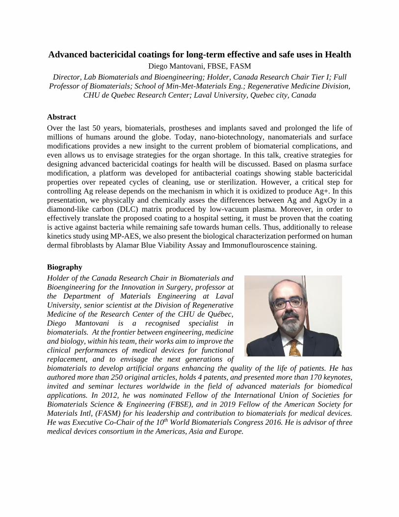
Advanced bactericidal coatings for long-term effective and safe uses in Health
Diego Mantovani, FBSE, FASM
Director, Lab Biomaterials and Bioengineering; Holder, Canada Research Chair Tier I; Full
Professor of Biomaterials; School of Min-Met-Materials Eng.; Regenerative Medicine Division,
CHU de Quebec Research Center; Laval University, Quebec city, Canada
Abstract
Over the last 50 years, biomaterials, prostheses and implants saved and prolonged the life of
millions of humans around the globe. Today, nano-biotechnology, nanomaterials and surface
modifications provides a new insight to the current problem of biomaterial complications, and
even allows us to envisage strategies for the organ shortage. In this talk, creative strategies for
designing advanced bactericidal coatings for health will be discussed. Based on plasma surface
modification, a platform was developed for antibacterial coatings showing stable bactericidal
properties over repeated cycles of cleaning, use or sterilization. However, a critical step for
controlling Ag release depends on the mechanism in which it is oxidized to produce Ag+. In this
presentation, we physically and chemically asses the differences between Ag and AgxOy in a
diamond-like carbon (DLC) matrix produced by low-vacuum plasma. Moreover, in order to
effectively translate the proposed coating to a hospital setting, it must be proven that the coating
is active against bacteria while remaining safe towards human cells. Thus, additionally to release
kinetics study using MP-AES, we also present the biological characterization performed on human
dermal fibroblasts by Alamar Blue Viability Assay and Immonuflouroscence staining.
Biography
Holder of the Canada Research Chair in Biomaterials and
Bioengineering for the Innovation in Surgery, professor at
the Department of Materials Engineering at Laval
University, senior scientist at the Division of Regenerative
Medicine of the Research Center of the CHU de Québec,
Diego Mantovani is a recognised specialist in
biomaterials. At the frontier between engineering, medicine
and biology, within his team, their works aim to improve the
clinical performances of medical devices for functional
replacement, and to envisage the next generations of
biomaterials to develop artificial organs enhancing the quality of the life of patients. He has
authored more than 250 original articles, holds 4 patents, and presented more than 170 keynotes,
invited and seminar lectures worldwide in the field of advanced materials for biomedical
applications. In 2012, he was nominated Fellow of the International Union of Societies for
Biomaterials Science & Engineering (FBSE), and in 2019 Fellow of the American Society for
Materials Intl, (FASM) for his leadership and contribution to biomaterials for medical devices.
He was Executive Co-Chair of the 10th World Biomaterials Congress 2016. He is advisor of three
medical devices consortium in the Americas, Asia and Europe.

Plasma Coatings – From Results to Innovation
Dirk Hegemann
Head of Plasma & Coating Group; Empa, Swiss Federal Laboratories for Materials Science and
Technology, Laboratory of Advanced Fibers, St.Gallen, Switzerland
Abstract
The use of plasma coatings is highly attractive to enhance biomaterials such as sensors, scaffolds,
antibacterial surfaces and others. Control over the formation of plasma coatings on the nanoscale
enables ultrathin films providing new surface properties. Investigations regarding mainly the
interaction with bacteria and proteins will be presented.
2 nm-thick hydrophobic cover layers on PDMS substrates of different crosslinking degree are used
to clarify the role of viscoelastic properties on bacterial growth indicating the lack of
mechanosensing abilities. Likewise, hydrophobic cover layers with varying film density are
explored to control water intrusion. Thus, a defined volume of water can be allowed to penetrate
a porous base layer. Protein adsorption of BSA is found to be affected by this hydration effect due
to orientation of water molecules in the subsurface. Moreover, controlled drug release from a Ag
reservoir is enabled for long-term antibacterial properties. On the contrary, undesired release from
conductive Ag-coated textile electrodes as used for ECG sensings can be avoided by passivation.
Recent progress in the understanding of plasma deposition processes enables increased control and
usability of functional plasma coatings at the nanoscale. Dry and environmentally friendly
processes can thus be implemented meeting the requirements for industrial applications.
Biography
Dr. Dirk Hegemann graduated in physics and earned a PhD
degree in materials science from TU Darmstadt, Germany.
As a scientist he worked with the Fraunhofer Institute for
Interfacial Engineering and Biotechnology in Stuttgart,
Germany, before moving to Empa, the Swiss Federal
Laboratories for Materials Science and Technology, in
2003. Currently, he is leading the group Plasma & Coating
at Empa's laboratory for Advanced Fibers in St.Gallen,
Switzerland. The main focus of his work concentrates on the
plasma treatment of polymeric substrates such as scaffolds,
membranes, textiles, packagings etc. by plasma etching,
plasma polymerization, and sputtering processes. Process
development and reactor design enable the transfer to industry.
Dirk Hegemann is appointed to the board of directors of the Swiss Physical Society (SPS) as well
as the International Plasma Chemistry Society (IPCS) and acts as Editor-in-chief for the journal
Plasma Processes and Polymers.

Melt Electrowriting: An Emerging Additive Biomanufacturing Technique for
Soft Tissue Engineering
Dr Elena De-Juan Pardo
Lab Head, Translational 3D Printing for Advanced Tissue Engineering Laboratory
(T3mPLATE)
Senior Lecturer, Harry Perkins Institute of Medical Research, The University of Western
Australia
Adjunct Associate Professor, Queensland University of Technology, Brisbane, Australia
Abstract
Melt electrowriting (MEW) is an emerging additive biomanufacturing technique capable of
printing fibrous constructs in ultra-high resolution, bringing down the jet diameter from the
micrometer to nanometer scale. Scaffolds manufactured by MEW are tailorable in terms of fibre
architecture, porosity and thickness. Moreover, MEW is a solvent-free process, compatible with
the use of medical grade thermoplastic polymers. These exclusive advantages make MEW an ideal
manufacturing technique for the production of scaffolds for a wide range of biomedical
applications. However, despite the great benefits of MEW, this technique is still at its infancy in a
few laboratory set-ups. In this talk, I will give an overview of the physical principles and unique
capabilities of MEW, followed by some examples on how to capitalize its potential to produce
scaffolds with controlled mechanical properties for soft tissue engineering. I will cover the
development of novel biomaterials capable of recapitulating the complex biomechanical features
of soft tissues. Native soft tissues are characterised by a strain-stiffening behaviour that makes
them very sensitive to load. Herein, the unique capabilities of MEW to produce biomimetic
scaffolds with controlled non-linearity, strain-stiffening behaviour, anisotropy and viscoelasticity
for soft tissue engineering will be presented.
Biography
Elena De-Juan-Pardo is a Senior Lecturer at The University
of Western Australia (UWA) and Laboratory Head at the
Harry Perkins Institute of Medical Research. She is a
materials engineer with 15 years of experience in biomaterials
and biofabrication for tissue engineering, regenerative
medicine and in vitro modelling. Her current research focuses
on the development of melt electrowriting, a pioneering 3D
printing technology that enables the production of highly
controlled fibrous scaffolds with tailored mechanical
properties for tissue engineering and biomedical applications.
From 2008-2012 she established and led the Tissue
Engineering and Biomaterials Group at the Centre of Studies
and Technical Research of Gipuzkoa (Spain) and served as Director of the Master in Biomedical
Engineering at the University of Navarra (Spain). In 2013 she joined the Centre in Regenerative
Medicine at the Institute of Health and Biomedical Innovation of the Queensland University of
Technology to further expand her interdisciplinary research skills in the areas of biomaterials,
tissue engineering and biofabrication. She served as Deputy Director of the Centre for the three
years prior to joining UWA.

Development of Biomimetic Nanostructured Antibacterial Titanium Surfaces
Elena Ivanova
School of Science, RMIT University, Melbourne, Victoria 3001, Australia
Abstract
Titanium is the material of choice for the manufacture of medical implants because of excellent
corrosion resistance and proven biocompatibility. The occurrence of premature implant failure is
due to implant-associated infections caused by the presence of pathogenic, often antibiotic resistant
bacteria, however, remains a prominent concern for clinicians.
Here we consider few examples of biomimetic antibacterial titanium surfaces. Naturally occurring
building strategies generate a remarkable compilation of technical equilibrium that is shared
amongst many biological materials. These principles of surface growth are rarely found on the
surfaces of conventional metals but have the potential to provide desirable properties when used
as templates for the natural materials. Antibacterial superhydrophobic quasi periodic self-
organized structures on titanium surfaces that possesses a surface topography mimicking that of
the surface of the lotus leaf, Nelumbo nucifera can be fabricated using femtosecond laser ablation
in a single processing step. A great deal of inspiration has resulted from studying the self-cleaning
insect wing surfaces. Hydrothermal etching of titanium surfaces, to produce random nanosheet
topologies that mimicked the surface architecture of dragonfly wings, has shown remarkable
ability to inactivate pathogenic bacteria via a physical mechanism. The results reported here
provide evidence that titanium surface nano-features can be engineered with a view to controlling
bacterial attachment.
Biography
Professor Elena P. Ivanova’s professional interests are
concentrated on development and research coordination in
fundamental and applied aspects of Nano/Biotechnology
including planar micro-devices, biomaterials,
immobilization of biomolecules and microorganisms in
micro/nano/environments, antimicrobial and bactericidal
surfaces, bacterial interactions with micro/nano-structured
surfaces. She received her Doctor of Philosophy from the
Institute of Microbiology and Virology, Ukraine, Doctor of
Science from the Pacific Institute of Bio-organic Chemistry,
Russian Federation, Juris Doctorate from the University of
Melbourne and Graduate Diploma from the Law Institute,
Victoria. Together with colleagues, Elena has published two books, edited 3 books, 26 book
chapters, 4 patents, in excess of 300 research papers. Professor Ivanova has been the recipient of
AIST and JSPS Fellowships (Japan), a UNESCO Biotechnology Fellowship, a Research
Excellence Award from the Governor of Primorye (Russia), the Prominent Young Doctor of
Science Award from the Russian Federation, the Morrison Rogosa Award from the American
Society for Microbiology (USA), the Australian Museum Eureka Prize for Scientific Research.
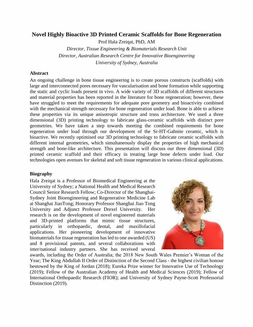
Novel Highly Bioactive 3D Printed Ceramic Scaffolds for Bone Regeneration
Prof Hala Zreiqat, PhD, AM
Director, Tissue Engineering & Biomaterials Research Unit
Director, Australian Research Centre for Innovative Bioengineering
University of Sydney, Australia
Abstract
An ongoing challenge in bone tissue engineering is to create porous constructs (scaffolds) with
large and interconnected pores necessary for vascularisation and bone formation while supporting
the static and cyclic loads present in vivo. A wide variety of 3D scaffolds of different structures
and material properties has been reported in the literature for bone regeneration; however, these
have struggled to meet the requirements for adequate pore geometry and bioactivity combined
with the mechanical strength necessary for bone regeneration under load. Bone is able to achieve
these properties via its unique anisotropic structure and truss architecture. We used a three
dimensional (3D) printing technology to fabricate glass-ceramic scaffolds with distinct pore
geometries. We have taken a step towards meeting the combined requirements for bone
regeneration under load through our development of the Sr-HT-Gahnite ceramic, which is
bioactive. We recently optimised our 3D printing technology to fabricate ceramic scaffolds with
different internal geometries, which simultaneously display the properties of high mechanical
strength and bone-like architecture. This presentation will discuss our three dimensional (3D)
printed ceramic scaffold and their efficacy in treating large bone defects under load. Our
technologies open avenues for skeletal and soft tissue regeneration in various clinical applications.
Biography
Hala Zreiqat is a Professor of Biomedical Engineering at the
University of Sydney; a National Health and Medical Research
Council Senior Research Fellow; Co-Director of the Shanghai-
Sydney Joint Bioengineering and Regenerative Medicine Lab
at Shanghai JiaoTong; Honorary Professor Shanghai Jiao Tong
University and Adjunct Professor Drexel University. Her
research is on the development of novel engineered materials
and 3D-printed platforms that mimic tissue structures,
particularly in orthopaedic, dental, and maxillofacial
applications. Her pioneering development of innovative
biomaterials for tissue regeneration has led to one awarded (US)
and 8 provisional patents, and several collaborations with
inter/national industry partners. She has received several
awards, including the Order of Australia; the 2018 New South Wales Premier’s Woman of the
Year; The King Abdullah II Order of Distinction of the Second Class - the highest civilian honour
bestowed by the King of Jordan (2018); Eureka Prize winner for Innovative Use of Technology
(2019); Fellow of the Australian Academy of Health and Medical Sciences (2019); Fellow of
International Orthopaedic Research (FIOR); and University of Sydney Payne-Scott Professorial
Distinction (2019).

Antimicrobial Biomaterials of Natural Origin and their Biomedical
Applications
Professor Ipsita Roy
Professor of Biomaterials; Department of Materials Science and Engineering; Faculty of
Engineering, University of Sheffield, Sheffield, UK
Abstract
Natural Polymers have the potential to be used in a variety of biomedical applications due to
their excellent biocompatibility, varied mechanical properties and sustainable resourcing.
There are a distinct class of natural polymers that are produced by controlled bacterial
fermentation including Polyhydroxyalkanoates (PHAs), Bacterial cellulose (BC), -
Polyglutamate (-PGA) and Alginate. The added advantage of this class of natural polymers
include the highly controlled production conditions resulting in repeatable properties.
PHAs are polymers of 3,4,5 and 6-hydroxyalkanoic acids produced by bacteria, mainly under
nutrient limiting conditions. In this work we have modified PHAs with various natural
antibacterial agents and active factors. In addition, a naturally antibacterial class of PHAs, thio-
PHAs have also been produced. All of these polymers have been characterised with respect to
their antibacterial activity against Staphylococcus aureus ATCC 6538 and Escherichia coli
ATCC 8739 following ISO22916. These antibacterial polymers have been used for the
development of tissue engineering scaffolds and medical devices.
BC is produced by several bacteria including Gluconacetobacter xylinus and has an inherent
hydrogel-like structure. In this work the surface of cellulose has been functionalized to produce
antibacterial BC. The cytotoxicity evaluation using HaCaT cells confirmed cytocompatibility
for both modified and unmodified BC.
Biography
Professor Ipsita Roy is an expert in microbial
biotechnology, natural biomaterials and their biomedical
applications. She is currently a Professor at the Department
of Materials Science and Engineering, Faculty of
Engineering, University of Sheffield, UK. Professor Roy
obtained her Ph.D. at the University of Cambridge and her
postdoctoral work was at the University of Minnesota, USA.
Subsequently, Professor Roy taught at Indian Institute of
Technology, India, and University of Westminster, London,
UK. Professor Roy has published over 100 papers in high
‘Impact Factor’ journals such as Biomaterials, Acta
Biomaterialia and ACS Applied Materials Interfaces and
has presented her work at numerous international conferences. Her group is currently focussed
on the production of novel polyhydroxyalkanoates (PHAs), a group of FDA-approved natural
polymers, their characterisation and application in hard/soft tissue engineering, wound healing
and drug delivery. She is an editor of the Journal of Chemical Technology and Biotechnology
(JCTB). Professor Roy has a total grant portfolio of 8.9 million pounds and has been the
scientific coordinator of two large EU projects REBIOSTENT and HYMEDPOLY.

Bioink Materials for Biomedical Applications
James J. Yoo
Professor, Wake Forest Institute for Regenerative Medicine; Wake Forest School of Medicine;
Winston-Salem, North Carolina, USA
Abstract
Tissue engineering and regenerative medicine has emerged as an innovative scientific field that
focuses on developing new approaches to repairing cells, tissues and organs. Over the years,
various engineering strategies have been developed to build functional tissues and organs for
clinical applications. In recent years, 3D bioprinting has gained an enormous attention as an
innovative tool that enables construction of complex 3D tissue structures for translational
applications. This developing field promises to revolutionize the field of medicine addressing the
dire need for tissues and organs suitable for surgical reconstruction. However, further development
is necessary to be able to bring this technology to the clinic. This session will discuss various
approaches to building complex tissue structures using the 3D printing technology. Development
efforts in the bioink materials unique to building 3D printed structures for biomedical applications
will also be discussed.
Biography
Dr. Yoo is Professor and Associate Director of the Wake
Forest Institute for Regenerative Medicine (WFIRM), with
a cross-appointment to the Departments of Urology,
Physiology and Pharmacology, and the Virginia Tech-Wake
Forest School of Biomedical Engineering and Sciences. Dr.
Yoo's research efforts have been directed toward the clinical
translation of tissue engineering technologies and cell-based
therapies. Dr. Yoo's background in cell biology and
medicine has facilitated the transfer of several cell-based
technologies from the bench-top to the bedside. A few
notable examples of successful clinical translation include
the bladder, urethra, vagina, and muscle cell therapy for incontinence. Dr. Yoo has been a lead
scientist in the bioprinting program at WFIRM and has been instrumental in developing skin
bioprinting and integrated tissue and organ printing (ITOP) systems for preclinical and clinical
applications.

Designing Hydrogel Inks for Extrusion Printing
Jason A. Burdick
Department of Bioengineering, University of Pennsylvania, Philadelphia, PA, USA
Abstract
Our laboratory is interested in developing new biomaterials for 3D printing, as well as new printing
processes to expand the utility of 3D printing in biomedical applications. This includes towards
the development of enabling technologies to advance our toolbox of materials and printing
approaches for 3D printing in precision medicine. Hydrogels represent a class of biomaterials that
have great promise for these applications, particularly due to our ability to engineer their
biophysical and biochemical properties and the potential for cell encapsulation during printing.
This presentation will provide an overview of our approaches to advance the applications of
hydrogels in extrusion-based 3D printing. This includes the engineering of shear-thinning and
self-healing hydrogels that can act as bioinks or as media for embedding printing technologies,
where various materials can be printed anywhere within 3D space and as sacrificial materials to
form channels. Further, we have developed both direct-curing and jammed microgel technologies
to print photocrosslinkable hydrogels that do not meet the stringent requirements (e.g., rheological
properties) for current printing techniques. Examples will be provided in how these new materials
and techniques have been used in the engineering of in vitro models (e.g., vessels) and for
translational tissue repair applications (e.g., cartilage repair).
Biography
Jason A. Burdick, PhD is the Robert D. Bent Professor of
Bioengineering at the University of Pennsylvania. Dr. Burdick’s
research involves the development of hydrogels through
techniques such as photocrosslinking and self-assembly and their
processing using approaches such as electrospinning and 3D
printing. The applications of his research range from controlling
stem cell differentiation through material cues to fabricating
scaffolding for regenerative medicine and tissue repair. Jason
currently has over 240 peer-reviewed publications and has been
awarded a K22 Scholar Development and Career Transition
Award through the National Institutes of Health, an Early Career Award through the Coulter
Foundation, a National Science Foundation CAREER award, a Packard Fellowship in Science and
Engineering, and an American Heart Association Established Investigator Award. He was recently
awarded the Clemson Award through the Society for Biomaterials and the George H.
Heilmeier Faculty Award for Excellence in Research. He is on the editorial boards of Tissue
Engineering, Biofabrication, and Advanced Healthcare Materials, and is an Associate Editor
for ACS Biomaterials Science & Engineering.

Interactions at the blood-material interface: reflections and reminiscences.
John L. Brash, FRSC
Distinguished University Professor,
School of Biomedical Engineering, Department of Chemical Engineering,
McMaster University, Hamilton, Ontario, Canada.
Abstract
First I want to thank Professors Kyla Sask and Hong Chen for their initiative and efforts in
organizing this workshop. I am greatly honoured to be seen, in a sense, as the “motivation” for the
inclusion of a session at the WBC (in Glasgow, where it all began for me) on the topic of blood-
material interactions, a topic on which I have spent by far the greatest part of my scientific career.
In this brief presentation I will look back on what I see as some of the highlights of my work,
which has focused on the role of protein adsorption in coagulation and thrombosis provoked by
blood-material contact, and on the exploitation/control of protein interfacial behaviour towards a
solution to these problems. It should be acknowledged that any success which may be attributed
to me must be shared with a long list of collaborators to whom I am most grateful and much
indebted.
Biography
John Brash is a Distinguished University Professor of
McMaster University. His main interest is in biomaterials
and biocompatibility with emphasis on materials for use in
blood contact. Both mechanistic and materials development
work have been pursued. The behaviour of proteins at the
blood-material interface has been an important underlying
theme. Collaborative research has been carried out with
laboratories in Canada, China, USA, France, Sweden and
Australia. He is a Fellow of the Royal Society of Canada
(2004) and recipient of the Clemson Award for Basic
Research (1994) and the Founders Award (2009), US
Society for Biomaterials; the C.P. Sharma Award, Indian
Society for Biomaterials and Artificial Organs (2016); and the Lifetime Achievement Award,
Canadian Biomaterials Society (2016).

3D Printing for Engineering Complex Tissues
John P. Fisher
NIBIB / NIH Center for Engineering Complex Tissue, Fischell Department of Bioengineering,
University of Maryland, College Park, MD, USA
Abstract
The generation of complex tissues has been an increasing focus in tissue engineering and
regenerative medicine. With recent advances in bioprinting technology, our laboratory has
focused on the development of platforms for the treatment and understanding of clinically
relevant problems ranging from congenital heart disease to preeclampsia. We utilize
stereolithography-based and extrusion-based additive manufacturing to generate patient-specific
vascular grafts, prevascular networks for bone tissue engineering, dermal dressings, cell-laden
models of preeclampsia, and bioreactors for expansion of stem cells. Furthermore, we have
developed a range of UV crosslinkable materials to provide clinically relevant 3D printed
biomaterials with tunable mechanical properties. Such developments demonstrate the ability to
generate biocompatible materials and fabricated diverse structures from natural and synthetic
biomaterials. In addition, one of the key challenges associated with the development of large
tissues is providing adequate nutrient and waste exchange. By combining printing and dynamic
culture strategies, we have developed new methods for generating macrovasculature that will
provide adequate nutrient exchange in large engineered tissues. This presentation will cover the
diverse range of materials and processes developed in our laboratory and their application to
relevant, emerging problems in tissue engineering.
Biography
Dr. John P. Fisher is the Director of NIBIB / NIH Center for
Engineering Complex Tissue, Fischell Family Distinguished Professor,
and Department Chair in the Fischell Department of Bioengineering at
the University of Maryland. As the Director of the Tissue Engineering
and Biomaterials Laboratory, Dr. Fisher’s group investigates
biomaterials, stem cells, bioprinting, and bioreactors for the
regeneration of lost tissues, particularly bone, cartilage, and
cardiovascular tissues. Dr. Fisher’s laboratory has published over 165
articles, book chapters, and proceedings (7500+ citations / 48 h-index)
as well as delivered over 340 invited and contributed presentations,
while utilizing over $15M in financial support from NIH, NSF, FDA,
NIST, DoD, and other institutions. Dr. Fisher has been elected Fellow of both the American
Institute for Medical and Biological Engineering (2012) and the Biomedical Engineering Society
(2016). Dr. Fisher is currently the Co-Editor-in-Chief of the journal Tissue Engineering and
Chair of the Tissue Engineering and Regenerative Medicine International Society - Americas
Chapter.

Augmentation Cystoplasty of Diseased Porcine Bladders
with Bi-Layer Silk Fibroin Grafts
Joshua R. Mauney
Associate Professor of Urology and Biomedical Engineering; Jerry D. Choate Presidential
Chair in Urology Tissue Engineering; University of California, Irvine, Irvine, CA
Abstract
Partial bladder outlet obstruction (pBOO) commonly results from neurogenic bladder and posterior
urethral valves. pBOO causes fibroproliferative remodeling of the detrusor, which results in
diminished bladder capacity, poor compliance, and incomplete emptying. Enterocystoplasty
presents the primary surgical procedure utilized to increase bladder capacity, reduce urinary
storage pressures, preserve renal function, and achieve urinary continence. Unfortunately, the
transposition of gastrointestinal (GI) segments into the urinary tract is associated with significant
complications, including chronic urinary tract infection (UTI), mucus production, and metabolic
abnormalities. Therefore, there is a significant need to develop alternative approaches for bladder
reconstruction. The search for an ideal "off-the-shelf" biomaterial for augmentation cystoplasty
remains elusive and current scaffold configurations are hampered by mechanical and
biocompatibility restrictions. In addition, preclinical evaluations of potential scaffold designs for
bladder repair are limited by the lack of tractable large animal models of obstructive bladder
disease that can mimic clinical pathology. The results of this study describe a novel, minimally
invasive, porcine model of pBOO that simulates clinically relevant phenotypes. Utilizing this
model, we demonstrate that acellular, bi-layer silk fibroin grafts can support the formation of
vascularized, innervated bladder tissues with functional properties.
Biography
Dr. Joshua Mauney is a tenured Associate Professor in the
Departments of Urology and Biomedical Engineering at the
University of California, Irvine. He holds the Jerry D. Choate
Presidential Endowed Chair in Urology Tissue Engineering. He
received his B.Sc. in Chemical Engineering and Ph.D. in
Biotechnology Engineering from Tufts University. Dr. Mauney’s
laboratory focuses on the development and evaluation of silk
fibroin grafts for the repair of visceral hollow organs including
the bladder, urethra, esophagus, and trachea. He also specializes
in the creation of novel large animal models of urinary tract and
gastrointestinal disease for preclinical medical device testing. Dr.
Mauney has been continuously funded from the National Institutes of Health since 2011 and
currently serves as the principal investigator on 2 R01 grants from NIDDK. Dr. Mauney has
authored >40 international peer-reviewed journal publications.

Novel Hydrogels and their Uses in Biomanufacturing
Justin Cooper-White
Professor of Bioengineering, School of Chemical Engineering; Senior Group Leader, Tissue
Engineering and Microfluidics Laboratory, Australian Institute for Bioengineering and
Nanotechnology; Co-Director, UQ Centre for Stem Cell Ageing and Regenerative Engineering;
Chair of School of Chemical Engineering; University of Queensland, Brisbane, Queensland,
Australia
Abstract
The clinical potential of stem cells in tissue engineering and regenerative medicine, and their
exploitation in in vitro disease models for drug toxicity and discovery, is recognised to be
significant, but remains a substantial challenge to realise and effect. Many of these challenges lie
in achieving high quality, high purity cellular products (whether stem cells, differentiated progeny
or neo-tissues) in sufficient numbers or volumes, reproducibly. Repurposed traditional culture
ware and FDA approved biomedical polymers for these applications has failed to address these
challenges.
In this workshop presentation, I will overview how new insights into the biology and physiology
of stem cells, their differentiated progeny and tissues, have been derived from synthetic hydrogels
that display physicochemical properties reminiscent of the natural cell microenvironment and that
can be engineered to display essential biological cues. I will also discuss how having learnt from
these insights we and others have developed next generation hydrogels that have seen applications
in cell bioprocessing and biomanufacturing. Lastly, I will highlight how high throughput
methodologies are now being used to, in an unbiased manner, identify novel ‘biomaterial-driven’
effectors of stem cell behaviours to unlock the clinical potential of stem cells in cell therapy, tissue
engineering and regenerative medicine.
Biography
Professor Cooper-White’s laboratory develops biomaterials
and biomicrodevices to decipher the roles of biophysical
and biochemical cues provided by the extracellular (niche)
microenvironment on stem cell fate. Such insights inform
the design of directive biomaterials that are applied to cell
therapy and musculoskeletal and cardiovascular
repair/regeneration. Prof. Cooper-White has over 200
publications in high impact journals, including Science
Advances, Nature Communications, Nature Protocols,
Biomaterials, Lab on a Chip, Stem Cells, Cell Stem Cell,
Stem Cells and Development, Integrative Biology and APL
Bioengineering. He has produced 6 Worldwide patents that
have reached National Phase Entry in USA, Europe and Australia in the areas of formulation
design for agriproducts, microbioreactor arrays (MBAs) and tissue engineering scaffolds. He has
received numerous awards, including most recently CSIRO OCE Science Leader Fellowship
(2013-2018), the AON Insurance and Life Sciences Queensland Regenerative Medicine Award
(2015) and the NHMRC Marshall and Warren Award for Research Excellence (2015).

3D Printing to Support Surgery and Interventions
Kawal Rhode
Professor of Biomedical Engineering; Head of Education, School of Biomedical Engineering &
Imaging Sciences, King’s College London, 4th Floor Lambeth Wing, St. Thomas’ Hospital,
London, United Kingdom
Abstract
3D printing offers many advantages including low cost, a wide range of materials, a direct link to
computer software and easy deployment. It is therefore not surprising that the use of this
technology in healthcare is an active area of research and development with many commercial
solutions already deployed, for example in dental practice. At King’s Health Partners, we are using
3D printing for a wide range of applications such as pre-operative planning, surgical and
interventional training, healthcare education, patient implants and medical robotics. This
presentation will highlight our experience and show how 3D printing is positively impacting
patient care, research and education at King’s.
Figure 1: (A) Digital CAD model of thoracic surgery training phantom (B) Physical model of
training phantom created using 3D printing.
Biography
Professor Rhode obtained his bachelor’s degree in Basic
Medical Sciences and Radiological Sciences at Guy's & St.
Thomas' Hospitals Medical School in 1992 and his
doctorate in Medical Physics from the Department of
Surgery, University College London in 2006. He has
worked in the field of healthcare technology research at
King’s College London since 2001 and is currently
Professor of Biomedical Engineering and Head of
Education for the School of Biomedical Engineering and
Imaging Sciences. His current research interests include
image-guided interventions, intelligent mechatronics
systems for interventions and ultrasound imaging, 3D
printing in healthcare and pedagogy for biomedical engineering. Prof. Rhode has more than 350
publications in journals, conference proceedings, book chapters and patents.
(B) (A)

Nanoengineering of plasma polymers for advanced biomedical devices
Krasimir Vasilev
School of Engineering, University of South Australia, Australia
Email: [email protected]
Abstract
In this keynote talk, I will give an overview of recent progress from my lab on development of
advanced surfaces capable of controlling infection, inflammation and stem cell differentiation.
Over the last few years, we have created the means to control the entire spectrum of surface
properties including chemical, physical, mechanical and topographical. We do that by
nanoengineering and tailoring traditional plasma polymer films using tools from nanotechnology.
By controlling surface properties, we are able to address medical challenges that are often
encountered with implantable devices such as infection and inflammation. We have developed
four distinct classes of antibacterial surfaces that are suitable for application on various medical
devices. These surfaces can be classified based on their mechanism of action as non-sticky, contact
killing, antimicrobial compound releasing and stimuli responsive. I will provide examples and
describe the strategies used to create these types surfaces, including such being translated onto
commercial devices in collaboration with industry. Recently, we have also revealed that the
mechanism of surface nanotopography induced modulation of inflammation is the unfolding of
adsorbed fibrinogen. This unfolding is surface nanotopography scale dependent and leads to the
exposure of (normally hidden) peptide sequences that activate the MAC-1 receptor of immune
cells. Finally, I will report on a bladder cancer diagnostic technology that we are currently
commercializing.
Biography
Professor Vasilev completed his PhD at the Max-Planck
Institute for Polymer Research in Mainz, Germany in 2005.
He is currently an NHMRC Fellow and a Humboldt Fellow,
and a Full Professor at the University of South Australia.
His research focuses on engineering and tailoring at a
molecular level of the disciplinary interphase, where
biological entities interact with biomaterials and devices.
He is the author of more than 200 publications, 5 patents and
has been awarded in excess of 20 million dollars of research
funding. His work results in translation of research
discoveries to tangible commercial outcomes such as device
for bladder cancer diagnostics and antibacterial surface for
hip and knee implants, both technologies being currently industrialized with commercial partners.
For his work, he has received various honors and awards such as the John A. Brodie Medal for
achievements in Chemical Engineering in 2016 and the International Association of Advanced
Materials Medal (IAAM medal) for contributions to the field of Advanced Materials in 2017. In
2017, he was elected a Fellow of the Royal Society of Chemistry (FRSC).

Mechanisms and analysis of blood-cell activation at biomaterials
Manfred F. Maitz, Claudia Sperling, Carsten Werner
Institute Biofunctional Polymer Materials
Leibniz Institute of Polymer Research Dresden, Hohe Strasse 6, 01069 Dresden, Germany
Abstract
Medical devices in contact with streaming blood, such as vascular stents, -grafts, heart valves,
tubes, or hemodialysis filters, have increasing applications in clinical medicine. As foreign
materials, they tend to activate plasmatic and cellular defense systems of coagulation and
inflammation. In blood, these reactions do not stay local but propagate into the whole body and
may pose risks to remote organs. Understanding and analysing these reactions, therefore, is
mandatory in order to improve the hemocompatibility of the materials.
Blood platelets and leukocytes are effector cells of blood coagulation and inflammatory responses,
respectively. Upon activation, platelets induce clot formation by aggregation, propagation of the
coagulation cascade, and exhibition of pro-coagulant properties. Activated leukocytes damage
tissue by the release of proteases and reactive oxygen species; they also attract and activate more
inflammatory cells via cytokine release.
This talk shall present the pathways of cell activation and their interplay. Test systems for in vitro
screening of cell activation and suitable parameters for analysis shall be presented.
Biography
Manfred Maitz is a medical doctor by education. He has
been working in the field of biomaterials for more than 20
years in institutes in Würzburg, Ulm, Rossendorf (Dresden),
Chengdu, and Dresden. He is currently group leader for
hemocompatible surfaces at the Leibniz Institute of Polymer
Research Dresden, Germany, and guest professor at
Southwest Jiaotong University, Chengdu, China.
With his co-workers he investigates pathways of blood-
biomaterial interaction and analyses materials for blood-
contacting devices. The focus of his research is on the
development of auto-controlled, feedback-responsive
materials which have high hemocompatibility due to their
interaction with physiological pathways.

Blood-material interactions from a bleeding perspective
Maud Gorbet1, Matthew Robichaud1, Kassem Ashe2 1Material Interactions with Biological Systems Laboratory
Systems Design Engineering, University of Waterloo, Ontario, Canada
2 St. Mary’s General Hospital, Kitchener, Ontario, Canada
Abstract
Despite years of research, we have yet to design materials that are truly blood compatible: cardiovascular
implant recipients continue to require lifelong anticoagulant therapy while heparin is also routinely needed
during cardiopulmonary bypass surgery and hemodialysis. Significant efforts have been undertaken to
characterize blood-material interactions and better understand the mechanisms involved in cell activation
and the interplay between the coagulation, fibrinolytic and complement systems. Due to the higher
prevalence of clotting complications, blood-material interactions are most often characterized from a
thrombotic perspective. However, the outcome of cardiopulmonary bypass surgery can also be excessive
bleeding. While the incidence of excessive postoperative bleeding may be reported to be as low as 6%,
these patients require a significant volume of blood transfusion, often need additional surgery and have a
higher risk of morbidity. In this talk, we will discuss how contact with biomaterials and the stresses that
blood cells are exposed to during CPB can result in a loss of functionality and contribute to bleeding
complications. To provide further context to the discussion, recent in vivo data will also be presented.
Biography
Maud Gorbet graduated from the Université de Technologie de Compiègne
(UTC) in biological engineering, option Biomateriaux, in 1993 and after
working in a canadian biomedical company pursued her PhD in chemical
engineering at the University of Toronto (Canada) investigating the
mechanisms of material-induced thrombosis in vitro. As a Senior Biomaterial
Scientist at Rimon Therapeutics Ltd, a Toronto startup, she worked on the
biological efficacy and safety of wound healing biomaterials. Her strong
research interests in understanding the mechanisms involved in material’s
biocompatibility led her to join the University of Waterloo (UW) in 2007. Her
research program involves the development of better in vitro models and tools
to assess biocompatibility and the study of the mechanisms involved in
material-induced cell activation. Over the past 10 years, she has collaborated
with start-up companies, ophthalmic industries, clinicians, scientists and engineers both nationally and
internationally (France, Australia and the USA). She also enjoys teaching about medical devices and
biocompatibility. She recently spearheaded the creation of the biomedical engineering undergraduate
program at UW and was appointed Director of the biomedical engineering program for its initial 5 years
and is currently the interim chair for Systems Design Engineering.

How to publish successfully: writing a scientific article
Neil Hammond
Executive Editor, Biomaterials Science, Royal Society of Chemistry, Thomas Graham House,
Milton Road, Cambridge CB40HF, United Kingdom.
Abstract
Writing and publishing journal articles is an unavoidable component of any modern research
career. Nevertheless, formal training in how to write effectively in order to maximize the chance
of success is frequently overlooked. Indeed, good science is often poorly communicated, even by
senior experienced researchers. In this workshop Dr Hammond will present his perspective, as
both an author of scientific articles and as a publishing professional, on the guiding principles
behind good science writing and on specific strategies and tips for improving written
communication. He will focus on the traditional format of the scientific article (abstract, title,
introduction, etc), but with reference to techniques and approaches that can be successfully
translated to other written formats (project proposals, PhD thesis, grant proposal, etc).
Biography
Dr Hammond received an MPhys degree in Physics from
Liverpool University in 1998, and a PhD in Nuclear
Structure Physics, also from Liverpool University, in 2002.
He completed a 2-year postdoctoral position at Argonne
National Laboratory, USA in heavy-ion physics research
before leaving research to begin a career in publishing. He
is currently an Executive Editor at the Royal Society of
Chemistry, the UKs professional body for chemical
scientists, and the not-for-profit publisher of 44 academic
journals. Neil is responsible at the RSC for the development
of 5 journals, including Biomaterials Science. He has held
various roles in academic publishing over the last 15 years,
and has worked with a diverse range of academic
communities and learned societies. He has co-authored more than 50 articles in peer-reviewed
journals.

Advances in bioprinting technology from layer-by-layer cell printing to
volumetric bioprinting
Riccardo Levato
Assistant Professor, Department of Orthopaedics, University Medical Center Utrecht,
Regenerative Medicine Center Utrecht, and Department of Equine Sciences, Utrecht University,
Utrecht, The Netherlands
Abstract
The function of living tissues is intimately linked to their complex architectures. Biofabrication
technologies are rapidly advancing as powerful tools capable to capture salient features of tissue
composition and thus guide the maturation of engineered construct into mimicking functionalities
of native organs. In biofabrication, multiple cell types and biomaterials are patterned in three
dimension through automated processes, either via bioprinting or bioassembly. The current
paradigm in bioprinting relies on the additive layer‐by‐layer deposition and assembly of repetitive
building blocks, typically cell‐laden hydrogel fibers or voxels, single cells, or cellular aggregates.
Since its initial conception and its first implementations through inkjet printing technologies,
bioprinting rapidly introduced a new toolset for bioengineers and material scientists to produce
new strategies to restore the function of impaired tissues. In this contribution, both currently
available and innovative bioprinting approaches will be reviewed, with a particular focus on how
these techniques can be combined to mimic the multi-material hierarchical composition of living
tissues. Key concepts underlying extrusion, laser and light-based technologies will be discussed,
together with the recent emergence of field-based printing methods. Finally, technological
advances and challenges towards the biofabrication of large, clinically-relevant multi-tissue
constructs via the development of volumetric bioprinting will be discussed.
Biography
Dr. Levato’s research focuses on the development of novel
biofabrication strategies to create lab-made tissue models
and transplantable engineered grafts, particularly for the
regeneration of the musculoskeletal system. His lab
integrates expertise in engineering, stem cell biology,
biomaterials and cartilage and bone pathophysiology, to
translate biofabricated structures towards novel treatments
for the regeneration of damaged articulating joints. The
application of said technologies for engineering soft tissues
is also explored. For his work on biofabrication, he received
several awards, including the 2018 Orthoregeneration
Network Fellowship by the International Cartilage Repair
Society, the 2016 Wake Forest Institute for Regenerative Medicine Young Investigator Award and
the 2015 Julia Polak award by the European Society for Biomaterials. Prior to his appointment at
UMCU, Dr. Levato also worked in several research groups in the field of Biomaterials: 3Bs,
University of Minho, (Portugal); BioMatLab, Technical University of Milan (Italy), Institute for
Bioengineering of Catalonia (Spain), and he holds a cum laude PhD in Biomedical Engineering
from the Technical University of Catalonia (Spain). Currently he is mentoring 6 PhD students.

Photopolymerization-Based 3D Printing for Medical Device Applications
Roger Narayan
Professor of Biomedical Engineering;
University of North Carolina and North Carolina State University, Raleigh, NC, USA
Abstract
In this talk, I will describe recent advances in photopolymerization-based 3D printing
technologies to process microstructured and nanostructured materials for medical applications.
For example, an additive manufacturing approach known as two photon polymerization has been
used for selective polymerization of photosensitive resins. Polymerization of structures with
microscale and nanoscale features is achievable since multiphoton absorption exhibits a
nonlinear relationship with the incident light intensity. We have used two photon polymerization
used to create several types of medically-relevant structures with microscale and nanoscale
features out of photosensitive polymers and organically-modified ceramic materials. Materials
testing and application-specific device testing, including in vivo studies, will be considered.
Biography
Dr. Roger Narayan is a Professor in the Joint Department of
Biomedical Engineering at the University of North Carolina
and North Carolina State University. He is an author of over
two hundred publications as well as several book chapters
on processing and characterization of biomedical materials.
Dr. Narayan has also edited several books, such as the
textbook Biomedical Materials (Springer) and the handbook
Materials for Medical Devices (ASM International). Dr.
Narayan has received many honors for his research
activities, such as the NCSU Alcoa Foundation Engineering
Research Achievement Award, the NCSU Sigma Xi Faculty
Research Award, the University of North Carolina
Jefferson-Pilot Fellowship in Academic Medicine, the University of North Carolina Junior Faculty
Development Award, the National Science Faculty Early Career Development Award, the Office
of Naval Research Young Investigator Award, and the American Ceramic Society Richard M.
Fulrath Award. Dr. Narayan has been elected as Fellow of the American Ceramic Society, AAAS,
ASM International, and AIMBE

Scalable Manufacturing of Human Pluripotent Stem Cell-Derived Cardiac
Microtissues
Sean P. Palecek
Director for Research, Center for Cell Manufacturing Technology; Milton J. and Maude
Shoemaker Professor; Department of Chemical and Biological Engineering; University of
Wisconsin – Madison, Madison, Wisconsin, USA.
Abstract
In recent years, tremendous advances have been made in differentiating human pluripotent stem
cells (hPSCs) to cardiomyocytes and evaluating these cells in preclinical models of heart disease.
While hPSC-derived cardiomyocytes have improved cardiac function in animals after myocardial
infarction, cell viability and immature phenotypes limit efficacy. Also, a lack of robust and
scalable manufacturing platforms plagues efforts to advance these cells into human clinical trials.
In an effort to improve cell quality and increase manufacturability, we have developed aggregates
consisting of iPSC-derived cardiomyocytes, cardiac fibroblasts, and endothelial cells that can be
maintained in suspension culture. We found that endothelial cells and cardiac fibroblasts enhance
distinct maturation phenotypes of cardiomyocytes including marker expression, morphology,
contractility, and electrophysiology in 2D and 3D models. These effects on maturation are most
pronounced if endothelial cells and fibroblasts are in direct contact with cardiomyocyte progenitors
during manufacturing. Finally, we have developed methods for scalable production of
therapeutically relevant numbers of cardiomyocytes (~109 cells) in 3D aggregates and found that
suspension manufacturing reduces costs by ~80% as compared to 2D manufacturing, largely due
to reductions in media costs. These 3D manufacturing platforms have the potential to advance
manufacturing of human cardiomyocytes for therapeutics and in vitro testing applications.
Biography
Sean Palecek is the Milton J. and Maude Shoemaker Professor
and Vilas Distinguished Achievement Professor in the
Department of Chemical & Biological Engineering at the
University of Wisconsin – Madison. Sean is the Bioengineering
Thrust Leader for the UW Stem Cell and Regenerative Medicine
Center, the Director for Research for the National Science
Foundation Center for Cell Manufacturing Technologies
(CMaT), and the Director for Research Innovation for the
Forward BIO Institute. Sean is also a Fellow at the Allen Institute
for Cell Science. Sean’s research lab studies how human
pluripotent stem cells (hPSCs) sense and respond to
microenvironmental cues in making fate choices, with a focus on
differentiation to cardiovascular lineages. Sean’s lab has
generated novel mechanistic insight and developed protocols for
differentiation of hPSCs to cardiovascular and neurovascular cell
types. They strive to engineer fully-defined, animal component-
free differentiation platforms, compatible with biomanufacturing of cells and tissues for in vitro
and in vivo diagnostic and regenerative medicine applications.

3D Bioprinting Applications - Opening New Pathways in Scientific Research Dr. Simon MacKenzie
Chief Executive Officer, REGENHU
Abstract
Bioprinting is galvanizing healthcare. From academia to research institutions, right through to the
biotech and pharmaceutical industries, more and more institutions are harnessing the power of
bioprinting technologies and their applications to carry out their research goals.
In this talk, I will give a brief overview of REGENHU, 3D bioprinting applications, from drug
discovery to tablet formulation, and examples of how our partners have advanced their research
using our instruments. The aim of this talk to create more awareness of bioprinting, an ever-
evolving discipline still in its infancy, its many applications across a wide range of fields, and how
REGENHU’s bioprinting technologies can help researchers & scientist across all domains achieve
their research objectives.
Biography
An entrepreneur, researcher, and scientist, Simon
MacKenzie serves as REGENHU’S Chief Executive
Officer. For more than 19 years, Dr. MacKenzie held senior
positions in strategic business development in the domains
of pharma, drug discovery, biotech and research, and has
published several scientific papers.
Dr. MacKenzie has a PhD in Biochemistry from the
University of Dundee and completed two industrial
collaborative post-docs in inflammatory, oncology and
metabolic disease.

3D Bioprinting for Regenerative Medicine
Tamer Mohamed
CEO, Aspect Biosystems
Vancouver, BC, Canada
Abstract
Aspect Biosystems is a Canadian biotechnology company operating at the leading edge of 3D
printing and regenerative medicine. Our proprietary microfluidic 3D bioprinting platform is
enabling advances in biology, disease research, drug discovery, and regenerative medicine.
Aspects current clinical pipeline of therapeutic tissues has two main areas of focus: orthopaedic
implants and cellularized implants for metabolic diseases. Our lead orthopaedic indication is a
personalized 3D printed meniscus tissue for treatment of patients with severe meniscal tears. Our
second lead indication in preclinical development is an implantable 3D printed allogeneic tissue
patch for treatment of Type 1 Diabetes. In addition to our internal therapeutic tissue programs, we
strategically partner with pharmaceutical and biotechnology companies, as well as academic
researchers, to create physiologically and commercially relevant tissues. These tissues are used to
advance and accelerate drug discovery and development, and enable the creation of cutting edge
tissue therapies of the future.
Biography
An entrepreneur, engineer, inventor, Tamer currently serves
as CEO of Aspect Biosystems. Tamer co-founded Aspect in
2013 and has played a leading role in the overall corporate,
business, and technology development. Under his
leadership, Aspect has secured significant funding, entered
strategic collaborations with best-in-class pharmaceutical
and biotechnology companies, and developed commercial
products. In his previous appointment as Chief Technology
Officer of the company, Tamer drove the innovation and
development of the company’s core technologies and
intellectual property.
As a leader in the field of 3D bioprinting, he has been invited to speak on this topic at venues
ranging from TEDx to industry, scientific and executive conferences. In 2017, he was awarded
BC's Top 40 under 40 award for demonstrating excellence in business, judgement, leadership, and
community contribution. Tamer serves on the Board of Directors for ACETECH, a non-profit
training and mentoring organization for CEOs of technology and life sciences companies, and on
the Board of Directors for The Stem Cell Network, an organization focused on building Canada’s
stem cell and regenerative medicine research sector.

Protein and Cell Interactions with Biomaterials Are the Basis for Device Acceptance or Rejection by the Body
Thomas Horbett Professor emeritus, Bioengineering and Chemical Engineering, University of Washington,
Seattle, Washington
Abstract Probably the single most important event of my scientific career was a visit to the McMaster University laboratories of Professors J. Fraser Mustard and Raelene L. Kinlough-Rathbone to learn about preparing and radiolabeling blood platelets. My visit was made possible because of the help of John Brash and Irwin Feuerstein. Many of my lab’s subsequent studies involved use of platelets. So in making my presentation at this workshop in honor of John Brash, I will emphasize some of the major findings about platelets and biomaterials made in my laboratory, probably the most important of which is the ability of platelets to adhere to polymeric surfaces having even very low levels of fibrinogen adsorbed from blood plasma. In addition, at the request of Professor Sask, I will spend some time explaining the methods we developed for quantitative measurement of protein adsorption to surfaces from complex mixtures of proteins such as plasma or serum using the 125I- radiolabeled protein method.
Biography Professor Horbett did research and teaching in biomaterials for over 40 years the University of Washington (UW) after receiving his PhD in biochemistry from the UW in 1970. He received many NIH grants and published many research articles related to his main research area of protein and cell interactions with biomaterials. He received the Society for Biomaterials Award (SFB) for Research in 1989 and became an SFB fellow 1994 and an International fellow in 1996. He became a fellow of the AIMBE in 1995. In 2018, he received a Founder’s Award from the SFB.

Designing bioink, bioresin and spheroid bioassembly platforms
Professor Tim Woodfield, PhD, FBSE
Director, Christchurch Regenerative Medicine and Tissue Engineering (CReaTE) Group
Director, Centre for Bioengineering & Nanomedicine
University of Otago, Christchurch, New Zealand
Abstract
Biofabrication technologies, including 3D bioprinting and bioassembly, enable the generation of
engineered constructs that replicate the complex 3D organization of native tissues via automated
hierarchical placement of cell-laden bioinks, tissue modules, and/or bioactive factors. Photo-initiated
radical polymerization combining light and photo-initiators to generate radicals for crosslinking photo-
polymerizable macromers, has been widely employed in 3D bioprinting of cell-laden hydrogels. Despite
rapid advances in biofabrication technologies, no universal bioink exists. The major challenge for
translational regenerative medicine is that the processing requirements (biofabrication window) of current
bioinks is narrow, requiring optimization of each bioink for each individual biofabrication technique and
specific tissue niche e.g. high viscosity “shear thinning” bioinks necessary for extrusion bioprinting versus
low viscosity bioinks for lithography-based bioprinting. This presentation discusses alternative strategies
to provide highly tunable bioinks that 1) promote a specific cell-instructive niche and 2) are printable across
multiple biofabrication technologies, including extrusion-, lithography- and microfluidic-based bioprinting.
This talk will describe the design of versatile, photo-clickable bioinks and bioresins for biofabrication of in
vitro models targeting cartilage, bone and vascular network regeneration, and how their convergence with
3D spheroid bioassembly and dual perfusion bioreactor (MPS) platforms offer new paradigms for high-
throughput screening, “on-chip” and osteochondral tissue repair applications.
Biography
Tim Woodfield is Professor of Regenerative Medicine at
the University of Otago Christchurch, New Zealand. He
holds a prestigious Rutherford Discovery Fellowship from
the Royal Society of New Zealand, and is Principal
Investigator within the Medical Technologies Centre of
Research Excellence (CoRE). His research technology
platform involves complex 3D Biofabrication and Additive
Manufacturing of biomaterial scaffolds and medical
devices applied to regenerative medicine of cartilage and
bone, including advanced 3D tissue culture models and
high throughput screening. He has published over 105 peer reviewed journal articles, book
chapters and published conference proceedings (h-index: 30). He is the current President Elect and
Executive Board Member of the International Society for Biofabrication (ISBF). He is the former
President of the Australasian Society for Biomaterials & Tissue Engineering (ASBTE), and was
recently awarded the ASBTE Award for Research Excellence. He also sits on the Tissue
Engineering and Regenerative Medicine International Society Asia Pacific (TERMIS-AP)
Council, and is an Editorial Board Member for Biofabrication, APL Bioengineering, and Frontiers
in Bioengineering & Biotechnology.

Growth of nanostructured patterns by plasmas for biomedical applications
Uroš Cvelbar
Head, Department for Gaseous Electronics (F-6), Jožef Stefan Institute, Jamova cesta 39,
Ljubljana, Slovenia
Abstract
Plasma as a discharge state of the gas is considered nowadays as a cutting-edge tool which can
manipulate objects at the atomic or molecular scale. Furthermore, plasma can also tailor the
surfaces beyond morphology by creating targeted chemical bonds or modifying underlaying
material. These kinds of surfaces possess certain properties which can control the interactions with
components of living systems, inducing favourable response from the biological entities, and as
such can direct the course of a therapy or diagnostic procedure. In this respect, plasmas can be
used to initiate even more favourable surface morphologies or selective responses, enabling the
biomaterials even more favourable or selective interaction.
In this talk, I will report the developments on the field of cold plasma nanotexturing of organic or
inorganic materials for targeted response of cells, recognition of bacteria or single organic
molecules relevant to biomedicine. The special attention will be given to cases like plasma
texturing and nanostructuring of polymers for targeted attraction of different cells or its
differentiation, or inorganic materials like metal oxides, metal alloys, etc. for targeted trapping,
adhesion and recognition of bacteria or other single molecules of interest. The aim of all these is
either to improve biocompatibility of plasma nanostructured material, e.g. before body
implantation, or use the surface for biomedical diagnostic purposes and detection, e.g. reusable
SERS substrates for detection and recognition of bacteria.
Biography
Professor Cvelbar’s research focusses on advancing
nanoscience, nanotechnology, biomaterials, biotechnology
and biomedicine in the cross-roads with plasma science.
Prof Cvelbar has authored +175 international peer-reviewed
journal publications, several books and +18 patents. Prof
Cvelbar’s research has been continuously supported by
different National Research Councils (e.g. ARRS, MIZŠ),
European Union, NATO, and also attracts significant
interest from industrial partners. He is executive board
member of DST at Electrochemical Society, the chair of
Plasma Nanoscience, fellow of WAAS and associated editor
of several journals. Prof Cvelbar has successfully
supervised to completion 7 PhD students and 10 MSc
students. He is currently involved in the supervision of 5
PhD students. Additionally, he has managed and mentored of 9 postdoctoral (PD) research fellows,
all of whom have remained in engineering/science and have secured employment in industry or
academia. Currently, advising 3 PD research fellows.

3D Bioprinting for in vitro Tissue Engineering
W. Sun
Department of Mechanical Engineering, Drexel University, USA
Biomanufacturing Center, Dept. of Mechanical Engineering, Tsinghua University, Beijing, China
Abstract
3D Bio-Printing uses living cells to build in vitro physiological models. The printed tissue models
have been widely applied to regenerative medicine, studying disease pathogenesis, developing
molecular therapeutics, and screening drugs. This presentation will review the basic principle of
cell printing and report our recent study on printing in vitro tissue models for regenerative
medicine, disease study and drug testing. An overview of a cell printing process and enabling
printing techniques will be introduced, followed by examples of printing different tissue models
for tissue engineering and drug testing, including printing ESCs for formation of embryonic body,
printing hiPSC cells for hepatocyte differentiation and integration with microfluidics for drug
testing, printing neuron cells to construct in vitro neural network and studying DNQX influence,
and printing cancer (Hela) cells to build 3D in vitro tumor models for studying chemoresistance
and expressions of genes and tumor markers. Comparison of biological data derived from 3D
printed models with 2D planar models in petri-dishes will be given wherever appropriate. A
personal opinion on challenges and opportunities of 3D bio-printing will also be shared.
Biography
Wei Sun, Albert Soffa Chair Professor of Mechanical
Engineering, Drexel University, and National “Thousand-
Talent” Distinguished Professor and Director of
Biomanufacturing Center, Tsinghua University, China. Dr.
Sun’s research has been on Biofabrication, 3D Bio-Printing,
and Tissue Engineering. Dr. Sun’s research has been funded
by NSF, DARPA, NASA, the Chinese Natural Science
Foundation, the Chinese Ministry of Science and
Technology, and the Chinese Ministry of Education. Dr.
Sun has published over 150+ peer-reviewed journal papers
with 9100+ SCI citations, 70+ patent applications, and 350+
invited presentations in the field of his research. Dr. Sun is
the Founding President for International Society of Biofabrication (2010-2014), and the Founding
Editor-in-Chief for journal Biofabrication (2009-present). Dr. Sun received Award of
Distinguished Visiting Fellow from the Royal Academy of Engineering, UK (2018), the Senior
Investigator Award from International Society of Biofabrication (2017), and MII / Fralin Visiting
Scholar Award from Virginia Tech (2015).

Personalized implants
Willem van Oeveren
Director Haemoscan. Laboratory for Blood Compatibility and biomarker detection
Stavangerweg 23-23, 9723JC Groningen, The Netherlands
Abstract
Thrombosis and inflammation are responses of blood components to any foreign body. With a few
exceptions these responses should be kept to a minimum.
Implant materials are being tested extensively before clinical use with random animal or human
blood, with the assumption that all species and recipients show a comparable response to a certain
type of material.
However, thrombotic and inflammatory reactions are highly different between individuals,
resulting in either tolerance or in unacceptable high levels of activation with the same material. In
particular the extend of thrombosis shows marked differences between individuals, which can be
visualized and quantified in detail by sensitive surface markers.
Many implant procedures are elective, which allows a screening protocol to be performed in
advance. This protocol should be performed with a small amount of blood from the patient in an
in vitro test system with the implant material. By intensive contact of blood with material is such
system data can be obtained in a short period of time. The outcome of the tests could result in
choice for another type of material, another coating, or a negative advice to consider implantation
of foreign body materials.
Biography
Mr Willem van Oeveren studied Medical Biology and
obtained a PhD in medicine on blood activation during use
of a heart-lung machine. As associate professor at the
university hospital in Groningen, The Netherlands, he
(co)authored more than 200 peer reviewed articles.
Since 1999 he participates in the ISO10993 working
committee.
In 2004 Willem was the founder of Haemoscan, which is a
privately owned laboratory for Blood Compatibility and
Biomarker analysis, which he is leading today. Haemoscan became a certified laboratory dedicated
to blood activation during contact with biomaterials, which is tested in fresh blood from healthy
human volunteers.