exploring the role of lhc protein structure and...
TRANSCRIPT

Exploring the Role of LHC Protein Structure and Function in the Evolution of NPQ Mechanisms in Eukaryotic Photosynthetic Organisms
by
Erika Marie Erickson
A dissertation submitted in partial satisfaction of the
requirements for the degree of
Doctor of Philosophy
in
Plant Biology
in the
Graduate Division
of the
University of California, Berkeley
Committee in charge:
Professor Krishna K. Niyogi, Chair Professor Peter H. Quail
Professor Anastasios Melis Professor Graham R. Fleming
Spring 2016


1
Abstract
Exploring the Role of LHC Protein Structure and Function in the Evolution of NPQ Mechanisms in Eukaryotic Photosynthetic Organisms
by
Erika Marie Erickson
Doctor of Philosophy in Plant Biology
University of California, Berkeley
Professor Krishna K. Niyogi, Chair
The initial step of photosynthesis occurs when light energy is absorbed by an array of pigments surrounding the photosynthetic reaction center. Chlorophyll and carotenoid molecules are coordinated by members of a family of intrinsic thylakoid membrane proteins known as Light-Harvesting Complex (LHC) proteins. LHCs are essential for stabilizing and tuning the spectroscopic characteristics of each individual pigment, as well as the pigment array, to allow for efficient energy transfer. Despite a high degree of sequence conservation within the protein family, each of the LHCs performs a different role in modulating the energy landscape of the thylakoid membrane. Two stress response LHC proteins, LHCSR and PSBS, are known to be essential for non-photochemical quenching in some photosynthetic eukaryotes. It has been shown that LHCSR is necessary for quenching in the green alga Chlamydomonas reinhardtii. Homologs of this stress-response protein are present in many photosynthetic eukaryotes, even those with chloroplasts that are of a red algal origin, but LHCSR is absent from terrestrial vascular plants. Likewise, PSBS is necessary for quenching in the plant Arabidopsis thaliana, but PSBS is only found in the green lineage of photosynthetic organisms. The molecular mechanism of both of these proteins is still unknown.
This work describes the relationship between protein structure and quenching function in the stress response protein LHCSR. I show the distinct contributions to non-photochemical quenching from each of the two LHCSR protein isoforms, LHCSR1 and LHCSR3, in C. reinhardtii. I also contributed to the identification of three lumen-exposed residues in LHCSR3 that are required for quenching function. By using directed mutagenesis and expression in heterologous and native organisms, this shows that while LHCSR is necessary for nearly all non-photochemical quenching in its native cellular environment within C. reinhardtii, it is not sufficient to activate quenching in plants when expressed heterlogously. By altering some of the pigment-binding sites within LHCSR3, however, the heterologously expressed mutant LHCSR3 can activate additional quenching in a plant system.
This also explores possible roles of the PSBS protein from C. reinhardtii, which has not previously been shown to have a function. Here, I present data showing that the algal PSBS protein can enhance non-photochemical quenching when expressed heterologously in plants. Since we do not see the same behavior in the alga, if PSBS is expressed in C. reinhardtii it most likely plays a

2
different role or is not sufficient to induce quenching alone. Heterologous co-expression of both LHCSR and PSBS from C. reinhardtii results in enhanced quenching in the tobacco, Nicotiana benthamiana. The enhanced quenching phenotype is dependent upon PSBS being pH-sensitive. While no evidence has yet been obtained showing an interaction between PSBS and LHCSR in C. reinhardtii, this motivates further exploration.
In order to better understand the evolution of photoprotective mechanisms employed by photosynthetic eukaryotes, I have also contributed to a genome annotation project aimed at establishing the marine heterokont alga, Nannochloropsis oceanica CCMP 1779, as a new model alga for studying photosynthesis. I identified three potential stress-response LHCs in the organism’s genome based on sequence homology, whose functionality and expression characteristics are being investigated by other researchers.

i
Contents
Contents . . . . . . . . . . . . . . . . . . . . . . . . . . . . . . . . . . . . . . . . . . . . . . . . . . . . . . . . . . . . . . . . . . . . . . . . . . . . . . . . . . . . . . . . . . . . . . . . . . . . . . . . . . . . . . . . i
List of Figures . . . . . . . . . . . . . . . . . . . . . . . . . . . . . . . . . . . . . . . . . . . . . . . . . . . . . . . . . . . . . . . . . . . . . . . . . . . . . . . . . . . . . . . . . . . . . . . . . . . . . . . i i i
List of Tables . . . . . . . . . . . . . . . . . . . . . . . . . . . . . . . . . . . . . . . . . . . . . . . . . . . . . . . . . . . . . . . . . . . . . . . . . . . . . . . . . . . . . . . . . . . . . . . . . . . . . . . . . v i
List of Symbols and Abbreviat ions . . . . . . . . . . . . . . . . . . . . . . . . . . . . . . . . . . . . . . . . . . . . . . . . . . . . . . . . . . . . . . . . . . . . . . . . . . . v i i
Acknowledgements . . . . . . . . . . . . . . . . . . . . . . . . . . . . . . . . . . . . . . . . . . . . . . . . . . . . . . . . . . . . . . . . . . . . . . . . . . . . . . . . . . . . . . . . . . . . . . . . . . . x
Chapter 1
Light stress and photoprotection in eukaryotic photosynthetic organisms . . . . . . . . . . . . . . . . . . . . . . . 1 1.1 Light stress ........................................................................................................................................... 1 1.2 Photoprotection ................................................................................................................................... 3 1.3 Significance of this research ................................................................................................................ 16 1.4 Acknowledgements ............................................................................................................................ 17
Chapter 2
Identif icat ion of a molecular mechanism of quenching in the Chlamydomonas reinhardti i s tress response LHC protein, LHCSR3 . . . . . . . . . . . . . . . . . . . . . . . . . . . . . . . . . . . . . . . . . . . . . . . . . . . . . . . . . . . . . . . . . . . . . 18
2.1 Abstract .............................................................................................................................................. 18 2.2 LHCSR is necessary for qE in Chlamydomonas reinhardtii .................................................................. 19 2.3 Modification of three lumen-exposed acidic residues of LHCSR3 affects the quenching phenotype of Chlamydomonas reinhardtii ....................................................................................................................... 29 2.4 Adaptation of a transient expression method in Nicotiana benthamiana for rapid screening of photosynthetic phenotypes ....................................................................................................................... 41 2.5 Disruption of pigment-binding sites in LHCSR3 ............................................................................... 52 2.6 An attempt to develop two additional fast screening methods for evaluating the photosynthetic function of Chlamydomonas genes ............................................................................................................ 60 2.7 Acknowledgements ............................................................................................................................ 67
Chapter 3
Assess ing the functionality of PSBS from Chlamydomonas reinhardti i . . . . . . . . . . . . . . . . . . . . . . . . . . . 68 3.1 Abstract .............................................................................................................................................. 68 3.2 PSBS in Chlamydomonas reinhardtii ................................................................................................... 68 3.3 Transient expression of Cr.PSBS in Nicotiana benthamiana ............................................................... 69 3.4 Stable Cr.PSBS expression partially rescues the NPQ defect of the Arabidopsis thaliana npq4-1 mutant ................................................................................................................................................................ 73 3.5 Discussion and future work ................................................................................................................ 76 3.6 Acknowledgements ............................................................................................................................ 77
Chapter 4
Exploring a functional re lat ionship between PSBS and LHCSR3 from Chlamydomonas reinhardti i . . . . . . . . . . . . . . . . . . . . . . . . . . . . . . . . . . . . . . . . . . . . . . . . . . . . . . . . . . . . . . . . . . . . . . . . . . . . . . . . . . . . . . . . . . . . . . . . . . . . . . . . . . . . 78
4.1 Abstract .............................................................................................................................................. 78

ii
4.2 LHCSR- and PSBS-mediated quenching pathways function independently and additively in the bryophyte moss, Physcomitrella patens ....................................................................................................... 78 4.3 Expression of Cr.LHCSR3 in Arabidopsis thaliana ............................................................................. 80 4.4 Transient co-expression of Cr.LHCSR3 and Cr.PSBS in Nicotiana benthamiana ............................... 81 4.5 Discussion and future work ................................................................................................................ 81 4.6 Acknowledgements ............................................................................................................................ 83
Chapter 5
Establishing a new model organism for studying the evolution of qE in eukaryotic photosynthetic organisms . . . . . . . . . . . . . . . . . . . . . . . . . . . . . . . . . . . . . . . . . . . . . . . . . . . . . . . . . . . . . . . . . . . . . . . . . . . . . . . . . . . . . . . 84
5.1 Building a toolkit for working with new model organisms ................................................................. 84 5.2 Acknowledgements ............................................................................................................................ 90
Chapter 6
Materia ls and Methods . . . . . . . . . . . . . . . . . . . . . . . . . . . . . . . . . . . . . . . . . . . . . . . . . . . . . . . . . . . . . . . . . . . . . . . . . . . . . . . . . . . . . . . . . . . 91 6.1 Growth Conditions ............................................................................................................................ 91 6.2 Transformation and complementation ............................................................................................... 92 6.3 Measurement of chlorophyll fluorescence ........................................................................................... 94 6.4 Immunoblot analysis .......................................................................................................................... 94 6.5 Generation of mutants ....................................................................................................................... 95 6.6 Evolutionary analysis .......................................................................................................................... 96
Bibliography . . . . . . . . . . . . . . . . . . . . . . . . . . . . . . . . . . . . . . . . . . . . . . . . . . . . . . . . . . . . . . . . . . . . . . . . . . . . . . . . . . . . . . . . . . . . . . . . . . . . . . . . . 98
Appendix S1
Primers used for c loning . . . . . . . . . . . . . . . . . . . . . . . . . . . . . . . . . . . . . . . . . . . . . . . . . . . . . . . . . . . . . . . . . . . . . . . . . . . . . . . . . . . . . . . 119

iii
List of Figures
Figure 1.1
The rate of l ight absorption exceeds the rate of photosynthesis in high l ight . . . . . . . . . . . . . . . . . . . 2
Figure 1.2
Time scales of long term and short term photoprotective responses in C. reinhardti i . . . . . . . . 4
Figure 1.3
Non-photochemical quenching mechanisms . . . . . . . . . . . . . . . . . . . . . . . . . . . . . . . . . . . . . . . . . . . . . . . . . . . . . . . . . . . . . . . . 8
Figure 2.1
qE in C. reinhardti i i s proportional to the concentrat ion of LHCSR3 . . . . . . . . . . . . . . . . . . . . . . . . . . . 20
Figure 2.2
The LHCSR genes of C. reinhardti i . . . . . . . . . . . . . . . . . . . . . . . . . . . . . . . . . . . . . . . . . . . . . . . . . . . . . . . . . . . . . . . . . . . . . . . . . . 21
Figure 2.3
Contribution of LHCSR1 and LHCSR3 to qE in C. reinhardti i . . . . . . . . . . . . . . . . . . . . . . . . . . . . . . . . . . . 23
Figure 2.4
Overexpress ion of LHCSR1 increases qE capacity . . . . . . . . . . . . . . . . . . . . . . . . . . . . . . . . . . . . . . . . . . . . . . . . . . . . . . 24
Figure 2.5
Structural model of LHCSR3 . . . . . . . . . . . . . . . . . . . . . . . . . . . . . . . . . . . . . . . . . . . . . . . . . . . . . . . . . . . . . . . . . . . . . . . . . . . . . . . . . . 31
Figure 2.6
NPQ measurements and immunoblot analysis of the LHCSR3 NQQ protonation s ite mutants . . . . . . . . . . . . . . . . . . . . . . . . . . . . . . . . . . . . . . . . . . . . . . . . . . . . . . . . . . . . . . . . . . . . . . . . . . . . . . . . . . . . . . . . . . . . . . . . . . . . . . . . . . . . . . . . 32
Figure 2.7
NPQ measurements and immunoblot analysis of the LHCSR3 NQQ and GGG protonation s ite mutants . . . . . . . . . . . . . . . . . . . . . . . . . . . . . . . . . . . . . . . . . . . . . . . . . . . . . . . . . . . . . . . . . . . . . . . . . . . . . . . . . . . . . . . . . . . . . . . . . . . . . . . . . . 34
Figure 2.8
View from the lumenal face of Asp-117, Glu-221, and Glu-224 in structural model of LHCSR3 . . . . . . . . . . . . . . . . . . . . . . . . . . . . . . . . . . . . . . . . . . . . . . . . . . . . . . . . . . . . . . . . . . . . . . . . . . . . . . . . . . . . . . . . . . . . . . . . . . . . . . . . . . . . . . . 35
Figure 2.9
Alignment of lumenal loops of LHCSR and LHCBM protein sequences from C. reinhardti i . . . . . . . . . . . . . . . . . . . . . . . . . . . . . . . . . . . . . . . . . . . . . . . . . . . . . . . . . . . . . . . . . . . . . . . . . . . . . . . . . . . . . . . . . . . . . . . . . . . . . . . . . . . . . . . . . . . . . . . . . . . 37
Figure 2.10
Alignment of lumen-exposed protein regions from a large evolutionary cross-sect ion of LHCSR protein sequences . . . . . . . . . . . . . . . . . . . . . . . . . . . . . . . . . . . . . . . . . . . . . . . . . . . . . . . . . . . . . . . . . . . . . . . . . . . . . . . . . . . . . . . 38

iv
Figure 2.11
Transient express ion of PSBS from A. thaliana in leaves of N. benthamiana . . . . . . . . . . . . . . . . . . 42
Figure 2.12
Biochemical analysis of thylakoid membranes from N. benthamiana leaves transiently express ing At.PSBS, Cr.LHCSR3, and At.CP29 . . . . . . . . . . . . . . . . . . . . . . . . . . . . . . . . . . . . . . . . . . . . . . . . . . . . . . . . . 43
Figure 2.13
Transient express ion analysis of Cr.CP29 . . . . . . . . . . . . . . . . . . . . . . . . . . . . . . . . . . . . . . . . . . . . . . . . . . . . . . . . . . . . . . . . . 44
Figure 2.14
Evolutionary hypothesis for the origin and expansion of LHC proteins . . . . . . . . . . . . . . . . . . . . . . . . 45
Figure 2.15
Selected CP29 mutants . . . . . . . . . . . . . . . . . . . . . . . . . . . . . . . . . . . . . . . . . . . . . . . . . . . . . . . . . . . . . . . . . . . . . . . . . . . . . . . . . . . . . . . . . . . 46
Figure 2.16
Transient express ion analysis of Cr.LHCSR3 . . . . . . . . . . . . . . . . . . . . . . . . . . . . . . . . . . . . . . . . . . . . . . . . . . . . . . . . . . . . 47
Figure 2.17
Effect of FLAG epitope tag on transient express ion of Cr.LHCSR3 in N. benthamiana . . . 48
Figure 2.18
Quenching phenotype of transiently expressed LHC proteins in N. benthamiana with s i lenced native Nb.PSBS . . . . . . . . . . . . . . . . . . . . . . . . . . . . . . . . . . . . . . . . . . . . . . . . . . . . . . . . . . . . . . . . . . . . . . . . . . . . . . . . . . . . . . . . . 49
Figure 2.19
Transient co-express ion of Cr.LHCSR3 with Cr.CP29 and Cr.LHCBM1 .. . . . . . . . . . . . . . . . . . . . . 50
Figure 2.20
Energy models of chlorophyll molecules in LHCII and CP29 . . . . . . . . . . . . . . . . . . . . . . . . . . . . . . . . . . . . . . 53
Figure 2.21
Transient express ion of se lected Cr.LHCSR3 chlorophyll and carotenoid binding s ite mutants . . . . . . . . . . . . . . . . . . . . . . . . . . . . . . . . . . . . . . . . . . . . . . . . . . . . . . . . . . . . . . . . . . . . . . . . . . . . . . . . . . . . . . . . . . . . . . . . . . . . . . . . . . . . . . . . 54
Figure 2.22
Chloroplast transformation strategy . . . . . . . . . . . . . . . . . . . . . . . . . . . . . . . . . . . . . . . . . . . . . . . . . . . . . . . . . . . . . . . . . . . . . . . . . 61
Figure 2.23
Basel ine NPQ and LHCSR express ion in psbH x npq4 lhcsr1 chloroplast transformation recipient strains, Line #13 and Line #20 . . . . . . . . . . . . . . . . . . . . . . . . . . . . . . . . . . . . . . . . . . . . . . . . . . . . . . . . . . . . . . . . . . . 62
Figure 2.24
NPQ and LHCSR express ion of chloroplast transformation parental strains . . . . . . . . . . . . . . . . . . 64
Figure 2.25
Transformation plasmids for se lect ion-dependent express ion in C. reinhardti i . . . . . . . . . . . . . . . 65

v
Figure 2.26
Zeocin select ion-l inked express ion of Cr.LHCSR3 and Cr.PSBS in C. reinhardti i . . . . . . . . . . 66
Figure 3.1
Alignment of PSBS protein sequences from C. reinhardti i and A. thaliana . . . . . . . . . . . . . . . . . . . . 69
Figure 3.2
Test of transient express ion of Cr.PSBS with and without the algal chloroplast transit peptide . . . . . . . . . . . . . . . . . . . . . . . . . . . . . . . . . . . . . . . . . . . . . . . . . . . . . . . . . . . . . . . . . . . . . . . . . . . . . . . . . . . . . . . . . . . . . . . . . . . . . . . . . . . . . . . . . 70
Figure 3.3
Analysis of thylakoid membrane complexes from N. benthamiana leaves transiently express ing Cr.PSBS and At.PSBS proteins . . . . . . . . . . . . . . . . . . . . . . . . . . . . . . . . . . . . . . . . . . . . . . . . . . . . . . . . . . . . . . . . . 71
Figure 3.4
Transient express ion of PSBS and non-functional PSBS constructs in N. benthamiana . . . . 72
Figure 3.5
Cr.PSBS part ia l ly rescues qE-defic ient npq4-1 phenotype in A. thaliana . . . . . . . . . . . . . . . . . . . . . . . 73
Figure 3.6
Analysis of thylakoid membrane complexes in transgenic A. thaliana npq4-1 l ine express ing Cr.PSBS . . . . . . . . . . . . . . . . . . . . . . . . . . . . . . . . . . . . . . . . . . . . . . . . . . . . . . . . . . . . . . . . . . . . . . . . . . . . . . . . . . . . . . . . . . . . . . . . 75
Figure 3.7
Si lencing of heterologous protein express ion in transgenic A. thaliana l ines at 2, 4, and 7 weeks . . . . . . . . . . . . . . . . . . . . . . . . . . . . . . . . . . . . . . . . . . . . . . . . . . . . . . . . . . . . . . . . . . . . . . . . . . . . . . . . . . . . . . . . . . . . . . . . . . . . . . . . . . . . . . . . . . . 76
Figure 4.1
Evolution of stress response LHC proteins in the green l ineage . . . . . . . . . . . . . . . . . . . . . . . . . . . . . . . . . . . 79
Figure 4.2
A. thaliana l ines express ing Cr.LHCSR3 . . . . . . . . . . . . . . . . . . . . . . . . . . . . . . . . . . . . . . . . . . . . . . . . . . . . . . . . . . . . . . . . . . . 80
Figure 4.3
Coexpress ion of Cr.LHCSR3 with PSBS results in increased quenching in N. benthamiana leaves . . . . . . . . . . . . . . . . . . . . . . . . . . . . . . . . . . . . . . . . . . . . . . . . . . . . . . . . . . . . . . . . . . . . . . . . . . . . . . . . . . . . . . . . . . . . . . . . . . . . . . . . . . . . . . . . . . . 82
Figure 5.1
Tree of identif ied N. oceanica VCP protein sequences . . . . . . . . . . . . . . . . . . . . . . . . . . . . . . . . . . . . . . . . . . . . . . . . 86
Figure 6.1
Video imager analysis schematic . . . . . . . . . . . . . . . . . . . . . . . . . . . . . . . . . . . . . . . . . . . . . . . . . . . . . . . . . . . . . . . . . . . . . . . . . . . . . . 91

vi
List of Tables
Table 2.1
Fluorescence l i fet ime measurements of C. reinhardti i NPQ mutants . . . . . . . . . . . . . . . . . . . . . . . . . . . . . 26
Table 2.2
Maximum photosynthetic yie ld and pigment analysis of C. reinhardti i NPQ mutants . . . . . 28
Table 2.3
Representative sequences and access ion information for evolutionary conservation analysis of LHCSR proteins . . . . . . . . . . . . . . . . . . . . . . . . . . . . . . . . . . . . . . . . . . . . . . . . . . . . . . . . . . . . . . . . . . . . . . . . . . . . . . . . . . . . . . . . . . . . . . . . 40
Table 2.4
Gene constructs for express ion in N. benthamiana and A. thaliana . . . . . . . . . . . . . . . . . . . . . . . . . . . . . . . 51
Table 2.5
Coordination of chlorophyll molecules in LHCII, CP29, and LHCSR3 proteins . . . . . . . . . . . . 57
Table 2.6
Chlorophyll disruption mutations in At.CP29 and Cr.LHCSR3 . . . . . . . . . . . . . . . . . . . . . . . . . . . . . . . . . . . 58
Table 2.7
Carotenoid-binding s ite mutations in Cr.LHCSR3 . . . . . . . . . . . . . . . . . . . . . . . . . . . . . . . . . . . . . . . . . . . . . . . . . . . . . 59
Table 2.8
Chloroplast transformation constructs . . . . . . . . . . . . . . . . . . . . . . . . . . . . . . . . . . . . . . . . . . . . . . . . . . . . . . . . . . . . . . . . . . . . . . 63
Table 5.1
Putative genes identif ied to be involved in photosynthetic e lectron transport in CCMP1779. . . . . . . . . . . . . . . . . . . . . . . . . . . . . . . . . . . . . . . . . . . . . . . . . . . . . . . . . . . . . . . . . . . . . . . . . . . . . . . . . . . . . . . . . . . . . . . . . . . . . . . . . . . 87
Table 5.2
Genes predicted to encode for violaxanthin-chlorophyll binding proteins (VCP) in CCMP1779 . . . . . . . . . . . . . . . . . . . . . . . . . . . . . . . . . . . . . . . . . . . . . . . . . . . . . . . . . . . . . . . . . . . . . . . . . . . . . . . . . . . . . . . . . . . . . . . . . . . . . . . . . . . 89
Table 5.3
Identif ied genes putatively involved in xanthophyll synthesis . . . . . . . . . . . . . . . . . . . . . . . . . . . . . . . . . . . . . . 90

vii
List of Symbols and Abbreviations
Mechanisms: NPQ – Non-photochemical quenching qE – Feedback de-excitation quenching qT – State transition-dependent quenching qZ – Zeaxanthin-dependent quenching qI – Photoinhibitory quenching qM – “Movement-based quenching” ΔpH – Proton gradient across thylakoid membrane Genes, Proteins, and Complexes: PSII – Photosystem II PSI – Photosystem I LHC – Light harvesting complex LHCI – LHC antenna associated with PSI LHCII – LHC antenna associated with PSII LHCBM – major LHCII antenna complex proteins from C. reinhardtii PSBS – PSII subunit S (aka CP22) Lhcb4/CP29 – PSII associated minor antenna gene/protein LHCSR – PSII associated stress response LHC protein CP43 – PSII associated minor antenne protein ELIP – Early light inducible protein HLIP – High light inducible protein SEP – Stress-enhanced protein VCP – Violaxanthin chlorophyll binding protein LHCX – Stress response LHCII protein D1/D2 – PSII reaction center core proteins (aka PsbA/PsbD) PsbH – PSII subunit H, reaction center protein PsaD – PSI subunit D, reaction center protein PSBR – PSII subunit R, reaction center protein PSBX – PSII subunit X, oxygen-evolving complex associated protein RbcL – RuBisCO large subunit Rbcs2 – RuBisCO small subunit gene PGR5 – Proton gradient regulator protein PGRL1 – PGR5-like protein, proposed ferredoxin-plastoquinone reductase PetA/PetM/ATPD – Cytochrome b6f subunits CAS – Thylakoid Ca2+-sensing receptor protein SOD – Superoxide dismutase APX – Ascorbate peroxidase CAT – Catalase

viii
CHR1 and CHR2 – Channel rhodopsin 1 and 2 proteins in C. reinhardtii PHOT1 and PHOT2 – Phototropin 1 and 2 proteins NAB1 – NGFI-A Binding Protein 1 VDE – Violaxanthin de-epoxidase ZEP – Zeaxanthin epoxidase LCYE – Lycopene ε-cyclase STT7 – State transition kinase; LHCII specific serine/threonine protein kinase SOQ1 – Suppressor of quenching FLV – Flavodiiron PTOX – Plastid terminal oxidase NDA2 – Plastidic NADPH dehydrogenase FtsH – Organellar AAA protease OCP – Orange carotenoid protein FRP – Fluorescence recovery protein GUS – β-glucuronidase GFP – Green fluorescent protein sh-ble/BleR – Streptoalloteichus hindustanus bleomycin resistance gene aaDa – Spectinomycin resistance gene aphA-6 – Kanamycin resistance gene FLAG – DYKDDDDK epitope tag Chemicals: Chl – Chlorophyll 1Chl* - Excited singlet state chlorophyll 3Chl* - Excited triplet state chlorophyll ROS – Reactive Oxygen Species O2 – Molecular oxygen H2O2 – Hydrogen Peroxide 1O2 – Singlet oxygen OH· – Hydroxyl radical O2
·- - Superoxide radical H+ - Proton NADPH – Nicotinamide adenosine dinucleotide phosphate ATP/ADP – Adenosine triphosphate/adenosine diphosphate DCMU - Electron transport inhibitor 3-(3,4-dichlorophenyl)-1,1-dimethylurea W7 – Calmodulin inhibitor DCCD – Dicyclohexylcarbodiimide EMS - Ethyl methanesulfonate PQ/PQH2 – Plastoquinone/Plastoquinol α-DM – n-dodecyl-α-D-maltopyranoside β-DM – n-dodecyl-β-D-maltopyranoside TAP – Tris acetate phosphate media HS – High salt minimal media

ix
Methods: TILLING – Targeting induced local lesions in genomes TCSPC – Time correlated single photon counting PAGE – Polyacrylamide gel electrophoresis OD – Optical density PCR – Polymerase chain reaction UV - Ultraviolet Mutants and Plasmids: WT – Wild type OEX – Overexpressor NQQ – Cr.LHCSR3 D117N E221Q E224Q mutant GGG – Cr.LHCSR3 D117G E221G E224G mutant QQ – At.PSBS E122Q E226Q mutant or Cr.PSBS E103Q E208Q mutant p72 – C. reinhardtii chloroplast transformation vector pBR9 – C. reinahrdtii nuclear transformation vector FMDV – Foot and mouth disease virus CaMV 35S –Cauliflower mosaic virus 35S promoter PAR4 – Tandem repeat of Hsp70A enhancer element and four copies of Rbcs2 promoter

x
Acknowledgements
I would first like to sincerely thank my advisor, Dr. Krishna Niyogi for the opportunity to explore so many questions, biological systems, and techniques – all at my own pace and in my own stubborn way, I might add – throughout this project. I was incredibly fortunate to have found such a generous advisor. I would also like to acknowledge the helpful feedback and guidance I have received from the other members of my committee, Dr. Peter Quail, Dr. Anastasios Melis, and Dr. Graham Fleming.
A very special thanks goes out to my wonderful labmates, past and present, all of whom have helped me in innumerable ways throughout graduate school. In particular, I want to mention Marilyn Kobayashi, without whom no work would ever have happened; Lauriebeth Leonelli, who taught me everything about everything (seriously); and Alizée Malnöe for her patience, encouragement and judgement-free French lessons. To all of my labmates: for the years of shared company, enlightening discussions, cakes, and novel ideas for how to waste time and materials, you guys are all the very best.
Finally, I want to recognize my family for their encouragement and support. This work would not have been possible without the funding assistance of the UC Berkeley
Graduate Fellowship, the National Science Foundation Graduate Research Fellowship, and the U.S. Department of Energy (DOE), Office of Science, Basic Energy Sciences (BES), under field work proposal 449B.

1
Chapter 1
Light stress and photoprotection in eukaryotic photosynthetic organisms
1.1 Light stress
For photosynthetic organisms, absorption of sunlight is required for growth, but too much light can be harmful. Natural environments exhibit a dynamic range of light conditions, from very high intensities in full sunlight (~2,000 µmol photons m-2 s-1) to heavily shaded conditions with little available light, and these conditions can fluctuate rapidly on a time scale of seconds, or over longer diurnal and seasonal time scales. In limiting light conditions, the rate of light absorption is well matched with the rate of photosynthesis (Figure 1.1), contributing to maximal photosynthetic efficiency (Björkman and Demmig, 1987). As light intensity increases, the rate of photosynthesis becomes saturated, but light in excess of what is required for the maximum photosynthetic rate (Pmax) is still absorbed (Figure 1.1). If the organism cannot manage this over-excitation, it can lead to the production of biologically damaging chemical intermediates and byproducts that cause photo-oxidative damage to the two photosynthetic reaction centers, photosystem (PS) II and PSI, or to other proteins, lipids, and nucleic acids in the cell (Niyogi, 1999). Thus, absorption of excess light can result in photoinhibition, defined as a decrease in the maximum efficiency and/or rate of photosynthesis (Kok, 1956; Long et al., 1994).
The most common reactive byproducts of photosynthesis that are formed in aerobic environments are reactive oxygen species (ROS), which include radical molecules such as superoxide (O2
·-) and the hydroxyl radical (OH·), and the non-radicals hydrogen peroxide (H2O2), and singlet oxygen (1O2). ROS are products of over-reduction of the photosynthetic electron transport chain and can cause cellular damage, but they also function as important signals in development and disease (Suzuki et al., 2012). The chloroplast contains a high concentration of chlorophyll (Chl), which can act as a photosensitizer of molecular oxygen, forming 1O2 through energy transfer (Krieger-Liszkay, 2005). When a Chl molecule enters the excited state upon photon absorption, it is in the short-lived singlet excited state (1Chl*). The 1Chl* can return to the ground state by transferring its excitation energy to another pigment, releasing the energy as fluorescence, or dissipating the energy as heat. The longer the excitation of 1Chl* lasts, which increases under saturating light conditions, the greater the chance that the molecule will enter the triplet excited state (3Chl*) via intersystem crossing. 3Chl* has a longer excitation lifetime and can transfer energy to the ground state of O2. The excited state molecule 1O2 is generated predominantly at the reaction center of PSII and, to a lesser extent, in the light-harvesting complexes (LHCs) (Krieger-Liszkay, 2005), and it is the main ROS responsible for photo-oxidative damage in plants (Triantaphylides et al., 2008). In contrast, H2O2, O2
·-, and OH· are formed mainly on the acceptor side of PSI by direct

2
electron transfer to O2 by the PSI reaction center or by NADPH and ferredoxin (Tjus et al., 2001; Asada, 2006).
Figure 1.1 The rate of l ight absorption exceeds the rate of photosynthesis in high light At low intensities of photosynthetically active radiation, the rate of light absorption is limiting and photosynthetic efficiency is at a maximum. As light levels increase, photosynthetic capacity becomes saturated, reaching a maximum photosynthetic rate (Pmax), but light absorption continues to rise beyond a level the cell can use for carbon fixation, resulting in excess light absorption. Under extreme light stress damage may occur, lowering Pmax. [Image from Erickson et al., 2015.] 1.1.1 Chlamydomonas reinhardtii
Stress response and light avoidance mechanisms used by the model organism Chlamydomonas reinhardtii enable this biflagellate, unicellular green alga to dynamically regulate photosynthesis in different and fluctuating light environments in order to minimize stress caused by excess light. C. reinhardtii has numerous attributes that make it ideal for studying responses to excess light. It is easily grown in liquid culture or on agar medium under a variety of controlled environmental conditions, enabling the study of changes in cellular biochemistry and gene expression in a uniform population of cells. This provides an advantage over vascular plants, in which leaves are composed of multiple cell types and layers that can exhibit different photoacclimation responses (Nishio et al., 1994). C. reinhardtii can be easily grown and uniformly exposed to different light intensities in the laboratory, allowing the investigator to obtain large amounts of homogeneous experimental material. Each C. reinhardtii cell contains a single chloroplast, and the photosynthetic apparatus of this alga is very similar to that of vascular plants. However, photosynthesis is dispensable for the growth of C. reinhardtii, which facilitates the isolation and analysis of mutants that are sensitive to light due to defects in photoprotection. 1.1.2 Arabidopsis thaliana Arabidopsis thaliana is widely viewed as the model plant species due to its genetic tractability and relatively short life cycle of about six weeks. The relatively small genome of 120 Mb does not

3
have the complexity of polyploidy like many economically important crop species, and a large mutant collection of gene knock-outs is available as a resource to the research community. While many photoprotective mechanisms employed by A. thaliana are similar to C. reinhardtii, plants are sessile, multicellular organisms with many complex tissues that change dynamically over the organism’s life cycle. From seedling to the production of seeds, developmental processes in the organism are intrinsically linked to the plant’s ability to harness and utilize light energy. A. thaliana cannot grow heterotrophically beyond the seedling stage, which allows for the identification of genes that are essential for proper photosynthetic function. 1.1.3 Light stress in evolutionarily distant photosynthetic eukaryotes
The majority of the research done to characterize mechanisms of photoprotection has been accomplished by studying mutants isolated from C. reinhardtii and A. thaliana, however many other eukaryotic and prokaryotic photosynthetic organisms also have mechanisms for dissipating excess light energy (Goss and Lepetit, 2015). One example amongst the less-well studied eukaryotic photosynthetic organisms includes the eustigmatophyte Nannochloropsis oceanica, which is a chromalveolate alga whose chloroplast is of red algal origin. Nannochloropsis not only has a very different physiology when compared to C. reinhardtii and A. thaliana, but also contains a variety of different carotenoids and proteins involved in light harvesting and photoprotection (Brown, 1987; Vieler et al., 2012b). Development of techniques to study novel organisms reveals a rich biodiversity of cellular processes and metabolisms, presenting an opportunity to discover new mechanisms of photoprotection, along with the potentially novel genes involved in these mechanisms. Advancements in sequencing have made it cheaper, faster, and more accessible to generate genomic information, from which gene predictions can be made. Basic biological tools to study gene function in emerging model organisms, however, are still in the developmental phase, but are vitally necessary to fully characterize newly identified genes of interest. 1.2 Photoprotection
Photosynthetic organisms have evolved multiple mechanisms to cope with light stress in order to avoid, minimize or repair potential damage caused by excess light. C. reinhardtii and A. thaliana cells can sense and respond to fluctuations in light level that occur over a range of time scales, including short-term stress events and long-term physiological acclimation to high light (Figure 1.2). Upon exposure to excess light in nature, one of C. reinhardtii’s first avoidance mechanisms is to move away from high intensities by negative phototaxis. Terrestrial plants like A. thaliana, however, are sessile and cannot move quickly away from high light intensities. Plants can employ strategies to minimize absorption through the leaves, however, by changing the leaf angle to reduce incident light at mid-day when light intensity is the highest, or by modifying the thickness and/or width of the leaves over long acclimation periods. Also, because plant cells have multiple chloroplasts per cell, unlike C. reinhardtii, chloroplasts can quickly change position within the cell to help mitigate excess light exposure. When high light cannot be avoided, on short time scales these organisms can dynamically regulate light harvesting at the organellar level by rearranging the light-harvesting antenna, altering electron transport, and thermally dissipating excess absorption in order to balance excitation with the cell’s capacity to use the light energy. Over longer time scales of

4
exposure to excess light, repair mechanisms mediate PSII turnover to maintain photosynthetic capacity, and changes in gene expression occur to help the cell acclimate to high light, while accumulation of antioxidants helps to eliminate ROS in the chloroplast. Once generated, some ROS are scavenged by enzymes such as superoxide dismutase (SOD), ascorbate peroxidase (APX), and catalase (CAT), whereas 1O2 is efficiently quenched, and also sensed and signaled to the nucleus to turn on defense mechanisms to minimize its deleterious effects.
Figure 1.2 Time scales of long term and short term photoprotective responses in C. reinhardtii Non-photochemical quenching processes (qE, qT, qZ, and qI) are shown in green. Changes in protein activity (such as the initiation of phototaxis or kinase activation) or protein expression (including degradation and synthesis of proteins, the PSII repair cycle, and regulation of light harvesting antenna and photosystem stoichiometry) are shown in orange. Transcriptional responses, like the up-regulation of high-light induced and stress-response genes and down-regulation of LHCB genes, are shown in red. Changes in the chloroplast energetic state are shown in blue, and changes in pigment properties or accumulation are shown in purple. [Image from Erickson et al., 2015.]

5
1.2.1 Avoiding excess l ight absorption Phototaxis
As a biflagellate, motile cell, C. reinhardtii can move towards light in limiting conditions, or avoid light in excess conditions by moving away from the undesirable light intensity (Witman, 1993). C. reinhardtii has the ability to sense the quantity and quality of photon flux using a number of photoreceptors, including two retinal-containing, light-gated ion channel proteins called channelrhodopsins (Berthold et al., 2008). These channelrhodopsin proteins, known as CHR1 and CHR2 in C. reinhardtii, which are homologous to archaeal rhodopsins (Nagel et al., 2002; Sineshchekov et al., 2002; Nagel et al., 2003), are localized near the eyespot apparatus (Suzuki et al., 2003) and mediate signal transduction from the eyespot to the flagella of the green alga. Ion translocation by the channelrhodopsins depolarizes the membrane, triggering an action potential that leads to either an abrupt short-term change in swimming direction to back away from high light intensity – known as a photophobic response – or a negative phototactic response that looks like smooth swimming away from the high light intensity (Hegemann and Bruck, 1989; Holland et al., 1997; Govorunova et al., 2004). CHR1 and CHR2 activate fast and slow photoreceptors current at high and low light intensities, respectively (Govorunova et al., 2004). CHR1 is selective for H+ and is believed to contribute most dominantly to photophobic responses, while both channelrhodopsins likely play a role in responding to environmental light fluctuations (Berthold et al., 2008). The direction of phototaxis in C. reinhardtii is regulated by the redox poise of the cytoplasm, allowing the cell a homeostatic negative-feedback mechanism for maintaining a slightly reducing environment in the cytoplasm so as to avoid oxidative damage in the cell via modulation of photosynthetic activity (Wakabayashi et al., 2011). Phototropism Phototropins, comprised of the PHOT1 and PHOT2 proteins, are blue light receptors that direct growth stimulus in response to light (phototropism), chloroplast movement within the cell, and stomatal opening in A. thaliana (Briggs & Christie, 2002; Lin, 2002). These responses are mediated by Ca2+ influx to the cytosol, and each receptor responds under different light conditions (Harada et al., 2003; Sakai & Haga, 2012). These receptors are important for photoprotection because they mediate chloroplast movement in the cell. When irradiance levels are too high, the chloroplasts will cluster against the cell wall to shade themselves, lowering the capacity to absorb excitation (Dall’Osto et al., 2014). This physical event can happen very quickly, in a matter of minutes, during the fluorescence measurements, and leads to a slowly relaxing fluorescence signature present in both the Columbia-0 wild type plants and the npq4-1 mutants. This was titled qM (Dall’Osto et al., 2014), for movement-based quenching of fluorescence, but it is not a photophysical component of photoprotection – an avenue for de-excitation – so not a true quenching component. When the phot2 mutant was crossed with npq4-1, the resulting double mutant no longer exhibits this slowly relaxing quenching that results from the shading of chloroplasts within the cells (Dall’Osto, et al., 2014). Reducing light absorption by decreasing l ight-harvesting antenna size
The sizes of Chl-containing antennae are dynamically regulated in response to the amount of light in the growth environment (Smith et al., 1990; Melis, 1991). The relationship between

6
antenna size and environmental light levels requires a balance between competing for light and minimizing damage. It has been proposed that increasing the number of pigments in the peripheral antenna might confer a competitive advantage to organisms living in environments where light levels can at times be limiting (Kirk, 1994), effectively shading competitors to prevent growth. Increasing the number of pigments in the peripheral antenna, however, also increases the likelihood of ROS formation. By instead reducing the absorption cross-section in high light, there is less excitation of Chl molecules to act as photosensitizers of 1O2 formation.
The genes involved in forming the photosynthetic reaction centers and antennae are encoded in both the nucleus and chloroplast, necessitating coordinated expression of two genomes and a mechanism for sensing and signaling environmental changes to expression machinery in both cellular compartments to control photoacclimatory changes in the cell. Studying the pathway(s) by which photosynthetic events in the chloroplast are communicated to the nucleus and vice-versa is an area of active research (Barkan & Goldschmidt-Clermont, 2000; Nott et al., 2006; Stern et al., 2010; Chi et al., 2013). Work in the field thus far has shown evidence that regulation of C. reinhardtii reaction centers and LHC antenna systems takes place on the transcriptional (Teramoto et al., 2002; Durnford et al., 2003) and the post-transcriptional (Durnford et al., 2003; McKim and Durnford, 2006) levels in response to changing light environments.
Experimental results show that C. reinhardtii cells grown in high light have a faster growth rate and higher biomass production when compared to lower light growth conditions (Melis et al., 1996; Bonente et al., 2012) and that cells acclimated to high light have much lower Chl content per cell (~50%) than those grown in low light (Neale and Melis, 1986; Durnford et al., 2003; Bonente et al., 2012). In fact, upon a shift from low light to high light, C. reinhardtii cells have been observed to halve their Chl content within 6 hours (Shapira et al., 1997). Carotenoids in the xanthophyll cycle - violaxanthin, antheraxanthin, and zeaxanthin - as well as lutein are increased up to 2-fold per Chl molecule in high-light-grown cells (Niyogi et al., 1997b; Baroli et al., 2003, Bonente et al., 2012). The dramatic reduction in Chl most likely reflects changes in expression of Chl-binding proteins as well, which has led to work aimed at understanding how the reaction center and LHC genes respond at the transcriptional and post-transcriptional levels under these conditions. The ratio of Chl a to Chl b, however, does not change with the changing light levels (Bonente et al., 2012). Since both LHCI and LHCII antenna proteins contain Chl a and Chl b, whereas the reaction centers only contain Chl a, this suggests that the number of LHC proteins containing Chl b remains proportional to the number of reaction centers. Because the ratio of Chl a to Chl b is maintained under different light conditions, Bonente et al. (2012) propose that the antenna sizes are not changing, and the decrease in Chl is due to a reduction in PS number. It has also been observed, however, that the Chl content of the PSII antenna in high-light-grown cells is reduced by approximately 25-40% when compared to low-light-grown cells, whereas the PSI antenna is decreased by greater than 50%, leading to the hypothesis that the decrease of Chl is due to the reduction of both PSI reaction center number and antenna size (Neale and Melis, 1986; Melis et al., 1996; Durnford et al., 2003). The regulation of antenna and reaction center in response to light stress that is seen in C. reinhardtii is different from the response seen in the plant Arabidopsis thaliana. Upon a shift from low light to high light, A. thaliana LHCII major antenna proteins are strongly downregulated, decreasing the size of the PSII antenna (Ballottari et al., 2007). The size of the A. thaliana PSI antenna relative to the core complex remains constant, however, and the function of PSI seems to be modulated by changing the ratio of PSI to PSII and the association of

7
LHCII antenna with PSI (Ballottari et al., 2007). These changes in antenna and reaction center levels in response to high light in plants are also thought to be regulated predominately at the post-transcriptional level (Floris et al., 2013).
In C. reinhardtii, transient, coordinated transcriptional repression of the genes encoding the major PSII-associated LHC antenna proteins, LHCBM1-9, as well as the minor antenna proteins, LHCB4, 5, and 7 (Elrad & Grossman, 2004), is seen for the first 1-2 hours after a shift from low-light growth to high-light growth, or even in low-light growth when the concentration of CO2 is limiting (Teramoto et al., 2002; Durnford et al., 2003). After 6-8 hours of high light exposure, however, transcript levels of all these genes are restored to previous (low light) levels (Durnford et al., 2003). Addition of the electron transport inhibitor 3-(3,4-dichlorophenyl)-1,1-dimethylurea (DCMU) partially blocked the high-light-mediated decrease in LHC mRNA levels, indicating that transcriptional regulation of the major and minor LHC genes is also at least partially controlled by the redox state of the plastoquinone pool (Teramoto et al., 2002), which has been described in plants as well (Frigerio et al., 2007). While transcript levels are being repressed in the first 2 hours after low light to high light transitions, the LHC mRNAs are also off-loaded from polysomes, indicating a transcript-specific arrest in translation (McKim and Durnford, 2006). This off-loading is possibly due to the cytosolic RNA-binding protein NAB1, whose regulatory role is to sequester LHC mRNA to prevent translation (Mussgnug et al., 2005; Beckman et al., 2009). This is followed by a large-scale translational inhibition period when polysomes are globally disassembled after about 4 hours in high light and must be reformed, an effect that can be mimicked with H2O2 treatment (McKim and Durnford, 2006). ELIP proteins play a role in photoprotection
High light intensities inhibit the transcription of LHC proteins, but activate transcription of another class of structurally similar proteins, also in the LHC protein superfamily, the early light inducible proteins, or ELIPs (Pötter & Kloppstech, 1993). ELIPs are thought to have the same structure as LHC antenna proteins (Green & Kühlbrandt, 1995; Jansson, 1999), and to bind Chl and carotenoids (Adamska et al., 1999). In addition to ELIPs, two other sets of proteins from the LHC protein superfamily, the two-helix stress-enhanced proteins (SEPs) and the one-helix high light induced proteins (HLIPs) are also transcriptionally induced immediately after exposure to high light (Jansson et al., 2000). LHCs, ELIPs, SEPs, and PSBS are thought to have shared a common two-helix ancestor, which shares a common ancestor with the single-helix HLIPs (Engelken et al., 2010). Using the chaos mutant, which has a non-functional chloroplast signal recognition particle (cpSRP) that inhibits rapid insertion of early-induced LHC proteins into the thylakoid membrane, Hutin and colleagues demonstrated that ELIPs play an important role in mediating stress response under high light, probably by holding Chl to prevent free photosensitizing pigments from being released during Chl-binding complex turnover and repair (Hutin et al., 2002). The light-induced response of ELIPs is similar to the stress response of LHCSR-like proteins, but based on sequence similarity in the transmembrane helices, they likely had a different two-helix progenitor, and LHCSR is part of the LHC class.

8
Figure 1.3 Non-photochemical quenching mechanisms qE in C. reinhardtii requires LHCSR and the formation of a PSII-LHCII-LHCSR supercomplex. In A. thaliana, PSBS is required for qE. PSBS and LHCSR become protonated as the lumen acidifies under excess light conditions. qZ is induced by zeaxanthin formation, which is catalyzed by violaxanthin de-epoxidation performed by the NPQ1 gene product. In A. thaliana, NPQ1 encodes the violaxanthin de-epoxidase protein (VDE), while the NPQ1 gene in C. reinhardtii is a novel enzyme that performs the same chemistry. This is reversed by zeaxathin epoxidase (ZEP) encoded by the NPQ2 gene. qT involves phosphorylation of LHCII antenna complexes by the STT7 kinase and their dissociation from the PSII core. qI is associated with photo-oxidative damage to PSII (represented as *PSII*). Repair of PSII is inhibited by reactive oxygen species (ROS), such as 1O2
– which can be quenched by carotenoids and tocopherols. [Image adapted from Erickson et al., 2015.] 1.2.2 Dissipation of excess absorbed l ight energy
When excitation exceeds the capacity for utilization, photoprotective mechanisms exist for quenching excess 1Chl* and dissipating the energy harmlessly as heat (Figure 1.3). These mechanisms are measured from the decrease of Chl fluorescence from PSII and are collectively referred to as non-photochemical quenching (NPQ). NPQ includes short-term responses to rapid fluctuations in light, as well as responses that occur over longer periods allowing for acclimation to high light exposure (Figure 1.2). Organisms differ in their capacity to dissipate excess energy, but measurements have shown that most of the light absorbed by some plants can be dissipated in full sunlight (Demmig-Adams et al., 1996). There are four types of NPQ described in the literature (Figure 1.3), which are distinguished by the time scales of their induction and relaxation: energy-dependent feedback de-excitation quenching (qE) (Müller et al., 2001; Ruban & Mullineaux, 2014), state transition-dependent quenching (qT) (Minagawa, 2011; Papageorgiou & Govindjee, 2014), zeaxanthin-dependent quenching (qZ) (Nilkens et al., 2010), and photoinhibitory quenching (qI)

9
(Krause, 1988; Adams et al., 2006). The most slowly reversible component of NPQ is qI, which represents sustained thermal dissipation of excitation that continues over the course of hours. The source of this quenching is somewhat controversial and may have more than one contributing mechanism (Lambrev et al., 2010). It has generally been attributed to photoinhibition of PSII (Somersalo & Krause, 1989), and is modulated by the PSII repair cycle (Aro et al., 1993; Long et al., 1994). qZ is induced on a time scale of minutes and represents the constitutive quenching of 1Chl* by accumulated zeaxanthin bound to LHCII antenna (Dall’Osto et al., 2005; Holub et al., 2007). Like qZ, qT acts on a moderate time scale and results from redox-regulated phosphorylation of migratory LHCII antenna complexes (state transitions). And finally, qE is the rapidly reversible component of NPQ that is the major short-term component of excess energy dissipation in C. reinhardtii under photoautotrophic growth conditions in excess light (Niyogi et al., 1997a). Rapidly reversible NPQ (qE)
The most rapid component of NPQ is also the most well-studied process. Rapid fluctuations in the level of light necessitate mechanisms to quickly dissipate excess absorbed light energy so as to avoid high light shock. qE can be induced in seconds and relaxes over the course of seconds to minutes. It is thought to protect the photosynthetic apparatus by (1) decreasing the average lifetime of 1Chl* in PSII, thereby decreasing 1O2 generation (Aro et al., 1993), (2) helping to prevent over-reduction of the electron carriers of the plastoquinone pool, and (3) preventing over-acidification of the thylakoid lumen (Kramer et al., 1999), which can damage the oxygen-evolving complex and result in the release of the manganese cluster (Virgin et al., 1998). It requires the formation of a proton gradient (ΔpH) across the thylakoid membrane (Briantais et al., 1979) and, in C. reinhardtii, a stress-related LHC protein, known as LHCSR (Peers et al., 2009).
Under excess illumination the thylakoid lumen becomes more acidic due to a build up in the ΔpH across the thylakoid membrane that is generated by photosynthetic electron transport (Müller et al., 2001). The ΔpH increases because ATP is not being consumed fast enough by assimilatory reactions, so the lack of ADP and Pi limits H+ efflux through ATP synthase. The decrease in lumen pH is a signal of excess light, which triggers feedback de-excitation of 1Chl* by protonation of PSII-associated proteins involved in qE (Horton et al., 1994, 1996). The necessity of the ΔpH for qE in C. reinhardtii and A. thaliana can be seen with the addition of nigericin (Niyogi et al., 1997a; Li et al., 2002c), an ionophore that prevents ΔpH formation.
Through isolation and characterization of npq mutants, two types of LHC proteins of C. reinhardtii have been identified as playing a role in qE. The npq4 mutation was mapped to an insertion interrupting the expression of two stress-related LHC genes, LHCSR3.1 and LHCSR3.2, which encode identical LHCSR3 proteins (Peers et al., 2009). These two genes have a nearly identical DNA sequence and are regulated by nearly identical promoter regions (Maruyama et al., 2014). A third isoform of LHCSR is encoded by the LHCSR1 gene, which is found upstream of LHCSR3.1 and LHCSR3.2 on the same chromosome. The LHCSR1 protein is similar to LHCSR3, but differs mostly at the C-terminus and the N-terminal chloroplast transit peptide, and it is paralogous to LHCSR3, probably originating from an earlier duplication event than the one that created the LHCSR3.1 and LHCSR3.2 genes. All three LHCSR genes are transcriptionally up-regulated in response to the shift from low light to high light, but there is differential expression in response to CO2 concentration. LHCSR3.1 is highly expressed in low CO2 conditions only, and LHCSR1 is highly expressed only when CO2 concentrations are high (Maruyama et al., 2014). The

10
thylakoid Ca2+-sensing receptor (CAS) protein is necessary for induction of LHCSR3 expression by high light (Petroutsos et al., 2011). The requirement for LHCSR explains why the qE capacity of C. reinhardtii is strongly dependent on growth conditions that induce LHCSR expression (Peers et al., 2009).
LHCSR3 has been shown to function as both a sensor of lumen pH and a site of 1Chl* quenching. The C-terminal domain of LHCSR3 is thought to control the pH-sensing capacity of the protein (Bailleul et al., 2010; Liguori et al., 2013). Reconstituted LHCSR3 protein has been shown to bind Chl and xanthophylls and can dissipate excitation energy in low pH buffer in vitro (Bonente et al., 2011). When the C-terminus is removed, however, this pH-dependent quenching is no longer observed for reconstituted LHCSR3 protein (Liguori et al., 2013). Transient absorption measurements in vitro showed evidence of a lutein radical cation, which could provide a path for rapid excitation dissipation (Bonente et al., 2011). Consistent with this finding, the lor1 mutant of C. reinhardtii, which lacks lutein and loroxanthin due to a defect in the lycopene ε-cyclase gene LCYE (Anwaruzzaman et al., 2004), is partially impaired in qE induction (Niyogi et al., 1997b). The double mutant, npq1 lor1, which makes no zeaxanthin or lutein, completely lacks qE (Niyogi et al., 1997b), indicating that either zeaxanthin or lutein is necessary for qE in C. reinhardtii. Measurements of Chl fluorescence lifetime snapshots during induction of qE in vivo showed the appearance of two components with short lifetimes, indicating that qE in C. reinhardtii involves two distinct underlying mechanisms (Amarnath et al., 2012).
LHCSR3 is found in a PSII-LHCII-LHCSR3 supercomplex (Figure 1.3) in which energy dissipation is induced by low pH in vitro (Tokutsu & Minagawa, 2013). This supercomplex includes the PSBR subunit of PSII and the LHCBM1 protein, which is a major component of the trimeric LHCII antenna complex. PSBR is necessary for efficient binding of LHCSR3 to PSII-LHCII (Xue et al., 2015). LHCBM1 is absent in the npq5 mutant of C. reinhardtii that was identified as having significantly impaired qE (Elrad et al., 2002), and it is possible that interaction between LHCSR3 and PSII-LHCII depends also on LHCBM1 (Bonente et al., 2011).
LHCSR is not found in most plants, which instead rely on PSBS, a different member of the LHC superfamily, for inducing qE (Li et al., 2000; Niyogi & Truong, 2013). Like LHCSR3, PSBS acts as a sensor of lumen pH for activating quenching (Li et al., 2004), however PSBS is not thought to bind pigments (Dominici et al., 2002). C. reinhardtii has two genes encoding homologs of PSBS (Anwaruzzaman et al., 2004), which show significant transcriptional up-regulation in response to nitrogen stress (Miller et al., 2010), but, as of yet, no data have been published indicating that the proteins are expressed in the alga (Bonente et al., 2008a; Peers et al., 2009). This is different from the moss Physcomitrella patens, which expresses both LHCSR and PSBS, and these two proteins function in qE independently and additively (Alboresi et al., 2010, Gerotto et al., 2012). Zeaxanthin-dependent NPQ (qZ)
In addition to its role in qE, zeaxanthin functions in a ΔpH-independent type of NPQ called qZ, which has been mainly described in plants (Figure 1.3; Nilkens et al., 2010). Zeaxanthin is formed when excess light activates violaxanthin de-epoxidase (VDE), a thylakoid enzyme that converts violaxanthin (containing 2 epoxides) to antheraxanthin (1 epoxide) and zeaxanthin (no epoxides) (Hieber et al., 2000). The npq1 mutant of C. reinhardtii is unable to synthesize zeaxanthin in high light (Niyogi et al., 1997a), indicating that it is impaired in VDE activity. The VDE in C. reinhardtii, however, differs from the enzyme found in plants and most algae, because an ortholog of

11
the plant-type VDE gene is absent from the genome (Anwaruzzaman et al., 2004). Upon exposure of dark-acclimated wild-type cells to high light, NPQ induction in C. reinhardtii exhibits biphasic kinetics (Niyogi et al., 1997a). The initial rapid rise in qE is attributed to the rapid increase in the ΔpH and the presence of lutein when levels of zeaxanthin are low (Niyogi, 2009). The second phase shows a slower rise in quenching over the course of a few minutes that corresponds to the accumulation of zeaxanthin (Niyogi et al., 1997a); this second phase is likely due to qZ. It is possible, however, that some zeaxanthin-dependent (and ΔpH-dependent) qE also contributes to this second phase of quenching. Consistent with either of these interpretations, the npq1 mutant shows a normal initial rise in qE upon illumination with high light, but it is deficient in the second phase (Niyogi et al., 1997a).
In limiting light, zeaxanthin is epoxidized, regenerating violaxanthin and completing the xanthophyll cycle (Hieber et al., 2000). The npq2 mutant of C. reinhardtii (Niyogi et al., 1997a) lacks the zeaxanthin epoxidase enzyme encoded by the ZEP1 gene and accumulates zeaxanthin constitutively (Baroli et al., 2003). In addition to showing rapid induction of qE, the npq2 mutant also exhibits sustained qZ quenching in the absence of a ΔpH (Holub et al., 2000; Holub et al., 2007). State transitions (qT)
Each photosystem contains a characteristic assortment of pigments and has a unique absorption spectrum that optimally drives charge separation (Allen et al., 1981). Because PSII and PSI work together in series, balancing the excitation rate of each photosystem relative to the other is important for maintaining efficient linear electron transport. PSII is particularly susceptible to inactivation under non-optimal conditions, because it generates a strong oxidant and is slower at quenching 1Chl* by photochemistry as compared to PSI (Papageorgiou & Govindjee, 2014). Plants and algae have a mechanism to reduce excessive excitation pressure on PSII by moving LHCII antenna from PSII to PSI, where excitation energy is quenched by PSI, resulting in qT. This mechanism, known as a state transition (Allen, 1992), helps to balance excitation of each photosystem.
The canonical description of state transitions in C. reinhardtii begins with the cells in State 1, where excitation of the photosystems is either in favor of PSI or balanced between the photosystems. In this state, LHCII antennae are associated with PSII and LHCI antennae are associated with PSI (Allen, 1992; Delosme et al., 1996). If the light quality changes to favor PSII, the plastoquinone pool becomes reduced by electrons coming from PSII, and plastoquinol binds the Qo site of the cytochrome b6f complex (Zito et al., 1999), which activates the STT7 serine/threonine kinase (Lemeille et al., 2009). STT7 phosphorylates LHCII major and minor antenna proteins causing disassociation from PSII (Figure 1.3) and subsequent association with PSI in what is known as State 2 (Depège et al., 2003; Lemeille et al., 2009). The process is reversible upon the action of a phosphatase (Pribil et al., 2010). Remarkably, up to 80% of LHCII antennae have been reported to dissociate from PSII in C. reinhardtii in State 2 conditions (Vallon et al., 1986; Delosme et al., 1996), whereas in plants only 15-20% of the LHCII antenna has been observed to be migratory in State 2 (Allen, 1992; Finazzi, 2005). This difference between organisms may be due to a difference in thylakoid architecture and photosystem localization between plants and C. reinhardtii (Goodenough & Levine, 1969; Chuartzman et al., 2008; Iwai et al., 2008; Fristedt et al., 2009) or the presence of CP29 in the PSI-LHCI-LHCII State 2 complex (Kargul et al., 2005; Tokutsu et al.,
12
2009; Drop et al. 2014), which is not seen in plants (Kouril et al., 2005). Wientjes et al. (2013a) also suggest that state transitions may be tuned for short-term responses to fluctuating light conditions in C. reinhardtii, whereas state transitions are more important for long-term acclimation in plants, because some LHCII antenna are associated with PSI in nearly all light conditions.
The study of state transitions has been an active area of ongoing research, with many recent observations and findings. One area of focus is the role of phosphorylation in driving migration of the LHCII antenna. Investigation of the C. reinhardtii phosphoproteome shows that the most heavily phosphorylated LHCII proteins include the major antenna proteins LHCBM1, 2, and 7, along with the minor antenna protein CP29 (Turkina et al., 2006), all of which have been found associated with the PSI-LHCI-LHCII State 2 complex (Drop et al., 2014). Phosphorylated forms of the major antenna protein LHCBM5 and the minor antenna protein CP26 have also been found to be associated with the PSI-LHCI-LHCII State 2 complex (Takahashi et al., 2006; Drop et al., 2014). RNAi knock-down lines of CP29 and two of the major LHCII antenna proteins, LHCBM2 and 7, show that CP29 is an important component in forming the PSI-LHCI-LHCII complexes in State 2 (Tokutsu et al., 2009), as are the LHCBM2/7 proteins (Ferrante et al., 2012). The absence of LHCBM1, however, does not affect state transitions (Elrad et al., 2002; Ferrante et al., 2012). While many LHCII antenna proteins are phosphorylated, not all dissociate from the PSII core (Iwai et al., 2008), such that some LHCII antenna proteins that have been phosphorylated remain associated with PSII and some are part of the migratory antenna (Wientjes et al., 2013b, Drop et al., 2014). This indicates that phosphorylation is not sufficient to result in dissociation of the LHCII antenna from the PSII core. In addition, some non-phosphorylated LHCII antennae are also included in the migratory LHCII antenna, specifically CP29, which in its non-phosphorylated form is in the migratory antenna and in its phosphorylated form remains associated with PSII (Drop et al., 2014).
Although up to 80% of the C. reinhardtii LHCII antenna proteins dissociate from PSII in State 2, the extent of their association with PSI is a matter of debate (Iwai et al., 2010; Ünlü et al., 2014). Based on independent quantitative analyses using non-invasive spectroscopic techniques, a population of LHCII antennae that are associated with neither photosystem has been proposed as the physiological mechanism that accounts for a dramatic decrease of antenna size for PSII without a corresponding increase of PSI antenna size in State 2 (Nagy et al., 2014; Ünlü et al., 2014; Wlodarcyzk et al., 2015). Based on these data, it is suggested by the authors that only a small fraction of the dissociated LHCII antenna proteins actually migrate to PSI, while the remainder form quenching antenna aggregates in the thylakoid membrane (Ünlü et al., 2014), which may contribute to qT.
Investigation of the relationship between state transitions and qE has revealed mechanistic links between qE and qT for photoprotection. In C. reinhardtii, the level of qE versus qT is variable and highly dependent on the growth conditions. Conditions that favor high qE capacity include photoautotrophic growth in ambient CO2 concentrations and high light (Niyogi et al., 1997a; Iwai et al., 2007), while conditions favoring qT involve high carbon availability, such as growth on acetate or photoautotrophic growth in high CO2 (Finazzi et al., 2006; Iwai et al., 2007). Study of an stt7 npq4 double mutant, which lacks both state transitions and LHCSR3-mediated qE, as compared to the single mutants, showed that state transitions also reduce photo-oxidative damage in high light, most likely by decreasing H2O2 production (Allorent et al., 2013). LHCSR3 can be phosphorylated by STT7 (Bonente et al., 2011), and the phospho-LHCSR3 detaches from PSII (Allorent et al.,

13
2013). Although the population of antenna proteins that is involved in qT differs from that in qE, the idea of separating LHC proteins from PSII is shared by some proposed models for these two types of NPQ. Similar to qT, detached LHCII aggregates have been suggested as sites of qE quenching (Ruban & Johnson, 2009; Tokutsu et al., 2009), and detached LHCII complexes have been associated with qE quenching in other organisms (Betterle et al., 2009; Miloslavina et al., 2009), indicating there might be a mechanistic similarity between qE and qT. Photoinhibitory quenching (qI) and repair of photodamage
PSII is particularly susceptible to high-light-induced oxidative damage to the D1 protein of the reaction center (Ohad et al., 1984; Aro et al., 1993), and photodamage to PSII exhibits a linear correlation with light intensity (Tyystjärvi & Aro, 1996; Baroli & Melis, 1996). PSII, however, has efficient and dynamically regulated repair machinery that can selectively degrade and replace photodamaged D1 (Aro et al., 1993), and photoinhibition of PSII only occurs if the rate of damage outpaces the rate of repair (Takahashi & Murata, 2005). The thylakoid FtsH protease is involved in degradation of photodamaged D1 (Malnoë et al., 2014). The speed of PSII repair is controlled by environmental cues and the energetic state of the chloroplast, and slows down under excess light (Murata et al., 2012). Photoinhibition of PSII, however, is associated with qI quenching (Figure 1.3) and helps control electron flow to PSI, which will become irreversibly damaged if the capacity of the electron acceptors is exceeded. Thus, photoinhibition can be viewed as a photoprotective mechanism for the photosynthetic machinery (Tikkanen et al., 2014), but the molecular basis for qI has not been elucidated. Non-photoinhibitory qI
In an effort to identify some of the molecular components responsible for the slower quenching processes, a slow-relaxing form of quenching was identified in the soq1 mutant of A. thaliana (Brooks et al., 2013). In the absence of PSBS, the npq4-1 soq1 double mutant has increased NPQ under high light as compared to the npq4-1 background, and the soq1 single mutant shows higher NPQ than its Columbia wild-type background. This additional quenching is not due to qE, qZ, or qT, however, and no change in the rate of damage or repair of the D1 protein has been measured to attribute it to photoinhibitory qI. Therefore, the SOQ1 protein is thought to inhibit a form of slowly reversible antenna quenching that is only needed during prolonged stress events, and would otherwise compete with photochemistry if it were to be activated more often or on a faster time scale. Atomic force microscopy of isolated thylakoid membranes from wild-type and soq1 tissue also shows that soq1 PSII reaction centers and antenna complexes preferentially adopt a configuration with higher molecular interaction between antennas, which SOQ1 prevents when present in the chloroplast (Onoa et al, 2014). These results support a hypothesis that qI is comprised of multiple mechanisms, not just that associated the damage and repair of PSII. 1.2.3 Alternative electron transport
Along with the NPQ mechanisms that provide sinks for excess excitation energy, there are multiple electron transport pathways that potentially serve as alternative routes for electrons (or ‘safety valves’ for photosynthesis) in excess light (Niyogi, 2000).

14
Cyclic electron flow The chloroplast must dynamically balance ATP and NADPH production for changing
carbon fixation demands in fluctuating environments. Cyclic electron flow can provide the extra ATP needed for carbon fixation and other cellular processes when the relative demand for ATP is higher than linear electron flow can provide (Lucker & Kramer, 2013). Linear electron flow works by transferring electrons from water to NADP+ via PSII, cytochrome b6f, and PSI, generating the ΔpH necessary for the synthesis of ATP. When the pH of the lumen gets too acidic, the turnover rate of plastoquinol (PQH2) at the cytochrome b6f complex is dramatically slowed since it must release two H+ into the already acidified lumen, resulting in an inhibition of linear electron flow (Finazzi & Rappaport, 1998). Cyclic electron flow shuttles electrons from the acceptor side of PSI to plastoquinone (PQ) then back to the donor side via cytochrome b6f and plastocyanin (Cardol et al., 2011). Protons are translocated into the lumen by the Q-cycle in cytochrome b6f, which increases the ΔpH and drives ATP production, but without the formation of NADPH as seen in linear electron flow. An increase in reduced acceptors for PSI boosts cyclic electron flow when the availability of ATP does not meet the requirement of the Calvin-Benson cycle (Alric et al., 2010). The precise pathways and regulation of cyclic electron flow, however, are still areas of debate (Eberhard et al., 2008; Alric, 2010; Minagawa, 2011), although there appear to be at least two cyclic pathways in C. reinhardtii. One pathway involves an NADPH-plastoquinone oxidoreductase encoded by the NDA2 gene (Jans et al., 2008), whereas the other depends on a PGR5-PGRL1 complex with ferredoxin-plastoquinone oxidoreductase activity (Tolleter et al., 2011; Hertle et al., 2013; Johnson et al., 2014). Although it is not really a valve for electrons, cyclic electron flow is thought to play a role in triggering qE by modulating the ΔpH across the thylakoid membrane, and both pgr5 (Johnson et al., 2014) and pgrl1 (Tolleter et al., 2011) mutants of C. reinhardtii are deficient in qE and hypersensitive to high light. The water-water cycle
In excess light conditions, electron flow to PSI may exceed the capacity of its electron acceptors, instead causing ferredoxin or PSI to directly reduce oxygen, forming O2
·- (Mehler, 1951). This can become toxic to the cell because O2
·- is enzymatically converted to H2O2 by superoxide dismutase (SOD), and H2O2 can either be converted back to H2O by various peroxidases (Dayer et al., 2008), or it can generate extremely reactive OH· (Asada, 2006). Flavodiiron (FLV) proteins in cyanobacteria have been shown to catalyze the photoreduction of O2 by NADPH to H2O (Helman et al., 2003; Allahverdiyeva et al., 2013), and homologs are encoded in the genome of C. reinhardtii. Consistent with a possible photoprotective role of the FLV-dependent photoreduction pathway, FLV protein expression is elevated in the pgrl1 mutant (Dang et al., 2014), but the role of FLV proteins in photoprotection of C. reinhardtii needs to be investigated further. Chlororespiration Oxygen promotes a positive feedback on photosynthetic oxygen evolution (Diner & Mauzerall, 1973), which led to the investigation of chlororespiration, a light-independent electron transport pathway in the chloroplast in which plastoquinone is the electron carrier for oxidation of NADPH by oxygen (Bennoun, 1982). Chlororespiration provides a mechanism to prevent the complete oxidation of the plastoquinone pool in the dark, as well as to prevent its complete reduction in excess light. Chlororespiration requires a chloroplast NADPH-dehydrogenase and a

15
plastoquinol oxidase, identified in C. reinhardtii as NDA2 (Jans et al., 2008) and two plastid terminal oxidases (PTOX1 and PTOX2), respectively (Cournac et al., 2000, Houille-Vernes et al., 2011). PTOX homologs in plants have been shown to play a role in carotenoid biosynthesis (McDonald et al., 2011), and also to be critical for relieving excitation pressure during the development of chloroplasts (Rosso et al., 2009). While these secondary roles have not been directly identified in C. reinhardtii, mutants without the major plastid terminal oxidase PTOX2 have significantly reduced fitness as compared to wild type, indicating the importance of chlororespiration for survival (Houille-Vernes et al., 2011). This is perhaps because the mechanism allows electrons to flow through PSII and PTOX in conditions when the level of PSI is very low, accumulating a ΔpH to drive ATP synthesis and induce qE in the absence of the linear electron flow (Bailey et al., 2008; Cardol et al., 2008). Mitochondrial respiration in excess l ight
Oxidative electron transport and phosphorylation in the mitochondria of plant cells has been shown to contribute to photosynthetic energy generation in the light (Hoefnagel et al., 1998; Raghavendra & Padmasree, 2003), in addition to its role in cellular respiration in the dark. The exact mechanism of the mitochondrial contribution to photosynthetic output is unclear, but the hypotheses under consideration include modulation of the cytosolic redox potential, or the cytosolic ATP concentration, or both (Krömer & Heldt, 1991; Krömer, 1995; Padmasree & Raghavendra 1998). Mitochondrial electron transport occurs via two pathways, distinguished by their sensitivity to cyanide: the cyanide-sensitive cytochrome pathway and the cyanide-insensitive alternative pathway (McIntosh, 1994). Disruption of either the mitochondrial electron transport pathways or oxidative phosphorylation by inhibitors has varying effects on photosynthetic carbon assimilation and regulation of the photosystems, depending on the environmental conditions (Bulté et al., 1990; Cardol et al., 2009). Mitochondrial mutants affecting various electron transport complexes in C. reinhardtii preferentially perform cyclic electron flow, and measurements of photosynthetic quantum yield correlate with mitochondrial respiration rate (Cardol et al., 2003). Work in plants has shown that the alternative electron transport pathway seems to be the most influential in regulating the activity of light-activated, chloroplast-localized Calvin-Benson cycle enzymes (Padmasree & Raghavendra, 2001).
The current model detailing the relationship between the mitochondria and the chloroplast suggests mitochondrial respiration allows for re-equilibration of the reducing and phosphorylating power in the different cellular compartments. Linear electron transport in the chloroplast creates a surplus of reducing equivalents relative to ATP for what is needed for the Calvin-Benson cycle (Zhu et al., 2008). A shared metabolite pool between chloroplast and mitochondrion can maintain a sink for excess reducing equivalents and a source for additional ATP needed to maintain carbon fixation (Lemaire et al., 1988) and protect the photosynthetic electron transport chain from damage by over-reduction of the chloroplast (Cardol et al., 2003). Proteomic analysis of C. reinhardtii has shown this to be especially true under excess light conditions, when an increased growth rate and increased amino acid production can act as a sink for excess reducing equivalents from the chloroplast (Davis et al., 2013). Mitochondrial respiration serves as a valve for electrons in the pgrl1 mutant (Dang et al., 2014) and especially in a rbcL pgr5 double mutant that is defective in both the Calvin-Benson cycle and cyclic electron flow (Johnson et al., 2014).

16
1.2.4 Antioxidants and singlet oxygen quenching 1O2 is the major damaging ROS in plants (Triantaphylides et al., 2008), and it is formed primarily by energy transfer from the triplet excited state of cellular photosensitizers such as Chl, other tetrapyrroles, retinals, and flavins (Knox & Dodge, 1985; Krieger-Liszkay, 2005). 1O2 can also be formed chemically with H2O2 and hypochlorite (Khan et al., 1994; Rosen & Klebanoff, 1977), or through lipid hydroperoxides (Miyamoto et al., 2014). Unlike O2
·- and H2O2, 1O2 is not detoxified through enzymatic reactions; it is quenched chemically or physically by various antioxidants (Triantaphylides & Havaux, 2009), such as tocopherols and carotenoids. Physical quenching occurs by transfer of energy without oxidation, which is the main mechanism used by carotenoids (carotenes and xanthophylls). β-carotene is presumed to quench 1O2 in the reaction center of PSII, which is the main site of 1O2 formation. In the LHCs, however, xanthophylls quench 3Chl* directly, thereby preventing 1O2 generation (reviewed by Krieger-Liszkay, 2005; Triantaphylides & Havaux, 2009; Fischer et al., 2013). These two classes of lipophilic antioxidants, tocopherols and carotenoids, have been well studied and established for their antioxidant and photoprotective roles. In addition to causing photo-oxidative damage, ROS also have important signaling roles in many organisms. C. reinhardtii exhibits a robust acclimation response specifically to 1O2: exposure to low dose of 1O2 confers resistance to subsequent higher and more toxic levels of 1O2 (Ledford et al., 2007). Because 1O2 is generated mainly in the chloroplast at PSII, the acclimation response involves retrograde signaling from the chloroplast to the nucleus, resulting in changes in nuclear gene expression that enhance resistance to 1O2 1.3 Significance of this research
Even conservative projections of population growth (Raftery, et al., 2012) over the next 30 years forecast increasing demands for plant biomass, for both nutritional and industrial uses - a need which must be met in part by improved growth efficiency (Wu et al., 2014). When considering the theoretical maximum yields of sunlight to biomass conversion, plants use only one third of the energy available for photochemistry (Zhu et al., 2010). This inefficient energy usage is the combined result of limited light penetrance as well as photo-protective mechanisms that dissipate absorbed energy as heat during periods of light saturation (Endo et al., 2014; Melis, 2009). Though NPQ can result in wasted energy, it is also vital for protecting plants during periods of high light stress, especially in environments with fluctuating light (Asada, 1996; Havaux & Niyogi, 1999). Modifications to NPQ and its regulation, however, present attractive targets for improving photosynthetic yields. The mechanism of qE is still not fully understood, though.
A. thaliana and C. reinhardtii as model organisms provide well-characterized systems for studying these dissipatory mechanisms and their regulation. Phytoplankton are responsible for approximately half of global carbon fixation (Falkowski, 1994) and have become a focal point for biofuel and sustainability research. A. thaliana is closely related to important food crops worldwide. These two biological systems provide a platform for study with broad application to important nutritional and biofuel crops. The research described herein explores the role of the stress response light harvesting complex proteins in photoprotection, and how evolution within this expansive protein family has influenced the diversity of light-energy quenching strategies in divergent eukaryotic photosynthetic organisms.

17
1.4 Acknowledgements With permission from the authors, much of Chapter 1 was adapted from the article: Erickson, E., Wakao, S., & Niyogi, K.K. (2015) Light stress and photoprotection in
Chlamydomonas reinhardtii. The Plant Journal 82(3): 449-465.
18
Chapter 2
Identif ication of a molecular mechanism of quenching in the Chlamydomonas reinhardtii stress response LHC protein, LHCSR3
2.1 Abstract Photosynthetic organisms must capture and store light energy to sustain life, but absorption in excess of the rate of the organism’s down-stream metabolic reactions can result in photo-oxidation damage and even death. In response to the potential harm of excess light, plants and algae have a plethora of mechanisms to manage the excess absorbed energy, collectively known as non-photochemical quenching (NPQ). The most rapid component of NPQ, known as qE, is triggered by the development of low pH conditions in the thylakoid lumen, where protons build up when the photosynthetic electron transport chain is working faster than the downstream metabolic reactions. Most of our current knowledge about photoprotection comes from genetic studies of Arabidopsis thaliana, in which the level of expression of key genes has been altered and the subsequent changes to protein level result in varied capacity and kinetics of photoprotection. This work allows us to better understand the physiological response to these processes, but the molecular mechanism of quenching is still a matter of debate. Here, I explore some potential molecular mechanisms of quenching in the unicellular green alga, Chlamydomonas reinhardtii. An exciting possibility of understanding the molecular mechanism of quenching is that photoprotective pathways can be manipulated and tuned to balance the need for photoprotection against the ability to enhance productivity.

19
2.2 LHCSR is necessary for qE in Chlamydomonas reinhardtii 2.2.1 LHCSR3 is responsible for the npq4 phenotype in C. reinhardtii
The npq4 mutant of C. reinhardtii has significantly impaired qE capacity after induction in high light (Niyogi et al., 1997a). This mutant was identified in an insertional mutagenesis screen (Niyogi et al., 1997a). A. thaliana npq4-1 lacks the PSBS protein, which is necessary for qE in the plant (Li et al., 2000). Unlike A. thaliana, however, the PSBS protein has been undetectable in C. reinhardtii under all tested conditions published to date, despite the presence of two orthologs of PSBS in the genome (Koziol et al., 2007). Orthologs of PSBS are also found in other green algal genomes (Koziol et al., 2007). The role of PSBS in C. reinhardtii is currently unknown. Work by Peers and colleagues led to the identification of the causative insertional lesion in npq4, showing that the LHCSR3 (light-harvesting complex stress-responsive) protein is essential for normal qE in C. reinhardtii (Peers et al., 2009). The npq4 mutant does not express the LHCSR2 and LHCSR3 genes (previously called LHCSR3.1 and LHCSR3.2), both of which code for identical LHCSR3 proteins. The mutant not only has diminished qE capacity but also suffers a lower survival rate compared to wild type under fluctuating light conditions (Peers et al., 2009). Contribution of LHCSR3 to qE
In low light growth conditions (~50 µmol photons m-2 s-1), C. reinhardtii cells have a decreased capacity for qE and show little to no expression of LHCSR proteins as compared to growth in high light conditions (~400 µmol photons m-2 s-1) (Peers et al., 2009). Complementation of the npq4 mutant with a cDNA-derived coding sequence of LHCSR2 expressed under the constitutive PsaD promoter resulted in partial rescue of the qE-deficient mutant phenotype (Truong et al., 2016). The PsaD::LHCSR2 complemented lines had a lower level of LHCSR3 protein accumulation as compared to the level observed in wild type cultures grown under high light conditions (Truong et al., 2016), which may explain why the phenotype is only partially rescued. In order to determine if the magnitude of qE is proportional to LHCSR3 protein concentration, multiple complemented lines were generated and lines showing varied levels of LHCSR3 expression were selected for characterization. These complemented lines were created by transforming the npq4 mutant with an LHCSR2 genomic clone that includes its native promoter. One transformant line (LHCSR2 OEX #9) expressed LHCSR3 protein at approximately the same level as wild type, and its qE capacity was very similar to that of wild type (Figure 2.1). LHCSR2 OEX #10 had expression below wild-type level and showed intermediate qE, between that of npq4 and wild type. When a transformant accumulated LHCSR3 above wild-type level, as in the case of LHCSR2 OEX #15, it also exhibited higher qE than wild type. Our results clearly show that the level of qE capacity is dependent on the level of LHCSR3 protein accumulation. This is similar to what has been shown in P. patens (Gerotto et al., 2012), in which LHCSR accumulation is positively correlated with quenching. Concentration-dependent quenching shown here for C. reinhardtii is also analogous to the concentration-dependent effect of PSBS on qE in A. thaliana (Li et al., 2002b).

20
Figure 2.1 qE in C. reinhardtii is proportional to the concentration of LHCSR3 Complementation of the npq4 mutant with a genomic clone of the LHCSR2 gene (also known as LHCSR3.1, which encodes the Cr.LHCSR3 protein) under the native promoter in the GwypBC1 plasmid A) Three complemented lines showing different levels of LHCSR3 over-expression were selected for characterization. OEX line #10 has slightly more NPQ than the mutant background, OEX line #9 has an equivalent NPQ phenotype to the wild type line, and OEX line #15 has a higher qE capacity than wild type C. reinhardtii after high light acclimation. B) Immunoblot analysis using an antibody that recognizes both isoforms of LHCSR shows that OEX line #10 has less LHCSR3 accumulation than wild type, OEX line #9 has the same accumulation as wild type, and OEX line #15 accumulates LHCSR3 in excess of wild type, relative to PSII accumulation as shown by the detection of D2. The LHCSR3 only blot in the middle is an over-exposure of the blot above it, showing only the lower band on the membrane. [Figure from Truong et al., 2016]

21
Figure 2.2 The LHCSR genes of C. reinhardtii A) There are three isoforms of LHCSR in C. reinhardtii on the same chromosome (Chr8). LHCSR3.1 and LHCSR3.2 are nearly identical at the nucleotide level and encode an identical protein sequence, LHCSR3. B) LHCSR1 and LHCSR3 are only 36% identical at the nucleotide level, but have 86% identity at the protein sequence level, where most of the variation is in the N-terminus of the protein. [Figure from Truong et al., 2016] 2.2.2 LHCSR1
C. reinhartdii contains a third, uncharacterized LHCSR gene, annotated as LHCSR1. It is located approximately 200 kilobases upstream from the other two LHCSR genes on the same chromosome (Figure 2.2A). LHCSR1 is 36% identical to both LHCSR2 and LHCSR3 at the nucleotide sequence level, but has greater than 80% identity in amino acid sequence (Figure 2.2B). Both LHCSR1 mRNA and protein level increase in high light in a manner similar to the expression

22
profiles of LHCSR2 and LHCSR3 (Peers et al., 2009), however low levels of LHCSR1 protein are present even under low light growth conditions in Chlamydomonas species (Peers et al, 2009, Mou et al., 2012). Analysis of transcript abundance of the LHCSR genes, published by Maruyama and colleagues, shows that, while all three LHCSR transcripts are up-regulated upon high light exposure, LHCSR1 transcription is not inhibited by the PSII inhibitor 3-(3,4-dichlorophenyl)-1,1-dimethylurea (DCMU) nor the calmodulin inhibitor W7, as are the LHCSR2 and LHCSR3 transcripts (Maruyama et al, 2014). Based on the protein sequence alignment (Figure 2.2B), it appears that most of the differences in the sequence between LHCSR1 and LHCSR3 are in the amino-terminal chloroplast transit peptide signal. Given the very high identity between the mature portions of the LHCSR1 and LHCSR3 isoforms, we hypothesize that LHCSR1 is also involved in qE.
Using the Targeting Induced Local Lesions in Genomes, or TILLING approach, in collaboration with the Seattle TILLING Project, the lhcsr1 mutant was generated, which lacks expression of the LHCSR1 protein isoform. TILLING is a method that allows for direct identification of mutations in specific genes (McCallum et al., 2000). This method uses mutagenesis by UV exposure or ethyl methanesulfonate (EMS) to induce small genomic lesions, and then identifies mutations in a specific gene of interest using a high-throughput screening system. This TILLING screen assessed 4,024 UV-mutagenized lines of wild type C. reinhardtii, yielding four point-mutant alleles in the LHCSR1 gene: two containing missense mutations, one a silent mutation in the coding region, and one an intron mutation. Immunoblot analysis of the mutants showed that one of the missense mutants, lhcsr1-1, did not express the LHCSR1 protein but had normal levels of LHCSR3 protein (Figure 2.3B). Contribution of LHCSR1 to qE
To assess the contribution of LHCSR1 to qE, we characterized the lhcsr1-1 loss-of-function mutation affecting the LHCSR1 gene. This lhcsr1-1 mutant has a substitution of nucleotides “GA” for “CT” in the third exon of the gene, resulting in the conversion of Asp-163 to a glutamate residue (D163E), and Tyr-164 to an asparagine residue (Y164N). The lhcsr1-1 mutant is unaffected in expression of LHCSR3 and its proposed phosphorylated form (Figure 2.3B). The role of phosphorylated LHCSR proteins is still uncertain, but it may be involved in quenching Chl* of detached LHC antenna proteins as the antennas move from PSII to PSI during state transition (Ferrante et al., 2012, Allorent et al., 2013). Immunoblot shows that LHCSR1 is most likely not phosphorylated, as there is no size-shifted band corresponding to LHCSR1 seen in the npq4 mutant (Peers et al., 2009), however, a low, undetectable level of phosphorylated LHCSR1 is possible.
As seen in Figure 2.3A, the lhcsr1 mutant has lower qE compared to wild type after high light induction, indicating LHCSR1 has a similar role in quenching as LHCSR3. The lhcsr1 mutant has slightly higher qE than npq4, suggesting LHCSR1 contributes less to total qE. This could simply be due to stoichiometry, since there is only a single copy of the LHCSR1 gene, while both LHCSR2 and LHCSR3 encode LHCSR3, whose absence is responsible for the npq4 mutation. The function of LHCSR1 is further confirmed by the overexpression of the protein in the npq4 mutant background. The overexpression line (LHCSR1 OEX) accumulated more LHCSR1 protein and concomitantly, displayed greater qE than npq4 (Figure 2.4). Additional LHCSR1 appears to compensate for the lack of LHCSR3, suggesting these proteins function additively and independently of each other. It also seems that LHCSR1 is a weaker quencher than LHCSR3, since

23
a sizeable overaccumulation of LHCSR1 only resulted in a modest increase in qE in the overexpressor line as compared to the npq4 background.
To create a mutant without any LHCSR to examine the overall contribution of LHCSRs to qE in C. reinhardtii, lhcsr1 was crossed to npq4. While the lhcsr1 mutant shows a similar deficiency in qE as npq4, the double mutant shows no detectable LHCSR protein and no quenching (Figure 2.3). This work supports the hypothesis that qE capacity is correlated with LHCSR quantity and all qE in C. reinhardtii is attributable to LHCSRs. This is unlike the situation in the moss P. patens, in which both PSBS and LHCSR contribute independently and additively to qE (Alboresi et al., 2010; Gerotto et al., 2012).
Figure 2.3 Contribution of LHCSR1 and LHCSR3 to qE in C. reinhardtii A) The npq4 mutant of C. reinhardtii, which lacks the LHCSR3 protein, has a significant deficiency in NPQ. The lhcsr1 mutant, which lacks expression of the LHCSR1 protein, also has lower NPQ than wild type. The npq4lhcsr1 mutant that expresses no LHCSR has virtually no NPQ. B) Immunoblot analysis with an antibody that recognizes both isoforms of LHCSR confirms that npq4 does not express LHCSR3, lhcsr1 does not express LHCSR1, and the npq4lhcsr1 mutant has no LHCSR expression. [Figure from Truong et al., 2016]

24
Figure 2.4 Overexpression of LHCSR1 increases qE capacity Complementation of the npq4 mutant with a genomic clone of LHCSR1 with its native promoter. A) Complementation of the npq4 mutant, which does not express LHCSR3, with an overexpression construct for LHCSR1 shows a modest increase in NPQ over the mutant background. B) The LHCSR1 overexpression line (LHCSR1 OEX) shows massive accumulation of LHCSR1, but only a modest increase over the mutant background level of NPQ. LHCSR1 does not seem to contribute as strongly as LHCSR3 to quenching in C. reinhardtii. [Figure from Truong et al., 2016] 2.2.3 Lack of LHCSR3 expression affects pigment composition in npq4 and npq4 lhcsr1 mutants
The photosynthetic efficiency and growth rate of qE-deficient mutants are equivalent to those of wild type in steady-state high light condition (Truong et al., 2016). It is likely these qE-deficient mutants could acclimate to the higher light condition by activating other mechanisms to deal with excessive light energy, such as decreasing the absorption cross section by down-regulation of light harvesting antenna, or by increasing antioxidant levels to combat generation of reactive

25
oxygen species. Work by Thuy Truong has shown that the two mutants lacking LHCSR3, npq4 and npq4 lhcsr1, compensate by using both mechanisms. Levels of neoxanthin and Chl a decrease in all strains when cultures are grown in high light as compared to low light, revealing that all strains down-regulate antenna size to avoid excessive light absorption. This is an effect that has been observed many times previously (Smith et al., 1990; Melis, 1991), and these results show that the mutants are not defective in light-induced regulation of antenna size. Mutants and wild type both increase the antioxidant pool in high light, but the npq4 and npq4 lhcsr1 qE-deficient mutants show a greater increase as compared to wild type. The npq4 and npq4 lhcsr1 mutants also generate higher amounts of lutein, have a larger xanthophyll cycle pool, and have a higher de-epoxidation state, allowing for increased removal of 3Chl* and 1O2. These two mutants also produced more α-tocopherol, which can quench 1O2, O2
·-, and H2O2. It seems that even in low light growth conditions npq4 and npq4 lhcsr1 accumulate almost twice the amount of α-tocopherol as wild type indicating a stress response even at low light growth levels.
Compared to the other two mutants, the lhcsr1 mutant showed a lesser response to high light, showing a modest increase in antioxidants and maintaining a pigment composition that is similar to that of wild type. This may be because lhcsr1 has a slightly higher qE capacity than npq4, clearing a threshold of protection so that a stress response is not triggered. 2.2.4 Fluorescence l i fetimes of C. reinhardtii qE mutants
To further evaluate the contribution of LHCSR1 and LHCSR3 to qE, Time Correlated Single Photon Counting (TCSPC) was used to measure the rate of fluorescence decay in the npq4 mutant. This method relies on ultra-fast spectroscopy and deconvolution of the fluorescence decay components to identify the magnitude and time-scale of Chl de-excitation that is attributable to the underlying mechanisms of qE and photochemistry. PSII reaction centers are closed during periods of light saturation, resulting in chlorophyll fluorescence lifetimes greater than 1 ns and increased opportunity for oxidative damage due to the formation of 3Chl* (Gilmore et al., 1995). Activation of qE reduces the fluorescence lifetime, thereby reducing the potential for damage.
In measurements done by Kapil Amarnath on live cells, wild type 4A+ C. reinhardtii cells exhibit two pH-dependent short fluorescence lifetimes, of 70 ps and 330 ps (Amarnath et al., 2012), which we hypothesize are signatures of a charge-transfer state in the minor LHC antenna complexes associated with the PSII reaction center (Ahn et al., 2008) and of quenching localized in LHCII antenna aggregates that are detached from the PSII reaction center (Iwai et al., 2010), respectively. A third lifetime of 1.2 ns is from unquenched reaction centers. The lhcsr1 mutant closely resembled wild type cells with similar amplitudes and fluorescence lifetimes (Truong et al., 2016). The npq4 mutant, however, shows a significant reduction in amplitude of the middle decay component, and the lifetime of that component increased from 330 ps to 520 ps, meaning the amplitude weighted average lifetime (τ) for npq4 cells increased (Table 2.1) (Truong et al., 2016). The τ value indicates how long excitation remains in the Chl system. The longer the system is exposed to excitation pressure, the more likely it is to experience damage. The npq4 lhcsr1 mutant showed a further decrease of the middle fluorescence lifetime component, an increase in amplitude of the longest component, and an even greater increase in τ value (Truong et al, 2016). The results from the npq4 lhcsr1 mutant indicate these cells are highly impaired in rapid pH-dependent quenching associated with the LHCII antenna and that both LHCSR1 and LHCSR3 play a role in this quenching.

26
Table 2.1 Fluorescence l ifetime measurements of C. reinhardtii NPQ mutants
Genotype Condition Lifet ime Component Amplitudes τ (ps) Wild Type
(4A) 70 ps 330 ps 1.2 ns
Light-acclimated 0.51 0.43 0.06 250 Nigericin-treated 0.35 0.30 0.35 544
npq4 70 ps 520 ps 1.5 ns Light-acclimated 0.56 0.23 0.21 474 Nigericin-treated 0.55 0.22 0.23 498
lhcsr1 50 ps 290 ps 1.14 ns Light-acclimated 0.50 0.43 0.07 230 Nigericin-treated 0.36 0.25 0.39 535
npq4lhcsr1 55 ps 340 ps 1.34 ns Light-acclimated 0.52 0.16 0.33 525 Nigericin-treated 0.53 0.24 0.26 459
2.2.5 Discussion and future work
Generation of the lhcsr1 mutant, and subsequent npq4 lhcsr1 mutant has allowed us to identify the contribution of the individual LHCSR gene products to qE. The capacity of qE after high light acclimation in C. reinhardtii is correlated with the amount of LHCSR protein that accumulates. When either LHCSR1 or LHCSR3 is overexpressed in the npq4 mutant, the qE capacity increases, which is similar to the behavior of PSBS in plants and in moss, where overexpression of PSBS also leads to greater qE capacity (Li et al., 2002c; Alboresi et al. 2010, Gerotto et al., 2011, Gerotto & Morosinotto, 2013). However, our data suggest that while LHCSR3 accumulation is linearly correlated with qE capacity, LHCSR1 accumulation is not. A robust gain in LHCSR1 protein in the npq4-complemented line (LHCSR1 OEX) did not yield an equivalent gain in qE. This finding is different from what has been found for LHCSR1 in moss. Gerotto et al. (2012) showed LHCSR1 accumulation level positively correlated with qE amplitude in P. patens. They could not, however, find an LHCSR1 overexpressing line. It seems possible that some post-translational regulation limits LHCSR1 accumulation in moss and its function in C. reinhardtii.
Despite the significant decrease in qE, the npq4 and npq4 lhcsr1 cells can acclimate to constant high light and even maintain healthy photosynthetic function, as seen by the photosynthetic efficiency (Fv/Fm, Table 2.2) and growth. These cells acclimate to high light by both down-regulating their antenna complexes to absorb less light and by up-regulating their anti-oxidant capacity to increase the amount of lutein, α-tocopherol, and xanthophyll cycle pigments. As shown by the Chl a: Chl b ratio in Table 2.2, npq4 and npq4 lhcsr1 did not reduce their antenna size any more than wild type in high light, instead increasing the anti-oxidant pool to compensate for a qE deficiency (Truong et al., 2016). These same mutants also had higher levels of α-tocopherol in low light, suggesting they may already experience excitation pressure at this light level due to the lack of qE. In contrast, the lhcsr1 mutant resembles wild type in its antioxidant regulation and fluorescence lifetime data, supporting that the absence of LHCSR1 alone does not put C. reinhardtii cells under much stress.
The qE component of NPQ in C. reinhardtii is dependent upon both the LHCSR1 and the LHCSR3 protein isoforms, but in different capacities, such that LHCSR1 appears to play a minor role compared to LHCSR3. Given that the LHCSR1 gene is present in a single copy, LHCSR1 most

27
likely does not accumulate to the same extent as LHCSR3 in the cell and therefore has less effect on total qE. LHCSR1 also appears to be a weaker quencher compared to LHCSR3, as suggested by the overexpression data in the npq4 background. Significant accumulation of LHCSR1 resulted in only a slight gain in qE. It is possible that some further modification or additional binding partner is needed for LHCSR1 function. The phosphorylation states of LHCSR1 and LHCSR3 may also play a role in their function that we have yet to understand.
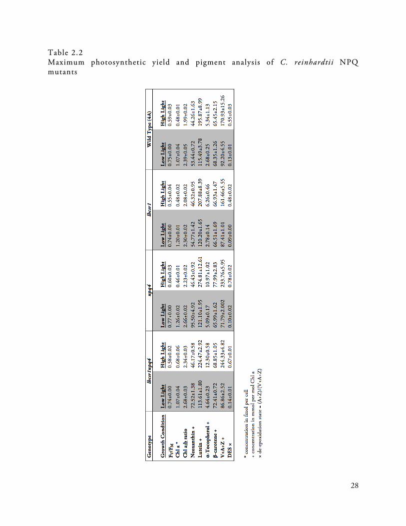
28
Table 2.2 Maximum photosynthetic yield and pigment analysis of C. reinhardtii NPQ mutants

29
2.3 Modification of three lumen-exposed acidic residues of LHCSR3 affects the quenching phenotype of Chlamydomonas reinhardtii 2.3.1 Stress response LHC proteins sense ΔpH across the thylakoid membrane
While PSBS is necessary for qE in A. thaliana under physiological conditions (Li et al., 2000), there is controversy over whether PSBS could be a site of quenching, since there is not consistent evidence that the protein binds pigments (Funk et al, 1995; Aspinall O’Dea et al., 2002, Dominici et al., 2002, Hieber et al., 2004; Fan et al., 2015). Instead, it is hypothesized that PSBS acts as a lumenal pH sensor to somehow activate qE induction. Biochemical and biophysical characterization of the PSBS protein from A. thaliana shows that two thylakoid lumen-exposed glutamate residues, Glu-122 and Glu-226, are important for qE induction (Li et al., 2002a), and that they are targeted for protonation when the lumen becomes acidified under excess light conditions (Li et al, 2004). When the two lumenal glutamate residues are mutated to non-acidic glutamine residues and expressed in npq4-1 mutant A. thaliana plants, despite protein accumulation and no change in xanthophyll cycle activity, the impaired qE phenotype is not restored (Li et al., 2002a, 2004). Furthermore, these PSBS mutant proteins do not bind dicyclohexylcarbodiimide (DCCD), a known inhibitor of qE, suggesting they are no longer susceptible to protonation. Mutation of only one of the glutamates has intermediate effects on qE induction and DCCD binding (Li et al., 2004). The crystal structure of PSBS from Spinacia oleracea shows DCCD is bound to the residue Glu-173, which is equivalent to the Glu-226 residue in A. thaliana PSBS (Fan et al., 2015). Biochemical and structural analysis of the protein shows that PSBS proteins form dimers in the thylakoid membrane in both the active form (Bergantino et al., 2003) and the inactive form where Glu-122 and Glu-226 have been modified (Fan et al., 2015). Based on the crystal structure of active and inactive PSBS, it is proposed that low pH protonation of the two glutamate residues drives a conformational change that allows four stabilizing hydrogen bonds to form at the lumenal dimer interface (Fan et al., 2015) and this conformation allows for qE.
To determine if LHCSR also has a mechanism for sensing acidification of the thylakoid lumen, a similar approach was taken to identify conserved acidic residues of lumen-exposed loops of the LHCSR3 protein and to ascertain their role in LHCSR3 function. 2.3.2 Expression of protonation mutants of LHCSR does not rescue impaired qE phenotype of the npq4 lhcsr1 mutant of C. reinhardtii
While the molecular mechanisms of quenching for both PSBS and LHCSR are hotly debated in the scientific literature, there are significant structural and biochemical differences between the proteins (Niyogi et al, 2005; Bonente et al., 2008a, Bonente et al., 2011, Xu et al., 2015, Croce, 2015). These differences would indicate the mechanism for inducing quenching must also be different, especially considering that LHCSR3 is a Chl- and xanthophyll-binding protein where quenching of Chl* can be observed in vitro in solutions of purified protein in the form of a reproducible short fluorescence lifetime (Bonente et al, 2011). In contrast, LHC-like pigment-binding sites are not conserved in PSBS, and only a non-canonical binding site for a single Chl molecule per PSBS dimer has been observed, along with a predicted non-canonical zeaxanthin

30
binding pocket (Fan et al., 2015). The residues that create these non-canonical binding sites are conserved across available PSBS sequences (Fan et al., 2015), which strongly supports an hypothesis where quenching is mediated by PSBS and an interaction partner (Fan et al., 2015).
A common feature of both LHCSR and PSBS, however, is the capacity for binding DCCD, a protein-modifying agent that covalently binds to acidic residues involved in reversible protonation events (Li, et al., 2004; Bonente et al, 2011; Ballottari et al., 2016). Measurement of DCCD binding is used to pinpoint which acidic residues are solvent accessible and could therefore sense low pH in the thylakoid lumen. Sequence analysis of available LHCSR protein sequences reveals multiple conserved acidic residues that are exposed to the lumen as potential sites of protonation (Figure 2.10). Mounting evidence for LHCSR3’s responsiveness to low pH has been observed in both in vitro and in vivo spectroscopic analyses (Tokutsu et al., 2004; Bonente et al., 2011). Recombinant LHCSR3 from C. reinhardtii reconstituted in solution has been shown to undergo a switch to a dissipative state in low pH solutions as measured by TCSPC (Bonente et al., 2011). In addition, isolated PSII supercomplexes containing LHCSR3 were reported to undergo a decrease in fluorescence lifetime when exposed to pH 5 solution (Tokutsu et al., 2004).
Structural prediction models of the LHCSR3 protein were created by threading the LHCSR3 protein sequence onto the available crystal structure templates of CP29 (Pan et al., 2011) and LHCII (Liu, et al., 2004), both from S. oleracea, using the PHYRE2 server (Kelley et al., 2015) and the I-TASSER tool (Zhang, 2008; Roy et al., 2010) (Figure 2.5). The predicted structure of LHCSR3 includes three trans-membrane helices (A, B, and C), typical of the general structure of LHCIIs (Liu et al., 2004; Amunts et al., 2007; Barros et al., 2009; Pan et al., 2011), as well as two amphipathic helices (D and E) packed against the lumen-exposed membrane, and a putative short highly-acid carboxy-terminal helix that is unique to LHCSR3. There are multiple acidic residues that are predicted to be lumen exposed, including the highly acidic carboxy-terminal tail that includes Glu-231, Glu-233, Glu-237, Asp-239, Asp- 240, Glu-242, Asp-244, and Asp-254. The carboxy-terminus has been reported to play a role in activating quenching based on in vitro measurements of fluorescence lifetimes (Liguori et al., 2013).
Work initiated by Thuy Truong focused on the additional acidic residues facing the thylakoid lumen: Glu-221 and Glu-224 in the amphipathic helix D, Asp-109 and Asp-117 in the lumenal loop between helix B and helix E, and Glu-218 in the lumenal loop between helix A and helix D. To identify residues that contribute to the general molecular mechanism of LHCSR3-mediated quenching, we wanted to focus on highly conserved motifs (Figure 2.10). Glu-221 and Glu-224 in LHCSR3 from C. reinhardtii were conserved in over half of the sequences analyzed (Figure 2.10), though notably absent from Ostreococcus tauri and Ostreococcus lucimarinus. As for the residue Glu-218, this is only found in C. reinhardtii and its very close relatives amongst the Volvocales, however a glutamate in a nearby position and shifted towards the N-terminus is found in all the other accessions, suggesting it might have a conserved functional role. Asp-109 is not highly conserved, while Asp-117 is present as either an aspartate or glutamate in most sequences. Those sequences that do not have Asp-117 do have one or more aspartate residues within 1-3 positions. On the basis of these results, Asp-109, Asp-117, Glu-218, Glu-221, Glu-224, Glu-231 and Glu-233 were selected for further investigation.
By systematically mutating each of the residues of interest as single, then double, and then triple mutants and expressing the protein using the mutant genomic LHCSR2 construct with its native promoter, we were able to identify three key residues that do not affect the expression or

31
stability of LHCSR3, but render the protein non-functional in qE: Asp-117, Glu-221, and Glu-224 (Ballottari et al., 2016) (Figure 2.8).
Figure 2.5 Structural model of LHCSR3 Structural model of LHCSR3 based on homology and a structural threading model over the LHCII (IRWT; Liu et al., 2004) and CP29 (3PLD; Pan et al., 2011) crystal structures. NQQ mutants of LHCSR3 do not rescue impaired qE phenotype of npq4 lhcsr1 mutants
When each individual acidic residue was mutated, the NPQ amplitude was reduced, but a significant level of quenching was still present, and the transformant lines exhibited an NPQ level proportional to the level of LHCSR3 protein accumulation as assessed by immunoblotting with anti-LHCSR antibodies, which is consistent with the results from Section 2.1. Strains transformed with LHCSR3 variants mutated at residue Asp-109 did not show any accumulation of LHCSR3, so this residue likely plays a major role in stabilizing protein folding. Lack of significant effect on quenching caused by single mutations supports that proton-sensing residues of LHCSR3 are likely redundant, therefore combinations of multiple mutations within the same protein were generated and tested. When Asp-117, Glu-221 and Glu-226 residues were mutated together, the triple mutant, which I refer to as the NQQ mutant, had a qE amplitude similar to that of the background npq4 mutant strain (Ballottari et al., 2016). Out of more than 300 colonies of the NQQ mutant in the npq4 background, none had higher NPQ induction than npq4, despite the accumulation of the mutant LHCSR3 protein at wild-type levels (Ballottari et al., 2016). Expression of the NQQ mutant LHCSR3 in the npq4 lhcsr1 mutant background, at varying levels of protein accumulation, also

32
shows no rescue over the background NPQ level (Figure 2.6). There is nearly no concentration dependence on the recovery of quenching observed in these NQQ mutants, which is in contrast to the results described in Section 2.1 with the functional genomic copy of LHCSR3.
Figure 2.6 NPQ measurements and immunoblot analysis of the LHCSR3 NQQ protonation site mutants None of the three LHCSR3 NQQ mutants analyzed here show much NPQ above the npq4 lhcsr1 background level, even when more protein is expressed. This is in contrast to the wild type LHCSR3 complement, which shows concentration dependent quenching in the same npq4 lhcsr1 mutant background. A) NPQ traces from high light acclimated C. reinhardtii cultures comparing the 4A wild type, the npq4 lhcsr1 qE-deficient mutant background, and four complemented lines: a wild type genomic construct expressing LHCSR3, and three mutant LHCSR3 constructs with the NQQ protonation site mutations. B) Western blot analysis comparing LHCSR expression in the wild type, mutant background, and four complemented lines. Wild type shows the presence of LHCSR1 as well as a poorly-resolved doublet of LHCSR3, while the transformed lines show only the LHCSR3 doublet.
The very minimal residual qE induction observed in the NQQ mutant could be related to the activity of one or more of the other protonatable sites that are still present in the mutant. Unfortunately, the addition of further mutations in the LHCSR3 gene resulted in a loss of protein accumulation, suggesting there is a threshold for mutation before destabilization of the protein or some impairment in protein import into the thylakoid membranes occurs (Ballottari et al., 2016).

33
A recombinant version of the NQQ mutant of LHCSR3 was reconstituted and compared to wild type protein for pigment binding and then the fluorescence lifetimes were evaluated. In work by Matteo Ballottari, Eleonora De Re, and colleagues, the reconstituted mutant and wild type proteins were shown to have similar pigment profiles and fluorescence emission spectra, suggesting the mutations did not affect the pigment arrangement within the protein. To see if the identified acidic residues are the targets of DCCD binding, comparison between the reconstituted wild type protein and the NQQ mutant protein reveals that 40% of the DCCD binding is attributable to Asp-117, Glu-221, and Glu-224. TCSPC of the wild type and mutant reconstituted proteins in a mild detergent solution showed that at pH 7.5 the fluorescence lifetime components of the two protein samples were comparable, whereas the short fluorescence lifetime signature of a dissipative state that is observed in the wild type protein at pH 5 could not be induced in the NQQ mutant at the same low pH (Ballottari et al., 2016). This was further confirmed by measuring the far-red fluorescence emission of Chl at 77 K. The far-red fluorescence signal has been associated with induction of NPQ in vivo and correlates with quenching localized in aggregates of LHCII antennas (Ballottari et al., 2010). The results of measuring a far-red spectral shift showed that at neutral pH the wild type and NQQ mutant protein samples were indistinguishable, but in pH 5 solution the wild type protein displayed significantly higher far-red emission (Ballottari et al., 2016), supporting that protonation of these lumenal residues plays a role in the induction of the LHCSR3-mediated quenching mechanism. GGG mutants of LHCSR3 partial ly rescue qE in npq4 lhcsr1 mutants
The glutamate residues of PSBS from A. thaliana described by Li and colleagues are intolerant to mutation, and resulted in the same inability to rescue the impaired qE phenotype when expressed in npq4-1 plants when Glu-122 and Glu-226 were mutated to either alanine residues or glutamine residues (Li et al., 2002a). These results support the hypothesis that an acidic residue, in those positions specifically, is essential for functionality in quenching. In order to test the tolerance to mutation of the proposed protonation sites in LHCSR3, Asp-117, Glu-221 and Glu-224 were mutated to glycine residues instead of asparagine and glutamine, as described above, which l refer to as the GGG mutant of LHCSR3. All of the major light harvesting complexes of C. reinhardtii, the LHCM proteins, have highly conserved glycine residues at the corresponding positions for Asp-117 and Glu-221, whereas the corresponding position for Glu-224 in the LHCBM proteins is either a glutamine or an alanine residue (Figure 2.9). Since LHCBM1 proteins do not induce pH-dependent quenching, this experiment set out to determine if the structure provided by the three lumen-exposed acidic residues is essential for quenching, or if by introducing increased flexibility into the lumenal α-helices of LHCSR3, to better match those of the LHCBM proteins, the LHCSR3 mutant can still adopt a permissive conformation for quenching.
The GGG mutant of LHCSR3 show a partially active qE response, though it is highly impaired compared to either of the single mutants, npq4 and lhcsr1, or its double mutant genetic background npq4 lhcsr1 (Figure 2.7). By analyzing multiple complemented lines of both the NQQ and the GGG mutants of LHCSR3, there does not appear to be a correlation between the dosage of the pH-insensitive mutants and quenching level (Figure 2.7), supporting the hypothesis that a conformational change driven by these three residues is critical for function in inducing qE. By introducing additional flexibility into the lumen-exposed loops of LHCSR3, the physical constraints of conformation change have been removed. This may allow LHCSR3 to adopt a low-efficiency or
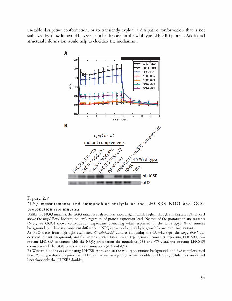
34
unstable dissipative conformation, or to transiently explore a dissipative conformation that is not stabilized by a low lumen pH, as seems to be the case for the wild type LHCSR3 protein. Additional structural information would help to elucidate the mechanism.
Figure 2.7 NPQ measurements and immunoblot analysis of the LHCSR3 NQQ and GGG protonation site mutants Unlike the NQQ mutants, the GGG mutants analyzed here show a significantly higher, though still impaired NPQ level above the npq4 lhcsr1 background level, regardless of protein expression level. Neither of the protonation site mutants (NQQ or GGG) shows concentration dependent quenching when expressed in the same npq4 lhcsr1 mutant background, but there is a consistent difference in NPQ capacity after high light growth between the two mutants. A) NPQ traces from high light acclimated C. reinhardtii cultures comparing the 4A wild type, the npq4 lhcsr1 qE-deficient mutant background, and five complemented lines: a wild type genomic construct expressing LHCSR3, two mutant LHCSR3 constructs with the NQQ protonation site mutations (#35 and #73), and two mutants LHCSR3 constructs with the GGG protonation site mutations (#28 and #71). B) Western blot analysis comparing LHCSR expression in the wild type, mutant background, and five complemented lines. Wild type shows the presence of LHCSR1 as well as a poorly-resolved doublet of LHCSR3, while the transformed lines show only the LHCSR3 doublet.

35
Figure 2.8 View from the lumenal face of Asp-117, Glu-221, and Glu-224 in structural model of LHCSR3 From left to right: Glu-224, Glu-221, and Asp-117, on the lumenal face of the LHCSR3 structural model. 2.3.3 Discussion and Future Work
Through the study of evolutionarily distant photosynthetic organisms, it seems that two strategies for initiating the rapid induction of NPQ in excess light conditions have been repeatedly observed: light-driven mechanisms, like the cyanobacterial OCP/FRP system (Boulay et al., 2010), and pH-driven mechanisms, as employed by PSBS and LHCSR in plants, algae, and moss species (Bonente et al., 2008b; Amarnath et al. 2012). LHCSR3 is unique when compared to PSBS in that it is potentially both the sensor of pH and the site of quenching, making it a simpler system for induction of rapid de-excitation.
The fact that this mechanism is lost in vascular terrestrial plants may indicate that it is not suitable for photoprotection in the environmental conditions of terrestrial growth. LHCSR-like sequences form an early branch in the molecular phylogeny of the three-helix LHC protein family (Figure 2.14B) (Dittami et al., 2010; Engelken et al., 2010). LHCSR-like proteins may represent an extant version of primitive members of the three-helix LHC protein family, which have maintained both a role as the site of quenching and as the trigger to induce quenching. However, it appears that LHCSRs may not be optimal in their roles. Bonente and colleagues (2011) observed that LHCSR3 protein samples in solution slightly quench even in non-acidic pH conditions. Some LHCSR proteins, including LHCSR1 in C. reinhardtii (Peers et al., 2009) accumulate even in low light conditions, resulting in wasteful quenching of excitation energy even when light is limiting for growth. In higher plants, however, these two roles are separated into different components. Other LHC stress response proteins, such as PSBS, may have evolved to function in qE thereby replacing LHCSR proteins, to become the “on/off” switch that activates cells to adopt a photoprotective state. To support this, it appears that PSBS is a better regulator of photosynthesis than the LHCSR proteins. The PSBS protein accumulates to some extent at all times (Li et al., 2000) allowing plants

36
like A. thaliana to respond immediately to high light stress, unlike C. reinhardtii, which requires high light acclimation over a long period of time. PSBS-dependent qE also has faster induction and recovery response compared to LHCSR-dependent qE in the moss P. patens (Gerotto et al., 2012). By increasing the complexity of the qE process, terrestrial plants may have the benefit of additional feedback for regulation, limiting losses in photosynthetic efficiency in highly dynamic light conditions, which C. reinhardtii is less equipped to handle.
A competing hypothesis concerning the pH-dependent response of LHCSR3 has been proposed, positing that the highly acidic carboxy-terminus of the protein is the regulator of quenching. The lumen-localized carboxy-termini of both LCHSR3 and LHCSR1 from C. reinhardtii are extended relative to other LHC proteins that comprise the major and minor antenna complexes of PSII, and contain many additional acidic residues (Figure 2.10). This highly acidic, extended carboxy-terminal domain of the C. reinhardtii LHCSR3 was reported to be the “knob of a dimmer switch” that controls a transition from a harvesting state to a dissipative state (Liguori et al., 2013). This work was reported based on in vitro spectroscopy studies of reconstituted recombinant LHCSR3 protein measured in solution, and shows that a de-excitation component seen in the wild type reconstituted protein under the same conditions is missing from the carboxy-terminal mutant. However, amongst the LHCSR-like sequences available, this highly acidic carboxy-terminus is present only in Chlamydomonas species, along with Volvox carterii and Aureococcus anophagefferens. Both the length and the number of acidic residues within this domain are variable in the other LHCSR proteins of different organisms (Figure 2.10). For example, the LHCSR sequences from both Ostreococcus species represented as well as Chlorella variabilis show only a single protonatable glutamate residue in the carboxy-terminus, and most LHCSR-like sequences do not have an extended carboxy-terminal domain at all, so that they more closely resemble CP29 and LHCII sequences. Furthermore, with mutation to only three acidic residues, as opposed to nine, we observe an equivalent reduction in quenching capacity. The loss of qE is not complete, nor does the mutation completely abolish DCCD binding. Perhaps LHCSR3 experiences a titratable conformation change that is influenced by many acidic residues, with these three residues contributing most heavily.
While the carboxy-terminal tail of the C. reinhardtii LHCSR3 protein may facilitate in an in vitro quenching pathway, it seems unlikely to be a universal molecular mechanism for LHCSR-mediated quenching. It would be prudent to examine the effects of this mutation in vivo, and this work is forthcoming.

37
Figure 2.9 Alignment of lumenal loops of LHCSR and LHCBM protein sequences from C. reinhardtii The major PSII antenna Lhcbm proteins of C. reinhardtii have significantly different lumenal loop sequences than the LHCSR proteins. The three key acidic residues are identified with red stars and red highlighting. The LHCBM proteins have small, non-polar residues in those positions instead. In addition, LHCSR has an extended, acidic C-terminal tail relative to the LHCBM proteins.

38
Figure 2.10 Alignment of lumen-exposed protein regions from a large evolutionary cross-section of LHCSR protein sequences (2 pages)

39
LHCSR-like protein homologs (from Table 2.3) show strong conservation of the three acidic residues (Asp-117, Glu-221, and Glu-224 in the Cr.LHCSR3 protein) that were identified in our mutant screen (shown with red stars and acidic residues in the right position are highlighted in red). Also labeled are the putative Chl and carotenoid binding site motifs that are conserved in LHCSR-like sequences (labeled above the sequences using the nomenclature of Kühlbrandt et al, 1994). Acidic residues in the C-terminal tail are highlighted in purple.

40
Table 2.3 Representative sequences and accession information for evolutionary conservation analysis of LHCSR proteins Protein Sequence Accession Organism XP_001696064.1 Chlamydomonas reinhardtii LHCSR3 XP_001696125.1 Chlamydomonas reinhardtii LHCSR1 XP_002948670.1 Volvox carterii f. nagariensis ADP89594.1 Chlamydomonas sp. ICE-L KXZ50176.1 Gonium pectorale KXZ42803.1 Gonium pectorale Q03965.1|L181_CHLMO Chlamydomonas moewusii XP_001768071.1 Physcomitrella patens LHCSR2 ABD58893.1 Acutodesmus obliquus GAQ89003.1 Klebsomordium flaccidum ADY38581.1 Ulva linza ADU04518.1 Ulva prolifera XP_005848576.1 Chlorella variabilis ABD37894.1 Mesostigma viride XP_002178699.1 Phaeodactylum tricornutum CCAP1055 XP_002295258.1 Thalassiosira pseudonana CCMP1335 XP_013902010.1 Monoraphidium neglectum XP_013900766.1 Monoraphidium neglectum CAA04404.1 Cyclotella cryptica EJK49165.1 Thalassiosira oceanica AHH80644.1 Durinskia baltica AAP79202.1 Bigelowiella natans XP_007511181.1 Bathycoccus prasinos KOO27643.1 Chrysochromulina sp. CCMP291 XP_009037936.1 Aureococcus anophagefferens XP_005756200.1 Emiliania huxleyii ABV22207.1 Karlodinium veneficum ABV22207.1 Ectocarpus siliculosus ABA55525.1 Isochrysis galbana XP_003079276.1 Ostreococcus tauri AAY27550.1 Ostreococcus tauri XP_001417976.1 Ostreococcus luminarinus XP_005647960.1 Coccomyxa subellipsoidea XP_003063244.1 Micromonas pusilla Micromonas sp. RCC299

41
2.4 Adaptation of a transient expression method in Nicotiana benthamiana for rapid screening of photosynthetic phenotypes
While C. reinhardtii has a well-established and tractable genetic system, there are some limitations with regard to the speed at which mutants can be generated, screened, and characterized. Furthermore, maintaining mutants over long periods of time poses additional challenge because C. reinhardtii may silence expression of exogenous genes, especially those that are highly expressed, but this can still occur even when the gene is expressed under its native endogenous promoter. Nuclear transformation, either by bombardment (Kindle et al, 1989), the glass bead method (Kindle, 1990) or electroporation (Shimogawara et al., 1998), accomplishes random insertion of the expression construct into the genome, which may result in variable expression between transformants, as well as disruptive mutations unrelated to the gene inserted. In collaboration with Lauriebeth Leonelli, I attempted to adapt an extensively used method from the field of plant pathology to create a rapid screening system for evaluating photosynthetic phenotypes caused by mutant C. reinhardtii LHC genes. This method would allow us to bypass complementation of each mutant into the native organism for screening, by instead taking advantage of transient Agrobacterium tumefaciens-mediated gene expression in the tobacco Nicotiana benthamiana (Rossi et al., 1993; Kapila et al., 1997; Van der Hoorn et al., 2000). This system exploits the natural ability of A. tumefaciens to genetically modify plant cells, allowing for in planta gene expression of algal genes in a matter of days. We hypothesized that this system would be particularly well suited for the study of algal LHC genes, because introduced genes can use endogenous pigments as substrates and can fit into the existing photosynthetically active membranes as a scaffold. In addition, the activity of multiples genes can be directly compared since proteins accumulate in discrete spots on the same leaf. 2.4.1 Method verif ication using PSBS from A. thaliana
Because fully developed leaves contain mature chloroplasts and assembled photosystems, we first wanted to test whether heterologous proteins could be introduced into the thylakoid membrane or noticeably alter photosynthesis in tobacco leaves at such a late stage of development. We first tested the method by introducing the PSBS gene from A. thaliana (At.PSBS), which is highly similar to the native PSBS of the plant (Nb.PSBS), in order to evaluate if NPQ dynamics change with the overexpression of a protein that is closely related to the endogenous PSBS protein of N. benthamiana. As expected, based on the previously described dosage effects on qE by overexpression of PSBS in A. thaliana (Li et al., 2002c), high levels of transient At.PSBS expression in mature leaves of N. benthamiana resulted in increased NPQ (Figure 2.11) (Leonelli et al, 2016). To ensure high expression levels, the gene is expressed under the cauliflower mosaic virus (CaMV) 35S promoter in the pEarleyGate100 vector (Earley et al., 2006), and to facilitate biochemical characterization, a carboxy-terminal FLAG epitope tag – DYKDDDDK (Hopp et al., 1988) - was included before the stop codon of the cDNA-derived coding sequence of At.PSBS (Table 2.4). To control for the potential stress response of infection, leaves of N. benthamiana were also infiltrated with a FLAG-epitope tagged β-glucuronidase (GUS) gene maintained under the same CaMV 35S promoter as the genes of interest, for use as a negative control. To ensure consistent stress response due to infection and high levels of protein accumulation amongst samples on the same leaf, the OD of A tumefaciens was equal across all spots to control for the load of infection. GUS expression does not alter photosynthetic membranes or activity.

42
Figure 2.11 Transient expression of PSBS from A. thaliana in leaves of N. benthamiana Expression of At.PSBS in leaf spots of N. benthamiana, shown here at 48 hours post-infiltration, enchances the level of NPQ, as shown here by false-colored video fluorescence imaging, when compared to the background of the leaf and the control spot expressing the GUS protein. Note, the blue color shown above represents fluorescence level and not GUS expression.
Expression of heterologous At.PSBS in N. benthamiana, even at high levels, does not disrupt the stability of solubilized thylakoid membrane complexes, as observed using blue native PAGE of complexes from purified N. benthamiana chloroplasts gently solubilized with the detergent n-dodecyl-α-D-maltopyranoside (Figure 2.12B). At.PSBS is associated with multiple solubilized complexes including PSI- and PSII-containing complexes, which is consistent with previous localization observations done by native electrophoresis (Thidolm et al., 2002; Teardo et al., 2007, Correa-Galvis et al., 2016a).

43
Figure 2.12 Biochemical analysis of thylakoid membranes from N. benthamiana leaves transiently expressing At.PSBS, Cr.LHCSR3, and At.CP29 A) Sucrose gradient analsysis of isolated chloroplasts from N. benthamiana 48 hours after infiltration, solubilized with n-dodecyl-α-D-maltopyranoside, shows that heterlogously expressed Cr.LHCSR3 and At.CP29 are incorporated in the thylakoid membrane and associate with PSII-containing complexes. B) Blue Native-PAGE of isolated thylakoid membrane tissue from N. benthamiana expressing At.PSBS, and solubilized with n-dodecyl-α-D-maltopyranoside, shows that At.PSBS does not disrupt the stability of the solubilized complexes, though the heterologous protein does associate with multiple complexes in the membrane. 2.4.2 CP29 transient expression To further analyze the efficacy of this method, the minor antenna complex protein CP29, encoded by the Lhcb4 gene, was also transiently expressed in mature leaves of N. bethamiana. It has been proposed that CP29 may be a site of quenching in plants (Ahn et al., 2008), therefore modifying CP29 for rapid phenotypic assessment may provide the opportunity to evaluate the minor antenna complex protein’s mechanistic role in qE. Leaf spots expressing the Lhcb4.1 gene from A. thaliana and the Lhcb4 gene from C. reinhardtii were evaluated for the effect of the expressed heterologous CP29 protein on qE in N. benthamiana. This also presented an opportunity to compare the transient expression system’s capacity to robustly express plant genes as well as algal genes. Using the pEarleyGate100 expression vector, expression of the cDNA-derived coding sequence of CP29 from A. thaliana (At.CP29) was compared to expression from the C. reinhardtii cDNA-derived coding sequence (Cr.CP29c), the genomic sequence (Cr.CP29g), and a codon-optimized construct (Cr.CP29) optimized for expression in vascular plants, all with a carboxy-terminal FLAG epitope tag included just before the stop codon (Table 2.4). The genes from C. reinhardtii were modified to include the chloroplast transit peptide from the A. thaliana PSBS protein, to ensure plant thylakoid localization. The amino-terminal transit peptide of CP29 from C. reinhardtii may be unique amongst plastid-targeted

44
genes (Turkina et al., 2004), and for this reason we chose to ensure plastid localization using a plant chloroplast transit peptide. The coding and codon optimized sequences, whether from the plant or algal Lhcb4 genes, expressed protein in the N. benthamiana leaf. The C. reinhardtii genomic expression construct, however, was not recognized by the plant system and no protein was accumulated (Figure 2.13). Despite expression, no quenching phenotype is induced by expression of CP29, neither from the plant nor the algal protein sequence, in the transient expression system (Figure 2.13). Transient expression of Cr.CP29 does not disrupt PSII
Expression of Cr.CP29, even at high levels, does not disrupt the assembly or the stability of the photosystem, as demonstrated by sucrose gradient analysis of solubilized isolated chloroplasts. Furthermore, transiently expressed Cr.CP29 is localized only in the stable PSII core and antenna band, most likely recognized by the plant and substituted into position for the native At.CP29 protein. (Figure 2.12A)
Figure 2.13 Transient expression analysis of Cr.CP29 CP29 does not induce quenching when transiently expressed in leaves of N. benthamiana. The Cr.CP29 genomic sequence is not expressed in the leaves, while the cDNA-derived coding sequence is expressed at a low level. The codon-optimized gene sequence for Cr.CP29 is transiently expressed at a high level 48 hours after infiltration.

45
Figure 2.14 Evolutionary hypothesis for the origin and expansion of LHC proteins A) PSBS and LHCSR-like proteins are all part of the LHC protein superfamily, and are thought to have originated from separate internal duplication events in the expanded family of two-helix progenitors already in the eukaryotic nuclear genome, supported by molecular phylogenetic analysis of the transmembrane helices of the extant representatives of each class of LHC protein. B) LHCSR-like proteins are found throughout the three-helix containing eukaryotic photosynthetic organisms (not Glaucophytes), and are thought to comprise a basal branch of the phylogenetic tree. Further adaptation led to the red-algal type LHCs (RedCAP and LHCR-type proteins), the Chl a/c type LHC that bind fucoxanthin or other accessory pigments (FCP and LHCF-type proteins), and the Lhca and Lhcb proteins in the green lineage. These hypotheses and figures are based on phylogenetic analyses described by Dittami and colleagues (2010) and Engelken and colleagues (2010) Modification of At.CP29 does not result in inducible quenching
We next attempted to identify whether At.CP29 could be susceptible to a gain-of-function mutation that would result in inducible quenching. Could random mutagenesis uncover a novel energetic sink within the protein? Or, by introducing elements of the sequence and structure of LHCSR3 from C. reinhardtii into the At.CP29 protein, could At.CP29 be converted into a quenching LHC protein? This line of questioning was motivated by the hypothesis that LHCSR-like
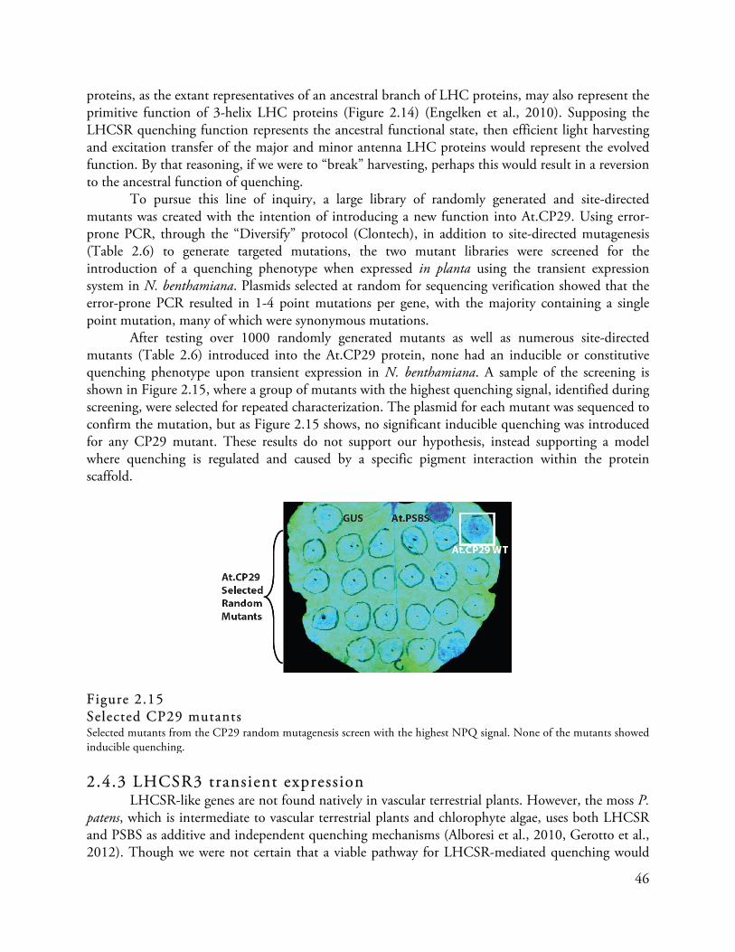
46
proteins, as the extant representatives of an ancestral branch of LHC proteins, may also represent the primitive function of 3-helix LHC proteins (Figure 2.14) (Engelken et al., 2010). Supposing the LHCSR quenching function represents the ancestral functional state, then efficient light harvesting and excitation transfer of the major and minor antenna LHC proteins would represent the evolved function. By that reasoning, if we were to “break” harvesting, perhaps this would result in a reversion to the ancestral function of quenching.
To pursue this line of inquiry, a large library of randomly generated and site-directed mutants was created with the intention of introducing a new function into At.CP29. Using error-prone PCR, through the “Diversify” protocol (Clontech), in addition to site-directed mutagenesis (Table 2.6) to generate targeted mutations, the two mutant libraries were screened for the introduction of a quenching phenotype when expressed in planta using the transient expression system in N. benthamiana. Plasmids selected at random for sequencing verification showed that the error-prone PCR resulted in 1-4 point mutations per gene, with the majority containing a single point mutation, many of which were synonymous mutations.
After testing over 1000 randomly generated mutants as well as numerous site-directed mutants (Table 2.6) introduced into the At.CP29 protein, none had an inducible or constitutive quenching phenotype upon transient expression in N. benthamiana. A sample of the screening is shown in Figure 2.15, where a group of mutants with the highest quenching signal, identified during screening, were selected for repeated characterization. The plasmid for each mutant was sequenced to confirm the mutation, but as Figure 2.15 shows, no significant inducible quenching was introduced for any CP29 mutant. These results do not support our hypothesis, instead supporting a model where quenching is regulated and caused by a specific pigment interaction within the protein scaffold.
Figure 2.15 Selected CP29 mutants Selected mutants from the CP29 random mutagenesis screen with the highest NPQ signal. None of the mutants showed inducible quenching. 2.4.3 LHCSR3 transient expression
LHCSR-like genes are not found natively in vascular terrestrial plants. However, the moss P. patens, which is intermediate to vascular terrestrial plants and chlorophyte algae, uses both LHCSR and PSBS as additive and independent quenching mechanisms (Alboresi et al., 2010, Gerotto et al., 2012). Though we were not certain that a viable pathway for LHCSR-mediated quenching would

47
exist in plants, we introduced transient expression of the C. reinhardtii LHCSR3 protein (Cr.LHCSR3) into N. benthamiana to characterize any subsequent inducible quenching.
Just as with Cr.CP29 described above, we tested Cr.LHCSR3 expression using the genomic sequence (Cr.LHCSR3g), the coding sequence (Cr.LHCSR3c), and a sequence that was codon-optimized for expression in vascular plants (Table 2.4). All of the LHCSR3 expression constructs had the 54-residue amino-terminal transit peptide from the At.PSBS protein replacing the 20-residue transit peptide sequence of the Cr.LHCSR3 gene, as well as a carboxy-terminal FLAG epitope tag (Table 2.4). The Cr.LHCSR3 coding sequence and the genomic sequence from C. reinhardtii were not expressed transiently, but the codon-optimized sequence had high expression in mature N. benthamiana leaves (Figure 2.16).
Figure 2.16 Transient expression analysis of Cr.LHCSR3 Transiently expressed Cr.LHCSR3 shows a high early quenching phenotype, within the first 30-45 seconds of illumination with actinic light. The genomic construct and cDNA-derived coding sequence constructs of Cr.LHCSR3 did not express when infiltrated into mature N. benthamiana leaves. The codon-optimized construct of LHCSR3, however, showed high transient expression 48 hours after infiltration.
Despite high transient expression 48 hours after transfection, there is only a modest effect on
quenching capacity upon the introduction of the Cr.LHCSR3 protein (Figure 2.16). Specifically, there is a consistent and reproducible early induction of qE in the first 30-45 seconds of exposure to the actinic light. This early effect is no longer seen over the dominating PSBS-mediated background qE in the leaf after the initial rapid induction, however (Figure 2.17). To determine if the FLAG

48
epitope tag was interfering with function, we compared a tagged and untagged construct of Cr.LHCSR3 in the pEarleyGate100 expression vector. All of the constructs led to transient protein expression in mature N. benthamiana leaves, but there was no difference in the quenching phenotype between any of the samples (Figure 2.17). The early high level of NPQ induction, at 30 seconds of actinic light exposure was maybe even a little more pronounced in the tagged constructs as compared to the untagged. To amplify the difference between the leaf’s background qE and the effect of Cr.LHCSR3 expression, we also measured the effect of Cr.LHCSR3 in N. benthamiana leaves where expression of the endogenous copy of PSBS (Nb.PSBS) was silenced by RNA interference delivered by A. tumefaciens transformation (Koscianska et al., 2005). There was no inducible quenching phenotype in the silenced leaves upon expression of Cr.LHCSR3 or At.CP29, whereas expression of At.PSBS was able to rescue quenching in the Nb.PSBS-silenced leaf (Figure 2.18)
Figure 2.17 Effect of FLAG epitope tag on transient expression of Cr.LHCSR3 in N. benthamiana To determine if the carboxy-terminal FLAG epitope tag interferes with function of Cr.LHCSR3 during transient expression in the N. benthamiana, we tested a tagged and untagged version. Both proteins result in the same early quenching phenotype observed in the first 30 seconds of actinic exposure, but no additional quenching was observed past that early induction for either construct. Constructs not containing FLAG were created by Lauriebeth Leonelli. 2.4.4 Co-Expression with LHCSR3 to identify interacting modulators
The inability of Cr.LHCSR3 to induce quenching in N. benthamiana led me to wonder if perhaps Cr.LHCSR3 requires an interacting partner that is present in C. reinhardtii cells, but is absent in the plant chloroplast. A few interaction candidates have previously been suggested in the literature based on attempts to determine the localization of LHCSR3 in C. reinhardtii thylakoids.

49
These candidates include the major antenna complex protein, LHCBM1 from C. reinhardtii (Elrad et al., 2002; Tokutsu & Minagawa, 2013), and the minor antenna complex CP29 (Tokutsu & Minagawa, 2013). More recently, however, the PSBR protein, which is a component of the oxygen-evolving complex (OEC), has been reported as essential for LHCSR3 binding to the PSII-LHCII reaction center (Xue et al., 2015). PSBR is present in the plant chloroplast, so it is possible that there is a difference between the plant and algal OEC which prevents Cr.LHCSR3 from recognizing PSBR from N. benthamiana, or another component in addition to PSBR is required to activate Cr.LHCSR3 for qE.
Figure 2.18 Quenching phenotype of transiently expressed LHC proteins in N. benthamiana with si lenced native Nb.PSBS To see if signal of Cr.LHCSR3 quenching could be better observed without the endogenous quenching system of N. benthamiana, RNA interference was used to silence the endogenous PSBS protein (Nb.PSBS). Wild type and Nb.PSBS-silenced leaves were infiltrated with At.PSBS, At.CP29, Cr.LHCSR3, and the negative control GUS. In the wild type leaves, Cr.LHCSR3 provides extra quenching in the early moments of high light induction, but the effect is no longer visible after a few minutes. At.PSBS, however, enhances the quenching capacity of the leaves over the course of qE induction. In the Nb.PSBS-silenced leaves, Cr.LHCSR3 cannot induce any early quenching phenotype, nor does it enhance the qE capacity after a few minutes of high light stress. At.PSBS, however, can rescue the quenching phenotype of the Nb.PSBS-silenced leaves, complementing the loss of the endogenous protein. Silencing was performed by Lauriebeth Leonelli.

50
Transient expression of either Cr.CP29 or Cr.LHCBM1 alone does not result in inducible quenching, nor does co-expression of either Cr.CP29 or Cr.LHCBM1 with Cr.LHCSR3 (Figure 2.19). Co-expression with the C. reinhardtii copy of PSBR was not tested in this work. Each spot was infiltrated with the same concentration of A. tumefaciens as determined by the optical density of the bacterial cell culture. To ensure equivalent load of infection in the co-expression spots, the bacterial cultures harboring plasmids for each individual gene were mixed with either the GUS control or each other for consistent infectivity from each construct in each test spot. While these genes may be important for qE in the native system, they are not sufficient to activate Cr.LHCSR3 in the N. benthamiana heterologous expression system.
Figure 2.19 Transient co-expression of Cr.LHCSR3 with Cr.CP29 and Cr.LHCBM1 Co-expression of Cr.LHCSR3 with Cr.CP29 did not enhance quenching in N. benthamiana leaves, nor did co-expression of Cr.LHCSR3 with Cr.LHBM1. 2.4.5 Discussion and Future Work
This method of analysis is not useful for gauging loss-of-function in LHCSR-mediated quenching, because the plant system does not appear to be permissive of the native quenching pathway with LHCSR alone. What this does allow, however, is gain-of-function analysis. Although our attempts at turning At.CP29 into a quenching LHC were ultimately unsuccessful, we certainly did not explore an exhaustive number of mutations to the protein. These suggest, however, that an “active-site” model for quenching cannot describe LHCSR-mediated quenching, and expressional, post-translational, or some other level of regulation of Cr.LHCSR3 is also required for qE.
Although our attempts to analyze the quenching function of Cr.LHCSR3 in a plant system were not very informative, this system could be useful for other questions. Work by Lauriebeth

51
Leonelli has shown that this system can be used to study novel carotenoid biosynthesis pathways (Leonelli et al., 2016). Further work related to carotenoid biosynthesis is underway in the lab, and this system offers a convenient platform for functional analysis and specificity determination for enzymes from formerly genetically intractable organisms. Table 2.4 Gene constructs for expression in N. benthamiana and A. thaliana Protein Gene constructs in pEARLEYGATE100 vector Construct name PSBS At.PSBS* + FLAG At.PSBS At.PSBS* + FLAG E122Q, E226Q At.PSBS QQ At.PSBS Transit Peptide (54) + Cr.PSBS* (Δ44) + FLAG Cr.PSBS At.PSBS Transit Peptide (54) + Cr.PSBS* (Δ44) + FLAG E103Q, E208Q Cr.PSBS QQ Cr.PSBS* + FLAG Cr.PSBS-nTP+ Cr.PSBS* Cr.PSBS-nTP- CP29 At.CP29* + FLAG At.CP29 At.PSBS Transit Peptide (54) + Cr.CP29[genomic] (Δ19) + FLAG Cr.CP29g At.PSBS Transit Peptide (54) + Cr.CP29* (Δ19) + FLAG Cr.CP29c At.PSBS Transit Peptide (54) + Cr.CP29≠ (Δ19) + FLAG Cr.CP29 LHCSR At.PSBS Transit Peptide (54) + LHCSR3.1[genomic] (Δ20) + FLAG Cr.LHCSR3g At.PSBS Transit Peptide (54) + LHCSR3.1* (Δ20) + FLAG Cr.LHCSR3c At.PSBS Transit Peptide (54) + LHCSR3.1≠ (Δ20) + FLAG Cr.LHCSR3 At.PSBS Transit Peptide (54) + LHCSR3.1≠ (Δ20) Cr.LHCSR3- LHCBM1 At.PSBS Transit Peptide (54) + LHCBM1≠ (Δ25) + FLAG Cr.LHCBM1 *cDNA-derived coding sequence ≠Codon-optimized for vascular plant expression

52
2.5 Disruption of pigment-binding sites in LHCSR3 2.5.1 Interactions between low energy chlorophyll molecules, or chlorophyll and a carotenoid could provide an energy sink for non-photochemical quenching
An area of photosynthesis attracting active research is the study of energy-transfer within and between the light-harvesting complexes of PSII. High-resolution crystal structures of an LHCII major antenna complex protein and the PSII-associated CP29 minor antenna complex protein, both from spinach, provide spatial information on pigment arrangements. In concert with further biochemical and biophysical characterization of isolated monomeric proteins and of complexes, it has been possible to develop and refine pigment site energy models for these two proteins (Novoderezhkin et al., 2011; Feng et al., 2013a; Feng et al., 2013b).
The photosynthetic functionality of an LHCII protein is characterized by efficient capture, and subsequent transfer, of excitation energy through the LHCII pigment-protein complex to the reaction center of PSII. The excitation will pass through other pigment-protein complexes, like the minor antenna complexes of CP43 and CP29, for example, en route (van Amerongen & van Grondelle, 2001). Photoprotection, however, is characterized by the presence of exciton-quenching sites, where a site may consist of either a single pigment or a cluster of pigments. Quenching sites receive energy from neighboring sites and then act as exciton traps, quenching the energy instead of transferring it to the reaction center. A possible configuration for a quenching molecule would include a site with a higher energy state, which can receive the exciton, and a site with a lower energy state, to trap the exciton. These sites would need to be strongly coupled to the LHC protein scaffold in order to dissipate the excess energy as heat through the protein backbone (Duffy et al., 2008). The low-energy site would be critical for quenching, and may be conformation dependent.
The site energy models of LHCII and CP29 have different conserved Chl molecules comprising the low energy clusters (Figure 2.20) (Novoderezhkin et al., 2011; Feng et al., 2013a; Feng et al., 2013b). Single-molecule spectroscopy of the major and minor complex LHCIIs from plants also shows that each protein has a different capacity to adopt a quenched state and a differential response to low pH buffer conditions (Krüger et al., 2012; Krüger et al., 2013; Schlau-Cohen et al., 2015). Many results support that quenching occurring within either CP29 (Cheng et al., 2008; Ahn et al., 2008) or LHCII (Krüger et al., 2013; Schlau-Cohen et al., 2015) could be the dominant site of quenching under low lumen pH conditions. A consensus on a mechanism has not been reached. LHCSR is, however, thought to be the dominant site of quenching in C. reinhardtii (Bonente et al., 2011; Liguori et al., 2013).
Using these energy models for reference, because Cr.LHCSR3 does not yet have a proposed crystal structure or site energy model, this work focuses on identifying which of the pigments associated with Cr.LHCSR3 are critical for maintaining a quenching site. As described earlier, the transient expression system in N. benthamiana is not suited to loss-of-function screening for Cr.LHCSR3-mediated quenching. Instead, the goal of this experiment was to identify mutations that either disrupt or abolish pigment binding, leading to an inducible quenching phenotype when expressed in the leaves of N. benthamiana – effectively identifying new quenching pathways that have been introduced within the pigment-protein complex.

53
Figure 2.20 Energy models of chlorophyll molecules in LHCII and CP29 Energy models based on published models (LHCII: from Novoderezhkin et al., 2011; CP29 from Feng et al., 2013a and 2013b). High-energy Chl b molecules can absorb excitation energy and transfer the exciton to the lower energy Chl a clusters, which are energetically and spatially clustered for interaction. Interacting clusters are indicated by shared colors and overlapping vertical alignments. In LHCII, quenching is thought to occur when excitation is passed from the high energy Chl b608-601-609 cluster to the low energy Chl a611/612-610 cluster. In CP29, Chl b608 is spatially coupled with Chl a609-603 dimer and in close proximity to the head group of violaxanthin, which may comprise the site of quenching. Chl b608 is also energetically coupled to the Chl a611-610-612-615 cluster, which form clusters around each head group of lutein and provide a low energy exciton trap, also creating a good candidate for the site of quenching (Pan et al., 2011). 2.5.2 Transient expression of LHCSR3 mutants targeting pigment-binding sites
Looking for conservation of specific residues in LHCSR sequences from different species can facilitate identification of residues that are crucial for protein function. When the LHCSR3 protein sequence from C. reinhardtii was compared to homologous sequences from other organisms, several residues appeared to be highly conserved (Figure 2.10). Most of the chlorophyll-binding sites previously proposed in Cr.LHCSR3 (Bonente et al., 2011) (Figure 2.10) can also be found in LHCSR proteins from other species (Figure 2.10), namely the residues for binding Chl at the A1, A4 and A5 sites (these pigment site references are consistent with the nomenclature from Kühlbrandt et al., 1994; see Table 2.5 for corresponding nomenclature consistent with Liu et al., 2004). Some variability in position can be found for the A2, B5 and A3 sites, and a poorly conserved glutamate residue at the B6 binding site position was only maintained in a few LHCSR-like protein sequences. The carotenoid-binding motifs of the three-helix LHC proteins are generally conserved, and known as L1, L2, V1, and N1 sites (Kühlbrandt et al., 1994), though there is variability in binding selectivity and preference for these sites which affords spectral diversity to different LHC proteins (Guiffra et al., 1996; Bassi et al., 1999; Remelli et al., 1999). In Cr.LHCSR3, the conserved motifs for the bipartite L1 and L2 sites are highlighted (Figure 2.10), though there is not strong evidence of a conserved N1 site. Analysis of reconstituted recombinant Cr.LHCSR3 supports the preferential binding of violaxanthin and lutein into two sites, along with a possible third site that was usually unoccupied in the reconstitution experiments (Bonente et al., 2011). No loroxanthin or neoxanthin was found bound to the reconstituted protein (Bonente et al., 2011). Despite the absence of neoxanthin binding in the refolding experiments, a tyrosine residue is found near the

54
position an N1 site would be located in other LHC protein. This tyrosine residue, Tyr-126 is conserved in C. reinhardtii and organisms very closely related to Chlamydomonas, but not widely conserved amongst LHCSR-like proteins. (See Figure 2.10).
Despite the inconclusive results of transient expression of Cr.LHCSR3 in N. benthamiana, as described in Section 2.3, we proceeded to test a library of site-directed Cr.LHCSR3 mutants targeting the conserved and predicted pigment binding sites of Cr.LHCSR3 by transient expression in N. benthamiana. (Tables 2.6 and 2.7) Mutants resulting in increased inducible quenching upon transient expression are described below (Figure 2.21).
Figure 2.21 Transient expression of selected Cr.LHCSR3 chlorophyll and carotenoid binding site mutants Selected pigment binding site mutants transiently expressed in N. benthamiana show higher quenching than the wild type Cr.LHCSR3 protein. Mutations to His-90, which is a highly conserved Chl a603 binding site, resulted in increased quenching. Disruption of this chlorophyll probably opens a new quenching pathway in the plant thylakoid membrane. Mutations to the putative L2a carotenoid-binding site, to make Cr.LHCSR3 more like CP29, LHCII, and LHCBM1 respectively, also increase quenching. Mutations to this area would also disrupt the orientation of Asp-117 in the lumen. Mutations to Tyr-126 also show an increased level of quenching when transiently expressed in N. benthamiana. Tyr-126 is near a putative conserved N1 binding site, but is also the neighbor of a conserved Chl a604 binding site, His-127. Mutations to His-127 do not affect quenching when transiently expressed.

55
His-90 A mutation at His-90 of Cr.LCHSR3, including a conversion of the histidine residue to
alanine, phenylalanine, lysine, arginine, tryptophan, and asparagine, results in enhanced quenching when the protein is transiently expressed in N. benthamiana. His-90 is a highly conserved residue amongst LHC proteins, and is predicted to be the ligand for binding the magnesium atom at the center of Chl a603 (or the A5 site). This Chl molecule would be embedded in the thylakoid membrane closest to the stromal side of the protein. In the LHCII energy model, Chl a603 is the higher-energy member of a stromal-side Chl a cluster (Chl a603-Chl a602; A4-A5) (Novoderezhkin et al., 2011). In the energy model for CP29, Chl a603 is strongly coupled to Chl a609 (the B5 site), where Chl a609 has the slightly higher energy state (Feng et al., 2013a; Feng et al., 2013b; Berman et al., 2015, Berman et al., 2016). Chl a609 is not strongly coupled to the protein scaffold, however, which limits our ability to make targeted disruptions to both members of this pigment cluster. Recently, a charge-transfer quenching model of quenching in CP29 was proposed, positing that qE occurs in either a Chl a dimer formed between Chl a603 and Chl a609 or between a carotenoid and Chl a603 (Berman et al., 2015). Because Cr.LHCSR3 most closely resembles the sequence of CP29, and this mutation results in a quenching phenotype in planta, it is possible that disruption of the Chl a609-Chl a603 cluster has opened a different exciton trap within the protein. We are unsure, however, if the energy model of Cr.LHCSR3 would be similar enough to CP29 to draw strong conclusions about the energetic coupling between this Chl a603 and any other pigments in this stress response LHC protein. Tyr-126
The N1 site of LHCII coordinates a neoxanthin molecule through an interaction between the carotenoid’s head group and a tyrosine residue in the lumenal loop of the protein. Observations made during study of LHCII have correlated a distortion in the neoxanthin molecule that is associated with increased quenching, and it has been proposed that the neoxanthin structural distortion is likely caused by overcrowding in the grana (Haferkamp et al., 2010). Cr.LHCSR3 does not have a conserved tyrosine in the exact N1 site position, nor does the reconstituted Cr.LHCSR3 protein stably bind neoxanthin when the carotenoid is provided in the reconstitution buffer (Bonente et al., 2011). There is, however, a tyrosine residue in the same lumenal loop of the Cr.LHCSR3 protein, near the N1 site position, and while the position is not fixed within the loop, the lumenal-loop tyrosine is well conserved among LHCSR proteins. Neoxanthin is in close proximity to Chl a604 (A6), and the magnesium-coordinating histidine residue for Chl a604 is conserved in Cr.LHCSR3 (His-127). In the structural model of Cr.LHCSR3 threaded onto the crystal structure of So.CP29, the aromatic ring of Tyr-126 would be in proximity to the face of Chl a604, assuming the pigment arrangement within Cr.LHCSR3 matches that of So.CP29.
A mutation of Tyr-126 of Cr.LHCSR3, including conversion to phenylalanine, histidine, valine, proline, and tryptophan, results in increased quenching when the mutant protein is transiently expressed in N. benthamiana. Mutation of the nearby His-127, the conserved Chl a604-liganding residue, does not change the quenching phenotype of transiently expressed Cr.LHCSR3 in leave of N. benthamiana. This screening method is not sensitive to the identification of loss-of-function mutations, which may explain why the His-127 mutation does not supply interesting results, but alteration of the Tyr-126 residue opens a novel quenching site in the mutant Cr.LHCSR3 proteins when transiently expressed in planta.

56
Asn-200 The lowest energy cluster in LHCII is another stromal-side Chl a cluster (Chl a610-Chl
a611- Chl a612; A1-A2) (Novoderezhkin et al., 2011). The higher energy pair in the cluster, Chl a611 and Chl a612 (A2) have nearly the same energy state, and they are coordinated to the protein by interaction with the same histidine residue and stabilized by each other. This conserved histidine is absent in Cr.LHCSR3, but instead corresponds to Asn-200 in the Cr.LHCSR3 sequence. In the CP29 energy model, Chl a611 and Chl a612 are still strongly coupled, but do not share equivalent energy state positions – Chl a612 is the lowest energy Chl in the cluster and Chl a611 is the highest energy Chl in the cluster (Feng et al., 2013a). In addition, they are part of this low-energy Chl a cluster along with Chl a615, whose energy state is intermediate to the other two, and which is not conserved in LHCII. Disruption of Asn-200 in Cr.LHCSR3 results in increased quenching when transiently expressed. This is true when Asn-200 is converted to a histidine (to match the CP29 sequence), as well as aspartate or glutamine. Disruption of Arg-202, which corresponds to the Chl a615 ligand from CP29, does not affect quenching during transient expression of Cr.LHCSR3. Other Cr.LHCSR3 mutations
Additional mutations were made to the putative L2 pigment-binding site, adding an insertion to make the Cr.LHCSR3 sequence more like CP29 and LHCII. Insertions in this region also disrupt the Asp-117 site – shifting the aspartate to a new position in the lumenal loop. These insertion site mutations also enhance quenching when the mutant protein is expressed transiently in N. benthamiana.
Another theoretical model for non-photochemical quenching has proposed that the B3 site chlorophyll, or Chl a614, whose liganding residue is a histidine within Helix D of the protein in the carboxy-terminal lumen-exposed region, is the chlorophyll molecule mediating inducible quenching (Ioannidis & Kotzabasis, 2015). The corresponding histidine in Cr.LHCSR3 is the His-225 residue. This model is based on homology of this region to hemoglobin, supporting a mechanism for conformation change of a protein scaffold that binds a heme-like molecule. This chlorophyll would be coordinated by the carboxy-terminal tail, allowing for greater conformational flexibility than would be possible in the transmembrane helices. The authors of this model indicate this could be a site of quenching in the major and minor antenna complexes of higher plants, but Cr.LHCSR3 does have a conserved tryptophan residue that is also a stabilizing ligand in this position. This mutant of Cr.LHCSR3 was not tested with the transient expression system; however, it would be a good candidate for testing in the native system to see if Cr.LHCSR3 becomes non-functional if this peripheral chlorophyll association is destabilized. 2.5.3 Future experiments
To better understand the effect of pigment-binding site disruption in Cr.LHCSR3, I would like to reintroduce these mutations into the native expression system to see how qE in an LHCSR-mediated quenching system is affected. Characterization of the expression of the selected mutants of interest (His-90, the L2 site, Tyr-126, His-127, Asn-200, and His-225) in C. reinhardtii is in progress. I would also like to better characterize the new quenching pathway that is introduced during in planta expression, so stable expression of select His-90 and Asn-200 mutants in A. thaliana is also in progress.

57
Table 2.5 Coordination of chlorophyll molecules in LHCII, CP29, and LHCSR3 proteins
LHCII Chlorophyll
Label*
Alternative LHCII Label ×
Spinach LHCII Ligand
Partner(s) *
Primary ligand
Stabilizing bonds
Corresponding CP29
Chlorophyll Label +
Spinach CP29 Ligand Partner
+
Primary ligand
Stabilizing bonds
Chlamydomonas LHCSR3 Ligand
Partner ≠
Primary Ligand
Stabilizing bonds
Chl b601 - Tyr-24 - - -
Chl a602 A4 Glu-65
Tyr-44, Trp-46
Chl a602 Glu-96 Glu-87
Chl a603 A5 His-68 Chl a603 His-99
Arg-91
His-90
Arg-82
Chl a604 A6 Water
Leu-133
Chl a604 Water
Leu-136
His-127
Ile-131
Chl b605 - Val-119
Gln-122, Ser-123
- - Val-120
Chl b606 B6 Water Chl b606 Water -
Chl b607 A7 Water
Gln-131
Chl b607 Water
Glu-151, Lys-126
-
Chl b608 B1 Water
Leu-148, Arg-70
Chl b608 Water
Gln-161, Arg-101
-
Chl b609 B5 Glu-139
Gln-131, His-68
Chl a609 Glu-159
His-99
-
His-90
Chl a610 A1 Glu 180
Gly-158
Chl a/b610 (mixed site)
Glu-197
Gly-175, Phe-178
Glu-197
Gly-173, Phe-177
Chl a611 B2 PG Phosphodiester Chl a611 G3P
His-200
-
Asn-200
Chl a612 A2 Asn-183 Chl a612 His-200 Asn-200
Chl a613 A3 Gln-197 Chl a613 Gln-214 Gln-214
Chl a614 B3 His-212 Chl b614 His-229
Trp-226
His-227
-
- - - Chl a615 G3P
Arg-202
-
Arg-202
*Nomenclature from Liu et al., 2004 ×Nomenclature from Kühlbrandt et al., 1994 +Nomenclature from Pan et al., 2011 ≠Using the protein sequence coordinates of Cr.LHCSR3

58
Table 2.6 Chlorophyll disruption mutations in At.CP29 and Cr.LHCSR3
LHCII Chlorophyll
Label*
Alternative LHCII Label ×
Corresponding CP29
Chlorophyll Label +
Targeted Chlamydomonas
LHCSR3 Residues ≠
Targeted Arabidopsis CP29 Residues ∞
Chl b601 - - - -
Chl a602 A4 Chl a602 Glu-87 Glu-140
Chl a603 A5 Chl a603 His-90
Arg-82
His-143
Arg-135
Chl a604 A6 Chl a604 His-127
Ile-131
-
Leu-178
Chl b605 - - Val-120 Val-157
Chl b606 B6 Chl b606 - -
Chl b607 A7 Chl b607 - Glu-195
Chl b608 B1 Chl b608 - -
Gln-205; Arg-145
Chl b609 B5 Chl a609 -
His-90
Glu-203
His-143
Chl a610 A1 Chl a/b610
(mixed site)
Glu-197
Gly-173, Phe-177, Asp-174
Glu-242
Gly-220, Phe-223, Asp-224
Chl a611 B2 Chl a611 -
Asn-200
-
His-245
Chl a612 A2 Chl a612 Asn-200 His-245
Chl a613 A3 Chl a613 Gln-214 Gln-259
Chl a614 B3 Chl b614 His-225 His-274
Trp-271
- - Chl a615 -
Arg-202
-
Arg-247
*Nomenclature from Liu et al., 2004 ×Nomenclature from Kühlbrandt et al., 1994 +Nomenclature from Pan et al., 2011 ≠Using the protein sequence coordinates of Cr.LHCSR3 ∞Using the protein sequence coordinates of the full length A. thaliana Lhcb4.1 gene
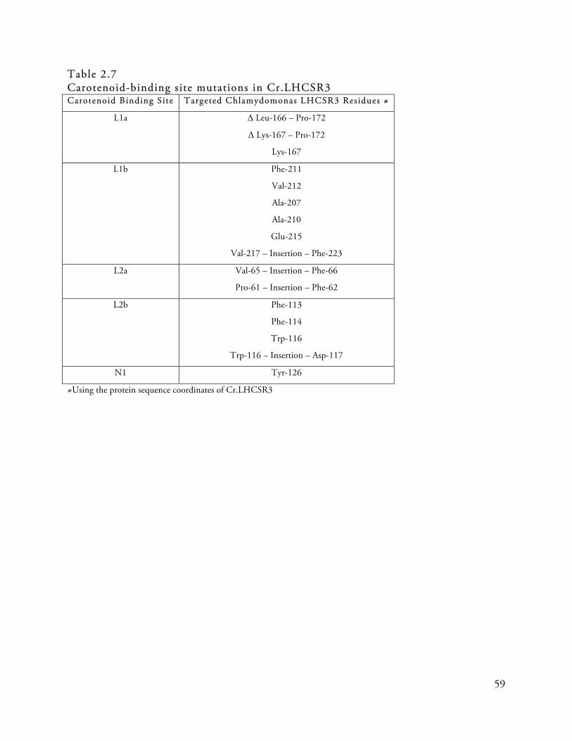
59
Table 2.7 Carotenoid-binding site mutations in Cr.LHCSR3 Carotenoid Binding Site Targeted Chlamydomonas LHCSR3 Residues ≠
L1a Δ Leu-166 – Pro-172
Δ Lys-167 – Pro-172
Lys-167
L1b Phe-211
Val-212
Ala-207
Ala-210
Glu-215
Val-217 – Insertion – Phe-223
L2a Val-65 – Insertion – Phe-66
Pro-61 – Insertion – Phe-62
L2b Phe-113
Phe-114
Trp-116
Trp-116 – Insertion – Asp-117
N1 Tyr-126
≠Using the protein sequence coordinates of Cr.LHCSR3

60
2.6 An attempt to develop two additional fast screening methods for evaluating the photosynthetic function of Chlamydomonas genes
While the transient expression system in N. benthamiana is a fast and easy way to screen large mutant libraries, it is not sufficient for finding loss-of-function mutations in Cr.LHCSR3 since the quenching signal is so minute. My goal was to find a way to normalize expression in transformed C. reinhardtii cultures for easy, consistently representative phenotypic assays. One way to do this would be to consistently express the mutant protein from the same location in the genome, eliminating positional effects from random insertion of the transformation cassette. C. reinhardtii has not been amenable to targeted nuclear genomic mutation techniques nor does it undergo homologous recombination in the nuclear genome. The chloroplast genome, however, can undergo homologous recombination and chloroplast transformation techniques are well established. The first approach I will describe is adapting a chloroplast transformation technique to assure consistent insertion and expression of thylakoid-localized genes from the chloroplast genome. Another way to assure consistent expression would be to couple the gene of interest to a regulatable expression system in order to tune expression levels regardless of the insertion site. To test this, I expressed Cr.LHCSR3 in C. reinhardtii using a specially designed nuclear transformation cassette where expression of the gene of interest is coupled to expression of a resistance gene required for survival in the presence of antibiotic, a method developed by Rasala and colleagues (2012). 2.6.1 Expression of LHC genes from the chloroplast genome
In order to establish a faster, more consistent screening strategy for assessing photosynthetic phenotypes, I set out to find a system that would ensure: 1) consistent expression levels for comparison between mutants, 2) a low chance of silencing the mutation, and 3) reduced risk of collateral damage to the photosystem or growth of the organism caused by random insertions. Chloroplast transformation offers these advantages because the gene is introduced into the prokaryotic-origin organellar genome, which does not have a rapidly adaptable silencing mechanism.
Chloroplast transformation was first demonstrated using particle bombardment (Boynton et al., 1988) and many strategies have been tested for selecting transformants by introducing antibiotic resistance genes into the organellar genome. Both the aadA gene, which confers resistance to spectinomycin (Goldschmidt-Clermont, 1991), and the aphA-6 gene, which confers resistance to kanamycin (Bateman & Purton, 2000) have proven to be successful selectable markers in C. reinhardtii, but there are limitations to this approach. If the transformed cells do not become homoplasmic, which is often difficult to assure, when the cells are removed from selection the transformation cassette will be diluted out over time and lost. Long-term exposure to antibiotics can increase the chances for mutations in the host, particularly to rRNA (Vester & Long, 2009).
Instead of using antibiotic selection to confirm successful chloroplast transformations, in the following experiments I chose to use a transformation system with a non-functional PsbH gene in the chloroplast genome (O’Connor et al., 1998), such that upon successful transformation photoautotrophic growth is restored. In this strain, known as the recipient strain, the aadA gene is disrupting PsbH in the chloroplast genome, preventing expression of the protein and thereby disrupting assembly of PSII and inhibiting photoautotrophic growth. By using a rescue vector, called

61
p72, a functional copy of the PsbH gene can be restored into the chloroplast genome by homologous recombination. Downstream of PsbH in the chloroplast genome is a neutral site providing an ideal space to introduce new genes into the chloroplast genome. The p72 vector has the functional copy of PsbH along with the gene of interest, in my case the cDNA-derived coding sequence of the Cr.LHCSR2 gene which encodes Cr.LHCSR3, stabilized by the 5’- and 3’-untranslated regions of the rbcL gene, all flanked by sequences homologous to the chloroplast genome surrounding PsbH (Figure 2.22). Successful transformation will yield a colony that can grow on minimal media and includes the gene of interest in the downstream neutral site. If the 5’- and 3’- untranslated regions of rbcL flanking the gene of interest were to incorrectly target the homologous recombination to remove rcbL, those transformants would be unable to grow on minimal media without the RbcL protein.
Figure 2.22 Chloroplast transformation strategy Based on the system developed by Purton and colleagues (19xx and 19xx). The recipient strain has the defective psbH chloroplast genome in the npq4lhcsr1 mutant nuclear genome. Transformation of the chloroplast genome using bombardment should rescue the disrupted PsbH gene to rescue autotrophic growth, and introduce the LHCSR2 gene into the chloroplast genome. The LHCSR2 gene is stabilized by the rcbL 5’ and 3’ untranslated regions for high expression.
In this strategy, I attempt to express a nuclear-encoded thylakoid-localized protein from the chloroplast genome. There are many reasons this could be unsuccessful for complementing function of the gene. The protein may be expressed, but not properly targeted to the thylakoid since it is not entering the organelle through the import machinery. Cleavage of the transit peptide may be necessary for activity, but does not occur when the protein is expressed from within the chloroplast. To address these potential pitfalls, I designed three different expression constructs to test the importance of import and signal peptide processing (Table 2.8). Each construct is designed with the cDNA-derived coding sequence of Cr.LHCSR2. Constructs p72-LHCSR3i and ii include only the coding sequence of LHCSR2, with or without a carboxy-terminal FLAG epitope tag, respectively. Constructs iii and iv encode a truncated copy of LHCSR3 that is lacking the first 20 residues which comprise the chloroplast transit peptide of LHCSR3. Constructs v and vi include the 35 residue membrane-targeting signal peptide from the petA gene, which encodes cytochrome b6f. This signal peptide is added to the amino-terminus of the Cr.LHCSR3 coding sequence. If the transit peptide interferes with proper localization for a protein expressed in the chloroplast, constructs iii and iv

62
should address this problem. The chloroplast-encoded petA gene’s 31-residue signal peptide has been shown to target proteins to the membrane (Gatenby et al., 1988; Pierre et al., 1995), though it is not a certainty that Cr.LHCSR3 proteins targeted to the thylakoid membrane using this signal peptide would adopt their native topology.
The goal was to use this transformation method to screen mutants of Cr.LHCSR3 for altered quenching function. To ensure the recipient chloroplast was in a qE-deficient genetic background, I performed a cross between the minus mating type of the npq4 lhcsr1 mutant, which lacks all LHCSR expression, and the recipient strain that has a disrupted psbH chloroplast genome. Using PCR to verify the genotype of the resulting tetrads, I selected a positive mating type strain with the recipient chloroplast genome psbH defect and the npq4 lhcsr1 mutations in the nuclear genome to perform the transformations (Line #13). I also selected a positive mating type strain with the recipient chloroplast genome, but with the recipient strain’s parental nuclear genomic background with functional LHCSR genes (Line #20) to compare the background qE-capacity of the parental strain to the transformants, and to evaluate overexpression of Cr.LHCSR3 resulting from both a nuclear- and a chloroplast-encoded copy of Cr.LHCSR2.
Figure 2.23 Baseline NPQ and LHCSR expression in psbH x npq4 lhcsr1 chloroplast transformation recipient strains, Line #13 and Line #20 Line #13 and Line #20 were generated by crossing the npq4 lhcsr1 qE-deficient mutant with the psbH recipient strain and screening for the psbH deletion in the chloroplast genome. #13 and #20 are the mt+ mating type progeny where Line #13 has the nuclear genetic background of the npq4 lhcsr1 parental strain and Line #20 has the nuclear genetic background of the psbH recipient strain. Both Line #13 and Line #20 were transformed with the empty p72 transformation vector to restore photoatutotrophic growth. Despite expression of LHCSR, Line #20 does not have qE.

63
To evaluate the qE-capacity of the parental background, I transformed both Line #13 and Line #20 with the empty rescue vector, which would only rescue the dysfunctional psbH gene and not introduce a gene into the neutral site. The transformation efficiency of this method was extremely low, but I did recover a few transformants that successfully rescued photoautotrophic growth. Unfortunately, while Line #20 does express some LHCSR in high light, it does not have any inducible qE (Figure 2.23), which means the photosystem defect or accumulated mutations elsewhere in the parental background of this strain are not permissive of LHCSR-mediated quenching. Despite transformation of both Line #13 and Line #20 with each of the rescue vector constructs, none of the transformations resulted in rescue of the quenching phenotype. To distinguish whether the parental background is not permissive of qE, or if perhaps only the particular progeny I selected were defective in some way, I also transformed the empty vector into the recipient strain that had not been crossed with npq4 lhcsr1. The parental background has low PSII accumulation, as seen by the low levels of D2 expression when evaluated on an equal Chl basis (Figure 2.24), which shows that rescue of PsbH that is sufficient to restore photoautotrophic growth does not necessarily perfectly restore PSII accumulation. The rescued recipient train does, however, expresses LHCSR proteins and has near 4A wild type levels of qE (Figure 2.24).
Because the parental strain is able to express LHCSR and has near wild-type levels of qE, the qE deficiency in Line #20 may represent an unlucky selection of progeny – perhaps this strain did not receive a permissive background for LHCSR-mediate quenching, for example. The qE deficiency in Line #13 could also be an unlucky selection of progeny, or that expression of LHCSR from the chloroplast genome is not capable of inducing NPQ in C. reinhardtii. Unfortunately, because neither of the recipient strains I selected is permissive of inducible quenching, it precludes my ability to use this experimental set-up to determine if expression of nuclear-encoded thylakoid-localized LHCSR genes from the chloroplast genome can functionally complement the npq4 lhcsr1 qE deficiency. If this method were to be pursued again, a larger set of progeny could be tested to see if any received a permissive background for LHCSR-mediated quenching. Alternatively, there are other chloroplast genome recipient strains available that may provide a better background for testing these genes (Chen & Melis, 2013). Table 2.8 Chloroplast transformation constructs Protein Gene constructs in p72 rescue vector Construct name Cr.LHCSR3 Cr.LHCSR3 + FLAG p72-LHCSR3i Cr.LCHSR3 p72-LHCSR3ii Cr.LHCSR3 (Δ20) + FLAG p72-LHCSR3iii Cr.LHCSR3 (Δ20) p72-LHCSR3iv petA (35) + Cr.LHCSR3 + FLAG p72-LHCSR3v petA (35) + Cr.LCHSR3 p72-LHCSR3vi The cDNA-derived coding sequence of Cr.LHCSR3 was used in each transformation cassette. In construct iii and iv, the first 20 residues comprising the transit peptide of LHCSR3 were not included, while in construct v and vi the signal peptide of the chloroplast-encoded petA gene, which encodes cytochrome f, was added to the amino-terminus of the Cr.LCHSR3 peptide sequence.

64
Figure 2.24 NPQ and LHCSR expression of chloroplast transformation parental strains After transformation with the empty p72 vector, the PsbH gene is restored and allows for photoautotrophic growth of the parental recipient strain. Despite the recovery of a functional PsbH gene, however, the parental recipient strains (#1-#3) do not accumulate very much PSII under high light growth condtions as shown by Western blotting against the PSII core protein, D2. The parental strains have near 4A wild type levels of NPQ and accumulate both LHCSR1 and LHCSR3 when grown in high light. 2.6.2 Control of LHC gene expression through fusion to a selectable marker gene In a separate attempt to achieve consistent, regulatable expression levels of mutant genes in C. reinhardtii, I adapted a method developed by Rasala and colleagues (2012) for selection-dependent expression. One of the major obstacles for using C. reinhardtii as a high-throughput platform is poor expression of heterologous genes from the nuclear genome. Rasala and colleagues used the foot-and-mouth-disease-virus (FMDV) 2A self-cleavage peptide to transcriptionally fuse heterologous gene expression to antibiotic resistance, assuring that survival on selection is linked to commensurate expression of the introduced gene of interest (2012). They used the Streptoalloteichus hindustanus bleomycin resistance gene (sh-ble), which confers resistance to the DNA double strand break-inducing bleomycin antibiotic family (Dumas et al., 1994). Zeocin resistance is achieved by binding and sequestration of the antibiotic agent, meaning resistance to varying levels of antibiotic selection is proportional to expression levels (Dumas et al., 1994). The FMDV 2A peptide is a 20-

65
residue sequence that promotes self-cleavage (Ryan et al, 1991). During translational elongation, a peptide bond fails to form between the 18th and 19th residue of the sequence, leaving the majority of the 2A sequence attached to the carboxy-terminus of the first protein encoded, yielding two independent proteins (Ryan et al., 1991). For these experiments we used the pBR9 plasmid (Rasala et al., 2012), which encodes the PAR4 promoter upstream of the expression construct and the Rbcs2-3’-untranslated region (Figure 2.25). PAR4 contains the Hsp70A promoter enhancer element along with four tandem copies of the Rbcs2 gene’s first intron, followed by the Rbcs2 promoter and 5’-untranslated region. In the expression construct, we inserted the cDNA-derived coding sequence for Cr.PSBS and Cr.LHCSR3, each with a carboxy-terminal FLAG-epitope tag, using the InFusion PCR kit (Clontech). The expression vector was introduced into npq4 lhcsr1 C. reinhardtii cells using electroporation for transformation following the published method (Rasala et al., 2012) and using 1 µg of DNA per 250 µL of cells. Successful transformants were identified by survival on TAP plates containing three different concentrations of zeocin (5, 10, and 15 µg/mL).
Figure 2.25 Transformation plasmids for selection-dependent expression in C. reinhardtii The pBR9 expression vector couples the Hsp70A and the RbcS promoters in the PAR4 promoter region upstream of the ble-sh antibiotic resistance gene that is linked to the coding sequence of the gene of interest by the foot-and-mouth-disease-virus (FMDV) 2A self-cleavage peptide linker (Rasala et al., 2012). Transformation efficiency was very low for the reactions selected on 15 µg/mL zeocin plates, and only about 25 colonies per µg of DNA were observed on 10µg/mL zeocin plates. The 5 µg/mL zeocin plates appeared to have high transformation efficiency of about 500 colonies per µg of DNA, but upon patching the colonies onto fresh 5 µg/mL zeocin, very few survived. Transformation of the whole expression cassette was confirmed by PCR, and only 20% of the colonies displaying zeocin resistance also had a full-length amplicon of both the sh-ble gene and LHCSR2 or PSBS. The transformants were maintained on TAP with zeocin, except to screen for photosynthetic phenotype, which is done in minimal media. Since this expression construct should lead to constitutive expression of the protein products, streaks of each transformant were screened on minimal media plates in both low light and high light acclimated conditions for restoration of qE. No rescue of the qE phenotype could be observed in low light, but a few colonies were selected for further characterization from the high light screen. Amongst these colonies, no photosynthetic phenotype

66
could be observed (Figure 2.26), but immunoblot analysis probing against the FLAG epitope showed that both Cr.LHCSR3 and Cr.PSBS expression is present at low levels after growth in high light conditions only. Transformed colonies not exposed to constant selection, even just for the course of a photosynthetic screen of 5 days, had all lost zeocin resistance when transferred from minimal media back to selection plates, confirming that the cells do not prefer to maintain the high-level of expression conferred by the PAR4 promoter. Over the course of six months, even under constant maintenance on zeocin, each of the colonies died. This suggests that either long-term zeocin exposure eventually leads to lethal double strand breaks, and/or that the expression construct is silenced even in the presence of persistent selection.
Figure 2.26 Zeocin selection-linked expression of Cr.LHCSR3 and Cr.PSBS in C. reinhardtii Expression of LHCSR and PSBS in C. reinhardtii via fusion to the sh-ble gene upon transformation into the npq4 mutant background. Protein only accumulates under high light growth conditions, but has no phenotypic effect. While the results published in the article by Rasala and colleagues showed high transformation efficiency and stable expression of the xylanase gene fused to GFP (2012), the pBR9 expression construct with either Cr.LCHSR2 or Cr.PSBS did not prove to have either high efficiency or stable expression for long-term analysis even under persistent selection. Zeocin is a potent mutagen, leading to double stranded DNA breaks, which may explain the slow but consistent loss of colony viability. BleR is the best selectable marker to use for this system because of the stoichiometric mechanism, as opposed to an enzymatic mechanism that is employed by most other genes conferring antibiotic resistance, however, because of the instability and rapid silencing of the expression construct, this system is not ideal for studying photosynthetic phenotypes in a semi-high-throughput method. The lack of a photosynthetic phenotype, even though protein expression had been confirmed, may be due to a number of reasons that I was not fully able to address because of the loss of colony viability. The FLAG epitope tag may interfere with function, particularly of the LHCSR3 protein since it would leave a highly acidic tail exposed to the lumen on the carboxy-terminus of the protein, but the effect of this epitope tag on protein function has not been confirmed. The epitope tag on Cr.PSBS, assuming the protein is localized to the thylakoid membrane in the same manner as

67
its plant homolog, would be exposed to the stroma and and would presumably be less likely to interfere with function. The fact that both proteins only accumulate under high light may indicate that stability of the two proteins is promoted in a high light- or stress-dependent manner, or that timing of expression is critical for function. The lack of rescue of the qE phenotype may also suggest that a translational or post-translational regulatory mechanism is required for function, but this mechanism is absent when the protein is translated from the coding sequence as opposed to the genomic sequence. If the pBR9 constructs had been stable some of these questions could have been addressed, but mutagenesis by random insertion is still the best method for screening photosynthetic phenotypes caused by C. reinhardtii genes for my questions. 2.7 Acknowledgements With permission from the authors, some elements of the chapter have been adapted from published articles and articles in preparation. Section 2.1 draws upon material in preparation: Truong, T., Erickson, E., Amarnath, K., Peers, G., Kobayashi, M., Inwood, W.,
Kustu, S., The Seattle TILLING Project, Fleming, G., and Niyogi, K.K. (expected 2016) Targeted reverse genetics demonstrates a strict LHCSR requirement for non-photochemical quenching in Chlamydomonas reinhardtii.
Section 2.2 draws upon material published in: Ballottari , M., Truong, T. B., De Re, E., Erickson, E., Stella, G.R., Fleming,
G.R., Bassi , R. and Niyogi, K. K. (2016) Identification of pH-sensing sites in the light harvesting complex stress-related 3 protein essential for triggering non-photochemical quenching in Chlamydomonas reinhardtii. Journal of Biological Chemistry, 291, 7334-7346.
Section 2.3 draws upon material submitted for publication: Leonell i , L., Erickson, E., Lyska, D., and Niyogi, K.K. (expected 2016) Transient
expression in Nicotiana benthamiana for rapid functional analysis of genes involved in non-photochemical quenching and carotenoid biosynthesis. Plant Journal. Manuscript submitted.
Section 2.4 draws upon material in preparation: Erickson, E., Leonell i , L., and Niyogi, K.K. (expected 2016) Disruption of predicted
pigment binding sites in LHCSR3 from Chlamydomonas reinhardtii enhances non-photochemical quenching. Manuscript in preparation.

68
Chapter 3
Assessing the functionality of PSBS from Chlamydomonas reinhardtii
3.1 Abstract Through study of the npq4-1 mutant of A. thaliana, it has been shown that expression of the PSBS protein is required for qE (Li et al., 2000), but a molecular mechanism of action for this four-transmembrane LHC protein is still unknown. In C. reinhardtii, however, contributions from LHCSR1 and LHCSR3 account for all of the rapidly inducible quenching observed after high light acclimation (Truong et al., 2016). Despite two copies of the PSBS gene in the genome of C. reinhardtii, no role has been proposed for the green algal homolog. Here I explore the potential functionality of PSBS from C. reinhardtii by studying the protein’s activity in N. benthamiana and A. thaliana. 3.2 PSBS in Chlamydomonas reinhardtii There are two PSBS homologs in the C. reinhardtii genome, PSBS1 and PSBS2. The genes are transcribed upon a shift from dark to light conditions, such that high levels of transcript have been measured within one hour of the onset of light (Duanmu et al., 2013), but rapidly return to a nearly undetectably low level shortly after (Zones et al., 2015). High levels of PSBS transcript have also been observed after prolonged nitrogen stress conditions (Miller et al., 2010). Despite the presence of two PSBS gene isoforms in the genome, no one had, until recently, reported finding expression of the protein. This may have been because the antibodies raised against the barley (Hordeum vulgare) PSBS protein (Bonente et al., 2008a) and the At.PSBS protein (Li et al., 2002a) do not recognize the natively expressed Cr.PSBS protein, or it may be because the Cr.PSBS protein is not expressed under the tested conditions (Bonente et al., 2008a). The anti-barley PSBS antibody does, however, recognize recombinant reconstituted Cr.PSBS using the native sequence (Bonente et al., 2008a). When the gene was expressed in C. reinhardtii under the strong constitutive PsaD promoter, protein was identified in the whole cell lysate, though none was detected in the thylakoid membrane fraction (Bonente et al., 2008a), and the authors of the study concluded that PSBS probably has a different function in C. reinhardtii than a role in photoprotection. The rapid transcriptional response observed upon light exposure supports that PSBS may still play a role in photoprotection, and when taken together with the protein over-expression results, these data support a model where photoprotective PSBS expression is regulated, transient, and may not require a high level of accumulation. But what is it doing?

69
3.2.1 Hypotheses regarding the possible role of PSBS Could PSBS be targeted somewhere other the thylakoid membrane of C. reinhardtii? And if so, what is its role in the cell? Could this role help us to understand how it became the dominant quenching mechanism in vascular terrestrial plants, whearas it is not required by unicellular green algal species for qE? Is C. reinhardtii just uniquely unable to use PSBS while the protein would be functional in a system that recognizes a PSBS-mediated quenching pathway? This could be the case, since the sequence of Cr.PSBS is quite similar to that of At.PSBS, and the two functional glutamate residues are conserved (Figure 3.1). The amino-terminal chloroplast transit peptide of Cr.PSBS, however is noticeably unique. This work set out to see if Cr.PSBS is functional for quenching in a system equipped to perform PSBS-mediated quenching.
Figure 3.1 Alignment of PSBS protein sequences from C. reinhardtii and A. thaliana The PSBS gene from C. reinhardtii has high protein sequence identity to At.PSBS, including conservation of the two essential lumenal glutamate residues, Glu-122 and Glu-226, indicated by red stars. All plant expression constructs described in this thesis that were used for testing transient expression in N. benthamiana and stable transgenic expression in A. thaliana use the chloroplast transit peptide sequence from At.PSBS, which is the first 54 residues of the sequence. The anti-PSBS antibody was raised against the At.PSBS peptide epitope labeled in black. 3.3 Transient expression of Cr.PSBS in Nicotiana benthamiana 3.3.1 Chloroplast transit peptide recognition across species Our first test was to see if Cr.PSBS, with its native chloroplast transit peptide (Cr.PSBS-nTP), could be expressed and functional in a plant system. N. benthamiana leaves were infiltrated with A. tumefaciens containing a pEarleyGate100 plasmid encoding the cDNA-derived coding sequence of Cr.PSBS. Two versions of Cr.PSBS-nTP were tested, one with a carboxy-terminal FLAG epitope tag (Cr.PBS-nTP+), and one without the FLAG tag (Cr.PSBS-nTP-). The native Cr.PSBS sequence was compared to transient expression of a construct that has the 54-residue chloroplast transit peptide from At.PSBS and the carboxy-terminal FLAG epitope tag (referred to as

70
Cr.PSBS). At.PSBS was used as a positive control, the Cr.PSBS QQ mutant was used as a negative control, and GUS as an expression control. Neither of the Chlamydomonas native chloroplast transit peptide constructs (Cr.PSBS-nTP+/-) was expressed in planta, whereas both At.PSBS and Cr.PSBS were expressed (Figure 3.2).
Figure 3.2 Test of transient expression of Cr.PSBS with and without the algal chloroplast transit peptide The native PSBS sequence from C. reinhardtii is not expressed by N. benthamiana, while the sequence containing the At.PSBS chloroplast transit peptide is expressed and results in additional quenching over the background level of the leaf. The level of quenching by Cr.PSBS is comparable to that of At.PSBS when transiently expressed for 48 hours in N. benthamiana leaves.

71
Expression of the Cr.PSBS construct with the At.PSBS chloroplast transit peptide in N. benthamiana leaves results in increased quenching relative to the background of the leaf and the GUS negative control (Figure 3.2). Just as was shown with expression of heterologous At.PSBS in N. benthamiana, even at high levels Cr.PSBS does not disrupt the stability of solubilized thylakoid membrane complexes when evaluated using blue native PAGE of isolated thylakoids purified from N. benthamiana chloroplasts that have been gently solubilized with the detergent n-dodecyl-α-D-maltopyranoside (Figure 3.3). Cr.PSBS is distributed throughout the solubilized complexes, even more so than At.PSBS. Also in contrast to the expression of At.PSBS in N. benthamiana leaves is that the Cr.PSBS protein appears to be processed or degraded as shown by SDS-PAGE analysis of the isolated solubilized thylakoid membrane complexes. Most likely these are alternative truncations that differ at the amino-terminal end of the protein, resulting in lower molecular weight versions (Figure 3.3). Together, these results show Cr.PSBS accumulates in the thylakoid membrane when expressed in a plant system and that it can perform some functional role in inducing quenching, but there are differences between At.PSBS and Cr.PSBS.
Figure 3.3 Analysis of thylakoid membrane complexes from N. benthamiana leaves transiently expressing Cr.PSBS and At.PSBS proteins The stability of solubilized thylakoid membrane complexes present in uninfiltarated leaves of N. benthamiana are the same in leaves expressing heterologous proteins, including At.PSBS, At.PSBS QQ, Cr.PSBS, Cr.PSBS QQ, and even Cr.LHCSR3. The localization of these proteins within those complexes varies, however. At.PSBS is associated with a few complexes, while Cr.PSBS is associated with many complexes throughtout the thylakoid membrane. LHCSR is only associated with a few high molecular-weight complexes. Denaturing SDS-PAGE analysis of the solubilized thylakoid membranes also shows that Cr.PSBS is differentially processed, resulting in multiple products that maintain the FLAG-epitope tag, while At.PSBS and Cr.LHCS3 are not processed or degraded.

72
3.3.2 Non-functional Cr.PSBS QQ and At.PSBS QQ mutants competitively inhibit native PSBS in N. benthamiana Because Cr.PSBS can induce quenching in planta, we also set out to see if alteration of the conserved lumenal glutamate residues that render At.PSBS non-functional would do the same for Cr.PSBS. When the At.PSBS QQ mutant is expressed in N. benthamiana, qE capacity of the infiltrated spot is significantly lower than the spots expressing the non-mutated copy (Figure 3.4). The Cr.PSBS QQ mutant works similarly, also showing reduced NPQ capacity in the infiltrated spots (Figure 3.4).
Figure 3.4 Transient expression of PSBS and non-functional PSBS constructs in N. benthamiana Leaf spots expressing At.PSBS or Cr.PSBS show increased quenching relative to the background of the leaf. Mutations changing the functional glutatmate residues in each protein construct to non-acidic glutamines results in diminished quenching, near the same level as the background quenching observed in the uninfiltarted leaf and the negative GUS control.

73
3.4 Stable Cr.PSBS expression partial ly rescues the NPQ defect of the Arabidopsi s thaliana npq4-1 mutant
Transient expression of Cr.PSBS showed that the algal PSBS protein could be functional in a system that is permissive to PSBS-mediated quenching. To confirm this quenching phenotype, we created homozygous transgenic lines of the npq4-1 mutant of A. thaliana expressing the Cr.PSBS construct with a plant chloroplast transit peptide. Using the same CaMV 35S promoter construct as was used for transient expression, plants expressing the Cr.PSBS protein were selected and then propagated to the homozygous T3 generation. The homozygous plants do show a partially rescued NPQ phenotype at four weeks old when compared to npq4-1 mutant plants and Columbia-0 wild type plants (Figure 3.5).
Figure 3.5 Cr.PSBS partial ly rescues qE-deficient npq4-1 phenotype in A. thaliana As observed with transient expression in the N. benthamiana leaves, stable transgenic npq4-1 A. thaliana lines expressing the same Cr.PSBS protein construct show a partial rescue of qE, intermediate between the Columbia-0 level and the npq4-1 mutant background level.
Although the transient expression assay in N. benthamiana showed Cr.PSBS to be nearly as good at quenching as the At.PSBS protein, in the stably tansformed lines Cr.PSBS is much less efficient at inducing quenching. To ensure that Cr.PSBS from this expression vector in transgenic A. thaliana lines is representative of the native protein, we wanted to compare rescue by Cr.PSBS to that of At.PSBS and the At.PSBS QQ non-functional protein expressed from the same expression vector and in the same genetic background. I only had to time to propagate the lines to the T2 generation, but discovered that the protein expression in these lines was silenced before the plants were even four weeks old (Figure 3.7). Comparable expression and fluorescence data is not yet available for these two lines. Previous studies evaluating the At.PSBS QQ mutant have not observed

74
any rescue of quenching from the non-functional mutant, however the expression vector used in that work had the wild-type gene and the mutant gene under the native promoter (Li et al., 2004), so there may be a difference using the CaMV 35S promoter, which we would like to further explore in the near future.
To look at the effect of expressing high levels of Cr.PSBS relative to the background npq4-1 and Columbia-0 wild type plants, we analyzed isolated solubilized thylakoid membrane complexes from transformed lines expressing Cr.PSBS, as well as those that had been expressing At.PSBS QQ as seedlings and compared them to each of the genetic background lines using BlueNative-PAGE of tissue from seven week-old plants. There is no discernible difference in the solubilized membrane complexes between the wild type Columbia-0 tissue, the npq4-1 mutant background, and each of the transformant lines when thylakoid membranes were solubilized with n-dodecyl-α-D-maltopyranoside or with n-dodecyl-β-D-maltopyraoside (Figure 3.6). Immunoblot of the native complexes of the npq4-1 tissue compared to the Cr.PSBS expressing line, using an anti-FLAG primary antibody shows that Cr.PSBS is associated with many complexes in the membrane (Figure 3.6), which is the same as was seen with the N. benthamiana tissue transiently expressing Cr.PSBS. High expression of the Cr.PSBS does not disrupt the solubilized membrane complexes, so the partial rescue caused by this protein is not due to widespread antenna complex rearrangements or instability in the membrane, but the protein is widely associated throughout all the complexes. Furthermore, the Cr.PSBS protein is again observed to be differentially truncated in A. thaliana thylakoids, as was seen in the biochemical analysis of N. benthamiana thylakoid membranes. The mechanism of quenching employed by the Cr.PSBS-expressing plants may not be the same as those that express the native At.PSBS.
The T2 lines transformed with the At.PSBS QQ construct were silenced as seedlings, which is confirmed by immunoblot analysis of the 1D Native-PAGE of solubilized isolated thylakoid tissue as well as the SDS-PAGE of both the solubilized thylakoids (Figure 3.6) and leaf punches from plants the same plants at two, four, and seven weeks old (Figure 3.7). Because there is no signal in either the isolated thylakoids or the whole cell lysate derived from leaf punches, the constructs were silenced, as opposed to faulty chloroplast import of the heterologous protein. This experiment will be redone with T3 generation homozygous lines in the future with plants at an earlier age to ensure expression.

75
Figure 3.6 Analysis of thylakoid membrane complexes in transgenic A. thaliana npq4-1 l ine expressing Cr.PSBS Lane 1 = Columbia-0 tissue Lane 2 = npq4-1 tissue Lane 3 = homozygous T3 line expressing Cr.PSBS in npq4-1 genetic background Lane 4 = T2 line expressing At.PSBS QQ in npq4-1 genetic background Separation of complexes from thylakoid membranes solubilized by n-dodecyl-α-D-maltopyranoside (α-DM) or by n-dodecyl-β-D-maltopyranoside (β-DM) detergents by BlueNative-PAGE shows no difference in overall membrane architecture between the wild type Columbia-0, the qE-deficient npq4-1 mutant, or the Cr.PSBS expressing transgenic line. Cr.PSBS is associated with multiple complexes in the membrane, a result that has been previously observed by others in DeriphatPAGE analysis (Teardo et al., 2007). While protein expression was observed in leaf punches of the At.PSBS QQ transgenic plant, and insertion of the At.PSBS QQ mutant gene sequence was verified by PCR, no protein accumulation is identified in the isolated thylakoid tissue as evaluated by anti-FLAG immunoblotting of the native samples. In case the epitope is occluded in the native samples, the same solubilized membrane samples were analyzed by SDS-PAGE 40 hours after preparation, and still only the Cr.PSBS line has detectable denatured protein accumulation recognized by anti-FLAG immunoblotting. Degradation of the Cr.PSBS protein appears to have begun during the 24-hour incubation in either detergent solution.
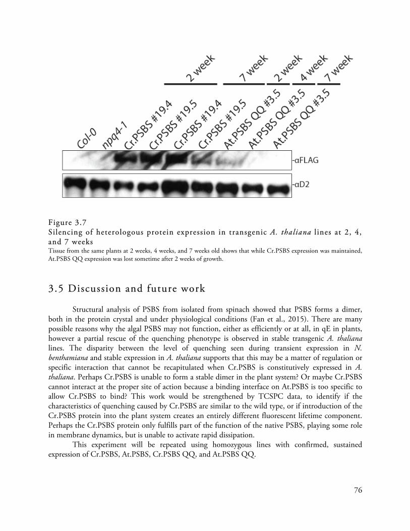
76
Figure 3.7 Silencing of heterologous protein expression in transgenic A. thaliana l ines at 2, 4, and 7 weeks Tissue from the same plants at 2 weeks, 4 weeks, and 7 weeks old shows that while Cr.PSBS expression was maintained, At.PSBS QQ expression was lost sometime after 2 weeks of growth. 3.5 Discussion and future work Structural analysis of PSBS from isolated from spinach showed that PSBS forms a dimer, both in the protein crystal and under physiological conditions (Fan et al., 2015). There are many possible reasons why the algal PSBS may not function, either as efficiently or at all, in qE in plants, however a partial rescue of the quenching phenotype is observed in stable transgenic A. thaliana lines. The disparity between the level of quenching seen during transient expression in N. benthamiana and stable expression in A. thaliana supports that this may be a matter of regulation or specific interaction that cannot be recapitulated when Cr.PSBS is constitutively expressed in A. thaliana. Perhaps Cr.PSBS is unable to form a stable dimer in the plant system? Or maybe Cr.PSBS cannot interact at the proper site of action because a binding interface on At.PSBS is too specific to allow Cr.PSBS to bind? This work would be strengthened by TCSPC data, to identify if the characteristics of quenching caused by Cr.PSBS are similar to the wild type, or if introduction of the Cr.PSBS protein into the plant system creates an entirely different fluorescent lifetime component. Perhaps the Cr.PSBS protein only fulfills part of the function of the native PSBS, playing some role in membrane dynamics, but is unable to activate rapid dissipation. This experiment will be repeated using homozygous lines with confirmed, sustained expression of Cr.PSBS, At.PSBS, Cr.PSBS QQ, and At.PSBS QQ.

77
3.6 Acknowledgements With permission from the authors, some elements of the chapter have been adapted from articles in preparation. Leonell i , L., Erickson, E., Lyska, D., and Niyogi, K.K. (expected 2016) Transient
expression in Nicotiana benthamiana for rapid functional analysis of genes involved in non-photochemical quenching and carotenoid biosynthesis. Plant Journal. Manuscript submitted.
Erickson, E., Leonell i , L., Garcia-Cerdan, J. , and Niyogi, K.K. (expected 2016)
Evidence of a functional role for the PSBS protein from Chlamydomonas reinhardtii. Manuscript in preparation.

78
Chapter 4
Exploring a functional relationship between PSBS and LHCSR3 from Chlamydomonas reinhardtii
4.1 Abstract LHCSR-mediated quenching pathways comprise the dominant quenching mechanism for unicellular green algal species, but LHCSR-like genes are no longer present in vascular terrestrial plant species. PSBS genes, however, are present throughout the green lineage, even though only terrestrial plants have been shown to use a PSBS-mediated quenching pathway. We have been exploring whether PSBS from C. reinhardtii may function in the same way as the plant protein in planta, but this does not account for any potentially different roles the protein may play in the more basal branches of the green lineage. Furthermore, it is not certain why LHCSR was lost from terrestrial plant species. In this chapter I explore the ability of Cr.LHCSR3 to influence quenching in A. thaliana, and subsequently, whether Cr.LHCSR3 and Cr.PSBS may both have a quenching function, as they do in the bryophyte moss, Physcomitrella patens. 4.2 LHCSR- and PSBS-mediated quenching pathways function independently and additively in the bryophyte moss, Physcomitrel la patens
An evolutionary analysis of photoprotective proteins in the green lineage shows that algal species utilize an LHCSR-mediated quenching pathway, but that homologs of LHCSR are no longer present in vascular plants (Figure 4.1) (Gerotto & Morosinotto, 2013). PSBS-mediated quenching pathways have been observed in vascular and non-vascular terrestrial plants, as well as the Charales and Zygnematales orders of algae (Bonente et al., 2008a; Gerotto & Morosinotto, 2013). The bryophyte moss, Physcomitrella patens, a non-vascular terrestrial plant uses both an LHCSR- and PSBS-mediated quenching pathway (Alboresi et al., 2010; Gerotto & Morosinotto, 2013). In P. patens, LHCSR-mediated quenching behaves in a similar manner to that of C. reinhardtii, and likewise PSBS-mediated quenching behaves in a similar manner to the PSBS of A. thaliana. No other organisms with both pathways have been characterized. While our results show that contributions from the two LHCSR proteins of C reinhardtii account for all of qE, it is unknown whether there is also a role for Cr.PSBS in photoprotection. New information about the expression

79
of PSBS in C. reinhardtii motivates us to take a fresh look at the role Cr.PSBS may be playing in the LHCSR-mediated quenching mechanism of the green alga.
Figure 4.1 Evolution of stress response LHC proteins in the green lineage Genome, transcript, and protein expression analysis from multiple sources show that a PSBS-like gene is present throughout the green lineage. While LHCSR-like genes are found amongst the algae of the green lineage, LHCSR-like genes are not present in vascular terrestrial plants. Genes for PSBS are present amongst the most basal chlorophyte algae, but no protein expression has been found.

80
4.3 Expression of Cr.LHCSR3 in Arabidopsi s thaliana Even though transient expression of Cr.LHCSR3 in N. benthamiana did not show a strong quenching phenotype, we generated homozygous transformant lines in A. thaliana to compare how a transient phenotype may differ from a stable transgenic line. Very similar work expressing the LHCSR protein from P. patens was recently published, where the quenching phenotype was not detected in transient expression in N. benthamiana but was noticeable in stably transformed N. tabaccum lines (Pinnola et al., 2015).
The Cr.LHCSR3 expression construct was introduced into npq4-1 mutant A. thaliana, and successful transformants were selected and propagated to the T2 generation for analysis. These transgenic lines showed no quenching phenotype (Figure 4.2). The Cr.LCHSR3 expression construct was also introduced into Columbia-0 wild type A. thaliana, and these transgenic lines showed a higher quenching capacity than the wild-type background line when the plants were seedlings (Figure 4.2). By introducing a heterologous stress response LHC protein, Cr.LHCSR3, into a plant system with its endogenous PSBS protein, we see enhanced quenching, leading to the hypothesis that perhaps LHCSR may require PSBS for its quenching function. This is further supported by the complete absence of signal in the Nb.PSBS-silenced leaves of N. benthamiana, which did not display the rapid early quenching phenotype seen in wild type N. benthamiana leaves (Figure 2.18). Like the PSBS-expressing T2 lines, however, the lines expressing LHCSR were also silenced between the age of two weeks and four weeks. Additional biochemical analysis will be done in the future.
Figure 4.2 A. thaliana l ines expressing Cr.LHCSR3 When Cr.LHCSR3 is expressed in the PSBS-deficient npq4-1 mutant background, there is no increase in quenching, but when Cr.LHCSR3 is expressed in the Columbia-0 wild type background additional quenching is observed over the background level.

81
4.4 Transient co-expression of Cr.LHCSR3 and Cr.PSBS in Nicotiana benthamiana As discussed in Chapter 3, if PSBS has two roles – a role in altering the arrangement of the thylakoid membrane complexes to accommodate quenching, and a role as the trigger for quenching – then maybe Cr.PSBS only plays one of these roles. Or, perhaps an interaction that occurs between Cr.PSBS and Cr.LHCSR3 would not be observed between At.PSBS and Cr.LHCSR3 due to species-level specificity of the binding interaction. To speed the time required for screening of a possible cooperative effect between Cr.LHCSR3 and PSBS, I transiently co-expressed Cr.LHCSR3 with Cr.PSBS and At.PSBS in mature leaves of N. benthamiana, to evaluate whether co-expression results in a significant phenotypic effect. I also co-expressed Cr.LHCSR3 with the At.PSBS QQ mutant and the Cr.PSBS QQ mutant to identify whether the pH-sensing sites are still necessary for quenching when the two proteins are together, or if solely a binding interaction can accomplish the same cooperative effect. As Figure 4.3 demonstrates, the combined expression of Cr.LHCSR3 and either PSBS protein results in enhanced quenching relative to the background of the leaf, the GUS negative control, and each protein expressed alone. Co-expression of Cr.LHCSR3 with either of the PSBS QQ mutants, however, did not enhance quenching in the N. benthamiana leaves. While the early induction seen with Cr.LHCSR3 expression alone is less apparent in the co-expression spots, the induction is more rapid than with either At.PSBS or Cr.PSBS expressed alone, and the final quenching capacity after induction is also higher than for any of proteins expressed alone. The qE capacity for spots expressing Cr.LHCSR3 alone is only slighter higher than the background quenching of the leaf, but when Cr.LHCSR3 is expressed together with either Cr.PSBS or At.PSBS, leaf spots show more than an additive effect in quenching, especially during the initial time points. 4.5 Discussion and future work
While the transient co-expression of Cr.LHCSR3 and Cr.PSBS results in a significant increase in quenching, we would like to see if this is still observed in a stable transgenic plant line. Crosses are underway to create homozygous lines expressing both Cr.LHCSR3 and Cr.PSBS in the npq4-1 genetic background of A. thaliana. Because addition of Cr.LHCSR3 to the Columbia-0 background showed higher NPQ capacity, we want to confirm the potential interaction between Cr.LHCSR3 and either Cr.PSBS or At.PSBS, in the absence of the native At.PSBS.
Although PSBS genes are present in C. reinhardtii, protein expression has not been observed, nor is PSBS thought to be necessary for qE in C. reinhardtii, as evidenced by the LHCSR1 and LHCSR3 results described in Section 2.1. Work by Correa-Galvis and colleagues (2016b), however, has turned these hypotheses over, by using a Cr.PSBS-specific antibody to measure expression of the Cr.PSBS protein in a transient burst at the onset of high light. Silencing of the gene using artificial micro-RNA results in lowered LHCSR3 protein expression and a qE defect (Correa-Galvis et al., 2016b). It is unclear if the artificial micro-RNA lines influence LHCSR expression, or if PSBS is somehow required for proper expression, function, or stability of LHCSR3. These results from Correa-Galvis er al. (2016b), along with the often-frustrating observations recorded in this thesis, support that high levels of expression of PSBS cannot enhance quenching in C. reinhardtii in the

82
same manner that it would in A. thaliana or N. benthamiana. These results provide motivation to look for additional regulatory steps in the quenching pathway that are not yet characterized.
Figure 4.3 Coexpression of Cr.LHCSR3 with PSBS results in increased quenching in N. benthamiana leaves Co-infiltration of Cr.LHCSR3 with either At.PSBS or Cr.PSBS results in enhanced quenching, significantly higher than what is observed when any of the constructs are expressed individually. These results motivate us to explore whether LHCSR3 and PSBS may act together for photoprotection in C. reinahardtii.

83
Because we observed a potentially novel quenching pathway in the mutants of Cr.LHCSR3 that target putative pigment-binding sites, I have also introduced these constructs into npq4-1 and Columbia-0 A. thaliana lines. These transgenic plants have not yet been characterized, but may provide a stable system for studying the effect of these mutations on Cr.LHCSR3. 4.6 Acknowledgements With permission from the authors, some elements of the chapter have been adapted from articles in preparation. Erickson, E., Leonell i , L., Garcia-Cerdan, J. , and Niyogi, K.K. (expected 2016)
Evidence of a functional role for the PSBS protein from Chlamydomonas reinhardtii. Manuscript in preparation.

84
Chapter 5
Establishing a new model organism for studying the evolution of qE in eukaryotic photosynthetic organisms
5.1 Building a toolkit for working with new model organisms 5.1.1 Nannochloropsis oceanica CCMP1779
Nannochloropsis species are of the class Eustigmatophyceae of the phylum Heterokontophyta (van den Hoek et al., 1995). Heterokonts are a diverse algal group that includes brown algae and diatoms. The plastid in this alga is derived from a secondary endosymbiotic event and is surrounded by four membranes (Reyes-Prieto et al, 2007). N. oceanica has received renewed attention in the scientific literature as a strain with industrial application, since Nannochloropsis species accumulate high concentrations of lipid per cell (Vieler et al, 2012a; Vieler et al., 2012b). In an attempt to gain greater insight into the biological mechanisms that allow for high lipid accumulation, and since N. oceanica is evolutionarily distant from the best-described model alga, C. reinhardtii, this organism is being developed as a new model alga. In order to provide a resource for the research community, the genome of the organism was sequenced and assembled (Vieler et al., 2012b). Since the downstream metabolic processes of N. oceanica are so valuable, the photosynthetic machinery of N. oceanica is a rich place to begin characterizing this organism. 5.1.2 Identif ication of photosynthesis related genes in Nannochloropsis oceanica
Photosynthesis is an essential physiological process in N. oceanica CCMP1779 because it is an obligate photoautotroph. Proteins involved in photosynthesis are encoded by both the nuclear and plastid genomes in eukaryotic algae, but the draft genome includes only the nuclear genomic sequences. I searched for and annotated nuclear genes that encode components of the photosynthetic linear electron transport chain, including components of the PSI reaction center, the PSII reaction center, the cytochrome b6/f complex, ATP synthase, and the electron carriers (Table 5.1). CCMP1779 contains ATPD, PETM, and PSBX genes in the nucleus, in contrast to other heterokont algae, for which these genes are found in the plastid genome.
CCMP1779 has integral thylakoid membrane hight-harvesting complexes, like green algae. The most abundant of these pigment-binding antenna proteins are part of the light-harvesting complex (LHC) superfamily of proteins (Green et al., 1991). Analysis of genes encoding proteins homologous to LHC proteins shows that CCMP1779 has genes for at least 20 members of the LHC superfamily. These members belong to three distinct clades of LHC proteins (Figure 5.1, Table 5.2):
85
one group related to the major fucoxanthin-chlorophyll protein (FCP)-like LHCs or the LHCFs of diatoms (Grossman et al., 1990), a second group related to the red-algal-like LHCs known as LHCRs (Nymark et al., 2009) and a third group of stress-responsive LHC proteins known as LHCSRs in green algae and bryophytes (Peers et al., 2009; Alboresi et al., 2010) and LHCXs in diatoms (Zhu & Green, 2008). However, no genes encoding the PSBS protein were identified, which is essential for the photoprotective qE component of NPQ in plants (Li et al., 2000), and contributes to qE in bryophytes (Alboresi et al., 2010).
CCMP1779 makes chlorophyll a but lacks any accessory chlorophyll pigments, and it produces predominantly violaxanthin and vaucheriaxanthin-esters as the major light-harvesting accessory carotenoid pigments, which are associated with the antenna proteins (Brown, 1987) (Table 5.3). Genes homologous to the carotenoid biosynthetic genes of terrestrial plants and green algae are present in the CCMP1779 genome, except for a clear ortholog of carotene isomerase (Table 5.3). Consistent with the exclusive presence of β-xanthophylls, only a single lycopene β-cyclase gene was found (Table 5.3). Furthermore, unlike plants, only a single carotene hydroxylase gene (of the cytochrome P450 type rather than a di-iron hydroxylase) is present (Table 5.3).
As described by Sukenik et al. (2000), the LHC superfamily proteins in N. oceanica are referred to as violaxanthin-chlorophyll proteins (VCPs). CCMP1779 contains a protein homolog with 96% identity and 95% coverage at the protein level of the VCP protein of another Nannochloropsis strain (Sukenik et al., 2000). A homolog of the VCP1 gene has also been described by Kilian et al. (2011) in another Nannochloropsis isolate. This group also identified a second LHC gene called VCP2. Homologs of both these genes are found in CCMP1779, which have 99% and 100% identity, respectively, at the nucleotide sequence level. Based on sequence similarity, there are also members of the LHC superfamily in CCMP1779 that might function in photoprotection as opposed to light harvesting. Four of the identified putative VCP proteins have higher similarity to LHCSR and LHCX protein sequences than the other LHC types, and we hypothesize that some or all of these function in qE. The role of these four LHCSR-like homologs is being investigated.
CCMP1779 has a gene encoding a highly conserved violaxanthin de-epoxidase (VDE) protein like that found in plants (Table 5.3). In A. thaliana, VDE is responsible for the conversion of violaxanthin to antheraxanthin and zeaxanthin, in a process known as the xanthophyll cycle. Violaxanthin and zeaxanthin have a well-established role as pigment ligands for plant LHCII complexes (Liu et al., 2004) and as quenchers of triplet chlorophyll and singlet oxygen in the thylakoid membrane (Demmig-Adams et al., 1996). Furthermore, zeaxanthin and/or antheraxanthin are necessary for maximum in vivo qE induction in A. thaliana (Niyogi et al., 1997a). Nannochloropsis has been shown to utilize the xanthophyll cycle in high light, and the activity appears to be somewhat dependent on temperature acclimation of the cells (Gentile & Blanch, 2001).
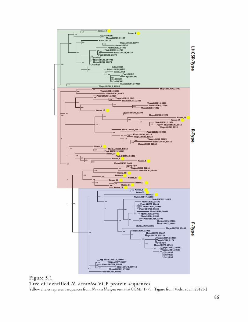
86
Figure 5.1 Tree of identified N. oceanica VCP protein sequences Yellow circles represent sequences from Nannochloropsis oceanica CCMP 1779. [Figure from Vieler et al., 2012b.]

87
Table 5.1 Putative genes identified to be involved in photosynthetic electron transport in CCMP1779.
Description Gene Name Gene Model ID
Cytochrome b6/f Complex
Rieske Fe-S subunit of Cytochrome b6-f Complex PETC CCMP1779_1077-mRNA-1 Cytochrome b6-f Complex subunit IV PETD1 nanno_2528:785..1264 1,2
PETD2 nanno_6709:10149..10859 2
Cytochrome b6-f Complex subunit PETM nanno_24:8196..7864 1,2
Cytochrome b6-f Complex subunit VIII PETN Not Found
Soluble Electron Carriers
Plastocyanin PETE1 nanno_963:17795..18553 2
Ferredoxin PETF1 NannoCCMP1779_7894-mRNA-1
PETF2 NannoCCMP1779_3054-mRNA-1
PETF3 NannoCCMP1779_2102-mRNA-1
Cytochrome c553
Not Found Ferredoxin-NADP oxidoreductase FNR1 nanno_1036:17495..20160 2
FNR2 nanno_427:12789..11620 2
FNR3 snap_masked-nanno_87-abinit-gene-0.5 4
FNR4 NannoCCMP1779_2013-mRNA-1
FNR5
augustus_masked-nanno_3186-abinit-gene-0.3
4
FNR6 CCMP1779_5041-mRNA-1
Photosystem I
PSI subunit II PSAD1 nanno_727:57421..58384 2
PSAD2 NannoCCMP1779_1336-mRNA-1
PSI subunit IV PSAE nanno_2655:6951..6687 2
PSI subunit III PSAF nanno_2570:15982..19244 2 PSI subunit V PSAG Not Found
PSI subunit VI PSAH nanno_798:86361..86944 2 PSI subunit X
Not Found
PSI subunit XI PSAL nanno_1707:204..719 2
PSI subunit N PSAN nanno_812:37141..36722 2
Photosystem II
PSII subunit M PSBM NannoCCMP1779_10463-mRNA-1
PSII Manganese-Stabilizing Protein PSBO augustus_masked-nanno_6323-abinit-gene-0.1
4 PSII Oxygen-Evolving Complex 23 PSBP NannoCCMP1779_11420-mRNA-1

88
PSII Oxygen-Evoloving Complex Enhancer Protein 3 PSBQ Not Found
PSII subunit R PSBR Not Found PSII 12KDa Extrinsic Protein PSBU CCMP1779_6260-mRNA-1
PSII 13KDa Protein PSBW CCMP1779_10160-mRNA-1
PSII subunit X PSBX nanno_243:28102..28984 2,3 Thylakoid Lumenal 17.4KDa Protein p17.4 CCMP1779_4019-mRNA-1
ATP Synthase
ATP Synthase CF1 γ chain ATPC1 augustus_masked-nanno_5244-abinit-gene-0.0
4
ATPC2 NannoCCMP1779_6999-mRNA-1
ATPC3
augustus_masked-nanno_1453-abinit-gene-0.0
4 ATP Synthase CF1 δ subunit ATPD1 NannoCCMP1779_10955-mRNA-1
ATPD2 nanno_6106:166..811 2
ATP Synthase CF0 subunit B' ATPG Not Found
1 partial sequence, 2 ambiguous structural annotation, coordinates are given (contigID:start.stop) 3 ambiguous functional annotation 4 these gene models are from augustus or snap gene annotation and deemed a better model after manual examination In cases where no gene model was structurally annotated, genome coordinates are given. [Table from Vieler et al., 2012b].

89
Table 5.2 Genes predicted to encode for violaxanthin-chlorophyll binding proteins (VCP) in CCMP1779
Name in Tree (Fig. 6)
ID LHC-Type
1 CCMP1779_8367-mRNA-1 F-type
2 CCMP1779_4699-mRNA-1 F-type 3 CCMP1779_3700-mRNA-1 R-type
4 CCMP1779_10562-mRNA-1 R-type
5 CCMP1779_10564-mRNA-1 F-type 6 CCMP1779_9414-mRNA-1 R-type
7 CCMP1779_11954-mRNA-1 R-type
8 CCMP1779_6809-mRNA-1 LHCSR-like 9 CCMP1779_6828-mRNA-1 R-type
10 CCMP1779_11487-mRNA-1 R-type 11 CCMP1779_8196-mRNA-1 R-type
12 CCMP1779_11886-mRNA-1 R-type
13 CCMP1779_8305-mRNA-1 R-type 14 CCMP1779_6226-mRNA-1 LHCSR-like
15 CCMP1779_35-mRNA-1 R-type
16 CCMP1779_904-mRNA-1 R-type 17 CCMP1779_10420-mRNA-1 R-type
18 CCMP1779_5333-mRNA-1 R-type 19 CCMP1779_6137-mRNA-1 F-type
20 nanno_220:23958..25565 1 R-type 1 ambiguous structural annotation, genome coordinates given Type designation of each VCP is from the phylogenetic tree (Figure 5.1). [Table from Vieler et al., 2012b].

90
Table 5.3 Identified genes putatively involved in xanthophyll synthesis Description Gene Name Gene Model ID
Carotenoid Biosysnthesis
Geranylgeranyl Pyrophosphate Synthase GGPS1 CCMP1779_9014-mRNA-1
GGPS2 CCMP1779_378-mRNA-1
GGPS3 CCMP1779_3156-mRNA-1
GGPS4 CCMP1779_3532-mRNA-1
Phytoene Syntase PSY CCMP1779_10288-mRNA-1 Phytoene Desaturase PDS CCMP1779_4801-mRNA-1
ζ-Carotene Isomerase ZISO maker-nanno_856-snap-gene-0.25-mRNA-1 1 ζ-Carotene Desaturase ZDS CCMP1779_501-mRNA-1 Carotene Isomerase Not found
Lycopene b-Cyclase LCYB CCMP1779_4993-mRNA1 Di-iron Carotene Hydroxylase Not found
Zeaxanthin Epoxidase ZEP CCMP1779_6822-mRNA-1
Violaxanthin DeEpoxidase VDE 2 CCMP1779_11477-mRNA-1 CCMP1779_11475-mRNA-1
Neoxanthin Synthase NXS CCMP1779_11764-mRNA-1 1 these gene models are from augustus or snap gene annotation and were deemed a better model after manual examination 2 gene found on two contigs, incomplete model [Table from Vieler et al., 2012b]. 5.2 Acknowledgements With permission from the authors, some elements of the chapter have been adapted from the following published article: Vieler, A., Wu, G., Tsai, C.H., Bullard, B., Cornish, A.J. , et al . (2012b) Genome,
Functional Gene Annotation, and Nuclear Transformation of the Heterokont Oleaginous Alga Nannochloropsis oceanica CCMP1779. PLoS Genetics 8(11): e1003064.

91
Chapter 6
Materials and Methods
6.1 Growth Conditions Algal Growth C. reinhardtii cultures were maintained on TAP plates, unless specified otherwise. Cells screened for fluorescence phenotype were grown on HS minimal media plates at high light (350-400 µmol photons m-2 s-1) or low light (~40 µmol photons m-2 s-1) for 5 days and then analyzed by Video Imager analysis (Figure 6.1). Cell lines were selected for chlorophyll fluorescence analysis by the Hansatech Fluorescence Monitoring System (FMS2) were grown in 50 mL HS at high light or low light for 5 days, and measured during early log phase while at a culture density of 1-3x10⁶ cells/mL.
Figure 6.1 Video imager analysis schematic The video imager system uses a blue actinic light to excite Chl in the sample, and measures the resulting fluorescence. [Adapted from Niyogi et al., 1997a]. Plant Growth
N. benthamiana seeds were planted in Sunshine soil mix #4 and grown in 12 hour day/night cycles in a Conviron growth chamber with light intensity ranging from 60-100 µmol photons m-2 s-1. Tobacco plants used in this study were between 4-6 weeks old.
A. thaliana seeds were sterilized in 70% EtOH for 10 minutes and planted on plates of MS media. Seeds were vernalized for 24-36 hours in the dark at 4°C, and then transferred to a short day

92
(8 hours light, 16 hours dark) growth cycle. Successful transformation was confirmed by growth on MS media containing BASTA. Seedlings with true leaves were transplanted to soil after 10 days of growth on MS media with BASTA. After two weeks on soil, chlorophyll fluorescence of single leaves was analyzed by Video Imager analysis to measure NPQ capacity. Leaf samples were processed for SDS-PAGE analysis of whole cell lysate and PCR amplification of the introduced gene using the Plant-PHIRE Direct Kit (Thermo Scientific) was used to confirm transformants had the proper sequence. 6.2 Transformation and complementation 6.2.1 Gene Sources Isolation of genetic material
RNA from A. thaliana Columbia-0 ecotype and C. reinhardtii 4A+ was isolated using the TRIzol reagent (Life technologies) as per the manufacturer’s instructions. cDNA was generated with the Omniscript RT kit (Qiagen), and genes of interest were amplified using the primers listed in Appendix S1. Nuclear genomic DNA from C. reinhardtii was isolated by phenol-chloroform extraction from 4A+ cells (Harris, 1989). Algal expression plasmid construction Genomic sequences for LHCSR1 and LHCSR2 were amplified from genomic DNA isolated from 4A+ wild type strain of C. reinhardtii, along with 1000bp of genomic sequence upstream and downstream of the genes, in order to capture the endogenous promoters. These genes were cloned into the entry vector pENTR-D according to the manufacturer’s directional TOPO cloning protocol (Life Technologies). The genes were then sequenced and cloned into a GATEWAY compatible vector using the GATEWAY LR Clonase II enzyme mix (Life Technologies). The GATWAY compatible vector, called pBC1 was created by cloning the GATEWAY compatible fragment from the pEarleyGate205 plasmid (Earley et al., 2006) into the XhoI sites of a vector made from pBluescript containing the paromomycin resistance gene for selection in C. reinhardtii. The GATEWAY compatible fragment is comprised of the ccdB suicide gene flanked by attR recombination sites. Plant expression plasmid construction
AttB adapter sites were added to genes of interest by PCR and recombined into gateway-compatible pDONR or pENTR entry vectors using BP Clonase II or the directional TOPO cloning kit, respectively (Life Technologies). These pDONR and pENTR vectors were used to recombine genes of interest into the pEarleyGate100 destination vector using LR Clonase II (Life Technologies). 6.2.2 Chlamydomonas reinhardtii Nuclear genomic complementation by glass-bead method
C. reinhardtii npq4 cells were transformed with the genomic clones of wild type LHCSR1 and LHCSR2 genes, as well as any mutant LHCSR2 genes by the glass bead method described previously (McCarthy et al., 2004) and grown on TAP plates containing 10 µg/mL paromomycin.
93
Colonies were picked after 10 days growth in very low light (~20 µmol photons m-2 s-1), grown on minimal media and screened for NPQ capacity by the Video Imager analysis (Niyogi et al., 1997a). For wild type gene transformations, colonies showing restored NPQ were then tested for protein presence, and chlorophyll fluorescence was quantified by pulse-amplitude-modulation (PAM) fluorometry using the FMS2 (Hansatech). For mutant gene transformations, which do not rescue the NPQ phenotype, random colonies were selected and screened for protein expression and chlorophyll fluorescence. Nuclear genomic complementation by electroporation
Coding sequence for LHCSR2 was PCR-amplified from cDNA stock extracted from a 4A+ wild-type culture of C. reinhardtii. A FLAG epitope tag was introduced onto the 3’-end of the gene directly before the stop codon. The coding sequence was cloned into the pBR9 plasmid (Rasala et al., 2012), between the PAR4 promoter and the 2A linker, upstream of the Streptoalloteichus hindustanus bleomycin resistance gene (sh-ble) using the InFusion PCR kit (Clontech). This plasmid expresses high levels of a fused mRNA encoding both the gene of interest and the zeocin resistance sh-ble gene. Electroporation protocol was done as described by Rasala et al. (2012). Chloroplast transformation by particle bombardment
Chloroplast transformation was done using the p72-rbcL plasmid, as described by O’Connor et al., (1998) and Bateman & Purton (2000), and the particle bombardment method recommended by the manufacturer for the BioRad Biolistic PDS-1000/He system. 6.2.3 Arabidopsis thaliana Inflorescence transformation using Agrobacterium tumefaciens A. thaliana inflorescences still with developing flowers were treated with A. tumefaciens cells containing the expression plasmid, resuspended in buffer with Silwet. Treated plants were kept covered for 18-24 hours, then grown in long day growth conditions (14 hours light, 10 hours dark) until seeds were collected. Successful transformation was confirmed by growth on MS media containing BASTA. 6.2.4 Transient expression in N. benthamiana Agrobacterium tumefaciens transformation
A. tumefaciens strain GV3101 was transformed with 400 ng of each plasmid by snap freezing in liquid nitrogen for 2 minutes, followed by heat shock at 37°C for 5 minutes. Transformants were rescued in 200 µL LB with shaking at 28°C for 2-3 hours and plated onto LB agar containing 30 µg/µL kanamycin and 50 µg/µL gentamycin. Single colonies appeared after 2 days of growth at 28°C. Infi ltration of N. benthamiana leaves
A. tumefaciens strains were resuspended in 1ml induction medium (10 mM MgCl, 10 mM MES pH5.6, 150 µM acetosyringeone) and incubated for 2 hours at 28°C. Cultures where then diluted to an OD600 of 0.5 with induction medium and injected into N. benthamiana leaves using a blunt-end 1 mL syringe. Plants were placed under constant light (~70 ) for 48-60 hours before chlorophyll fluorescence measurements were done, and the collection of protein samples and HPLC
94
samples. In addition to technical replicates on the same leaf, at least three biological replicates were collected for light treatments and pigment samples. 6.3 Measurement of chlorophyll f luorescence Chlorophyll f luorescence measurements in C. reinhardtii
C. reinhardtii cells were grown photoautotrophically in minimal (HS) media in high light (400 µmol photons m-2 s-1) and harvested while in early log phase. Chlorophyll fluorescence measurements of the cells were performed with a Fluorescence Monitoring System (FMS) from Hansatech, the FMS2. Before FMS measurements, cells were incubated in the dark for 20 to 30 minutes and filtered onto a glass-fiber filter and placed on the instrument’s filter clip. During measurement, the samples were exposed to actinic light with a light intensity of 630 µmol photons m-2 s-1 for 10 minutes, followed by 10 minutes of dark recovery. Chlorophyll f luorescence measurements of N. benthamiana leaf spots
Video imaging was performed on detached N. benthamiana leaves with the IMAGING-PAM M-Series from Heinz Walz. Measurements were recorded during 15 minutes of blue actinic light (600 µmol photons m−2 s−1), followed by 10 minutes of dark recovery. Samples were incubated in the dark for 20 minutes prior to measurements. Photosynthetic parameters were calculated as described in Brooks & Niyogi (2011). Chlorophyll f luorescence measurements of A. thaliana leaves Video imaging was performed on seedlings growing on MS plates, on detached leaves of two-week old plants, and leaf discs (0.5 cm2) of four-to-five-week old plants, using the IMAGING-PAM M-Series from Heinz Walz. Measurements were recorded during 15 minutes of blue actinic light (600 µmol photons m-2 s-1), followed by 10 minutes of dark recovery. Samples were incubated in the dark for 20 minutes prior to measurements and photosynthetic parameters were calculated as described in Brooks & Niyogi (2011). Chlorophyll fluorescence measurements were also performed on attached leaves of four-to-five-week old plants using a Fluorescence Monitoring System (FMS) from Hansatech, the FMS2 using a leaf clip to hold the sample. The full plants were incubated in the dark for 20 minutes prior to measurement. During measurement, the samples were exposed to actinic light with a light intensity of 630 µmol photons m-2 s-1 for 10 minutes, followed by 10 minutes of dark recovery. 6.4 Immunoblot analysis 6.4.1 Thylakoid membrane complex analysis Analysis of thylakoid membrane complexes of A. thaliana and N. benthamiana tissue Between 1.5 g and 2 g of leaf tissue was collected and homogenized at 4°C in pH 8.0 Isolation Buffer (330 mM Sorbitol, 20 mM HEPES, 10 mM EDTA, 5 mM MgCl2, and 10 mM

95
NaHCO3). Homogenized tissue was strained through cheesecloth and Miracloth, then centrifuged for 10 minutes at 1300 x g in a high speed centrifuge in the JA-20 rotor. The pellet was resuspended in cold Resuspension Buffer (330 mM Sorbitol, 20 mM HEPES, 5 mM MgCl2, an 10 mM NaHCO3). Samples were adjusted to 0.5µg Chl per mL of sample in a solution of 50mM BisTris and 40% (w/v) glycerol with either 1% n-dodecyl-α-D-maltopyranoside or 1% n-dodecyl-β-D-maltopyranoside detergent. 8 µL of sample was loaded into each well of a precast 4-16% gradient Novex Native gel, and run at 5 mA at 4°C for 3-3.5 hours to achieve separation of native complexes. 6.4.2 Protein Expression Analysis Measurement of expression of LHCSR1 and LHCSR3 in C. reinhardtii
Cells were harvested in early log phase and collected via centrifugation with 0.01% Tween-20 at 3500 rpm/2500 x g for 3 minutes in a tabletop centrifuge. They were resuspended in SDS denaturing buffer for 15 minutes at room temperature and centrifuged again at 14,000 rpm in a microfuge to remove insoluble material. Samples were measured for Chl a concentration and diluted to a concentration of 0.02 µg Chl a per µl of sample. Protein samples were treated with β-mercaptoethanol to 1 M final concentration and solubilized for 30 minutes at room temperature, heated at 55°C for 1 minute and then centrifuged for 1 minute at top speed of 14,000 rpm in a tabletop microfuge before loading. Proteins were separated on freshly-poured 15% bis-acrylamide 6 M urea denaturing gels. Proteins were transferred to 0.45 µm PVDF membrane via semi-dry method and the membranes were blocked with 5% milk in TBS-T. The anti-LHCSR primary antibody (Richard et al., 2000) used was in a 1:3000 dilution with 10% BSA in TBS with Tween-20 and donkey anti-rabbit HRP secondary antibody (Amersham) was used in a dilution of 1:10,000 in 5% milk in TBS with Tween-20. Measurement of heterologous protein expression in plant tissue
For both A. thaliana and N. benthmaiana tissue, leaf discs (0.5 cm2) from tissue expressing different protein constructs were collected and flash frozen in liquid nitrogen. Leaf discs are solubilized in lysis buffer (100 mM EDTA pH 8.0, 120 mM TrisHCl pH 6.8, 4% (w/v) SDS, 12% (w/v) sucrose, 200 mM dithiothretol) and heated to 100°C for 10 minutes. Cell debris was pelleted by centrifugation at 14,000 rpm for 5 minutes, and 8 µL of the soluble fraction was loaded into precast Novex 10-20% tris-glycine gels. Proteins were transferred to 0.45 µm PVDF membranes. Membranes were blocked overnight at 4°C in 5% milk in TBS with Tween-20, probed for 1 hour at room temperature with rabbit anti-FLAG (1:5000) primary antibody (Sigma) in 10% BSA in TBS with Tween-20. Secondary antibody was a donkey anti-rabbit-HRP conjugate (Amersham) used at a dilution of 1:20,000 in 5% milk in TBS with Tween-20. 6.5 Generation of mutants 6.5.1 Generation of C. reinhardtii mutants lhcsr1 TILLING mutant
In brief, Chlamydomonas cells were UV-mutagenized and DNA was extracted from individual colonies resulting from mutagenesis using a kit from Stratagene. The DNA from individuals was then pooled into sets of 16 in master plates. PCR and Cel1 digestion were done on

96
the plate and products were run on a Li-Cor model 4300 DNA Analyzer. For an introduction to the TILLING process, please refer to McCallum et al. (2000). The LHCSR1F and LHCSR1R primers were used for PCR amplification of the LHCSR1 gene (see appendix S1). npq4 lhcsr1 mutant
To obtain the npq4 lhcsr1 mutant, npq4 was crossed to the lhcsr1 mutant and tetrad progeny were restreaked onto HS plates and grown in high light. These plates were then assayed by video imaging of chlorophyll fluorescence (Niyogi et al., 1997a) using an Imaging PAM instrument (Walz). One progeny that exhibited lower NPQ compared to npq4 was genotyped by PCR using primers LHCSR1F and LHCS1R, and the product was sequenced to confirm the presence of the mutation in LHCSR1. The primer sets for LHCSR3.1 and LHCSR3.2 were also used to test the presence of the LHCSR2/3 isoforms. Subsequent immunoblot analysis was done using an anti-LHCSR antibody (Richard et al., 2000) to test for presence of the protein. Chloroplast transformation recipient strain psbH npq4 lhcsr1 To obtain the psbH recipient chloroplast in the npq4 lhcsr1 nuclear genetic background, the recipient strain provided to the lab by Saul Purton was crossed to the minus mating type of npq4 lhcsr1 and tetrad progeny were screened by PCR to confirm genotype and replica plating on HS minimal media to confirm phenotype. Cells harboring the psbH recipient chloroplast were unable to grow on minimal media due to the psbH defect. 6.5.2 Creation of mutagenesis l ibraries Site-directed mutagenesis l ibraries of At.CP29 and Cr.LHCSR3
Point mutations were introduced into PSBS and LHCSR using either the QuikChange Lightning Multi site-directed mutagenesis kit (Agilent Technologies) or the 3SM (also known as “Round-the-horn”) method described in Follo and Isidoro (2008). The PSBS transit peptide was added to algal genes using the splicing overlap extension PCR method described in (Warrens et al., 1997). Primers used are available in Appendix S1. Random mutagenesis l ibrary of At.CP29
Random mutants were generated using error-prone PCR optimized by the “Diversify” kit (Clontech), based on the method developed by Leung et al. (1989) and Cadwell & Joyce (1992). Mutagenesis was done to the At.CP29 gene, but not its chloroplast transit peptide, so primers were designed so as not to include the first 19 residues of the protein product. 6.6 Evolutionary analysis Sequence alignment and phylogenetic tree construction Sequences were aligned used the MUSCLE multiple sequence alignment tool (Edgar, 2004). The phylogenetic tree of N. oceanica LHC genes was generated using the Phylogeny.fr software (Dereeper et al., 2008) with gene blocks analysis and visualized with the FigTree program (http://tree.bio.ed.ac.uk/software/figtree/).

97
Molecular models Molecular models were generated using the I-TASSER server (Zhang, 2008; Roy et al., 2010) and the PHYRE2 software (Kelley et al., 2015). Visualization was done using the Chimera molecular visualization software package (Pettersen et al., 2004). Gene annotation Gene annotation in N. oceanica CCMP 1779 was done using the gBrowse platform (Stein et al., 2002) and the BLAST tool (Altschul et al., 1997).

98
Bibliography
Adams III, W., Zarter, C. R., Mueh, K., Amiard, V. and Demmig-Adams, B. (2006)
Energy dissipation and photoinhibition: A continuum of photoprotection. In (B. Demmig-Adams, W. Adams III and A. Mattoo, eds) Springer Netherlands.
Adamska, I. , Roobol-Bóza, M., Lindahl, M. and Andersson, B. (1999) Isolation of pigment-binding early light-inducible proteins from pea. European Journal of Biochemistry, 260(2), 453-460.
Ahn, T.K., Avenson, T. J. , Ballottari , M., Cheng, Y., Niyogi, K. K., Bassi , R. and Fleming, G. R. (2008) Architecture of a charge-transfer state regulating light harvesting in a plant antenna protein. Science, 320(5877), 794-797.
Alboresi, A., Gerotto, C., Giacometti, G. M., Bassi , R. and Morosinotto, T. (2010) Physcomitrella patens mutants affected on heat dissipation clarify the evolution of photoprotection mechanisms upon land colonization. Proceedings of the National Academy of Sciences, 107(24), 11128-11133.
Allahverdiyeva, Y., Mustila, H., Ermakova, M., Bersanini, L., Richaud, P., Ajlani, G., Battchikova, N., Cournac, L. and Aro, E. (2013) Flavodiiron proteins Flv1 and Flv3 enable cyanobacterial growth and photosynthesis under fluctuating light. Proceedings of the National Academy of Sciences, 110(10), 4111-4116.
Allen, J.F. (1992) Protein phosphorylation in regulation of photosynthesis. Biochimica Et Biophysica Acta (BBA) - Bioenergetics, 1098(3), 275-335.
Allen, J.F., Bennett, J. , Steinback, K. E. and Arntzen, C. J. (1981) Chloroplast protein phosphorylation couples plastoquinone redox state to distribution of excitation energy between photosystems. Nature, 291(5810), 25-29.
Allorent, G., Tokutsu, R., Roach, T., Peers, G., Cardol, P., Girard-Bascou, J. , Seigneurin-Berny, D., Petroutsos, D., Kuntz, M., Breyton, C., Franck, F., Wollman, F., Niyogi, K. K., Krieger-Liszkay, A., Minagawa, J. and Finazzi, G. (2013) A dual strategy to cope with high light in Chlamydomonas reinhardtii. The Plant Cell Online, 25(2), 545-557.
Alric, J. (2010) Cyclic electron flow around PSI in unicellular green algae. Photosyn Res, 106(1-2), 47-56.
Alric, J. , Lavergne, J. and Rappaport, F. (2010) Redox and ATP control of photosynthetic cyclic electron flow in Chlamydomonas reinhardtii (I) aerobic conditions. Biochimica Et Biophysica Acta (BBA) - Bioenergetics, 1797(1), 44-51.
Altschul, S.F., Madden, T. L., Schäffer, A. A., Zhang, J. , Zhang, Z., Miller, W. and Lipman, D. J. (1997) Gapped BLAST and PSI-BLAST: A new generation of protein database search programs. Nucleic Acids Research, 25(17), 3389-3402.
Amarnath, K., Zaks, J. , Park, S. D., Niyogi, K. K. and Fleming, G. R. (2012) Fluorescence lifetime snapshots reveal two rapidly reversible mechanisms of photoprotection in live cells of Chlamydomonas reinhardtii. Proceedings of the National Academy of Sciences, 109(22), 8405-8410.

99
Amunts, A., Drory, O. and Nelson, N. (2007) The structure of a plant photosystem I supercomplex at 3.4Å resolution. Nature, 447(7140), 58-63.
Anwaruzzaman, M., Chin, B. L., Li, X., Lohr, M., Martinez, D. A. and Niyogi, K. K. (2004) Genomic analysis of mutants affecting xanthophyll biosynthesis and regulation of photosynthetic light harvesting in Chlamydomonas reinhardtii. Photosynthesis Research, 82(3), 265-276.
Aro, E., Virgin, I. and Andersson, B. (1993) Photoinhibition of photosystem II. inactivation, protein damage and turnover. Biochimica Et Biophysica Acta (BBA) - Bioenergetics, 1143(2), 113-134.
Asada, K. (2006) Production and scavenging of reactive oxygen species in chloroplasts and their functions. Plant Physiology, 141(2), 391-396.
Aspinall-O'Dea, M., Wentworth, M., Pascal, A., Robert, B., Ruban, A. and Horton, P. (2002) In vitro reconstitution of the activated zeaxanthin state associated with energy dissipation in plants. Proceedings of the National Academy of Sciences, 99(25), 16331-16335.
Bailey, S., Melis, A., Mackey, K. R. M., Cardol, P., Finazzi, G., van Dijken, G., Berg, G. M., Arrigo, K., Shrager, J. and Grossman, A. (2008) Alternative photosynthetic electron flow to oxygen in marine Synechococcus. Biochimica Et Biophysica Acta (BBA) - Bioenergetics, 1777(3), 269-276.
Ballottari , M., Dall 'Osto, L., Morosinotto, T. and Bassi, R. (2007) Contrasting behavior of higher plant photosystem I and II antenna systems during acclimation. Journal of Biological Chemistry, 282(12), 8947-8958.
Ballottari , M., Truong, T. B., De Re, E., Erickson, E., Stella, G. R., Fleming, G. R., Bassi , R. and Niyogi, K. K. (2016) Identification of pH-sensing sites in the light harvesting complex stress-related 3 protein essential for triggering non-photochemical quenching in Chlamydomonas reinhardtii. Journal of Biological Chemistry, 291, 7334-7346.
Barkan, A. and Goldschmidt-Clermont, M. (2000) Participation of nuclear genes in chloroplast gene expression. Biochimie, 82(6–7), 559-572.
Baroli , I . and Melis, A. (1996) Photoinhibition and repair in Dunaliella salina acclimated to different growth irradiances. Planta, 198(4), 640-646.
Baroli , I . , Do, A. D., Yamane, T. and Niyogi, K. K. (2003) Zeaxanthin accumulation in the absence of a functional xanthophyll cycle protects Chlamydomonas reinhardtii from photooxidative stress. The Plant Cell Online, 15(4), 992-1008.
Barros, T., Royant, A., Standfuss, J. , Dreuw, A. and Kühlbrandt, W. (2008) Crystal structure of plant light-harvesting complex shows the active, energy-transmitting state. EMBO J., 28(3), 298-306.
Bassi, R., Croce, R., Cugini, D. and Sandonà , D. (1999) Mutational analysis of a higher plant antenna protein provides identification of chromophores bound into multiple sites. Proceedings of the National Academy of Sciences, 96(18), 10056-10061.
Bateman,J.M., Purton,S. (2000) Tools for chloroplast transformation in Chlamydomonas: Expression vectors and a new dominant selectable marker. Molecular Genetics and Genomics : MGG, 263(3), 404-410.

100
Beckmann, J. , Lehr, F., Finazzi, G., Hankamer, B., Posten, C., Wobbe, L. and Kruse, O. (2009) Improvement of light to biomass conversion by de-regulation of light-harvesting protein translation in Chlamydomonas reinhardtii. J. Biotechnol., 142(1), 70-77.
Bennoun, P. (1982) Evidence for a respiratory chain in the chloroplast. Proceedings of the National Academy of Sciences, 79, 4352-4356.
Bergantino, E., Segalla, A., Brunetta, A., Teardo, E., Rigoni, F., Giacometti, G. M. and Szabò, I. (2003) Light- and pH-dependent structural changes in the PsbS subunit of photosystem II. Proceedings of the National Academy of Sciences, 100(25), 15265-15270.
Berman, G.P., Nesterov, A. I. , Sayre, R. T. and Stil l , S. (2016) On improving the performance of nonphotochemical quenching in CP29 light-harvesting antenna complex. Physics Letters A, 380(13), 1279-1283.
Berman, G.P., Nesterov, A. I. , Gurvitz, S. and Sayre, R. T. (2015,) Possible role of interference and sink effects in nonphotochemical quenching in photosynthetic complexes. Arxiv:1412.3499, , 35.
Berthold, P., Tsunoda, S. P., Ernst, O. P., Mages, W., Gradmann, D. and Hegemann, P. (2008) Channelrhodopsin-1 initiates phototaxis and photophobic response in Chlamydomonas by immediate light-induced depolarization. The Plant Cell Online, 20(6), 1665-1677.
Betterle, N., Ballottari , M., Zorzan, S., de Bianchi, S., Cazzaniga, S., Dall 'Osto, L., Morosinotto, T. and Bassi, R. (2009) Light-induced dissociation of an antenna hetero-oligomer is needed for non-photochemical quenching induction. Journal of Biological Chemistry, 284(22), 15255-15266.
Bjorkman, O. and Demmig, B. (1987) Photon yield of O2 evolution and chlorophyll fluorescene characteristics at 77K among vascular plants of diverse origins. Planta, 170(4), 489-504.
Bonente, G., Pippa, S., Castellano, S., Bassi, R. and Ballottari, M. (2012) Acclimation of Chlamydomonas reinhardtii to different growth irradiances. Journal of Biological Chemistry, 287(8), 5833-5847.
Bonente, G., Howes, B. D., Caffarri , S., Smulevich, G. and Bassi, R. (2008b) Interactions between the photosystem II subunit PsbS and xanthophylls studied in vivo and in vitro. Journal of Biological Chemistry, 283(13), 8434-8445.
Bonente, G., Ballottari , M., Truong, T. B., Morosinotto, T., Ahn, T. K., Fleming, G. R., Niyogi, K. K. and Bassi, R. (2011) Analysis of LhcSR3, a protein essential for feedback de-excitation in the green alga Chlamydomonas reinhardtii. PLoS Biol, 9(1), e1000577.
Bonente, G., Passarini, F., Cazzaniga, S., Mancone, C., Buia, M. C., Tripodi, M., Bassi , R. and Caffarri , S. (2008a) The occurrence of the psbS gene product in Chlamydomonas reinhardtii and in other photosynthetic organisms and its correlation with energy quenching? Photochem. Photobiol., 84(6), 1359-1370.
Boulay, C., Wilson, A., D'Haene, S. and Kirilovsky, D. (2010) Identification of a protein required for recovery of full antenna capacity in OCP-related photoprotective mechanism in cyanobacteria. Proceedings of the National Academy of Sciences, 107(25), 11620-11625.

101
Boynton, J. , Gillham, N., Harris, E., Hosler, J. , Johnson, A., Jones, A., Randolph-Anderson, B., Robertson, D., Klein, T., Shark, K. and et, a. (1988) Chloroplast transformation in Chlamydomonas with high velocity microprojectiles. Science, 240(4858), 1534-1538.
Briantais, J.- . , Vernotte, C., Picaud, M. and Krause, G. H. (1979) A quantitative study of the slow decline of chlorophyll a fluorescence in isolated chloroplasts. Biochimica Et Biophysica Acta (BBA) - Bioenergetics, 548(1), 128-138.
Briggs, W.R. and Christie, J. M. (2002) Phototropins 1 and 2: Versatile plant blue-light receptors. Trends Plant Sci., 7(5), 204-210.
Brooks, M.D. and Niyogi, K. K. (2011) Use of a pulse-amplitude modulated chlorophyll fluorometer to study the efficiency of photosynthesis in Arabidopsis plants. In Chloroplast Research in Arabidopsis: Methods and Protocols, Volume II (P.R. Jarvis, ed) Totowa, NJ: Humana Press.
Brooks, M.D., Sylak-Glassman, E. J. , Fleming, G. R. and Niyogi, K. K. (2013) A thioredoxin-like/ß-propeller protein maintains the efficiency of light harvesting in Arabidopsis. Proceedings of the National Academy of Sciences, 110(29), E2733-E2740.
Brown, J.S. (1987) Functional organization of chlorophyll a and carotenoids in the alga, nannochloropsis salina. Plant Physiology, 83(2), 434-437.
Bulté, L., Gans, P., Rebéil lé, F. and Wollman, F. (1990) ATP control on state transitions in vivo in Chlamydomonas reinhardtii. Biochimica Et Biophysica Acta (BBA) - Bioenergetics, 1020(1), 72-80.
Cadwell, R.C. and Joyce, G. F. (1992) Randomization of genes by PCR mutagenesis. Genome Research, 2(1), 28-33.
Cardol, P., Forti , G. and Finazzi, G. (2011) Regulation of electron transport in microalgae. Biochimica Et Biophysica Acta (BBA) - Bioenergetics, 1807(8), 912-918.
Cardol, P., Alric, J. , Girard-Bascou, J. , Franck, F., Wollman, F. and Finazzi, G. (2009) Impaired respiration discloses the physiological significance of state transitions in Chlamydomonas. Proceedings of the National Academy of Sciences, 106(37), 15979-15984.
Cardol, P., Gloire, G., Havaux, M., Remacle, C., Matagne, R. and Franck, F. (2003) Photosynthesis and state transitions in mitochondrial mutants of Chlamydomonas reinhardtii affected in respiration. Plant Physiology, 133(4), 2010-2020.
Cardol, P., Bail leul, B., Rappaport, F., Derelle, E., Béal, D., Breyton, C., Bailey, S., Wollman, F. A., Grossman, A., Moreau, H. and Finazzi, G. (2008) An original adaptation of photosynthesis in the marine green alga Ostreococcus. Proceedings of the National Academy of Sciences, 105(22), 7881-7886.
Chen, H. and Melis, A. (2013) Marker-free genetic engineering of the chloroplast in the green microalga Chlamydomonas reinhardtii. Plant Biotechnology Journal, 11(7), 818-828.
Cheng, Y., Ahn, T. K., Avenson, T. J. , Zigmantas, D., Niyogi, K. K., Ballottari , M., Bassi , R. and Fleming, G. R. (2008) Kinetic modeling of charge-transfer quenching in the CP29 minor complex. J Phys Chem B, 112(42), 13418-13423.
Chi, W., Sun, X. and Zhang, L. (2013) Intracellular signaling from plastid to nucleus. Annu. Rev. Plant Biol., 64(1), 559-582.
102
Chuartzman, S.G., Nevo, R., Shimoni, E., Charuvi, D., Kiss, V., Ohad, I. , Brumfeld, V. and Reich, Z. (2008) Thylakoid membrane remodeling during state transitions in Arabidopsis. The Plant Cell Online, 20(4), 1029-1039.
Correa-Galvis, V., Poschmann, G., Melzer, M., Stühler, K. and Jahns, P. (2016a) PsbS interactions involved in the activation of energy dissipation in arabidopsis. Nature Plants, 2, 15225.
Correa-Galvis, V., Redekop, P., Wakao, S., Guan, K., Grieß, A., Niyogi, K.K., and Jahns, P. (2016b) PSBS is essential for the activation of energy dissipation in Chlamydomonas reinhardtii. Manuscript in preparation.
Cournac, L., Josse, E., Joët, T., Rumeau, D., Redding, K., Kuntz, M. and Peltier, G. (2000) Flexibility in photosynthetic electron transport: A newly identified chloroplast oxidase involved in chlororespiration. Philosophical Transactions of the Royal Society B Biological Sciences, 355, 1447-1454.
Croce, R. (2015) PsbS is the plants' pick for sun protection. Nat Struct Mol Biol, 22(9), 650-652. Dall 'Osto, L., Caffarri , S. and Bassi, R. (2005) A mechanism of nonphotochemical energy
dissipation, independent from PsbS, revealed by a conformational change in the antenna protein CP26. The Plant Cell Online, 17(4), 41217-41232.
Dall 'Osto, L., Cazzaniga, S., Wada, M. and Bassi, R. (2014) On the origin of a slowly reversible fluorescence decay component in the Arabidopsis npq4 mutant. Philosophical Transactions of the Royal Society of London B: Biological Sciences, 369(1640).
Dang, K., Plet, J . , Tolleter, D., Jokel, M., Cuiné, S., Carrier, P., Auroy, P., Richaud, P., Johnson, X., Alric, J. , Allahverdiyeva, Y. and Peltier, G. (2014) Combined increases in mitochondrial cooperation and oxygen photoreduction compensate for deficiency in cyclic electron flow in Chlamydomonas reinhardtii. The Plant Cell Online, 26(7), 3036-3050.
Davis, M.C., Fiehn, O. and Durnford, D. G. (2013) Metabolic acclimation to excess light intensity in Chlamydomonas reinhardtii. Plant, Cell Environ., 36(7), 1391-1405.
Dayer, R., Fischer, B. B., Eggen, R. I. L. and Lemaire, S. D. (2008) The peroxiredoxin and glutathione peroxidase families in Chlamydomonas reinhardtii. Genetics, 179(1), 41-57.
Delosme, R., Olive, J. and Wollman, F. (1996) Changes in light energy distribution upon state transitions: An in vivo photoacoustic study of the wild type and photosynthesis mutants from Chlamydomonas reinhardtii. Biochimica Et Biophysica Acta (BBA) - Bioenergetics, 1273(2), 150-158.
Demmig-Adams, B., Adams III, W. W., Barker, D. H., Logan, B. A., Bowling, D. R. and Verhoeven, A. S. (1996) Using chlorophyll fluorescence to assess the fraction of absorbed light allocated to thermal dissipation of excess excitation. Physiol. Plantarum, 98(2), 253-264.
Depège, N., Bellafiore, S. and Rochaix, J. (2003) Role of chloroplast protein kinase Stt7 in LHCII phosphorylation and state transition in Chlamydomonas. Science, 299(5612), 1572-1575.
Dereeper, A., Guignon, V., Blanc, G., Audic, S., Buffet, S., Chevenet, F., Dufayard, J. - . , Guindon, S., Lefort, V., Lescot, M., Claverie, J. - . and Gascuel, O. (2008) Phylogeny.fr: Robust phylogenetic analysis for the non-specialist. Nucleic Acids Research, 36(suppl 2), W465-W469.

103
Diner, B. and Mauzerall , D. (1973) Feedback controlling oxygen production in a cross-reaction between two photosystems in photosynthesis. Biochimica Et Biophysica Acta (BBA) - Bioenergetics, 305(2), 329-352.
Dittami, S.M., Michel, G., Collén, J. , Boyen, C. and Tonon, T. (2010) Chlorophyll-binding proteins revisited - a multigenic family of light-harvesting and stress proteins from a brown algal perspective. BMC Evolutionary Biology, 10(1), 1-14.
Dominici, P., Caffarri , S., Armenante, F., Ceoldo, S., Crimi, M. and Bassi, R. (2002) Biochemical properties of the PsbS subunit of photosystem II either purified from chloroplast or recombinant. Journal of Biological Chemistry, 277(25), 22750-22758.
Drop, B., Yadav K.N., S., Boekema, E. J. and Croce, R. (2014) Consequences of state transitions on the structural and functional organization of photosystem I in the green alga Chlamydomonas reinhardtii. The Plant Journal, 78(2), 181-191.
Duanmu, D., Casero, D., Dent, R. M., Gallaher, S., Yang, W., Rockwell, N. C., Martin, S. S., Pellegrini, M., Niyogi, K. K., Merchant, S. S., Grossman, A. R. and Lagarias, J. C. (2013) Retrograde bilin signaling enables chlamydomonas greening and phototrophic survival. Proceedings of the National Academy of Sciences of the United States of America, 110(9), 3621-6.
Duffy, C.D.P., Ruban, A. V. and Barford, W. (2008) Theoretical investigation of the role of strongly coupled chlorophyll dimers in photoprotection of LHCII. J Phys Chem B, 112(39), 12508-12515.
Dumas, P., Bergdoll, M., Cagnon, C. and Masson, J. M. (1994) Crystal structure and site-directed mutagenesis of a bleomycin resistance protein and their significance for drug sequestering. EMBO J., 13(11), 2483-2492.
Durnford, D.G., Price, J. A., McKim, S. M. and Sarchfield, M. L. (2003) Light-harvesting complex gene expression is controlled by both transcriptional and post-transcriptional mechanisms during photoacclimation in Chlamydomonas reinhardtii. Physiol. Plantarum, 118(2), 193-205.
Earley, K.W., Haag, J. R., Pontes, O., Opper, K., Juehne, T., Song, K. and Pikaard, C. S. (2006) Gateway-compatible vectors for plant functional genomics and proteomics. The Plant Journal, 45(4), 616-629.
Eberhard, S., Finazzi, G. and Wollman, F. (2008) The dynamics of photosynthesis. Annu Rev Genetics, 42, 463-515.
Edgar, R.C. (2004) MUSCLE: Multiple sequence alignment with high accuracy and high throughput. Nucleic Acids Research, 32(5), 1792-1797.
Elrad, D. and Grossman, A. R. (2004) A genome's-eye view of the light-harvesting polypeptides of Chlamydomonas reinhardtii. Curr Genet, 45, 61-75.
Elrad, D., Niyogi, K. K. and Grossman, A. R. (2002) A major light-harvesting polypeptide of photosystem II functions in thermal dissipation. The Plant Cell Online, 14(8), 1801-1816.
Endo, T., Uebayashi, N., Ishida, S., Ikeuchi, M. and Sato, F. (2014) Light energy allocation at PSII under field light conditions: How much energy is lost in NPQ-associated dissipation? Plant Physiology and Biochemistry, 81, 115-120.

104
Engelken, J. , Brinkmann, H. and Adamska, I. (2010) Taxonomic distribution and origins of the extended LHC (light-harvesting complex) antenna protein superfamily. BMC Evol. Biol., 10, 233.
Erickson, E., Wakao, S., & Niyogi, K.K. (2015) Light stress and photoprotection in Chlamydomonas reinhardtii. The Plant Journal 82(3): 449-465.
Falkowski, P.G. (1994) The role of phytoplankton photosynthesis in global biogeochemical cycles. Photosynthesis Res., 39(3), 235-258.
Fan, M., Li, M., Liu, Z., Cao, P., Pan, X., Zhang, H., Zhao, X., Zhang, J. and Chang, W. (2015) Crystal structures of the PsbS protein essential for photoprotection in plants. Nat Struct Mol Biol, 22(9), 729-735.
Feng, X., Kell, A., Pieper, J. and Jankowiak, R. (2013b) Modeling of optical spectra of the light-harvesting CP29 antenna complex of photosystem II - Part II. J Phys Chem B, 117(22), 6593-6602.
Feng, X., Pan, X., Li, M., Pieper, J. , Chang, W. and Jankowiak, R. (2013a) Spectroscopic study of the light-harvesting CP29 antenna complex of photosystem II - Part I. J Phys Chem B, 117(22), 6585-6592.
Ferrante, P., Ballottari , M., Bonente, G., Giuliano, G. and Bassi, R. (2012) LHCBM1 and LHCBM2/7 polypeptides, components of major LHCII complex, have distinct functional roles in photosynthetic antenna system of Chlamydomonas reinhardtii. Journal of Biological Chemistry, 287(20), 16276-16288.
Finazzi, G. (2005) The central role of the green alga Chlamydomonas reinhardtii in revealing the mechanism of state transitions. Journal of Experimental Botany, 56(411), 383-388.
Finazzi, G. and Rappaport, F. (1998) In vivo characterization of the electrochemical proton gradient generated in darkness in green algae and its kinetic effects on cytochrome b6f Turnover. Biochemistry, 37(28), 9999-10005.
Finazzi, G., Johnson, G. N., Dall 'Osto, L., Zito, F., Bonente, G., Bassi , R. and Wollman, F. (2006) Nonphotochemical quenching of chlorophyll fluorescence in Chlamydomonas reinhardtii. Biochemistry, 45, 1490-1498.
Fischer, B.B., Hideg, E. and Krieger-Liszkay, A. (2013) Production, detection, and signaling of singlet oxygen in photosynthetic organisms. Antioxidants & Redox Signaling, 18(16), 2145-2162.
Floris, M., Bassi , R., Robaglia, C., Alboresi, A. and Lanet, E. (2013) Post-transcriptional control of light-harvesting genes expression under light stress. Plant Mol. Biol., 82(1-2), 147-154.
Follo, C. and Isidoro, C. (2008) A fast and simple method for simultaneous mixed site-specific mutagenesis of a wide coding sequence. Biotechnol. Appl. Biochem., 49(2), 175-183.
Frigerio, S., Campoli, C., Zorzan, S., Fantoni, L. I. , Crosatti , C., Drepper, F., Haehnel, W., Cattivell i , L., Morosinotto, T. and Bassi, R. (2007) Photosynthetic antenna size in higher plants is controlled by plastoquinone redox state at post-transcriptional rather than transcriptional level. Journal of Biological Chemistry, 282(40), 29457-29469.
Fristedt, R., Will ig, A., Granath, P., Crèvecoeur, M., Rochaix, J. and Vener, A. V. (2009) Phosphorylation of photosystem II controls functional macroscopic folding of photosynthetic membranes in Arabidopsis. The Plant Cell Online, 21(12), 3950-3964.

105
Funk, C., Adamska, I. , Green, B. R., Andersson, B. and Renger, G. (1995) The nuclear-encoded chlorophyll-binding photosystem II-S protein is stable in the absence of pigments. Journal of Biological Chemistry, 270(50), 30141-30147.
Gatenby, A.A., Rothstein, S. J. and Nomura, M. (1989) Translational coupling of the maize chloroplast atpB and atpE genes. Proceedings of the National Academy of Sciences, 86(11), 4066-4070.
Gentile, M. and Blanch, H. W. (2001) Physiology and xanthophyll cycle activity of Nannochloropsis gaditana. Biotechnol. Bioeng., 75(1), 1-12.
Gerotto, C. and Morosinotto, T. (2013) Evolution of photoprotection mechanisms upon land colonization: Evidence of PSBS-dependent NPQ in late streptophyte algae. Physiol. Plantarum, 149(4), 583-598.
Gerotto, C., Alboresi, A., Giacometti, G. M., Bassi , R. and Morosinotto, T. (2012) Coexistence of plant and algal energy dissipation mechanisms in the moss Physcomitrella patens. New Phytol., 196(3), 763-773.
Gerotto, C., Alboresi, A., Giacometti, G. M., Bassi , R. and Morosinotto, T. (2011) Role of PSBS and LHCSR in Physcomitrella patens acclimation to high light and low temperature. Plant, Cell Environ., 34(6), 922-932.
Gilmore, A.M., Hazlett, T. L. and Govindjee (1995) Xanthophyll cycle-dependent quenching of photosystem II chlorophyll a fluorescence: Formation of a quenching complex with a short fluorescence lifetime. Proceedings of the National Academy of Sciences, 92(6), 2273-2277.
Giuffra, E., Cugini, D., Croce, R. and Bassi, R. (1996) Reconstitution and pigment-binding properties of recombinant CP29. European Journal of Biochemistry, 238(1), 112-120.
Goldschmidt-Clermont, M. (1991) Transgenic expression of aminoglycoside adenine transferase in the chloroplast: A selectable marker of site-directed transformation of Chlamydomonas. Nucleic Acids Res., 19(15), 4083-4089.
Goodenough, U.W. and Levine, R. P. (1969) Chloroplast ultrastructure in mutant strains of Chlamydomonas reinhardtii lacking components of the photosynthetic apparatus. Plant Physiology, 44(7), 990-1000.
Goss, R. and Lepetit, B. (2015) Biodiversity of NPQ. J. Plant Physiol., 172, 13-32. Govorunova, E.G., Jung, K., Sineshchekov, O. A. and Spudich, J. L. (2004)
Chlamydomonas sensory rhodopsins A and B: Cellular content and role in photophobic response. Biophysical Journal, 86(4), 2342-2349.
Green, B.R. and Kühlbrandt, W. (1995) Sequence conservation of light-harvesting and stress-response proteins in relation to the three-dimensional molecular structure of LHCII. Photosynth Res, 44, 139-148.
Green, B.R., Pichersky, E. and Kloppstech, K. (1991) Chlorophyll a/b-binding proteins: An extended family. Trends Biochem. Sci., 16(5), 181-186.
Grossman, A., Manodori, A. and Snyder, D. (1990) Light-harvesting proteins of diatoms: Their relationship to the chlorophyll a/b binding proteins of higher plants and their mode of transport into plastids. Mol. Gen. Genet., 224, 91-100.

106
Haferkamp, S., Haase, W., Pascal, A. A., van Amerongen, H. and Kirchhoff, H. (2010) Efficient light harvesting by photosystem II requires an optimized protein packing density in grana thylakoids. Journal of Biological Chemistry, 285(22), 17020-17028.
Harada, A., Sakai, T. and Okada, K. (2003) phot1 and phot2 mediate blue light-induced transient increases in cytosolic Ca2+ differently in Arabidopsis leaves. Proceedings of the National Academy of Sciences, 100(14), 8583-8588.
Harris, E.H. (1989) The Chlamydomonas Sourcebook New York, NY: Academic Press. Havaux, M. and Niyogi, K. K. (1999) The violaxanthin cycle protects plants from
photooxidative damage by more than one mechanism. Proceedings of the National Academy of Sciences, 96(15), 8762-8767.
Hegemann, P. and Bruck, B. (1989) Light-induced stop response in Chlamydomonas reinhardtii: Occurrence and adaptation phenomena. Cell Motil. Cytoskeleton, 14(4), 501-515.
Helman, Y., Tchernov, D., Reinhold, L., Shibata, M., Ogawa, T., Schwarz, R., Ohad, I. and Kaplan, A. (2003) Genes encoding A-type flavoproteins are essential for photoreduction of O2 in cyanobacteria. Current Biology, 13(3), 230-235.
Hertle, A., Blunder, T., Wunder, T., Pesaresi, P., Pribil , M., Armbruster, U. and Leister, D. (2013) PGRL1 is the elusive ferredoxin-plastoquinone reductase in photosynthetic cyclic electron flow. Mol. Cell, 49(3), 511-523.
Hieber, A.D., Kawabata, O. and Yamamoto, H. Y. (2004) Significance of the lipid phase in the dynamics and functions of the xanthophyll cycle as revealed by PsbS overexpression in tobacco and in-vitro de-epoxidation in monogalactosyldiacylglycerol micelles. Plant and Cell Physiology, 45(1), 92-102.
Hieber, A.D., Bugos, R. C. and Yamamoto, H. Y. (2000) Plant lipocalins: Violaxanthin de-epoxidase and zeaxanthin epoxidase. Biochimica Et Biophysica Acta (BBA) - Protein Structure and Molecular Enzymology, 1482(1–2), 84-91.
Hoefnagel, M.H.N., Atkin, O. K. and Wiskich, J. T. (1998) Interdependence between chloroplasts and mitochondria in the light and the dark. Biochimica Et Biophysica Acta (BBA) - Bioenergetics, 1366(3), 235-255.
Holland, E.M., Harz, H., Uhl, R. and Hegemann, P. (1997) Control of phobic behavioral responses by rhodopsin-induced photocurrents in Chlamydomonas. Biophysical Journal, 73(3), 1395-1401.
Holub, O., Seufferheld, M. J. , Gohlke, C., Heiss, G. J. and Clegg, R. M. (2007) Fluorescence lifetime imaging microscopy of Chlamydomonas reinhardtii: Non-photochemical quenching mutants and the effect of photosynthetic inhibitors on the slow chlorophyll fluorescence transient. J. Microsc., 226(2), 90-120.
Holub, O., Seufferheld, M. J. , Gohlke, C., Govindjee and Clegg, R. M. (2000) Fluorescence lifetime imaging (FLI) in real-time - a new technique in photosynthesis research. Photosynthetica, 38(4), 581-599.
Hopp, T.P., Prickett, K. S., Price, V. L., Libby, R. T., March, C. J. , Pat Cerretti , D., Urdal, D. L. and Conlon, P. J. (1988) A short polypeptide marker sequence useful for recombinant protein identification and purification. Nat Biotech, 6(10), 1204-1210.

107
Horton, P., Ruban, A. V. and Walters, R. G. (1996) Regulation of light harvesting in green plants. Annu. Rev. Plant Physiol. Plant Mol. Biol., 47(1), 655-684.
Horton, P., Ruban, A. and Walters, R. (1994) Regulation of light harvesting in green plants. Plant Physiology, 106, 415-420.
Houille-Vernes, L., Rappaport, F., Wollman, F., Alric, J. and Johnson, X. (2011) Plastid terminal oxidase 2 (PTOX2) is the major oxidase involved in chlororespiration in Chlamydomonas. Proceedings of the National Academy of Sciences, 108(51), 20820-20825.
Hutin, C., Nussaume, L., Moise, N., Moya, I. , Kloppstech, K. and Havaux, M. (2003) Early light-induced proteins protect Arabidopsis from photooxidative stress. Proceedings of the National Academy of Sciences, 100(8), 4921-4926.
Ioannidis, N.E. and Kotzabasis, K. (2015) Could structural similarity of specific domains between animal globins and plant antenna proteins provide hints important for the photoprotection mechanism? J. Theor. Biol., 364, 71-79.
Iwai, M., Takahashi, Y. and Minagawa, J. (2008) Molecular remodeling of photosystem II during state transitions in Chlamydomonas reinhardtii. The Plant Cell Online, 20(8), 2177-2189.
Iwai, M., Kato, N. and Minagawa, J. (2007) Distinct physiological responses to a high light and low CO2 environment revealed by fluorescence quenching in photoautotrophically grown Chlamydomonas reinhardtii. Photosyn Res, 94, 307-314.
Iwai, M., Yokono, M., Inada, N. and Minagawa, J. (2010) Live-cell imaging of photosystem II antenna dissociation during state transitions. Proceedings of the National Academy of Sciences, 107(5), 2337-2342.
Jans, F., Mignolet, E., Houyoux, P., Cardol, P., Ghysels, B., Cuiné, S., Cournac, L., Peltier, G., Remacle, C. and Franck, F. (2008) A type II NAD(P)H dehydrogenase mediates light-independent plastoquinone reduction in the chloroplast of Chlamydomonas. Proceedings of the National Academy of Sciences, 105(51), 20546-20551.
Jansson, S. (1999) A guide to the lhc genes and their relatives in Arabidopsis. Trends Plant Sci., 4(6), 236-240.
Jansson, S., Andersson, J. , Jung Kim, S. and Jackowski, G. (2000) An Arabidopsis thaliana protein homologous to cyanobacterial high-light-inducible proteins. Plant Mol. Biol., 42(2), 345-351.
Johnson, X., Steinbeck, J. , Dent, R. M., Takahashi, H., Richaud, P., Ozawa, S., Houille-Vernes, L., Petroutsos, D., Rappaport, F., Grossman, A. R., Niyogi, K. K., Hippler, M. and Alric, J. (2014) PGR5-mediated cyclic electron flow under ATP- or redox-limited conditions: A study of ΔATPase pgr5 and ΔrbcL pgr5 mutants in Chlamydomonas reinhardtii. Plant Physiology, 165(1), 438-452.
Kühlbrandt, W., Wang, D. N. and Fujiyoshi, Y. (1994) Atomic model of plant light-harvesting complex by electron crystallography. Nature, 367, 614-621.
Kapila, J. , De Rycke, R., Van Montagu, M. and Angenon, G. (1997) An agrobacterium-mediated transient gene expression system for intact leaves. Plant Science, 122(1), 101-108.
Kargul, J. , Turkina, M. V., Nield, J. , Benson, S., Vener, A. V. and Barber, J. (2005) Light-harvesting complex II protein CP29 binds to photosystem I of Chlamydomonas reinhardtii under state 2 conditions. FEBS Journal, 272(18), 4797-4806.

108
Kelley, L.A., Mezulis, S., Yates, C. M., Wass, M. N. and Sternberg, M. J. E. (2015) The Phyre2 web portal for protein modeling, prediction and analysis. Nat. Protocols, 10(6), 845-858.
Khan, U.A. and Kasha, M. (1994) Singlet molecular oxygen evolution upon simple acidification of aqueous hypochlorite: Application to studies on the deleterious health effects of chlorinated drinking water. Proceedings of the National Academy of Sciences, 91, 12362-12364.
Kilian, O., Benemann, C. S. E., Niyogi, K. K. and Vick, B. (2011) High-efficiency homologous recombination in the oil-producing alga Nannochloropsis sp. Proceedings of the National Academy of Sciences, 108(52), 21265-21269.
Kindle, K.L. (1990) High-frequency nuclear transformation of Chlamydomonas reinhardtii. Proc. Natl. Acad. Sci. U. S. A., 87(3), 1228-1232.
Kindle, K.L., Schnell , R. A., Fernández, E. and Lefebvre, P. A. (1989) Stable nuclear transformation of Chlamydomonas using the Chlamydomonas gene for nitrate reductase. The Journal of Cell Biology, 109(6), 2589-2601.
Kirk, J.T.O. (1994) Light and photosynthesis in aquatic ecosystems 2nd Edition edn. Cambridge: Cambridge University Press.
Knox, P.J. and Dodge, A. D. (1985) Singlet oxygen and plants. Phytochemistry, 24(5), 889-896; 889.
Kok, B. (1956) On the inhibition of photosynthesis by intense light. Biochim. Biophys. Acta, 21(2), 234-244.
Koscianska, E., Kalantidis, K., Wypijewski, K., Sadowski, J. and Tabler, M.Analysis of RNA silencing in agroinfiltrated leaves of Nicotiana benthamiana and Nicotiana tabacum. Plant Mol. Biol., 59(4), 647-661.
Kouril , R., Zygadlo, A., Arteni, A. A., de Wit, C. D., Dekker, J. P., Jensen, P. E., Scheller, H. V. and Boekema, E. J. (2005) Structural characterization of a complex of photosystem I and light-harvesting complex II of Arabidopsis thaliana. Biochemistry (N. Y. ), 44(33), 10935-10940.
Koziol, A.G., Borza, T., Ishida, K. -. I . , Keeling, P., Lee, R. W. and Durnford, D. G. (2007) Tracing the evolution of the light-harvesting antennae in chlorophyll a/b-containing organisms. Plant Physiol., 143(4):1802-16.
Kramer, D., Sacksteder, C. and Cruz, J. (1999) How acidic is the lumen? Photosynthesis Res., 60(2-3), 151-163.
Krause, G.H. (1988) Photoinhibition of photosynthesis. an evaluation of damaging and protective mechanisms. Physiol. Plantarum, 74(3), 566-574.
Krieger-Liszkay, A. (2005) Singlet oxygen production in photosynthesis. Journal of Experimental Botany, 56(411), 337-46.
Kromer, S. (1995) Respiration during photosynthesis. Annu. Rev. Plant Physiol. Plant Mol. Biol., 46(1), 45-70.
Krömer, S. and Heldt, H. W. (1991) Respiration of pea leaf mitochondria and redox transfer between the mitochondrial and extramitochondrial compartment. Biochimica Et Biophysica Acta (BBA) - Bioenergetics, 1057(1), 42-50.

109
Krüger, T., Il ioaia, C., Johnson, M., Belgio, E., Horton, P., Ruban, A. and van Grondelle, R. (2013) The specificity of controlled protein disorder in the photoprotection of plants. Biophys. J., 105(4), 1018-1026.
Krüger, T.J. , I l ioaia, C., Johnson, M., Ruban, A., Papagiannakis, E., Horton, P. and van Grondelle, R. (2012) Controlled disorder in plant light-harvesting complex II explains its photoprotective role. Biophys. J., 102(11), 2669-2676.
Lambrev, P.H., Nilkens, M., Miloslavina, Y., Jahns, P. and Holzwarth, A. R. (2010) Kinetic and spectral resolution of multiple nonphotochemical quenching components in Arabidopsis leaves. Plant Physiology, 152, 1611-1624.
Ledford, H.K., Chin, B. L. and Niyogi, K. K. (2007) Acclimation to singlet oxygen stress in Chlamydomonas reinhardtii. Eukaryotic Cell, 6(6), 919-30.
Lemaire, C., Wollman, F. A. and Bennoun, P. (1988) Restoration of phototrophic growth in a mutant of Chlamydomonas reinhardtii in which the chloroplast atpB gene of the ATP synthase has a deletion: An example of mitochondria-dependent photosynthesis. Proceedings of the National Academy of Sciences, 85(5), 1344-1348.
Lemeille, S., Will ig, A., Depège-Fargeix, N., Delessert, C., Bassi , R. and Rochaix, J. (2009) Analysis of the chloroplast protein kinase Stt7 during state transitions. PLoS Biol, 7(3), e1000045.
Leonell i , L., Erickson, E., Lyska, D., and Niyogi, K.K. (expected 2016) Transient expression in Nicotiana benthamiana for rapid functional analysis of genes involved in non-photochemical quenching and carotenoid biosynthesis. Plant Journal. Manuscript submitted.
Leung, D.W., Chen, E. and Goeddel, D. V. (1989) A method for random mutagenesis of a defined DNA segment using a modified polymerase chain reaction. . Technique, 1, 11-15.
Li, X., Gilmore, A. M. and Niyogi, K. K. (2002c) Molecular and global time-resolved analysis of a psbSGene dosage effect on pH- and xanthophyll cycle-dependent nonphotochemical quenching in photosystem II. Journal of Biological Chemistry, 277(37), 33590-33597.
Li, X., Müller-Moulé, P., Gilmore, A. M. and Niyogi, K. K. (2002b) PsbS-dependent enhancement of feedback de-excitation protects photosystem II from photoinhibition. Proceedings of the National Academy of Sciences, 99(23), 15222-15227.
Li, X., Phippard, A., Pasari, J. and Niyogi, K. K. (2002a) Structure–function analysis of photosystem II subunit S (PsbS) in vivo. Functional Plant Biol., 29(10), 1131-1139.
Li, X., Gilmore, A. M., Caffarri , S., Bassi , R., Golan, T., Kramer, D. and Niyogi, K. K. (2004) Regulation of photosynthetic light harvesting involves intrathylakoid lumen pH sensing by the PsbS protein. Journal of Biological Chemistry, 279(22), 22866-22874.
Li, X., Bjorkman, O., Shih, C., Grossman, A. R., Rosenquist, M., Jansson, S. and Niyogi, K. K. (2000) A pigment-binding protein essential for regulation of photosynthetic light harvesting. Nature, 403(6768), 391-395.
Liguori, N., Roy, L. M., Opacic, M., Durand, G. and Croce, R. (2013) Regulation of light harvesting in the green alga Chlamydomonas reinhardtii: The C-terminus of LHCSR is the knob of a dimmer switch. J. Am. Chem. Soc., 135(49), 18339-18342.
Lin, C. (2002) Blue light receptors and signal transduction. Plant Cell, 14, s207-s225.

110
Liu, Z., Yan, H., Wang, K., Kuang, T., Zhang, J. , Gui, L., An, X. and Chang, W. (2004) Crystal structure of spinach major light-harvesting complex at 2.72 A resolution. Nature, 428.
Long, S.P., Humphries, S. and Falkowski, P. G. (1994) Photoinhibition of photosynthesis in nature. Annual Reviews Plant Physiology Plant Molecule Biology, 45, 633-662.
Lucker, B. and Kramer, D. M. (2013) Regulation of cyclic electron flow in Chlamydomonas reinhardtii under fluctuating carbon availability. Photosyn Res, 117(1-3), 449-459.
Malnoë, A., Wang, F., Girard-Bascou, J. , Wollman, F. and de Vitry, C. (2014) Thylakoid FtsH protease contributes to photosystem II and cytochrome b6f remodeling in Chlamydomonas reinhardtii under stress conditions. The Plant Cell Online, 26(1), 373-390.
Maruyama, S., Tokutsu, R. and Minagawa, J. (2014) Transcriptional regulation of the stress-responsive light harvesting complex genes in Chlamydomonas reinhardtii. Plant and Cell Physiology, 55(7), 1304-1310.
McCallum, C.M., Comai, L., Greene, E. A. and Henikoff, S. (2000) Targeting induced LocalLesions IN genomes (TILLING) for plant functional genomics. Plant Physiology, 123(2), 439-442.
McCarthy, S.S., Kobayashi, M. C. and Niyogi, K. K. (2004) White mutants of Chlamydomonas reinhardtii are defective in phytoene synthase. Genetics, 168(3), 1249-57.
McDonald, A.E., Ivanov, A. G., Bode, R., Maxwell, D. P., Rodermel, S. R. and Hüner, N. P. A. (2011) Flexibility in photosynthetic electron transport: The physiological role of plastoquinol terminal oxidase (PTOX). Biochimica Et Biophysica Acta (BBA) - Bioenergetics, 1807(8), 954-967.
McIntosh, L. (1994) Molecular biology of the alternative oxidase. Plant Physiology, 105(3), 781-786.
McKim, S.M. and Durnford, D. G. (2006) Translational regulation of light-harvesting complex expression during photoacclimation to high-light in Chlamydomonas reinhardtii. Plant Physiology and Biochemistry, 44(11–12), 857-865.
Mehler, A.H. (1951) Studies on reactions of illuminated chloroplasts. II. stimulation and inhibition of the reaction with molecular oxygen. Arch. Biochem. Biophys., 34(2), 339-351.
Melis, A. (2009) Solar energy conversion efficiencies in photosynthesis: Minimizing the chlorophyll antennae to maximize efficiency. Plant Science, 177(4), 272-280.
Melis, A. (1991) Dynamics of photosynthetic membrane composition and function. Biochimica Et Biophysica Acta (BBA) - Bioenergetics, 1058(2), 87-106.
Melis, A., Murakami, A., Nemson, J. , Aizawa, K., Ohki, K. and Fujita, Y. (1996) Chromatic regulation in Chlamydomonas reinhardtii alters photosystem stoichiometry and improves the quantum efficiency of photosynthesis. Photosynthesis Res., 47(3), 253-265.
Miller, R., Wu, G., Deshpande, R. R., Vieler, A., Gärtner, K., Li, X., Moellering, E. R., Zäuner, S., Cornish, A. J. , Liu, B., Bullard, B., Sears, B. B., Kuo, M., Hegg, E. L., Shachar-Hill , Y., Shiu, S. and Benning, C. (2010) Changes in transcript abundance in Chlamydomonas reinhardtii following nitrogen deprivation predict diversion of metabolism. Plant Physiology, 154(4), 1737-1752.
Miloslavina, Y., Grouneva, I. , Lambrev, P. H., Lepetit, B., Goss, R., Wilhelm, C. and Holzwarth, A. R. (2009) Ultrafast fluorescence study on the location and

111
mechanism of non-photochemical quenching in diatoms. Biochimica Et Biophysica Acta (BBA) - Bioenergetics, 1787(10), 1189-1197.
Minagawa, J. (2011) State transitions—The molecular remodeling of photosynthetic supercomplexes that controls energy flow in the chloroplast. Biochimica Et Biophysica Acta (BBA) - Bioenergetics, 1807(8), 897-905.
Miyamoto, S., Martinez, G. R., Medeiros, M. H. G. and Di Mascio, P. (2014) Singlet molecular oxygen generated by biological hydroperoxides. Journal of Photochemistry and Photobiology B: Biology, 139(0), 24-33.
Mou, S., Zhang, X., Ye, N., Dong, M., Liang, C., Liang, Q., Miao, J. , Xu, D. and Zheng, Z. (2012) Cloning and expression analysis of two different LhcSR genes involved in stress adaptation in an antarctic microalga, Chlamydomonas sp. ICE-L. Extremophiles, 16(2), 193-203.
Müller, P., Li, X. and Niyogi, K. K. (2001) Non-photochemical quenching. A response to excess light energy. Plant Physiology, 125(4), 1558-1566.
Murata, N., Allakhverdiev, S. I. and Nishiyama, Y. (2012) The mechanism of photoinhibition in vivo: Re-evaluation of the roles of catalase, α-tocopherol, non-photochemical quenching, and electron transport. Biochimica Et Biophysica Acta (BBA) - Bioenergetics, 1817(8), 1127-1133.
Mussgnug, J.H., Wobbe, L., Elles, I. , Claus, C., Hamilton, M., Fink, A., Kahmann, U., Kapazoglou, A., Mullineaux, C. W., Hippler, M., Nickelsen, J. , Nixon, P. J. and Kruse, O. (2005) NAB1 is an RNA binding protein involved in the light-regulated differential expression of the light-harvesting antenna of Chlamydomonas reinhardtii. The Plant Cell Online, 17(12), 3409-3421.
Nagel, G., Ollig, D., Fuhrmann, M., Kateriya, S., Musti, A. M., Bamberg, E. and Hegemann, P. (2002) Channelrhodopsin-1: A light-gated proton channel in green algae. Science, 296(5577), 2395-2398.
Nagel, G., Szellas, T., Huhn, W., Kateriya, S., Adeishvil i , N., Berthold, P., Ollig, D., Hegemann, P. and Bamberg, E. (2003) Channelrhodopsin-2, a directly light-gated cation-selective membrane channel. Proceedings of the National Academy of Sciences, 100(24), 13940-13945.
Nagy, G., Ünnep, R., Zsiros, O., Tokutsu, R., Takizawa, K., Porcar, L., Moyet, L., Petroutsos, D., Garab, G., Finazzi, G. and Minagawa, J. (2014) Chloroplast remodeling during state transitions in Chlamydomonas reinhardtii as revealed by noninvasive techniques in vivo. Proceedings of the National Academy of Sciences, 111(13), 5042-5047.
Neale, P.J. and Melis, A. (1986) Algal photosynthetic membrane complexes and the photosynthesis-irradiance curve: A comparison of light-adaptation responses in Chlamydomonas reinhardtii (chlorophyta)1. J. Phycol., 22(4), 531-538.
Nilkens, M., Kress, E., Lambrev, P., Miloslavina, Y., Müller, M., Holzwarth, A. R. and Jahns, P. (2010) Identification of a slowly inducible zeaxanthin-dependent component of non-photochemical quenching of chlorophyll fluorescence generated under steady-state conditions in Arabidopsis. Biochimica Et Biophysica Acta (BBA) - Bioenergetics, 1797(4), 466-475.
112
Nishio, J.N., Sun, J. and Vogelmann, T. C. (1994) Photoinhibition and the light environment within leaves. In Photoinhibition of Photosynthesis - From Molecular Mechanisms to the Field (N.R. Baker and J. Bowyer, eds) Oxford: Bios Scientific, Publishers Ltd.
Niyogi, K.K., Bjorkman, O. and Grossman, A. R. (1997a) Chlamydomonas xanthophyll cycle mutants identified by video imaging of chlorophyll fluorescence quenching. The Plant Cell Online, 9(8), 1369-1380.
Niyogi, K.K. (2009) Chapter 23 - photoprotection and high light responses. In The Chlamydomonas Sourcebook (Second Edition) (E.H.H.B.S.B. Witman, ed) London: Academic Press.
Niyogi, K.K. (2000) Safety valves for photosynthesis. Curr. Opin. Plant Biol., 3(6), 455-460. Niyogi, K.K. (1999) Photoprotection revisited: Genetic and molecular approaches. Annu Rev
Plant Physiol Plant Mol Biol, 50, 33-59. Niyogi, K.K. and Truong, T. B. (2013) Evolution of flexible non-photochemical quenching
mechanisms that regulate light harvesting in oxygenic photosynthesis. Curr. Opin. Plant Biol., 16(3), 307-314.
Niyogi, K.K., Bjorkman, O. and Grossman, A. R. (1997b) The roles of specific xanthophylls in photoprotection. Proceedings of the National Academy of Sciences, 94, 14162-14167.
Niyogi, K.K., Li, X., Rosenberg, V. and Jung, H. (2005) Is PsbS the site of non-photochemical quenching in photosynthesis? Journal of Experimental Botany, 56(411), 375-382.
Nott, A., Jung, H., Koussevitzky, S. and Chory, J. (2006) Plastid-to-nucleus retrograde signaling. Annu. Rev. Plant Biol., 57(1), 739-759.
Novoderezhkin, V., Marin, A. and van Grondelle, R. (2011) Intra- and inter-monomeric transfers in the light harvesting LHCII complex: The redfield-forster picture. Phys. Chem. Chem. Phys., 13(38), 17093-17103.
Nymark, M., Valle, K. C., Brembu, T., Hancke, K., Winge, P., Andresen, K., Johnsen, G. and Bones, A. M. (2009) An integrated analysis of molecular acclimation to high light in the marine diatom Phaeodactylum tricornutum. PloS One, 4(11): e7743.
O'Connor, H.E., Ruffle, S. V., Cain, A. J. , Deak, Z., Vass, I. , Nugent, J. H. A. and Purton, S. (1998) The 9-kDa phosphoprotein of photosystem: II. generation and characterisation of Chlamydomonas mutants lacking PSII-H and a site-directed mutant lacking the phosphorylation site. Biochimica Et Biophysica Acta (BBA) - Bioenergetics, 1364(1), 63-72.
Ohad, I. , Kyle, D. J. and Arntzen, C. J. (1984) Membrane protein damage and repair: Removal and replacement of inactivated 32-kilodalton polypeptides in chloroplast membranes. The Journal of Cell Biology, 99(2), 481-485.
Onoa, B., Schneider, A. R., Brooks, M. D., Grob, P., Nogales, E., Geissler, P. L., Niyogi, K. K. and Bustamante, C. (2014) Atomic force microscopy of photosystem II and its unit cell clustering quantitatively delineate the mesoscale variability in Arabidopsis thylakoids. Plos One, 9(7), e101470.
Padmasree, K. and Raghavendra, A. S. (2001) Consequence of restricted mitochondrial oxidative metabolism on photosynthetic carbon assimilation in mesophyll protoplasts:

113
Decrease in light activation of four chloroplastic enzymes. Physiol. Plantarum, 112(4), 582-588.
Padmasree, K. and Raghavendra, A. S. (1998) Interaction with respiration and nitrogen metabolism. In Photosynthesis: A Comprehensive Treatise (A.S. Raghavendra, ed) Cambridge: Cambridge University Press.
Pan, X., Li, M., Wan, T., Wang, L., Jia, C., Hou, Z., Zhao, X., Zhang, J. and Chang, W. (2011) Structural insights into energy regulation of light-harvesting complex CP29 from spinach. Nat Struct Mol Biol, 18(3), 309-315.
Papageorgiou, G. and Govindjee (2014) The non-photochemical quenching of the electronically excited state of chlorophyll a in plants: Definitions, timelines, viewpoints, open questions. In (B. Demmig-Adams, G. Garab, I.I. Adams and Govindjee, eds) Springer Netherlands.
Peers, G., Truong, T. B., Ostendorf, E., Busch, A., Elrad, D., Grossman, A. R., Hippler, M. and Niyogi, K. K. (2009) An ancient light-harvesting protein is critical for the regulation of algal photosynthesis. Nature, 462(7272), 518-521.
Petroutsos, D., Busch, A., Janßen, I. , Trompelt, K., Bergner, S. V., Weinl, S., Holtkamp, M., Karst, U., Kudla, J. and Hippler, M. (2011) The chloroplast calcium sensor CAS is required for photoacclimation in Chlamydomonas reinhardtii. The Plant Cell Online, 23(8), 2950-2963.
Pettersen, E.F., Goddard, T. D., Huang, C. C., Couch, G. S., Greenblatt, D. M., Meng, E. C. and Ferrin, T. E. (2004) UCSF Chimera—A visualization system for exploratory research and analysis. Journal of Computational Chemistry, 25(13), 1605-1612.
Pierre, Y., Breyton, C., Kramer, D. and Popot, J. (1995) Purification and characterization of the cytochrome b6 f complex from Chlamydomonas reinhardtii. Journal of Biological Chemistry, 270(49), 29342-29349.
Pinnola, A., Ghin, L., Gecchele, E., Merlin, M., Alboresi, A., Avesani, L., Pezzotti , M., Capaldi, S., Cazzaniga, S. and Bassi, R. (2015) Heterologous expression of moss light-harvesting complex stress-related 1 (LHCSR1), the chlorophyll a-xanthophyll pigment-protein complex catalyzing non-photochemical quenching, in Nicotiana sp. Journal of Biological Chemistry, 290(40), 24340-24354.
Pötter, E. and Kloppstech, K. (1993) Effects of light stress on the expression of early light-inducible proteins in barley. European Journal of Biochemistry, 214(3), 779-786.
Pribil , M., Pesaresi, P., Hertle, A., Barbato, R. and Leister, D. (2010) Role of plastid protein phosphatase TAP38 in LHCII dephosphorylation and thylakoid electron flow. PLoS Biol, 8(1), e1000288.
Raftery, A.E., Li, N., Ševcíková, H., Gerland, P. and Heilig, G. K. (2012) Bayesian probabilistic population projections for all countries. Proceedings of the National Academy of Sciences, 109(35), 13915-13921.
Raghavendra, A.S. and Padmasree, K. (2003) Beneficial interactions of mitochondrial metabolism with photosynthetic carbon assimilation. Trends Plant Sci., 8(11), 546-553.
Rasala, B.A., Lee, P. A., Shen, Z., Briggs, S. P., Mendez, M. and Mayfield, S. P. (2012) Robust expression and secretion of Xylanase1 in Chlamydomonas reinhardtii by fusion to a selection gene and processing with the FMDV 2A peptide. Plos One, 7(8), e43349.

114
Remelli , R., Varotto, C., Sandonà, D., Croce, R. and Bassi, R. (1999) Chlorophyll binding to monomeric light-harvesting complex: A MUTATION ANALYSIS OF CHROMOPHORE-BINDING RESIDUES. Journal of Biological Chemistry, 274(47), 33510-33521.
Reyes-Prieto, A., Hackett, J. D., Soares, M. B., Bonaldo, M. F. and Bhattacharya, D. (2006) Cyanobacterial contribution to algal nuclear genomes is primarily limited to plastid functions. Curr. Biol., 16, 2320-2325.
Richard, C., Ouellet, H. and Guertin, M. (2000) Characterization of the LI818 polypeptide from the green unicellular alga Chlamydomonas reinhardtii. Plant Mol. Biol., 42(2), 303-316.
Rosen, H. and Klebanoff, S. J. (1977) Formation of singlet oxygen by the myeloperoxidase-mediated antimicrobial system. Journal of Biological Chemistry, 252(14), 4803-4810.
Rossi, L., Escudero, J. , Hohn, B. and Tinland, B.Efficient and sensitive assay for T-DNA-dependent transient gene expression. Plant Mol. Biol. Rep., 11(3), 220-229.
Rosso, D., Bode, R., Li, W., Krol, M., Saccon, D., Schil laci, L. A., Rodermel, S. R., Maxwell, D. P. and Hüner, N. P. A. (2009) Photosynthetic redox imbalance governs leaf sectoring in the Arabidopsis thaliana Variegation Mutants immutans, spotty, var1, and var2. The Plant Cell Online, 21(11), 3473-3492.
Roy, A., Kucukural, A. and Zhang, Y. (2010) I-TASSER: A unified platform for automated protein structure and function prediction. Nature Protocols, 5(4), 725-738.
Ruban, A. and Mullineaux, C. (2014) Non-photochemical fluorescence quenching and the dynamics of photosystem II structure. In (B. Demmig-Adams, G. Garab, I.I. Adams and Govindjee, eds) Springer Netherlands.
Ruban, A. and Johnson, M. (2009) Dynamics of higher plant photosystem cross-section associated with state transitions. Photosynthesis Res., 99(3), 173-183.
Ryan, M.D., King, A. M. Q. and Thomas, G. P. (1991) Cleavage of foot-and-mouth disease virus polyprotein is mediated by residues located within a 19 amino acid sequence. J. Gen. Virol., 72(11), 2727-2732.
Sakai, T. and Haga, K. (2012) Molecular genetic analysis of phototropism in Arabidopsis. Plant and Cell Physiology, 53(9), 1517-1534.
Schlau-Cohen, G., Yang, H., Krüger, T. P. J. , Xu, P., Gwizdala, M., van Grondelle, R., Croce, R. and Moerner, W. E. (2015) Single-molecule identification of quenched and unquenched states of LHCII. J. Phys. Chem. Lett., 6(5), 860-867.
Shapira, M., Lers, A., Heifetz, P., Irihimovitz, V., Barry Osmond, C., Gillham, N. and Boynton*, J. (1997) Differential regulation of chloroplast gene expression in Chlamydomonas reinhardtii during photoacclimation: Light stress transiently suppresses synthesis of the rubisco LSU protein while enhancing synthesis of the PS II D1 protein. Plant Mol. Biol., 33(6), 1001-1001.
Shimogawara, K., Fujiwara, S., Grossman, A. and Usuda, H. (1998) High-efficiency transformation of Chlamydomonas reinhardtii by electroporation. Genetics, 148(4), 1821-1828.
Sineshchekov, O.A., Jung, K. and Spudich, J. L. (2002) Two rhodopsins mediate phototaxis to low- and high-intensity light in Chlamydomonas reinhardtii. Proceedings of the National Academy of Sciences, 99(13), 8689-8694.

115
Smith, B.M., Morrissey, P. J. , Guenther, J. E., Nemson, J. A., Harrison, M. A., Allen, J. F. and Melis, A. (1990) Response of the photosynthetic apparatus in Dunaliella salina (green algae) to irradiance stress. Plant Physiology, 93(4), 1433-1440.
Somersalo, S. and Krause, G. H. (1989) Photoinhibition at chilling temperature. Planta, 177(3), 409-416.
Stein, L.D., Mungall , C., Shu, S., Caudy, M., Mangone, M., Day, A., Nickerson, E., Stajich, J. E., Harris, T. W., Arva, A. and Lewis, S. (2002) The generic genome browser: A building block for a model organism system database. Genome Research, 12(10), 1599-1610.
Stern, D.B., Goldschmidt-Clermont, M. and Hanson, M. R. (2010) Chloroplast RNA metabolism. Annu. Rev. Plant Biol., 61(1), 125-155.
Sukenik, A., Livne, A., Apt, K. E. and Grossman, A. R. (2000) Characterization of a gene encoding the light-harvesting violaxanthin-chlorophyll protein of Nannochloropsis sp. (eustigmatophyceae). J. Phycol., 36(3), 563-570.
Suzuki, N., Koussevitzky, S., Mittler, R. and Miller, G. (2012) ROS and redox signalling in the response of plants to abiotic stress. Plant, Cell Environ., 35(2), 259-270.
Suzuki, T., Yamasaki, K., Fujita, S., Oda, K., Iseki, M., Yoshida, K., Watanabe, M., Daiyasu, H., Toh, H., Asamizu, E., Tabata, S., Miura, K., Fukuzawa, H., Nakamura, S. and Takahashi, T. (2003) Archaeal-type rhodopsins in Chlamydomonas: Model structure and intracellular localization. Biochem. Biophys. Res. Commun., 301(3), 711-717.
Takahashi, H., Iwai, M., Takahashi, Y. and Minagawa, J. (2006) Identification of the mobile light-harvesting complex II polypeptides for state transitions in Chlamydomonas reinhardtii. Proceedings of the National Academy of Sciences of the United States of America, 103(2), 477-482.
Takahashi, S. and Murata, N. (2005) Interruption of the calvin cycle inhibits the repair of photosystem II from photodamage. Biochimica Et Biophysica Acta (BBA) - Bioenergetics, 1708(3), 352-361.
Teardo, E., de Laureto, P. P., Bergantino, E., Dalla Vecchia, F., Rigoni, F., Szabò, I. and Giacometti, G. M. (2007) Evidences for interaction of PsbS with photosynthetic complexes in maize thylakoids. Biochimica Et Biophysica Acta (BBA) - Bioenergetics, 1767(6), 703-711.
Teramoto, H., Nakamori, A., Minagawa, J. and Ono, T. (2002) Light-intensity-dependent expression of lhc Gene family encoding light-harvesting chlorophyll-a/b Proteins of photosystem II in Chlamydomonas reinhardtii. Plant Physiology, 130(1), 325-333.
Thidholm, E., Lindström, V., Tissier, C., Robinson, C., P. Schröder, W. and Funk, C. (2002) Novel approach reveals localisation and assembly pathway of the PsbS and PsbW proteins into the photosystem II dimer. FEBS Lett., 513(2-3), 217-222.
Tikkanen, M., Mekala, N. R. and Aro, E. (2014) Photosystem II photoinhibition-repair cycle protects photosystem I from irreversible damage. Biochimica Et Biophysica Acta (BBA) - Bioenergetics, 1837(1), 210-215.
Tjus, S.E., Scheller, H. V., Andersson, B. and Møller, B. L. (2001) Active oxygen produced during selective excitation of photosystem I is damaging not only to photosystem I, but also to photosystem II. Plant Physiology, 125(4), 2007-2015.
116
Tokutsu, R. and Minagawa, J. (2013) Energy-dissipative supercomplex of photosystem II associated with LHCSR3 in Chlamydomonas reinhardtii. Proceedings of the National Academy of Sciences, 110(24), 10016-10021.
Tokutsu, R., Iwai, M. and Minagawa, J. (2009) CP29, a monomeric light-harvesting complex II protein, is essential for state transitions in Chlamydomonas reinhardtii. Journal of Biological Chemistry, 284(12), 7777-7782.
Tokutsu, R., Teramoto, H., Takahashi, Y., Ono, T. and Minagawa, J. (2004) The light-harvesting complex of photosystem I in Chlamydomonas reinhardtii: Protein composition, gene structures and phylogenic implications. Plant and Cell Physiology, 45(2), 138-145.
Tolleter, D., Ghysels, B., Alric, J. , Petroutsos, D., Tolstygina, I. , Krawietz, D., Happe, T., Auroy, P., Adriano, J. , Beyly, A., Cuiné, S., Plet, J. , Reiter, I. M., Genty, B., Cournac, L., Hippler, M. and Peltier, G. (2011) Control of hydrogen photoproduction by the proton gradient generated by cyclic electron flow in Chlamydomonas reinhardtii. The Plant Cell Online, 23(7), 2619-2630.
Triantaphylides, C. and Havaux, M. (2009) Singlet oxygen in plants: Production, detoxification and signaling. Trends in Plant Science, 14(4), 219-28.
Triantaphylides, C., Krischke, M., Hoeberichts, F. A., Ksas, B., Gresser, G., Havaux, M., Van Breusegem, F. and Mueller, M. J. (2008) Singlet oxygen is the major reactive oxygen species involved in photooxidative damage to plants. Plant Physiology, 148(2), 960-8.
Truong, T., Erickson, E., Amarnath, K., Peers, G., Kobayashi, M., Inwood, W., Kustu, S., The Seattle TILLING Project, Fleming, G., and Niyogi, K.K. (expected 2016) Targeted reverse genetics demonstrates a strict LHCSR requirement for non-photochemical quenching in Chlamydomonas reinhardtii. Manuscript in preparation.
Turkina, M.V., Kargul, J. , Blanco-Rivero, A., Villarejo, A., Barber, J. and Vener, A. V. (2006) Environmentally modulated phosphoproteome of photosynthetic membranes in the green alga Chlamydomonas reinhardtii. Molecular Cellular Proteomics, 5(8), 1412-1425.
Tyystjärvi, E. and Aro, E. M. (1996) The rate constant of photoinhibition, measured in lincomycin-treated leaves, is directly proportional to light intensity. Proceedings of the National Academy of Sciences, 93(5), 2213-2218.
Ünlü, C., Drop, B., Croce, R. and van Amerongen, H. (2014) State transitions in Chlamydomonas reinhardtii strongly modulate the functional size of photosystem II but not of photosystem I. Proceedings of the National Academy of Sciences, 111(9), 3460-3465.
Vallon, O. and Wollman,Francis-André,Olive,J. (1986) Lateral distribution of the main protein complexes of the photosynthetic apparatus in Chlamydomonas reinhardtii and in spinach: An immunocytochemical study using intact thylakoid membranes and a PS II enriched membrane preparation. Photobiochem Photobiophys, 12, 203-220.
van Amerongen, H. and van Grondelle, R. (2001) Understanding the energy transfer function of LHCII, the major light-harvesting complex of green plants. J Phys Chem B, 105(3), 604-617.
van den Hoek, C., Mann, D. and Jahns, H. (1995) Heterokontophyta: Class eustigmatophyta. . In Algae: An Introduction to Phycology (C. ven den Hoek, D. Mann and H. Jahns, eds) Cambridge, UK: University Press.

117
van der Hoorn, R.A., Laurent, F., Roth, R. and De Wit, Pierre J. G. M. (2000) Agroinfiltration is a versatile tool that facilitates comparative analyses of Avr9/Cf-9-induced and Avr4/Cf-4-induced necrosis. Mol. Plant-Microbe Interact., 13(4), 439-446.
Vester, B. and Long, K. S. (2009) Antibiotic resistance in bacteria caused by modified nucleosides in 23S ribosomal RNA. In DNA and RNA Modification Enzymes: Structure, Mechanism, Function and Evolution (Henri Grosjean, ed) New York, NY: Plenum Publishing Co.
Vieler, A., Brubaker, S. B., Vick, B. and Benning, C. (2012a) A lipid droplet protein of nannochloropsis with functions partially analogous to plant oleosins. Plant Physiology, 158(4), 1562-1569.
Vieler, A., Wu, G., Tsai, C., Bullard, B., Cornish, A. J. , Harvey, C., Reca, I. , Thornburg, C., Achawanantakun, R., Buehl, C. J. , Campbell , M. S., Cavalier, D., Childs, K. L., Clark, T. J. , Deshpande, R., Erickson, E., Armenia Ferguson, A., Handee, W., Kong, Q., Li, X., Liu, B., Lundback, S., Peng, C., Roston, R. L., Sanjaya, Simpson, J. P., TerBush, A., Warakanont, J. , Zäuner, S., Farre, E. M., Hegg, E. L., Jiang, N., Kuo, M., Lu, Y., Niyogi, K. K., Ohlrogge, J. , Osteryoung, K. W., Shachar-Hill , Y., Sears, B. B., Sun, Y., Takahashi, H., Yandell , M., Shiu, S. and Benning, C. (2012b) Genome, functional gene annotation, and nuclear transformation of the heterokont oleaginous alga Nannochloropsis oceanica CCMP1779. PLoS Genet, 8(11), e1003064.
Virgin, I. , Styring, S. and Andersson, B. (1988) Photosystem II disorganization and manganese release after photoinhibition of isolated spinach thylakoid membranes. FEBS Lett., 233(2), 408-412.
Wakabayashi, K., Misawa, Y., Mochiji , S. and Kamiya, R. (2011) Reduction-oxidation poise regulates the sign of phototaxis in Chlamydomonas reinhardtii. Proceedings of the National Academy of Sciences, 108(27), 11280-11284.
Warrens, A.N., Jones, M. D. and Lechler, R. I. (1997) Splicing by overlap extension by PCR using asymmetric amplification: An improved technique for the generation of hybrid proteins of immunological interest. Gene, 186(1), 29-35.
Wientjes, E., van Amerongen, H. and Croce, R. (2013b) LHCII is an antenna of both photosystems after long-term acclimation. Biochimica Et Biophysica Acta (BBA) - Bioenergetics, 1827(3), 420-426.
Wientjes, E., Drop, B., Kouril , R., Boekema, E. J. and Croce, R. (2013a) During state 1 to state 2 transition in Arabidopsis thaliana, the photosystem II supercomplex gets phosphorylated but does not disassemble. Journal of Biological Chemistry, 288(46), 32821-32826.
Witman, G.B. (1993) Chlamydomonas phototaxis. Trends Cell Biol, 3, 403-408. Wlodarczyk, L., Snellenburg, J. , Ihalainen, J. , van Grondelle, R., van Stokkum, I.
M. and Dekker, J.Functional rearrangement of the light-harvesting antenna upon state transitions in a green alga. Biophys. J., 108(2), 261-271.
Wu, G., Fanzo, J. , Miller, D. D., Pingali , P., Post, M., Steiner, J. L. and Thalacker-Mercer, A. E. (2014) Production and supply of high-quality food protein for human consumption: Sustainability, challenges, and innovations. Ann. N. Y. Acad. Sci., 1321(1), 1-19.

118
Xue, H., Tokutsu, R., Bergner, S. V., Scholz, M., Minagawa, J. and Hippler, M. (2015) PSBR is required for efficient binding of LHCSR3 to photosystem II - light-harvesting supercomplexes in Chlamydomonas reinhardtii. Plant Physiology, 167(4), 1566-1578.
Zhang, Y. (2008) I-TASSER server for protein 3D structure prediction. BMC Bioinformatics, 9(1), 1-8.
Zhu, S.-.H. and Green, B. R. (2008) Light-harvesting and photoprotection in diatoms: Identification and expression of L818-like proteins. In Photosynthesis. Energy from the Sun (J.F. Allen, E. Gantt, J.H. Golbeck and B. Osmond, eds) Dordrecht: Springer.
Zhu, X., Long, S. P. and Ort, D. R. (2010) Improving photosynthetic efficiency for greater yield. Annu. Rev. Plant Biol., 61(1), 235-261.
Zhu, X., Long, S. P. and Ort, D. R. (2008) What is the maximum efficiency with which photosynthesis can convert solar energy into biomass? Curr. Opin. Biotechnol., 19(2), 153-159.
Zito, F., Finazzi, G., Delosme, R., Nitschke, W., Picot, D. and Wollman, F. (1999) The Qo site of cytochrome b6f complexes controls the activation of the LHCII kinase. The EMBO Journal, 18(11), 2961-2969.
Zones, J.M., Blaby, I. K., Merchant, S. S. and Umen, J. G. (2015) High-resolution profiling of a synchronized diurnal transcriptome from Chlamydomonas reinhardtii reveals continuous cell and metabolic differentiation. The Plant Cell, 27(10), 2743-2769.

119
Appendix S1
Primers used for cloning
Description Purpose Primer (5’ à 3 ’) LHCSR2 (LHCSR3.1) LHCSR2 Promoter Forward Genomic Complementation TTCAAGGGATGAGCAAGTT LHCSR2 3’ UTR Reverse Complementation CACCGCTGACTCCCCTGTCTTCAG LHCSR2 Start Forward Coding Sequence ATGCTCGCGAACGTCGTTTCC LHCSR2 Reverse Coding Sequence TTACAGGCTCTTGAGGTTGT LHCSR2 Δ20 -TP Coding Sequence ACTGTGGCCGTCAAGT Cr.LHCSR3+FLAG Reverse Codon Optimized TTACTTATCATCATCATCCTT
ATAATCCAAAGACTTCAAATTGTC LHCSR1 LHCSR1 Promoter Forward Genomic Complementation CACCCATGCTGGCTGTCGTAACTG LHCSR1 3’ UTR Reverse Genomic Complementation TGAAGTTATCCGACTGCGT Cr.CP29 Cr.Lhcb4 Start Forward Coding Sequence TCAGTCGTTTCTCTAA Cr.Lhcb4 Reverse Coding Sequence CATCTATCTGGATTACA Cr.Lhcb4 Δ19 Forward -TP Coding Sequence ACCAAGCCGGCCCCTAA Cr.CP29+FLAG Reverse Codon Optimized TTACTTATCATCATCATCCT
TATAATCCAATCCCTTACCGTTG At.CP29 At.Lhcb4.1 Start Forward Coding Sequence ATGGCCGCAACATCCGC At.Lhcb4.1 Reverse Coding Sequence TTAAGATGAGGAGAAGGTATC At.PSBS At.PSBS Start Forward Coding Sequence ATGGCTCAAACCATGCTG At.PSBS Reverse Coding Sequence TTAGCTTTCTTCACCATC At.PSBS Transit Peptide Forward Codon Optimized ATGGCACAGACAATGTTG At.PSBS TP Reverse 54 residue Transit Peptide TAGCGAGAGGAACGAATGA Cr.LHCBM1 Cr.LHCBM1+FLAG Reverse Codon Optimized ATGGCTCAGACTATGTTG Cr.PSBS Cr.PSBS Start Forward Coding Sequence ATGGCCATGACTCTGTCG Cr.PSBS Reverse Coding Sequence TAAGGCGGACTCCTCGTCCA