expression of active trypsin-like serine peptidases in the ... · research open access expression...
TRANSCRIPT
Dias-Lopes et al. Parasites & Vectors (2015) 8:296 DOI 10.1186/s13071-015-0908-0
RESEARCH Open Access
Expression of active trypsin-like serinepeptidases in the midgut of sugar-feedingfemale Anopheles aquasalis
Geovane Dias-Lopes1, Andre Borges-Veloso1, Leonardo Saboia-Vahia3, Gilberto B. Domont2, Constança Britto1,Patricia Cuervo3* and Jose Batista De Jesus1,4*Abstract
Background: Anopheles aquasalis is a dipteran of the family Culicidae that is widely distributed in the coastalregions of South and Central America. This species acts as a vector of Plasmodium vivax, an important etiologicalagent of malaria, which represents a serious public health problem. In mosquitoes, trypsin-like serine proteases areimportant in blood meal digestion, immune responses and reproductive functions. The study of peptidasesexpressed in the mosquito midgut is essential to understanding the mechanisms of parasite-host interaction andthe physiological process of nutrient digestion.
Methods: Our study aimed to identify and characterize the proteolytic activities in the midgut of sugar-fedAn. aquasalis females using zymographic analyses (substrate-SDS-PAGE), in-solution assays and mass spectrometry.
Results: Here, we used a zymographic analysis to further biochemically characterize the proteolytic profile of themidgut of sugar-feeding An. aquasalis females. The trypsin peptidases migrated between ~17 and ~76 kDa anddisplayed higher proteolytic activities between pH 7.5 and 10 and at temperatures between 37 °C and 50 °C. Fourputative trypsin-like serine peptidases were identified using mass spectrometry and data mining. The molecularmasses of these peptidases were similar to those observed using zymography, which suggested that thesepeptidases could be responsible for some of the observed proteolytic bands.
Conclusions: Taken together, our results contribute to the gene annotation of the unknown genome of thisspecies, to the tissue location of these peptidases, and to the functional prediction of these crucial enzymes, whichall impact further studies of this species.
Keywords: Anopheles aquasalis, Midgut, Zymography, Trypsin-like serine peptidases, Proteomics, Mass spectrometry
BackgroundAnopheles (Nyssorhynchus) aquasalis is a dipteran of thefamily Culicidae that is widely distributed in the coastal re-gions of South and Central America [1]. In these regions,this species acts as a vector of Plasmodium vivax [2, 3], animportant etiological agent of malaria, which represents aserious public health problem [4–6]. The prevention ofmalaria transmission relies primarily on vector controlstrategies, which depend on the understanding of mosquito
* Correspondence: [email protected]; [email protected]ório de Pesquisa em Leishmaniose – Instituto Oswaldo Cruz,FIOCRUZ, Av. Brasil 4365, Manguinhos, Pav. 26, Sala 509, Rio de Janeiro, Brazil1Laboratório de Biologia Molecular e Doenças Endêmicas – InstitutoOswaldo Cruz, FIOCRUZ, Rio de Janeiro, BrazilFull list of author information is available at the end of the article
© 2015 Dias-Lopes et al. This is an Open AcceLicense (http://creativecommons.org/licenses/medium, provided the original work is propercreativecommons.org/publicdomain/zero/1.0/
biology [7–10]. Understanding the physiology and expres-sion of specific proteins in mosquito tissues that are in-volved in the pathogen-host interaction can providerelevant data for developing alternative measures to con-trol the disease [11–16]. Currently, An. aquasalis is theonly Neotropical anopheline for which an experimental au-tonomous colony can be established [17], which allows fortheir use as a controlled model for various studies.Peptidases are proteolytic enzymes that can be subdi-
vided into endopeptidases and exopeptidases [18]. Serinepeptidases are a class of endopeptidases that are charac-terized by the presence of three invariant residues at theactive site: serine, aspartic acid and histidine [19, 20]. Inmosquitoes, serine peptidases are related primarily to
ss article distributed under the terms of the Creative Commons Attributionby/4.0), which permits unrestricted use, distribution, and reproduction in anyly credited. The Creative Commons Public Domain Dedication waiver (http://) applies to the data made available in this article, unless otherwise stated.

Dias-Lopes et al. Parasites & Vectors (2015) 8:296 Page 2 of 10
food digestion, and blood feeding specifically regulatessome of these enzymes in female adults [21–23]. Infact, this regulation is an important characteristic ofhematophagic activity and is involved in host-pathogeninteraction [24–26]. In Aedes aegypti, the midgut tryp-sin activity provides nutrients that sustain the denguevirus (DENV-2) replication and possibly act in viralsurface processing, which enhances infection [27]. Ex-perimental infections of Ae. aegypti with Plasmodiumgallinaceum demonstrated that a mosquito’s trypsin-like peptidase activates the parasite’s prochitinase,which allows the Plasmodium to traverse through theperitrophic matrix [16]. In addition, serine peptidasesare essential for mosquito immune responses, such ashemolymph coagulation, antimicrobial peptide synthe-sis, and the melanization of the pathogen [15, 27–29].Furthermore, the serine peptidases of the female lowerreproductive tissues of An. gambiae are involved inthe processing of male products transferred to femalesduring mating [30].Despite the availability of data on the role of serine
peptidases in the mosquitoes’ blood digestion, fewstudies have examined the active peptidases expressedin the midgut of anophelines females that feed onsugar [31–33]. Our study aimed to identify and cha-racterize the proteolytic activities in the midgut ofsugar-fed An. aquasalis females using zymographicanalyses (substrate-SDS-PAGE), in-solution assays andmass spectrometry. We demonstrated that sugar-feeding An. aquasalis females exhibit a complex profileof trypsin-like serine peptidases with high activity in al-kaline pH, and we identified four trypsin genes that areexpressed at the protein level in the midgut of the fe-males. Given that the expression at the protein level ofpeptidase genes and the demonstration that they areactive in female mosquitoes fed on sugar are contro-versial issues, our results contribute to clarifying suchsubjects as we characterized the active peptidases profileand identified trypsin-like serine peptidases by mass spec-trometry. The role of the enzymes expressed undersuch conditions should be clarified in future works.
MethodsChemicalsStock solutions of 1,10-phenantroline (200 mM) andpepstatin A (1 mg/ml) were prepared in ethanol, whereastrans-epoxysuccinyl L-leucylamido-(4-guanidino) butane(E-64, 1 mM) was prepared in water. Phenyl-methylsulfonyl-fluoride (PMSF, 250 mM) was diluted in iso-propanol, and N-a-Tosyl-L-lysine chloromethyl ketonehydrochloride (TLCK, 100 mM) and N-p-Tosyl-L-phenylalanine chloromethyl ketone (TPCK, 100 mM)were prepared in methanol. All protease inhibitors weremaintained at - 20 °C.
InsectsThe experiments were conducted with female An. aqua-salis adults that were obtained from a colony establishedand maintained in the Laboratório de Fisiologia e Con-trole de Artrópodes Vetores of the Instituto OswaldoCruz (Rio de Janeiro). The mosquitoes were fed a 10 %sugar solution ad libitum and maintained in laboratoryconditions at 26–28 °C and 70–80 % relative humidity.For each experiment, the female adults (3–5 days old)were cold-anesthetized on ice and decapitated. The mid-gut was dissected as previously described [34].
Zymographic analysisPools of 20 midguts were washed twice with phosphate-buffered saline (PBS), pH 7.2, and homogenized in lysisbuffer containing 10 % glycerol, 0.6 % Triton X-100,100 mM Tris–HCl pH 6.8, and 150 mM NaCl [35]. Thehomogenates were centrifuged at 14,000 × g and 4 °C for30 min to remove insoluble material, and the proteinswere resolved as previously described [36]. Briefly, thesupernatants were mixed with sodium dodecyl sulfatepolyacrylamide gel electrophoresis (SDS-PAGE) samplebuffer (125 mM Tris, pH 6.8, 4 % SDS, 20 % glycerol,0.002 % bromophenol blue) and loaded onto 10 % SDS-PAGE co-polymerized with 0.1 % porcine gelatin. Thegels were loaded with 5 μg of protein per well, and elec-trophoresis was carried out at 4 °C with a constant volt-age of 110 V. Subsequently, the gels were washed twicefor 30 min at 4 °C in either 100 mM sodium acetate buf-fer (pH 3.5 or 5.5) containing 2.5 % Triton X-100 or100 mM Tris–HCl buffer (pH 7.5 or 10.0) plus 2.5 %Triton X-100. The protease activities were detected byincubating the gels in a reaction buffer containing100 mM sodium acetate (pH 3.5 or 5.5) or 100 mMTris–HCl buffer (pH 7.5 or 10.0) at 37 °C for 30 min, 2,4, and 6 h. The hydrolyzed gelatin bands were visualizedby staining the gels in a solution of 0.2 % Coomassieblue R-250, 40 % methanol, and 10 % acetic acid and de-staining them with 10 % acetic acid. The molecularmasses of the peptidases were estimated by comparingthem with the mobility of molecular mass standards(PageRuler™ Protein Ladder, Fermentas). All results arederived from three independent experiments carried outin triplicate.
Effect of temperature and peptidase inhibitors onproteolytic activityAfter the electrophoresis, gels of female midgut homoge-nates were incubated at 10, 25, 37, 50, 65 and 80 °C for2 h in preheated 100 mM Tris–HCl pH 7.5 reaction buf-fer. The peptidases were resolved as described above. Toanalyze the sensitivity of peptidases to inhibitors, the fe-male midgut homogenates were pre-incubated (beforeelectrophoresis) for 30 min at 37 °C with one of the

Dias-Lopes et al. Parasites & Vectors (2015) 8:296 Page 3 of 10
following peptidase inhibitors: 10 μM E-64, 1 mM PMSF,100 μM TLCK, 100 μM TPCK, 10 μM pepstatin-A, or10 mM 1,10-phenanthroline. These inhibitors were alsoadded to the reaction buffer at the same concentration.The peptidase activities were resolved as describedabove.
In-solution enzymatic assaysThe in-solution assays were performed at 37 °C andpH 7.5 using the fluorogenic substrate Z-carbobenzoxy-L-phenylalanyl-L-arginine-(7-amino-4-methylcoumarin)[Z-Phe-Arg-AMC]. The substrate was prepared at a con-centration of 3 mM in dimethyl sulfoxide (DMSO) anddiluted to a 100 μM working concentration for eachassay. The reactions proceeded by adding 5 μg of pro-teins from the female midgut diluted in 100 mM sodiumacetate (at pH 3.5 or 5.5) or 100 mM Tris–HCl (pH 7.5or 10.0) for pH evaluation or peptidase inhibitors:10 μM E-64, 1 mM PMSF, 100 μM TLCK, 100 μMTPCK. The fluorescence intensity was measured con-tinuously by spectrophotofluorometry for a 45 minperiod (Molecular Devices) using excitation and emis-sion wavelengths of 380 and 460 nm, respectively. All as-says were performed at 37 °C. Controls without enzymeor without substrate were also included. All results arederived from three independent experiments carried outin triplicate.
Protein extraction and mass spectrometry analysisFifty pooled midguts were mechanically disrupted with apestle and a motor drive in 100 μL of lysis buffer (2 MThiourea, 7 M Urea, 4 % CHAPS and 65 mM DTT) anda protease inhibitor cocktail. To remove the insolublematerial, the samples were centrifuged at 14,000 × g for15 min at 4 °C. Subsequently, the samples were precipi-tated with a 3:1 solution of methanol:chloroform. Theproteins were re-suspended in buffer containing 0.5 MHepes and 7 M urea, and the protein concentration wasdetermined using Qubit. The proteins were enzymati-cally digested in a solution following previously estab-lished protocols [37] with some modifications. Briefly,the proteins were reduced in 65 mM DTT for 30 min at56 °C and then alkylated with 200 mM iodoacetamide at25 °C in the dark for 30 min. Trypsin (200 ng) wasadded, and the mixture was incubated at 37 °C over-night. The peptides were purified and concentratedusing a column packed in-house with Poros oligo R3resin (Applied Biosystems). The peptides were finallyeluted with a solution of 0.1 % formic acid in 50 % v/vacetronitrile. Three biological replicates were performedin technical triplicates.For each sample, 4 μL of peptides were applied to an
EASY II-nano LC system (Thermo Fisher Scientific)coupled online to an ESI-LTQ-Orbitrap Velos mass
spectrometer (Thermo Fisher Scientific). The peptideswere eluted through a trap column (150 μm× 2 cm)packed in-house with C-18 ReproSil 5 μm resin and ananalytical column (100 μm× 15 cm) packed in-housewith C-18 ReproSil 3 μm resin. Mobile phase A con-sisted of 0.1 % (v/v) formic acid in water, and mobilephase B consisted of 0.1 % (v/v) formic acid in aceto-nitrile. The gradient conditions were as follows: 5–40 %B in 180 min. Mass spectra were acquired in positivemode using the data-dependent automatic (DDA) surveyMS scan and tandem mass spectra (MS/MS) acquisition.Each DDA consisted of a survey scan of the m/z rangeof 300–2000 and resolution of 60 000 with a target valueof 1 × 10⁻⁶ ions. Survey scan was followed by the MS/MS of the 10 most intense ions in the LTQ using thecollision-induced dissociation (CID), and previouslyfragmented ions were dynamically excluded for 60 s.
Protein identificationThe data were searched using the ProLuCID 1.3 searchengine in the PatternLab platform [38] against a custom-ized database that included non-redundant sequences ofthe Culicidae family retrieved from UniRef100 (approxi-mately 81,000 entries, downloaded June 2014, http://www.uniprot.org/). Searches were performed with onemissed cleavage, with the carbamidomethylation of cyst-eine residues as a fixed modification, methionine oxida-tion as a variable modification and mass tolerances of50 ppm and 500 ppm as precursor and fragment ions,respectively. The false discovery rate (FDR) was esti-mated using the identifications obtained with the re-versed decoy database. The validity of the peptidesequence matches (PSMs) was assessed using the SearchEngine Processor (SEPro) of the PatternLab platform[39]. This software uses the XCorr, DeltaCN and ZScorevalues from the ProLuCID analysis to create a discrimin-ator score. The peptide identification was based on afalse-discovery rate (FDR) of 1 % and post-processed toonly accept PSMs mass tolerances less than 10 ppm.The protein identification is supported by at least twoindependent pieces of evidence: (i) the identification of apeptide with different charge states; (ii) a modified and anon-modified version of the same peptide; (iii) morethan two spectra for peptide; or (iv) two differentpeptides.
Multiple sequence alignment analysisThe amino acid sequences of trypsin peptidases identi-fied by MS/MS were aligned using CLUSTAL Omega[40] against the well-annotated sequences of Ae. aegyptitrypsin 3 (TRY3_AEDAE) and An. gambie trypsin 6(TRY6_ANOGA). The trypsin sequences were scannedfor the active site (His, Asp, Ser), the signal peptide andthe conserved cysteine residues of disulfide bounds
Dias-Lopes et al. Parasites & Vectors (2015) 8:296 Page 4 of 10
using the PROSCAN function of the PROSITE suite(http://prosite.expasy.org) [41]. The signal peptide wasalso predicted by SignalP 4.0 (http://cbs.dtu.dk/services/SignalP) [42].
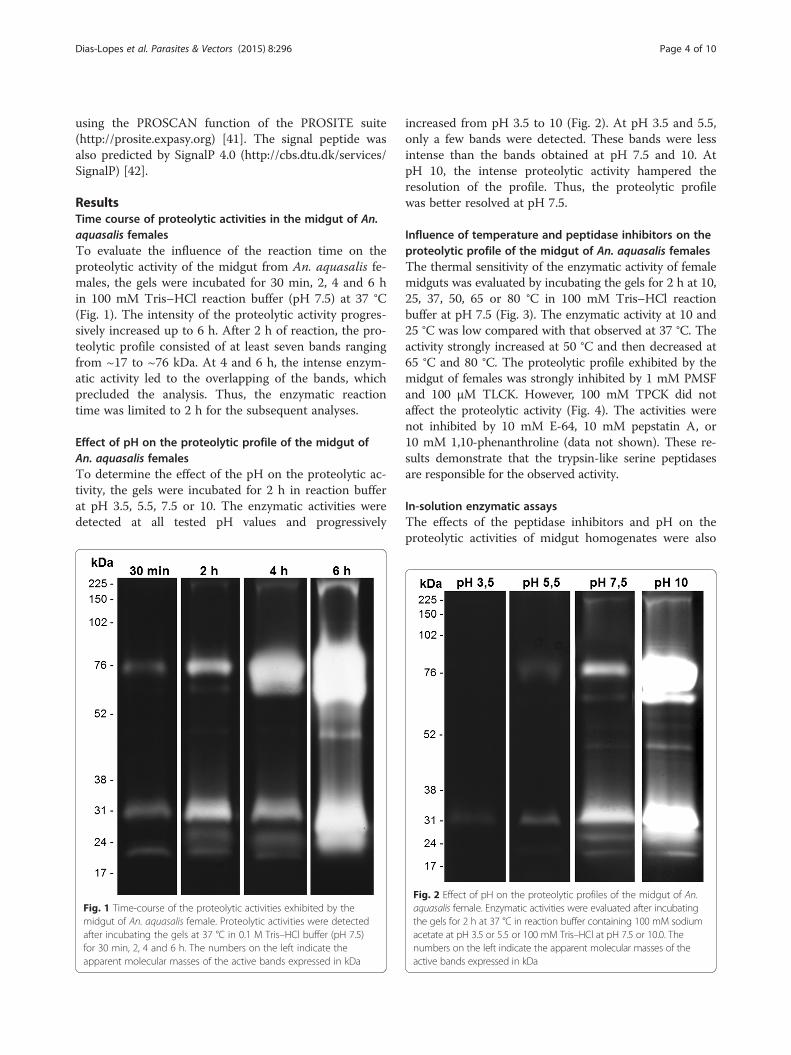
ResultsTime course of proteolytic activities in the midgut of An.aquasalis femalesTo evaluate the influence of the reaction time on theproteolytic activity of the midgut from An. aquasalis fe-males, the gels were incubated for 30 min, 2, 4 and 6 hin 100 mM Tris–HCl reaction buffer (pH 7.5) at 37 °C(Fig. 1). The intensity of the proteolytic activity progres-sively increased up to 6 h. After 2 h of reaction, the pro-teolytic profile consisted of at least seven bands rangingfrom ~17 to ~76 kDa. At 4 and 6 h, the intense enzym-atic activity led to the overlapping of the bands, whichprecluded the analysis. Thus, the enzymatic reactiontime was limited to 2 h for the subsequent analyses.
Effect of pH on the proteolytic profile of the midgut ofAn. aquasalis femalesTo determine the effect of the pH on the proteolytic ac-tivity, the gels were incubated for 2 h in reaction bufferat pH 3.5, 5.5, 7.5 or 10. The enzymatic activities weredetected at all tested pH values and progressively
Fig. 1 Time-course of the proteolytic activities exhibited by themidgut of An. aquasalis female. Proteolytic activities were detectedafter incubating the gels at 37 °C in 0.1 M Tris–HCl buffer (pH 7.5)for 30 min, 2, 4 and 6 h. The numbers on the left indicate theapparent molecular masses of the active bands expressed in kDa
increased from pH 3.5 to 10 (Fig. 2). At pH 3.5 and 5.5,only a few bands were detected. These bands were lessintense than the bands obtained at pH 7.5 and 10. AtpH 10, the intense proteolytic activity hampered theresolution of the profile. Thus, the proteolytic profilewas better resolved at pH 7.5.
Influence of temperature and peptidase inhibitors on theproteolytic profile of the midgut of An. aquasalis femalesThe thermal sensitivity of the enzymatic activity of femalemidguts was evaluated by incubating the gels for 2 h at 10,25, 37, 50, 65 or 80 °C in 100 mM Tris–HCl reactionbuffer at pH 7.5 (Fig. 3). The enzymatic activity at 10 and25 °C was low compared with that observed at 37 °C. Theactivity strongly increased at 50 °C and then decreased at65 °C and 80 °C. The proteolytic profile exhibited by themidgut of females was strongly inhibited by 1 mM PMSFand 100 μM TLCK. However, 100 mM TPCK did notaffect the proteolytic activity (Fig. 4). The activities werenot inhibited by 10 mM E-64, 10 mM pepstatin A, or10 mM 1,10-phenanthroline (data not shown). These re-sults demonstrate that the trypsin-like serine peptidasesare responsible for the observed activity.
In-solution enzymatic assaysThe effects of the peptidase inhibitors and pH on theproteolytic activities of midgut homogenates were also
Fig. 2 Effect of pH on the proteolytic profiles of the midgut of An.aquasalis female. Enzymatic activities were evaluated after incubatingthe gels for 2 h at 37 °C in reaction buffer containing 100 mM sodiumacetate at pH 3.5 or 5.5 or 100 mM Tris–HCl at pH 7.5 or 10.0. Thenumbers on the left indicate the apparent molecular masses of theactive bands expressed in kDa
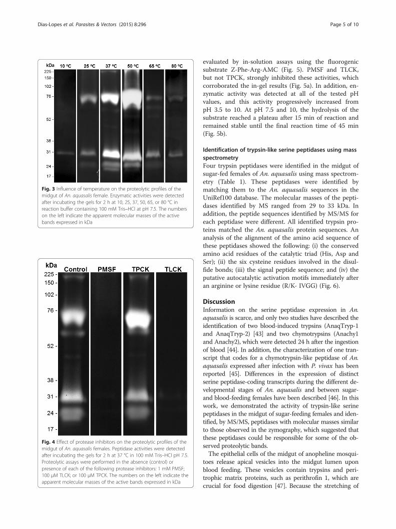
Fig. 3 Influence of temperature on the proteolytic profiles of themidgut of An. aquasalis female. Enzymatic activities were detectedafter incubating the gels for 2 h at 10, 25, 37, 50, 65, or 80 °C inreaction buffer containing 100 mM Tris–HCl at pH 7.5. The numberson the left indicate the apparent molecular masses of the activebands expressed in kDa
Fig. 4 Effect of protease inhibitors on the proteolytic profiles of themidgut of An. aquasalis females. Peptidase activities were detectedafter incubating the gels for 2 h at 37 °C in 100 mM Tris–HCl pH 7.5.Proteolytic assays were performed in the absence (control) orpresence of each of the following protease inhibitors: 1 mM PMSF;100 μM TLCK; or 100 μM TPCK. The numbers on the left indicate theapparent molecular masses of the active bands expressed in kDa
Dias-Lopes et al. Parasites & Vectors (2015) 8:296 Page 5 of 10
evaluated by in-solution assays using the fluorogenicsubstrate Z-Phe-Arg-AMC (Fig. 5). PMSF and TLCK,but not TPCK, strongly inhibited these activities, whichcorroborated the in-gel results (Fig. 5a). In addition, en-zymatic activity was detected at all of the tested pHvalues, and this activity progressively increased frompH 3.5 to 10. At pH 7.5 and 10, the hydrolysis of thesubstrate reached a plateau after 15 min of reaction andremained stable until the final reaction time of 45 min(Fig. 5b).
Identification of trypsin-like serine peptidases using massspectrometryFour trypsin peptidases were identified in the midgut ofsugar-fed females of An. aquasalis using mass spectrom-etry (Table 1). These peptidases were identified bymatching them to the An. aquasalis sequences in theUniRef100 database. The molecular masses of the pepti-dases identified by MS ranged from 29 to 33 kDa. Inaddition, the peptide sequences identified by MS/MS foreach peptidase were different. All identified trypsin pro-teins matched the An. aquasalis protein sequences. Ananalysis of the alignment of the amino acid sequence ofthese peptidases showed the following: (i) the conservedamino acid residues of the catalytic triad (His, Asp andSer); (ii) the six cysteine residues involved in the disul-fide bonds; (iii) the signal peptide sequence; and (iv) theputative autocatalytic activation motifs immediately afteran arginine or lysine residue (R/K- IVGG) (Fig. 6).
DiscussionInformation on the serine peptidase expression in An.aquasalis is scarce, and only two studies have described theidentification of two blood-induced trypsins (AnaqTryp-1and AnaqTryp-2) [43] and two chymotrypsins (Anachy1and Anachy2), which were detected 24 h after the ingestionof blood [44]. In addition, the characterization of one tran-script that codes for a chymotrypsin-like peptidase of An.aquasalis expressed after infection with P. vivax has beenreported [45]. Differences in the expression of distinctserine peptidase-coding transcripts during the different de-velopmental stages of An. aquasalis and between sugar-and blood-feeding females have been described [46]. In thiswork, we demonstrated the activity of trypsin-like serinepeptidases in the midgut of sugar-feeding females and iden-tified, by MS/MS, peptidases with molecular masses similarto those observed in the zymography, which suggested thatthese peptidases could be responsible for some of the ob-served proteolytic bands.The epithelial cells of the midgut of anopheline mosqui-
toes release apical vesicles into the midgut lumen uponblood feeding. These vesicles contain trypsins and peri-trophic matrix proteins, such as perithrofin 1, which arecrucial for food digestion [47]. Because the stretching of
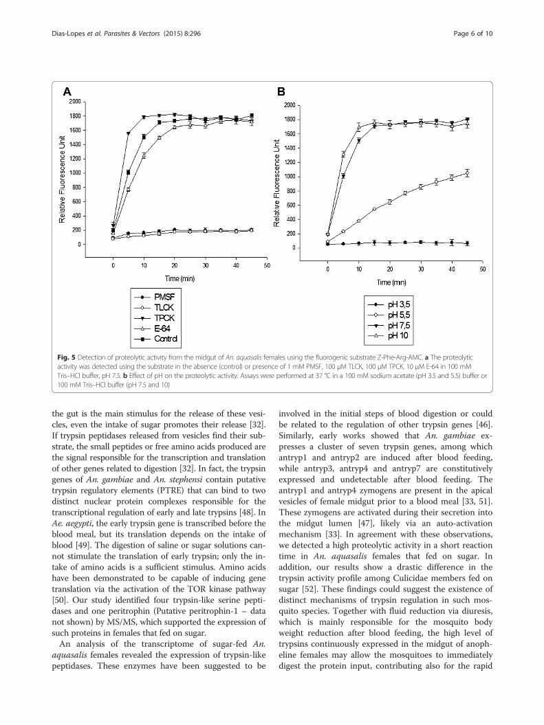
Fig. 5 Detection of proteolytic activity from the midgut of An. aquasalis females using the fluorogenic substrate Z-Phe-Arg-AMC. a The proteolyticactivity was detected using the substrate in the absence (control) or presence of 1 mM PMSF, 100 μM TLCK, 100 μM TPCK, 10 μM E-64 in 100 mMTris–HCl buffer, pH 7.5. b Effect of pH on the proteolytic activity. Assays were performed at 37 °C in a 100 mM sodium acetate (pH 3.5 and 5.5) buffer or100 mM Tris–HCl buffer (pH 7.5 and 10)
Dias-Lopes et al. Parasites & Vectors (2015) 8:296 Page 6 of 10
the gut is the main stimulus for the release of these vesi-cles, even the intake of sugar promotes their release [32].If trypsin peptidases released from vesicles find their sub-strate, the small peptides or free amino acids produced arethe signal responsible for the transcription and translationof other genes related to digestion [32]. In fact, the trypsingenes of An. gambiae and An. stephensi contain putativetrypsin regulatory elements (PTRE) that can bind to twodistinct nuclear protein complexes responsible for thetranscriptional regulation of early and late trypsins [48]. InAe. aegypti, the early trypsin gene is transcribed before theblood meal, but its translation depends on the intake ofblood [49]. The digestion of saline or sugar solutions can-not stimulate the translation of early trypsin; only the in-take of amino acids is a sufficient stimulus. Amino acidshave been demonstrated to be capable of inducing genetranslation via the activation of the TOR kinase pathway[50]. Our study identified four trypsin-like serine pepti-dases and one peritrophin (Putative peritrophin-1 – datanot shown) by MS/MS, which supported the expression ofsuch proteins in females that fed on sugar.An analysis of the transcriptome of sugar-fed An.
aquasalis females revealed the expression of trypsin-likepeptidases. These enzymes have been suggested to be
involved in the initial steps of blood digestion or couldbe related to the regulation of other trypsin genes [46].Similarly, early works showed that An. gambiae ex-presses a cluster of seven trypsin genes, among whichantryp1 and antryp2 are induced after blood feeding,while antryp3, antryp4 and antryp7 are constitutivelyexpressed and undetectable after blood feeding. Theantryp1 and antryp4 zymogens are present in the apicalvesicles of female midgut prior to a blood meal [33, 51].These zymogens are activated during their secretion intothe midgut lumen [47], likely via an auto-activationmechanism [33]. In agreement with these observations,we detected a high proteolytic activity in a short reactiontime in An. aquasalis females that fed on sugar. Inaddition, our results show a drastic difference in thetrypsin activity profile among Culicidae members fed onsugar [52]. These findings could suggest the existence ofdistinct mechanisms of trypsin regulation in such mos-quito species. Together with fluid reduction via diuresis,which is mainly responsible for the mosquito bodyweight reduction after blood feeding, the high level oftrypsins continuously expressed in the midgut of anoph-eline females may allow the mosquitoes to immediatelydigest the protein input, contributing also for the rapid
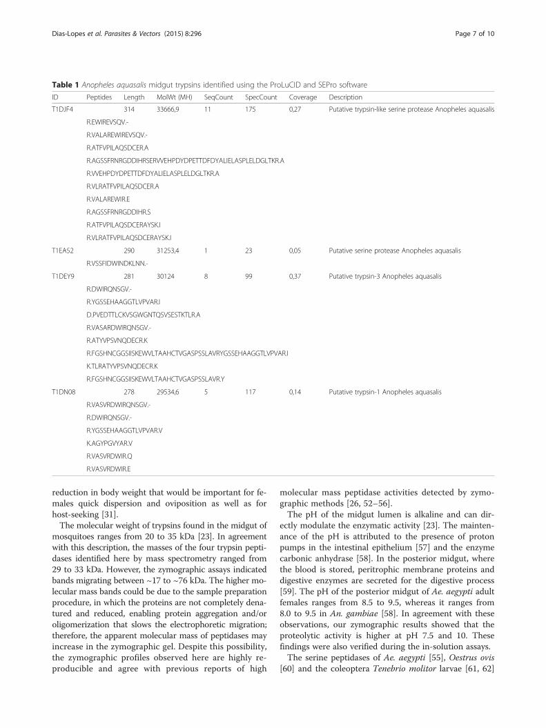
Table 1 Anopheles aquasalis midgut trypsins identified using the ProLuCID and SEPro software
ID Peptides Length MolWt (MH) SeqCount SpecCount Coverage Description
T1DJF4 314 33666,9 11 175 0,27 Putative trypsin-like serine protease Anopheles aquasalis
R.EWIREVSQV.-
R.VALAREWIREVSQV.-
R.ATFVPILAQSDCER.A
R.AGSSFRNRGDDIHRSERVVEHPDYDPETTDFDYALIELASPLELDGLTKR.A
R.VVEHPDYDPETTDFDYALIELASPLELDGLTKR.A
R.VLRATFVPILAQSDCER.A
R.VALAREWIR.E
R.AGSSFRNRGDDIHR.S
R.ATFVPILAQSDCERAYSK.I
R.VLRATFVPILAQSDCERAYSK.I
T1EA52 290 31253,4 1 23 0,05 Putative serine protease Anopheles aquasalis
R.VSSFIDWINDKLNN.-
T1DEY9 281 30124 8 99 0,37 Putative trypsin-3 Anopheles aquasalis
R.DWIRQNSGV.-
R.YGSSEHAAGGTLVPVAR.I
D.PVEDTTLCKVSGWGNTQSVSESTKTLR.A
R.VASARDWIRQNSGV.-
R.ATYVPSVNQDECR.K
R.FGSHNCGGSIISKEWVLTAAHCTVGASPSSLAVRYGSSEHAAGGTLVPVAR.I
K.TLRATYVPSVNQDECR.K
R.FGSHNCGGSIISKEWVLTAAHCTVGASPSSLAVR.Y
T1DN08 278 29534,6 5 117 0,14 Putative trypsin-1 Anopheles aquasalis
R.VASVRDWIRQNSGV.-
R.DWIRQNSGV.-
R.YGSSEHAAGGTLVPVAR.V
K.AGYPGVYAR.V
R.VASVRDWIR.Q
R.VASVRDWIR.E
Dias-Lopes et al. Parasites & Vectors (2015) 8:296 Page 7 of 10
reduction in body weight that would be important for fe-males quick dispersion and oviposition as well as forhost-seeking [31].The molecular weight of trypsins found in the midgut of
mosquitoes ranges from 20 to 35 kDa [23]. In agreementwith this description, the masses of the four trypsin pepti-dases identified here by mass spectrometry ranged from29 to 33 kDa. However, the zymographic assays indicatedbands migrating between ~17 to ~76 kDa. The higher mo-lecular mass bands could be due to the sample preparationprocedure, in which the proteins are not completely dena-tured and reduced, enabling protein aggregation and/oroligomerization that slows the electrophoretic migration;therefore, the apparent molecular mass of peptidases mayincrease in the zymographic gel. Despite this possibility,the zymographic profiles observed here are highly re-producible and agree with previous reports of high
molecular mass peptidase activities detected by zymo-graphic methods [26, 52–56].The pH of the midgut lumen is alkaline and can dir-
ectly modulate the enzymatic activity [23]. The mainten-ance of the pH is attributed to the presence of protonpumps in the intestinal epithelium [57] and the enzymecarbonic anhydrase [58]. In the posterior midgut, wherethe blood is stored, peritrophic membrane proteins anddigestive enzymes are secreted for the digestive process[59]. The pH of the posterior midgut of Ae. aegypti adultfemales ranges from 8.5 to 9.5, whereas it ranges from8.0 to 9.5 in An. gambiae [58]. In agreement with theseobservations, our zymographic results showed that theproteolytic activity is higher at pH 7.5 and 10. Thesefindings were also verified during the in-solution assays.The serine peptidases of Ae. aegypti [55], Oestrus ovis
[60] and the coleoptera Tenebrio molitor larvae [61, 62]
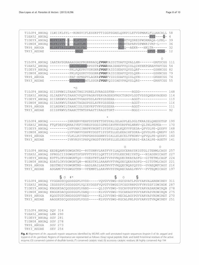
Fig. 6 Alignment of An. aquasalis trypsin sequences identified by MS/MS with well annotated trypsin sequences (trypsin-3 of Ae. aegypti andtrypsin-6 of An. gambiae). Regions of importance are represented as follows: (Gray) signal peptide; (Italic and bold) N-terminal residues of the activeenzyme; (O) conserved cysteine of disulfide bonds; (*) conserved catalytic triad; (§) accessory catalytic residues; (#) highly conserved Asp 194
Dias-Lopes et al. Parasites & Vectors (2015) 8:296 Page 8 of 10

Dias-Lopes et al. Parasites & Vectors (2015) 8:296 Page 9 of 10
are reportedly thermally sensitive. We observed that thetrypsin-like serine peptidases of An. aquasalis are activeover a large temperature range, but the proteolytic activ-ity is markedly decreased at lower temperatures. Thesefindings are in agreement with that observed in O. ovis[60]. The authors of this previous study suggested thatthese enzymes could be partially inactivated at low tem-peratures, which could affect the degradation of food inthe larval stages and culminate in the low rates of larvaldevelopment that are characteristic of the cold monthsin temperate climates [60]. The possibility that these en-zymes retain some level of activity at low temperaturesmay have ecological implications in mosquitoes, such asduring diapause, as suggested by other authors [63].
ConclusionThe study of peptidases expressed in the mosquito midgutis essential to understand the mechanisms of parasite-hostinteraction and the physiological process of nutrient di-gestion. Here, we biochemically characterized the activeproteolytic profile of An. aquasalis and confirmed the ex-pression of four putative trypsin-like serine peptidases ofsimilar molecular mass as those observed with zymogra-phy. These findings contribute to the gene annotation ofthe unknown genome of this species, to the tissue locationof these peptidases, and to the prediction of the functionof these crucial enzymes, impacting further studies of thisspecies.
Competing interestsThe authors declare that they have no competing interests.
Authors’ contributionsJBJ, GDL and PC designed the study. GDL, LSV, ABV and performed theexperimental work. GDL, PC and JBJ analyzed the data and prepared themanuscript with the critical input of CB and GBD. All authors read andapproved the final manuscript.
AcknowledgementsThis work was supported by FAPEMIG (Edital Universal Process No. APQ-01070-12),FIOCRUZ-IOC, CNPq (J.B.J. PQ Process No. 308679/2012-1) and CAPES. We thankProf. Dr. José Bento Pereira Lima and Denise Valle (Laboratório de Fisiologia eControle de Artrópodes Vetores of the Instituto Oswaldo Cruz) for kindly providingthe insects.
Author details1Laboratório de Biologia Molecular e Doenças Endêmicas – InstitutoOswaldo Cruz, FIOCRUZ, Rio de Janeiro, Brazil. 2Laboratório de Química deProteínas, Departamento de Química, UFRJ, Rio de Janeiro, Brazil.3Laboratório de Pesquisa em Leishmaniose – Instituto Oswaldo Cruz,FIOCRUZ, Av. Brasil 4365, Manguinhos, Pav. 26, Sala 509, Rio de Janeiro, Brazil.4Departamento de Medicina, Faculdade de Medicina – Universidade Federalde São João Del Rey, São João del Rei, MG, Brazil.
Received: 23 February 2015 Accepted: 20 May 2015
References1. Sinka ME, Rubio-Palis Y, Manguin S, Patil AP, Temperley WH, et al. The dominant
Anopheles vectors of human malaria in the Americas: occurrence data, distributionmaps and bionomic précis. Parasit Vectors. 2010;3:72.
2. Laubach HE, Validum L, Bonilla JA, Agar A, Cummings R, Mitchell C, et al.Identification of anopheles aquasalis as a possible vector of malaria inGuyana, south America. West Indian Med J. 2001;50:319–21.
3. Pina-Costa A, Brasil P, Di-Santi SM, Araujo MP, Suárez-Mutis MC, Santelli AC,et al. Malaria in Brazil: what happens outside the Amazonian endemicregion. Mem Inst Oswaldo Cruz. 2014;109:618–33.
4. Gallup JL, Sachs JD. The economic burden of malaria. Am J Trop Med Hyg.2001;64:85–96.
5. Sachs J, Malaney P. The economic and social burden of malaria. Nature.2002;415:680–5.
6. WHO - Word Health Organization. World Malaria Report 2013. 2013.7. Hemingway J, Ranson H. Insecticide resistance in insect vectors of human
disease. Annu Rev Entomol. 2000;45:371–91.8. White NJ. Antimalarial drug resistance. J Clin Invest. 2004;113:1084–92.9. Catteruccia F. Malaria vector control in the third millennium: progress and
perspectives of molecular approaches. Pest Manag Sci. 2007;63:634–40.10. Raghavendra K, Barik TK, Reddy BP, Sharma P, Dash AP. Malaria vector
control: from past to future. Parasitol Res. 2011;108:757–79.11. Chertemps T, Mitri C, Perrot S, Sautereau J, Jacques JC, Thiery I, et al.
Anopheles gambiae PRS1 modulates Plasmodium development at bothmidgut and salivary gland steps. PLoS One. 2010;5, e11538.
12. Blandin S, Shiao SH, Moita LF, Janse CJ, Waters AP, Kafatos FC, et al.Complement-like protein TEP1 is a determinant of vectorial capacity in themalaria vector Anopheles gambiae. Cell. 2004;116:661–70.
13. Osta MA, Christophides GK, Kafatos FC. Effects of mosquito genes onPlasmodium development. Science. 2004;303:2030–2.
14. Yassine H, Kamareddine L, Chamat S, Christophides GK, Osta MA. A serineprotease homolog negatively regulates TEP1 consumption in systemic infectionsof the malaria vector Anopheles gambiae. J Innate Immun. 2014;6:806–18.
15. Gorman MJ, Paskewitz SM. Serine proteases as mediators of mosquitoimmune responses. Insect Biochem Mol Biol. 2001;31:257–62.
16. Shahabuddin M, Lemos FJA, Kaslow DC, Jacobs-Lorena M. Antibody-mediated inhibition of Aedes aegypti midgut trypsins blocks sporogonicdevelopment of Plasmodium gallinaceum. Infect Immun. 1996;64:739–43.
17. Rios-Velásquez CM, Martins-Campos KM, Simões RC, Izzo T, dos Santos EV,Pessoa FAC, et al. Experimental Plasmodium vivax infection of key Anophelesspecies from the Brazilian Amazon. Malar J. 2013;12:460–9.
18. Rawlings ND, Tolle DP, Barrett AJ. MEROPS: the peptidase database. NucleicAcids Res. 2004;32:160–4.
19. Polgár L. The catalytic triad of serine peptidases. Cell Mol Life Sci.2005;62:2161–72.
20. Rawlings ND, Barrett AJ. Families of serine peptidases. Methods Enzymol.1994;244:19–61.
21. Lu SJ, Pennington JE, Stonehouse AR, Mobula MM, Wells MA. Reevaluationof the role of early trypsin activity in the transcriptional activation of thelate trypsin gene in the mosquito Aedes aegypti. Insect Biochem Mol Biol.2006;36:336–43.
22. Shen Z, Edwards MJ, Jacobs-Lorena M. A gut-specific serine protease fromthe malaria vector Anopheles gambiae is downregulated after blood ingestion.Insect Mol Biol. 2000;9:223–9.
23. Terra WR, Ferreira C. Insect digestive enzymes: properties, compartmentalizationand function. Comp Biochem Physiol B. 1994;109:1–62.
24. Wu DD, Wang GD, Irwin DM, Zhang YP. A profound role for the expansionof trypsin-like serine protease family in the evolution of hematophagy inmosquito. Mol Biol Evol. 2009;26:2333–41.
25. Shahabuddin M, Costero A. Spatial distribution of factors that determinesporogonic development of malaria parasites in mosquitoes. InsectBiochem Mol Biol. 2001;31:231–40.
26. Telleria EL, Araújo APO, Secundino NF, d’Avila-Levy CM, Traub-Csekö YM.Trypsin-like serine proteases in Lutzomyia longipalpis – expression, activityand possible modulation by Leishmania infantum chagasi. PLoS One.2010;5:e10697.
27. Molina-Cruz A, Gupta L, Richardson J, Bennett K, Black 4th W, Barillas-MuryC. Effect of mosquito midgut trypsin activity on dengue-2 virus infectionand dissemination in Aedes aegypti. Am J Trop Med Hyg. 2005;72:631–7.
28. Paskewitz SM, Andreev O, Shi L. Gene silencing of serine proteases affectsmelanization of Sephadex beads in Anopheles gambiae. Insect Biochem MolBiol. 2006;36:701–11.
29. Volz J, Muller HM, Zdanowicz A, Kafatos FC, Osta MA. A genetic moduleregulates the melanization response of Anopheles to Plasmodium. CellMicrobiol. 2006;8:1392–405.

Dias-Lopes et al. Parasites & Vectors (2015) 8:296 Page 10 of 10
30. Mancini E, Tammaro F, Baldini F, Via A, Raimondo D, George P, et al.Molecular evolution of a gene cluster of serine proteases expressed in theAnopheles gambiae female reproductive tract. BMC Evol Biol. 2011;11:72–89.
31. Hörler E, Briegel H. Proteolytic enzymes of female Anopheles: biphasicsynthesis, regulation and multiple feeding. Arch Insect Biochem Physiol.1995;28:189–205.
32. Lemos FJA, Cornel AJ, Jacobs-Lorena M. Trypsin and aminopeptidase geneexpression is affected by age and food composition in Anopheles gambiae.Insect Biochem Mol Biol. 1996;26:651–8.
33. Müller HM, Catteruccia F, Vizioli J, della Torre A, Crisanti A. Constitutive andblood meal-induced trypsin genes in Anopheles gambiae. Exp Parasitol.1995;81:371–85.
34. Saboia-Vahia L, Borges-Veloso A, Cuervo P, Junqueira M, Mesquita-Rodrigues C,Britto C, et al. Protein expression in the midgut of sugar-fed Aedes albopictusfemales. Parasit Vectors. 2012;5:290–314.
35. Galán JE, Pace J, Hayman MJ. Involvement of the epidermal growth factorreceptor in the mammalian cells by Salmonella typhimurium. Nature.1992;357:588–9.
36. Cuervo P, Mesquita-Rodrigues C, D’avila Levy CM, Britto C, Pires FA, Gredilha R,et al. Serine protease activities in Oxysarcodexia thornax (Walker) (Diptera:Sarcophagidae) first instar larva. Mem Inst Oswaldo Cruz. 2008;103:504–6.
37. Caballero NG, Rodríguez-Vega A, Dias-Lopes G, Valenzuela JG, Ribeiro JM,Carvalho PC, et al. Expression of the mevalonate pathway enzymes in theLutzomyia longipalpis (Diptera: Psychodidae) sex pheromone glanddemonstrated by an integrated proteomic approach. J Proteomics.2014;96:117–32.
38. Carvalho PC, Yates 3rd JR, Barbosa VC. Analyzing shotgun proteomic datawith patternLab for proteomics. Curr Protoc Bioinformatics. 2010;30:13.13.1–15.
39. Carvalho PC, Fischer JSG, Xu T, Cociorva D, Balbuena TS, Valente RH, et al.Search engine processor: filtering and organizing peptide spectrummatches. Proteomics. 2012;12:944–9.
40. Sievers F, Wilm A, Dineen D, Gibson TJ, Karplus K, Li W, et al. Fast, scalablegeneration of high-quality protein multiple sequence alignments usingClustal Omega. Mol Syst Biol. 2011;7:539.
41. Sigrist CJ, de Castro E, Cerutti L, Cuche BA, Hulo N, Bridge A, et al. New andcontinuing developments at PROSITE. Nucleic Acids Res. 2013;41:344–7.
42. Petersen TN, Brunak S, von Heijne G, Nielsen H. SignalP 4.0: discriminatingsignal peptides from transmembrane regions. Nat Methods. 2011;8:785–6.
43. Caroci AS, Calvo E, Ribolla PE, De Biachi AG, Marinotti O. Two digestivetrypsins occur in three species of neotropical anophelines. J Med Entomol.2003;40:991–5.
44. de Almeida RW, Tovar FJ, Ferreira II, Leoncini O. Chymotrypsin genes in themalaria mosquitoes Anopheles aquasalis and Anopheles darlingi. InsectBiochem Mol Biol. 2003;33:307–15.
45. Bahia AC, Kubota MS, Tempone AJ, Pinheiro WD, Tadei WP, Secundino NFC,et al. Anopheles aquasalis infected by Plasmodium vivax displays uniquegene expression profiles when compared to other malaria vectors andplasmodia. PLoS One. 2010;5:e9795.
46. Costa-da-Silva AL, Marinotti O, Ribeiro JMC, Silva MCP, Lopes AR, Barros MS,et al. Transcriptome sequencing and developmental regulation of geneexpression in Anopheles aquasalis. PLoS Negl Trop Dis. 2014;8:e3005.
47. Devenport M, Fujioka H, Jacobs-Lorena M. Storage and secretion of theperitrophic matrix protein Ag-Aper1 and trypsin in the midgut of Anophelesgambiae. Insect Mol Biol. 2004;13:349–58.
48. Giannoni F, Müller HM, Vizioli J, Catteruccia F, Kafatos FC, Crisanti A. Nuclearfactors bind to a conserved DNA element that modulates transcription ofAnopheles gambiae trypsin genes. J Biol Chem. 2001;276:700–7.
49. Noriega FG, Pennington JE, Barillas-Mury C, Wang X, Wells MA. Early trypsin,an Aedes aegypti female specific protease, is post-transcriptionally regulatedby the blood meal. Insect Mol Biol. 1996;5:25–9.
50. Brandon MC, Pennington JE, Isoe J, Zamora J, Schillinger AS, Miesfeld RL.TOR signaling is required for amino acid stimulation of early trypsin proteinsynthesis in the midgut of Aedes aegypti mosquitoes. Insect Biochem MolBiol. 2008;38:916–22.
51. Müller HM, Crampton JM, della Torre A, Sinden R, Crisanti A. Members of atrypsin gene family in Anopheles gambiae are induced in the gut by bloodmeal. EMBO J. 1993;12:2891–900.
52. Saboia-Vahia L, Cuervo P, Borges-Veloso A, Souza NP, Britto C, Lopes GD,et al. The midgut of Aedes albopictus females expresses active trypsin-likeserine peptidases. Parasit Vectors. 2014;7:253.
53. Nauen R, Sorge D, Sterner A, Borovsky D. TMOF-like factor controls thebiosynthesis of serine proteases in the larval gut of Heliothis virescens.Arch Insect Biochem Physiol. 2001;47:169–80.
54. Vandooren J, Geurts N, Martens E, Van den Steen PE, Opdenakker G.Zymography methods for visualizing hydrolytic enzymes. Nat Methods.2013;10:211–20.
55. Mesquita-Rodrigues C, Saboia-Vahia L, Cuervo P, Levy CM, Honório NA,Domont GB, et al. Expression of trypsin-like serine peptidases inpre-imaginal stages of Aedes aegypti (Diptera: Culicidae). Arch InsectBiochem Physiol. 2011;76:223–35.
56. Borges-Veloso A, Saboia-Vahia L, Cuervo P, Pires RC, Britto C, Fernandes N,et al. Proteolytic profiling and comparative analyses of active trypsin-likeserine peptidases in preimaginal stages of Culex quinquefasciatus. ParasitVectors. 2012;5:123.
57. Filippova M, Ross LS, Gill SS. Cloning of the V-ATPase B subunit cDNA fromCulex quinquefasciatus and expression of the B and C subunits in mosquitoes.Insect Mol Biol. 1998;7:223–32.
58. Corena MDP, VanEkeris L, Salazar MI, Bowers D, Fiedler MM, Silverman D,et al. Carbonic anhydrase in the adult mosquito midgut. J Exp Biol.2005;208:3263–73.
59. Okuda K, Caroci AS, Ribolla PEM, de Bianchi AG, Bijovsky AT. Functionalmorphology of adult female Culex quinquefasciatus midgut during blooddigestion. Tissue Cell. 2002;34:210–9.
60. Angulo-Valadez CE, Cepeda-Palacios R, Ascencio F, Jacquiet P, Dorchies P,Romero MJ, et al. Proteolytic activity in salivary gland products of sheep botfly (Oestrus ovis) larvae. Vet Parasitol. 2007;149:117–25.
61. Elpidina EN, Tsybina TA, Dunaevsky YE, Belozersky MA, Zhuzhikov DP,Oppert B. A chymotrypsin-like proteinase from the midgut ofTenebrio molitor larvae. Biochimie. 2005;87:771–9.
62. Tsybina TA, Dunaevsky YE, Belozersky MA, Zhuzhikov DP, Oppert B,Elpidina EN. Digestive proteinases of yellow mealworm (Tenebrio molitor)larvae: purification and characterization of a trypsin-like proteinase.Biochemistry. 2005;70:300–5.
63. Robich RM, Denlinger DL. Diapause in the mosquito Culex pipiens evokes ametabolic switch from blood feeding to sugar gluttony. Proc Natl Acad SciU S A. 2005;44:15912–7.
Submit your next manuscript to BioMed Centraland take full advantage of:
• Convenient online submission
• Thorough peer review
• No space constraints or color figure charges
• Immediate publication on acceptance
• Inclusion in PubMed, CAS, Scopus and Google Scholar
• Research which is freely available for redistribution
Submit your manuscript at www.biomedcentral.com/submit