expression of cannabinoid receptors type 1 and type 2 in ... cancer/expression of... · expression...
TRANSCRIPT
Expression of cannabinoid receptors type 1 and type 2 in non-Hodgkin
lymphoma: Growth inhibition by receptor activation
Kristin Gustafsson1, Xiao Wang1, Denise Severa1, Maeve Eriksson1, Eva Kimby2, Mats Merup2,Birger Christensson1, Jenny Flygare1 and Birgitta Sander1*
1Department of Laboratory Medicine, Division of Pathology, Karolinska Institutet and KarolinskaUniversity Hospital Huddinge, F-46, SE-14186 Stockholm, Sweden2Department of Medicine, Division of Hematology, Karolinska Institutet and Karolinska UniversityHospital Huddinge, SE-14186 Stockholm, Sweden
Endogenous and synthetic cannabinoids exert antiproliferativeand proapoptotic effects in various types of cancer and in mantlecell lymphoma (MCL). In this study, we evaluated the expressionof cannabinoid receptors type 1 and type 2 (CB1 and CB2) in non-Hodgkin lymphomas of B cell type (n 5 62). A majority of thelymphomas expressed higher mRNA levels of CB1 and/or CB2 ascompared to reactive lymphoid tissue. With the exception ofMCL, which uniformly overexpresses both CB1 and CB2, the lev-els of cannabinoid receptors within other lymphoma entities werehighly variable, ranging from 0.1 to 224 times the expression in re-active lymph nodes. Low levels of the splice variant CB1a, previ-ously shown to have a different affinity for cannabinoids thanCB1, were detected in 44% of the lymphomas, while CB1b expres-sion was not detected. In functional studies using MCL, Burkittlymphoma (BL), chronic lymphatic leukemia (CLL) and plasmacell leukemia cell lines, the stable anandamide analog R(þ)-meth-anandamide (R(þ)-MA) induced cell death only in MCL and CLLcells, which overexpressed both cannabinoid receptors, but not inBL. In vivo treatment with R(þ)-MA caused a significant reduc-tion of tumor size and mitotic index in mice xenografted withhuman MCL. Together, our results suggest that therapies usingcannabinoid receptor ligands will have efficiency in reducing tu-mor burden in malignant lymphoma overexpressing CB1 andCB2.' 2008 Wiley-Liss, Inc.
Key words: cannabinoid; lymphoma; cancer; apoptosis; xenograft
Non-Hodgkin lymphomas represent clonal expansions of malig-nant B cells at various stages of maturation. Our understanding ofthe molecular biology and pathogenesis of malignant lymphomahas improved substantially during the last few years. As a result,targeted therapies and interference with critical signaling path-ways are evolving as new treatment modalities.1 The malignant Bcells might also be dependent on interactions with the tumormicroenvironment for their survival.2,3 This is supported by thefact that it is very difficult to grow lymphoma cells in vitro in theabsence of stroma or CD40 stimulation. In follicular lymphoma(FL), interactions with T cells, macrophages and dendritic cellshave been suggested to influence the clinical behavior of themalignancy.2,4,5 The expression pattern of cytokines, chemokines,prostaglandins, leukotrienes and their receptors may also promoteor inhibit the survival of malignant B cells.
We have previously studied the role of the endocannabinoidsystem in MCL.6–8 The endocannabinoid system encompasses theG-protein coupled receptors, cannabinoid receptor type 1 (CB1)and type 2 (CB2), the endocannabinoids and the enzymes respon-sible for endocannabinoid synthesis and degradation.9 The endo-cannabinoids are synthesized from arachidonic acid by many celltypes including macrophages and dendritic cells. They have prop-erties similar to compounds of Cannabis sativa and have recentlybeen shown to have effects on various types of cancer.9 Many ofthe effects of natural and synthetic cannabinoids are mediatedthrough the cannabinoid receptors. CB1 is expressed at high lev-els in the central nervous system and at lower levels in immunecells. In addition to the full-length CB1 transcript, 2 splice var-iants denoted CB1a and CB1b have been reported in humanbrain.10 Both splice variants have different ligand binding
capacity compared with that of the full length receptor.10 TheCB2 receptor is expressed in lymphocytes and at lower levels inthe brain. Accordingly, resting B lymphocytes express the CB2receptor and low levels of the CB1 receptor.11,12 Polyclonal acti-vation has been reported to modulate expression of cannabinoidreceptors on lymphocytes.13–16 The function of the cannabinoidreceptors in the immune system is not fully known, but a role inregulation of cytokine production and cell migration has beensuggested.16
In MCL, CB1 and CB2 were found to be consistently overex-pressed compared with reactive lymphoid tissue or normal purifiedB lymphocytes.8,17 Ligation of the receptors with the endocanna-binoid arachidonoyl-ethanolamide (anandamide) as well as withthe synthetic cannabinoids WIN 55,212-2 and R(þ)-methananda-mide (R(þ)-MA) inhibited cell proliferation and induced celldeath in MCL cell lines and patient samples.6,7 Normal B cells,which express lower levels of cannabinoid receptors than MCL,were not affected by cannabinoids, suggesting that ligation of can-nabinoid receptors could selectively target the malignant cells.7 Inaddition to MCL, high expression of cannabinoid receptors com-pared to normal epithelium has been reported in a number of dif-ferent epithelial malignancies (recently reviewed in Ref. 18).Pharmacological doses of cannabinoids are emerging as suppres-sors of signaling pathways involved in cancer cell growth, sug-gesting that the endocannabinoid system might be targeted to con-trol cancer (reviewed in Refs. 18–21).
Little is known about the role of the cannabinoid system inmalignant non-Hodgkin B cell lymphomas other than MCL.Ligation of CB2 induced programmed cell death in acute T celllymphoblastic leukemia (T-ALL).22,23 Immunoreactivity for CB2has been reported in both B and T non-Hodgkin lymphomas.However, the 2 different antibodies used gave divergent results,possibly due to different phosphorylation of the receptor, and nofunctional studies were reported.24
The last two authors contributed equally to this work.Grant sponsors: The Swedish Cancer Society, The Swedish Research
Council, The Gunnar Nilsson Cancer Foundation, The Cancer Society inStockholm, The Magnus Bergvall Foundation, Karolinska Institutet Funds,Stockholm County Council.*Correspondence to: Department of Laboratory Medicine, Division of
Pathology, Karolinska University Hospital Huddinge, F-46, SE-14186Stockholm, Sweden. Fax: þ468-5858-1020.E-mail: [email protected] 20 February 2008; Accepted after revision 29 February 2008DOI 10.1002/ijc.23584Published online 10 June 2008 in Wiley InterScience (www.interscience.
wiley.com).
Abbreviations: B-CLL, B cell chronic lymphocytic leukemia; BL, Bur-kitt lymphoma; CB1, cannabinoid receptor type 1; CB2, cannabinoid re-ceptor type 2; DLBCL, diffuse large B cell lymphoma; FL, follicular lym-phoma; MCL, mantle cell lymphoma; MZL, marginal zone lymphoma;pre-B ALL, precursor-B acute lymphoblastic lymphoma; RFI, relative foldincrease; R(þ)-MA, R(þ)-methanandamide; SLL/CLL, small lymphocyticlymphoma/B cell chronic lymphocytic leukemia; SR1, SR141716A; SR2,SR144528; T-ALL, acute T cell lymphoblastic leukemia.
Int. J. Cancer: 123, 1025–1033 (2008)' 2008 Wiley-Liss, Inc.
Publication of the International Union Against Cancer

In the present study, we analysed CB1 and CB2 expression byquantitative real time PCR in a panel of non-Hodgkin lymphomasof B cell type, including small lymphocytic lymphoma/B cellchronic lymphocytic leukemia (SLL/CLL), marginal zone lym-phoma (MZL), diffuse large B cell lymphoma (DLBCL), FL, pre-cursor-B acute lymphoblastic lymphoma (pre-B ALL), MCL,immunocytoma (IC) and BL. Our results demonstrate that the ma-jority of malignant B cell lymphomas express CB1 and/or CB2but at a highly variable level. Treatment with the anandamide ana-log R(þ)-MA induced apoptosis in MCL and in B CLL cell lines,expressing higher levels of CB1 and CB2 than reactive lympho-cytes. However, BL cell lines and a plasma cell line were resistant.In vivo, treatment with R(þ)-MA caused a 40% reduction of tu-mor weight in mice xenografted with MCL. Reduced proliferation,measured as mitotic index, was observed in the tumor cells fromthe treated animals. The antiproliferative and proapoptotic effectsof cannabinoids make the endocannabinoid system a potentialnew therapeutic target for individualized therapy in lymphomaswhich overexpress cannabinoid receptors such as MCL and BCLL.
Material and methods
Reagents
SR141716A (SR1) and SR144528 (SR2) were kind gifts fromSanofi-Reserche (Montpellier, France). R(þ)-methanandamide(R(þ)-MA) was purchased from Tocris Cookson (Bristol, UK).AIM-V medium was purchased from Invitrogen (Carlsbad, CA).
Tumor and control tissue
Non-Hodgkin lymphomas of B cell type and reactive lymphoidtissues were retrieved from the files of the Department of Pathol-ogy. Tissue removed for diagnostic purposes was obtained unfixedat the Department of Pathology and immediately processed. Rep-resentative parts of the tissue was fixed in formalin and paraffinembedded. Other parts were snap-frozen and subsequently storedat 270�C. All cases (Table I) were diagnosed and classified usingWorld Health Organization Classification tumors of Haemato-poietic and Lymphoid Tissues.25
Cell lines
The MCL cell line Rec-126 was a kind gift from Dr. ChristianBastard, Ronan, France. The CLL cell lines MEC1 and MEC2, theBL cell lines Raji and Namalwa, the plasma cell line SK-MM-2and the MCL cell line JEKO-1 were obtained from DeutscheSammlung von Microorganismen und Zellkulturen (DSMZ;Braunschweig, Germany). Cell lines were maintained in RPMI1640 medium (Invitrogen) supplemented with 2 mM l-glutamineand 10% fetal calf serum (FCS) and 50 lg of gentamicin (Invi-trogen) under standard conditions (humidified atmosphere, 95%air, 5% CO2, 37�C).
Mouse xenograft model of human MCL
Ten-week-old female SCID/NOD mice (bred at KarolinskaInstitutet, Southern Campus, Stockholm, Sweden) were injectedsubcutaneously on the back with a suspension of 10 3 106 JEKO-1 cells in 0.4 ml PBS. A stock solution of R(þ)-MA in EtOH wasdiluted to 500 lg/ml in PBS. Three days after injecting the JEKO-1 cells, the mice within the group of treated animals (n 5 11)were injected subcutaneously with R(þ)-MA, 5 mg/kg, at the pre-vious injection site. The mice included in the group of mock-treated animals (n 5 12) were injected subcutaneously with PBScontaining the same amount of EtOH as the diluted R(þ)-MA. Tu-mor diameters were measured daily. Animals were sacrificed atday 33 when the diameter of the largest tumor in the mock-treatedgroup reached 1 cm. The tumors were excised and tumor weightwas measured. No visible signs of toxicity were observed in theanimals during the treatment period. The animals were maintained
at the animal facility of the Karolinska Institutet, Southern Cam-pus, according to rules and regulations of European standard.
RNA isolation and quality control
Total RNA was prepared using Gentra RNA purification systemas directed by the supplier (Ambion, Austin, TX). The sampleswere treated with Deoxyribonuclease I Amplification Grade toeliminate DNA (Invitrogen Life Technologies, Carlsbad, CA).RNA quality was assessed using an Agilent Bioanalyzer 2100(Agilent Technologies, Palo Alto, CA).
cDNA synthesis and real-time quantitative RT-PCR
First strand cDNA synthesis was carried out according to theprotocol for Omniscript Reverse Transcription (Qiagen, GmbH,Hilden, Germany). One or two micrograms of RNA were used inthe reactions for RT-PCR or PCR, respectively. The Beacon De-signer 3 program (Biosoft International, Palo Alto, CA) wasemployed for design of CB1, CB1a and CB2 primers. The follow-ing primers were used: CB1 forward-TCATTAAGACGGTGTTTGCATTCT, CB1 reverse-CGTGTCGCAGGTCCTTAC TC,CB2 forward-TGACTATGACCTTCACAGCCTCT, CB2 reverse-AGGACCCACATGATGCCCAG, CB1a forward-GACATCAAAGGAGAATGAGGAGAAC and CB1a reverse-GAGGGACAGGACTGCAATGG. The primers for b-actin were described pre-viously in Ref. 27. Primers were synthesized by Eurogentec(Southampton, Hampshire, UK). The quantification of CB1, CB1aand CB2 compared to b-actin was carried out with an iCycler iQ(Bio-Rad Laboratories, Sundbyberg, Sweden). The iCycler iQreaction detection system software from the same company wasused for data analysis. cDNA was amplified using the qPCR KitPlatinum SYBR Green qPCR SuperMix-UGD with FITC (Invi-trogen) according to the manufacturer’s instructions. The sampleswere divided into triplicates in a 96-well PCR plate (Abgene,Hamburg, Germany) and run at 95�C for 10 min followed by 40cycles, each cycle consisting of 15 sec at 95�C and 1 min at 55�C.Threshold (Ct) cycle numbers were obtained from amplification ofb-actin, CB1, CB1a and CB2. The PCR products were verified bysequencing. DCt values were calculated by subtracting the Ctvalue of b-actin from the Ct value of CB1, CB1a or CB2. The rel-ative fold increase (RFI) of CB1, CB1a and CB2 was calculated asfollows. The DCt for controls (reactive lymph nodes) and patientsamples was first determined. The DDCt value was calculated bysubtracting the DCt value for pooled controls from the DCt valuefor the sample. The RFI of CB1, CB1a or CB2 was calculated bythe equation: RFI 5 2-DDCt. To use this calculation, the PCR effi-ciencies of the target and control assays must be similar. This wasachieved by adjusting primer concentrations. The criterion forusing the DCt method was fulfilled because by graphing serialdilutions of input cDNA of a random sample against DCt values(CB1–b-actin, CB1a–b-actin or CB2–b-actin) the slope of theline was�0.1 (data not shown).
PCR for CB1a and CB1b
Expression of CB1a and CB1b was investigated using 2 roundsof PCR as described by Ryberg et al.10 The following forwardCB1-splice (50-CCTAATCAAAGACTGAGGTTATGAAGTC-30)and reverse CB1-splice (50-AGTTCTCCCCACACTGGATGTT-30) primers were used in a PCR mixture of 50 ll, containing 50 ngcDNA from MCL patient samples. Platinum Taq polymerase(Invitrogen) was used according to the manufacturer’s instruc-tions. Amplification was carried out in a PTC-200 (MJ ResearchWalthem, MA) for 40 cycles: 95�C for 30 sec, 57�C for 45 sec,and 68�C for 1 min. The resulting products were resolved on 2%agarose gels and a major band at 324 bp corresponding to thehuman unspliced CB1 receptor was detected. The 0.12–0.25-kbgel area was excised and melted (at 95�C for 5 min) and used astemplate for further PCR amplification. This second round of PCRwas performed using the same conditions as described above with
1026 GUSTAFSSON ET AL.
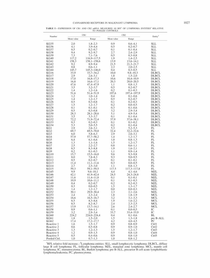
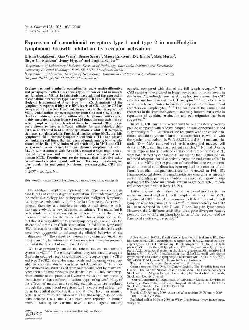
TABLE I – EXPRESSION OF CB1 AND CB2 mRNA MEASURED AS RFI1 OF LYMPHOMA ENTITIES2 RELATIVETO POOLED CONTROLS
NumberCB1 CB2
Entity2
Mean value Range Mean value Range
KG35 2,0 1,8–2,3 0,9 0,6–4,1 SLLKG36 4,1 3,9–4,4 0,5 0,2–0,7 SLLKG37 0,5 0,2–0,7 0,1 0,1–0,4 SLLKG38 9,4 9,2–9,7 2,7 2,4–2,9 SLLKG39 7,3 7,1–7,6 0,5 0,3–0,8 SLLKG40 117,2 116,9–117,5 1,9 1,4–2,5 SLLKG41 158,3 158,1–158,6 13,9 13,6–14,1 SLLKG42 9,2 8,9–9,4 21,5 21,3–21,7 SLLKG43 0,8 0,6–1,1 2,3 2,0–2,5 SLLKG44 145,7 145,3–146,0 0,4 0,3–0,5 SLLKG16 33,9 33,7–34,2 10,0 9,8–10,3 DLBCLKG17 2,9 2,6–3,1 1,8 1,5–2,0 DLBCLKG18 17,0 16,8–17,3 10,6 10,4–10,9 DLBCLKG19 16,8 16,6–17,1 20,3 20,0–20,5 DLBCLKG20 47,6 47,4–47,9 1,1 0,8–1,3 DLBCLKG21 3,5 3,2–3,7 0,5 0,2–0,7 DLBCLKG22 1,4 1,2–1,6 0,2 0,1–0,3 DLBCLKG23 51,6 51,4–51,9 197,6 197,4–197,9 DLBCLKG24 1,2 1,0–1,4 0,4 0,1–0,6 DLBCLKG25 1,4 1,2–1,7 0,5 0,2–0,7 DLBCLKG26 0,5 0,3–0,8 0,1 0,2–0,3 DLBCLKG27 1,5 1,2–1,7 0,2 0,0–0,5 DLBCLKG28 0,1 0,1–0,1 0,3 0,1–0,6 DLBCLKG29 0,5 0,3–0,8 3,2 2,9–3,4 DLBCLKG30 28,3 28,1–28,6 5,1 4,9–5,4 DLBCLKG31 3,5 3,3–3,7 0,1 0,1–0,4 DLBCLKG32 72,2 71,9–72,4 37,9 37,6–38,1 DLBCLKG33 0,3 0,2–0,3 0,1 0,1–0,2 DLBCLKG34 5,3 5,0–5,5 0,2 0,1–0,4 DLBCLKG1 2,9 2,6–3,1 5,3 5,2–5,3 FLKG2 69,7 69,5–70,0 32,4 32,2–32,6 FLKG3 6,0 5,8–6,3 2,9 2,6–3,1 FLKG4 57,9 57,7–58,2 1,4 1,2–1,7 FLKG5 6,4 6,1–6,6 1,0 0,8–1,3 FLKG6 1,3 1,1–1,6 1,5 1,2–1,7 FLKG7 2,5 2,2–2,7 0,8 0,6–1,1 FLKG8 8,5 8,3–8,8 1,9 1,6–2,1 FLKG9 0,2 0,1–0,3 1,1 0,9–1,4 FLKG10 15,7 15,5–16,0 9,6 9,3–9,8 FLKG11 8,0 7,8–8,3 9,3 9,0–9,5 FLKG12 0,5 0,2–0,7 0,1 0,1–0,1 FLKG13 11,4 11,1–11,6 0,3 0,0–0,5 FLKG14 2,7 2,5–3,0 1.1 0,8–1,3 FLKG15 59,3 59,1–59,5 117.5 117,3–117,8 FLKG45 9,9 9,6–10,1 4,4 4,1–4,6 MZLKG46 42,1 41,9–42,4 24,5 24,3–24,8 MZLKG47 11,6 11,4–11,8 0,1 0,1–0,1 MZLKG48 10,9 10,6–11,1 0,1 0,1–0,3 MZLKG49 0,4 0,2–0,7 0,2 0,2–0,3 MZLKG50 0,3 0,0–0,5 1,5 1,3–1,7 MZLKG51 1,4 1,2–1,7 0,0 0,0–0,3 MZLKG52 30,1 29,9–30,4 2,3 2,1–2,6 MZLKG53 2,4 2,3–2,4 1,8 1,8–1,9 MCLKG54 16,6 16,5–16,7 3,2 3,1–3,3 MCLKG55 8,5 8,3–8,6 1,9 1,6–2,2 MCLKG56 8,5 8,2–8,7 2,4 2,3–2,5 MCLKG57 13,9 13,7–14,1 2,6 2,4–2,7 MCLKG58 0,9 0,6–1,1 0,1 0,0–0,5 ICKG59 3,2 2,9–3,4 33,7 33,4–33,9 ICKG60 224,2 224,0–224,4 0,4 0,1–0,6 BLKG61 1,8 1,5–2,0 1,5 1,3–1,8 pre B-ALLKG62 17,4 17,2–17,7 4,2 4,0–4,5 PCReactive 1 1,6 1,5–1,7 0,9 0,8–0,9 Ctrl1Reactive 2 0,6 0,5–0,8 0,9 0,9–1,0 Ctrl2Reactive 3 1,3 1,2–1,3 1,5 1,2–1,7 Ctrl3Reactive 4 1,2 0,9–1,5 0,9 0,8–1,0 Ctrl4Reactive 5 0,5 0,4–0,6 0,9 0,8–1,0 Ctrl5Pooled Ctrl. 1,0 0,7–1,3 1,0 0,8–1,2
1RFI, relative fold increase.–2Lymphoma entities: SLL, small lymphocytic lymphoma; DLBCL, diffuselarge B cell lymphoma; FL, follicular lymphoma; MZL, marginal zone lymphoma; MCL, mantle celllymphoma; IC, immunocytoma; BL, Burkitt lymphoma; pre B-ALL, precursor B cell acute lymphoblasticlymphoma/leukemia; PC, plasmocytoma.
1027CANNABINOID RECEPTORS IN MALIGNANT LYMPHOMA

4 ll of the melted material as template. The resulting PCR productwas again resolved on a 2% agarose gel. The PCR products wereexcised and purified using the GFXTM PCR DNA and Gel BandPurification Kit (GE Healthcare, United Kingdom) prior to verifi-cation by sequencing.
Western blotting
Cell extracts were prepared using ice-cold lysis buffer (50 mMHepes, 500 mM NaCl, 0.05% Tween 20 and 1% NP40) to whichprotease inhibitor cocktail p8340 (Sigma, St. Louis, MO) had beenadded. Tumor pieces were disrupted for protein extraction using aSample Grinding Kit (GE Healthcare, Piscataway, NJ). Proteinconcentration was determined by means of the bicichoninic acid(BCA) assay (Pierce, Rockford, IL). Proteins were resolved by10% SDS/PAGE and transferred onto PVDF membrane, whichwas incubated with a rabbit polyclonal antibody to CB1 (CaymanChemicals Ab 101500, Ann Arbor, MI). Antibodies to actin (SantaCruz) were used as loading controls. Antibody binding wasdetected by enhanced chemiluminescence using Supersignal WestPico (Pierce) chemiluminescent substrate.
Immunohistochemistry for CB1
All stainings were semiautomated and performed on a BondMax robot by using the Vision BiosystemsTM bond PolymerRefine, and Bond DAB Enhance, as recommended by the manu-facturer (Leica Microsystems, Wetzlar, Germany). Primary poly-clonal antibody to CB1 (Cayman, Catalog no. 10006590) wasdiluted to 1:100 with Bond primary Ab Diluent (AR 9352, VisionBiosystems, Newcastle, UK). Pretreatment protocol for antigen re-trieval was citrate buffer pH 7.2 at 60�C overnight.
Caspase-3 assay
DEVD-dependent caspase activity was measured using a Cas-pase-3/CPP32 Fluorimetric Assay (MCL International, Woburn,MA) according to the manufacturer’s instructions. The assay isbased on fluorimetric detection of cleavage of the substrateDEVD-AFC. Uncleaved substrate emits blue light (kmax 400 nm),whereas free AFC emits yellow-green light fluorescence (kmax
505 nm). In brief, cells from cell lines were washed and resus-pended in AIM-V medium (Invitrogen). Further, cells were pre-treated with the CB1 and CB2 inhibitors SR1 or SR2, respectively,for 30 min prior to incubation with 10 lM of R(þ)-MA for 24 hr.Cells were then harvested, lysed and incubated with buffer con-taining DEVD-AFC for 2 hr at 37�C. Emission was measuredusing a fluorimeter at 400 nm excitation and 505 nm emission.
Cell death ELISA
Cell death ELISA (Roche, Mannhein, Germany) is a quantita-tive sandwich ELISA that detects histone and intranucleosomalDNA fragmentation by binding to 2 different monoclonal antibod-ies. It allows specific determination of mononucleosomes and oli-gonucleosomes in the cytoplasmatic fraction in cell lysates. Theantihistone-biotin antibody binds to histones H1, H2A, H2B, H3and H4. The anti-DNA-POD antibody reacts with double or singlestranded DNA in the cytoplasm. In brief, cells were washed andresuspended in AIM-V medium. Following pretreatment with theinhibitors SR141716 or SR144528 for 30 min, the cells were incu-bated with 10 lM R(þ)-MA for 24 hr. Cells were harvested andlysed with lysis buffer. The cell lysate was allowed to bind to theenzyme immunoassay plate for 2 hr together with immunoreagentcontaining anti-DNA-POD and antihistone-biotin and incubationbuffer. Thereafter, ABTS substrate was added for 20 min. Addingstop solution terminated the reaction, and the emission was deter-mined at 405 nm.
Mitotic index
For each tumor, a total of 5 3 6,000 cells were evaluated in dif-ferent areas of the tissue sections at 4003 magnification using amicroscope grid and the number of mitoses was counted. The mi-
totic index is expressed as the percentage of cells in mitosis of thetotal number of cells.
Statistical analysis
Caspase-3 assay and cell death ELISA were evaluated using theKruskal-Wallis test comparing control and treated cells. p-Valuesare presented in figure legends. The software Statistica (StatsoftAB, Tulsa, OK) was used. Correlation analysis was performedusing SAS software (version 9.1; SAS Institute, Cary, NC).
Ethical permission
This study was approved by the ethics committees at KarolinskaInstitutet (Forskningsetikkommitt�e syd and Stockholms S€odraDjurf€ors€oksetiska n€amnd).
Results
Expression of CB1 and CB2 mRNA in non-Hodgkinlymphomas of B cell type
In the present study, we have analyzed CB1 and CB2 mRNAexpression in non-Hodgkin lymphomas of the B-cell type (n 562) (Table I). For some lymphoma entities, such as BL and pre B-ALL, only few frozen samples were available. We still choose toinclude those entities to have a representation, as broad as possi-ble, of the spectrum of B cell lymphomas.
Using quantitative real-time PCR, we found that the majority ofthe lymphoma samples expressed higher levels of CB1 (51/62,80%) and CB2 (38/62, 60%) compared with pooled control tissuefrom 5 reactive lymph nodes (Table I). With the exception ofMCL, all the other lymphoma entities showed a highly variableexpression of cannabinoid receptors. Cannabinoid receptor expres-sion did not correlate with morphological variants of DLBCL orwith FL tumor grade (data not shown). Importantly, while sometumors expressed lower levels of cannabinoid receptors than reac-tive lymphoid tissue, no single lymphoma entity uniformly lackedexpression of cannabinoid receptors.
Since studies in the T cell line Jurkat have suggested that signal-ing through CB2 might upregulate functional CB1 receptors,28 weanalyzed a possible correlation between expression levels of CB1and CB2 in the B cell lymphomas. However, statistical analysis inour series of lymphomas showed only a moderate correlationbetween expression of CB1 and CB2 (correlation coefficient: 0.52and p < 0.001).
mRNA expression of the splice variant CB1a in MCL andother B cell lymphomas
To detect differences in the expression of CB1 transcript var-iants in MCL, primers that would give rise to products of 324, 157and 225 bp corresponding to CB1, CB1a and CB1b were used(Fig. 1a). An expanded set of MCL samples (MCL 6-17, n 5 12)expressed full length CB1 (Fig. 1b, top). However, no productscorresponding to CB1a or CB1b were detected after a first roundof PCR (Fig. 1b, top) similar to previous reports demonstratinglow levels of these transcripts in brain tissue.10 The regionbetween 125 and 250 bp was therefore excised, the gel wasmelted, and an aliquot was used as a template in a second round ofPCR. CB1a was now clearly detected in 8 out of 12 patient sam-ples, while no expression of CB1b was observed (Fig. 1b, bottom).We further investigated the expression CB1a by quantitative real-time PCR in MCL and the whole panel of lymphoma samples rep-resenting different entities. CB1a could be detected in 27 of the 62samples (44 %) at expression levels ranging from 1 to 227 timesthe expression in control lymph node (Table II). No correlationbetween expression of CB1 and CB1a was observed (Fig. 1b andTable II).
1028 GUSTAFSSON ET AL.

Protein expression and distribution of CB1 in non-Hodgkinlymphomas of B cell type
The data from the quantitative real-time PCR was validated byWestern blot (Fig. 2a) and immunohistochemistry (Fig. 2b) inpatient samples from different entities, expressing varying CB1mRNA levels. Immunostainings demonstrated CB1 expression notonly in the lymphoma cells and but also in a few nonmalignantcells such as endothelium (Fig. 2b). In some lymphomas, CB1expression was highly variable within the tumor cell population(Fig. 2b).
R(þ)-MA induces cell death via CB1 and CB2 in CLL
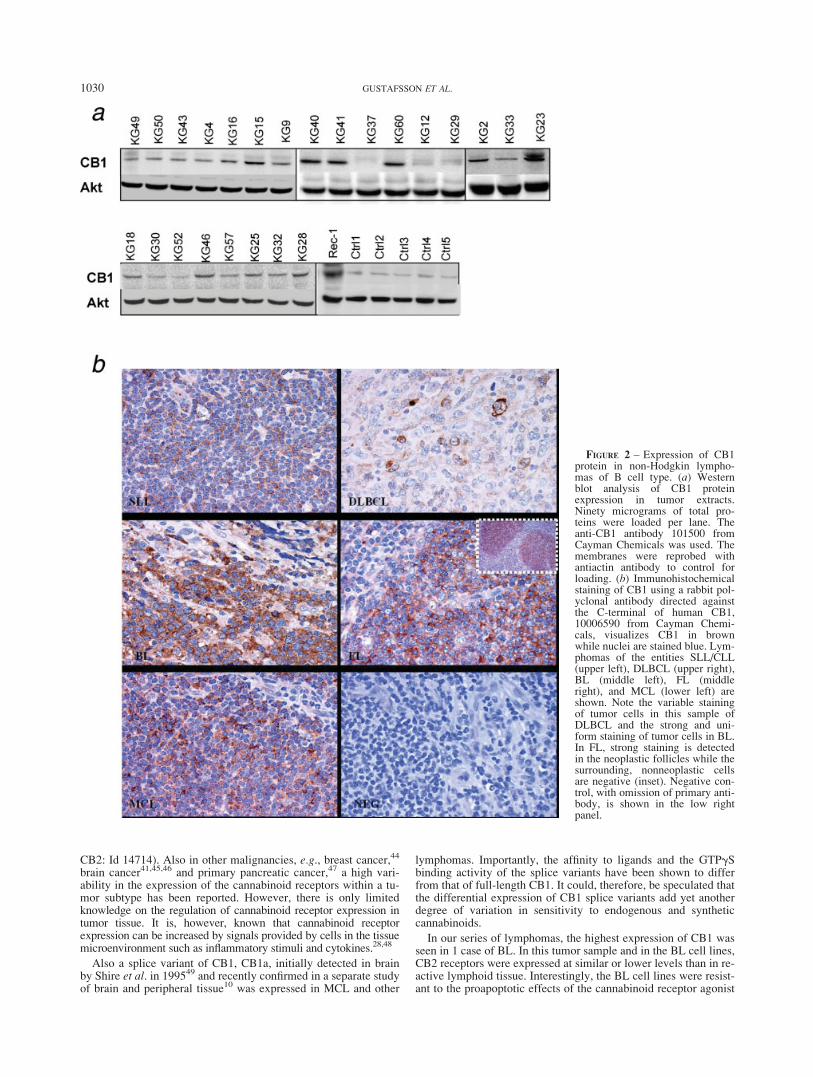
Pharmacological doses of cannabinoids have been reported toreduce cancer growth in cell lines, in xenografted cancers and in apilot study on human glioblastoma multiforme.29–43 We previ-ously reported that treatment with micromolar doses of cannabi-noid receptor ligands such as the endocannabinoid anandamideinduces apoptosis in MCL.6,7 There is very limited knowledge onthe effects of cannabinoids in other lymphoma entities. We, there-fore, investigated levels of CB receptors on well-characterizedcell lines, derived from plasma cell leukemia (SK-MM-2), BL(Raji and Namalwa), CLL (MEC1 and MEC2), and MCL (Rec-1)(Fig. 3a), and the cells were exposed to the metabolically stableanalog of anandamide R(þ)-MA. To assess the relative contribu-tion of CB1 and CB2, cells were pretreated with receptor antago-nists SR141716 (SR1) and SR144528 (SR2), acting on CB1 andCB2 respectively. R(þ)-MA induced caspase-3 activity (Fig. 3b)and cell death (Fig. 3c) in cell lines from MCL and CLL but not inBL cell lines (expressing low levels of CB2) or in the plasma cellline SK-MM-2 (expressing low levels of CB1). In the MCL andCLL cell lines, the R(þ)-MA-induced cell death was abrogated bypretreatment with either of the receptor antagonists SR1 or SR2,suggesting that both CB receptors are needed for the induction ofcell death.
Effect of R(þ)-MA on tumors in xenotransplanted mice
To determine whether cannabinoids can be used to treat malig-nant lymphoma in vivo, xenograft tumors were established by sub-cutaneous inoculation of SCID/NOD mice with human MCL cell-line JEKO-1, expressing CB1 and CB2. Treatment with 5 mg/kgof the stable CB1 agonist R(þ)-MA by subcutaneous inoculationevery 12 hr at the previous injection site was initiated 3 days aftertumor cell injection. The tumors in R(þ)-MA-treated animals
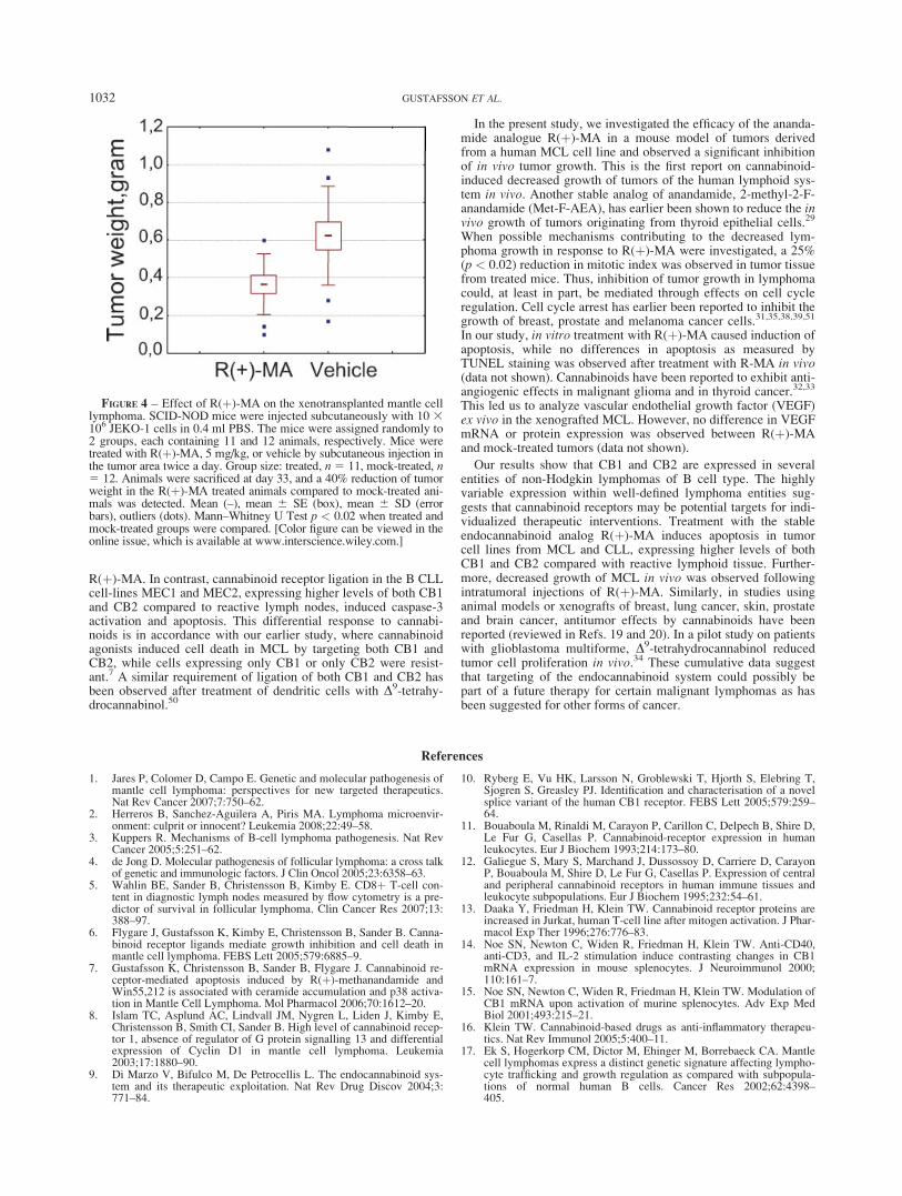
grew slower and were significantly smaller compared with mock-treated control tumors at termination of the experiment (Fig. 4).
We have previously shown that treatment of MCL in vitro withnanomolar doses of cannabinoids during several days leads todecreased growth, while applying micromolar doses of cannabi-noids during a short time causes apoptosis.6,7 In the xenograftedtumors, we found a 25% reduction in the mitotic index (p < 0.02)in the tumors from R(þ)-MA treated mice (treated mice, average1.35, control mice average 1.80) but no significant difference inapoptosis as assayed by TUNEL staining (data not shown).
Discussion
Recently, the endocannabinoid system has been recognized asderegulated in various types of epithelial malignancies, and thereis an increasing interest in the antineoplastic potential of cannabi-noids.9,18,19 Most, but not all,18 of the effects of endogenous andsynthetic cannabinoids are mediated via the cannabinoid recep-tors, CB1 and CB2. To investigate whether cannabinoids may beof potential use in lymphoma therapy, we analyzed the expressionof these receptors in a panel of malignant B cell lymphomas. Bothcannabinoid receptors were expressed in the vast majority of ma-lignant lymphomas investigated, albeit with a large span ofexpression levels within most entities. The highly variable expres-sion levels in most of the B cell lymphoma entities might explainwhy these receptors have not previously been recognized asderegulated using array-based gene expression analysis of B cellmalignancies. A variation in the mRNA expression of CB1 andCB2 in FL and of CB2 in DLBCL can indeed be found in experi-ments available in public databases (data from the National Centerfor Biotechnology Information (NCBI) Gene Expression Omnibus(GEO) database (FL: GDS1839, DLBCL: GDS75 CB1: Id 34333,
FIGURE 1 – Detection of full-length CB1 and CB1a in cDNAderived from MCL. (a) Schematic picture of the length of the PCRproducts representing the different transcripts. Primers are indicatedwith arrows. (b) A 324-bp product corresponding to full-length CB1was detected from a first round of PCR (upper lane). A region between120- and 250-bp was excised and melted, and used as template for fur-ther PCR amplification. A 157-bp product corresponding to CB1a wasdetected from a second round of PCR (lower lane). CB1b, predictedproduct 225 bp, was not expressed.
TABLE II – EXPRESSION OF CB1A mRNA MEASURED AS RFI1 OFLYMPHOMA ENTITIES2 RELATIVE TO POOLED CONTROLS
Number CB1a Range Entity
KG 35 6,9 6,8–7,0 SLLKG 36 7,8 7,5–8,1 SLLKG 43 14,6 14,4–14,8 SLLKG 18 19,2 19,0–19,5 DLBCLKG 21 2,4 2,1–2,6 DLBCLKG 22 5,7 5,4–5,9 DLBCLKG 31 28,2 27,9–28,4 DLBCLKG 33 1 0,7–1,2 DLBCLKG 45 28,5 28,4–28,6 MZLKG 46 228,1 227,9–228,3 MZLKG 47 22,9 22,7–23,1 MZLKG 48 10,6 10,3–10,8 MZLKG 58 11,1 10,8–11,3 ICKG 62 83,5 83,3–83,7 PCKG 2 36,8 36,5–37,0 FLKG 3 45,3 45,0–45,5 FLKG 4 10,2 9,9–10,5 FLKG 5 41,3 41,0–41,5 FLKG 6 19 18,8–19,3 FLKG 7 5,7 5,4–5,9 FLKG 8 40,3 40,1–40,5 FLKG 12 1,7 1,6–1,8 FLKG 13 21,1 20,9–21,4 FLKG 14 10,4 10,3–10,5 FLKG 53 52,6 52,3–52,8 MCLKG 56 128 127,8–128,2 MCLCtrl1 0,9 0,8–1,0 ReactiveCtrl2 0,6 0,4–0,8 ReactiveCtrl3 1,1 1,0–1,2 ReactiveCtrl4 1,5 1,2–1,7 ReactiveCtrl5 0,9 0,7–1.1 ReactivePooled Ctrl 1 0,8–1,2
1RFI, relative fold increase.–2Lymphoma entities: SLL, small lym-phocytic lymphoma; DLBCL, diffuse large B cell lymphoma; MZL,marginal zone lymphoma; IC, immunocytoma; PC, plasmacytoma;FL, follicular lymphoma; MCL, mantle cell lymphoma.
1029CANNABINOID RECEPTORS IN MALIGNANT LYMPHOMA
CB2: Id 14714). Also in other malignancies, e.g., breast cancer,44
brain cancer41,45,46 and primary pancreatic cancer,47 a high vari-ability in the expression of the cannabinoid receptors within a tu-mor subtype has been reported. However, there is only limitedknowledge on the regulation of cannabinoid receptor expression intumor tissue. It is, however, known that cannabinoid receptorexpression can be increased by signals provided by cells in the tissuemicroenvironment such as inflammatory stimuli and cytokines.28,48
Also a splice variant of CB1, CB1a, initially detected in brainby Shire et al. in 199549 and recently confirmed in a separate studyof brain and peripheral tissue10 was expressed in MCL and other
lymphomas. Importantly, the affinity to ligands and the GTPgSbinding activity of the splice variants have been shown to differfrom that of full-length CB1. It could, therefore, be speculated thatthe differential expression of CB1 splice variants add yet anotherdegree of variation in sensitivity to endogenous and syntheticcannabinoids.
In our series of lymphomas, the highest expression of CB1 wasseen in 1 case of BL. In this tumor sample and in the BL cell lines,CB2 receptors were expressed at similar or lower levels than in re-active lymphoid tissue. Interestingly, the BL cell lines were resist-ant to the proapoptotic effects of the cannabinoid receptor agonist
FIGURE 2 – Expression of CB1protein in non-Hodgkin lympho-mas of B cell type. (a) Westernblot analysis of CB1 proteinexpression in tumor extracts.Ninety micrograms of total pro-teins were loaded per lane. Theanti-CB1 antibody 101500 fromCayman Chemicals was used. Themembranes were reprobed withantiactin antibody to control forloading. (b) Immunohistochemicalstaining of CB1 using a rabbit pol-yclonal antibody directed againstthe C-terminal of human CB1,10006590 from Cayman Chemi-cals, visualizes CB1 in brownwhile nuclei are stained blue. Lym-phomas of the entities SLL/CLL(upper left), DLBCL (upper right),BL (middle left), FL (middleright), and MCL (lower left) areshown. Note the variable stainingof tumor cells in this sample ofDLBCL and the strong and uni-form staining of tumor cells in BL.In FL, strong staining is detectedin the neoplastic follicles while thesurrounding, nonneoplastic cellsare negative (inset). Negative con-trol, with omission of primary anti-body, is shown in the low rightpanel.
1030 GUSTAFSSON ET AL.
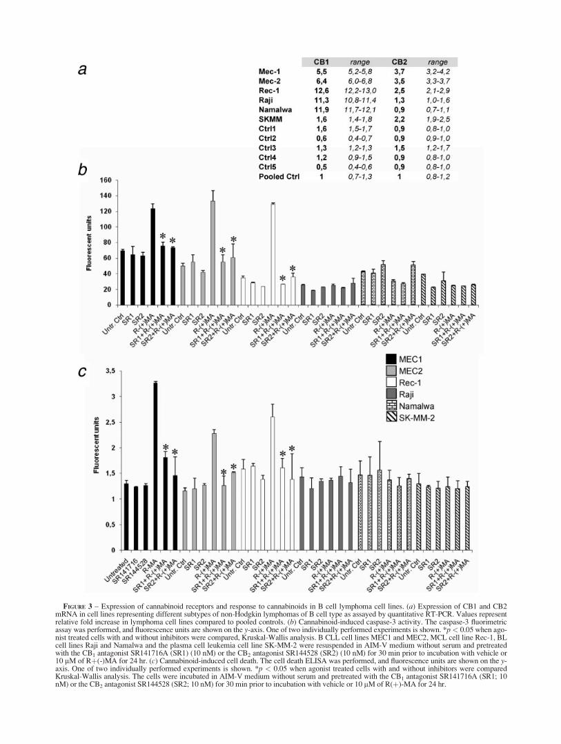
FIGURE 3 – Expression of cannabinoid receptors and response to cannabinoids in B cell lymphoma cell lines. (a) Expression of CB1 and CB2mRNA in cell lines representing different subtypes of non-Hodgkin lymphomas of B cell type as assayed by quantitative RT-PCR. Values representrelative fold increase in lymphoma cell lines compared to pooled controls. (b) Cannabinoid-induced caspase-3 activity. The caspase-3 fluorimetricassay was performed, and fluorescence units are shown on the y-axis. One of two individually performed experiments is shown. *p< 0.05 when ago-nist treated cells with and without inhibitors were compared, Kruskal-Wallis analysis. B CLL cell lines MEC1 and MEC2, MCL cell line Rec-1, BLcell lines Raji and Namalwa and the plasma cell leukemia cell line SK-MM-2 were resuspended in AIM-V medium without serum and pretreatedwith the CB1 antagonist SR141716A (SR1) (10 nM) or the CB2 antagonist SR144528 (SR2) (10 nM) for 30 min prior to incubation with vehicle or10 lM of Rþ(-)MA for 24 hr. (c) Cannabinoid-induced cell death. The cell death ELISA was performed, and fluorescence units are shown on the y-axis. One of two individually performed experiments is shown. *p < 0.05 when agonist treated cells with and without inhibitors were comparedKruskal-Wallis analysis. The cells were incubated in AIM-V medium without serum and pretreated with the CB1 antagonist SR141716A (SR1; 10nM) or the CB2 antagonist SR144528 (SR2; 10 nM) for 30 min prior to incubation with vehicle or 10 lM of R(þ)-MA for 24 hr.
R(þ)-MA. In contrast, cannabinoid receptor ligation in the B CLLcell-lines MEC1 and MEC2, expressing higher levels of both CB1and CB2 compared to reactive lymph nodes, induced caspase-3activation and apoptosis. This differential response to cannabi-noids is in accordance with our earlier study, where cannabinoidagonists induced cell death in MCL by targeting both CB1 andCB2, while cells expressing only CB1 or only CB2 were resist-ant.7 A similar requirement of ligation of both CB1 and CB2 hasbeen observed after treatment of dendritic cells with D9-tetrahy-drocannabinol.50
In the present study, we investigated the efficacy of the ananda-mide analogue R(þ)-MA in a mouse model of tumors derivedfrom a human MCL cell line and observed a significant inhibitionof in vivo tumor growth. This is the first report on cannabinoid-induced decreased growth of tumors of the human lymphoid sys-tem in vivo. Another stable analog of anandamide, 2-methyl-2-F-anandamide (Met-F-AEA), has earlier been shown to reduce the invivo growth of tumors originating from thyroid epithelial cells.29
When possible mechanisms contributing to the decreased lym-phoma growth in response to R(þ)-MA were investigated, a 25%(p < 0.02) reduction in mitotic index was observed in tumor tissuefrom treated mice. Thus, inhibition of tumor growth in lymphomacould, at least in part, be mediated through effects on cell cycleregulation. Cell cycle arrest has earlier been reported to inhibit thegrowth of breast, prostate and melanoma cancer cells.31,35,38,39,51
In our study, in vitro treatment with R(þ)-MA caused induction ofapoptosis, while no differences in apoptosis as measured byTUNEL staining was observed after treatment with R-MA in vivo(data not shown). Cannabinoids have been reported to exhibit anti-angiogenic effects in malignant glioma and in thyroid cancer.32,33
This led us to analyze vascular endothelial growth factor (VEGF)ex vivo in the xenografted MCL. However, no difference in VEGFmRNA or protein expression was observed between R(þ)-MAand mock-treated tumors (data not shown).
Our results show that CB1 and CB2 are expressed in severalentities of non-Hodgkin lymphomas of B cell type. The highlyvariable expression within well-defined lymphoma entities sug-gests that cannabinoid receptors may be potential targets for indi-vidualized therapeutic interventions. Treatment with the stableendocannabinoid analog R(þ)-MA induces apoptosis in tumorcell lines from MCL and CLL, expressing higher levels of bothCB1 and CB2 compared with reactive lymphoid tissue. Further-more, decreased growth of MCL in vivo was observed followingintratumoral injections of R(þ)-MA. Similarly, in studies usinganimal models or xenografts of breast, lung cancer, skin, prostateand brain cancer, antitumor effects by cannabinoids have beenreported (reviewed in Refs. 19 and 20). In a pilot study on patientswith glioblastoma multiforme, D9-tetrahydrocannabinol reducedtumor cell proliferation in vivo.34 These cumulative data suggestthat targeting of the endocannabinoid system could possibly bepart of a future therapy for certain malignant lymphomas as hasbeen suggested for other forms of cancer.
References
1. Jares P, Colomer D, Campo E. Genetic and molecular pathogenesis ofmantle cell lymphoma: perspectives for new targeted therapeutics.Nat Rev Cancer 2007;7:750–62.
2. Herreros B, Sanchez-Aguilera A, Piris MA. Lymphoma microenvir-onment: culprit or innocent? Leukemia 2008;22:49–58.
3. Kuppers R. Mechanisms of B-cell lymphoma pathogenesis. Nat RevCancer 2005;5:251–62.
4. de Jong D. Molecular pathogenesis of follicular lymphoma: a cross talkof genetic and immunologic factors. J Clin Oncol 2005;23:6358–63.
5. Wahlin BE, Sander B, Christensson B, Kimby E. CD8þ T-cell con-tent in diagnostic lymph nodes measured by flow cytometry is a pre-dictor of survival in follicular lymphoma. Clin Cancer Res 2007;13:388–97.
6. Flygare J, Gustafsson K, Kimby E, Christensson B, Sander B. Canna-binoid receptor ligands mediate growth inhibition and cell death inmantle cell lymphoma. FEBS Lett 2005;579:6885–9.
7. Gustafsson K, Christensson B, Sander B, Flygare J. Cannabinoid re-ceptor-mediated apoptosis induced by R(þ)-methanandamide andWin55,212 is associated with ceramide accumulation and p38 activa-tion in Mantle Cell Lymphoma. Mol Pharmacol 2006;70:1612–20.
8. Islam TC, Asplund AC, Lindvall JM, Nygren L, Liden J, Kimby E,Christensson B, Smith CI, Sander B. High level of cannabinoid recep-tor 1, absence of regulator of G protein signalling 13 and differentialexpression of Cyclin D1 in mantle cell lymphoma. Leukemia2003;17:1880–90.
9. Di Marzo V, Bifulco M, De Petrocellis L. The endocannabinoid sys-tem and its therapeutic exploitation. Nat Rev Drug Discov 2004;3:771–84.
10. Ryberg E, Vu HK, Larsson N, Groblewski T, Hjorth S, Elebring T,Sjogren S, Greasley PJ. Identification and characterisation of a novelsplice variant of the human CB1 receptor. FEBS Lett 2005;579:259–64.
11. Bouaboula M, Rinaldi M, Carayon P, Carillon C, Delpech B, Shire D,Le Fur G, Casellas P. Cannabinoid-receptor expression in humanleukocytes. Eur J Biochem 1993;214:173–80.
12. Galiegue S, Mary S, Marchand J, Dussossoy D, Carriere D, CarayonP, Bouaboula M, Shire D, Le Fur G, Casellas P. Expression of centraland peripheral cannabinoid receptors in human immune tissues andleukocyte subpopulations. Eur J Biochem 1995;232:54–61.
13. Daaka Y, Friedman H, Klein TW. Cannabinoid receptor proteins areincreased in Jurkat, human T-cell line after mitogen activation. J Phar-macol Exp Ther 1996;276:776–83.
14. Noe SN, Newton C, Widen R, Friedman H, Klein TW. Anti-CD40,anti-CD3, and IL-2 stimulation induce contrasting changes in CB1mRNA expression in mouse splenocytes. J Neuroimmunol 2000;110:161–7.
15. Noe SN, Newton C, Widen R, Friedman H, Klein TW. Modulation ofCB1 mRNA upon activation of murine splenocytes. Adv Exp MedBiol 2001;493:215–21.
16. Klein TW. Cannabinoid-based drugs as anti-inflammatory therapeu-tics. Nat Rev Immunol 2005;5:400–11.
17. Ek S, Hogerkorp CM, Dictor M, Ehinger M, Borrebaeck CA. Mantlecell lymphomas express a distinct genetic signature affecting lympho-cyte trafficking and growth regulation as compared with subpopula-tions of normal human B cells. Cancer Res 2002;62:4398–405.
FIGURE 4 – Effect of R(þ)-MA on the xenotransplanted mantle celllymphoma. SCID-NOD mice were injected subcutaneously with 10 3106 JEKO-1 cells in 0.4 ml PBS. The mice were assigned randomly to2 groups, each containing 11 and 12 animals, respectively. Mice weretreated with R(þ)-MA, 5 mg/kg, or vehicle by subcutaneous injection inthe tumor area twice a day. Group size: treated, n 5 11, mock-treated, n5 12. Animals were sacrificed at day 33, and a 40% reduction of tumorweight in the R(þ)-MA treated animals compared to mock-treated ani-mals was detected. Mean (–), mean 6 SE (box), mean 6 SD (errorbars), outliers (dots). Mann–Whitney U Test p < 0.02 when treated andmock-treated groups were compared. [Color figure can be viewed in theonline issue, which is available at www.interscience.wiley.com.]
1032 GUSTAFSSON ET AL.
18. Flygare J, Sander B. The endocannabinoid system in cancer-Potentialtherapeutic target? Semin Cancer Biol; in press.
19. Bifulco M, Di Marzo V. Targeting the endocannabinoid system incancer therapy: a call for further research. Nat Med 2002;8:547–50.
20. Guzman M. Cannabinoids: potential anticancer agents. Nat Rev Can-cer 2003;3:745–55.
21. Bifulco M, Laezza C, Pisanti S, Gazzero P. Cannabinoids and cancer:pros and cons of an antitumour strategy. Br J Pharmacol 2006;148:123–25.
22. Herrera B, Carracedo A, Diez-Zaera M, Guzman M, Velasco G. p38MAPK is involved in CB2 receptor-induced apoptosis of human leu-kaemia cells. FEBS Lett 2005;579:5084–8.
23. McKallip RJ, Lombard C, Fisher M, Martin BR, Ryu S, Grant S,Nagarkatti PS, Nagarkatti M. Targeting CB2 cannabinoid receptors asa novel therapy to treat malignant lymphoblastic disease. Blood 2007;100:627–34.
24. Rayman N, Lam KH, Van Leeuwen J, Mulder AH, Budel LM, Low-enberg B, Sonneveld P, Delwel R. The expression of the peripheralcannabinoid receptor on cells of the immune system and non-Hodg-kin’s lymphomas. Leuk Lymphoma 2007;48:1389–99.
25. Jaffe ES, Harris NL, Stein H, Vardiman JW, eds. World Health Orga-nization classification of tumours: pathology and genetics of tumoursof haematopoietic and lymphoid tissues. Lyon: IARC Press, 2001.
26. Rimokh R, Berger F, Bastard C, Klein B, French M, Archimbaud E,Rouault JP, Santa Lucia B, Duret L, Vuillaume M, Coiffier B, BryonP-A, Magaud JP. Rearrangement of CCND1 (BCL1/PRAD1) 30untranslated region in mantle-cell lymphomas and t(11q13)-associ-ated leukemias. Blood 1994;83:3689–96.
27. Rundlof AK, Fernandes AP, Selenius M, Babic M, Shariatgorji M,Nilsonne G, Ilag LL, Dobra K, Bjornstedt M. Quantification of alter-native mRNA species and identification of thioredoxin reductase 1isoforms in human tumor cells. Differentiation 2007;75:123–32.
28. Borner C, Hollt V, Sebald W, Kraus J. Transcriptional regulation ofthe cannabinoid receptor type 1 gene in T cells by cannabinoids.J Leukoc Biol 2007;81:336–43.
29. Bifulco M, Laezza C, Portella G, Vitale M, Orlando P, De PetrocellisL, Di Marzo V. Control by the endogenous cannabinoid system of rasoncogene-dependent tumor growth. FASEB J 2001;15:2745–7.
30. Bifulco M, Laezza C, Valenti M, Ligresti A, Portella G, Di Marzo V.A new strategy to block tumor growth by inhibiting endocannabinoidinactivation. FASEB J 2004;18:1606–8.
31. Blazquez C, Carracedo A, Barrado L, Real PJ, Fernandez-Luna JL,Velasco G, Malumbres M, Guzman M. Cannabinoid receptors asnovel targets for the treatment of melanoma. FASEB J 2006;20:2633–5.
32. Blazquez C, Casanova ML, Planas A, Del Pulgar TG, Villanueva C,Fernandez-Acenero MJ, Aragones J, Huffman JW, Jorcano JL, Guz-man M. Inhibition of tumor angiogenesis by cannabinoids. FASEB J2003;17:529–31.
33. Casanova ML, Blazquez C, Martinez-Palacio J, Villanueva C, Fernan-dez-Acenero MJ, Huffman JW, Jorcano JL, Guzman M. Inhibition ofskin tumor growth and angiogenesis in vivo by activation of cannabi-noid receptors. J Clin Invest 2003;111:43–50.
34. Guzman M, Duarte MJ, Blazquez C, Ravina J, Rosa MC, Galve-Roperh I, Sanchez C, Velasco G, Gonzalez-Feria L. A pilot clinicalstudy of Delta9-tetrahydrocannabinol in patients with recurrent glio-blastoma multiforme. Br J Cancer 2006;95:197–203.
35. Laezza C, Pisanti S, Crescenzi E, Bifulco M. Anandamide inhibitsCdk2 and activates Chk1 leading to cell cycle arrest in human breastcancer cells. FEBS Lett 2006;580:6076–82.
36. Ligresti A, Bisogno T, Matias I, De Petrocellis L, Cascio MG,Cosenza V, D’Argenio G, Scaglione G, Bifulco M, Sorrentini I, Di
Marzo V. Possible endocannabinoid control of colorectal cancergrowth. Gastroenterology 2003;125:677–87.
37. Ligresti A, Moriello AS, Starowicz K, Matias I, Pisanti S, De Petrocel-lis L, Laezza C, Portella G, Bifulco M, Di Marzo V. Antitumor activityof plant cannabinoids with emphasis on the effect of cannabidiol onhuman breast carcinoma. J Pharmacol Exp Ther 2006;318:1375–87.
38. Melck D, De Petrocellis L, Orlando P, Bisogno T, Laezza C, BifulcoM, Di Marzo V. Suppression of nerve growth factor Trk receptors andprolactin receptors by endocannabinoids leads to inhibition of humanbreast and prostate cancer cell proliferation. Endocrinology 2000;141:118–26.
39. Melck D, Rueda D, Galve-Roperh I, De Petrocellis L, Guzman M, DiMarzo V. Involvement of the cAMP/protein kinase A pathway and ofmitogen-activated protein kinase in the anti-proliferative effects ofanandamide in human breast cancer cells. FEBS Lett 1999;463:235–40.
40. Portella G, Laezza C, Laccetti P, De Petrocellis L, Di Marzo V,Bifulco M. Inhibitory effects of cannabinoid CB1 receptor stimulationon tumor growth and metastatic spreading: actions on signals involvedin angiogenesis and metastasis. FASEB J 2003;17:1771–3.
41. Sanchez C, de Ceballos ML, del Pulgar TG, Rueda D, Corbacho C,Velasco G, Galve-Roperh I, Huffman JW, Ramon y Cajal S, GuzmanM. Inhibition of glioma growth in vivo by selective activation of theCB(2) cannabinoid receptor. Cancer Res 2001;61:5784–9.
42. Sarfaraz S, Afaq F, Adhami VM, Malik A, Mukhtar H. Cannabinoidreceptor agonist-induced apoptosis of human prostate cancer cellsLNCaP proceeds through sustained activation of ERK1/2 leading toG1 cell cycle arrest. J Biol Chem 2006;281:39480–91.
43. Sarfaraz S, Afaq F, Adhami VM, Mukhtar H. Cannabinoid receptor asa novel target for the treatment of prostate cancer. Cancer Res2005;65:1635–41.
44. Caffarel MM, Sarrio D, Palacios J, Guzman M, Sanchez C. Delta9-tetrahydrocannabinol inhibits cell cycle progression in human breastcancer cells through Cdc2 regulation. Cancer Res 2006;66:6615–21.
45. Ellert-Miklaszewska A, Grajkowska W, Gabrusiewicz K, KaminskaB, Konarska L. Distinctive pattern of cannabinoid receptor type II(CB2) expression in adult and pediatric brain tumors. Brain Res2007;1137:161–9.
46. Held-Feindt J, Dorner L, Sahan G, Mehdorn HM, Mentlein R. Canna-binoid receptors in human astroglial tumors. J Neurochem 2006;98:886–93.
47. Carracedo A, Gironella M, Lorente M, Garcia S, Guzman M, VelascoG, Iovanna JL. Cannabinoids induce apoptosis of pancreatic tumorcells via endoplasmic reticulum stress-related genes. Cancer Res2006;66:6748–55.
48. Maresz K, Pryce G, Ponomarev ED, Marsicano G, Croxford JL,Shriver LP, Ledent C, Cheng X, Carrier EJ, Mann MK, GiovannoniG, Pertwee RG, et al. Direct suppression of CNS autoimmune inflam-mation via the cannabinoid receptor CB1 on neurons and CB2 onautoreactive T cells. Nat Med 2007;13:492–7.
49. Shire D, Carillon C, Kaghad M, Calandra B, Rinaldi-Carmona M, LeFur G, Caput D, Ferrara P. An amino-terminal variant of the centralcannabinoid receptor resulting from alternative splicing. J Biol Chem1995;270:3726–31.
50. Do Y, McKallip RJ, Nagarkatti M, Nagarkatti PS. Activation throughcannabinoid receptors 1 and 2 on dendritic cells triggers NF-kappaB-dependent apoptosis: novel role for endogenous and exogenous can-nabinoids in immunoregulation. J Immunol 2004;173:2373–82.
51. De Petrocellis L, Melck D, Palmisano A, Bisogno T, Laezza C,Bifulco M, Di Marzo V. The endogenous cannabinoid anandamideinhibits human breast cancer cell proliferation. Proc Natl Acad SciUSA 1998;95:8375–80.
1033CANNABINOID RECEPTORS IN MALIGNANT LYMPHOMA