expression of pax8 in normal and neoplastic...
TRANSCRIPT

www.biocare.net
Expression of PAX8 in Normal and Neoplastic Tissues:A Comprehensive Immunohistochemical Study
Authors: D Tacha, L Cheng, D Zhou and RL Henshall-Powell.
Biocare Medical, Concord, CA, United States and Indiana Univ School of Med, Indianapolis, IN, United States.
As Presented at USCAP 2010, Abstract #991

BackgroundPAX genes are a family of cell-lineage transcription factors that play
fundamental roles during organogenesis and are regulatory proteins
expressed in embryonic or neoplastic cells of the same lineage.
PAX8 is a nephric-lineage transcription factor that is crucial for
organogenesis of the thyroid gland, kidney, and Müllerian system.
These proteins are required for cell growth and differentiation in
embryonic tissues and can be expressed in adult tissues in specific
cell-lineage neoplastic tissues. Recently PAX8 has been shown to be
expressed in a high percentage of kidney and ovarian carcinomas.
Little is known about the specificity of PAX8 in various neoplastic
tumors. This detailed study examines the immunohistochemical
expression of PAX8 in over 1100 cases of normal and neoplastic
tissues. MethodsHuman formalin-fixed paraffin embedded tissue microarrays
(TMAs) were both constructed in-house and commercially
purchased (US Biomax and Pantomics). TMAs included both
normal and neoplastic tissues including kidney, bladder, prostate,
lung, breast, colon, liver, pancreas, uterus, cervix, ovary, testis,
brain, stomach, esophagus, melanoma, sarcoma, and lymphoma.
Tissue sections were cut at 5 microns (5µm) thickness and dried
for 30 minutes at 60°C. Slides were deparaffinized in Slide Brite
(Biocare Medical) and hydrated in a series of graded alcohols to
water. Tissues were treated with 3% H202 in buffer, placed into
a modified citrate buffer (DIVA™, Biocare Medical), loaded into
a Decloaking Chamber™ (antigen retrieval unit, Biocare Medical)
and heated to 125°C. Tissues were then allowed to cool for 10
minutes, and then rinsed in deionized water.
ResultsA summary of PAX8 expression for a broad array of cancer types
is presented in Table 1. PAX8 expression was positive in renal cell
carcinoma (90%). Normal kidney (photo 1) stains positive for PAX8
in all cases. In clear cell RCC, PAX8 expression was observed in
91% (86/94) of cases. Of these, 100% (8/8) of metastatic cases,
including a metastasis in the thyroid gland (photo 2), 100%
(14/14) of papillary RCC cases, and 57% (4/7) of chromophobe
RCC cases (photo 3) (Table 2) exhibited PAX8 expression. In ovarian
cancers, PAX8 stained 79% (181/229) of all ovarian carcinomas.
When differentiating ovarian tumors by tumor type, positive cases
were observed in 92% (101/109) of serous adenocarcinomas
(photo 4), 83% (49/59) of endometrioid carcinomas (photo 5),
100% (3/3) of clear cell carcinomas, 50% (27/54) of mucinous
cystadenocarcinomas (photo 6), and in 31/32 (97%) of all ovarian
metastatic cases (Table 2). Other cancer types, positive for PAX8
included thyroid cancers (90%) and endometrial carcinomas (84%)
(Table 1). In lung cancer, PAX8 staining was found in only 1 case (1+
staining). PAX 8 expression was also seen in normal brain, normal
tonsil and lymph node. Expression in lymph tissues appears to be
confined to B-cells and/or B-cell lineage. The vast majority of other
cancers were 100% negative (Table 1).
Normal kidney
1
Serous ovarian adenocarcinoma
4
Clear cell RCC metastasized to thyroid
2
Endometrioid ovarian cancer
5
Chromophobe RCC
3
Ovarian mucinous cystadenocarcinoma
6
ConclusionIn summary, PAX8 specificity and sensitivity was demonstrated in
over 1100 cases of normal and neoplastic tissues, in 19 different
major cancer types and subtypes, and in 33 different normal
tissues. PAX8 proved to be a specific and a sensitive marker
for renal cell carcinoma (90%) and ovarian carcinomas (79%),
with serous and endometrioid ovarian carcinomas having 89%
sensitivity. We propose that PAX8 be recommended for use in a
clinical histopathology laboratory setting.
Photos 1-8 stained with PAX-8
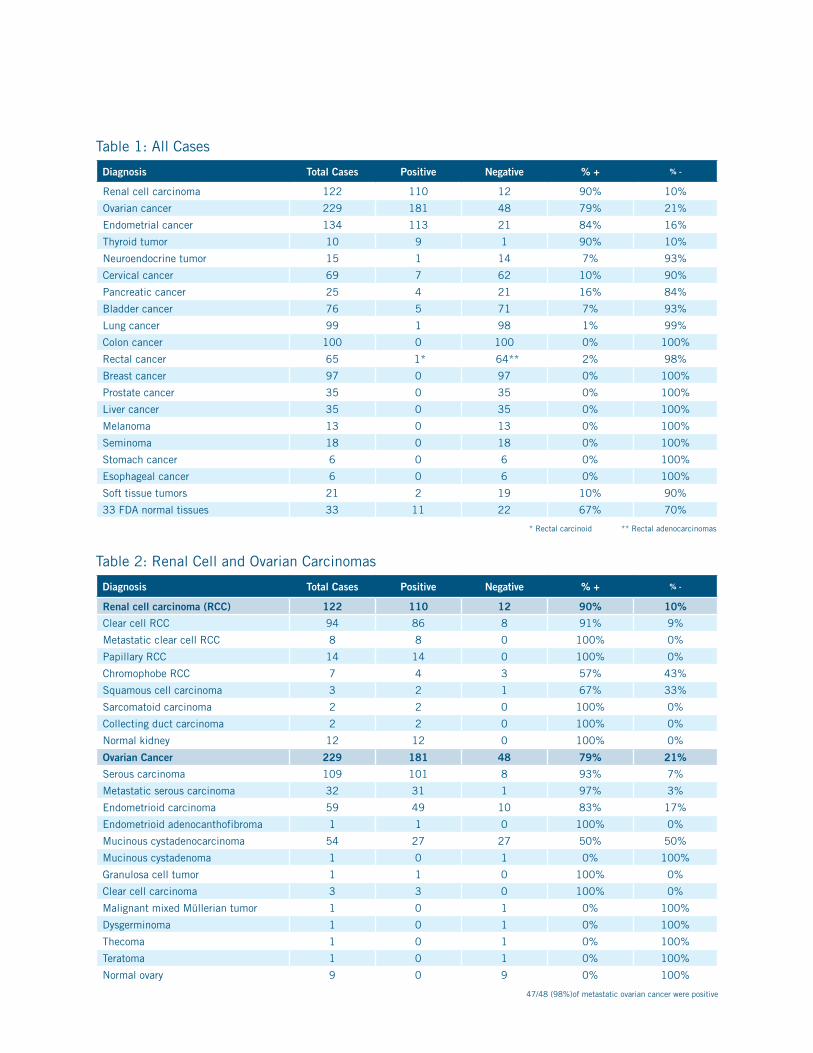
Table 1: All Cases
Diagnosis Total Cases Positive Negative % + % -
Renal cell carcinoma 122 110 12 90% 10%
Ovarian cancer 229 181 48 79% 21%
Endometrial cancer 134 113 21 84% 16%
Thyroid tumor 10 9 1 90% 10%
Neuroendocrine tumor 15 1 14 7% 93%
Cervical cancer 69 7 62 10% 90%
Pancreatic cancer 25 4 21 16% 84%
Bladder cancer 76 5 71 7% 93%
Lung cancer 99 1 98 1% 99%
Colon cancer 100 0 100 0% 100%
Rectal cancer 65 1* 64** 2% 98%
Breast cancer 97 0 97 0% 100%
Prostate cancer 35 0 35 0% 100%
Liver cancer 35 0 35 0% 100%
Melanoma 13 0 13 0% 100%
Seminoma 18 0 18 0% 100%
Stomach cancer 6 0 6 0% 100%
Esophageal cancer 6 0 6 0% 100%
Soft tissue tumors 21 2 19 10% 90%
33 FDA normal tissues 33 11 22 67% 70%
* Rectal carcinoid ** Rectal adenocarcinomas
Table 2: Renal Cell and Ovarian Carcinomas
Diagnosis Total Cases Positive Negative % + % -
Renal cell carcinoma (RCC) 122 110 12 90% 10%
Clear cell RCC 94 86 8 91% 9%
Metastatic clear cell RCC 8 8 0 100% 0%
Papillary RCC 14 14 0 100% 0%
Chromophobe RCC 7 4 3 57% 43%
Squamous cell carcinoma 3 2 1 67% 33%
Sarcomatoid carcinoma 2 2 0 100% 0%
Collecting duct carcinoma 2 2 0 100% 0%
Normal kidney 12 12 0 100% 0%
Ovarian Cancer 229 181 48 79% 21%
Serous carcinoma 109 101 8 93% 7%
Metastatic serous carcinoma 32 31 1 97% 3%
Endometrioid carcinoma 59 49 10 83% 17%
Endometrioid adenocanthofibroma 1 1 0 100% 0%
Mucinous cystadenocarcinoma 54 27 27 50% 50%
Mucinous cystadenoma 1 0 1 0% 100%
Granulosa cell tumor 1 1 0 100% 0%
Clear cell carcinoma 3 3 0 100% 0%
Malignant mixed Müllerian tumor 1 0 1 0% 100%
Dysgerminoma 1 0 1 0% 100%
Thecoma 1 0 1 0% 100%
Teratoma 1 0 1 0% 100%
Normal ovary 9 0 9 0% 100%
47/48 (98%)of metastatic ovarian cancer were positive

800.799.9499
4040 Pike Lane
Concord CA 94520 www.biocare.net
References1. Bowen NJ et al. Emerging roles for PAX8 in ovarian cancer and endosalpingeal development. Gynecol Oncol. 2007 Feb;104(2):331-7. Epub 2006 Oct 24.
2. Nonaka D et al. Diagnostic utility of thyroid transcription factors Pax8 and TTF-2 (FoxE1) in thyroid epithelial neoplasms. Mod Pathol. 2008 Feb; 21(2):192-200. Epub 2007 Dec 14.
3. Köbel M et al. Ovarian carcinoma subtypes are different diseases: implications for biomarker studies. PLoS Med. 2008 Dec 2;5(12):e232.
4. Nonaka D et al. Expression of pax8 as a useful marker in distinguishing ovarian carcinomas from mammary carcinomas. Am J Surg Pathol. 2008 Oct; 32(10):1566-71.
5. Tong GX et al. Expression of PAX8 in nephrogenic adenoma and clear cell adenocarcinoma of the lower urinary tract: evidence of related histogenesis?Am J Surg Pathol. 2008 Sep; 32(9):1380-7.
6. Tong GX et al. Expression of PAX8 in normal and neoplastic renal tissues: an immunohistochemical study. Mod Pathol. 2009 Sep; 22(9):1218-27. Epub 2009 Jun 12.
7. American Cancer Society
8. Geramizadeh B et al. Useful markers for differential diagnosis of oncocytoma, chromophobe renal cell carcinoma and conventional renal cell carcinoma.Indian J Pathol Microbiol. 2008 Apr-Jun; 51(2):167-71.
9. Zou H et al. [Study on clinicopathologic features and immunophenotype of 114 cases of renal cell carcinoma] Zhonghua Bing Li Xue Za Zhi. 2008 Nov; 37(11):726-31.
10. Avery AK et al. Use of antibodies to RCC and CD10 in the differential diagnosis of renal neoplasms. Am J Surg Pathol. 2000 Feb; 24(2):203-10.
11. Zhou M et al. The usefulness of immunohistochemical markers in the differential diagnosis of renal neoplasms. Clin Lab Med. 2005 Jun; 25(2):247-57.
12. Kuehn A et al. Expression analysis of kidney-specific cadherin in a wide spectrum of traditional and newly recognized renal epithelial neoplasms: diagnostic and histogenetic implications. Am J Surg Pathol. 2007 Oct; 31(10):1528-33.
14. Mazal PR et al. Expression of kidney-specific cadherin distinguishes chromophobe renal cell carcinoma from renal oncocytoma. Hum Pathol. 2005 Jan; 36(1):22-8.
15. Robbins, Pathology Basis of Disease; Six Edition: pp1066-72
16. Zhu W, Michael CW. WT1, monoclonal CEA, TTF1, and CA125 antibodies in the differential diagnosis of lung, breast, and ovarian adenocarcinomas in serous effusions. Diag Cytopathol. 2007 Jun; 35(6):370-5.
17. Tornos C et al. Expression of WT1, CA 125, and GCDFP-15 as useful markers in the differential diagnosis of primary ovarian carcinomas versus metastatic breast cancer to the ovary. Am J Surg Pathol. 2005 Nov; 29(11):1482-9.
18. Lee AH et al. The expression of Wilms’ tumour-1 and CA125 in invasive micropapillary carcinoma of the breast. Histopathology. 2007 Dec; 51(6):824-8.
19. Leake J et al. Immunocytochemical and serological expression of CA 125: a clinicopathological study of 40 malignant ovarian epithelial tumours. Histopathology. 1994 Jan; 24(1):57-64.
20. Koelma IA et al. The value of tumours marker CA 125 in surgical pathology.Histopathology. 1987 Mar; 11(3):287-94.
21. Reid-Nicholson M et al. Immunophenotypic diversity of endometrial adenocarcinomas: implications for differential diagnosis. Mod Pathol. 2006 Aug; 19(8):1091-100. Epub 2006 Apr
22. Sosa-Pineda B. The gene Pax4 is an essential regulator of pancreatic beta-cell development. Mol Cells. 2004 Dec 31;18(3):289-94.
PAX8Poster